Fibroblast Growth Factors/Fibroblast Growth Factor Receptors as Targets for the Development of...
20
Current Pharmaceutical Design, 2007, 13, 2025-2044 2025 1381-6128/07 $50.00+.00 © 2007 Bentham Science Publishers Ltd. Fibroblast Growth Factors/Fibroblast Growth Factor Receptors as Targets for the Development of Anti-Angiogenesis Strategies M. Rusnati and M. Presta * Unit of General Pathology and Immunology, Department of Biomedical Sciences and Biotechnology, School of Medicine, University of Brescia, Italy Abstract: Angiogenesis, the process of new blood vessel formation from pre-existing ones, plays a key role in various physiological and pathological conditions, including embryonic development, wound repair, inflammation, and tumor growth. The 1980s saw for the first time the identification, purification, and sequencing of the two prototypic heparin-binding angiogenic fibroblast growth factors (FGF) 1 and 2. Since then, 22 structurally-related members of the FGF family and differenent classes of FGF receptors have been identified. Several experimental evidences point to a role for various FGFs in the neovascularization process that takes place in inflammation, angioproliferative diseases, and tumor growth. Thus, the FGF/FGF receptor system represents a target for the development of anti- angiogenic therapies. Purpose of this review is to summarize the different modalities that have been approached to impair the pro- angiogenic activity of the FGF/FGF receptor system and discuss their possible therapeutic implications. Key Words: Angiogenesis; endothelium; FGF; FGF receptors; inhibitors. 1. THE FGF/FGF RECEPTOR SYSTEM IN ANGIOGENESIS Angiogenesis is the process of new blood vessel formation from pre-existing ones. Neovascularization is involved in embryonic development, wound repair, and inflammation [1]. Also, the local, uncontrolled release of angiogenic growth factors contributes to neovascularization that takes place during angiogenesis-dependent diseases, including cancer [2]. The 1980s saw the purification of the pro-angiogenic proteins fibroblast growth factor-1 (FGF1) and FGF2 [3]. Since then, 22 structurally-related members of the FGF family have been identified [4]. Among them, FGF1, FGF2, FGF4, FGF5, and FGF8 have been demonstrated to be endowed with angiogenic potential [5]. FGFs are pleiotropic factors that act on different cell types, including endothelial cells (ECs), by interacting with tyrosine kinase (TK) FGF receptors (TK-FGFRs), heparan-sulfate proteo- glycans (HSPGs), integrins, and gangliosides. Several experimental evidences point to a role for FGFs in tumor angiogenesis, infla- mmation, and angio-proliferative diseases (discussed in [5]). Thus, the FGF/FGF receptor system may represent a target for anti- angiogenic therapies. FGFs induce a complex “pro-angiogenic phenotype” in cultured ECs (Fig. (1)) that recapitulates the angiogenesis process in vivo, including expression of proteases, integrins, and cadherins and the stimulation of EC proliferation and migration (summarized in [6]). Extracellular matrix (ECM) degradation, mainly by the plas- min-plasminogen activator (PA) system and matrix metalloprotei- nases (MMPs), represents an important step of the angiogenic process [7]. FGFs upregulate urokinase-type PA (uPA) and MMPs production in ECs [8, 9]. uPA converts plasminogen into plasmin that degrades different matrix proteins and activate MMPs [10]. FGF2 stimulates chemotaxis/chemokinesis in ECs [11]. When cultured on permissive three-dimensional matrix, ECs invade the substratum and organize capillary-like structures with a hollow lumen [12]. FGF2 enhances this response in collagen I [13] and fibrin [14] gels in a CD44- [15] and integrin- [16] dependent manner. Also, FGF2 promotes EC reorganization on Matrigel [17] that requires MMPs [18] and uPA [19] activity as well as 6 1 integrin engagement [20], thus underlying the thigh cross-talk among FGFs and the integrin receptor system (see below). *Address correspondence to this author at the General Pathology and Immunology, Department of Biomedical Sciences and Biotechnology, Viale Europa 11, 25123 Brescia, Italy; Tel: ++39-30-3717311; Fax: ++39-30- 3701157; E-mail: [email protected] EC migration and proliferation are limited by lateral cell-cell adhesion and ECM interactions [21] mediated by cadherin and integrin engagement. Interestingly, FGF2 regulates the expression of different cadherins [21] and integrins [22] and the production of various ECM components in ECs [23], contributing to the maturation of the new blood vessels (Fig. (1)). The angiogenic activity of various members of the FGF family has been demonstrated in vivo in different experimental models, including the chick embryo chorion-allantoic membrane assay [24], the avascular rabbit [25] or mouse [26] cornea assays, and the subcutaneous Matrigel implantation assay [27]. In these experi- mental models a potent angiogenic response can be obtained by the delivery of FGFs as recombinant proteins, via retroviral, adenoviral, lentiviral, and adeno-associated viral vector transduction, or via implantation of FGF-overexpressing cell transfectants. The latter approach allows the continuous delivery of FGF produced by a limited number of cells, thus mimicking more closely the in vivo situation [28]. For instance, the release of 1.0 pg FGF2 per day from viable cells triggers an angiogenic response in the chick embryo chorion-allantoid membrane assay quantitatively similar to that elicited by 1.0 μg of the recombinant molecule [29]. These considerations may impact the design of FGF-antagonist strategies. FGFs establish a complex interaction with EC surface [5]. As stated above, FGFs interact with TK-FGFRs and HSPGs [5]. Also, FGFs may require the engagement of the integrin receptor v 3 [30] and of cell surface-associated gangliosides [31] (Fig. (2)). The four members of the TK-FGFR family [TK-FGFR1 (flg), TK-FGFR2 (bek), TK-FGFR3, and TK-FGFR4] are encoded by distinct genes and their structural variability is increased by alternative splicing [32]. TK-FGFR1 is expressed by ECs in vivo [33] and in vitro [6]. Less frequently, cultured ECs can express TK- FGFR2 [34], whereas the expression of TK-FGFR3 or TK-FGFR4 has never been reported in endothelium. The interactions of FGFs with TK-FGFRs occur with high affinity [dissociation constant (K d ) = 10-550 pM] and causes receptor dimerization and autophos- phorylation of specific tyrosine residues located in the TK-FGFR intra-cytoplasmic tail. This in turn leads to the recruitment of intracellular messengers/adaptors that bind to phosphorylated tyrosine residues on the activated receptor [for further details see [35] and (Fig. (2))]. HSPGs are associated with the surface of ECs at densities ranging between 10 5 -10 6 molecules/cell. They consist of a core protein and of glycosaminoglycan (GAG) chains represented by Not For Distribution
-
Upload
independent -
Category
Documents
-
view
0 -
download
0
Transcript of Fibroblast Growth Factors/Fibroblast Growth Factor Receptors as Targets for the Development of...

Current Pharmaceutical Design, 2007, 13, 2025-2044 2025
1381-6128/07 $50.00+.00 © 2007 Bentham Science Publishers Ltd.
Fibroblast Growth Factors/Fibroblast Growth Factor Receptors as Targets for the Development of Anti-Angiogenesis Strategies
M. Rusnati and M. Presta*
Unit of General Pathology and Immunology, Department of Biomedical Sciences and Biotechnology, School of Medicine, University
of Brescia, Italy
Abstract: Angiogenesis, the process of new blood vessel formation from pre-existing ones, plays a key role in various physiological and
pathological conditions, including embryonic development, wound repair, inflammation, and tumor growth. The 1980s saw for the first time the identification, purification, and sequencing of the two prototypic heparin-binding angiogenic fibroblast growth factors (FGF) 1
and 2. Since then, 22 structurally-related members of the FGF family and differenent classes of FGF receptors have been identified. Several experimental evidences point to a role for various FGFs in the neovascularization process that takes place in inflammation,
angioproliferative diseases, and tumor growth. Thus, the FGF/FGF receptor system represents a target for the development of anti-angiogenic therapies. Purpose of this review is to summarize the different modalities that have been approached to impair the pro-
angiogenic activity of the FGF/FGF receptor system and discuss their possible therapeutic implications.
Key Words: Angiogenesis; endothelium; FGF; FGF receptors; inhibitors.
1. THE FGF/FGF RECEPTOR SYSTEM IN ANGIOGENESIS
Angiogenesis is the process of new blood vessel formation from pre-existing ones. Neovascularization is involved in embryonic development, wound repair, and inflammation [1]. Also, the local, uncontrolled release of angiogenic growth factors contributes to neovascularization that takes place during angiogenesis-dependent diseases, including cancer [2].
The 1980s saw the purification of the pro-angiogenic proteins fibroblast growth factor-1 (FGF1) and FGF2 [3]. Since then, 22 structurally-related members of the FGF family have been identified [4]. Among them, FGF1, FGF2, FGF4, FGF5, and FGF8 have been demonstrated to be endowed with angiogenic potential [5]. FGFs are pleiotropic factors that act on different cell types, including endothelial cells (ECs), by interacting with tyrosine kinase (TK) FGF receptors (TK-FGFRs), heparan-sulfate proteo- glycans (HSPGs), integrins, and gangliosides. Several experimental evidences point to a role for FGFs in tumor angiogenesis, infla- mmation, and angio-proliferative diseases (discussed in [5]). Thus, the FGF/FGF receptor system may represent a target for anti-angiogenic therapies.
FGFs induce a complex “pro-angiogenic phenotype” in cultured ECs (Fig. (1)) that recapitulates the angiogenesis process in vivo, including expression of proteases, integrins, and cadherins and the stimulation of EC proliferation and migration (summarized in [6]).
Extracellular matrix (ECM) degradation, mainly by the plas-min-plasminogen activator (PA) system and matrix metalloprotei-nases (MMPs), represents an important step of the angiogenic process [7]. FGFs upregulate urokinase-type PA (uPA) and MMPs production in ECs [8, 9]. uPA converts plasminogen into plasmin that degrades different matrix proteins and activate MMPs [10].
FGF2 stimulates chemotaxis/chemokinesis in ECs [11]. When cultured on permissive three-dimensional matrix, ECs invade the substratum and organize capillary-like structures with a hollow lumen [12]. FGF2 enhances this response in collagen I [13] and fibrin [14] gels in a CD44- [15] and integrin- [16] dependent manner. Also, FGF2 promotes EC reorganization on Matrigel [17] that requires MMPs [18] and uPA [19] activity as well as 6 1 integrin engagement [20], thus underlying the thigh cross-talk among FGFs and the integrin receptor system (see below).
*Address correspondence to this author at the General Pathology and
Immunology, Department of Biomedical Sciences and Biotechnology, Viale
Europa 11, 25123 Brescia, Italy; Tel: ++39-30-3717311; Fax: ++39-30-3701157; E-mail: [email protected]
EC migration and proliferation are limited by lateral cell-cell adhesion and ECM interactions [21] mediated by cadherin and integrin engagement. Interestingly, FGF2 regulates the expression of different cadherins [21] and integrins [22] and the production of various ECM components in ECs [23], contributing to the maturation of the new blood vessels (Fig. (1)).
The angiogenic activity of various members of the FGF family has been demonstrated in vivo in different experimental models, including the chick embryo chorion-allantoic membrane assay [24], the avascular rabbit [25] or mouse [26] cornea assays, and the subcutaneous Matrigel implantation assay [27]. In these experi-mental models a potent angiogenic response can be obtained by the delivery of FGFs as recombinant proteins, via retroviral, adenoviral, lentiviral, and adeno-associated viral vector transduction, or via implantation of FGF-overexpressing cell transfectants. The latter approach allows the continuous delivery of FGF produced by a limited number of cells, thus mimicking more closely the in vivo situation [28]. For instance, the release of 1.0 pg FGF2 per day from viable cells triggers an angiogenic response in the chick embryo chorion-allantoid membrane assay quantitatively similar to that elicited by 1.0 μg of the recombinant molecule [29]. These considerations may impact the design of FGF-antagonist strategies.
FGFs establish a complex interaction with EC surface [5]. As stated above, FGFs interact with TK-FGFRs and HSPGs [5]. Also, FGFs may require the engagement of the integrin receptor v 3 [30] and of cell surface-associated gangliosides [31] (Fig. (2)).
The four members of the TK-FGFR family [TK-FGFR1 (flg), TK-FGFR2 (bek), TK-FGFR3, and TK-FGFR4] are encoded by distinct genes and their structural variability is increased by alternative splicing [32]. TK-FGFR1 is expressed by ECs in vivo [33] and in vitro [6]. Less frequently, cultured ECs can express TK-FGFR2 [34], whereas the expression of TK-FGFR3 or TK-FGFR4 has never been reported in endothelium. The interactions of FGFs with TK-FGFRs occur with high affinity [dissociation constant (Kd) = 10-550 pM] and causes receptor dimerization and autophos-phorylation of specific tyrosine residues located in the TK-FGFR intra-cytoplasmic tail. This in turn leads to the recruitment of intracellular messengers/adaptors that bind to phosphorylated tyrosine residues on the activated receptor [for further details see [35] and (Fig. (2))].
HSPGs are associated with the surface of ECs at densities ranging between 10
5-10
6 molecules/cell. They consist of a core
protein and of glycosaminoglycan (GAG) chains represented by
Not For Distribution

2026 Current Pharmaceutical Design, 2007, Vol. 13, No. 20 Rusnati and Presta
Fig. (2). Signal transduction pathways triggered by the interaction of
FGF2 with EC integrins, TK-FGFRs, and HSPGs. Only second
messengers converging to the Raf/MEK/MAPK pathway are shown. For
more details about the second messengers activated by TK-FGFRs,
integrins, and HSPGs see [35], [288], and [289], respectively. No data are
available about the possibility that FGF2/ganglioside interaction may
directly activate intracellular second messengers.
unbranched heparin-like anionic polysaccharides [36]. The interaction of HSPGs with FGFs occurs with low affinity (Kd = 2-200 nM) and is mediated by the negatively charged sulfated groups of the GAG chain [37] that bind to basic amino acid motifs present in the growth factor molecule [38]. FGF/HSPG interaction
modulate angiogenesis in vitro and in vivo by direct activation of phosphatidylinositol 4,5-bisphosphate (PIP2) and protein kinase C (PKC)- [39] that eventually lead to the activation of mitogen activated protein kinases (MAPKs) [40]. Also, HSPGs promote FGFs internalization [41] and present FGFs to TK-FGFRs in a proper conformation, thus facilitating the formation of productive HSPG/FGF/TK-FGFR ternary complexes [42]. Finally, HSPGs act as a reservoir for extracellular FGFs that are protected from degradation [43] and accumulate in the microenvironment to sustain a long-term stimulation of ECs [44]. Interestingly, FGF2 regulates the synthesis and release of proteases and glycosidases that digest HSPGs and induce the mobilization of free HSPG/HS chains [45]. Also, ECM degradation leads to the mobilization of entrapped FGF2 (Fig. (3)) with consequent activation of an angiogenic response [46]. The capacity of FGFs to complex HSPGs (as well as other ECM or serum components [5]) may modify their accessi-bility to neutralizing antibodies or antagonist compounds.
Integrins are transmembrane receptor heterodimers comprised of and subunits that mediate cell adhesion to a variety of adhesive proteins of the ECM [47]. Integrins regulate also the response of ECs to growth factors, including FGF2 [48]. In parti-cular, v 3 integrin is expressed on ECs where it plays a central role in neovascularization. For this reason, v 3 is considered as a target for the development of anti-angiogenic therapies [49]. Similar to classical adhesive proteins, FGF2 binds v 3 [30] with a Kd equal to 20 nM (M. Rusnati, unpublished observations). Conse-quently, immobilized FGF2 promotes EC adhesion and spreading, leading to uPA upregulation, cell migration, proliferation, and morphogenesis [50]. v 3/FGF2 interaction and EC adhesion to immobilized FGF2 lead to the assembly of focal adhesion plaques containing v 3 and TK-FGFR1 [50]. Consistently, a direct
v 3/TK-FGFR1 interaction is required for a full response to FGF2 [51]. Unlike TK-FGFRs, integrins lack intrinsic TK activity. Yet, an early event during integrin signaling is the tyrosine phosphory-lation of the non-receptor TK focal adhesion kinase (FAK) [52] that, in turn, leads to the activation of the RhoA GTPase and/or pp60
src [53-55]. In ECs, this signal transduction pathway can be
activated upon integrin engagement by adhesive proteins and leads
Fig. (1). Events triggered by FGF/FGF receptor interaction in ECs that contribute to the acquisition of the angiogenic phenotype in vitro and
neovascularization in vivo.
Not For Distribution
FGF/FGF Receptor Inhibitors in Angiogenesis Current Pharmaceutical Design, 2007, Vol. 13, No. 20 2027
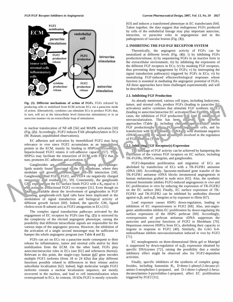
Fig. (3). Different mechanisms of action of FGFs. FGFs released by
producing cells or mobilized from ECM activate ECs via a paracrine mode
of action. Alternatively, cytokines can stimulate ECs to produce FGFs that,
in turn, will act at the intracellular level (intracrine stimulation) or in an
autocrine manner via an extracellular loop of stimulation.
to nuclear translocation of NF- B [56] and MAPK activation [50] (Fig. (2)). Accordingly, FGF2 induces FAK phosphorylation in ECs (M. Rusnati, unpublished observations).
EC adhesion and activation by immobilized FGF2 may have relevance in vivo since FGF2 accumulates as an immobilized protein in the ECM, mainly by binding to HSPGs. Accordingly, heparin-bound FGF2 retains it cell-adhesive capacity [57]. Thus, HSPGs may facilitate the interaction of ECM with FGF2 that, in turn, promotes EC adhesion and activation.
Gangliosides are neuraminic acid-containing glycosphingo-lipids mainly found associated to the EC membrane, where they modulate cell growth, adhesion, and cell-cell interaction [58]. Gangliosides bind FGF1, FGF2, and FGF4 via negatively charged neuraminic acid residues [31, 59]. Consistently, the ganglioside GM1 expressed on the EC surface binds FGF2 with a Kd equal to 3 nM, acting as a functional FGF2 co-receptor [31]. Even though no data are available about the involvement of gangliosides in FGF signaling, ganglioside-rich lipid rafts have been implicated in the modulation of signal transduction and biological activity of different growth factors [60]. Indeed, the specific GM1 ligand cholera toxin B subunit acts as FGF2 antagonists in ECs [31].
The complex signal transduction pathways activated by the engagement of EC receptors by FGFs (see Fig. (2)) is mirrored by the complexity of the elicited angiogenic phenotype, raising the possibility that different intracellular signals are responsible for the various steps of the angiogenic process. However, the inhibition of the activation of a single second messenger may be sufficient to hamper the whole angiogenic program (see Table 4 and 5).
FGFs can act on ECs via a paracrine mode consequent to their release by inflammatory, tumor and stromal cells and/or by their mobilization from the ECM. On the other hand, FGFs play autocrine/intracrine roles in ECs (see [61] and references therein). Relevant to this point, the single-copy human fgf2 gene encodes multiple FGF2 isoforms (from 18 to 24 kDa) that play different functions possibly related to differences in their release and/or subcellular localization [62]. Indeed, high molecular weight FGF2 isoforms contain a nuclear localization sequence, are mostly recovered in the nucleus, and lead to cell immortalization when overexpressed in ECs. In contrast, 18 kDa FGF2 is mostly cytosolic
[63] and induces a transformed phenotype in EC transfectants [64]. Taken together, the data suggest that endogenous FGFs produced by cells of the endothelial lineage may play important autocrine, intracrine, or paracrine roles in angiogenesis and in the pathogenesis of vascular lesions (Fig. (3)).
2. INHIBITING THE FGF/FGF RECEPTOR SYSTEM
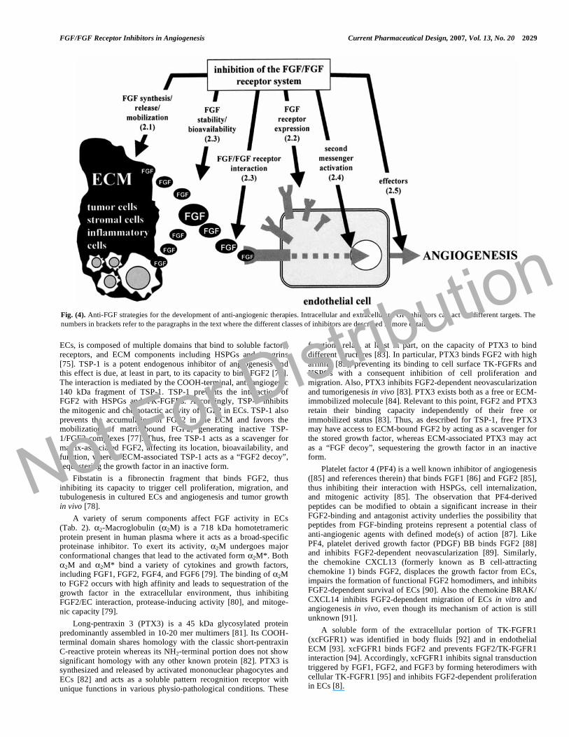
Theoretically, the angiogenic activity of FGFs can be neutralized at different levels (Fig. (4)): i) by inhibiting FGFs production/release; ii) by sequestering FGFs in an inactive form in the extracellular environment; iii) by inhibiting the expression of the different FGF receptors in ECs; iv) by masking FGF receptors, thus preventing their engagement by FGFs; v) by interrupting the signal transduction pathway(s) triggered by FGFs in ECs; vi) by neutralizing FGF-induced effectors/biological responses whose function is essential in mediating the angiogenic potential of FGFs. All these approaches have been challenged experimentally and will be described below.
2.1. Inhibiting FGF Production
As already mentioned, various cell types, including leukocytes, tumor, and stromal cells, produce FGFs (leading to paracrine EC activation) and/or cytokines that stimulate FGF synthesis in ECs (leading to autocrine/intracrine EC activation) (see Fig. (3)). In both cases, the inhibition of FGF production will lead to inhibition of neovascularization. This has been achieved with different approaches (Table 1), including chemotherapeutics, that inhibit FGF production by killing FGF-producing tumor cells, and by transfection with FGF antisense cDNAs or with dominant negative cDNAs encoding for second messengers involved in the regulation of FGF synthesis (Table 1).
2.2. Inhibiting FGF Receptor(s) Expression
The blockage of FGF activity can be achieved by hampering the expression of the various FGF receptors on EC surface, including TK-FGFRs, HSPGs, integrins, and gangliosides.
FGF2-dependent proliferation and migration of ECs are abolished by transfection of ECs with a TK-FGFR1 antisense cDNA [40]. Accordingly, liposome-mediated gene transfer of the TK-FGFR1 antisense cDNA blocks intratumoral angiogenesis in human melanomas grafted in nude mice [65]. Also, the synthetic retinoid fenretinide inhibits FGF2-induced angiogenesis in vivo and EC proliferation in vitro by reducing the expression of TK-FGFR2 on the EC surface [66]. Finally, EC surface expression of TK-FGFR1 and TK-FGFR2 can be inhibited by antibodies directed against v 3 and 5 1 integrins or by exposure to fibrin [67].
Lead exposure causes HSPG down-regulation, leading to inhibition of EC responsiveness to FGF2 [68]. Also, anti-angio-genic antithrombin inhibits EC proliferation by down-regulating the surface expression of the HSPG perlecan [69]. Accordingly, overexpression of perlecan antisense cDNA suppresses the autocrine and paracrine functions of FGF2 in fibroblasts [70]. Heparinase removes HSPGs from ECs, abolishing their capacity to migrate in response to FGF2 [40]. Similarly, the GAG 6-0-endosulfatase inhibits neovascularization induced in vivo by FGF2 [71].
EC morphogenesis on three-dimensional fibrin gel or Matrigel is suppressed by down-regulation of v 3 expression obtained by specific DNAzymes [72], raising the possibility that a similar inhibitory effect might be observed also for FGF2-dependent activities.
Finally, specific inhibitors of the synthesis of complex gang-liosides, including fumonisin B1, D-threo-1-phenyl-2-decano-yl-amino-3-morpholino-1-propanol, and D-1-threo-1-phenyl-2-hexa-decanoylamino-3-pyrrolidino-1-propanol, affect EC proliferation triggered by FGF2 [31].
Not For Distribution

2028 Current Pharmaceutical Design, 2007, Vol. 13, No. 20 Rusnati and Presta
Table 1. Inhibition of FGF Production in Tumor and ECs
Cell Type Experimental Approach Inhibitor Reference
FGF2 antisense cDNA transfection [65]
STAT1 knockout [175]
dominant negative STAT3 transfection [73]
modulation of gene expression
dominant negative Akt transfection [73]
taxane IDN 5109 (BAY59-8862) [176]
docetaxel [177]
epidermal growth factor receptor TK inhibitor ZD1839 (Iressa) [178]
doxycycline [179]
thalidomide [180]
chemotherapeutics
zoledronic acid [181]
JAK inhibitor AG490 [73]
PI3K inhibitor LY294002 [73] second messenger inhibitors
PKA inhibitor 8-chloro-cyclic AMP [182]
genistein [183]
fumagillin and its analog TNP-470 [184]
curcumin [132]
natural products
green tea (epigallocatechin-3-gallate) [134]
dipeptidyl peptidase IV [185]
tumor cells
endogenous molecules
INF- [186]
c-jun antisense cDNA transfection [187]
dominant negative ERK1/2 transfection [188]
dominant negative JNK transfection [188]
anti-early growth response-1 (Egr-1) DNA-cleaving deoxyribozymes [189]
modulation of gene expression
anti-FGF2 antisense oligonucleotides [190]
PI3K inhibitor LY294002 [191] second messenger inhibitors
PKC inhibitor calphostin C [192]
ECs
natural products green tea (epigallocatechin-3-gallate) [134]
2.3. Inhibiting FGF Interaction with EC Receptors
In the presence of FGFs and their EC receptors, it is still possible to block neovascularization by sequestering FGFs in the extracellular environment or by concealing the receptors to their ligands.
2.3.1. Sequestering FGFs in the Extracellular Environment
Classically, the interaction with target cells can be prevented by means of specific antibodies raised against the growth factor. This is the case also for FGF2, whose functions can be inhibited by neutralizing antibodies in different experimental conditions [73, 74].
Once released in the extracellular environment, FGFs interact with several partners that modulate their bioavailability, stability, local concentration, interaction with EC receptors, and intracellular fate [5]. The identification of these molecules and the biochemical characterization of their FGF-binding/antagonist capacity may allow the design of selective inhibitors. Since the bulk of data refer to FGF2, we will focus on this member of the FGF family, even though many of the interactions described below may apply to various FGFs.
Several ECM components or their degradation products affect FGF-driven angiogenesis (Table 2). Thrombospondin-1 (TSP-1), a modular glycoprotein secreted by different cell types, including
Not For Distribution
FGF/FGF Receptor Inhibitors in Angiogenesis Current Pharmaceutical Design, 2007, Vol. 13, No. 20 2029
ECs, is composed of multiple domains that bind to soluble factors, receptors, and ECM components including HSPGs and integrins [75]. TSP-1 is a potent endogenous inhibitor of angiogenesis and this effect is due, at least in part, to its capacity to bind FGF2 [76]. The interaction is mediated by the COOH-terminal, anti-angiogenic 140 kDa fragment of TSP-1. TSP-1 prevents the interaction of FGF2 with HSPGs and TK-FGFRs. Accordingly, TSP-1 inhibits the mitogenic and chemotactic activity of FGF2 in ECs. TSP-1 also prevents the accumulation of FGF2 in the ECM and favors the mobilization of matrix-bound FGF2, generating inactive TSP-1/FGF2 complexes [77]. Thus, free TSP-1 acts as a scavenger for matrix-associated FGF2, affecting its location, bioavailability, and function, whereas ECM-associated TSP-1 acts as a “FGF2 decoy”, sequestering the growth factor in an inactive form.
Fibstatin is a fibronectin fragment that binds FGF2, thus inhibiting its capacity to trigger cell proliferation, migration, and tubulogenesis in cultured ECs and angiogenesis and tumor growth in vivo [78].
A variety of serum components affect FGF activity in ECs (Tab. 2). 2-Macroglobulin ( 2M) is a 718 kDa homotetrameric protein present in human plasma where it acts as a broad-specific proteinase inhibitor. To exert its activity, 2M undergoes major conformational changes that lead to the activated form 2M*. Both
2M and 2M* bind a variety of cytokines and growth factors, including FGF1, FGF2, FGF4, and FGF6 [79]. The binding of 2M to FGF2 occurs with high affinity and leads to sequestration of the growth factor in the extracellular environment, thus inhibiting FGF2/EC interaction, protease-inducing activity [80], and mitoge-nic capacity [79].
Long-pentraxin 3 (PTX3) is a 45 kDa glycosylated protein predominantly assembled in 10-20 mer multimers [81]. Its COOH-terminal domain shares homology with the classic short-pentraxin C-reactive protein whereas its NH2-terminal portion does not show significant homology with any other known protein [82]. PTX3 is synthesized and released by activated mononuclear phagocytes and ECs [82] and acts as a soluble pattern recognition receptor with unique functions in various physio-pathological conditions. These
functions relay, at least in part, on the capacity of PTX3 to bind different structures [83]. In particular, PTX3 binds FGF2 with high affinity [83], preventing its binding to cell surface TK-FGFRs and HSPGs with a consequent inhibition of cell proliferation and migration. Also, PTX3 inhibits FGF2-dependent neovascularization and tumorigenesis in vivo [83]. PTX3 exists both as a free or ECM-immobilized molecule [84]. Relevant to this point, FGF2 and PTX3 retain their binding capacity independently of their free or immobilized status [83]. Thus, as described for TSP-1, free PTX3 may have access to ECM-bound FGF2 by acting as a scavenger for the stored growth factor, whereas ECM-associated PTX3 may act as a “FGF decoy”, sequestering the growth factor in an inactive form.
Platelet factor 4 (PF4) is a well known inhibitor of angiogenesis ([85] and references therein) that binds FGF1 [86] and FGF2 [85], thus inhibiting their interaction with HSPGs, cell internalization, and mitogenic activity [85]. The observation that PF4-derived peptides can be modified to obtain a significant increase in their FGF2-binding and antagonist activity underlies the possibility that peptides from FGF-binding proteins represent a potential class of anti-angiogenic agents with defined mode(s) of action [87]. Like PF4, platelet derived growth factor (PDGF) BB binds FGF2 [88] and inhibits FGF2-dependent neovascularization [89]. Similarly, the chemokine CXCL13 (formerly known as B cell-attracting chemokine 1) binds FGF2, displaces the growth factor from ECs, impairs the formation of functional FGF2 homodimers, and inhibits FGF2-dependent survival of ECs [90]. Also the chemokine BRAK/ CXCL14 inhibits FGF2-dependent migration of ECs in vitro and angiogenesis in vivo, even though its mechanism of action is still unknown [91].
A soluble form of the extracellular portion of TK-FGFR1 (xcFGFR1) was identified in body fluids [92] and in endothelial ECM [93]. xcFGFR1 binds FGF2 and prevents FGF2/TK-FGFR1 interaction [94]. Accordingly, xcFGFR1 inhibits signal transduction triggered by FGF1, FGF2, and FGF3 by forming heterodimers with cellular TK-FGFR1 [95] and inhibits FGF2-dependent proliferation in ECs [8].
Fig. (4). Anti-FGF strategies for the development of anti-angiogenic therapies. Intracellular and extracellular FGF inhibitors can act on different targets. The
numbers in brackets refer to the paragraphs in the text where the different classes of inhibitors are described in more details.
Not For Distribution

2030 Current Pharmaceutical Design, 2007, Vol. 13, No. 20 Rusnati and Presta
Table 2. Endogenous Inhibitors of FGFs in ECs
Localization Molecule Mechanism of Action
homeobox gene GAX inhibition of NF-kB activation [193]
sprouty proteins inhibition of TK-FGFR signaling [194]
intracellular
heat shock proteins (Hsp) 70 and 90 pAkt, c-Raf-1, and ERK1/2 down modulation [195]
collagen I unknown [196]
TSP-1 FGF2 sequestration [76], CD36 engagement [155], integrin occupancy (?),
HSPG occupancy (?)
alphastatin (fibrinogen fragment) unknown [197]
endostatin cytoskeleton organization [198], Shb activation [199]
ECM
fibstatin (fibronectin fragment) FGF2 sequestration [78]
CXCL13 FGF2 sequestration [90]
CXCL14 unknown [91]
PDGF FGF2 sequestration [88]
2M FGF2 sequestration [79]
PTX3 FGF2 sequestration [83]
heparin FGF2 sequestration [42]
gangliosides FGF2 sequestration [59]
PF4 FGF2 sequestration [86], HSPG occupancy [109], unknown [153]
xcFGFR1 FGF2 sequestration [94], formation of heterodimers with TK-FGFR1 [95]
histidine-rich glycoprotein HSPG occupancy [109], tropomyosin engagement [200]
antithrombin HSPG down-regulation [69]
thromboxane inhibition of TK-FGFR1 internalization [201]
angiostatin (fragment of plasminogen) inhibition of ERK cascade [202]
prolactin (16 kDa fragment) unknown [203]
vitamin D3-binding protein CD36 engagement [204]
ghrelin inhibition of TK/MAPK cascades [205]
lysophosphatidylcholine inhibition of ras/ERK1/2 cascades [206]
cleaved HMW kininogen tropomyosin engagement [207]
IL-4 alteration of cell cycle [208]
IL-12 unknown [209]
IP-10 unknown [210]
pigment epithelium-derived factor inhibition of Fyn [211]
vasculostatin (fragment of brain angiogenesis inhibitor-1) unknown [212]
vasostatin unknown [213]
kininostatin (fragment of kininogen) inhibition of cyclin D1 expression [214]
kallistatin HSPG occupancy, inhibition of FGF-induced proteases [112]
blood
TGF- 1 unknown [14]
Not For Distribution

FGF/FGF Receptor Inhibitors in Angiogenesis Current Pharmaceutical Design, 2007, Vol. 13, No. 20 2031
(Table 2) Contd….
Localization Molecule Mechanism of Action
TIMP-2, 4 inhibition of FGF-induced proteases [215]
IFN- TK-FGFR down-regulation [216]
IL-1 TK-FGFR down-regulation [216]
TNF- , unknown [217]
somatostatin unknown [218]
retinoids unknown [66]
apolipoprotein(a) unknown [219]
heparan sulfate 6-0-endosulfatase HSPG desulfation [71]
heparinase HSPG degradation [40]
extracellular
micro-
environment semaphorin-3F inhibition of ERK1/2 cascade [220]
FGFs bind free heparin, a negatively charged GAG released in the blood stream during inflammation. At variance with HSPGs, that act as FGF co-receptors (see above), free heparin sequesters FGFs in the extracellular environment exerting an antagonist effect. However, due to its anticoagulant activity and its capacity to bind a wide array of growth factors, cytokines, enzymes, and proteases, unmodified heparin can not be used as an anti-angiogenic drug. This prompted a series of studies aimed at identifying heparin derivatives and/or heparin-like molecules endowed with a more specific FGF antagonist activity and a more favorable therapeutic window (reviewed in [96]). A list of polyanionic compounds able to bind FGFs and to inhibit their biological activity in ECs is shown in Table 3.
It must be pointed out that polyanionic compounds may exert also co-stimulatory effects on FGF activity depending on various experimental conditions, including: i) the member of the FGF family under investigation and/or the utilized biological assay; ii) the molar ratio of the FGF:polyanion interaction and medium composition [97]; iii) the EC type under study ([42] and references therein); iv) the structural properties of the polyanion under test [98]. Taken together, these considerations call for an extreme caution in the design of this class of anti-angiogenic compounds and in the evaluation of their biological activity.
Given the structural similarity among the various members of the FGF family and the heparin-binding capacity shared by a variety of angiogenic growth factors and cytokines, it may be difficult to envisage the design of selective polyanionic antagonists. Nevertheless, recent observations have shown the possibility to achieve a certain degree of specificity by selective structural modifications of the E. coli K5 polysaccharide [99, 100]. It must be pointed out, however, that the “multitarget” activity of certain polyanionic compounds may increase their efficacy in vivo. Indeed, tumor angiogenesis and growth are often the result of the synergistic action of more than one angiogenic growth factor ([5] and references therein). Relevant to this point, pentosan polysulfate (PPS) efficiently inhibits the biological activity of the angiogenic HIV-1 transactivating factor (Tat) [101] as well as of FGF2 [102]. Interestingly, phase I and II clinical trials have shown that PPS leads to stabilization of Kaposi’s sarcoma [103], a lesion in which HIV-1 Tat and FGF2 act synergistically [104].
A peculiar class of polyanionic compounds is represented by sialo-gangliosides that act as functional FGF2 co-receptors when associated to the EC surface [31]. During tumor growth, sialo-gangliosides are shed in the microenvironment, where they bind and sequester FGF2, inhibiting its EC interaction and mitogenic
activity [59]. Sialo-gangliosides may therefore represent the basis for the design of novel anti-angiogenic FGF-antagonists.
2.3.2. Masking FGF Receptors
Neutralizing anti-TK-FGFR antibodies have been shown to block FGF2-mediated angiogenesis in vivo [105]. Also, TK-FGFRs can be bound by synthetic peptides and masked to their ligands. For instance, the interaction of FGF2 with TK-FGFR1 can be inhibited by peptides derived from the amino acid sequence 112-155 of the growth factor [8]. Also, a structural analysis carried out on FGF2 identified a region encompassing residues 48-58 as involved in FGF2 dimerization. Accordingly, the derived peptide FREG-(48-58) prevents dimerization of the growth factor and its interaction with TK-FGFR1, thus inhibiting TK-FGFR1 phosphorylation, FGF2-dependent EC proliferation and migration in vitro and angiogenesis in vivo [106]. Furthermore, a polyclonal antibody directed against FREG-(48-58) blocks FGF2 action in vitro [106]. In contrast, a FGF2 peptide derived from the amino-terminal extension of the high molecular weight 24 kDa FGF2 isoform plus the first 31 amino acids from the canonic 18 kDa isoform, inhibits FGF2-dependent migration of ECs without affecting FGF2/TK-FGFR1 interaction nor extracellular regulated kinase1/2 (ERK1/2) activation [107]. Finally, FGF/TK-FGFR interaction can be disrupted by protamine, an arginine-rich polypeptide that inhibits FGF2-dependent proliferation of ECs [8] possibly by binding and masking TK-FGFRs [108].
Besides masking TK-FGFRs, protamine interacts with and masks HSPGs [108]. Similarly, the histidine-rich glycoprotein and PF4 bind and mask cell surface HSPGs, hindering these receptors to FGF2 and FGF1 [109]. Also, the anti-angiogenic collagen XVIII fragment endostatin prevents FGF2/HSPG interaction [110]. In keeping with these observations, a liposome-based peptide vaccine targeting the heparin-binding domain of FGF2 generates a specific anti-FGF2 antibody that inhibits FGF2 binding to HSPGs and FGF2-dependent angiogenesis in vivo [111]. Finally, kallistatin, a serpin originally identified as a specific inhibitor of tissue kallikrein, inhibits FGF2-induced proliferation, migration, and adhesion of cultured ECs and neovascularization in vivo possibly by hindering HSPGs to FGF2 binding [112].
Besides TK-FGFRs and HSPGs, integrins may represent a target for anti-angiogenic compounds. For instance, synthetic peptides representing two regions of the FGF2 molecule [FGF2(61-73) and FGF2(82-101)] inhibit FGF2-dependent proliferation of ECs [113]. These regions contain an Asp-Gly-Arg (DGR) sequence that is the inverse of the integrin-recognition sequence RGD present in many adhesive proteins. Actually, the two FGF2-derived peptides
Not For Distribution

2032 Current Pharmaceutical Design, 2007, Vol. 13, No. 20 Rusnati and Presta
Table 3. Heparin-Like Polyanionic Compounds that Inhibit
FGF2 Activity in ECs
Polyanionic Compound Inhibited EC Response
sulfated malto-oligosaccharides proliferation, morphogenesis
[221]
sulfated beta-(1-->4)-galacto-
oligosaccharides
angiogenesis [222]
RG-13577 (non sulfated aromatic
compound)
proliferation, morphogenesis
[223]
heparin-derived oligosaccharides proliferation [97], angiogenesis,
tumor growth [224]
fucoidan proliferation, migration [225],
morphogenesis, integrin
expression [226]
suramin motogenesis [105]
suramin derivatives angiogenesis, proliferation [227],
migration, uPA expression [228],
tumorigenesis [229]
PPS proliferation, migration [102]
TMPP (porphyrin analogue) morphogenesis [230]
K5 derivatives (chemically sulfated
polysaccharides from E. coli)
proliferation, FGF2-dependent
cell-cell interaction,
morphogenesis, angiogenesis
[99], cell adhesion [57]
suleparoide (heparan sulfate analog) angiogenesis [231]
undersulfated glycol-split heparins proliferation, FGF2-dependent
cell-cell interaction, angiogenesis
[232]
synthetic sulfonic acid polymers FGF2-dependent cell-cell
interaction [233], proliferation,
angiogenesis, morphogenesis
[234]
-cyclodextrin polysulfate angiogenesis [235]
ATA (aurintricarboxylic acid) angiogenesis [236]
PS-ODN (phosphorothioate
oligodeoxynucleotides)
morphogenesis, angiogenesis
[237]
gangliosides proliferation, angiogenesis [238]
carrageenan proliferation [143]
inositol hexaphosphate angiogenesis [239]
inhibit v 3-mediated EC adhesion to immobilized FGF2 without affecting FGF2/TK-FGFR interaction [30]. Accordingly, RGD-containing tetra or eptapeptides, and monoclonal anti- v 3 antibodies inhibit FGF2-dependent EC adhesion, proliferation, and uPA production [30, 113]. Following these observations, we have demonstrated that RGD-pepti-domimetics inhibit FGF2-dependent neovascularization and tumori-genesis in vivo [114, 115]. A similar mechanism of action may be shared by disintegrins, a class of naturally occurring integrin antagonists that inhibit different aspects of FGF2 biology [116].
Finally, the cholera toxin B subunit inhibits FGF2-dependent proliferation of ECs by binding the cell surface GM1 ganglioside [31].
2.4. Inhibiting FGF Receptor Signal Transduction
Intracellular signals activated by FGFs in ECs (Fig. (2)) might be considered as a target for angiogenesis inhibitors [35]. Actually, FGF activity can be inhibited in vitro and in vivo by synthetic compounds (Table 4) and selective dominant negative mutants or antisense cDNAs (Table 5) targeting various signal transduction pathways triggered by FGFs. Also, different endogenous inhibitors of angiogenesis have been shown to affect FGF signaling (Table 2). Among them, several cytokines modulate EC activation and/or neovascularization induced by FGF2. It is possible to hypothesize that these cytokines, by interacting with their cognate receptors on ECs, may interfere with the signal transduction pathway(s) activated by the angiogenic growth factor. However, the therapeutic exploitation of this approach is greatly limited by the fact that several among the second messengers activated by FGFs during pathological neovascularization are implicated in various physio-logical processes. Their inhibition may thus cause undesired side effects.
2.5. Inhibiting FGF-Activated EC Responses/Effectors of Angiogenesis
FGFs induce a complex “pro-angiogenic phenotype” in ECs characterized by an increase in ECM degradation and in EC motility, proliferation, and morphogenesis (see Fig. (1)). These processes are mediated by distinct effectors induced/activated by FGFs, and their blockage may result in the inhibition of FGF-dependent angiogenesis.
For instance, in order to degrade ECM, FGFs upregulate the production of several proteases in ECs (see above). Tissue inhibitors of MMPs (TIMPs) and synthetic MMP inbitors [117] inhibit FGF2 neovascularization [118]. Interestingly, a MMP-independent mechanism of inhibition of FGF-dependent angio-genesis has been proposed for TIMP-2 [118]. Also, MMP production and FGF2-dependent angiogenesis can be inhibited by endogenous mediators, like interferons (IFNs) [119]. Similarly, PA/plasmin inhibitors affect FGF2-dependent angiogenesis in vitro and in vivo [120]. Finally, inhibition of proteases has been proposed to contribute to the FGF2-inhibitory effect exerted by kallistatin [112].
The epidermal growth factor-like domain of murine uPA alone or fused to the Fc portion of human IgG acts as high-affinity uro-kinase receptor antagonist and inhibits FGF2-induced angiogenesis in vivo [121]. Accordingly, medroxyprogesterone acetate exerts an angiostatic effect by increasing the expression of PA inhibitor-1, thus counteracting the uPA-inducing activity of FGF2 [122].
The properties of neovasculature differ from those of quiescent endothelium. Vascular targeting agents exploit differences in cell proliferation, permeability, maturation, and reliance on tubulin cytoskeleton to induce selective blood vessel occlusion and destruction [123]. In particular, microtubule-destabilizing agents, including combretastatin-derived prodrugs and analogues, disrupt rapidly proliferating and immature tumor endothelium, leading to reduced blood flow and hypoxia [124]. Interestingly, microtubule-destabilizing agents, e.g. combretastatin A-4 and vinblastine, may also show a distinct anti-angiogenic activity [125]. Accordingly, the trans-resveratrol derivative 3,5,4’-trimethoxystilbene acts as a microtubule-destabilizing agent endowed with both anti-angiogenic FGF2-antagonist activity and vascular targeting capacity [126]. Similarly, microtubule-stabilizing agents, including paclitaxel and taxane derivatives [127, 128], affect FGF2-triggered angiogenesis in vitro and in vivo. Also, by preventing the formation of stress fibers, the antifungal polyether macrolide goniodomin-A inhibits FGF2-induced migration and morphogenesis in ECs, leaving unaffected their proliferation [129]. These findings are of importance
Not For Distribution

FGF/FGF Receptor Inhibitors in Angiogenesis Current Pharmaceutical Design, 2007, Vol. 13, No. 20 2033
Table 4. Chemical Inhibitors of FGF2-Mediated Intracellular Signaling
Inhibitor Second
Messenger Inhibited EC Response
SU5416
SU5402
Z24
PD173074
CP-547,632
FGFR-TK
FGFR-TK
FGFR-TK
FGFR-TK
FGFR-TK
survival [240], angiogenesis [241], EC monolayer wound repaira
proliferation [240]
angiogensis [241]
morphogenesis, angiogenesis [242]
proliferation, angiogenesis [243]
PD 098059
U0126
apigenin
ERK1/2
ERK1/2
ERK1/2
proliferation [50], survival [244], uPA expression [245], MMP3 expression [246], migration [247], CD13
expression [248], morphogenesis [248], angiogenesis [248], survival, integrin activation [249], Egr-1
expression [250], KDR expression [251]
morphogenesis [245], survival [252], MMP3 expression [246], motogenesisa
proliferation [253]
SB203580 P38 morphogenesis [254]
LY294002
neutralizing antibodies
apigenin
PI3K
PI3K
PI3K
survival [252], CD13 expression, morphogenesis [248], migration [162], proliferation [253], cytoskeleton
organization [255], motogenesisa, FGF2 production [191]
proliferation [256]
proliferation [253]
Bis I
GO6983
GFX
chelerythrine
H7
NSC 639366
calphostin C
PKC
PKC
PKC
PKC
PKC
PKC
PKC
survival [252]
survival [252]
KDR expression [251]
proliferation [257]
proliferation [258], survival [259]
migration, uPA expression, angiogenesis [260]
angiogenesis [261], FGF2 production [192]
manumycin A
FTS
FPT inhibitor III
Ras
Ras
Ras
CD13 expression [248], morphogenesis [248], proliferation [262]
proliferation [262]
proliferation [253]
tyrphostin 23
genistein
herbimycin A
Pan-TK
Pan-TK
Pan-TK
proliferation [50], EC monolayer wound repaira
proliferation [263]
proliferation [263]
PP1
PP2
c-Src
c-Src
migration [264], morphogenesis [262]
angiogenesis, morphogenesis, cytoskeleton organization [265]
neutralizing antibodies
aristolochic acid
ONO-RS-082
PLC-
PLC- 2
PLC- 2
proliferation [266]
migration [267]
migration [267]
rapamycin P70S6K proliferation [253]
C3 RhoA ICAM-1 expression [268]
Grb2–Src homology 2
domain binding antagonist
Grb2 proliferation, migration, angiogenesis [269]
Not For Distribution
2034 Current Pharmaceutical Design, 2007, Vol. 13, No. 20 Rusnati and Presta
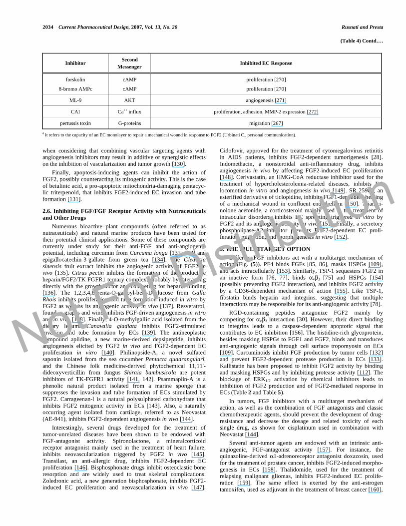
(Table 4) Contd….
Inhibitor Second
Messenger Inhibited EC Response
forskolin
8-bromo AMPc
cAMP
cAMP
proliferation [270]
proliferation [270]
ML-9 AKT angiogenesis [271]
CAI Ca++ influx proliferation, adhesion, MMP-2 expression [272]
pertussis toxin G-proteins migration [267]
a it refers to the capacity of an EC monolayer to repair a mechanical wound in response to FGF2 (Urbinati C., personal communication).
when considering that combining vascular targeting agents with angiogenesis inhibitors may result in additive or synergistic effects on the inhibition of vascularization and tumor growth [130].
Finally, apoptosis-inducing agents can inhibit the action of FGF2, possibly counteracting its mitogenic activity. This is the case of betulinic acid, a pro-apoptotic mitochondria-damaging pentacyc-lic triterpenoid, that inhibits FGF2-induced EC invasion and tube formation [131].
2.6. Inhibiting FGF/FGF Receptor Activity with Nutraceuticals
and Other Drugs
Numerous bioactive plant compounds (often referred to as nutraceuticals) and natural marine products have been tested for their potential clinical applications. Some of these compounds are currently under study for their anti-FGF and anti-angiogenic potential, including curcumin from Curcuma longa [132, 133] and epigallocatechin-3-gallate from green tea [134]. The Gleditsia sinensis fruit extract inhibits the angiogenic activity of FGF2 in vivo [135]. Citrus pectin inhibits the formation of the productive heparin/FGF2/TK-FGFR1 ternary complex, probably by interacting directly with the growth factor and competing for heparin binding [136]. The 1,2,3,4,6-penta-O-galloyl-beta-D-glucose from Galla Rhois inhibits proliferation and tube formation induced in vitro by FGF2 as well as its angiogenic activity in vivo [137]. Resveratrol, found in grapes and wine, inhibits FGF-driven angiogenesis in vitro and in vivo [138]. Finally, 4-O-methylgallic acid isolated from the dietary legume Canavalia gladiata inhibits FGF2-stimulated invasion and tube formation by ECs [139]. The antineoplastic compound aplidine, a new marine-derived depsipeptide, inhibits angiogenesis elicited by FGF2 in vivo and FGF2-dependent EC proliferation in vitro [140]. Philinopside-A, a novel sulfated saponin isolated from the sea cucumber Pentacta quadrangulari, and the Chinese folk medicine-derived phytochemical 11,11'-dideoxyverticillin from fungus Shiraia bambusicola are potent inhibitors of TK-FGFR1 activity [141, 142]. Psammaplin-A is a phenolic natural product isolated from a marine sponge that suppresses the invasion and tube formation of ECs stimulated by FGF2. Carrageenan-l is a natural polysulphated carbohydrate that inhibits FGF2 mitogenic activity in ECs [143]. Also, a naturally occurring agent isolated from cartilage, referred to as Neovastat (AE-941), inhibits FGF2-dependent angiogenesis in vivo [144].
Interestingly, several drugs developed for the treatment of tumor-unrelated diseases have been shown to be endowed with FGF-antagonist activity. Spironolactone, a mineralcorticoid receptor antagonist mainly used in the treatment of heart failure, inhibits neovascularization triggered by FGF2 in vivo [145]. Transilast, an anti-allergic drug, inhibits FGF2-dependent EC proliferation [146]. Bisphosphonate drugs inhibit osteoclastic bone resorption and are widely used to treat skeletal complications. Zoledronic acid, a new generation bisphosphonate, inhibits FGF2-induced EC proliferation and neovascularization in vivo [147].
Cidofovir, approved for the treatment of cytomegalovirus retinitis in AIDS patients, inhibits FGF2-dependent tumorigenesis [28]. Indomethacin, a nonsteroidal anti-inflammatory drug, inhibits angiogenesis in vivo by affecting FGF2-induced EC proliferation [148]. Cerivastatin, an HMG-CoA reductase inhibitor used for the treatment of hypercholesterolemia-related diseases, inhibits EC locomotion in vitro and angiogenesis in vivo [149]. SR 25989, an esterified derivative of ticlopidine, inhibits FGF1-dependent healing of a mechanical wound in confluent endothelium [150]. Triamci-nolone acetonide, a corticosteroid mainly used in the treatment of intraocular disorders, inhibits EC sprouting triggered in vitro by FGF2 and its angiogenic activity in vivo [151]. Finally, a secretory phospholipase-A2 inhibitor prevents FGF2-dependent EC proli-feration, migration, and morphogenesis in vitro [152].
3. THE MULTITARGET OPTION
Different FGF inhibitors act with a multitarget mechanism of action (Fig. (5)). PF4 binds FGFs [85, 86], masks HSPGs [109], and acts intracellularly [153]. Similarly, TSP-1 sequesters FGF2 in an inactive form [76, 77], binds v 3 [75] and HSPGs [154] (possibly preventing FGF2 interaction), and inhibits FGF2 activity by a CD36-dependent mechanism of action [155]. Like TSP-1, fibstatin binds heparin and integrins, suggesting that multiple interactions may be responsible for its anti-angiogenic activity [78].
RGD-containing peptides antagonize FGF2 mainly by competing for v 3 interaction [30]. However, their direct binding to integrins leads to a caspase-dependent apoptotic signal that contributes to EC inhibition [156]. The histidine-rich glycoprotein, besides masking HSPGs to FGF1 and FGF2, binds and transduces anti-angiogenic signals through cell surface tropomyosin on ECs [109]. Curcuminoids inhibit FGF production by tumor cells [132] and prevent FGF2-dependent protease production in ECs [133]. Kallistatin has been proposed to inhibit FGF2 activity by binding and masking HSPGs and by inhibiting protease activity [112]. The blockage of ERK1/2 activation by chemical inhibitors leads to inhibition of FGF2 production and of FGF2-mediated response in ECs (Table 2 and Table 5).
In tumors, FGF inhibitors with a multitarget mechanism of action, as well as the combination of FGF antagonists and classic chemotherapeutic agents, should prevent the development of drug-resistance and decrease the dosage and related toxicity of each single drug, as shown for cisplatinum used in combination with Neovastat [144].
Several anti-tumor agents are endowed with an intrinsic anti-angiogenic, FGF-antagonist activity [157]. For instance, the quinazoline-derived 1-adrenoreceptor antagonist doxazosin, used for the treatment of prostate cancer, inhibits FGF2-induced morpho-genesis in ECs [158]. Thalidomide, used for the treatment of relapsing malignant gliomas, inhibits FGF2-induced EC prolife-ration [159]. The same effect is exerted by the anti-estrogen tamoxifen, used as adjuvant in the treatment of breast cancer [160],
Not For Distribution

FGF/FGF Receptor Inhibitors in Angiogenesis Current Pharmaceutical Design, 2007, Vol. 13, No. 20 2035
Fig. (5). Multitarget activity of selected FGF inhibitors. Possible
mechanisms of action of anti-angiogenic ERK1/2 inhibitors, TSP-1, and PF4.
See text for further details.
and by the functionally related medroxyprogesterone-acetate that inhibits the release of uPA induced by FGF2 in ECs [161]. The topoisomerase-I inhibitor topotecan possesses an indirect anti-tumor effect in vivo mediated by angiosuppression due, at least in part, to inhibition of FGF2-induced EC migration [162]. Aplidine, that exerts a cytotoxic effect in tumor cells and is currently tested in early phase clinical trials, possesses FGF2-antagonist activity [140]. The same dual effect has been demonstrated for Neovastat [144]. The chemotherapic 6-methylmercaptopurine-riboside inhibits FGF2-dependent angiogenesis in vitro and in vivo [163]. Combination of tegafur and uracil (UFT), utilized for the treatment of a variety of malignant tumors, inhibits EC proliferation induced by FGF2 [164]. The antimetabolite 6-thioguanine, utilized in the management of acute myelogenous leukemia, inhibits EC proliferation and angiogenesis triggered by FGF2 [163]. Finally, Atiprimod, an azaspirane cationic amphiphilic drug, activates
caspases and induces apoptosis in various tumor cell lines and, simultaneously, inhibits FGF2-induced proliferation and migration of ECs [165].
It must be pointed out that, due to their pleiotropic nature, FGFs may contribute to cancer progression not only as pro-angiogenic growth factors but also by acting directly on tumor cells (Fig. (6)). For instance, the co-expression of FGF7/KGF and its receptor TK-FGFR2 IIIb/KGFR correlates with the high proliferative activity and poor prognosis in lung adenocarcinoma [166]. Also, high levels of FGF8 [167] or FGF17 [168] are associated with less favorable prognosis in human prostate cancer. Thus, targeting the FGF/FGF receptor system in cancer may provide benefits not only in terms of angiosuppression but also by a direct inhibition of tumor cell proliferation (Fig. (6)). For instance, inhibition of the FGF/FGF receptor system in glioma cells by dominant negative TK-FGFR transfection [169] or in prostate cancer cells by fgf2 gene knockout [170] results in inhibition of tumor growth by both angiogenesis-dependent and angiogenesis-independent mechanisms.
Table 5. “Modulation of Gene Expression” Approach for the
Inhibition of FGF2-Mediated Intracellular Signalinga
Target Inhibited EC Response
FGF2 b cell proliferation [273], angiogenesis [65, 274]
FGFR-TK proliferation [50], cytoskeleton organization
[275], migration, angiogenesis [276],
uPA expression [8]
Syndecan docking sites proliferation, migration, morphogenesis [39]
FAK angiogenesis [277]
c-Src chemotaxis [264], angiogenesis [265]
Rac proliferation [278]
Ras CD13 expression [248], angiogenesis [277]
Raf CD13 expression [248], survival [244],
angiogenesis [277]
MEK CD13 expression [248], proliferation,
migration [206]
ERK 1/2 CD13 expression [248]
SH2 cytoskeleton organization [255],
proliferation [279]
PKC proliferation, morphogenesis [280]
c-FES chemotaxis [281]
PI3K survival [282]
PAK angiogenesis [277]
AKT survival [283], morphogenesis [284]
Egr-1 c proliferation [189]
c-Fyn morphogenesis [285]
Ets-1 angiogenesis [286]
NF-kB d angiogenesis [287]
aInhibition was obtained by overexpression of dominant negative forms of the indi-
cated target with the exception for b, c, d where antisense oligonucleotides, neutralizing
single-stranded DNA, and IkB-2A overexpression were used, respectively.
Not For Distribution

2036 Current Pharmaceutical Design, 2007, Vol. 13, No. 20 Rusnati and Presta
4. CONCLUDING REMARKS
The bulk of experimental data summarized in this review clearly indicate that the FGF/FGF receptor system may represent a target for anti-angiogenic strategies in different pathological settings, including cancer. At present, cancer clinical trials are in progress to assess the safety and efficacy of various compounds with a potential capacity to affect the FGF/FGF receptor system at different levels [171, 172]. In several cases, however, the main rationale for testing these compounds was independent of their putative FGF/FGF receptor antagonist activity. For instance, heparin derivatives have been tested in cancer patients because of their anti-thrombotic effect rather than for their capacity to bind angiogenic FGFs. Similarly, the humanized anti- v 3 monoclonal antibody vitaxin [173, 174] has been investigated for its ability to affect the cell-adhesive function of this integrin receptor rather than for its potential role in angiogenesis and FGF activity. Also, as stated above, numerous cytotoxic drugs can affect the FGF/FGF receptor system and angiogenesis. Novel strategies aimed at inhibiting multiple targets, including the FGF/FGF receptor system, may represent an efficacious approach for the treatment of angiogenesis-dependent diseases, including cancer.
ACKNOWLEDGEMENTS
We wish to thank Dr. P. Dell’Era for helpful discussion. This work was supported by grants from MIUR (Centro di Eccellenza IDET, FIRB 2001, Cofin 2004), Fondazione Berlucchi, ISS (Progetto Oncotecnologico), and AIRC to MP, and by grants from AIRC, ISS (AIDS Project) and «60%» to MR.
ABBREVIATIONS
2M = 2-Macroglobulin
ECs = Endothelial cells
ECM = Extracellular matrix
ERK = Extracellular regulated kinase
FGF = Fibroblast growth factor
TK-FGFR = Tyrosine kinase FGF receptor
FAK = Focal adhesion kinase
GAG = Glycosaminoglycan
HS = Heparan sulfate
HSPGs = HS Proteoglycans
IFN = Interferon
Kd = Dissociation constant
MAPK = Mitogen activated protein kinase
MMP = Metalloproteinase
PF4 = Platelet factor-4
PDGF = Platelet derived growth factor
PIP2 = Phosphatidylinositol 4,5-bisphosphate
PPS = Pentosan polysulfate
PTX3 = Long-pentraxin 3
PKC = Protein kinase C
Tat = HIV-1 Transactivating factor
TIMP = Tissue inhibitors of MMP
TK = Tyrosine kinase
TSP-1 = Thrombospondin-1
uPA = Urokinase-type plasminogen activator
xcFGFR1 = Extracellular portion of FGFR1
Fig. (6). Multiple effects of FGF antagonists and antineoplastic drugs on tumor growth and neovascularization. FGF antagonists can affect tumor
growth indirectly by decreasing blood supply and directly by blocking FGF-dependent tumor cell proliferation. On the other hand, cytotoxic drugs can inhibit
EC proliferation and decrease the amount of FGF available to ECs by killing FGF-producing tumor cells.
Not For Distribution

FGF/FGF Receptor Inhibitors in Angiogenesis Current Pharmaceutical Design, 2007, Vol. 13, No. 20 2037
REFERENCES
References 290-292 are related articles recently published in Current Pharmaceutical Design.
[1] Carmeliet P, Jain RK. Angiogenesis in cancer and other diseases.
Nature 2000; 407: 249-57.
[2] Folkman J. Angiogenesis in cancer, vascular, rheumatoid and other
disease. Nat Med 1995; 1: 27-31.
[3] Maciag T, Mehlman T, Friesel R, Schreiber AB. Heparin binds
endothelial cell growth factor, the principal endothelial cell
mitogen in bovine brain. Science 1984; 225: 932-5.
[4] Itoh N, Ornitz DM. Evolution of the Fgf and Fgfr gene families.
Trends Genet 2004; 20: 563-9.
[5] Presta M, Dell'era P, Mitola S, Moroni E, Ronca R, Rusnati M.
Fibroblast growth factor/fibroblast growth factor receptor system in
angiogenesis. Cytokine Growth Factor Rev 2005; 16: 159-78.
[6] Javerzat S, Auguste P, Bikfalvi A. The role of fibroblast growth
factors in vascular development. Trends Mol Med 2002; 8: 483-9.
[7] Liotta LA, Steeg PS, Stetler-Stevenson WG. Cancer metastasis and
angiogenesis: an imbalance of positive and negative regulation.
Cell 1991; 64: 327-36.
[8] Rusnati M, Dell'Era P, Urbinati C, Tanghetti E, Massardi ML,
Nagamine Y, et al. A distinct basic fibroblast growth factor (FGF-
2)/FGF receptor interaction distinguishes urokinase-type
plasminogen activator induction from mitogenicity in endothelial
cells. Mol Biol Cell 1996; 7: 369-81.
[9] Taraboletti G, D'Ascenzo S, Borsotti P, Giavazzi R, Pavan A, Dolo
V. Shedding of the matrix metalloproteinases MMP-2, MMP-9,
and MT1-MMP as membrane vesicle-associated components by
endothelial cells. Am J Pathol 2002; 160: 673-80.
[10] Hiraoka N, Allen E, Apel IJ, Gyetko MR, Weiss SJ. Matrix
metalloproteinases regulate neovascularization by acting as
pericellular fibrinolysins. Cell 1998; 95: 365-77.
[11] Terranova VP, DiFlorio R, Lyall RM, Hic S, Friesel R, Maciag T.
Human endothelial cells are chemotactic to endothelial cell growth
factor and heparin. J Cell Biol 1985; 101: 2330-4.
[12] Montesano R, Orci L, Vassalli P. In vitro rapid organization of
endothelial cells into capillary-like networks is promoted by
collagen matrices. J Cell Biol 1983; 97: 1648-52.
[13] Montesano R, Vassalli JD, Baird A, Guillemin R, Orci L. Basic
fibroblast growth factor induces angiogenesis in vitro. Proc Natl
Acad Sci USA 1986; 83: 7297-301.
[14] Pepper MS, Belin D, Montesano R, Orci L, Vassalli JD.
Transforming growth factor-beta 1 modulates basic fibroblast
growth factor-induced proteolytic and angiogenic properties of
endothelial cells in vitro. J Cell Biol 1990; 111: 743-55.
[15] Henke CA, Roongta U, Mickelson DJ, Knutson JR, McCarthy JB.
CD44-related chondroitin sulfate proteoglycan, a cell surface
receptor implicated with tumor cell invasion, mediates endothelial
cell migration on fibrinogen and invasion into a fibrin matrix. J
Clin Invest 1996; 97: 2541-52.
[16] Takei A, Tashiro Y, Nakashima Y, Sueishi K. Effects of fibrin on
the angiogenesis in vitro of bovine endothelial cells in collagen gel.
In Vitro Cell Dev Biol Anim 1995; 31: 467-72.
[17] Kumar R, Yoneda J, Bucana CD, Fidler IJ. Regulation of distinct
steps of angiogenesis by different angiogenic molecules. Int J
Oncol 1998; 12: 749-57.
[18] Schnaper HW, Grant DS, Stetler-Stevenson WG, Fridman R,
D'Orazi G, Murphy AN, et al. Type IV collagenase(s) and TIMPs
modulate endothelial cell morphogenesis in vitro. J Cell Physiol
1993; 156: 235-46.
[19] Schnaper HW, Barnathan ES, Mazar A, Maheshwari S, Ellis S,
Cortez SL, et al. Plasminogen activators augment endothelial cell
organization in vitro by two distinct pathways. J Cell Physiol 1995;
165: 107-18.
[20] Davis GE, Camarillo CW. Regulation of endothelial cell
morphogenesis by integrins, mechanical forces, and matrix
guidance pathways. Exp Cell Res 1995; 216: 113-23.
[21] Underwood PA, Bean PA, Gamble JR. Rate of endothelial
expansion is controlled by cell:cell adhesion. Int J Biochem Cell
Biol 2002; 34: 55-69.
[22] Klein S, Giancotti FG, Presta M, Albelda SM, Buck CA, Rifkin
DB. Basic fibroblast growth factor modulates integrin expression in
microvascular endothelial cells. Mol Biol Cell 1993; 4: 973-82.
[23] Gerritsen ME, Soriano R, Yang S, Zlot C, Ingle G, Toy K, et al.
Branching out: a molecular fingerprint of endothelial
differentiation into tube-like structures generated by Affymetrix
oligonucleotide arrays. Microcirculation 2003; 10: 63-81.
[24] Ribatti D, Vacca A, Roncali L, Dammacco F. The chick embryo
chorioallantoic membrane as a model for in vivo research on anti-
angiogenesis. Curr Pharm Biotechnol 2000; 1: 73-82.
[25] Herbert JM, Laplace MC, Maffrand JP. Effect of heparin on the
angiogenic potency of basic and acidic fibroblast growth factors in
the rabbit cornea assay. Int J Tissue React 1988; 10: 133-9.
[26] Seghezzi G, Patel S, Ren CJ, Gualandris A, Pintucci G, Robbins
ES, et al. Fibroblast growth factor-2 (FGF-2) induces vascular
endothelial growth factor (VEGF) expression in the endothelial
cells of forming capillaries: an autocrine mechanism contributing to
angiogenesis. J Cell Biol 1998; 141: 1659-73.
[27] Passaniti A, Taylor RM, Pili R, Guo Y, Long PV, Haney JA, et al.
A simple, quantitative method for assessing angiogenesis and
antiangiogenic agents using reconstituted basement membrane,
heparin, and fibroblast growth factor. Lab Invest 1992; 67: 519-28.
[28] Liekens S, Neyts J, De Clercq E, Verbeken E, Ribatti D, Presta M.
Inhibition of fibroblast growth factor-2-induced vascular tumor
formation by the acyclic nucleoside phosphonate cidofovir. Cancer
Res 2001; 61: 5057-64.
[29] Ribatti D, Nico B, Morbidelli L, Donnini S, Ziche M, Vacca A, et
al. Cell-mediated delivery of fibroblast growth factor-2 and
vascular endothelial growth factor onto the chick chorioallantoic
membrane: endothelial fenestration and angiogenesis. J Vasc Res
2001; 38: 389-97.
[30] Rusnati M, Tanghetti E, Dell'Era P, Gualandris A, Presta M.
alphavbeta3 integrin mediates the cell-adhesive capacity and
biological activity of basic fibroblast growth factor (FGF-2) in
cultured endothelial cells. Mol Biol Cell 1997; 8: 2449-61.
[31] Rusnati M, Urbinati C, Tanghetti E, Dell'Era P, Lortat-Jacob H,
Presta M. Cell membrane GM1 ganglioside is a functional
coreceptor for fibroblast growth factor 2. Proc Natl Acad Sci USA
2002; 99: 4367-72.
[32] Johnson DE, Williams LT. Structural and functional diversity in
the FGF receptor multigene family. Adv Cancer Res 1993; 60: 1-
41.
[33] Yoon SY, Tefferi A, Li CY. Cellular distribution of platelet-
derived growth factor, transforming growth factor-beta, basic
fibroblast growth factor, and their receptors in normal bone
marrow. Acta Haematol 2000; 104: 151-7.
[34] Dell'Era P, Belleri M, Stabile H, Massardi ML, Ribatti D, Presta M.
Paracrine and autocrine effects of fibroblast growth factor-4 in
endothelial cells. Oncogene 2001; 20: 2655-63.
[35] Eswarakumar VP, Lax I, Schlessinger J. Cellular signaling by
fibroblast growth factor receptors. Cytokine Growth Factor Rev
2005; 16: 139-49.
[36] Lindahl U, Lidholt K, Spillmann D, Kjellen L. More to "heparin"
than anticoagulation. Thromb Res 1994; 75: 1-32.
[37] Pellegrini L. Role of heparan sulfate in fibroblast growth factor
signalling: a structural view. Curr Opin Struct Biol 2001; 11: 629-
34.
[38] Eriksson AE, Cousens LS, Weaver LH, Matthews BW. Three-
dimensional structure of human basic fibroblast growth factor. Proc
Natl Acad Sci USA 1991; 88: 3441-5.
[39] Horowitz A, Tkachenko E, Simons M. Fibroblast growth factor-
specific modulation of cellular response by syndecan-4. J Cell Biol
2002; 157: 715-25.
[40] Chua CC, Rahimi N, Forsten-Williams K, Nugent MA. Heparan
sulfate proteoglycans function as receptors for fibroblast growth
factor-2 activation of extracellular signal-regulated kinases 1 and 2.
Circ Res 2004; 94: 316-23.
[41] Rusnati M, Urbinati C, Presta M. Internalization of basic fibroblast
growth factor (bFGF) in cultured endothelial cells: role of the low
affinity heparin-like bFGF receptors. J Cell Physiol 1993; 154:
152-61.
[42] Rusnati M, Presta M. Interaction of angiogenic basic fibroblast
growth factor with endothelial cell heparan sulfate proteoglycans.
Biological implications in neovascularization. Int J Clin Lab Res
1996; 26: 15-23.
[43] Coltrini D, Rusnati M, Zoppetti G, Oreste P, Grazioli G, Naggi A,
et al. Different effects of mucosal, bovine lung and chemically
Not For Distribution

2038 Current Pharmaceutical Design, 2007, Vol. 13, No. 20 Rusnati and Presta
modified heparin on selected biological properties of basic
fibroblast growth factor. Biochem J 1994; 303 (Pt 2): 583-90.
[44] Presta M, Maier JA, Rusnati M, Ragnotti G. Basic fibroblast
growth factor is released from endothelial extracellular matrix in a
biologically active form. J Cell Physiol 1989; 140: 68-74.
[45] Vlodavsky I, Korner G, Ishai-Michaeli R, Bashkin P, Bar-Shavit R,
Fuks Z. Extracellular matrix-resident growth factors and enzymes:
possible involvement in tumor metastasis and angiogenesis. Cancer
Metastasis Rev 1990; 9: 203-26.
[46] Ribatti D, Leali D, Vacca A, Giuliani R, Gualandris A, Roncali L,
et al. In vivo angiogenic activity of urokinase: role of endogenous
fibroblast growth factor-2. J Cell Sci 1999; 112 (Pt 23): 4213-21.
[47] Ruegg C, Mariotti A. Vascular integrins: pleiotropic adhesion and
signaling molecules in vascular homeostasis and angiogenesis. Cell
Mol Life Sci 2003; 60: 1135-57.
[48] Eliceiri BP. Integrin and growth factor receptor crosstalk. Circ Res
2001; 89: 1104-10.
[49] Kumar CC. Integrin alpha v beta 3 as a therapeutic target for
blocking tumor-induced angiogenesis. Curr Drug Targets 2003; 4:
123-31.
[50] Tanghetti E, Ria R, Dell'Era P, Urbinati C, Rusnati M, Ennas MG,
et al. Biological activity of substrate-bound basic fibroblast growth
factor (FGF2): recruitment of FGF receptor-1 in endothelial cell
adhesion contacts. Oncogene 2002; 21: 3889-97.
[51] Sahni A, Francis CW. Stimulation of endothelial cell proliferation
by FGF-2 in the presence of fibrinogen requires {alpha}v{beta}3.
Blood 2004; 104: 3635-41.
[52] Schlaepfer DD, Hauck CR, Sieg DJ. Signaling through focal
adhesion kinase. Prog Biophys Mol Biol 1999; 71: 435-78.
[53] Palazzo AF, Eng CH, Schlaepfer DD, Marcantonio EE, Gundersen
GG. Localized stabilization of microtubules by integrin- and FAK-
facilitated Rho signaling. Science 2004; 303: 836-9.
[54] Zhai J, Lin H, Nie Z, Wu J, Canete-Soler R, Schlaepfer WW, et al.
Direct interaction of focal adhesion kinase with p190RhoGEF. J
Biol Chem 2003; 278: 24865-73.
[55] Sharma-Walia N, Naranatt PP, Krishnan HH, Zeng L, Chandran B.
Kaposi's sarcoma-associated herpesvirus/human herpesvirus 8
envelope glycoprotein gB induces the integrin-dependent focal
adhesion kinase-Src-phosphatidylinositol 3-kinase-rho GTPase
signal pathways and cytoskeletal rearrangements. J Virol 2004; 78:
4207-23.
[56] Scatena M, Almeida M, Chaisson ML, Fausto N, Nicosia RF,
Giachelli CM. NF-kappaB mediates alphavbeta3 integrin-induced
endothelial cell survival. J Cell Biol 1998; 141: 1083-93.
[57] Presta M, Oreste P, Zoppetti G, Belleri M, Tanghetti E, Leali D, et
al. Antiangiogenic activity of semisynthetic biotechnological
heparins: low-molecular-weight-sulfated Escherichia coli K5
polysaccharide derivatives as fibroblast growth factor antagonists.
Arterioscler Thromb Vasc Biol 2005; 25: 71-6.
[58] Birkle S, Zeng G, Gao L, Yu RK, Aubry J. Role of tumor-
associated gangliosides in cancer progression. Biochimie 2003; 85:
455-63.
[59] Rusnati M, Tanghetti E, Urbinati C, Tulipano G, Marchesini S,
Ziche M, et al. Interaction of fibroblast growth factor-2 (FGF-2)
with free gangliosides: biochemical characterization and biological
consequences in endothelial cell cultures. Mol Biol Cell 1999; 10:
313-27.
[60] Miljan EA, Bremer EG. Regulation of growth factor receptors by
gangliosides. Sci STKE 2002; 2002: RE15.
[61] Gualandris A, Rusnati M, Belleri M, Nelli EE, Bastaki M,
Molinari-Tosatti MP, et al. Basic fibroblast growth factor
overexpression in endothelial cells: an autocrine mechanism for
angiogenesis and angioproliferative diseases. Cell Growth Differ
1996; 7: 147-60.
[62] Bikfalvi A, Klein S, Pintucci G, Quarto N, Mignatti P, Rifkin DB.
Differential modulation of cell phenotype by different molecular
weight forms of basic fibroblast growth factor: possible
intracellular signaling by the high molecular weight forms. J Cell
Biol 1995; 129: 233-43.
[63] Florkiewicz RZ, Baird A, Gonzalez AM. Multiple forms of bFGF:
differential nuclear and cell surface localization. Growth Factors
1991; 4: 265-75.
[64] Quarto N, Talarico D, Florkiewicz R, Rifkin DB. Selective
expression of high molecular weight basic fibroblast growth factor
confers a unique phenotype to NIH 3T3 cells. Cell Regul 1991; 2:
699-708.
[65] Wang Y, Becker D. Antisense targeting of basic fibroblast growth
factor and fibroblast growth factor receptor-1 in human melanomas
blocks intratumoral angiogenesis and tumor growth. Nat Med
1997; 3: 887-93.
[66] Ribatti D, Alessandri G, Baronio M, Raffaghello L, Cosimo E,
Marimpietri D, et al. Inhibition of neuroblastoma-induced
angiogenesis by fenretinide. Int J Cancer 2001; 94: 314-21.
[67] Tsou R, Isik FF. Integrin activation is required for VEGF and FGF
receptor protein presence on human microvascular endothelial
cells. Mol Cell Biochem 2001; 224: 81-9.
[68] Fujiwara Y, Kaji T. Possible mechanism for lead inhibition of
vascular endothelial cell proliferation: a lower response to basic
fibroblast growth factor through inhibition of heparan sulfate
synthesis. Toxicology 1999; 133: 147-57.
[69] Zhang W, Chuang YJ, Swanson R, Li J, Seo K, Leung L, et al.
Antiangiogenic antithrombin down-regulates the expression of the
proangiogenic heparan sulfate proteoglycan, perlecan, in
endothelial cells. Blood 2004; 103: 1185-91.
[70] Aviezer D, Iozzo RV, Noonan DM, Yayon A. Suppression of
autocrine and paracrine functions of basic fibroblast growth factor
by stable expression of perlecan antisense cDNA. Mol Cell Biol
1997; 17: 1938-46.
[71] Wang S, Ai X, Freeman SD, Pownall ME, Lu Q, Kessler DS, et al.
QSulf1, a heparan sulfate 6-O-endosulfatase, inhibits fibroblast
growth factor signaling in mesoderm induction and angiogenesis.
Proc Natl Acad Sci USA 2004; 101: 4833-8.
[72] Cieslak M, Niewiarowska J, Nawrot M, Koziolkiewicz M, Stec
WJ, Cierniewski CS. DNAzymes to beta 1 and beta 3 mRNA
down-regulate expression of the targeted integrins and inhibit
endothelial cell capillary tube formation in fibrin and matrigel. J
Biol Chem 2002; 277: 6779-87.
[73] Jee SH, Chu CY, Chiu HC, Huang YL, Tsai WL, Liao YH, et al.
Interleukin-6 induced basic fibroblast growth factor-dependent
angiogenesis in basal cell carcinoma cell line via JAK/STAT3 and
PI3-kinase/Akt pathways. J Invest Dermatol 2004; 123: 1169-75.
[74] Ribatti D, Urbinati C, Nico B, Rusnati M, Roncali L, Presta M.
Endogenous basic fibroblast growth factor is implicated in the
vascularization of the chick embryo chorioallantoic membrane.
Dev Biol 1995; 170: 39-49.
[75] Bornstein P, Armstrong LC, Hankenson KD, Kyriakides TR, Yang
Z. Thrombospondin 2, a matricellular protein with diverse
functions. Matrix Biol 2000; 19: 557-68.
[76] Taraboletti G, Belotti D, Borsotti P, Vergani V, Rusnati M, Presta
M, et al. The 140-kilodalton antiangiogenic fragment of
thrombospondin-1 binds to basic fibroblast growth factor. Cell
Growth Differ 1997; 8: 471-9.
[77] Margosio B, Marchetti D, Vergani V, Giavazzi R, Rusnati M,
Presta M, et al. Thrombospondin 1 as a scavenger for matrix-
associated fibroblast growth factor 2. Blood 2003; 102: 4399-406.
[78] Bossard C, Van den Berghe L, Laurell H, Castano C, Cerutti M,
Prats AC, et al. Antiangiogenic properties of fibstatin, an
extracellular FGF-2-binding polypeptide. Cancer Res 2004; 64:
7507-12.
[79] Asplin IR, Wu SM, Mathew S, Bhattacharjee G, Pizzo SV.
Differential regulation of the fibroblast growth factor (FGF) family
by alpha(2)-macroglobulin: evidence for selective modulation of
FGF-2-induced angiogenesis. Blood 2001; 97: 3450-7.
[80] Dennis PA, Saksela O, Harpel P, Rifkin DB. Alpha 2-
macroglobulin is a binding protein for basic fibroblast growth
factor. J Biol Chem 1989; 264: 7210-6.
[81] Bottazzi B, Vouret-Craviari V, Bastone A, De Gioia L, Matteucci
C, Peri G, et al. Multimer formation and ligand recognition by the
long pentraxin PTX3. Similarities and differences with the short
pentraxins C-reactive protein and serum amyloid P component. J
Biol Chem 1997; 272: 32817-23.
[82] Breviario F, d'Aniello EM, Golay J, Peri G, Bottazzi B, Bairoch A,
et al. Interleukin-1-inducible genes in endothelial cells. Cloning of
a new gene related to C-reactive protein and serum amyloid P
component. J Biol Chem 1992; 267: 22190-7.
[83] Rusnati M, Camozzi M, Moroni E, Bottazzi B, Peri G, Indraccolo
S, et al. Selective recognition of fibroblast growth factor-2 by the
long pentraxin PTX3 inhibits angiogenesis. Blood 2004; 104: 92-9.
Not For Distribution

FGF/FGF Receptor Inhibitors in Angiogenesis Current Pharmaceutical Design, 2007, Vol. 13, No. 20 2039
[84] Salustri A, Garlanda C, Hirsch E, De Acetis M, Maccagno A,
Bottazzi B, et al. PTX3 plays a key role in the organization of the
cumulus oophorus extracellular matrix and in in vivo fertilization.
Development 2004; 131: 1577-86.
[85] Perollet C, Han ZC, Savona C, Caen JP, Bikfalvi A. Platelet factor
4 modulates fibroblast growth factor 2 (FGF-2) activity and inhibits
FGF-2 dimerization. Blood 1998; 91: 3289-99.
[86] Lozano RM, Redondo-Horcajo M, Jimenez MA, Zilberberg L,
Cuevas P, Bikfalvi A, et al. Solution structure and interaction with
basic and acidic fibroblast growth factor of a 3-kDa human platelet
factor-4 fragment with antiangiogenic activity. J Biol Chem 2001;
276: 35723-34.
[87] Hagedorn M, Zilberberg L, Wilting J, Canron X, Carrabba G,
Giussani C, et al. Domain swapping in a COOH-terminal fragment
of platelet factor 4 generates potent angiogenesis inhibitors. Cancer
Res 2002; 62: 6884-90.
[88] Russo K, Ragone R, Facchiano AM, Capogrossi MC, Facchiano A.
Platelet-derived growth factor-BB and basic fibroblast growth
factor directly interact in vitro with high affinity. J Biol Chem
2002; 277: 1284-91.
[89] De Marchis F, Ribatti D, Giampietri C, Lentini A, Faraone D,
Scoccianti M, et al. Platelet-derived growth factor inhibits basic
fibroblast growth factor angiogenic properties in vitro and in vivo
through its alpha receptor. Blood 2002; 99: 2045-53.
[90] Spinetti G, Camarda G, Bernardini G, Romano Di Peppe S,
Capogrossi MC, Napolitano M. The chemokine CXCL13 (BCA-1)
inhibits FGF-2 effects on endothelial cells. Biochem Biophys Res
Commun 2001; 289: 19-24.
[91] Shellenberger TD, Wang M, Gujrati M, Jayakumar A, Strieter RM,
Burdick MD, et al. BRAK/CXCL14 is a potent inhibitor of
angiogenesis and a chemotactic factor for immature dendritic cells.
Cancer Res 2004; 64: 8262-70.
[92] Hanneken A, Baird A. Soluble forms of the high-affinity fibroblast
growth factor receptor in human vitreous fluid. Invest Ophthalmol
Vis Sci 1995; 36: 1192-6.
[93] Hanneken A, Maher PA, Baird A. High affinity immunoreactive
FGF receptors in the extracellular matrix of vascular endothelial
cells--implications for the modulation of FGF-2. J Cell Biol 1995;
128: 1221-8.
[94] Bergonzoni L, Caccia P, Cletini O, Sarmientos P, Isacchi A.
Characterization of a biologically active extracellular domain of
fibroblast growth factor receptor 1 expressed in Escherichia coli.
Eur J Biochem 1992; 210: 823-29.
[95] Ueno H, Gunn M, Dell K, Tseng A Jr, Williams L. A truncated
form of fibroblast growth factor receptor 1 inhibits signal
transduction by multiple types of fibroblast growth factor receptor.
J Biol Chem 1992; 267: 1470-6.
[96] Presta M, Leali D, Stabile H, Ronca R, Camozzi M, Coco L, et al.
Heparin derivatives as angiogenesis inhibitors. Curr Pharm Des
2003; 9: 553-66.
[97] Barzu T, Lormeau JC, Petitou M, Michelson S, Choay J. Heparin-
derived oligosaccharides: affinity for acidic fibroblast growth
factor and effect on its growth-promoting activity for human
endothelial cells. J Cell Physiol 1989; 140: 538-48.
[98] Ishihara M, Shaklee PN, Yang Z, Liang W, Wei Z, Stack RJ, et al.
Structural features in heparin which modulate specific biological
activities mediated by basic fibroblast growth factor. Glycobiology
1994; 4: 451-8.
[99] Leali D, Belleri M, Urbinati C, Coltrini D, Oreste P, Zoppetti G, et
al. Fibroblast growth factor-2 antagonist activity and angiostatic
capacity of sulfated Escherichia coli K5 polysaccharide
derivatives. J Biol Chem 2001; 276: 37900-8.
[100] Urbinati C, Bugatti A, Oreste P, Zoppetti G, Waltenberger J,
Mitola S, et al. Chemically sulfated Escherichia coli K5
polysaccharide derivatives as extracellular HIV-1 Tat protein
antagonists. FEBS Lett 2004; 568: 171-7.
[101] Rusnati M, Urbinati C, Caputo A, Possati L, Lortat-Jacob H,
Giacca M, et al. Pentosan polysulfate as an inhibitor of
extracellular HIV-1 Tat. J Biol Chem 2001; 276: 22420-5.
[102] Herbert JM, Cottineau M, Driot F, Pereillo JM, Maffrand JP.
Activity of pentosan polysulphate and derived compounds on
vascular endothelial cell proliferation and migration induced by
acidic and basic FGF in vitro. Biochem Pharmacol 1988; 37: 4281-
8.
[103] Schwartsmann G, Sprinz E, Kalakun L, Yamagushi N, Sander E,
Grivicich I, et al. Phase II study of pentosan polysulfate (PPS) in
patients with AIDS-related Kaposi's sarcoma. Tumori 1996; 82:
360-3.
[104] Ensoli B, Gendelman R, Markham P, Fiorelli V, Colombini S,
Raffeld M, et al. Synergy between basic fibroblast growth factor
and HIV-1 Tat protein in induction of Kaposi's sarcoma. Nature
1994; 371: 674-80.
[105] Schilling-Schon A, Pleyer U, Hartmann C, Rieck PW. The role of
endogenous growth factors to support corneal endothelial migration
after wounding in vitro. Exp Eye Res 2000; 71: 583-9.
[106] Facchiano A, Russo K, Facchiano AM, De Marchis F, Facchiano F,
Ribatti D, et al. Identification of a novel domain of fibroblast
growth factor 2 controlling its angiogenic properties. J Biol Chem
2003; 278: 8751-60.
[107] Ding L, Donate F, Parry GC, Guan X, Maher P, Levin EG.
Inhibition of cell migration and angiogenesis by the amino-terminal
fragment of 24kD basic fibroblast growth factor. J Biol Chem
2002; 277: 31056-61.
[108] Neufeld G, Gospodarowicz D. Protamine sulfate inhibits mitogenic
activities of the extracellular matrix and fibroblast growth factor,
but potentiates that of epidermal growth factor. J Cell Physiol
1987; 132: 287-94.
[109] Brown KJ, Parish CR. Histidine-rich glycoprotein and platelet
factor 4 mask heparan sulfate proteoglycans recognized by acidic
and basic fibroblast growth factor. Biochemistry 1994; 33: 13918-
27.
[110] Reis RC, Schuppan D, Barreto AC, Bauer M, Bork JP, Hassler G,
et al. Endostatin competes with bFGF for binding to heparin-like
glycosaminoglycans. Biochem Biophys Res Commun 2005; 333:
976-83.
[111] Plum SM, Vu HA, Mercer B, Fogler WE, Fortier AH. Generation
of a specific immunological response to FGF-2 does not affect
wound healing or reproduction. Immunopharmacol Immunotoxicol
2004; 26: 29-41.
[112] Miao RQ, Agata J, Chao L, Chao J. Kallistatin is a new inhibitor of
angiogenesis and tumor growth. Blood 2002; 100: 3245-52.
[113] Presta M, Rusnati M, Urbinati C, Sommer A, Ragnotti G.
Biologically active synthetic fragments of human basic fibroblast
growth factor (bFGF): identification of two Asp-Gly-Arg-
containing domains involved in the mitogenic activity of bFGF in
endothelial cells. J Cell Physiol 1991; 149: 512-24.
[114] Kumar CC, Malkowski M, Yin Z, Tanghetti E, Yaremko B,
Nechuta T, et al. Inhibition of angiogenesis and tumor growth by
SCH221153, a dual alpha(v)beta3 and alpha(v)beta5 integrin
receptor antagonist. Cancer Res 2001; 61: 2232-8.
[115] Belvisi L, Riccioni T, Marcellini M, Vesci L, Chiarucci I, Efrati D,
et al. Biological and molecular properties of a new
alpha(v)beta3/alpha(v)beta5 integrin antagonist. Mol Cancer Ther
2005; 4: 1670-80.
[116] Yeh CH, Peng HC, Yang RS, Huang TF. Rhodostomin, a snake
venom disintegrin, inhibits angiogenesis elicited by basic fibroblast
growth factor and suppresses tumor growth by a selective
alpha(v)beta(3) blockade of endothelial cells. Mol Pharmacol 2001;
59: 1333-42.
[117] Collen A, Hanemaaijer R, Lupu F, Quax PH, van Lent N,
Grimbergen J, et al. Membrane-type matrix metalloproteinase-
mediated angiogenesis in a fibrin-collagen matrix. Blood 2003;
101: 1810-7.
[118] Seo DW, Li H, Guedez L, Wingfield PT, Diaz T, Salloum R, et al.
TIMP-2 mediated inhibition of angiogenesis: an MMP-independent
mechanism. Cell 2003; 114: 171-80.
[119] Bauvois B, Dumont J, Mathiot C, Kolb JP. Production of matrix
metalloproteinase-9 in early stage B-CLL: suppression by
interferons. Leukemia 2002; 16: 791-8.
[120] Bastaki M, Nelli EE, Dell'Era P, Rusnati M, Molinari-Tosatti MP,
Parolini S, et al. Basic fibroblast growth factor-induced angiogenic
phenotype in mouse endothelium. A study of aortic and
microvascular endothelial cell lines. Arterioscler Thromb Vasc Biol
1997; 17: 454-64.
[121] Min HY, Doyle LV, Vitt CR, Zandonella CL, Stratton-Thomas JR,
Shuman MA, et al. Urokinase receptor antagonists inhibit
angiogenesis and primary tumor growth in syngeneic mice. Cancer
Res 1996; 56: 2428-33.
Not For Distribution

2040 Current Pharmaceutical Design, 2007, Vol. 13, No. 20 Rusnati and Presta
[122] Blei F, Wilson EL, Mignatti P, Rifkin DB. Mechanism of action of
angiostatic steroids: suppression of plasminogen activator activity
via stimulation of plasminogen activator inhibitor synthesis. J Cell
Physiol 1993; 155: 568-78.
[123] Thorpe PE. Vascular targeting agents as cancer therapeutics. Clin
Cancer Res 2004; 10: 415-27.
[124] Tozer GM, Prise VE, Wilson J, Cemazar M, Shan S, Dewhirst
MW, et al. Mechanisms associated with tumor vascular shut-down
induced by combretastatin A-4 phosphate: intravital microscopy
and measurement of vascular permeability. Cancer Res 2001; 61:
6413-22.
[125] Vacca A, Iurlaro M, Ribatti D, Minischetti M, Nico B, Ria R, et al.
Antiangiogenesis is produced by nontoxic doses of vinblastine.
Blood 1999; 94: 4143-55.
[126] Belleri M, Ribatti D, Nicoli S, Cotelli F, Forti L, Vannini V, et al.
Antiangiogenic and vascular-targeting activity of the microtubule-
destabilizing trans-resveratrol derivative 3,5,4'-trimethoxystilbene.
Mol Pharmacol 2005; 67: 1451-9.
[127] Belotti D, Vergani V, Drudis T, Borsotti P, Pitelli MR, Viale G, et
al. The microtubule-affecting drug paclitaxel has antiangiogenic
activity. Clin Cancer Res 1996; 2: 1843-9.
[128] Taraboletti G, Micheletti G, Rieppi M, Poli M, Turatto M, Rossi C,
et al. Antiangiogenic and antitumor activity of IDN 5390, a new
taxane derivative. Clin Cancer Res 2002; 8: 1182-8.
[129] Abe M, Inoue D, Matsunaga K, Ohizumi Y, Ueda H, Asano T, et
al. Goniodomin A, an antifungal polyether macrolide, exhibits
antiangiogenic activities via inhibition of actin reorganization in
endothelial cells. J Cell Physiol 2002; 190: 109-16.
[130] Wildiers H, Ahmed B, Guetens G, De Boeck G, de Bruijn EA,
Landuyt W, et al. Combretastatin A-4 phosphate enhances CPT-11
activity independently of the administration sequence. Eur J Cancer
2004; 40: 284-90.
[131] Kwon HJ, Shim JS, Kim JH, Cho HY, Yum YN, Kim SH, et al.
Betulinic acid inhibits growth factor-induced in vitro angiogenesis
via the modulation of mitochondrial function in endothelial cells.
Jpn J Cancer Res 2002; 93: 417-25.
[132] Shao ZM, Shen ZZ, Liu CH, Sartippour MR, Go VL, Heber D, et
al. Curcumin exerts multiple suppressive effects on human breast
carcinoma cells. Int J Cancer 2002; 98: 234-40.
[133] Mohan R, Sivak J, Ashton P, Russo LA, Pham BQ, Kasahara N, et
al. Curcuminoids inhibit the angiogenic response stimulated by
fibroblast growth factor-2, including expression of matrix
metalloproteinase gelatinase B. J Biol Chem 2000; 275: 10405-12.
[134] Sartippour MR, Heber D, Zhang L, Beatty P, Elashoff D, Elashoff
R, et al. Inhibition of fibroblast growth factors by green tea. Int J
Oncol 2002; 21: 487-91.
[135] Chow LM, Chui CH, Tang JC, Lau FY, Yau MY, Cheng GY, et al.
Anti-angiogenic potential of Gleditsia sinensis fruit extract. Int J
Mol Med 2003; 12: 269-73.
[136] Liu Y, Ahmad H, Luo Y, Gardiner DT, Gunasekera RS, McKeehan
WL, et al. Citrus pectin: characterization and inhibitory effect on
fibroblast growth factor-receptor interaction. J Agric Food Chem
2001; 49: 3051-7.
[137] Huh JE, Lee EO, Kim MS, Kang KS, Kim CH, Cha BC, et al.
Penta-O-galloyl-beta-D-glucose suppresses tumor growth via
inhibition of angiogenesis and stimulation of apoptosis: roles of
cyclooxygenase-2 and mitogen-activated protein kinase pathways.
Carcinogenesis 2005; 26: 1436-45.
[138] Brakenhielm E, Cao R, Cao Y. Suppression of angiogenesis, tumor
growth, and wound healing by resveratrol, a natural compound in
red wine and grapes. FASEB J 2001; 15: 1798-800.
[139] Jeon KS, Na HJ, Kim YM, Kwon HJ. Antiangiogenic activity of 4-
O-methylgallic acid from Canavalia gladiata, a dietary legume.
Biochem Biophys Res Commun 2005; 330: 1268-74.
[140] Taraboletti G, Poli M, Dossi R, Manenti L, Borsotti P, Faircloth
GT, et al. Antiangiogenic activity of aplidine, a new agent of
marine origin. Br J Cancer 2004; 90: 2418-24.
[141] Tong Y, Zhang X, Tian F, Yi Y, Xu Q, Li L, et al. Philinopside A,
a novel marine-derived compound possessing dual anti-angiogenic
and anti-tumor effects. Int J Cancer 2005; 114: 843-53.
[142] Tong Y, Zhang X, Zhao W, Zhang Y, Lang J, Shi Y, et al. Anti-
angiogenic effects of Shiraiachrome A, a compound isolated from a
Chinese folk medicine used to treat rheumatoid arthritis. Eur J
Pharmacol 2004; 494: 101-9.
[143] Hoffman R, Burns WW 3rd, Paper DH. Selective inhibition of cell
proliferation and DNA synthesis by the polysulphated carbohydrate
l-carrageenan. Cancer Chemother Pharmacol 1995; 36: 325-34.
[144] Dupont E, Falardeau P, Mousa SA, Dimitriadou V, Pepin MC,
Wang T, et al. Antiangiogenic and antimetastatic properties of
Neovastat (AE-941), an orally active extract derived from cartilage
tissue. Clin Exp Metastasis 2002; 19: 145-53.
[145] Klauber N, Browne F, Anand-Apte B, D'Amato RJ. New activity
of spironolactone. Inhibition of angiogenesis in vitro and in vivo.
Circulation 1996; 94: 2566-71.
[146] Koyama S, Takagi H, Otani A, Suzuma K, Nishimura K, Honda Y.
Tranilast inhibits protein kinase C-dependent signalling pathway
linked to angiogenic activities and gene expression of retinal
microcapillary endothelial cells. Br J Pharmacol 1999; 127: 537-45.
[147] Wood J, Bonjean K, Ruetz S, Bellahcene A, Devy L, Foidart JM, et
al. Novel antiangiogenic effects of the bisphosphonate compound
zoledronic acid. J Pharmacol Exp Ther 2002; 302: 1055-61.
[148] Pai R, Szabo IL, Kawanaka H, Soreghan BA, Jones MK,
Tarnawski AS. Indomethacin inhibits endothelial cell proliferation
by suppressing cell cycle proteins and PRB phosphorylation: a key
to its antiangiogenic action? Mol Cell Biol Res Commun 2000; 4:
111-6.
[149] Vincent L, Soria C, Mirshahi F, Opolon P, Mishal Z, Vannier JP, et
al. Cerivastatin, an inhibitor of 3-hydroxy-3-methylglutaryl
coenzyme a reductase, inhibits endothelial cell proliferation
induced by angiogenic factors in vitro and angiogenesis in in vivo
models. Arterioscler Thromb Vasc Biol 2002; 22: 623-9.
[150] Klein-Soyer C, Cazenave JP, Herbert JM, Maffrand JP. SR 25989
inhibits healing of a mechanical wound of confluent human
saphenous vein endothelial cells which is modulated by standard
heparin and growth factors. J Cell Physiol 1994; 160: 316-22.
[151] Spandau UH, Sauder G, Schubert U, Hammes HP, Jonas JB. Effect
of triamcinolone acetonide on proliferation of retinal endothelial
cells in vitro and in vivo. Br J Ophthalmol 2005; 89: 745-7.
[152] Chen W, Li L, Zhu J, Liu J, Soria J, Soria C, et al. Control of
angiogenesis by inhibitor of phospholipase A2. Chin Med Sci J
2004; 19: 6-12.
[153] Sulpice E, Bryckaert M, Lacour J, Contreres JO, Tobelem G.
Platelet factor 4 inhibits FGF2-induced endothelial cell
proliferation via the extracellular signal-regulated kinase pathway
but not by the phosphatidylinositol 3-kinase pathway. Blood 2002;
100: 3087-94.
[154] Feitsma K, Hausser H, Robenek H, Kresse H, Vischer P.
Interaction of thrombospondin-1 and heparan sulfate from
endothelial cells. Structural requirements of heparan sulfate. J Biol
Chem 2000; 275: 9396-402.
[155] Dawson DW, Volpert OV, Pearce SF, Schneider AJ, Silverstein
RL, Henkin J, et al. Three distinct D-amino acid substitutions
confer potent antiangiogenic activity on an inactive peptide derived
from a thrombospondin-1 type 1 repeat. Mol Pharmacol 1999; 55:
332-8.
[156] Aguzzi MS, Giampietri C, De Marchis F, Padula F, Gaeta R,
Ragone G, et al. RGDS peptide induces caspase 8 and caspase 9
activation in human endothelial cells. Blood 2004; 103: 4180-7.
[157] Ribatti D, Vacca A, Merchionne F, Presta M. Antiangiogenesis by
chemotherapeutic agents. Mini Rev Med Chem 2005; 5: 313-7.
[158] Keledjian K, Garrison JB, Kyprianou N. Doxazosin inhibits human
vascular endothelial cell adhesion, migration, and invasion. J Cell
Biochem 2005; 94: 374-88.
[159] Gelati M, Corsini E, Frigerio S, Pollo B, Broggi G, Croci D, et al.
Effects of thalidomide on parameters involved in angiogenesis: an
in vitro study. J Neurooncol 2003; 64: 193-201.
[160] Gagliardi AR, Hennig B, Collins DC. Antiestrogens inhibit
endothelial cell growth stimulated by angiogenic growth factors.
Anticancer Res 1996; 16: 1101-6.
[161] Ashino-Fuse H, Takano Y, Oikawa T, Shimamura M, Iwaguchi T.
Medroxyprogesterone acetate, an anti-cancer and anti-angiogenic
steroid, inhibits the plasminogen activator in bovine endothelial
cells. Int J Cancer 1989; 44: 859-64.
[162] Nakashio A, Fujita N, Tsuruo T. Topotecan inhibits VEGF- and
bFGF-induced vascular endothelial cell migration via
downregulation of the PI3K-Akt signaling pathway. Int J Cancer
2002; 98: 36-41.
[163] Presta M, Belleri M, Vacca A, Ribatti D. Anti-angiogenic activity
of the purine analog 6-thioguanine. Leukemia 2002; 16: 1490-9.
Not For Distribution

FGF/FGF Receptor Inhibitors in Angiogenesis Current Pharmaceutical Design, 2007, Vol. 13, No. 20 2041
[164] Basaki Y, Aoyagi K, Chikahisa L, Miyadera K, Hashimoto A,
Yonekura K, et al. UFT and its metabolites inhibit cancer-induced
angiogenesis. Via a VEGF-related pathway. Oncology (Huntingt)
2000; 14: 68-71.
[165] Shailubhai K, Dheer S, Picker D, Kaur G, Sausville EA, Jacob GS.
Atiprimod is an inhibitor of cancer cell proliferation and
angiogenesis. J Exp Ther Oncol 2004; 4: 267-79.
[166] Yamayoshi T, Nagayasu T, Matsumoto K, Abo T, Hishikawa Y,
Koji T. Expression of keratinocyte growth factor/fibroblast growth
factor-7 and its receptor in human lung cancer: correlation with
tumour proliferative activity and patient prognosis. J Pathol 2004;
204: 110-8.
[167] Gnanapragasam VJ, Robinson MC, Marsh C, Robson CN, Hamdy
FC, Leung HY. FGF8 isoform b expression in human prostate
cancer. Br J Cancer 2003; 88: 1432-8.
[168] Heer R, Douglas D, Mathers M, Robson C, Leung H. Fibroblast
growth factor 17 is over-expressed in human prostate cancer. J
Pathol 2004; 204: 578-86.
[169] Auguste P, Gursel DB, Lemiere S, Reimers D, Cuevas P, Carceller
F, et al. Inhibition of fibroblast growth factor/fibroblast growth
factor receptor activity in glioma cells impedes tumor growth by
both angiogenesis-dependent and -independent mechanisms.
Cancer Res 2001; 61: 1717-26.
[170] Polnaszek N, Kwabi-Addo B, Peterson LE, Ozen M, Greenberg
NM, Ortega S, et al. Fibroblast growth factor 2 promotes tumor
progression in an autochthonous mouse model of prostate cancer.
Cancer Res 2003; 63: 5754-60.
[171] Hagedorn M, Bikfalvi A. Target molecules for anti-angiogenic
therapy: from basic research to clinical trials. Crit Rev Oncol
Hematol 2000; 34: 89-110.
[172] Ziche M, Donnini S, Morbidelli L. Development of new drugs in
angiogenesis. Curr Drug Targets 2004; 5: 485-93.
[173] Patel SR, Jenkins J, Papadopolous N, Burgess MA, Plager C,
Gutterman J, et al. Pilot study of vitaxin--an angiogenesis inhibitor-
in patients with advanced leiomyosarcomas. Cancer 2001; 92:
1347-8.
[174] Posey JA, Khazaeli MB, DelGrosso A, Saleh MN, Lin CY, Huse
W, et al. A pilot trial of Vitaxin, a humanized anti-vitronectin
receptor (anti alpha v beta 3) antibody in patients with metastatic
cancer. Cancer Biother Radiopharm 2001; 16: 125-32.
[175] Huang S, Bucana CD, Van Arsdall M, Fidler IJ. Stat1 negatively
regulates angiogenesis, tumorigenicity and metastasis of tumor
cells. Oncogene 2002; 21: 2504-12.
[176] Cassinelli G, Lanzi C, Supino R, Pratesi G, Zuco V, Laccabue D, et
al. Cellular bases of the antitumor activity of the novel taxane IDN
5109 (BAY59-8862) on hormone-refractory prostate cancer. Clin
Cancer Res 2002; 8: 2647-54.
[177] Hotchkiss KA, Ashton AW, Mahmood R, Russell RG, Sparano JA,
Schwartz EL. Inhibition of endothelial cell function in vitro and
angiogenesis in vivo by docetaxel (Taxotere): association with
impaired repositioning of the microtubule organizing center. Mol
Cancer Ther 2002; 1: 1191-200.
[178] Ciardiello F, Caputo R, Bianco R, Damiano V, Fontanini G,
Cuccato S, et al. Inhibition of growth factor production and
angiogenesis in human cancer cells by ZD1839 (Iressa), a selective
epidermal growth factor receptor tyrosine kinase inhibitor. Clin
Cancer Res 2001; 7: 1459-65.
[179] Ginns LC, Roberts DH, Mark EJ, Brusch JL, Marler JJ. Pulmonary
capillary hemangiomatosis with atypical endotheliomatosis:
successful antiangiogenic therapy with doxycycline. Chest 2003;
124: 2017-22.
[180] Dmoszynska A, Bojarska-Junak A, Domanski D, Rolinski J, Hus
M, Soroka-Wojtaszko M. Production of proangiogenic cytokines
during thalidomide treatment of multiple myeloma. Leuk
Lymphoma 2002; 43: 401-6.
[181] Ferretti G, Fabi A, Carlini P, Papaldo P, Cordiali Fei P, Di Cosimo
S, et al. Zoledronic-acid-induced circulating level modifications of
angiogenic factors, metalloproteinases and proinflammatory
cytokines in metastatic breast cancer patients. Oncology 2005; 69:
35-43.
[182] Bianco C, Tortora G, Baldassarre G, Caputo R, Fontanini G, Chine
S, et al. 8-Chloro-cyclic AMP inhibits autocrine and angiogenic
growth factor production in human colorectal and breast cancer.
Clin Cancer Res 1997; 3: 439-48.
[183] Sasamura H, Takahashi A, Miyao N, Yanase M, Masumori N,
Kitamura H, et al. Inhibitory effect on expression of angiogenic
factors by antiangiogenic agents in renal cell carcinoma. Br J
Cancer 2002; 86: 768-73.
[184] Fujimoto J, Hori M, Ichigo S, Hirose R, Sakaguchi H, Tamaya T.
Plausible novel therapeutic strategy of uterine endometrial cancer
with reduction of basic fibroblast growth factor secretion by
progestin and O-(chloroacetyl-carbamoyl) fumagillol (TNP-470;
AGM-1470). Cancer Lett 1997; 113: 187-94.
[185] Wesley UV, McGroarty M, Homoyouni A. Dipeptidyl peptidase
inhibits malignant phenotype of prostate cancer cells by blocking
basic fibroblast growth factor signaling pathway. Cancer Res 2005;
65: 1325-34.
[186] Huang SF, Kim SJ, Lee AT, Karashima T, Bucana C, Kedar D, et
al. Inhibition of growth and metastasis of orthotopic human
prostate cancer in athymic mice by combination therapy with
pegylated interferon-alpha-2b and docetaxel. Cancer Res 2002; 62:
5720-6.
[187] Yoshida S, Ono M, Shono T, Izumi H, Ishibashi T, Suzuki H, et al.
Involvement of interleukin-8, vascular endothelial growth factor,
and basic fibroblast growth factor in tumor necrosis factor alpha-
dependent angiogenesis. Mol Cell Biol 1997; 17: 4015-23.
[188] Faris M, Ensoli B, Kokot N, Nel AE. Inflammatory cytokines
induce the expression of basic fibroblast growth factor (bFGF)
isoforms required for the growth of Kaposi's sarcoma and
endothelial cells through the activation of AP-1 response elements
in the bFGF promoter. AIDS 1998; 12: 19-27.
[189] Khachigian LM. Early growth response-1: blocking angiogenesis
by shooting the messenger. Cell Cycle 2004; 3: 10-1.
[190] Maier JA, Morelli D, Menard S, Colnaghi MI, Balsari A. Tumor-
necrosis-factor-induced fibroblast growth factor-1 acts as a survival
factor in a transformed endothelial cell line. Am J Pathol 1996;
149: 945-52.
[191] Lee HT, Lee JG, Na M, Kay EP. FGF-2 induced by interleukin-1
beta through the action of phosphatidylinositol 3-kinase mediates
endothelial mesenchymal transformation in corneal endothelial
cells. J Biol Chem 2004; 279: 32325-32.
[192] Albuquerque ML, Akiyama SK, Schnaper HW. Basic fibroblast
growth factor release by human coronary artery endothelial cells is
enhanced by matrix proteins, 17beta-estradiol, and a PKC signaling
pathway. Exp Cell Res 1998; 245: 163-9.
[193] Patel S, Leal AD, Gorski DH. The homeobox gene Gax inhibits
angiogenesis through inhibition of nuclear factor-kappaB-
dependent endothelial cell gene expression. Cancer Res 2005; 65:
1414-24.
[194] Hanafusa H, Torii S, Yasunaga T, Nishida E. Sprouty1 and
Sprouty2 provide a control mechanism for the Ras/MAPK
signalling pathway. Nat Cell Biol 2002; 4: 850-8.
[195] Kaur G, Belotti D, Burger AM, Fisher-Nielson K, Borsotti P,
Riccardi E, et al. Antiangiogenic properties of 17-(dimethylamino-
ethylamino)-17-demethoxygeldanamycin: an orally bioavailable
heat shock protein 90 modulator. Clin Cancer Res 2004; 10: 4813-
21.
[196] Kroon ME, van Schie ML, van der Vecht B, van Hinsbergh VW,
Koolwijk P. Collagen type 1 retards tube formation by human
microvascular endothelial cells in a fibrin matrix. Angiogenesis
2002; 5: 257-65.
[197] Staton CA, Brown NJ, Rodgers GR, Corke KP, Tazzyman S,
Underwood JC, et al. Alphastatin, a 24-amino acid fragment of
human fibrinogen, is a potent new inhibitor of activated endothelial
cells in vitro and in vivo. Blood 2004; 103: 601-6.
[198] Dixelius J, Cross M, Matsumoto T, Sasaki T, Timpl R, Claesson-
Welsh L. Endostatin regulates endothelial cell adhesion and
cytoskeletal organization. Cancer Res 2002; 62: 1944-7.
[199] Dixelius J, Larsson H, Sasaki T, Holmqvist K, Lu L, Engstrom A,
et al. Endostatin-induced tyrosine kinase signaling through the Shb
adaptor protein regulates endothelial cell apoptosis. Blood 2000;
95: 3403-11.
[200] Guan X, Juarez JC, Qi X, Shipulina NV, Shaw DE, Morgan WT, et
al. Histidine-proline rich glycoprotein (HPRG) binds and
transduces anti-angiogenic signals through cell surface
tropomyosin on endothelial cells. Thromb Haemost 2004; 92: 403-
12.
[201] Ashton AW, Cheng Y, Helisch A, Ware JA. Thromboxane A2
receptor agonists antagonize the proangiogenic effects of fibroblast
Not For Distribution

2042 Current Pharmaceutical Design, 2007, Vol. 13, No. 20 Rusnati and Presta
growth factor-2: role of receptor internalization, thrombospondin-1,
and alpha(v)beta3. Circ Res 2004; 94: 735-42.
[202] Redlitz A, Daum G, Sage EH. Angiostatin diminishes activation of
the mitogen-activated protein kinases ERK-1 and ERK-2 in human
dermal microvascular endothelial cells. J Vasc Res 1999; 36: 28-
34.
[203] Duenas Z, Torner L, Corbacho AM, Ochoa A, Gutierrez-Ospina G,
Lopez-Barrera F, et al. Inhibition of rat corneal angiogenesis by 16-
kDa prolactin and by endogenous prolactin-like molecules. Invest
Ophthalmol Vis Sci 1999; 40: 2498-505.
[204] Kanda S, Mochizuki Y, Miyata Y, Kanetake H, Yamamoto N.
Effects of vitamin D(3)-binding protein-derived macrophage
activating factor (GcMAF) on angiogenesis. J Natl Cancer Inst
2002; 94: 1311-9.
[205] Baiguera S, Conconi MT, Guidolin D, Mazzocchi G, Malendowicz
LK, Parnigotto PP, et al. Ghrelin inhibits in vitro angiogenic
activity of rat brain microvascular endothelial cells. Int J Mol Med
2004; 14: 849-54.
[206] Rikitake Y, Kawashima S, Yamashita T, Ueyama T, Ishido S,
Hotta H, et al. Lysophosphatidylcholine inhibits endothelial cell
migration and proliferation via inhibition of the extracellular
signal-regulated kinase pathway. Arterioscler Thromb Vasc Biol
2000; 20: 1006-12.
[207] Zhang JC, Donate F, Qi X, Ziats NP, Juarez JC, Mazar AP, et al.
The antiangiogenic activity of cleaved high molecular weight
kininogen is mediated through binding to endothelial cell
tropomyosin. Proc Natl Acad Sci USA 2002; 99: 12224-9.
[208] Kim J, Cheon IS, Won YJ, Na HJ, Kim YM, Choe J. IL-4 inhibits
cell cycle progression of human umbilical vein endothelial cells by
affecting p53, p21(Waf1), cyclin D1, and cyclin E expression. Mol
Cells 2003; 16: 92-6.
[209] Sgadari C, Angiolillo AL, Tosato G. Inhibition of angiogenesis by
interleukin-12 is mediated by the interferon-inducible protein 10.
Blood 1996; 87: 3877-82.
[210] Angiolillo AL, Sgadari C, Taub DD, Liao F, Farber JM,
Maheshwari S, et al. Human interferon-inducible protein 10 is a
potent inhibitor of angiogenesis in vivo. J Exp Med 1995; 182: 155-
62.
[211] Kanda S, Mochizuki Y, Nakamura T, Miyata Y, Matsuyama T,
Kanetake H. Pigment epithelium-derived factor inhibits fibroblast-
growth-factor-2-induced capillary morphogenesis of endothelial
cells through Fyn. J Cell Sci 2005; 118: 961-70.
[212] Kaur B, Brat DJ, Devi NS, Van Meir EG. Vasculostatin, a
proteolytic fragment of brain angiogenesis inhibitor 1, is an
antiangiogenic and antitumorigenic factor. Oncogene 2005; 24:
3632-42.
[213] Pike SE, Yao L, Jones KD, Cherney B, Appella E, Sakaguchi K, et
al. Vasostatin, a calreticulin fragment, inhibits angiogenesis and
suppresses tumor growth. J Exp Med 1998; 188: 2349-56.
[214] Guo YL, Wang S, Colman RW. Kininostatin, an angiogenic
inhibitor, inhibits proliferation and induces apoptosis of human
endothelial cells. Arterioscler Thromb Vasc Biol 2001; 21: 1427-
33.
[215] Lafleur MA, Handsley MM, Knauper V, Murphy G, Edwards DR.
Endothelial tubulogenesis within fibrin gels specifically requires
the activity of membrane-type-matrix metalloproteinases (MT-
MMPs). J Cell Sci 2002; 115: 3427-38.
[216] Norioka K, Mitaka T, Mochizuki Y, Hara M, Kawagoe M,
Nakamura H. Interaction of interleukin-1 and interferon-gamma on
fibroblast growth factor-induced angiogenesis. Jpn J Cancer Res
1994; 85: 522-9.
[217] Sato N, Nariuchi H, Tsuruoka N, Nishihara T, Beitz JG, Calabresi
P, et al. Actions of TNF and IFN-gamma on angiogenesis in vitro.
J Invest Dermatol 1990; 95: 85S-89S.
[218] Grant MB, Caballero S, Millard WJ. Inhibition of IGF-I and b-FGF
stimulated growth of human retinal endothelial cells by the
somatostatin analogue, octreotide: a potential treatment for ocular
neovascularization. Regul Pept 1993; 48: 267-78.
[219] Schulter V, Koolwijk P, Peters E, Frank S, Hrzenjak A, Graier WF,
et al. Impact of apolipoprotein(a) on in vitro angiogenesis.
Arterioscler Thromb Vasc Biol 2001; 21: 433-8.
[220] Kessler O, Shraga-Heled N, Lange T, Gutmann-Raviv N, Sabo E,
Baruch L, et al. Semaphorin-3F is an inhibitor of tumor
angiogenesis. Cancer Res 2004; 64: 1008-15.
[221] Foxall C, Wei Z, Schaefer ME, Casabonne M, Fugedi P, Peto C, et
al. Sulfated malto-oligosaccharides bind to basic FGF, inhibit
endothelial cell proliferation, and disrupt endothelial cell tube
formation. J Cell Physiol 1996; 168: 657-67.
[222] Kasbauer CW, Paper DH, Franz G. Sulfated beta-(1-->4)-galacto-
oligosaccharides and their effect on angiogenesis. Carbohydr Res
2001; 330: 427-30.
[223] Miao HQ, Ornitz DM, Aingorn E, Ben-Sasson SA, Vlodavsky I.
Modulation of fibroblast growth factor-2 receptor binding,
dimerization, signaling, and angiogenic activity by a synthetic
heparin-mimicking polyanionic compound. J Clin Invest 1997; 99:
1565-75.
[224] Hasan J, Shnyder SD, Clamp AR, McGown AT, Bicknell R, Presta
M, et al. Heparin octasaccharides inhibit angiogenesis in vivo. Clin
Cancer Res 2005; 11: 8172-9.
[225] Giraux JL, Matou S, Bros A, Tapon-Bretaudiere J, Letourneur D,
Fischer AM. Modulation of human endothelial cell proliferation
and migration by fucoidan and heparin. Eur J Cell Biol 1998; 77:
352-9.
[226] Chabut D, Fischer AM, Colliec-Jouault S, Laurendeau I, Matou S,
Le Bonniec B, et al. Low molecular weight fucoidan and heparin
enhance the basic fibroblast growth factor-induced tube formation
of endothelial cells through heparan sulfate-dependent alpha6
overexpression. Mol Pharmacol 2003; 64: 696-702.
[227] Gagliardi AR, Taylor MF, Collins DC. Uptake of suramin by
human microvascular endothelial cells. Cancer Lett 1998; 125: 97-
102.
[228] Takano S, Gately S, Neville ME, Herblin WF, Gross JL, Engelhard
H, et al. Suramin, an anticancer and angiosuppressive agent,
inhibits endothelial cell binding of basic fibroblast growth factor,
migration, proliferation, and induction of urokinase-type
plasminogen activator. Cancer Res 1994; 54: 2654-60.
[229] Sola F, Gualandris A, Belleri M, Giuliani R, Coltrini D, Bastaki M,
et al. Endothelial cells overexpressing basic fibroblast growth
factor (FGF-2) induce vascular tumors in immunodeficient mice.
Angiogenesis 1997; 1: 102-16.
[230] Aviezer D, Cotton S, David M, Segev A, Khaselev N, Galili N, et
al. Porphyrin analogues as novel antagonists of fibroblast growth
factor and vascular endothelial growth factor receptor binding that
inhibit endothelial cell proliferation, tumor progression, and
metastasis. Cancer Res 2000; 60: 2973-80.
[231] Benelli U, Bocci G, Danesi R, Lepri A, Bernardini N, Bianchi F, et
al. The heparan sulfate suleparoide inhibits rat corneal
angiogenesis and in vitro neovascularization. Exp Eye Res 1998;
67: 133-42.
[232] Casu B, Guerrini M, Guglieri S, Naggi A, Perez M, Torri G, et al.
Undersulfated and glycol-split heparins endowed with
antiangiogenic activity. J Med Chem 2004; 47: 838-48.
[233] Liekens S, Leali D, Neyts J, Esnouf R, Rusnati M, Dell'Era P, et al.
Modulation of fibroblast growth factor-2 receptor binding, signal-
ing, and mitogenic activity by heparin-mimicking polysulfonated
compounds. Mol Pharmacol 1999; 56: 204-13.
[234] Liekens S, Neyts J, Degreve B, De Clercq E. The sulfonic acid
polymers PAMPS [poly(2-acrylamido-2-methyl-1-propanesulfonic
acid)] and related analogues are highly potent inhibitors of
angiogenesis. Oncol Res 1997; 9: 173-81.
[235] Sakairi N, Kuzuhara H, Okamoto T, Yajima M. Synthesis and
biological evaluation of 2-amino-2-deoxy- and 6-amino-6-deoxy-
cyclomaltoheptaose polysulfates as synergists for angiogenesis
inhibition. Bioorg Med Chem 1996; 4: 2187-92.
[236] Gagliardi AR, Collins DC. Inhibition of angiogenesis by
aurintricarboxylic acid. Anticancer Res 1994; 14: 475-9.
[237] Kitajima I, Unoki K, Maruyama I. Phosphorothioate
oligodeoxynucleotides inhibit basic fibroblast growth factor-
induced angiogenesis in vitro and in vivo. Antisense Nucleic Acid
Drug Dev 1999; 9: 233-9.
[238] Ziche M, Morbidelli L, Alessandri G, Gullino PM. Angiogenesis
can be stimulated or repressed in vivo by a change in GM3:GD3
ganglioside ratio. Lab Invest 1992; 67: 711-5.
[239] Vucenik I, Passaniti A, Vitolo MI, Tantivejkul K, Eggleton P,
Shamsuddin AM. Anti-angiogenic activity of inositol
hexaphosphate (IP6). Carcinogenesis 2004; 25: 2115-23.
[240] Heryanto B, Lipson KE, Rogers PA. Effect of angiogenesis
inhibitors on oestrogen-mediated endometrial endothelial cell
Not For Distribution

FGF/FGF Receptor Inhibitors in Angiogenesis Current Pharmaceutical Design, 2007, Vol. 13, No. 20 2043
proliferation in the ovariectomized mouse. Reproduction 2003;
125: 337-46.
[241] Wang LL, Li JJ, Zheng ZB, Liu HY, Du GJ, Li S. Antitumor
activities of a novel indolin-2-ketone compound, Z24: more potent
inhibition on bFGF-induced angiogenesis and bcl-2 over-
expressing cancer cells. Eur J Pharmacol 2004; 502: 1-10.
[242] Dimitroff CJ, Klohs W, Sharma A, Pera P, Driscoll D, Veith J, et
al. Anti-angiogenic activity of selected receptor tyrosine kinase
inhibitors, PD166285 and PD173074: implications for combination
treatment with photodynamic therapy. Invest New Drugs 1999; 17:
121-35.
[243] Beebe JS, Jani JP, Knauth E, Goodwin P, Higdon C, Rossi AM, et
al. Pharmacological characterization of CP-547,632, a novel
vascular endothelial growth factor receptor-2 tyrosine kinase
inhibitor for cancer therapy. Cancer Res 2003; 63: 7301-9.
[244] Alavi A, Hood JD, Frausto R, Stupack DG, Cheresh DA. Role of
Raf in vascular protection from distinct apoptotic stimuli. Science
2003; 301: 94-6.
[245] Giuliani R, Bastaki M, Coltrini D, Presta M. Role of endothelial
cell extracellular signal-regulated kinase1/2 in urokinase-type
plasminogen activator upregulation and in vitro angiogenesis by
fibroblast growth factor-2. J Cell Sci 1999; 112 ( Pt 15): 2597-606.
[246] Pintucci G, Yu PJ, Sharony R, Baumann FG, Saponara F, Frasca A,
et al. Induction of stromelysin-1 (MMP-3) by fibroblast growth
factor-2 (FGF-2) in FGF-2-/- microvascular endothelial cells
requires prolonged activation of extracellular signal-regulated
kinases-1 and -2 (ERK-1/2). J Cell Biochem 2003; 90: 1015-25.
[247] Naik MU, Vuppalanchi D, Naik UP. Essential role of junctional
adhesion molecule-1 in basic fibroblast growth factor-induced
endothelial cell migration. Arterioscler Thromb Vasc Biol 2003;
23: 2165-71.
[248] Bhagwat SV, Petrovic N, Okamoto Y, Shapiro LH. The angiogenic
regulator CD13/APN is a transcriptional target of Ras signaling
pathways in endothelial morphogenesis. Blood 2003; 101: 1818-26.
[249] Kuzuya M, Satake S, Ramos MA, Kanda S, Koike T, Yoshino K, et
al. Induction of apoptotic cell death in vascular endothelial cells
cultured in three-dimensional collagen lattice. Exp Cell Res 1999;
248: 498-508.
[250] Santiago FS, Lowe HC, Day FL, Chesterman CN, Khachigian LM.
Early growth response factor-1 induction by injury is triggered by
release and paracrine activation by fibroblast growth factor-2. Am J
Pathol 1999; 154: 937-44.
[251] Hata Y, Rook SL, Aiello LP. Basic fibroblast growth factor induces
expression of VEGF receptor KDR through a protein kinase C and
p44/p42 mitogen-activated protein kinase-dependent pathway.
Diabetes 1999; 48: 1145-55.
[252] Langford D, Hurford R, Hashimoto M, Digicaylioglu M, Masliah
E. Signalling crosstalk in FGF2-mediated protection of endothelial
cells from HIV-gp120. BMC Neurosci 2005; 6: 8.
[253] Zubilewicz A, Hecquet C, Jeanny JC, Soubrane G, Courtois Y,
Mascarelli F. Two distinct signalling pathways are involved in
FGF2-stimulated proliferation of choriocapillary endothelial cells:
a comparative study with VEGF. Oncogene 2001; 20: 1403-13.
[254] Tanaka K, Abe M, Sato Y. Roles of extracellular signal-regulated
kinase 1/2 and p38 mitogen-activated protein kinase in the signal
transduction of basic fibroblast growth factor in endothelial cells
during angiogenesis. Jpn J Cancer Res 1999; 90: 647-54.
[255] Lu L, Holmqvist K, Cross M, Welsh M. Role of the Src homology
2 domain-containing protein Shb in murine brain endothelial cell
proliferation and differentiation. Cell Growth Differ 2002; 13: 141-
8.
[256] Kay EP, Park SY, Ko MK, Lee SC. Fibroblast growth factor 2 uses
PLC-gamma1 for cell proliferation and PI3-kinase for alteration of
cell shape and cell proliferation in corneal endothelial cells. Mol
Vis 1998; 4: 22.
[257] Kent KC, Mii S, Harrington EO, Chang JD, Mallette S, Ware JA.
Requirement for protein kinase C activation in basic fibroblast
growth factor-induced human endothelial cell proliferation. Circ
Res 1995; 77: 231-8.
[258] Presta M, Tiberio L, Rusnati M, Dell'Era P, Ragnotti G. Basic
fibroblast growth factor requires a long-lasting activation of protein
kinase C to induce cell proliferation in transformed fetal bovine
aortic endothelial cells. Cell Regul 1991; 2: 719-26.
[259] Haimovitz-Friedman A, Balaban N, McLoughlin M, Ehleiter D,
Michaeli J, Vlodavsky I, et al. Protein kinase C mediates basic
fibroblast growth factor protection of endothelial cells against
radiation-induced apoptosis. Cancer Res 1994; 54: 2591-7.
[260] Takano S, Gately S, Jiang JB, Brem S. A diaminoantraquinone
inhibitor of angiogenesis. J Pharmacol Exp Ther 1994; 271: 1027-
33.
[261] Hu DE, Fan TP. Protein kinase C inhibitor calphostin C prevents
cytokine-induced angiogenesis in the rat. Inflammation 1995; 19:
39-54.
[262] Klint P, Kanda S, Kloog Y, Claesson-Welsh L. Contribution of Src
and Ras pathways in FGF-2 induced endothelial cell
differentiation. Oncogene 1999; 18: 3354-64.
[263] van Hinsbergh VW, Vermeer M, Koolwijk P, Grimbergen J,
Kooistra T. Genistein reduces tumor necrosis factor alpha-induced
plasminogen activator inhibitor-1 transcription but not urokinase
expression in human endothelial cells. Blood 1994; 84: 2984-91.
[264] Shono T, Kanetake H, Kanda S. The role of mitogen-activated
protein kinase activation within focal adhesions in chemotaxis
toward FGF-2 by murine brain capillary endothelial cells. Exp Cell
Res 2001; 264: 275-83.
[265] Kilarski WW, Jura N, Gerwins P. Inactivation of Src family
kinases inhibits angiogenesis in vivo: implications for a mechanism
involving organization of the actin cytoskeleton. Exp Cell Res
2003; 291: 70-82.
[266] Lee HT, Kim TY, Kay EP. Cdk4 and p27Kip1 play a role in PLC-
gamma1-mediated mitogenic signaling pathway of 18 kDa FGF-2
in corneal endothelial cells. Mol Vis 2002; 8: 17-25.
[267] Sa G, Fox PL. Basic fibroblast growth factor-stimulated endothelial
cell movement is mediated by a pertussis toxin-sensitive pathway
regulating phospholipase A2 activity. J Biol Chem 1994; 269:
3219-25.
[268] Anwar KN, Fazal F, Malik AB, Rahman A. RhoA/Rho-associated
kinase pathway selectively regulates thrombin-induced intercellular
adhesion molecule-1 expression in endothelial cells via activation
of I kappa B kinase beta and phosphorylation of RelA/p65. J
Immunol 2004; 173: 6965-72.
[269] Soriano JV, Liu N, Gao Y, Yao ZJ, Ishibashi T, Underhill C, et al.
Inhibition of angiogenesis by growth factor receptor bound protein
2-Src homology 2 domain bound antagonists. Mol Cancer Ther
2004; 3: 1289-99.
[270] D'Angelo G, Lee H, Weiner RI. cAMP-dependent protein kinase
inhibits the mitogenic action of vascular endothelial growth factor
and fibroblast growth factor in capillary endothelial cells by
blocking Raf activation. J Cell Biochem 1997; 67: 353-66.
[271] Forough R, Weylie B, Patel C, Ambrus S, Singh US, Zhu J. Role of
AKT/PKB signaling in fibroblast growth factor-1 (FGF-1)-induced
angiogenesis in the chicken chorioallantoic membrane (CAM). J
Cell Biochem 2005; 94: 109-16.
[272] Hoffmann S, He S, Jin ML, Masiero L, Wiedemann P, Ryan SJ, et
al. Carboxyamido-triazole modulates retinal pigment epithelial and
choroidal endothelial cell attachment, migration, proliferation, and
MMP-2 secretion of choroidal endothelial cells. Curr Eye Res
2005; 30: 103-13.
[273] Wijelath ES, Carlsen B, Cole T, Chen J, Kothari S, Hammond WP.
Oncostatin M induces basic fibroblast growth factor expression in
endothelial cells and promotes endothelial cell proliferation,
migration and spindle morphology. J Cell Sci 1997; 110 (Pt 7):
871-9.
[274] Danesi R, Del Bianchi S, Soldani P, Campagni A, La Rocca RV,
Myers CE, et al. Suramin inhibits bFGF-induced endothelial cell
proliferation and angiogenesis in the chick chorioallantoic
membrane. Br J Cancer 1993; 68: 932-8.
[275] Cross MJ, Hodgkin MN, Roberts S, Landgren E, Wakelam MJ,
Claesson-Welsh L. Tyrosine 766 in the fibroblast growth factor
receptor-1 is required for FGF-stimulation of phospholipase C,
phospholipase D, phospholipase A(2), phosphoinositide 3-kinase
and cytoskeletal reorganisation in porcine aortic endothelial cells. J
Cell Sci 2000; 113 ( Pt 4): 643-51.
[276] Lee SH, Schloss DJ, Swain JL. Maintenance of vascular integrity
in the embryo requires signaling through the fibroblast growth
factor receptor. J Biol Chem 2000; 275: 33679-87.
[277] Hood JD, Frausto R, Kiosses WB, Schwartz MA, Cheresh DA.
Differential alphav integrin-mediated Ras-ERK signaling during
two pathways of angiogenesis. J Cell Biol 2003; 162: 933-43.
[278] Mettouchi A, Klein S, Guo W, Lopez-Lago M, Lemichez E,
Westwick JK, et al. Integrin-specific activation of Rac controls
Not For Distribution

2044 Current Pharmaceutical Design, 2007, Vol. 13, No. 20 Rusnati and Presta
progression through the G(1) phase of the cell cycle. Mol Cell
2001; 8: 115-27.
[279] Cross MJ, Lu L, Magnusson P, Nyqvist D, Holmqvist K, Welsh M,
et al. The Shb adaptor protein binds to tyrosine 766 in the FGFR-1
and regulates the Ras/MEK/MAPK pathway via FRS2
phosphorylation in endothelial cells. Mol Biol Cell 2002; 13: 2881-
93.
[280] Murakami M, Horowitz A, Tang S, Ware JA, Simons M. Protein
kinase C (PKC) delta regulates PKCalpha activity in a Syndecan-4-
dependent manner. J Biol Chem 2002; 277: 20367-71.
[281] Kanda S, Lerner EC, Tsuda S, Shono T, Kanetake H, Smithgall TE.
The nonreceptor protein-tyrosine kinase c-Fes is involved in
fibroblast growth factor-2-induced chemotaxis of murine brain
capillary endothelial cells. J Biol Chem 2000; 275: 10105-11.
[282] Tan J, Hallahan DE. Growth factor-independent activation of
protein kinase B contributes to the inherent resistance of vascular
endothelium to radiation-induced apoptotic response. Cancer Res
2003; 63: 7663-7.
[283] Gu Q, Wang D, Wang X, Peng R, Liu J, Jiang T, et al. Basic
fibroblast growth factor inhibits radiation-induced apoptosis of
HUVECs. I. The PI3K/AKT pathway and induction of
phosphorylation of BAD. Radiat Res 2004; 161: 692-702.
[284] Kanda S, Miyata Y, Kanetake H. Fibroblast growth factor-2-
mediated capillary morphogenesis of endothelial cells requires
signals via Flt-1/vascular endothelial growth factor receptor-1:
possible involvement of c-Akt. J Biol Chem 2004; 279: 4007-16.
[285] Tsuda S, Ohtsuru A, Yamashita S, Kanetake H, Kanda S. Role of
c-Fyn in FGF-2-mediated tube-like structure formation by murine
brain capillary endothelial cells. Biochem Biophys Res Commun
2002; 290: 1354-60.
[286] Pourtier-Manzanedo A, Vercamer C, Van Belle E, Mattot V,
Mouquet F, Vandenbunder B. Expression of an Ets-1 dominant-
negative mutant perturbs normal and tumor angiogenesis in a
mouse ear model. Oncogene 2003; 22: 1795-806.
[287] Klein S, de Fougerolles AR, Blaikie P, Khan L, Pepe A, Green CD,
et al. Alpha 5 beta 1 integrin activates an NF-kappa B-dependent
program of gene expression important for angiogenesis and
inflammation. Mol Cell Biol 2002; 22: 5912-22.
[288] Guo W, Giancotti FG. Integrin signalling during tumour
progression. Nat Rev Mol Cell Biol 2004; 5: 816-26.
[289] Simons M, Horowitz A. Syndecan-4-mediated signalling. Cell
Signal 2001; 13: 855-62.
[290] Benelli R, Lorusso G, Albini A, Noonan DM. Cytokines and
chemokines as regulators of angiogenesis in health and disease.
Curr Pharm Des 2006; 12(24): 3101-15.
[291] Furness MS, Robinson TP, Ehlers T, Hubbard RB 4th, Arbiser JL,
Goldsmith DJ, et al. Antiangiogenic agents: studies on fumagillin
and curcumin analogs. Curr Pharm Des 2005; 11(3): 357-73.
[292] Verhoef C, de Wilt JH, Verheul HM. Angiogenesis inhibitors:
perspectives for medical, surgical and radiation oncology. Curr
Pharm Des 2006; 12(21): 2623-30.
Not For Distribution