Stage-Specific Changes in Neurogenic and Glial Markers in Alzheimer's Disease
Upload
independentCategory
view
3download
0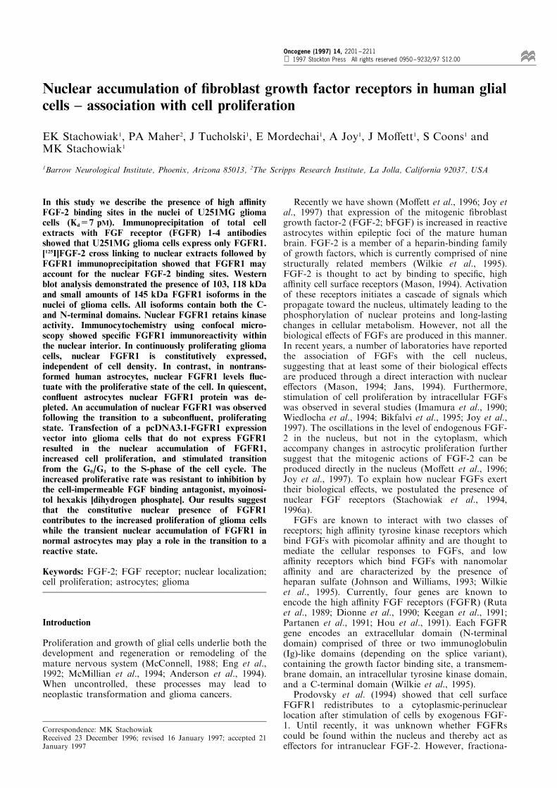
Nuclear accumulation of ®broblast growth factor receptors in human glialcells ± association with cell proliferation
EK Stachowiak1, PA Maher2, J Tucholski1, E Mordechai1, A Joy1, J Mo�ett1, S Coons1 andMK Stachowiak1
1Barrow Neurological Institute, Phoenix, Arizona 85013, 2The Scripps Research Institute, La Jolla, California 92037, USA
In this study we describe the presence of high a�nityFGF-2 binding sites in the nuclei of U251MG gliomacells (Kd=7 pM). Immunoprecipitation of total cellextracts with FGF receptor (FGFR) 1-4 antibodiesshowed that U251MG glioma cells express only FGFR1.[125I]FGF-2 cross linking to nuclear extracts followed byFGFR1 immunoprecipitation showed that FGFR1 mayaccount for the nuclear FGF-2 binding sites. Westernblot analysis demonstrated the presence of 103, 118 kDaand small amounts of 145 kDa FGFR1 isoforms in thenuclei of glioma cells. All isoforms contain both the C-and N-terminal domains. Nuclear FGFR1 retains kinaseactivity. Immunocytochemistry using confocal micro-scopy showed speci®c FGFR1 immunoreactivity withinthe nuclear interior. In continuously proliferating gliomacells, nuclear FGFR1 is constitutively expressed,independent of cell density. In contrast, in nontrans-formed human astrocytes, nuclear FGFR1 levels ¯uc-tuate with the proliferative state of the cell. In quiescent,con¯uent astrocytes nuclear FGFR1 protein was de-pleted. An accumulation of nuclear FGFR1 was observedfollowing the transition to a subcon¯uent, proliferatingstate. Transfection of a pcDNA3.1-FGFR1 expressionvector into glioma cells that do not express FGFR1resulted in the nuclear accumulation of FGFR1,increased cell proliferation, and stimulated transitionfrom the G0/G1 to the S-phase of the cell cycle. Theincreased proliferative rate was resistant to inhibition bythe cell-impermeable FGF binding antagonist, myoinosi-tol hexakis [dihydrogen phosphate]. Our results suggestthat the constitutive nuclear presence of FGFR1contributes to the increased proliferation of glioma cellswhile the transient nuclear accumulation of FGFR1 innormal astrocytes may play a role in the transition to areactive state.
Keywords: FGF-2; FGF receptor; nuclear localization;cell proliferation; astrocytes; glioma
Introduction
Proliferation and growth of glial cells underlie both thedevelopment and regeneration or remodeling of themature nervous system (McConnell, 1988; Eng et al.,1992; McMillian et al., 1994; Anderson et al., 1994).When uncontrolled, these processes may lead toneoplastic transformation and glioma cancers.
Recently we have shown (Mo�ett et al., 1996; Joy etal., 1997) that expression of the mitogenic ®broblastgrowth factor-2 (FGF-2; bFGF) is increased in reactiveastrocytes within epileptic foci of the mature humanbrain. FGF-2 is a member of a heparin-binding familyof growth factors, which is currently comprised of ninestructurally related members (Wilkie et al., 1995).FGF-2 is thought to act by binding to speci®c, higha�nity cell surface receptors (Mason, 1994). Activationof these receptors initiates a cascade of signals whichpropagate toward the nucleus, ultimately leading to thephosphorylation of nuclear proteins and long-lastingchanges in cellular metabolism. However, not all thebiological e�ects of FGFs are produced in this manner.In recent years, a number of laboratories have reportedthe association of FGFs with the cell nucleus,suggesting that at least some of their biological effectsare produced through a direct interaction with nucleare�ectors (Mason, 1994; Jans, 1994). Furthermore,stimulation of cell proliferation by intracellular FGFswas observed in several studies (Imamura et al., 1990;Wiedlocha et al., 1994; Bikfalvi et al., 1995; Joy et al.,1997). The oscillations in the level of endogenous FGF-2 in the nucleus, but not in the cytoplasm, whichaccompany changes in astrocytic proliferation furthersuggest that the mitogenic actions of FGF-2 can beproduced directly in the nucleus (Mo�ett et al., 1996;Joy et al., 1997). To explain how nuclear FGFs exerttheir biological e�ects, we postulated the presence ofnuclear FGF receptors (Stachowiak et al., 1994,1996a).FGFs are known to interact with two classes of
receptors; high a�nity tyrosine kinase receptors whichbind FGFs with picomolar a�nity and are thought tomediate the cellular responses to FGFs, and lowa�nity receptors which bind FGFs with nanomolara�nity and are characterized by the presence ofheparan sulfate (Johnson and Williams, 1993; Wilkieet al., 1995). Currently, four genes are known toencode the high a�nity FGF receptors (FGFR) (Rutaet al., 1989; Dionne et al., 1990; Keegan et al., 1991;Partanen et al., 1991; Hou et al., 1991). Each FGFRgene encodes an extracellular domain (N-terminaldomain) comprised of three or two immunoglobulin(Ig)-like domains (depending on the splice variant),containing the growth factor binding site, a transmem-brane domain, an intracellular tyrosine kinase domain,and a C-terminal domain (Wilkie et al., 1995).Prodovsky et al. (1994) showed that cell surface
FGFR1 redistributes to a cytoplasmic-perinuclearlocation after stimulation of cells by exogenous FGF-1. Until recently, it was unknown whether FGFRscould be found within the nucleus and thereby act ase�ectors for intranuclear FGF-2. However, fractiona-
Correspondence: MK StachowiakReceived 23 December 1996; revised 16 January 1997; accepted 21January 1997
Oncogene (1997) 14, 2201 ± 2211 1997 Stockton Press All rights reserved 0950 ± 9232/97 $12.00

tion of human astrocytes or bovine adrenal medullarycells revealed the presence of FGFR1 in isolated nuclei,associated with both the nucleoplasm and nuclearmatrix (Stachowiak et al., 1996a,b). Immunoconfocalmicroscopy con®rmed the intranuclear presence ofFGFR1 and showed colocalization with FGF-2.Nuclear FGFR1 binds FGF-2 and retains tyrosinekinase activity. Thus the presence of functional FGFRin the nucleus suggests that the receptors may mediatethe e�ects of nuclear FGF-2. In the present study wedemonstrate: (1) the nuclear localization of functionalFGFR1 in human glioma cells, and (2) provideevidence for the mitogenic role of nuclear receptorsin glial cell proliferation.
Results
Scatchard analysis of [125I]FGF-2 binding to U251MGglioma cells
To ascertain whether the nucleus contains functionalFGFR capable of interacting with FGF-2, weexamined the binding of [125I]FGF-2 to subcellularfractions obtained from U251MG glioma cells. TheU251MG surface binding data, analysed by themethod of Scatchard (1949), was consistent with atwo component binding curve. Accordingly, the cellsurface contains both high a�nity (Kd=6 pM) and lowa�nity (Kd=17 nM) binding sites for [125I]FGF-2(Table 1). Low and high a�nity binding sites werealso observed with the cytoplasmic fraction, while onlyhigh a�nity binding sites were found in the nuclearfraction. In all cases, speci®c binding was eliminated bya 100-fold molar excess of unlabeled FGF-2.
FGFR1 is the predominant subtype of FGFR expressedin U251MG glioma cells
In human astrocytes, FGFR1 was identi®ed as thepredominant type of FGF-2 receptor (Stachowiak etal., 1996a). To determine the types of high a�nityFGFR expressed in U251MG glioma cells, FGFRswere immunoprecipitated from total cell extracts withantibodies speci®c to the C-terminal domains ofFGFR1-4 (Santa Cruz Biotechnology). The immuno-precipitates were subjected to Western analysis with amonoclonal FGFR antibody (McAb6) that recognizesa common sequence in the N-terminal, extracellulardomains of FGFR1-4 (Hanneken et al., 1995). Theresults show that FGFR1 is the predominant FGFR in
U251MG glioma cells (Figure 1). FGFR1 isoforms of103, 118 and 145 kDa were observed, consistent withthe earlier reported sizes for FGFR1 (Xu et al., 1992).Since all these isoforms contain both the C-terminaland N-terminal domains they are likely to represent thefull length receptors.
Subcellular distribution of FGFR1
To determine the subcellular localization of FGFR1 inglioma cells, we isolated nuclear and cytoplasmicfractions as described previously (Boyle et al., 1985;Stachowiak et al., 1994; 1996a,b). Nuclei isolated bythis method contain nearly 90% of the total TCA-precipitable cellular DNA. Analysis of the isolatednuclei by phase-constant microscopy showed nocontamination with cytoplasmic membranes andorganelles. The purity of the isolated nuclei wascon®rmed using biochemical assays of marker en-zymes. The nuclear fraction contained 5+2% of thetotal cellular activity of acid phosphatase (lysosomalmarker) and 8+3% of the 5'-nucleotidase (cellmembrane marker). Western blot analysis of nuclearextracts of U251MG cells using the monoclonal FGFRantibody (McAb6) detected proteins of 103 and118 kDa. An additional lower abundance 145 kDaprotein was detected after longer exposure (not shown).The speci®c content of FGFR1 in the nuclear fractionwas approximately 2 ± 3-fold higher than in the totalcell lysates (Figure 2a). Only trace amounts of FGFR1were detected in the cytoplasmic fraction. The apparentmolecular weights of FGFR1 isoforms in the nuclei ofU251MG cells are identical to those found in the nucleiof human astrocytes (Stachowiak et al., 1996a).We used immunohistochemistry in combination with
confocal microscopy to provide a topological evalua-tion of the distribution of FGFR1 in glioma cells andastrocytes. Cells were stained with the C-term FGFR1Ab. The distribution of FGFR1 immuno¯uorescencewas analysed using 2-dimensional projections of aseries of confocal images (Figure 2b). Astrocytes(Figure 2b,II) and U251MG glioma cells (Figure2b,III) showed weak cytoplasmic FGFR1 immuno-reactivity with several cells showing cell-perimeter
R1 R2 R3 R4 — 200
— 116
— 97
pre sup sup sup supip ip ip ip
(kDa)
Figure 1 U251MG glioma cells express the 103, 118 and145 kDa FGFR1 forms containing both the extracellular (N-terminal) and intracellular (C-terminal) domains. FGFR wasimmunoprecipitated with antibodies speci®c to the C-terminaldomains of FGFR1± 4 (Santa Cruz, Biotechnology) fromapproximately 56106 cells. The immunoprecipitates weresubjected to Western analysis with McAb6 which recognizes theN-terminal extracellular domains of FGFR1± 4. Pre-whole celllysate from 0.46106 cells prior to immunoprecipitation; Sup-material remaining after FGFR immunoprecipitation; Ip-immu-noprecipitated FGFR. Migration of molecular weight standards isindicated at right
Table 1 Affinity of [125I]FGF-2 binding sites on the surface and inthe cytoplasmic and nuclear fractions of glioma U251MG cells
Kd
Low affinity High affinity
Cell surfaceCytoplasmNuclei
17 nM79 nMND
6pM13 pM7 pM
The binding a�nity was determined using Scatchard analysis asdescribed in Materials and methods. Data analysis revealed that thecell surface contains both low (r=0.94) and high a�nity (r=0.89)FGF-2 binding sites. The nuclear fraction contains only the higha�nity sites with the r=0.91. ND ± not detected. The valuesrepresent a mean of six samples in two independent experiments
Nuclear FGF receptors in human glial cellsEK Stachowiak et al
2202

staining. This pattern of immunoreactivity may be dueto plasma membrane-associated FGFR1. Both astro-cytes and U251MG glioma cells showed a distinctnuclear presence of FGFR1 immuno¯uorescence. Thesame pattern of staining was observed with McAb6which recognizes the extracellular domain of FGFR1(not shown). Omitting the FGFR1 antibody (Figure2b,I) or replacing it with preimmune serum abolishednuclear staining (not shown). Nuclear FGFR1immuno¯uorescence also was eliminated after the C-term FGFR1 Ab was preincubated with an excess ofits cognate peptide (Stachowiak et al., 1996a). Using
the same staining protocol, we detected non-nuclearglial acidic ®brillary protein (GFAP) exclusively in thecytoplasm (Joy et al., 1997).The observed pattern of immuno¯uorescence could
be due to perinuclear, nuclear membrane or intra-nuclear staining. To distinguish between these localiza-tions, we analysed a series of consecutive 2 mmconfocal laser sections through cells stained with theC-term FGFR1 antibodies (Figure 2b,IV). The patternseen in these sections shows that FGFR1 immuno-¯uorescence is present within the nuclear interior.Therefore, based on the results of biochemical
b
Figure 2 Subcellular distribution of FGFR1 proteins in human glioma cells and astrocytes. (a) Analysis of FGFR1 in total cellularlysate and in cytoplasmic (cyt) or nuclear (nuc) fractions of the U251MG glioma cells. Blots were probed with monoclonal McAb6antibody. Molecular weights of FGFR1 bands are indicated at left. (b) Immunocytochemical localization of FGFR1 in humanastrocytes (I, II-QG strain, IV-SC strain) and U251MG glioma cells (III). Cells were incubated with C-term FGFR1 Ab and theimmune complexes stained with CY3-conjugated IgG. I-III ± photographs represent stacked confocal laser sections taken 1 mmapart. No staining was observed when primary antibody was omitted (I). IV ± shows individual consecutive (top to bottom)confocal sections taken 2 mm apart to illustrate intranuclear localization of FGFR1
Total Cyt Nuc
— 145
— 118
— 103
kDa
U251 MGa
Nuclear FGF receptors in human glial cellsEK Stachowiak et al
2203

subfractionation and confocal analysis, we concludethat FGFR1 is located within the nucleus of bothastrocytes and glioma cells.To ascertain that the FGFR detected in glioma cells
binds FGF-2, intact cells (Figure 3a) or nuclear extracts(Figure 3b) were incubated with [125I]FGF-2, cross-linked and immunoprecipitated with either the C-terminal FGFR1-4 antibodies for the cell surfacereceptors or with the C-term FGFR1Ab for the nuclearfraction. Only FGFR1-[125I]FGF-2 cell surface com-plexes were detected on U251MG cells (Figure 3a),consistent with the results shown in Figure 2. Themolecular weight of the main complex (approximately140 kDa) is consistent with the binding of FGF-2 to the118 kDa isoform of FGFR1. An additional 165 kDacomplex may represent the binding of FGF-2 to the lowabundance 145 kDa FGFR1 isoform (Figure 1). Innuclear extracts which contain only trace amounts of145 kDa FGFR1 (Figure 2a) we detected only a140 kDa FGFR1-[125I]FGF-2 complex (Figure 3b).
The nuclear content of FGFR1 in astrocytes is regulatedby cell density
In con¯uent, quiescent astrocytes, transcription of theFGF-2 gene is down-regulated and nuclear FGF-2 isdepleted (Mo�ett et al., 1996; Joy et al., 1997). Aftertransition to a subcon¯uent state, astrocytes prolifer-ate, express the FGF-2 gene, and accumulate nuclearFGF-2. We examined whether the nuclear content ofFGFR1 is also co-regulated with cell density inastrocytes. The reduction of nuclear FGFR1 contentconcomitant with an increase in cell density wasinitially detected using immunostaining, as visualizedwith peroxidase and diaminobenzidine. One week afterplating, subcon¯uent astrocytes exhibited intensenuclear FGFR1 immunoreactivity (Figure 4a,I). Threeweeks after plating, when a con¯uent monolayercovered the dish, no nuclear FGFR1 immunoreactiv-ity could be detected in the astrocytes (Figure 4a,II).As with FGF-2, the depletion of nuclear FGFR1 wasreversible. In the vacant surfaces left by detached,con¯uent astrocytes, we observed nuclear FGFR1immunoreactivity in astrocytes invading those areas(not shown). Similar results were observed with
primary astrocytic cultures obtained from the frontallobes of two di�erent subjects (not shown).In addition to immunocytochemistry, the cell
density-dependent regulation of nuclear FGFR1content in astrocytes was con®rmed by Western blotanalysis. Figure 4b shows the time dependentaccumulation of FGFR1 in astrocytic nuclei followinga reduction in cell density. We used the C-term FGFR1Ab for these experiments, which recognizes predomi-nantly the 103 kDa hypoglycosylated isoform ofFGFR1 (Hanneken et al., 1995, Maher unpublishedobservations). The level of the 103 kDa FGFR1isoform in nuclei from con¯uent astrocytes was lowand remained reduced for at least 6 h after cells were
Figure 3 Cross linking of [125I]FGF-2 to cell surface and nuclearreceptor. Intact U251MG cells (a) or nuclear extracts (b) wereincubated with 106 c.p.m./ml of [125I]FGF-2, cross linked with0.15 mM DSS and immunoprecipitated overnight with antibodiesagainst the C-terminal domains of FGFR1± 4 (cell surface) orFGFR1 (nuclei). The immunoprecipitates were analysed by SDS-polyacrylamide gel electrophoresis and autoradiography. 7noDSS used; +0.15 mM DSS
C N C N C N C N
➝103 kDa
0 6 18 24 -Hours
a
b
C N C N— 200
— 116— 97
— 68
— 43(kDa)
c2h 18h
Figure 4 Inhibition of nuclear FGFR1 expression in con¯uentastrocytes. (a) Immunocytochemical light microscope analysis ofFGFR1 in human astrocytes (SC strain). Cells were incubatedwith C-term FGFR1 Ab. Immune complexes were stained byincubating with biotinylated secondary antibody, avidin-biotinperoxidase, and diaminobenzidine-peroxide solution. I ± subcon-¯uent astrocytes (1 week in culture) show nuclear FGFR1immunoreactivity; II ± con¯uent astrocytes (3 weeks) ± nonuclear FGFR1 staining is detected. (b) Time-dependentaccumulation of FGFR1 after reduction of cell density.Astrocytes (SS strain) were maintained in con¯uent state for48 h. Some cultures were replated at lower density (approx. 30%of con¯uence). Nuclear (N) and cytoplasmic (C) extracts wereanalysed for FGFR1 using C-term FGFR1 Ab. (c) Time-dependent increase in FGFR1-associated kinase activity afterreduction of cell density. Cytoplasmic (C) and nuclear (N)extracts from cultures maintained in subcon¯uent state for 2and 18 h were immunoprecipitated with the C-terminal FGFR1antibody. Immunoprecipitates were incubated with [g-32P]ATPand resolved on SDS-7.5% polyacrylamide gels. Bands (approxi-mately 103, 118 and 145 kDa) indicate phosphorylated FGFR1.The strong band at approximately 50 kDa is IgG heavy chain
R1 R2 R3 R4
— 200
— 100
— 68
(kDA)
202 —
180 —
140 —
(kDa)
– + DSS
a b
Nuclear FGF receptors in human glial cellsEK Stachowiak et al
2204

replated at a subcon¯uent density. Between 6 and 18 hafter the cell density was reduced, a marked increase innuclear FGFR1 content occurred followed by a smalldecline after an additional 6 h.Next we examined whether the FGFR1 which
accumulates in the nuclei of subcon¯uent astrocyteshas tyrosine kinase activity. FGFR1-associated kinaseactivity was examined 2 and 18 h after replating theastrocytes; before and while the increased nuclearaccumulation of FGFR1 protein is detectable. Im-munoprecipitation of nuclear FGFR1 followed byincubation with [g32P]ATP showed a phosphorylatedband at 103 kDa (Figure 4c), similar in size to theFGFR1 bands detected by immunoblotting (Figure 4b)and a broader band between 118 and 145 kDa,consistent with the migration of the higher molecularweight, glycosylated isoforms of FGFR1. In addition,unidenti®ed, coprecipitating products, which migratedslower than the IgG heavy chain, became phosphory-lated. They could represent potential targets fornuclear FGFR1. Phosphorylated FGFR1 bands werenot detected in the nuclear extracts incubated with
control IgG (not shown). Thus, nuclear accumulationof FGFR1 protein is accompanied by an increase inreceptor kinase activity.
Deregulation of nuclear FGFR1 distribution in U251MGglioma cells
In contrast to astrocytes, the U251MG glioma cellsexhibited similar amounts of nuclear FGFR1 immu-noreactivity in both the con¯uent and subcon¯uentstate (Figure 5a, I and II, respectively). The impairedcell density-dependent depletion of nuclear FGFR1 inglioma cells was also con®rmed by Western blotanalysis. The C-term FGFR1Ab detected a highcontent of nuclear 103 kDa FGFR1 in dense culturesof U251MG cells (Figure 5b). While the C-termFGFR1 Ab recognizes predominantly the 103 kDahypoglycosylated isoform of FGFR1, McAb6 reactswith both the 103 kDa FGFR1 isoform as well as thehyperglycosylated 118 and 145 kDa isoforms. Figure5c compares the nuclear content of FGFR1 insubcon¯uent and con¯uent glioma cells using McAb6.
103 —
Cyt Cyt Cyt Cyt NucNuc Nuc Nuc NucCytkDaAstrocytes
Low High HighCell Density
subconfluent confluent
— 145
— 118
— 103
kDa
b c
a
U251 MGU251 MG
Figure 5 The nuclear content of FGFR1 in U251MG glioma cells is not a�ected by cell density. (a) I ± subcon¯uent (1 week inculture) and II ± con¯uent (3 weeks in culture) glioma cells were stained for FGFR1 as in Figure 4a. Cells show nuclear andcytoplasmic staining. (b) Western analysis of FGFR1 in con¯uent U251MG glioma cells and in con¯uent or subcon¯uent astrocytes.Equal amounts of cytoplasmic and nuclear proteins were analysed using C-term FGFR1Ab. (c) Western analysis of nuclear andcytoplasmic FGFR1 in U251MG cells using McAb6. Cells were maintained in subcon¯uent or con¯uent state
Nuclear FGF receptors in human glial cellsEK Stachowiak et al
2205

Intracellular FGFR1 stimulates proliferation of glial cells
In an e�ort to determine the function of nuclearFGFR1 we have identi®ed a glioma cell line, SF-763,that does not express FGFR1. Immunoprecipitationwith antibodies speci®c to the C-terminal of FGFR1 ± 4showed that FGFR2 and FGFR3 are the predominanttypes of FGFR in SF-763 glioma cells, while FGFR1and FGFR4 are not detected (Figure 6a). Weexamined whether ecoptic expression of FGFR1 inSF-763 glioma cells can result in nuclear accumulationof FGFR1 and a�ect cell proliferation. SF-763 cellswere transfected with the pcDNA3.1-FGFR1 plasmidcontaining full length FGFR1 cDNA or with thecontrol plasmid pcDNA3.1, lacking the FGFR1 cDNAinsert. A population of G418 resistant, stablytransfected cells was obtained. The cells were lysedwith 0.6% NP-40 in hypotonic bu�er, and nuclei wereseparated from the extranuclear material (cytoplasmand cell membranes) by centrifugation. The nuclearfraction obtained by this method showed a similar lackof signi®cant contamination with plasma membrane or
lysosomal proteins as the nuclei obtained by theisotonic lysis method (Figure 2a). Fractions wereanalysed by Western blot with FGFR1-speci®c anti-body (Santa Cruz Biotechnology; Figure 6b). A major,145 kDa band was detected in the nuclei of SF-763-pcDNA3.1-FGFR1 cells while only trace amounts ofFGFR1 were found in the extranuclear fractioncontaining both the cytoplasm and cell membranes.No FGFR1 was detected in SF-763-pcDNA3.1 cells(Figure 6b). Consistent with these results, we observedthe presence of FGFR1-associated kinase activity inSF-763 cell lines expressing recombinant FGFR1 butnot in pcDNA3.1 transfected cells (Figure 6c).The subcellular localization of FGFR1 in transfected
SF-763 cells was also examined by immunocytochem-istry. The SF-763-pcDNA3.1-FGFR1 cells exhibitedFGFR1-immunoreactivity, predominantly within thenucleus (Figure 7a). Staining was not detected whenthe primary antibody was omitted (not shown). Incontrast, the SF-763-pcDNA3.1 cells did not showspeci®c FGFR1 staining with the C-term-FGFR1Ab(Figure 7c).The SF-763-pcDNA3.1-FGFR1 cells also exhibited
an increased proliferation rate as compared to the SF-763-pcDNA3.1 cells (Figure 8a). This acceleratedproliferation is likely to represent an increased mitoticactivity of individual cells. Flow-cytometry showed thatin cell lines transfected with pcDNA3.1-FGFR1 thenumber of cells residing in S phase increased 30% witha concomitant decrease in the proportion of cells in G0/G1 phases relative to cells transfected with thepcDNA3.1 plasmid (not shown). To determine if themitogenic e�ect of transfected FGFR1 is mediatedintracellularly we used myo-Inositol hexakis [dihydro-gen phosphate] (IP6), a cell-impermeable antagonistwhich prevents the interaction of FGF-2 with cellsurface FGFR but does not inhibit the e�ects ofintracellular FGF-2 (Sherman et al., 1993; Morrison etal., 1994a). IP6 preferentially inhibited the proliferationof the SF-763-pcDNA3.1 cells (IC50=251 mM) ascompared with the SF-763-pcDNA3.1-FGFR1 cells(IC50=3013 mM) cells (Figure 8b). The resistance of theSF-763-pcDNA3.1-FGFR1 cells to IP6 suggests that
Figure 6 Expression of nuclear FGFR1 in SF-763 cellstransfected with pcDNA3.1-FGFR1. (a) Native SF-763 gliomacells express FGFR2 and FGFR3. Cells were incubated with[125I]FGF-2, cross linked with DSS and immunoprecipitated withantibodies against the C-terminal domains of FGFR1± 4. Theimmunoprecipitates were analysed by SDS-polyacrylamide gelelectrophoresis and autoradiography. (b) Western analysis ofFGFR1 in the nuclear and cytoplasmic fractions of SF-763 cellsstably transfected with plasmids expressing full length FGFR1(pcDNA3.1-FGFR1) or control plasmid (pcDNA3.1) using a C-term FGFR1 Ab from Santa Cruz Biotechnology. Cells werelysed with 0.6% NP-40 in hypotonic HEPES bu�er and nuclei(N) were separated from the extra-nuclear material (EN) bycentrifugation as described in Materials and methods. (c)FGFR1-associated tyrosine kinase activity. SF-763 cell lineexpressing FGFR1 and nonexpressing cells were lysed andFGFR1 was immunoprecipitated using 1 mg of the C-terminalFGFR1 antibody (Santa Cruz Biotechnology). Immunoprecipi-tates were incubated with [g32P]ATP and were resolved on SDS-7.5% polyacrylamide gels. Band (approximately 145 kDa)indicate phosphorylated FGFR1
pcDNA3.1 pcDNA3.1-FGFR1
Figure 7 Immunostaining of SF-763 glioma cells transfected withpcDNA3.1-FGFR1 expressing FGFR1 or control plasmidpcDNA3.1. SF-763-pcDNA3.1-FGFR1 cells showed no stainingwhen the primary C-term FGFR1 Ab was omitted (not shown)
R1 R2 R3 R4
— 200
— 100
— 68
(kDa)
(kDa)
200 —
116 —
68 —
C N C N
pcDNApcDNA-FGFR1
+ ++ +
++
<145
a
b c
Nuclear FGF receptors in human glial cellsEK Stachowiak et al
2206

their increased proliferative rate is independent of cellsurface FGFR and could be mediated by the nuclearFGFR1.
Discussion
Recently, we showed that the expression of nuclearFGF-2 is induced in reactive astrocytes within theepileptic foci of the mature human brain and in gliomatumors (Joy et al., 1997). In order to determine if higha�nity FGFR could mediate the e�ects of nuclearFGF-2 in astrocytes and glioma cells we examined theintracellular distribution of FGFR within these two celltypes.The studies reported here demonstrate an intra-
nuclear localization for FGFR1 in human glioma cellsand con®rm our earlier ®nding that the nuclei of humanastrocytes contain functional FGFR1 (Stachowiak etal., 1996). Furthermore, nuclear FGFR1 is a full lengthprotein since its size is similar to the largest FGFR1predicted by the cDNA (approximately 96 kDa; Dionneet al., 1990; Hou et al., 1991) and is recognized byantibodies to both the extracellular domain (McAb6)and the C terminal domain of the receptor. Consistentwith the presence of full length receptor in the nucleusare the observations that nuclear FGFR1 binds FGF-2and retains tyrosine kinase activity. In a separate studywe found that treatment of nuclear extracts with N-glycanase converts the 103, 118 and 145 kDa isoformsinto a single approximately 100 kDa protein (ourunpublished observations). Thus, the 103, 118 and145 kDa bands appear to represent di�erent degrees ofglycosylation of FGFR1. The results of our cross-linking experiments suggest that the 118 and 145 kDabind e�ciently FGF-2.Recently, Johnston et al. (1995) reported that nuclei-
containing subcellular fractions isolated from breastcancer cell lines contain FGFR3 and suggested that the
association of FGFR3 with the nuclear fractions maybe caused by a deletion of its transmembrane domain.In our study, we show that transfection of cells withthe pcDNA3.1-FGFR1 plasmid containing the fulllength FGFR1 cDNA, into SF-763 cells results in anaccumulation of FGFR1 in the nuclei. This suggeststhat the full length receptor can translocate to thenucleus. Cell surface labeling of U251MG cells with animpermeable analog of biotin followed by extendedincubation at 378C did not result in any biotin-labeledFGFR1 in the nuclei indicating that FGFR1 can moveto the nucleus without prior insertion into plasmamembrane (P Maher and MK Stachowiak, inpreparation). The import of many proteins into thenucleus is mediated by nuclear localization signals(NLSs). Most characterized NLSs consist of a stretchof 3 ± 5 basic residues, often ¯anked by proline orglycine (Chelsky et al., 1989). Basic amino acidspresent in FGFR1 do not form typical NLS-likeclusters. It is possible that the movement of FGFR1to the nucleus is mediated by a chaperon protein.Whether FGFR1 is translocated to the nucleuscomplexed with cytoplasmic FGF-2 or other proteinsis under investigation. A distinct, NLS-independentnuclear transport system that imports glycosylatedproteins from the cytoplasm into the nucleus in asugar-dependent manner has been identi®ed (Duvergeret al., 1995). Unlike the NLS-mediated transport thatoften direct proteins to the nucleoli, the sugar-mediatedtransport results in general nuclear localization (asobserved for FGFR1).The intranuclear content of FGFR1 oscillates in
association with cell proliferation. Quiescent astrocytesmaintained in high-density cultures had little or nonuclear FGFR1 (Figure 4). After the cell density wasreduced, cell hypertrophy and transient proliferationwere observed accompanied by an induction of nuclearFGFR1. In contrast to the transient burst of controlledgrowth experienced by reactive astrocytes in gliotic
a b
Figure 8 Proliferation of SF-763 glioma cells transfected with pcDNA3.1 and pcDNA3.1-FGFR1 plasmids. (a) proliferation of SF-763-pcDNA3.1 and SF-763-pcDNA3.1-FGFR1 cells. Symbols represent mean of quadruplicate samples +s.e.m. Equal amounts ofSF-763-pcDNA3.1 and SF-763-pcDNA3.1-FGFR1 cells were plated at day 0. Between days 6 and 10 cells transfected withpcDNA3.1-FGFR1 proliferated at a rate approximately ®vefold greater than cells transfected with control plasmid. (b) Cells wereincubated with indicated concentrations of IP6 for 7 days. The IC50 values (251 mM for the SF-763-pcDNA3.1 and 3013 mM for theSF-763-pcDNA3.1-FGFR1) were determined as described in Materials and methods
Nuclear FGF receptors in human glial cellsEK Stachowiak et al
2207
tissue, neoplastic glioma cells proliferate continuously,unresponsive to external cues such as cell contactinhibition. This constitutive proliferation was alsoobserved in vitro. After con¯uent monolayers wereformed, cultured U251MG cells continued to produceadditional layers. Nuclear FGFR1 decreased onlyslightly as the glioma cells reached con¯uence. In ourother reports, we show that the nuclear content ofFGF-2 is regulated in a manner similar to FGFR1 inastrocytes and glioma cells (Mo�ett et al., 1996; Joy etal., 1997). Also FGF-2 and FGFR1 are bothassociated with the nuclear matrix (Stachowiak et al.,1996a; Joy et al., 1997) and nuclear FGFR1 is capableof binding FGF-2 (Stachowiak et al., 1996a; presentstudy). Thus, nuclear FGFR1 could serve as an e�ectorfor nuclear FGF-2.To address the role of nuclear FGFR1 in cell
proliferation, we transfected SF-763 glioma cells witha pcDNA3.1-FGFR1 plasmid and examined prolif-eration rate in the absence or presence of IP6, a cell-impermeable FGF binding antagonist (Sherman et al.,1993; Morrison et al., 1994a). Unlike U251MG, theSF-763 glioma cells express FGFR2 but not FGFR1,consistent with earlier reported di�erences in FGFRexpression in human glioma tumors (Morrison et al.,1994b). FGFR1 was found intracellularly in trans-fected cells where it was localized almost exclusivelyto the nucleus. The expression of nuclear FGFR1 wasaccompanied by an increase in spontaneous cellproliferation in the absence of exogenously addedFGF-2. The increase in proliferation of the SF-763-pcDNA3.1-FGFR1 cells was associated with in-creased mitotic activity of the individual cells. Ourresults indicate that nuclear FGFR1 stimulates thetransition from the G0/G1 to the S phase of the cellcycle. IP6 inhibited the proliferation of the SF-763-pcDNA3.1 cells suggesting that these cells proliferatein response to an extracellular growth factor whichacts through cell surface FGFR. Since we could notdetect FGF-2 in the medium conditioned by SF-763cells (MK Stachowiak and R Florkiewicz, unpub-lished observations), other members of the FGFfamily may be responsible for this activity and mayact through FGFR2 and/or FGFR3 (Figure 6a). Incontrast to the SF-763-pcDNA3.1 cells, the sponta-neous proliferation of the SF763-pcDNA3.1-FGFR1cells was resistant to IP6 (Figure 8b) providing strongevidence that the increase in the proliferation of theSF-763-pcDNA3.1-FGFR1 cells occurs independentlyof cell surface FGFR and may depend on nuclearFGFR1.Our study suggests that the cellular e�ects of FGFs
can be mediated by an intracellular (intracrine)pathway that operates through nuclear FGFR1.Thus, cells could regulate their responsiveness toFGFs by controlling both the expression and thesubcellular localization of FGFR. The presence offunctional FGFR1 within the nucleus may explain howintracellular FGFs exert the mitogenic e�ects observedin this (Joy et al., 1997) and in other laboratories(Imamura et al., 1990; Wiedlocha et al., 1994; Bikfalviet al., 1995).We propose that the regulation of both FGFR1
(present study) and FGF-2 (Mo�ett et al. 1996; Joy etal., 1997) content in the nucleus could serve as a novelmechanism controlling proliferation in glial cells.
Thus, transient nuclear accumulation of FGF-2 andFGFR1 may mediate the reversible mitotic activationof quiescent astrocytes. In glioma cells, the constitu-tive nuclear presence of FGF-2 and FGFR1 maypromote proliferation that is insensitive to cell contactinhibition. Further analysis of the nuclear transloca-tion and function of FGF-2/FGFR1 may reveal themechanisms controlling normal and neoplastic growthof glial cells.
Materials and methods
Materials
Culture media were from Grand Island Biologicals (GrandIsland, NY). The remaining reagents were from BoehringerMannheim (Indianapolis, IN), Sigma Chemical Co. (St.Louis, Mo), Bio-Rad (Richmond, CA), Stratagene (La Jolla,CA), Du Pont NEN (Boston, MA), Pharmacia (Alameda,CA); Santa Cruz Biotechnology (Santa Cruz, CA), Genzyme(Cambridge, MA) and Pierce (Rockford, IL).
Astrocytic and glioma cell cultures and cell fractionation
Two normal astrocyte cultures (QG and SC) obtained fromthe dissociation of brain tissue taken from di�erent traumapatients are described in (Mo�ett et al., 1996; Stachowiaket al., 1996a). Cells were prepared and maintained inWaymouth 87/3 medium (MAB) supplemented with 20%fetal bovine serum. Established glioma cell lines U251MG(Binger et al., 1981) and SF-763 (Berens et al., 1990) wereused. Cell fractionation was performed by the isotonicmethod of Boyle et al. (1985) or by the hypotonic/NP-40lysis method of Schreiber et al. (1989) as described inStachowiak et al. (1994, 1996a,b; Joy et al., 1997). In theisotonic protocol cells were lysed in nuclear bu�ercontaining 0.1% digitonin, 5 mM sodium phosphate pH7.4, 50 mM NaCl, 150 mM sucrose, 5 mM KCl, 2 mM
dithiothreitol, 1 mM MgCl2, 0.5 mM CaCl2, 0.1 mM
PMSF, 10 mM leupeptin, 1 mg/ml aprotinin, 25 mg/mlsoybean trypsin inhibitor, and 5 mg/ml pepstatin A. Nucleiwere collected by centrifuging at 500 g for 10 min at 48C.The postnuclear supernatant was further centrifuged at40 000 g for 30 min at 48C and the supernatants werereserved for analysis of cytoplasmic FGFR. The crudenuclear pellet was further puri®ed by centrifugationthrough a cushion of TN bu�er (2.5 mM Tris HCl, pH7.4, 10 mM NaCl) containing 30% sucrose at 1000 g for10 min at 48C. In the hypotonic protocol, cells werewashed in Tris-bu�ered saline, swollen in homogenizationbu�er containing 10 mM HEPES, pH 7.9, 10 mM KCl,0.1 mM EDTA, 0.1 mM EGTA, 1 mM DTT, 0.5 mM
PMSF, and vortexed in homogenization bu�er containing0.6% NP-40. Nuclei were pelleted by centrifugation for30 s in a microfuge, and washed in homogenization bu�ercontaining NP-40. The nuclear pellet and the supernatantcontaining both cytoplasm and plasma membranes wereused for further analyses. The isolated nuclei were washedthree times in the isotonic nuclear bu�er. The purity ofisolated fractions was evaluated by measuring the activityof plasma membrane 5'-nucleotidase and lysosomal acidphosphatase using Sigma Chem. Co. assay kits assuggested by the manufacturer.
Binding of [125I]human recombinant FGF-2 to cell surface andto the nuclear and cytoplasmic fractions of U251MG gliomacells
Nuclei and cytoplasmic fractions were isolated as describedabove. The binding of [125I]FGF-2 was examined using the
Nuclear FGF receptors in human glial cellsEK Stachowiak et al
2208
method of Moscatelli et al. (1987). Cells were platedovernight at 16106/well in 12-well plates in serum-freeMEM containing 0.15% gelatin. Subsequently cells werewashed twice with ice-cold PBS and were incubated for 4 hat 48C in 100 ml MEM containing 25 mM HEPES, 0.15%gelatin and increasing concentrations (0 ± 50 nM) of[125I]FGF-2 with or without 100-fold molar excess ofunlabeled FGF-2. Cells were washed three times withPBS and the low binding a�nity fraction was extractedwith 1 ml of 2 M NaCl in 20 mM HEPES, pH 7.5. The higha�nity binding fraction was obtained by solubilizing thecells in 250 ml of 0.5% Triton-X100 in 0.1 M sodiumphosphate, pH 8.1. Each fraction was counted for[125I]FGF-2 using a Tracor Gamma counter. Nuclear andcytoplasmic fractions were collected from 106 cells anddiluted with binding bu�er (25 mM HEPES, pH 7.5, 0.15%gelatin) and the indicated concentrations of [125I]FGF-2(speci®c activity 70 mCi/mg). The binding was carried out insuspension at room temperature for 2 h in gently rotatingtubes in the presence or absence of 100-fold excess ofunlabeled FGF-2. The reaction mixtures were transferredto Amicon-50 Microconcentrators and centrifuged 10 min,6000 g, at room temperature. The ligand-bound, ®lter-retained fraction of [125I]FGF-2 was counted and analysedutilizing the Mac Ligand program according to Munsonand Rodbard (1980). Presence of di�erent a�nity bindingsites on the cell surface or in the cytoplasmic or nuclearfraction was evaluated by computing the `r' coe�cient (astatistical value which describes the accuracy betweeninter-sample variance).
Cross linking of [125I]FGF-2 to receptors and immunoprecipi-tation of cross-linked FGFR
[125I]FGF-2 was prepared as described using lactoperox-idase (Schubert et al., 1987). The free iodine was separatedfrom the iodinated growth factor by passage over aheparan-Sepharose column. Speci®c activity was 4 ±66105 c.p.m./ng. For cross-linking, cells were incubatedwith 106 c.p.m./ml of [125I]FGF-2 in 150 mM NaCl, 20 mM
HEPES, pH 7.5 for 2 h with rocking and were rinsed twicewith PBS. Cells, cytoplasmic fractions or nuclear extractswere incubated with 0.15 mM disuccinimidyl suberate(DSS; Sigma Chemical Co.) in PBS for 15 min at roomtemperature with rotation. The cross-linking was stoppedby the addition of 200 mM ethanolamine. The samples wererinsed twice with PBS and then solubilized in 500 ml ofTriton X-100 bu�er (1% Triton X-100, 50 mM HEPES, pH7.5, 50 mM NaCl, 5 mM EDTA, 1 mM Na3VO4). FGFRswere immunoprecipitated at 48C overnight with antibodiesagainst the C-terminal domains of the di�erent types ofFGFR (FGFR1 ± 4 ± Santa Cruz Biotechnology) as de-scribed below. The immunoprecipitates were collected onprotein A-sepharose, washed twice with 0.1% Triton X-100bu�er in 20 mM HEPES, pH 7.5, 150 mM NaCl, once withphosphate-bu�ered saline (PBS) and solubilized in46SDS-sample bu�er. The samples were separated onSDS-7.5% polyacrylamide gel and the gel was dried andautoradiographed.
Western analysis of FGFR in subcellular fractions
Western analysis of FGFR was performed as described inStachowiak et al. (1996). Brie¯y, protein concentrationswere determined using the Bio-Rad protein assay (Bio-Rad,Richmond, CA). Equal amounts (50 mg) of proteins fromtotal cell lysate, cytoplasmic or nuclear fractions weresolubilized in 46SDS-sample bu�er and separated on SDS-7.5% polyacrylamide gel and transferred to nitrocellulosemembrane. The transfers were stained with amido black tocon®rm the presence of equal amounts of protein in eachlane. Blots were probed using a polyclonal, a�nity-puri®ed
rabbit antibody to the carboxyl terminus of FGFR1 (C-term-FGFR1Ab; Hanneken et al., 1995). The C-termFGFR1 Ab does not cross-react with FGFR2, FGFR3 orFGFR4 receptors (Hanneken et al., 1995). The secondantibody used was a monoclonal antibody (McAb6) againstthe extracellular domain of FGFR1 produced in baculo-virus which was characterized as described in (Hanneken etal., 1995). Nitrocellulose membranes were blocked with0.1% Tween 20 (C-term FGFR1 Ab) or 5% nonfat milk(McAb6) in Tris-bu�ered saline overnight prior toincubation with antibodies. Membranes were incubatedwith C-term FGFR1 Ab (2 mg/ml) for 4 h, washed and thentreated with 125I-labeled protein A (0.2 mCi/ml). ForMcAb6, the transfers were incubated overnight with theantibody (3 mg/ml), washed and then incubated with 1 mg/ml of rabbit anti-mouse IgG for 2 h prior to the addition of[125I]protein A as described above. The transfers wereautoradiographed overnight at 7708C. The apparentmolecular sizes of FGFR forms were determined bycomparison to concurrently run protein molecular weightstandards. FGFR1 protein levels in the fractions wereestimated by densitometric scanning of autoradiogramsusing a Beckman DU70 spectrophotometer.
Immunoprecipitation of FGFR and in vitro phosphorylationassays
Nuclear proteins were solubilized by sonication of theisolated nuclei in Triton X-100 bu�er to give a ®nal proteinconcentration of 1 mg/ml. FGFR were immunoprecipitatedfrom the total cell lysates or from the cytoplasmic ornuclear extracts at 48C overnight with the commercial anti-FGFR1 antibody (Santa Cruz Biotechnology; 0.5 mg/500 mlnuclear extract). The immunoprecipitates were collected onprotein A-Sepharose, washed 36 with 0.1% Triton X-100in 20 mM HEPES, pH 7.5, 1 mM Na3VO4, 150 mM NaCland resuspended in 50 ml in vitro phosphorylation bu�er(150 mM NaCl, 20 mM HEPES, pH 7.5, 0.1% Triton X-100) containing 10 mM MgCl2, 50 mM MnCl2, 1 mM
Na3VO4, 50 nM ATP and 10 mCi[g32P]ATP (speci®c activity6000 Ci/mmol). The reaction was carried out for 20 min atroom temperature with continuous rotation and thenterminated by the addition of an equal volume of46SDS-sample bu�er. The samples were separated onSDS-7.5% polyacrylamide gels and the gels were ®xedovernight in 25% methanol, 10% acetic acid, dried andautoradiographed for 12 ± 18 h.
Immunocytochemical staining
Cultured cells were ®xed, permeabilized with 1.0% TritonX-100, and stained immunocytochemically as in (Stacho-wiak et al., 1994, 1996) with the same FGFR1 antibodies(C-term FGFR1Ab or McAb6; Hanneken et al., 1995) thatwere used in Western blot assays. Immune complexes weredetected with biotinylated secondary antibody, followed byavidin-linked peroxidase and diaminobenzidine-hydrogenperoxide solution (Stachowiak et al., 1994). Endogenousperoxidase activity was exhausted by treating cells with0.1% H2O2. Immuno¯uorescent staining of FGFR1 wasperformed using CY3-conjugated secondary antibodies(Jackson Immuno Research Labs., West Grove, PA) asdescribed in (Stachowiak et al., 1996a). Digitized images of1 mm confocal optical sections were acquired using aBiorad MRC 600 confocal microscope with a YHS568 nm ®lter and 15 mW Crypton/Argon laser. Theimages were recorded directly onto 35 mm ®lm.
The speci®city of FGFR1 immunostaining was indicatedby several observations: (1) staining was not observed whenthe primary antibody was omitted or replaced withpreimmune serum; (2) similar staining was observed usingtwo di�erent antibodies (C-term FGFR1 Ab and McAb6)
Nuclear FGF receptors in human glial cellsEK Stachowiak et al
2209

and di�erent staining techniques; (3) the same stainingtechnique detected FGFR1 in the nuclei (Figure 7) and thecytoplasmic glial acidic ®brillary protein in the cytoplasm(Joy et al., 1997); (4) neutralization of the C-term FGFR1 Abby an excess of the C-terminal FGFR1 peptide reducedFGFR1 staining (Stachowiak et al., 1996a); (5) the presence,absence and di�erences in the levels of nuclear FGFR1immunoreactivity were con®rmed by Western blot analysis ofFGFR1 in subcellular fractions; and (6) transfection ofFGFR1 expressing plasmid into glioma cells that do notexpress FGFR1 led to an induction of nuclear FGFR1immunoreactivity.
Proliferation of glioma cells expressing recombinant FGFR1
pcDNA3.1-FGFR1 vector was constructed by cloning the2494 bp XbaI cDNA fragment containing the entire codingsequence of the three-loop form of human FGFR1 (Hou etal., 1991) into the XbaI site 3' to the cytomegalovirus(CMV) immediate early promoter of the pcDNA3.1plasmid (Invitrogen). pcDNA3.1 contains a G418 resis-tance gene for selection of stable transfectants. ThepcDNA3.1-FGFR1 directs the expression of a completeFGFR1. SF-763 Glioma cells (36106 cells/ml) weretransfected with 100 mg of pcDNA3.1-FGFR1 or controlpcDNA3.1 plasmids using electroporation. Two days aftertransfection the cells were maintained with 400 mg/ml ofG418 (Stratagene) to select transfectants. G418-resistantcells were pooled to establish populations of stable-transfected cells. The proliferation of transfected gliomacells was examined according to Brasaemle and Attie(1988). Brie¯y, cells were plated in 96-well plates at 26103
cells/well in medium containing 5% serum. To determinecell densities at the indicated times, the cells were ®xed andstained with crystal violet (0.01%) and solubilized with10% SDS. The changes in cell number were quanti®ed byreading the absorbance at 540 nm on an automated ELISAplate reader. The inhibition of cell proliferation by IP6 was
analysed by interactive nonlinear least square analysis togive the accurate concentration of IP6 needed to inhibitcell proliferation by 50% (ID50) (Draper and Smith, 1966).
Cell cycle analysis by ¯ow cytometry was carried outaccording to Latham et al. (1996) as described in Joy et al.(1997). DNA histograms were generated using EPICS Pro®le¯ow cytometer (Coulter Co. Hialeh, Fl) and were analysedusing Multicycle cell cycle analysis program (Phoenix FlowSystems, San Diego, CA).
AbbreviationsDSS, disuccinimidyl suberate; FGF-2, ®broblast growthfactor 2 or basic FGF; FGFR, FGF receptor; C-termFGFR1 Ab, polyclonal antibody against C-terminaldomain of FGFR1; McAb6, monoclonal antibody againstextracellular domain of FGFR1; FGFR1-IR, FGFR1-immunoreactivity; GFAP, glial ®brillary acidic protein;IgG-immunoglobulin; QG, SC, SS-human astrocyticstrains from di�erent individuals; U251MG, SF-763,human glioma cell lines; IP6, myo-inositol hexakis[dihydrogen phosphate].
AcknowledgementsWe wish to thank Dr Michael Berens for the gift of gliomacell lines, Dr Leslie Tolbert and Mrs Patty Jansma for theassistance with confocal microscopy and Dr Shelley Kickfor editorial help. SF-763 cell line was from the BrainTumor Research Center, University of California, SanFrancisco. This study was supported by National ScienceFoundation (94-11226, MKS), American Parkinson Dis-ease Association (MKS), National Institutes of Health(HL49376-01A1, MKS and NS28121, PAM) and byArizona Disease Control Research Commission (JM, AJand MKS).
References
Anderson BA, Li X, Alcantara AA, Isaacs K, Black JE andGreenough WT. (1994). Glia, 11, 73 ± 80.
Boyle WT, Lampert MA, Li AC and Baluda MA. (1985).Mol. Cell. Biol., 11, 3017 ± 3023.
Berens ME, Weisman AS, Spencer DR, Dougherty DV,Elliger SS and Rosenblum ML. (1990). Proc. Amer. Assoc.Cancer Res., 31, 46.
Bikfalvi A, Klein S, Pintucci G, Quatro N, Mignatti P andRifkin DB. (1995). J. Cell. Biol., 129, 233 ± 243.
Binger DD, Binger SH and Ponten J. (1981). J. Neuropathol.Exp. Neurol., 40, 201 ± 209.
Brasaemle DL and Attie AD. (1988). BioFeedback 6, 418 ±419.
Chelsky D, Ralph R and Jonak G. (1989).Mol. Cell. Biol., 9,2487 ± 2492.
Dionne CA, Crumley G, Bellot F, Kaplow JM, Searfoss G,Ruta M, Burgess WH, Jaye M and Schlessinger J. (1990).EMBO J., 9, 2685 ± 2692.
Draper NR and Smith H. (1966). Applied regression analysis.Wiley: New York, pp. 263 ± 285.
Duverger E, Pellerin-Mendes C, Mayer R, Roche AC andMonsigny M. (1995). J. Cell Sci., 108, 1325 ± 1332.
Eng LF, Yu ACH and Lee YL. (1992). Prog. Brain Res., 94,353 ± 365.
Hanneken A, Maher PA and Baird A. (1995). J. Cell. Biol.,128, 1221 ± 1228.
Hou J, Kan M, McKeehan K, McBride G, Adams P andMcKeehan WL. (1991). Science, 251, 665 ± 668.
Immamura T, Engelka K, Zhan X, Tokita Y, Forough R,Roeder D, Jackson D, Maier JAM, Hla T and Maciag T.(1990). Science, 249, 1567 ± 1570.
Jans DA. (1994). FASEB J., 8, 841 ± 847.Johnson DE and Williams LT. (1993). Adv. Cancer Res., 60,1 ± 41.
Johnston CL, Cox HC, Gomm JJ and Coombes RC. (1995).J. Biol. Chem., 270, 30643 ± 30650.
Joy A, Mo�ett J, Neary K, Mordechai E, Stachowiak EK,Coons S, Rankin-Shapiro J, Florkiewicz RZ andStachowiak MK. (1997). Oncogene, 14, 171 ± 183.
Keegan K, Johnson DE, Williams LT and Hayman MJ.(1991). Proc. Natl. Acad. Sci. USA, 88, 1095 ± 1099.
Latham KE, Cosenza S, Reichenbach NL, Mordechai E,Adelson ME, Kon N, Horvath SE, Charubala R,Mikhailov SN, Pfeiderer W and Suhadolnik RJ. (1996).Oncogene, 12, 827 ± 837.
Mason IJ. (1994). Cell, 78, 547 ± 552.McConnel SK. (1988). Brain Res. Rev., 472, 1 ± 23.McMillian MK, Thai L, Hong J-S, O'Callaghan PO andPennypacker KR. (1994). TINS, 17, 138 ± 142.
Mo�ett J, Kratz E, Florkiewicz R and Stachowiak MK.(1996). Proc. Natl. Acad. Sci. USA, 93, 2470 ± 2475.
Morrison RS, Shi E, Kan M, Yamaguchi F, McKeehan W,Rudnicka-Nawrot M and Palczewski K. (1994a). In vitroCell Dev. Biol., 30A, 783 ± 789.
Nuclear FGF receptors in human glial cellsEK Stachowiak et al
2210

Morrison RS, Yamaguchi F, Bruner JM, Tang M,McKeehan W and Berger MS. (1994b). Cancer Res., 54,2794 ± 2799.
Moscatelli D. (1987). J. Cell Physiol., 131, 123 ± 130.Munson PJ and Rodbard D. (1980). Analytical Biochem.,107, 220 ± 239.
Partanen J, Makela TP, Eerola E, Korhonen J, Hirvonen H,Claesson-Welsh L and Alitalo K. (1991). EMBO J., 10,1347 ± 1354.
Prudovsky I, Savion N, Zhan X, Friesel R, Xu J, Hou J,McKeehan WL and Maciag T. (1994). J. Cell Biol., 269,31720 ± 31724.
Ruta M, Burgess W, Givol D, Epstein J, Neiger N, Kaplow J,Crumley G, Dionne C, Jaye M and Schlessinger J. (1989).Proc. Natl. Acad. Sci. USA, 86, 8722 ± 8726.
Scatchard G. (1949). Ann. NY Acad. Sci., 51, 660 ± 672.Schreiber E, Mathias P, Muller MM and Scha�ner W.(1989). Nuclei Acid Res., 17, 6419.
Schubert D, Ling N and Baird A. (1987). J. Cell Biol., 104,635 ± 643.
Sherman K, Stocker KM, Morrison R and Ciment G. (1993).Development, 24, 791 ± 802.
Stachowiak MK, Maher PA, Joy A, Mordechai E andStachowiak EK. (1996a).Molec. Brain. Res., 38, 161 ± 165.
Stachowiak MK, Maher PA, Joy A, Mordechai E andStachowiak EK. (1996b).Molec. Biol. Cell, 7, 1299 ± 1317.
Stachowiak MK, Mo�ett J, Joy A, Puchacz E, FlorkiewiczR, Stachowiak EK. (1994). J. Cell Biol., 127, 203 ± 223.
Wiedlocha A, Falnes PO, Madshus IH, Sandvig K andOlsnes S. (1994). Cell, 76, 1039 ± 1051.
Wilkie AOM, Morriss-Kay GM, Jones EY and Hearg JK.(1995). Current Biol., 5, 500 ± 507.
Xu J, Nakahara M, Crabb JW, Shi E, Matuo Y, Fraser M,Kan M, Hou J and McKeehan LW. (1992). J. Biol. Chem.,267, 17792 ± 17803.
Nuclear FGF receptors in human glial cellsEK Stachowiak et al
2211
Copyright © 2022 FDOKUMEN