Extremely low-frequency electromagnetic fields affect the immune response of monocyte-derived...
10
Journal of Cellular Biochemistry 93:83–92 (2004) Extremely Low Frequency Electromagnetic Fields as Effectors of Cellular Responses In Vitro: Possible Immune Cell Activation Myrtill Simko ´ 1 * and Mats-Olof Mattsson 2 1 Division of Environmental Physiology, Institute of Cell Biology and Biosystems Technology, University of Rostock, Albert-Einstein-Strasse 3, D-18059 Rostock, Germany 2 Cell Biology Laboratory, Department of Natural Sciences, O ¨ rebro University, SE-701 82 O ¨ rebro, Sweden Abstract There is presently an intense discussion if electromagnetic field (EMF) exposure has consequences for human health. This include exposure to structures and appliances that emit in the extremely low frequency (ELF) range of the electromagnetic spectrum, as well as emission coming from communication devices using the radiofrequency part of the spectrum. Biological effects of such exposures have been noted frequently, although the implication for specific health effects is not that clear. The basic interaction mechanism(s) between such fields and living matter is unknown. Numerous hypotheses have been suggested, although none is convincingly supported by experimental data. Various cellular components, processes, and systems can be affected by EMF exposure. Since it is unlikely that EMF can induce DNA damage directly, most studies have examined EMF effects on the cell membrane level, general and specific gene expression, and signal transduction pathways. In addition, a large number of studies have been performed regarding cell proliferation, cell cycle regulation, cell differentiation, metabolism, and various physiological characteristics of cells. Although 50/60 Hz EMF do not directly lead to genotoxic effects, it is possible that certain cellular processes altered by exposure to EMF indirectly affect the structure of DNA causing strand breaks and other chromosomal aberrations. The aim of this article is to present a hypothesis of a possible initial cellular event affected by exposure to ELF EMF, an event which is compatible with the multitude of effects observed after exposure. Based on an extensive literature review, we suggest that ELF EMF exposure is able to perform such activation by means of increasing levels of free radicals. Such a general activation is compatible with the diverse nature of observed effects. Free radicals are intermediates in natural processes like mitochondrial metabolism and are also a key feature of phagocytosis. Free radical release is inducible by ionizing radiation or phorbol ester treatment, both leading to genomic instability. EMF might be a stimulus to induce an ‘‘activated state’’ of the cell such as phagocytosis, which then enhances the release of free radicals, in turn leading to genotoxic events. We envisage that EMF exposure can cause both acute and chronic effects that are mediated by increased free radical levels: (1) Direct activation of, for example macrophages (or other cells) by short-term exposure to EMF leads to phagocytosis (or other cell specific responses) and consequently, free radical production. This pathway may be utilized to positively influence certain aspects of the immune response, and could be useful for specific therapeutic applications. (2) EMF-induced macrophage (cell) activation includes direct stimulation of free radical production. (3) An increase in the lifetime of free radicals by EMF leads to persistently elevated free radical concentrations. In general, reactions in which radicals are involved become more frequent, increasing the possibility of DNA damage. (4) Long-term EMF exposure leads to a chronically increased level of free radicals, subsequently causing an inhibition of the effects of the pineal gland hormone melatonin. Taken together, these EMF induced reactions could lead to a higher incidence of DNA damage and therefore, to an increased risk of tumour development. While the effects on melatonin and the extension of the lifetime of radicals can explain the link between EMF exposure and the incidence of for example leukaemia, the two additional mechanisms described here specifically for mouse macrophages, can explain the possible correlation between immune cell system stimulation and EMF exposure. J. Cell. Biochem. 93: 83–92, 2004. ß 2004 Wiley-Liss, Inc. Key words: ELF EMF; free radicals; melatonin ß 2004 Wiley-Liss, Inc. Received 8 March 2004; Accepted 10 May 2004 DOI 10.1002/jcb.20198 Published online 26 July 2004 in Wiley InterScience (www.interscience.wiley.com). *Correspondence to: Myrtill Simko ´, Division of Environ- mental Physiology, Institute of Cell Biology and Biosystems Technology, University of Rostock, Albert-Einstein-Strasse 3, D-18059 Rostock, Germany. E-mail: [email protected]
Transcript of Extremely low-frequency electromagnetic fields affect the immune response of monocyte-derived...
Journal of Cellular Biochemistry 93:83–92 (2004)
Extremely Low Frequency Electromagnetic Fields asEffectors of Cellular Responses In Vitro:Possible Immune Cell Activation
Myrtill Simko1* and Mats-Olof Mattsson2
1Division of Environmental Physiology, Institute of Cell Biology and Biosystems Technology,University of Rostock, Albert-Einstein-Strasse 3, D-18059 Rostock, Germany2Cell Biology Laboratory, Department of Natural Sciences, Orebro University, SE-701 82 Orebro, Sweden
Abstract There is presently an intense discussion if electromagnetic field (EMF) exposure has consequences forhuman health. This include exposure to structures and appliances that emit in the extremely low frequency (ELF) range ofthe electromagnetic spectrum, as well as emission coming from communication devices using the radiofrequency partof the spectrum. Biological effects of such exposures have been noted frequently, although the implication for specifichealth effects is not that clear. The basic interaction mechanism(s) between such fields and living matter is unknown.Numerous hypotheses have been suggested, although none is convincingly supported by experimental data. Variouscellular components, processes, and systems can be affected by EMF exposure. Since it is unlikely that EMF can induceDNA damage directly, most studies have examined EMF effects on the cell membrane level, general and specific geneexpression, and signal transduction pathways. In addition, a large number of studies have been performed regarding cellproliferation, cell cycle regulation, cell differentiation, metabolism, and various physiological characteristics of cells.Although 50/60 Hz EMF do not directly lead to genotoxic effects, it is possible that certain cellular processes altered byexposure to EMF indirectly affect the structure of DNA causing strand breaks and other chromosomal aberrations. The aimof this article is to present a hypothesis of a possible initial cellular event affected by exposure to ELF EMF, an event which iscompatible with the multitude of effects observed after exposure. Based on an extensive literature review, we suggest thatELF EMF exposure is able to perform such activation by means of increasing levels of free radicals. Such a generalactivation is compatible with the diverse nature of observed effects. Free radicals are intermediates in natural processeslike mitochondrial metabolism and are also a key feature of phagocytosis. Free radical release is inducible by ionizingradiation or phorbol ester treatment, both leading to genomic instability. EMF might be a stimulus to induce an ‘‘activatedstate’’ of the cell such as phagocytosis, which then enhances the release of free radicals, in turn leading to genotoxicevents. We envisage that EMF exposure can cause both acute and chronic effects that are mediated by increased freeradical levels: (1) Direct activation of, for example macrophages (or other cells) by short-term exposure to EMF leads tophagocytosis (or other cell specific responses) and consequently, free radical production. This pathway may be utilized topositively influence certain aspects of the immune response, and could be useful for specific therapeutic applications.(2) EMF-induced macrophage (cell) activation includes direct stimulation of free radical production. (3) An increase in thelifetime of free radicals by EMF leads to persistently elevated free radical concentrations. In general, reactions in whichradicals are involved become more frequent, increasing the possibility of DNA damage. (4) Long-term EMF exposure leadsto a chronically increased level of free radicals, subsequently causing an inhibition of the effects of the pineal glandhormone melatonin. Taken together, these EMF induced reactions could lead to a higher incidence of DNA damage andtherefore, to an increased risk of tumour development. While the effects on melatonin and the extension of the lifetime ofradicals can explain the link between EMF exposure and the incidence of for example leukaemia, the two additionalmechanisms described here specifically for mouse macrophages, can explain the possible correlation between immunecell system stimulation and EMF exposure. J. Cell. Biochem. 93: 83–92, 2004. � 2004 Wiley-Liss, Inc.
Key words: ELF EMF; free radicals; melatonin
� 2004 Wiley-Liss, Inc.
Received 8 March 2004; Accepted 10 May 2004
DOI 10.1002/jcb.20198Published online 26 July 2004 in Wiley InterScience(www.interscience.wiley.com).
*Correspondence to: Myrtill Simko, Division of Environ-mental Physiology, Institute of Cell Biology and BiosystemsTechnology, University of Rostock, Albert-Einstein-Strasse3, D-18059 Rostock, Germany.E-mail: [email protected]
The question whether electromagnetic fields(EMFs) induce biological effects which might beharmful to human health and the environmentremains a controversial issue. Of interest is theexposure to structures and appliances that emitin the extremely low frequency (ELF) range ofthe electromagnetic spectrum, such as powerlines and ordinary household appliances, aswell as the emission coming from communica-tion devices using the radiofrequency part of thespectrum (e.g., mobile phones and their basestations). Numerous investigations have shownthe presence of a multitude of biological effects,from whole organisms down to the sub-cellularlevel. However, the situation regarding morespecific health effects is not that clear.
Expert groups have recently classified ELFelectric and magnetic field exposure as apossible carcinogen, class 2B [NIEHS, 1998;IARC, 2002]. This is primarily based on the riskfor childhood cancer and living close to power-lines, and on occupational exposure and anincreased risk for leukemia and brain tumors.
Although a large number of reports have beenpublished regarding biological effects caused bypower line-frequency EMFs (50/60 Hz) duringthe last few years [NIEHS, 1998; IARC, 2002,for extensive reviews], there is still need formore basic research in this area, since knowl-edge of the underlying molecular mechanismswill allow an estimate of potential health risks.The question whether EMFs cause neoplasticdevelopment leading to carcinogenesis is ofspecial interest.
However, the basic interaction mechanismsbetween these relatively weak fields and livingmatter are presently unclear. Numerous hy-potheses have been put forward, although noneis convincingly supported by experimental data.There is, furthermore no model for the biologicalmechanism(s) involved, although our own work,among many others, support that molecules(yet unidentified) in or close to the plasma mem-brane are involved very early on in the biologicalresponse.
There are many observations of cellular re-sponses induced by EMFs in vitro. A largenumber of cellular components, cellular pro-cesses, and cellular systems can conceivably beaffected by EMF exposure. However, becauseevidence from theoretical and experimentalstudies suggest that EMFs are unlikely toinduce DNA damage directly, most studies havebeen conducted to examine EMF effects on the
cell membrane level, general and specific geneexpression, and signal transduction pathways.In addition, a large number of studies have beenperformed on processes such as cell prolifera-tion, cell cycle regulation, cell differentiation,metabolism, and various physiological char-acteristics of cells. In addition to studies withobserved effects after EMF exposure, it must betaken into account that several observationshave been difficult to replicate in other studies,and that many studies have reported no effectsdue to exposure.
It is generally accepted that 50/60 Hz EMFdo not transfer energy to cells in sufficientamounts to directly damage DNA leading togenotoxic effects. However, it is possible thatcertain cellular processes altered by exposure toEMF, such as free radicals, indirectly affect thestructure of DNA causing strand breaks andother chromosomal aberrations, includingsister chromatid exchange, micronucleus for-mation, effects on DNA repair, or leading tocytotoxic effects inducing cell death. Genotoxiceffects after EMF exposure, such as micronu-cleus formation in vitro [Simko et al., 1998a,b,2001a], chromosomal aberration [Nordensonet al., 1994] or sister chromatid exchange[Rosenthal and Obe, 1989; Khalil and Quassem,1991] and chromosomal breaks [D’Ambrosioet al., 1988] were reported by different authors.After using the comet assay, dose-dependentDNA strand breaks have been noted [Lai andSingh, 1997; Ivancsits et al., 2002]. In contrast,a number of other studies reported that EMFexposure does not induce chromosomal damagein vitro [Garcia-Sagredo et al., 1990; Livingstonet al., 1991; Scarfi et al., 1993; Fairbairn andO’Neill, 1994].
Numerous studies have addressed the inter-action between EMF and calcium fluxes, be-cause calcium is a principal regulator of severalcellular processes. Modulations in intracellularCa2þ concentrations during exposure to EMFwere reported by various investigators [Liburdyet al., 1993a; Lindstrom et al., 1993; Loschingeret al., 1999; Mattsson et al., 2001]. It has beenconcluded that modulation of intracellular Ca2þ
concentrations is possible only in stimulatedimmune cells [Walleczek, 1992].
In accordance, findings regarding effects onsignal transduction processes have also beenreported [Korzh-Sleptsova et al., 1995; Uckunet al., 1995; Dibirdik et al., 1998, among others].This also includes stimulation of activity of the
84 Simko and Mattsson
enzyme PKC, which plays a key role in trans-ferring external signals to the cells interior inorder to regulate proliferation and differentia-tion, as well as other processes. When PKC isactivated either naturally or synthetically (e.g.,with the phorbol ester TPA), the enzyme istranslocated from the cytosol to the cell mem-brane. Several studies with 50/60 Hz MF ondifferent human cells have shown that treat-ment with suboptimal TPA concentrations isaugmented by EMF exposure, whereas EMF initself do not cause PKC translocation [Tuinstraet al., 1998; Tao and Henderson, 1999; Richardet al., 2002]. A possible explanation for theseresults is that cells exposed to suboptimalconcentrations of TPA shift from a normal intoan activated state without saturating the phy-siological capacity to respond to TPA, so allow-ing an additional effect caused by magneticfield exposure.
Alterations in DNA and RNA synthesis rateswere found to be produced by EMF; effects onspecific gene transcription have also beenreported [cf. Lacy-Hulbert et al., 1998]. On theother hand, certain results have not beenpossible to reproduce at other laboratories,causing intense discussion about the generalityof the observations [Lacy-Hulbert et al., 1995;Saffer and Thurston, 1995].
Increases in ornithine decarboxylase (ODC)activity in cultured mammalian cells afterexposure to EMF, were reported by severalauthors [Litovitz et al., 1991; Valtersson et al.,1997; Mullins et al., 1999]. This is of interestsince this enzyme is the first and rate-limitingstep in the synthesis of polyamines, multi-charged low-molecular weight compounds in-volved in cell growth.
Altered proliferation of cells after EMF-exposure has been observed in a number ofstudies in vitro. An increase in cell cycleprogression of human lymphocytes exposed toa 5 mT (50 Hz) were shown [Rosenthal and Obe,1989; Antonopoulos et al., 1995] and Westet al. [1994] demonstrated increased colonygrowth in anchorage-independent JB6 cellsafter 10–14 days exposure to a 1.1 mT (60 Hz)MF. In a study by Katsir and Parola [1998], anincrease in cell proliferation with exposure overthe frequency range of 50–100 Hz and intensityrange of 0.1–0.7 mT was demonstrated. Bothfrequency- and intensity-dependent responseswere observed, with a maximum enhancementof proliferation of 70% seen with exposure to
100 Hz at 0.7 mT. Studies have shown theproliferative effect of EMF with regard tospecific cell cycle parameters. In general, thesedata are consistent with a small decrease in therate of progression through the early part of thecell cycle. Cridland et al. [1999] revealed thatexposure to weak MF (50 Hz, 20 and 200 mT)lead to a delay in the onset of DNA synthesisin synchronized normal human fibroblasts,which indicates a lengthening of the G1 phase.Schimmelpfeng and Dertinger [1993] reportedthat exposure to 50 Hz, 2 mT caused alterationsof several indicators of cell proliferation, includ-ing an accumulation of G1 phase cells. Langeet al. [2002] showed that exposure to EMFs(1 mT) modifies the passage of amniotic fluidcells through the cell cycle, leading to a retarda-tion of DNA synthesis, which was indicated by aless rapid increase in the rate of BrdU incor-poration. They also described alterations in theexpression of cell cycle relevant proteins(cyclins, cdks, p21, p16).
However, in other studies no or oppositeeffects were found. Zhao et al. [1999] failed todemonstrate any significant differences in therate of DNA synthesis after EMF exposure(60 Hz, 0.1–0.8 mT) in INIT/10T1/2 cells.Results of Wei et al. [2000] suggest that 60 Hzmagnetic fields at intensities of 90 and 120 mT,respectively, can increase [3H] thymidine incor-poration into DNA of astrocytoma cells in a timedependent manner, while 60 mT had no effect onthe DNA synthesis.
There are several studies concerning celldifferentiation under EMF-exposure conditions.Rodemann et al. [1989] described that long-termEMF exposure of fibroblasts in vitro induces thedifferentiation of mitotic to postmitotic cells thatare characterized by differentiation-specific pro-teins and differentiation-dependent enhancedmetabolic activities. Landry et al. [1997] showedthat EMFs can affect osteogenesis by increasingthe rate of differentiation, whereas Tao andHenderson [1999] reported that only co-expo-sure to EMF and a phorbol ester (TPA) influencethe time-schedule of differentiation in HL-60cells. A flux density dependent inhibition ofdifferentiation coupled with an increased pro-liferation rate in the undifferentiated Frienderythroleukemia cells was presented by Chenet al. [2000].
It has been suggested that cells respond toEMF as to an unspecific stressor, causing onsetof transcription, and subsequent translation, of
Possible Immune Cell Activation 85
inducible heat shock protein genes (includinghsp70) or of heat-shock transcription factor(hsf1) [Goodman and Blank, 1998; Lin et al.,1998; Junkersdorf et al., 2000].
Taken together, these and other studies haveshown that the wide variety of effects of EMFexposure on different cell types are dependenton cell age, differentiation status, activationand/or metabolic state, etc. [see also Eremenkoet al., 1997; Nindl et al., 1997; Simko et al.,1998a]. Moreover, the fact that most of theobserved effects are of a very small magnitude(often in the order of 20–30%) additionallycomplicates the evaluation of the controversialresults and the verification of published studies.
The aim of this article is to present a hypo-thesis of a possible initial cellular event affectedby exposure to ELF EMF, an event which iscompatible with the multitude of effects ob-served after exposure. The interaction of EMFwith the biological system must include activa-tion of a cellular process, which is generallyoccurring, and capable of activating signaltransduction pathways that are causing thepreviously mentioned observations. Based onseveral lines of investigations, we here suggestthat ELF EMF exposure is able to perform suchactivation by means of increasing levels of freeradicals. Such a general activation is compatiblewith the diverse nature of observed effects, andis furthermore not excluding other initial inter-action processes, possibly due to EMF withother physical characteristics. A considerableamount of the literature supporting this notionrelates to experiments on cells of the immunesystem, which is covered in the next section ofthis article.
IMMUNE CELL ACTIVATION
Cell regulatory processes, involving theinduction of free radical production, which areable to interact with DNA or other cellularcomponents, can lead to a potentiation of freeradical-dependent effects. Such processes cantherefore be likely targets of EMF-inducedcellular responses. In macrophages, physiologi-cal activation is associated with the onset ofphagocytosis and leads to increased formationof reactive oxygen species (ROS). EMF has beenshown to increase ROS levels in phorbol ester-activated neutrophils [Roy et al., 1995], but untilrecently it was unclear whether non-activatedmacrophages can be activated by short-termEMF stimulation. Thus, Simko et al. [2001b]
found that 1 mT MF for 45 min is able to activatemacrophages and to increase the internali-zation rate of latex beads with 35%. Particleuptake was measured as a function of fluxdensity, showing a significant increase at dosesas low as 0.5 mT. In a previous study [Flipo et al.,1998] using pre-stimulated cells, a decrease inphagocytic uptake of beads combined withincreased intracellular Ca2þ-levels wasreported. However, in this study cells wereexposed to static and much higher (25–106 mT)magnetic fields, as well as for prolonged periodsof time (24 h).
It is not known why various cell types respondto EMF with different efficacy, but it seems to beclear that several cell types are responsive toEMF by different mechanisms. The responsive-ness might depend on the number and thedistribution of cell-specific membrane recep-tors, which are variably expressed on differentcell types. It is known that TPA enhances thestimulation of macrophages for phagocytosisvia the phospholipid dependent protein kinaseC pathway. It is noteworthy that the TPA-effecton differentiation of HL-60 cells was reported tobe mimicked by EMF [Tao and Henderson,1999], similar to the TPA-effect on activation ofmacrophage phagocytosis in the study of Simkoet al. [2001b].
ROS are unstable reactive molecules pro-duced continuously in several cell types, and areinvolved in intracellular signal transductionpathways, regulation of gene expression deter-mining the anti-inflammatory response, cellgrowth, differentiation, proliferation and stressresponse. A significantly increased super oxideproduction in the absence of phagocytic activitywas detected after exposure to EMF in mousemacrophages [Simko et al., 2001b; Rollwitzet al., 2004] and also in human monocytes[Lupke et al., 2004], which is an indication ofcell activation processes. These investigationsdemonstrate a low, but significant induction ofsuper oxide production indicating a specificactivation of the NADH-oxidase after EMF-exposure. On the other hand, the activation ofNADPH-oxidase would result in a higher pro-duction of free radicals leading to an oxidativeburst. Interestingly, Roy et al. [1995] reportedan increased oxidative burst after exposure to0.1 mT MF in TPA-activated rat peritonealneutrophils; however the authors proposed anincreased lifetime of free radicals due to themagnetic fields as a possible mechanism.
86 Simko and Mattsson
SYNTHESIS AND PROPOSALOF A HYPOTHESIS
The experimental set-up of the exposure con-ditions to investigate the influence of EMF onbiological systems and its biological effective-ness, are extremely heterogeneous, regardingthe field strength, frequency, and the use ofpulsed or modulated fields. However, therapeuticapplication of EMF ranges from bone fracturehealing up to cancer treatments [Glaser, 1992]and, therefore the question whether EMFs arebiologically effective can be answered with yes.Nevertheless, the nature of the underlyingmechanism and their relevance to health riskhas to be revealed. It should be taken intoaccount that continuous environmental expo-sure to 50 Hz EMFs is well below 50 mT, whereasmost of the experimental work and all of themedical applications use field strengths beyond50 mT.
There are several reports published to eluci-date possible correlation between EMF andcancer or other diseases but no causal relation-ship is supported by the discussed mechanisms.Valberg et al. [1997] outlined five main physicalpossibilities of interaction: (1) energy transferby acceleration of ions and charged proteinsmodifies cell membranes and receptor proteins.However, 50 Hz EMF energies are far belowthose of biomolecules. (2) Electric fields inducedinside the body exert forces on electric chargesand electric moments, but these forces seem tobe smaller than biological forces. (3) Magneticmoments of ferromagnetic particles and freeradicals interact with EMF physically, butmagnetic field sensory cells have not been foundin humans and modification of radical recombi-nation rates in biological systems are veryproblematic. (4) Resonant interactions includeEMFs driving transitions in ion-biomoleculecomplexes. This mechanism conflicts withaccepted physics and experimental tests failedto find effects. (5) Temporal averaging or spatialsummation can improve the ratio of signal tonoise in any system but this requires structuresand processes having the capabilities of EMFdetection that have been not found in humans.
It seems that the interaction between EMFand cellular systems is not based on only one ofthe mentioned hypotheses. Moreover, multipletriggering events seem to amplify cellularreactions, causing different effects, dependingon the affected cell type. Externally applied
EMFs are able to shift the equilibrium distribu-tions of ions slightly, as described by Blank[1988] in his surface compartment model. Smalldeviation of the local concentrations nearmembrane associated enzymes and proteinsmay trigger their function and induce connectedcascades.
The combination of different biological condi-tions and physical parameters may result in‘‘multiple windows’’ which interact by second-ary mechanisms causing cellular responsesthat lead to, for example, genotoxicity or othercellular changes. The cell shape seems also to beof importance, for example, fibroblast cells arespindle-shaped whereas blood cells are round inshape. The induced electric fields can be focusedin different regions on the cell surface or withinthe cell causing ‘‘hot spots’’ with very highelectric field intensities. Depending on theorientation of exposed cells to the E-field, iden-tical physical parameters can induce differentcellular responses [Bernhardt, 1979].
The induced field densities or magnetic fieldscan influence enzymatic activities by mimickingreceptor binding, which affects proteins andother biomolecules to activate biochemical cas-cades and ‘‘out of schedule’’ cellular reactions.These induced physiological reactions causecellular effects through secondary or indirectmechanisms such as ‘‘cell activation’’ processes,for example, through the release of free radicalsand/or the out of schedule switching-on oftransduction pathways.
It has been hypothesized by Stevens [1987]that exposure to 50/60 Hz EMF may suppressthe nocturnal production of the pineal hormonemelatonin. A later report suggests that reducedlevels of melatonin could lead to an increasedrisk of breast cancer [Stevens and Davis, 1996].A number of studies have been done to elucidatethe role of melatonin and the mechanism of itsphysiological activity. Melatonin acts also as areceptor independent free radical scavengerand antioxidant, and is able to reduce theincidence of mutations and likelihood of devel-oping cancer. Moreover, melatonin inhibits thegrowth of different types of established tumours[Dubocovich, 1995; Reiter, 1997]. It seems thatmelatonin inhibits mammary tumour genesis inanimals and in vitro, and acts in an oncostaticmanner in different cancer cells [Reiter, 1997].Several studies described reduced levels ofmelatonin in EMF exposed animals [Katoet al., 1994; Yellon, 1994] as well as in vitro
Possible Immune Cell Activation 87
[Lerchl et al., 1991; Liburdy et al., 1993b;Loscher and Mevissen, 1994], but short-termexposure to human volunteers failed to pro-vide evidence for EMF-effects on melatonin[Graham et al., 1996; Selmaoui et al., 1996].However, epidemiological studies reported sup-pressed levels of melatonin in occupational orresidential EMF exposed humans [Pfluger andMinder, 1996; Burch et al., 1998]. It remains tobe determined whether EMF exposure is able toreduce the melatonin synthesis or its physiolo-gical effects to levels where an increased cancerincidence would be a consequence.
In order to clarify the question of a possiblecorrelation between cancer and EMF, reprodu-cible effects at the cellular level have to beobtained and the underlying mechanisms haveto be elucidated. It is important to note thatmost experimental designs of EMF exposureand hence investigations are focused only onsimple well defined questions. However, theinteraction of EMF with an organism is verycomplex. We, therefore set out to describemechanisms, which take into account the wholeorganism.
We have shown in previous studies that 50 HzEMF induce genotoxic effects in humanamniotic cells. However, it is obvious that EMFsare not able to induce genotoxic effects by directinteraction with DNA, but only due to an in-directly mediated mechanism. The shape,origin, and the amount of cell-specific mem-brane receptors seem to play a key role in theeffectiveness of EMF. Depending on the re-ceptors, cell specific responses can be induced,leading to cell specific activity.
We also studied the PKC mediated signaltransduction pathway in human amniotic fluidcells, in SCL II cells as well as in mouse bonemarrow derived macrophages and did not findany direct correlation between EMF-exposureand PKC activation [Simko et al., 2001b;Richard et al., 2002]. However, further studiesat the molecular level showed disturbances andchanges in the expression of cell cycle relatedproteins [Lange et al., 2002]. Therefore, weconclude that genotoxicity after exposure toEMF is due to secondary induced mechanisms,such as, the production of free radicals, whichmust be induced by different signal transduc-tion pathways.
Free radicals are important intermediates innatural processes and are released duringnatural cell metabolism. These intermediates
arise in mitochondrial respiration and are also akey feature of the phagocytic process. It is wellknown that the release of free radicals isinducible by ionizing radiation or TPA, bothleading to genomic instability. EMF might be astimulus to induce cell activating processescausing an ‘‘activated state’’ of the cell such asin phagocytic activity, which then enhances therelease of free radicals, which in turn can lead togenotoxic events.
Scaiano et al. [1994] proposed that MF isable to stabilize free radicals in such a way asto increase their lifetime and permit a widerdispersion rather than their return to the basallevel. The hypothesis of a prolonged lifetime offree radicals will increase the probability ofradical-mediated DNA damage and thereforethe overall amount of DNA damage, leadingto clastogenic damage. The suppression of theEMF enhanced cell proliferation in the presenceof radical scavengers, shown by Katsir andParola [1998], is another supportive finding forthis proposed model of interaction betweenEMF and biological systems.
Macrophages play an essential role in theimmune system. Activated macrophages pos-sess enhanced phagocytosis and elevated phy-siological production of toxic oxygen as well asnitrogen intermediates. In our studies, an in-creased phagocytic activity in mouse macro-phages after exposure to EMF was detectedin a dose-dependent manner [Simko et al.,2001b]. This cellular activation was shown inthe absence of any pre- or co-stimulation. We,furthermore showed increased super oxideradical production during exposure to 1 mTEMF in the absence of phagocytosis. This istaken for evidence of a direct activation ofmacrophages by EMF.
Tan et al. [1999] identified markedly elevatedlevels of melatonin in rat bone marrow, which isan endogenous free radical scavenger and animmune-enhancer. It has been suggested thatthe high level of melatonin may provide pro-tection by reducing oxidative damage and toenhance the immune capacity. Therefore, mel-atonin is supposed to be oncostatic as well. As ithas been hypothesized that EMF may suppressmelatonin synthesis [Stevens and Davis, 1996],reduced levels of melatonin could also lead to anincreased cancer risk. The following reactionmay be induced in an organism after chronicexposure to EMF: an increased ‘‘cell activation’’process concomitant with an increased free
88 Simko and Mattsson
radical production including a prolonged life-time of radicals, coupled with a decrease of theradical scavenger melatonin leading to in-creased ROS levels in organs generally, butespecially in lymph nodes and in the bonemarrow including stem cells. Such changescould lead to an increased risk of cancers suchas lymphomas and leukemia, but also to breastcancer. On the other hand, short term ex-posure to EMF may have the beneficial effectof activating immune response cells such asmacrophages. These findings have potentialimplications for therapeutic applications.
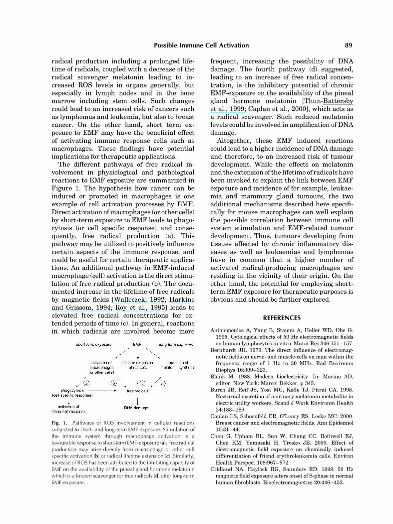
The different pathways of free radical in-volvement in physiological and pathologicalreactions to EMF exposure are summarized inFigure 1. The hypothesis how cancer can beinduced or promoted in macrophages is oneexample of cell activation processes by EMF.Direct activation of macrophages (or other cells)by short-term exposure to EMF leads to phago-cytosis (or cell specific response) and conse-quently, free radical production (a). Thispathway may be utilized to positively influencecertain aspects of the immune response, andcould be useful for certain therapeutic applica-tions. An additional pathway in EMF-inducedmacrophage (cell) activation is the direct stimu-lation of free radical production (b). The docu-mented increase in the lifetime of free radicalsby magnetic fields [Walleczek, 1992; Harkinsand Grissom, 1994; Roy et al., 1995] leads toelevated free radical concentrations for ex-tended periods of time (c). In general, reactionsin which radicals are involved become more
frequent, increasing the possibility of DNAdamage. The fourth pathway (d) suggested,leading to an increase of free radical concen-tration, is the inhibitory potential of chronicEMF-exposure on the availability of the pinealgland hormone melatonin [Thun-Battersbyet al., 1999; Caplan et al., 2000], which acts asa radical scavenger. Such reduced melatoninlevels could be involved in amplification of DNAdamage.
Altogether, these EMF induced reactionscould lead to a higher incidence of DNA damageand therefore, to an increased risk of tumourdevelopment. While the effects on melatoninand the extension of the lifetime of radicals havebeen invoked to explain the link between EMFexposure and incidence of for example, leukae-mia and mammary gland tumours, the twoadditional mechanisms described here specifi-cally for mouse macrophages can well explainthe possible correlation between immune cellsystem stimulation and EMF-related tumourdevelopment. Thus, tumours developing fromtissues affected by chronic inflammatory dis-eases as well as leukaemias and lymphomashave in common that a higher number ofactivated radical-producing macrophages areresiding in the vicinity of their origin. On theother hand, the potential for employing short-term EMF exposure for therapeutic purposes isobvious and should be further explored.
REFERENCES
Antonopoulos A, Yang B, Stamm A, Heller WD, Obe G.1995. Cytological effects of 50 Hz electromagnetic fieldson human lymphocytes in vitro. Mutat Res 346:151–157.
Bernhardt JH. 1979. The direct influence of electromag-netic fields on nerve- and muscle-cells on man within thefrequency range of 1 Hz to 30 MHz. Rad EnvironmBiophys 16:309–323.
Blank M. 1988. Modern bioelectricity. In: Marino AD,editor. New York: Marcel Dekker. p 345.
Burch JB, Reif JS, Yost MG, Keffe TJ, Pitrat CA. 1998.Nocturnal excretion of a urinary melatonin metabolite inelectric utility workers. Scand J Work Environm Health24:183–189.
Caplan LS, Schoenfeld ER, O’Leary ES, Leske MC. 2000.Breast cancer and electromagnetic fields. Ann Epidemiol10:31–44.
Chen G, Upham BL, Sun W, Chang CC, Rothwell EJ,Chen KM, Yamasaki H, Trosko JE. 2000. Effect ofelectromagnetic field exposure on chemically induceddifferentiation of friend erythroleukemia cells. EnvironHealth Perspect 108:967–972.
Cridland NA, Haylock RG, Saunders RD. 1999. 50 Hzmagnetic field exposure alters onset of S-phase in normalhuman fibroblasts. Bioelectromagnetics 20:446–452.
Fig. 1. Pathways of ROS involvement in cellular reactionssubjected to short- and long-term EMF exposure. Stimulation ofthe immune system through macrophage activation is afavourable response to short-term EMF-exposure (a). Free radicalproduction may arise directly from macrophage or other cellspecific activation (b) or radical lifetime extension (c). Similarly,increase of ROS has been attributed to the inhibiting capacity ofEMF on the availability of the pineal gland hormone melatoninwhich is a known scavenger for free radicals (d) after long-termEMF-exposure.
Possible Immune Cell Activation 89
D’Ambrosio G, Massa R, Di Berardino D, Lioi MB,Scaglione A, Scarfi MR. 1988. Chromosomal aberrationsin bovine lymphocytes exposed to 50 Hz electric current.Bioelectromagnetics 7:239–245.
Dibirdik I, Kristupaitis D, Kurosaki T, Tuel-Ahlgren L,Chu A, Pond D, Tuong D, Luben R, Uckun FM. 1998.Stimulation of Src family protein-tyrosine kinases as aproximal and mandatory step for SYK kinase-dependentphospholipase Cg2 activation in lymphoma B cellsexposed to low energy electromagnetic fields. J BiolChem 273:4035–4039.
Dubocovich ML. 1995. Melatonin receptors: Are theremultiple subtypes? Trends Pharmacol Sci 16:50–56.
Eremenko T, Esposito C, Pasquarelli A, Pasquali E, VolpeP. 1997. Cell cycle kinetics of friend erythroleukemiacells in a magnetically shielded room and in a low-frequency/low-intensity magnetic field. Bioelectromag-netics 18:58–66.
Fairbairn DW, O’Neill KL. 1994. The effect of electro-magnetic field exposure on the formation of DNA singlestrand breaks in human cells. Cell Mol Biol (Noisy-le-grand) 40:561–567.
Flipo D, Fournier M, Benquet C, Roux P, Le Boulaire C,Pinsky C, LaBella FS, Krzystyniak K. 1998. Increasedapoptosis, changes in intracellular Ca2þ, and functionalalterations in lymphocytes and macrophages after in vitroexposure to static magnetic field. J Toxicol EnvironHealth 54:63–76.
Garcia-Sagredo JM, Parada LA, Monteagudo JL. 1990.Effect on SCE in human chromosomes in vitro of low-level pulsed magnetic field. Environ Mol Mutagen 16:185–188.
Glaser R. 1992. Current concepts of the interaction of weakelectromagnetic fields with cells. Bioelectrochem andBioenerg 27:255–268.
Goodman R, Blank M. 1998. Magnetic field stress inducesexpression of hsp70. Cell Stress Chaperones 3:79–88.
Graham C, Cook MR, Riffle DW, Gerkovich MM, CohenHD. 1996. Nocturnal melatonin levels in human volun-teers exposed to intermittent 60 Hz magnetic fields.Bioelectromagnetics 17:263–273.
Harkins TT, Grissom CB. 1994. Magnetic field effects onB12 ethanolamine ammonia lyase: Evidence for a radicalmechanism. Science 263:958–960.
IARC 2002. Monographs on the evaluation of carcinogenicrisks to humans. Non-ionizing radiation, Part 1:staticand extremely low-frequency (ELF) electric and mag-netic fields, 429 pages ISBN 92 832 1280 0.
Ivancsits S, Diem E, Pilger A, Rudiger HW, Jahn O. 2002.Induction of DNA strand breaks by intermittentexposure to extremely-low-frequency electromagneticfields in human diploid fibroblasts. Mutat Res 519:1–13.
Junkersdorf B, Bauer H, Gutzeit HO. 2000. Electromag-netic fields enhance the stress response at elevatedtemperatures in the nematode Caenorhabditis elegans.Bioelectromagnetics 21:100–106.
Kato M, Honma K, Shigemitsu T, Shiga Y. 1994. Circularlypolarized 50-Hz magnetic field exposure reduces pinealgland and blood melatonin concentrations of Long-Evansrats. Neurosci Lett 166:59–62.
Katsir G, Parola AH. 1998. Enhanced proliferation causedby a low frequency weak magnetic field in chick embryofibroblasts is suppressed by radical scavengers. BiochemBiophys Res Commun 252:753–756.
Khalil AM, Quassem W. 1991. Cytogenetic effects of pulsingelectromagnetic field on human lymphocytes in vitro:Chromosome aberrations, sister-chromatid exchanges.Mutat Res 247:141–146.
Korzh-Sleptsova IL, Lindstrom E, Mild KH, Berglund A,Lundgren E. 1995. Low frequency MFs increased inositol1,4,5-trisphosphate levels in the Jurkat cell line. FEBSLett 359:151–154.
Loscher W, Mevissen M. 1994. Animal studies on the role of50/60-Hertz magnetic fields in carcinogenesis. Life Sci54:1531–1543.
Loschinger M, Thumm S, Hammerle H, Rodemann HP.1999. Induction of intracellular calcium oscillations inhuman skin fibroblast populations by sinusoidal extre-mely low-frequency magnetic fields (20 Hz, 8 mT) isdependent on the differentiation state of the single cell.Radiat Res 151:195–200.
Lacy-Hulbert A, Wilkins RC, Hesketh TR, Metcalfe JC.1995. No effect of 60 Hz electromagnetic fields on MYC orbeta-actin expression in human leukemic cells. RadiatRes 144:9–17.
Lacy-Hulbert A, Metcalfe JC, Hesketh R. 1998. Biologicalresponses to electromagnetic fields. FASEB J 12:395–420.
Lai H, Singh NP. 1997. Acute exposure to a 60 Hz magneticfield increases DNA strand breaks in rat brain cells.Bioelectromagnetics 18:156–165.
Landry PS, Sadasivan KK, Marino AA, Albright JA. 1997.Electromagnetic fields can affect osteogenesis by increas-ing the rate of differentiation. Clin Orthop 338:262–270.
Lange S, Richard D, Viergutz T, Kriehuber R, Simko M.2002. Alterations in the cell cycle and in the protein levelof cyclin D1, p21CIP1, and p16INK4a after exposure to50 Hz MF in human cells. Radiat Environ Biophys41:131–137.
Lerchl A, Reiter RJ, Howes KA, Nonaka KO, Stokkan KA.1991. Evidence that extremely low frequency Ca(2þ)-cyclotron resonance depresses pineal melatonin synth-esis in vitro. Neurosci Lett 124:213–215.
Liburdy RP, Callahan DE, Harland J, Dunham E, SlomaTR, Yaswen P. 1993a. Experimental evidence for 60 Hzmagnetic fields operating through the signal transduc-tion cascade. Effects on calcium influx and c-MYC mRNAinduction. FEBS Lett 334:301–308.
Liburdy RP, Sloma TR, Sokolic R, Yaswen P. 1993b. ELFmagnetic fields, breast cancer, and melatonin: 60 Hzfields block melatonin’s oncostatic action on ERþ breastcancer cell proliferation. J Pineal Res 14:89–97.
Lin H, Head M, Blank M, Han L, Jin M, Goodman R. 1998.Myc-mediated transactivation of HSP70 expressionfollowing exposure to magnetic fields. J Cell Biochem69:181–188.
Lindstrom E, Lindstrom P, Berglund A, Mild KH,Lundgren E. 1993. Intracellular calcium oscillationsinduced in a T-cell line by weak 50-Hz magnetic-field.J Cell Physiol 156:395–398.
Litovitz TA, Krause D, Mullins JM. 1991. Effect ofcoherence time of the applied magnetic field on ornithinedecarboxylase activity. Biochem Biophys Res Commun178:862–865.
Livingston GK, Witt KL, Gandhi OP, Chatterjee I, Roti JL.1991. Reproductive integrity of mammalian cells exposedto power frequency electromagnetic fields. Environ MolMutagen 17:49–58.
90 Simko and Mattsson
Lupke M, Rollwitz J, Simko M. 2004. 50 Hz magneticfields induce reactive oxygen intermediates in humanmonocytes and in Mono-Mac 6 cells. (in press).
Mattsson MO, Lindstrom E, Still M, Lindstrom P, Mild KH,Lundgren E. 2001. [Ca2þ](i) rise in Jurkat E6-1 cell linesfrom different sources as a response to 50 Hz magneticfield exposure is a reproducible effect and independent ofpoly-L-lysine treatment. Cell Biol Int 25:901–907.
Mullins JM, Penafiel LM, Juutilainen J, Litovitz TA.1999. Dose-response of electromagnetic field-enhancedornithine decarboxylase activity. Bioelectrochem Bio-energ 48:193–199.
National Institute of Environmental Health Sciences(NIEHS). 1998. Working group report: Assessment ofhealth effects from exposure to power-line frequencyelectric and magnetic fields. In: Portier CJ, Wolfe MS,editors. U. S. National Institutes of Health. NIH Publica-tion No. 98-3981. Research Triangle Park: NIEHS .
Nindl G, Swez JA, Miller JM, Balcavage WX. 1997. Growthstage dependent effects of electromagnetic fields onDNA synthesis of Jurkat cells. FEBS Lett 414:501–506.
Nordenson I, Mild KH, Andersson G, Sandstrom M. 1994.Chromosomal aberrations in human amniotic cells afterintermittent exposure to fifty hertz magnetic fields.Bioelectromagnetics 15:293–301.
Pfluger DH, Minder CE. 1996. Effects of exposure to 16.7 Hzmagnetic fields on urinary 6-hydroxymelatonin sulfateexcretion of swiss railway workers. J Pineal Research 21:91–100.
Reiter RJ. 1997. Antioxidant actions of melatonin. AdvPharmacol 38:103–117.
Richard D, Lange S, Viergutz T, Kriehuber R, Weiss DG,Simko M. 2002. Influence of 50 Hz magnetic fields incombination with a tumour promoting phorbol ester onprotein kinase C and cell cycle in human cells. Mol CellBiochem 232:133–141.
Rodemann HP, Bayreuther K, Pfleiderer G. 1989. Thedifferentiation of normal and transformed human fibro-blasts in vitro is influenced by electromagnetic fields.Exp Cell Res 182:610–621.
Rollwitz J, Lupke M, Simko M. 2004. 50 Hz magnetic fieldsinduce free radical formation in mouse bonemarrow-derived macrophages. (in press).
Rosenthal M, Obe G. 1989. Effects of 50-Hertz electro-magnetic fields on proliferation and on chromosomalalterations in human peripheral lymphocytes untreatedor pre-treated with chemical mutagens. Mutation Res210:329–335.
Roy S, Noda Y, Eckert V, Traber MG, Mori A, Liburdy RP,Packer L. 1995. The phorbol 12-myristate 13-acetate(PMA)-induced oxidative burst in rat peritoneal neutro-phils is increased by a 0.1 mT (60 Hz) magnetic field.FEBS Lett 376:164–166.
Saffer JD, Thurston SJ. 1995. Short exposures to 60 Hzmagnetic fields do not alter MYC expression in HL60 orDaudi cells. Radiat Res 144:18–25.
Scaiano JC, Mohtat N, Cozens FL, McLean J, ThansandoteA. 1994. Application of the radical pair mechanism to freeradicals in organized systems: Can the effects of 60 Hz bepredicted from studies under static fields? Bioelectro-magnetics 15:549–554.
Scarfi MR, Bersani F, Cossarizza A, Monti D, Zeni O,Lioi MB, Franceschetti G, Capri M, Franceschi C. 1993.
50 Hz AC sinusoidal electric fields do not exert genotoxiceffects (micronucleus formation) in human lymphocytes.Radiat Res 135:64–68.
Schimmelpfeng J, Dertinger H. 1993. The action of 50 Hzmagnetic and electric fields upon cell proliferationand cyclic AMP content of cultured mammalian cells.Bioelectrochem Bioenerg 30:143–150.
Selmaoui B, Bogdan A, Auzeby A, Lambrozo J, Touitou Y.1996. Acute exposure to 50 Hz magnetic field does notaffect hematologic or immunologic functions in healthyyoung men: A circadian study. Bioelectromagnetics 17:364–372.
Simko M, Kriehuber R, Weiss DG, Luben R. 1998a. Theeffects of 50 Hz EMF exposure on micronucleus forma-tion and apoptosis in transformed and non transformedhuman cell lines. Bioelectromagnetics 19:85–91.
Simko M, Kriehuber R, Lange S. 1998b. Micronucleusformation in human amnion cells after exposure to 50 HzMF applied horizontally and vertically. Mutat Res 418:101–111.
Simko M, Richard D, Kriehuber R, Weiss DG. 2001a.Micronucleus induction in SHE cells following exposureto 50 Hz magnetic fields, benzo(a)pyrene and TPAin vitro. Mutat Res 495:43–50.
Simko M, Droste S, Kriehuber R, Weiss DG. 2001b.Stimulation of phagocytosis and free radical productionin murine macrophages by 50 Hz electromagnetic fields.Eur J Cell Biol 80:562–566.
Stevens RG. 1987. Electric power use and breast cancer:A hypothesis. American J Epidemiol 125:556–561.
Stevens RG, Davis S. 1996. The melatonin hypothesis:Electric power and breast cancer. Environ HealthPerspect 104(Suppl 1):135–140.
Tan DX, Manchester LC, Reiter RJ, Qi WB, Zhang M,Weintraub ST, Cabrera J, Sainz RM, Mayo JC. 1999.Identification of highly elevated levels of melatonin inbone marrow: Its origin and significance. BiochimBiophys Acta 1472:206–214.
Tao Q, Henderson A. 1999. EMF induces differentiation inHL-60 cells. J Cell Biochem 73:212–217.
Thun-Battersby S, Mevissen M, Loscher W. 1999. Exposureof Sprague-Dawley rats to a 50-Hertz, 100-microTeslamagnetic field for 27 weeks facilitates mammary tumor-igenesis in the 7,12-dimethylbenz[a]-anthracene model ofbreast cancer. Cancer Res 59:3627–3633.
Tuinstra R, Goodman E, Greenebaum B. 1998. Pro-tein kinase C activity following exposure to mag-netic field and phorbol ester. Bioelectromagnetics 19:469–476.
Uckun FM, Kurosaki T, Jin J, Jun X, Morgan A, Takata M,Bolen J, Luben RA. 1995. Exposure of B-lineage lymphoidcells to low energy electromagnetic fields stimulates Lynkinase. J Biol Chem 270:27666–27670.
Valberg PA, Kavet R, Rafferty CN. 1997. Can low-level 50/60 Hz electric and magnetic fields cause biologicaleffects? Radiat Res 148:2–21.
Valtersson U, Hansson Mild K, Mattsson MO. 1997.Ornithine decarboxylase activity and polyamine levelsare different in Jurkat and CEM-CM3 cells after exposureto a 50 Hz magnetic field. Bioelectrochem Bioenerg43:169–172.
Walleczek J. 1992. Electromagnetic field effects on cells ofthe immune system: The role of calcium signalling,FASEB J 6:3177–3185.
Possible Immune Cell Activation 91

Wei LX, Goodman R, Henderson A. 1990. Changes in levelof c-myc and histone H2B following exposure of cells tolow-frequency sinusoidal electromagnetic fields: Eviden-ce for a window effect. Bioelectromagnetics 11:269–272.
Wei M, Guizzetti M, Yost M, Costa LG. 2000. Exposure to60-Hz magnetic fields and proliferation of human astro-cytoma cells in vitro. Toxicol Appl Pharmacol 162:166–176.
West RW, Hinson WG, Lyle BD, Swicord MC. 1994.Enhancement of anchorage-independent growth in JB6
cells exposed to 60 Hz magnetic fields. Bioelectrochemand Bioenerg 34:39–43.
Yellon SM. 1994. Acute 60 Hz magnetic field exposureeffects on the melatonin rhythm in the pineal gland andcirculation of the adult Djungarian hamster. J Pineal Res16:136–144.
Zhao YL, Johnson PG, Jahreis GP, Hui SW. 1999.Increased DNA synthesis in INIT/10T1/2 cells afterexposure to a 60 Hz magnetic field: A magnetic field ora thermal effect? Radiat Res 151:201–208.
92 Simko and Mattsson