Expression Patterns of Two Murine Homologs of Drosophila Single-MindedSuggest Possible Roles in...
16
Molecular and Cellular Neuroscience 7, 1 –16 (1996) Article No. 0001 MCN Expression Patterns of Two Murine Homologs of Drosophila Single-Minded Suggest Possible Roles in Embryonic Patterning and in the Pathogenesis of Down Syndrome Chen-Ming Fan,* ,1 Ellen Kuwana,* , ² Alessandro Bulfone,² Colin F. Fletcher,‡ Neal G. Copeland,‡ Nancy A. Jenkins,‡ Stephen Crews,§ Salvador Martinez, Ø Luis Puelles, Ø John L. R. Rubenstein,² and Marc Tessier-Lavigne* *Howard Hughes Medical Institute, Programs in Cell Biology, Developmental Biology and Neuroscience, Department of Anatomy, University of California, San Francisco, 513 Parnassus Avenue, San Francisco, California 94143-0452; ²Nina Ireland Laboratory of Developmental Neurobiology, Programs in Developmental Biology and Neuroscience, Center for Neurobiology and Psychiatry, Department of Psychiatry and Langley Porter Psychiatric Institute, University of California, San Francisco, San Francisco, California 94143-0984; ‡Mammalian Genetics Laboratory, ABL-Basic Research Program, NCI-Frederick Cancer Research and Development Center, Frederick, Maryland 21702; §Department of Biochemistry and Biophysics, University of North Carolina at Chapel Hill, Chapel Hill, North Carolina 27599-7260; and Ø Department of Morphological Sciences, Faculty of Medicine, University of Murcia, 30100 Murcia, Spain The single-minded (sim) gene encodes a transcriptional regionalization of the diencephalon prior to any overt mor- phological differentiation in this region. Outside the CNS, regulator that functions as a key determinant of central nervous system (CNS) midline development in Drosophila. Sim1 is expressed in mesodermal and endodermal tis- sues, including developing somites, mesonephric duct, We report here the identification of two murine homologs of sim, Sim1 and Sim2, whose products show a high de- and foregut. Sim2 is expressed in facial and trunk carti- lage, as well as trunk muscles. Both murine Sim genes gree of sequence conservation with Drosophila SIM in their amino-terminal halves, with each containing a basic are also expressed in the developing kidney. Our data suggest that the Sim genes play roles in directing the helix–loop–helix domain as well as a PAS domain. Sim1 maps to the proximal region of mouse chromosome 10, regionalization of tissues where they are expressed. Moreover, the expression pattern documented for Sim2 whereas Sim2 maps to a portion of the distal end of chro- mosome 16 that is syntenic to the Down syndrome critical may provide insights into its potential roles in Down syn- drome. region of human chromosome 21. Recent exon-trapping studies have identified in the critical region several exons of a human sim homolog which appears to be the homolog of murine Sim2; this has led to the hypothesis that in- INTRODUCTION creased dosage of this sim homolog in cases of trisomy 21 might be a causal factor in the pathogenesis of Down In Drosophila, the single-minded gene (sim) (Thomas et syndrome. We have examined the expression patterns of al., 1988; Crews et al., 1988) directs the development of the Sim genes during embryogenesis. Both genes are ex- cells located at the midline of the developing central ner- pressed in dynamic and selective fashion in specific neu- vous system (CNS) (reviewed by Crews et al., 1992). In romeric compartments of the developing forebrain, and sim mutant embryos these cells fail to differentiate and the expression pattern of Sim2 provides evidence for early eventually die (Thomas et al., 1988). Conversely, when sim is expressed ubiquitously (under control of a heat- 1 Present address: Carnegie Institute of Washington, Department of Embryology, 115 West University Parkway, Baltimore, MD 21210. shock promoter) in transgenic flies, many other cells of 1044-7431/96 $18.00 Copyright q 1996 by Academic Press, Inc. All rights of reproduction in any form reserved. 1
-
Upload
independent -
Category
Documents
-
view
0 -
download
0
Transcript of Expression Patterns of Two Murine Homologs of Drosophila Single-MindedSuggest Possible Roles in...
Molecular and Cellular Neuroscience 7, 1–16 (1996)
Article No. 0001 MCNExpression Patterns of Two Murine Homologs ofDrosophila Single-Minded Suggest PossibleRoles in Embryonic Patterning and in thePathogenesis of Down Syndrome
Chen-Ming Fan,*,1 Ellen Kuwana,*,† Alessandro Bulfone,†Colin F. Fletcher,‡ Neal G. Copeland,‡ Nancy A. Jenkins,‡Stephen Crews,§ Salvador Martinez,Ø Luis Puelles,Ø
John L. R. Rubenstein,† and Marc Tessier-Lavigne**Howard Hughes Medical Institute, Programs in Cell Biology, Developmental Biology andNeuroscience, Department of Anatomy, University of California, San Francisco, 513 ParnassusAvenue, San Francisco, California 94143-0452; †Nina Ireland Laboratory of DevelopmentalNeurobiology, Programs in Developmental Biology and Neuroscience, Center for Neurobiologyand Psychiatry, Department of Psychiatry and Langley Porter Psychiatric Institute, Universityof California, San Francisco, San Francisco, California 94143-0984; ‡Mammalian GeneticsLaboratory, ABL-Basic Research Program, NCI-Frederick Cancer Research and DevelopmentCenter, Frederick, Maryland 21702; §Department of Biochemistry and Biophysics, Universityof North Carolina at Chapel Hill, Chapel Hill, North Carolina 27599-7260; and ØDepartment ofMorphological Sciences, Faculty of Medicine, University of Murcia, 30100 Murcia, Spain
The single-minded (sim) gene encodes a transcriptional regionalization of the diencephalon prior to any overt mor-phological differentiation in this region. Outside the CNS,regulator that functions as a key determinant of central
nervous system (CNS) midline development in Drosophila. Sim1 is expressed in mesodermal and endodermal tis-sues, including developing somites, mesonephric duct,We report here the identification of two murine homologs
of sim, Sim1 and Sim2, whose products show a high de- and foregut. Sim2 is expressed in facial and trunk carti-lage, as well as trunk muscles. Both murine Sim genesgree of sequence conservation with Drosophila SIM in
their amino-terminal halves, with each containing a basic are also expressed in the developing kidney. Our datasuggest that the Sim genes play roles in directing thehelix– loop–helix domain as well as a PAS domain. Sim1
maps to the proximal region of mouse chromosome 10, regionalization of tissues where they are expressed.Moreover, the expression pattern documented for Sim2whereas Sim2 maps to a portion of the distal end of chro-
mosome 16 that is syntenic to the Down syndrome critical may provide insights into its potential roles in Down syn-drome.region of human chromosome 21. Recent exon-trapping
studies have identified in the critical region several exonsof a human sim homolog which appears to be the homologof murine Sim2; this has led to the hypothesis that in- INTRODUCTIONcreased dosage of this sim homolog in cases of trisomy21 might be a causal factor in the pathogenesis of Down In Drosophila, the single-minded gene (sim) (Thomas etsyndrome. We have examined the expression patterns of al., 1988; Crews et al., 1988) directs the development ofthe Sim genes during embryogenesis. Both genes are ex- cells located at the midline of the developing central ner-pressed in dynamic and selective fashion in specific neu-
vous system (CNS) (reviewed by Crews et al., 1992). Inromeric compartments of the developing forebrain, andsim mutant embryos these cells fail to differentiate andthe expression pattern of Sim2 provides evidence for earlyeventually die (Thomas et al., 1988). Conversely, whensim is expressed ubiquitously (under control of a heat-1 Present address: Carnegie Institute of Washington, Department of
Embryology, 115 West University Parkway, Baltimore, MD 21210. shock promoter) in transgenic flies, many other cells of
1044-7431/96 $18.00Copyright q 1996 by Academic Press, Inc.All rights of reproduction in any form reserved. 1
AID MCN 0530 / 6p06$$$$61 03-12-96 08:56:13 mcnal AP: MCN

2 Fan et al.
the ventral neurogenic region differentiate into CNS during mouse embryogenesis by in situ hybridization. Likesim, the mammalian homologs show restricted patterns ofmidline cells (Nambu et al., 1991). Thus the sim gene
product appears to be both necessary and sufficient for expression during embryonic development. Sim1 and Sim2have restricted distributions in the developing brain, par-the differentiation of CNS midline cells within the ven-
tral neurogenic region. In addition to its function in CNS axial mesoderm, intermediate mesoderm, and gut that sug-gest their involvement in the regionalization of these tissuesmidline cells, sim is expressed in a subset of myoblasts,
the gut, and the brain, where it may also play roles in during early development. Importantly, during the finalstages of this study, two studies reported the identificationregionalization and cell fate determination (Crews et al.,
1988, Lewis and Crews, 1994). of fragments of a candidate human SIM gene by exon-trapping of DNA sequences within the Down syndromeThe SIM protein contains a basic helix– loop–helix
(bHLH) motif and localizes to the cell nucleus where it critical region of human chromosome 21 (Chen et al., 1995;Dahmane et al., 1995). The open reading frame of the sixmay act as a transcriptional activator (Nambu et al., 1991;
Franks and Crews, 1994). bHLH transcription factors ap- trapped exons comprises a bHLH and a partial PAS do-main highly homologous to those of the mouse Sim2 gene.pear to play important roles in directing cell fates and
controlling cell proliferation and differentiation during Our results describing the expression pattern of the murinehomolog confirm and extend a report of the expressionembryonic development (see Weintraub et al., 1991; Jan
and Jan, 1993; Olson and Klein, 1994, for review). Mem- pattern of the SIM2 gene in human and rat embryos (Dah-mane et al., 1995) and may provide insights to possiblebers of this family include, for example, the products of
the myogenic determination genes like Myf5, MyoD, and links between SIM2 function and Down syndrome.Myogenin, and the neurogenic genes of the aechete–scutecomplex. These proteins function by forming heterodi- RESULTSmers through their helix– loop–helix (HLH) domains
Isolation of cDNAs for Two Murine Homologues ofand bind to specific DNA sequences through their basicDrosophila simdomains to transactivate target genes. Several of these
To search for murine homologues of Drosophila sim,genes are evolutionarily conserved in sequence, and inwe screened a mouse genomic library at low stringencymany cases homologs in widely divergent species fromusing a probe corresponding to the sim bHLH domainarthropods to vertebrates have been found to have ap-(Nambu et al., 1991). Seven clones were isolated. Theparently similar patterns of expression and embryonicsequence of each hybridizing fragment contained a shortfunctions (reviewed by Jan and Jan, 1993).open reading frame that was highly homologous over aIn addition to its bHLH domain, Drosophila SIM pos-171-nt stretch to the sequence coding for the bHLH do-sesses a domain that shares homology with a domain inmain of sim. Five of the seven clones contained fragmentsthe Drosophila PER protein (which plays a role in control-of one gene, designated Sim1, and the other two con-ling circadian rhythms) and the two subunits of the hu-tained fragments of a second gene, designated Sim2. Be-man aromatic hydrocarbon (dioxin) receptor, ARNT (thecause in situ hybridization demonstrated the expressionaromatic-hydrocarbon receptor nuclear translocator) andof these two genes in the caudal diencephalon and rostralAHR (the aromatic-hydrocarbon receptor) (Crews et al.,mesencephalon of E11.5 mouse embryos (see below), this1988; Hoffman et al., 1991; Burbach et al., 1992). Thesetissue was microdissected and used as a source of mRNAdomains have been termed PAS domains (for PER,for Northern analysis and for the isolation of cDNAs. AnARNT, SIM). The PAS domain in PER has been shown8.0-kb transcript for Sim1 and a 4.2 kb transcript for Sim2to function as a dimerization domain (Huang et al., 1993;were detected in this tissue by Northern analysis (dataReisz-Porszasz et al., 1994; Jain et al., 1994; Lindebro etnot shown). A cDNA library made from this tissue wasal., 1995), whereas that of AHR functions as a ligandscreened for Sim1 and Sim2 cDNAs. A 4.0-kb clone con-(dioxin)-binding and heat-shock protein association do-taining the entire coding region of Sim2 was isolated.main (Burbach et al., 1992; Whitelaw et al., 1993). TheSeveral overlapping cDNA clones and a genomic clonerecently identified hypoxia-inducible factor 1 (HIF-1)were used to assemble a 7.2-kb stretch of sequence ofalso contains both bHLH and PAS domains and is a newthe Sim1 cDNA containing the entire coding region ofmember of this gene family (Wang et al., 1995).the gene (see Experimental Methods).In searching for genes related to sim in the mammalian
genome by low-stringency hybridization, we isolated twoThe bHLH and PAS Domains of the Drosophila simmurine homologs of sim. Like the Drosophila SIM protein,Gene Product Are Highly Conserved in the Mousethe two predicted murine proteins each possess a bHLH
and a PAS domain. To explore the possible functions of the Figure 1 compares the deduced amino acid sequencesof the two murine gene products to that of Drosophila SIM.mammalian Sim genes, we characterized their expression
AID MCN 0530 / 6p06$$$$61 03-12-96 08:56:13 mcnal AP: MCN

3Mammalian Single-Minded Genes
FIG. 1. Relationships between the predicted products of the Drosophila sim and the murine Sim1 and Sim2 genes. (A) Structural organization ofthe predicted SIM, SIM1, and SIM2 proteins. Each protein has a basic helix– loop–helix (bHLH) domain at its extreme N-terminus (dark shading).Each protein also has a PAS domain; the portions of the PAS domains that are conserved across species are indicated by boxes with light shading(the direct repeats within the PAS domain are indicated by black boxes). The three proteins diverge significantly after the PAS domain. Otherstructural motifs in the SIM protein are also illustrated: AAQ repeats (checker board), proline (P) rich region (horizonal lines), and a glutamine(Q) rich region (black box). (B) Alignment of the predicted amino acid sequences of the SIM, SIM1, and SIM2 proteins. Amino acids conservedbetween all three proteins are boxed. The nucleotide sequences of Sim1 and Sim2 have been submitted to GenBank (see Experimental Methods).The partial human SIM2 sequence (Chen et al., 1995; Dahmane et al., 1995) is aligned underneath the mouse SIM2 sequence: the differences betweenthe predicted human SIM2 and mouse SIM2 amino acid sequences are shown, whereas dots indicate amino acids that are identical between humanand mouse SIM2. Note the presence of two gaps in the human sequence compared to the murine seuqences.
The amino-terminal halves of the three proteins have simi- VVA) is strongly conserved in SIM1 and SIM2. In particu-lar, valine 92 of SIM is conserved in both murine proteins;lar structural domains (Fig. 1A). Like SIM, SIM1 and SIM2
have predicted bHLH domains at their extreme amino ter- mutation of the homologous valine in the PER protein toan aspartic acid in the perL allele alters the biological func-mini. The amino acid sequences of the bHLH domains are
highly conserved between fly and mouse: there are only tion of PER and dramatically reduces the ability of the PERPAS domain to dimerize (Huang et al., 1993). A secondfive substitutions in the SIM1 bHLH domain, and six in
the SIM2 bHLH domain; for both genes, two of the substi- region that is very conserved between all PAS domain pro-teins (residues 277–283 of SIM: GYEPQDL) is identical intutions are conservative (Fig. 1B).
Each of the murine proteins also has a PAS domain, the fly and murine proteins.The homology between the two murine proteins andwhich is highly homologous to that of Drosophila SIM. One
difference between the murine and fly proteins is the pres- SIM is also seen in the 40 amino-acid stretch linkingthe bHLH and PAS domains in each protein. Overall,ence, in the PAS domain of SIM, of a 24 amino acid insert
(residues 155–178) between the two so-called PAS repeats throughout the amino terminal halves of the proteins(the first 375, 350, and 351 residues of SIM, SIM1, and(Fig. 1B). Interestingly, a hydrophobic region just amino
terminal to the first PAS repeat that is conserved between SIM2, respectively), SIM1 and SIM2 are slightly moreclosely related to each other than they are to SIM atPER, ARNT, AHR, and SIM (residues 89–94 of SIM: FIF-
AID MCN 0530 / 6p06$$$$61 03-12-96 08:56:13 mcnal AP: MCN

4 Fan et al.
FIG. 2. Murine chromosomal location of Sim1 and Sim2. The two genes were mapped to mouse chromosomes by interspecific backcross analysis.The segregation patterns of Sim1, Sim2, and the flanking genes in, respectively, 93 and 180 backcross animals that were typed for all loci are shownat the top of the figure. Each column represents the chromosome identified in the backcross progeny that was inherited from the (C57BL/6J 1 M.spretus)F1 parent. The black boxes represent the presence of the C57BL/6J allele and the white boxes represent the presence of the M. spretus allele.The number of offspring inheriting each type of allele is listed at the bottom of each column. Partial chromosome linkage maps showing thelocations of the Sim loci in relation to linked markers (Erg: Rao et al., 1987; Fyn, Ros1, and Gja1: Justice et al., 1990; Haeflinger et al., 1992; App,Taim1, and Mx1: Bae et al., 1994; Habets et al., 1995) are shown. The number of recombinant N2 animals over the total number of N2 animalstyped plus the recombination frequencies expressed as genetic distance in centimorgans ({1 standard error) is shown for each pair of loci to theleft of each chromosome. The upper 95% confidence limit of the recombination distance is given in parentheses when no recombinants are foundbetween loci. Gene order was determined by minimizing the number of recombination events required to explain the allele distribution pattern.The positions of loci mapped to human chromosomes are shown to the right of the chromosome maps. References for the map positions of mosthuman loci can be obtained from GDB (Genome Data Base), a computerized database of human linkage information maintained by the WilliamH. Welch Medical Library of The Johns Hopkins University (Baltimore, MD).
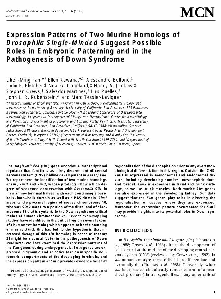
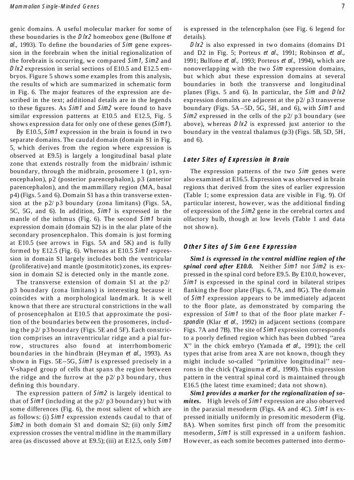
FIG. 3. Early expression pattern of Sim2 in the forebrain primordium. (A) Sim2 expression (bracketed by black lines) can be detected as early asthe 2-somite stage (E8.0), as shown in a side view of an embryo whole-mount. (B– D) A comparison with En1 expression shows that Sim2 isexpressed in a narrow band in the caudal portion of the forebrain, immediately rostral to the midbrain– forebrain boundary (indicated by thewhite lines). Three consecutive sections through a 10-somite (E8.5) embryo were hybridized with antisense probes to En1 (B), Sim2 (C), and En1again (D). The rostral limit of En1 expression and the caudal limit of Sim-2 expression are approximately at the same point (the white lines). Atthis stage, the rostral limit of En1 expression defines the midbrain– forebrain boundary (McMahon et al., 1992). Scale: 155 mm (A), 100 mm (B–D).FIG. 4. Expression patterns of Sim1 and Sim2 in the E9.5 mouse. Transcripts were detected by in situ hybridization in embryo whole-mounts (A,B) and in tissue sections (C–F): Sim1 expression is visualized in a lateral view of a whole-mounted embryo (A) and in a parasagittal section (C)and a transverse section (E) through the caudal diencephalon. At E9.5, Sim1 expression is seen in the basal portion of the caudal diencephalon,the mesonephric ducts, and head mesenchyme. Note that expression in differentiated somites is restricted to the dermomyotome (see also Fig. 8).Sim2 expression is visualized in a lateral view of a whole-mounted embryo (B) and a near midsagittal section (D) from the same embryo that wasused in C. Sim2 expression is confined to the caudal portion of the diencephalon in the basal plate and crosses the midline at the mammillaryarea (F). Weak expression in the mesonepheric tubules can also be detected by E9.5 (not shown). Note that in the mammillary area, Sim1 isexpressed in the basal half but is excluded from the midline (C), whereas Sim2 is expressed in the midline (F). Abbreviations: dm, dermomyotome;fb, forebrain; hm, head mesenchyme; mb, midbrain; md, mesonephric duct; s, somite; psm, presomitic mesoderm. Scale, 420 (A, B), 360 (C, D),100 (E, F) mm.FIG. 5. Expression of Sim1 compared to that of Dlx2 at E10.5, as detected by radioactive in situ RNA hybridization of tissue sections. (Top) Sim1expression; (bottom) Dlx2 expression. (A, B) Parasagittal sections. (C– N) Cross sections. Planes of cross sections in C –N are shown in A and B,as follows. Plane w: sections C– H; plane x: sections I and J; plane y: sections K and L; plane z: sections M and N. The boxed region in E is shownat fourfold higher magnification in F. The regions roughly corresponding to the p1/p2 and p2/p3 interprosomeric boundaries are indicated in F.G and H show details of the diencephalon from C and D, respectively. Sim1 is expressed in two domains; domain S1 is in the ventricular zoneand mantle of the basal plate of the diencephalon (BP DI) and mesencephalon (A, I, K, M), whereas domain S2 (a separatehypothalamic domain) is in the mantle (see arrows in A, K, M). Domain S1 has a dorsal extension at the zona limitans (zl; A, C, G). Sim1 is alsoexpressed in a restricted region of head mesenchyme (hm; A, M). Dlx2 is expressed in two domains (see Bulfone et al., 1993). Briefly, domain D1is largely an alar plate zone that begins just anterior of the zona limitans (zl), extends along a longitudinal strip of hypothalamus (labeled as D1in B, L, N; the domain extends from the ventral thalamus (VT), to the anterior midline just ventral to the optic stalk). Domain D2 is in the basaltelencephalon and includes the medial and lateral ganglionic eminences (MGE, LGE) and preoptic area (POA) (B, D, H, J, L, N). There is a
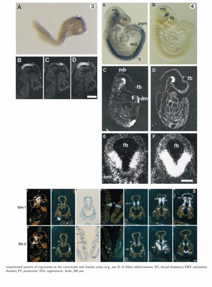
complicated pattern of expression in the ventricular and mantle zones (e.g., see D, J). Other abbreviations: DT, dorsal thalamus; EMT, eminentiathalami; PT, pretectum; TEG, tegmentum. Scale, 200 mm.
03-12-96 08:56:13 mcnal AP: MCN

6 Fan et al.
the amino acid level (SIM:SIM1, 69%; SIM:SIM2, 65%; important role at early stages of neurogenesis in Drosoph-ila, we first describe the expression patterns of the twoSIM1:SIM2, 86%). However, no significant amino acid
(19%) or nucleotide homology is seen among the car- murine genes in the developing nervous system. Expres-sion outside the nervous system is discussed later.boxy-terminal halves of the three proteins (after their
PAS domains). In particular, neither SIM1 nor SIM2 ap-pears to have a glutamine-rich region like those found The Murine Sim Genes Are Expressed Early within SIM, ARNT, and AHR (Fig. 1A), which appear to act
Restricted Patterns in the Developing Forebrainas transcriptional activation domains (Reisz-Porszasz etal., 1994; Jain et al., 1994; Franks and Crews, 1994). Sim2 is a marker of early regionalization of the ante-
rior neural plate. The earliest expression of Sim2 wasChromosomal Locations of the Sim1 and observed in the anterior neural plate in 2-somite stageSim2 Genes embryos (E8.0; Fig. 3A); it was not detected at E7.5 (data
not shown). Sim2 expression is restricted to a narrowThe chromosomal locations of the murine Sim1 andSim2 genes were determined using an interspecific back- transverse band of cells located near the junction of the
midbrain (mesencephalon) and forebrain (prosencepha-crossing panel derived from crosses of [(C57BL/6J 1Mus Spretus)F1 1 C57BL/6J)] mice (Copeland and Jen- lon). This localization was confirmed by comparing the
expression of Sim2 and En1. In serial sections of a 10-kins, 1991; and unpublished; see Experimental Methods).Informative restriction-fragment-length polymorphisms somite embryo, the caudal boundary of Sim2 expression
was found roughly to coincide with the rostral border(RFLPs) were used to map these two genes. Sim1 mappedto the proximal region of mouse chromosome 10, 3 cM of En1 expression (Figs. 3B–3D), which at this stage is
known to approximate the mesencephalon–prosenceph-distal of Fyn, and 0.9 and 1.9 cM proximal of Ros andGjai1, respectively (Fig. 2). Several mutations map in this alon boundary (McMahon et al., 1992). This zone of ex-
pression is also restricted in the mediolateral dimension,interval, including jackson circler (jc), waltzer (v), downless(dl), and kidney disease (kd) (Green, 1989). Sim2 mapped being excluded from the lateral edge of the neural plate
(data not shown). By E9.5 (24-somite stage), Sim2 expres-to the very distal end of chomosome 16. App and Tiam1map 5.3 and 2.6 cM proximal, respectively. Sim2 is 3.4 sion remains restricted to a ventral domain of the caudal
diencephalon (Figs. 4B and 4D), that extends anteriorlycM proximal of Mx1, and has an identical strain distribu-tion as Erg. The only mutation that maps in this vicinity into the mammillary region, where Sim2 expression
crosses the ventral midline (Fig. 4F).is weaver (wv) (Fig. 2). According to the mouse–humanlinkage homologies, it is likely that Sim1 maps to human Sim1 is also expressed in the caudal diencephalon.
Sim1 expression overlaps partially with that of Sim2 in6q21 and Sim2 to 21q22. Indeed, using this murine Sim2cDNA as a probe, a human SIM2 gene has been mapped the caudal diencephalon, but has a slightly later onset
(E9.0; Figs. 4A, 4C, and 4E show expression at E9.5). Theto this region of chromosome 21 (21q22.2–q22.3)(Muenke et al., 1995), which corresponds to the Down rostrocaudal extent of expression is similar to that of
Sim2, although it appears to extend slightly more cau-syndrome critical region. Similarly, exon-trapping stud-ies in this region have recently led to the isolation of six dally into the mesencephalon (Figs. 4A and 4C). As seen
in transverse sections across the diencephalon, Sim1 ex-exons of a human sim-related gene (Chen et al., 1995;Dahmane et al., 1995). The ORF of these exons codes for pression is also restricted to the basal half of the caudal
diencephalon, but, unlike Sim2, is excluded from the ven-252 amino acids of a SIM homolog; the predicted se-quence is highly homologous to the amino terminal 40% tral midline (Fig. 4E). In addition to expression in neural
tube, Sim1 is also expressed in the mesenchyme underly-of the murine SIM2 sequence (Fig. 1B), with identicalbHLH domains and only seven amino acid substitutions ing the neural tube in the cephalic flexure (Figs. 4C, 4E).in the remainder of the human sequence. There are, how-ever, two short gaps in the human sequence compared to
The Sim Genes and Dlx2 Are Expressed inthe murine SIM2 sequence (Fig. 1B). These may represent
Adjacent Domains during Regionalizationbona fide differences; alternatively, it is possible that one
of the Forebrainor more intervening exons in this region of the humangene have not been identified. It has been proposed that the forebrain is divided into
several neuromeres (Bulfone et al., 1993; Figdor andExpression of Murine Sim Genes During Stern, 1993) called prosomeres (Puelles and Rubenstein,Embryonic Development 1993; Rubenstein et al., 1994). According to this model,
the forebrain is subdivided by transverse (neuromeric)The expression of Sim1 and Sim2 was studied by RNAin situ hybridization. Because sim plays a particularly and longitudinal boundaries that separate distinct histo-
AID MCN 0530 / 6p06$$$$61 03-12-96 08:56:13 mcnal AP: MCN
7Mammalian Single-Minded Genes
genic domains. A useful molecular marker for some of is expressed in the telencephalon (see Fig. 6 legend fordetails).these boundaries is the Dlx2 homeobox gene (Bulfone et
al., 1993). To define the boundaries of Sim gene expres- Dlx2 is also expressed in two domains (domains D1and D2 in Fig. 5; Porteus et al., 1991; Robinson et al.,sion in the forebrain when the initial regionalization of
the forebrain is occurring, we compared Sim1, Sim2 and 1991; Bulfone et al., 1993; Porteus et al., 1994), which arenonoverlapping with the two Sim expression domains,Dlx2 expression in serial sections of E10.5 and E12.5 em-
bryos. Figure 5 shows some examples from this analysis, but which abut these expression domains at severalboundaries in both the transverse and longitudinalthe results of which are summarized in schematic form
in Fig. 6. The major features of the expression are de- planes (Figs. 5 and 6). In particular, the Sim and Dlx2expression domains are adjacent at the p2/p3 transversescribed in the text; additional details are in the legends
to these figures. As Sim1 and Sim2 were found to have boundary (Figs. 5A–5D, 5G, 5H, and 6), with Sim1 andSim2 expressed in the cells of the p2/p3 boundary (seesimilar expression patterns at E10.5 and E12.5, Fig. 5
shows expression data for only one of these genes (Sim1). above), whereas Dlx2 is expressed just anterior to theboundary in the ventral thalamus (p3) (Figs. 5B, 5D, 5H,By E10.5, Sim1 expression in the brain is found in two
separate domains. The caudal domain (domain S1 in Fig. and 6).5, which derives from the region where expression isobserved at E9.5) is largely a longitudinal basal plate
Later Sites of Expression in Brainzone that extends rostrally from the midbrain/isthmicboundary, through the midbrain, prosomere 1 (p1, syn- The expression patterns of the two Sim genes were
also examined at E16.5. Expression was observed in brainencephalon), p2 (posterior parencephalon), p3 (anteriorparencephalon), and the mammillary region (MA, basal regions that derived from the sites of earlier expression
(Table 1; some expression data are visible in Fig. 9). Ofp4) (Figs. 5 and 6). Domain S1 has a thin transverse exten-sion at the p2/p3 boundary (zona limitans) (Figs. 5A, particular interest, however, was the additional finding
of expression of the Sim2 gene in the cerebral cortex and5C, 5G, and 6). In addition, Sim1 is expressed in themantle of the isthmus (Fig. 6). The second Sim1 brain olfactory bulb, though at low levels (Table 1 and data
not shown).expression domain (domain S2) is in the alar plate of thesecondary prosencephalon. This domain is just formingat E10.5 (see arrows in Figs. 5A and 5K) and is fully
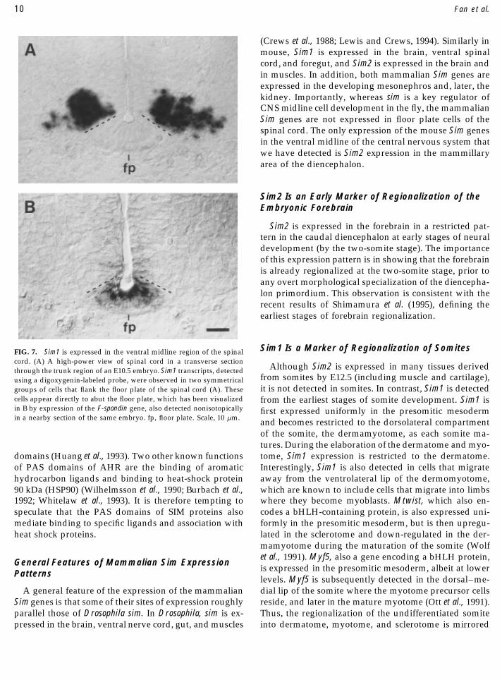
Other Sites of Sim Gene Expressionformed by E12.5 (Fig. 6). Whereas at E10.5 Sim1 expres-sion in domain S1 largely includes both the ventricular Sim1 is expressed in the ventral midline region of the
spinal cord after E10.0. Neither Sim1 nor Sim2 is ex-(proliferative) and mantle (postmitotic) zones, its expres-sion in domain S2 is detected only in the mantle zone. pressed in the spinal cord before E9.5. By E10.0, however,
Sim1 is expressed in the spinal cord in bilateral stripesThe transverse extension of domain S1 at the p2/p3 boundary (zona limitans) is interesting because it flanking the floor plate (Figs. 6, 7A, and 8G). The domain
of Sim1 expression appears to be immediately adjacentcoincides with a morphological landmark. It is wellknown that there are structural constrictions in the wall to the floor plate, as demonstrated by comparing the
expression of Sim1 to that of the floor plate marker F-of prosencephalon at E10.5 that approximate the posi-tion of the boundaries between the prosomeres, includ- spondin (Klar et al., 1992) in adjacent sections (compare
Figs. 7A and 7B). The site of Sim1 expression correspondsing the p2/p3 boundary (Figs. 5E and 5F). Each constric-tion comprises an intraventricular ridge and a pial fur- to a poorly defined region which has been dubbed ‘‘area
X’’ in the chick embryo (Yamada et al., 1991); the cellrow, structures also found at interrhombomericboundaries in the hindbrain (Heyman et al., 1993). As types that arise from area X are not known, though they
might include so-called ‘‘primitive longitudinal’’ neu-shown in Figs. 5E–5G, Sim1 is expressed precisely in aV-shaped group of cells that spans the region between rons in the chick (Yaginuma et al., 1990). This expression
pattern in the ventral spinal cord is maintained throughthe ridge and the furrow at the p2/p3 boundary, thusdefining this boundary. E16.5 (the latest time examined; data not shown).
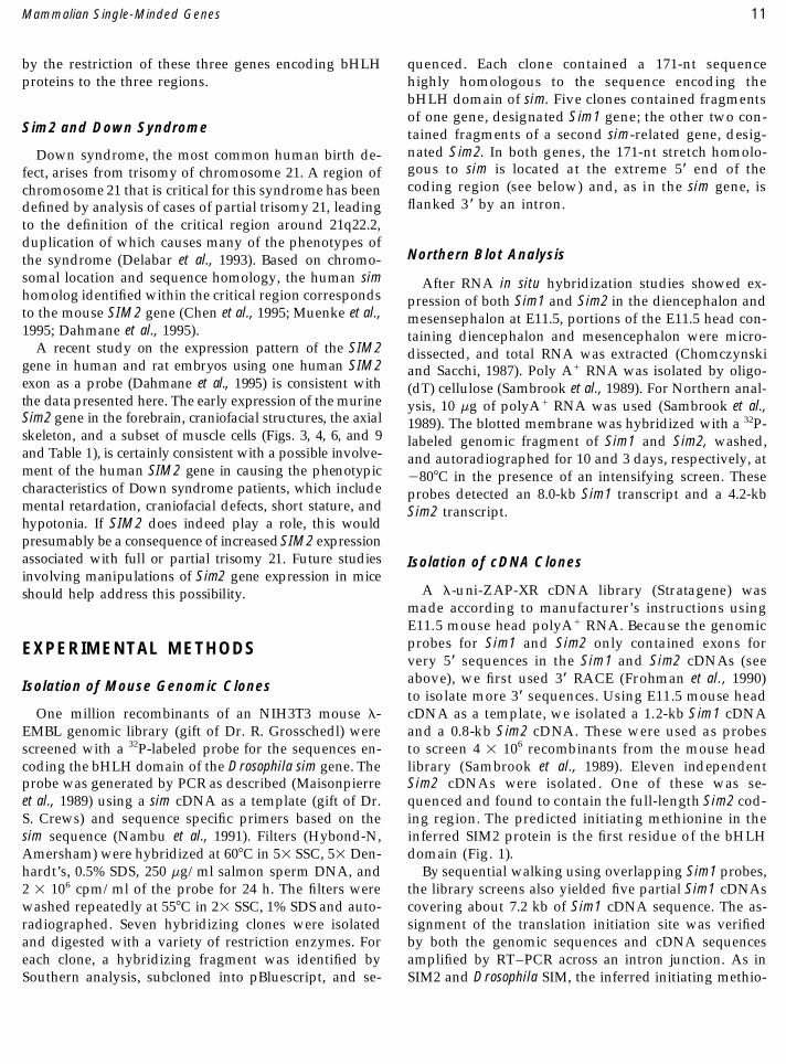
Sim1 provides a marker for the regionalization of so-The expression pattern of Sim2 is largely identical tothat of Sim1 (including at the p2/p3 boundary) but with mites. High levels of Sim1 expression are also observed
in the paraxial mesoderm (Figs. 4A and 4C). Sim1 is ex-some differences (Fig. 6), the most salient of which areas follows: (i) Sim1 expression extends caudal to that of pressed initially uniformly in presomitic mesoderm (Fig.
8A). When somites first pinch off from the presomiticSim2 in both domain S1 and domain S2; (ii) only Sim2expression crosses the ventral midline in the mammillary mesoderm, Sim1 is still expressed in a uniform fashion.
However, as each somite becomes patterned into dermo-area (as discussed above at E9.5); (iii) at E12.5, only Sim1
AID MCN 0530 / 6p06$$$$61 03-12-96 08:56:13 mcnal AP: MCN

8 Fan et al.
FIG. 6. Schema showing expression of Sim1, Sim2, and Dlx2 at E10.5 and E12.5. Adjacent sections were used for this study to compare theexpression of each gene in order to refine the map of their expression patterns. The domains of expression respect transverse and longitudinalboundaries that are incorportated within the framework of the Prosomeric model (Puelles and Rubenstein, 1993; Rubenstein et al., 1994). Expressionsin the mantle and ventricular zone (VZ) are indicated in different colors: Sim1 is shown in dark orange (VZ) and pale orange (mantle), Sim2 inmagenta (VZ) and pink (mantle), and Dlx2 in purple (VZ) and blue (mantle). Abbreviations: M, midbrain; p, prosomere; SC, spinal cord.
myotome and sclerotome, and subsequently into derma- later stages, Sim1 expression becomes restricted to thelateral aspect of the dermatome (data not shown).tome, myotome, and sclerotome, Sim1 expression be-
comes restricted first to the dermomyotome and then the Expression in derivatives of somites. Between E9.5and E10.5, Sim1 is also expressed in cells migrating awaydermatome (Figs. 8C and 8E). Interestingly, this pattern
of Sim1 expression is the mirror image of that of a differ- from the ventrolateral margin of the dermatome, which,at the limb level, contain the presumed limb myoblastent bHLH protein-encoding gene, Mtwist, which is also
expressed uniformly in presomitic mesoderm but be- precursors (Chevallier et al., 1977; Christ et al., 1977). Itis, however, detected at only very low levels in the limbcomes gradually enriched in the sclerotome (though it is
not altogether excluded from the other two components proper (Fig. 8G). In contrast, from E10.5, Sim2 is ex-pressed strongly in the limbs in regions that appear toof the somite, Figs. 8B, 8D, and 8F) (Wolf et al., 1991). At
AID MCN 0530 / 6p06$$$$61 03-12-96 08:56:13 mcnal AP: MCN
9Mammalian Single-Minded Genes
TABLE 1
Expression Pattern of the Sim Genes at E16.5
Sim1 Sim2
Forebrain ForebrainDomain 1 Domain 1
Mammillary nuclei (lateral mammillary) Mammillary nuclei (lateral mammillary)Zona limitans Zona limitansBasal plate domains of p1, p2 Basal plate domains of p1, p2
Domain 2 Domain 2Anterior hypothalamic nuclei Anterior hypothalamic nucleiAmygdala Cortex
Midbrain Olfactory bulbBasal plate Midbrain
Spinal cord Basal plateCells next to the floor plate Cartilage/bone
Head mesenchyme RibsDermis (face, chest, and back) VertebraeGenital eminence PalatalKidney tubules Mandibular
HyoidLimb/digits
Skeletal musclesSubsets of the trunk muscles at the limb level
Oral epitheliumKidney tubules
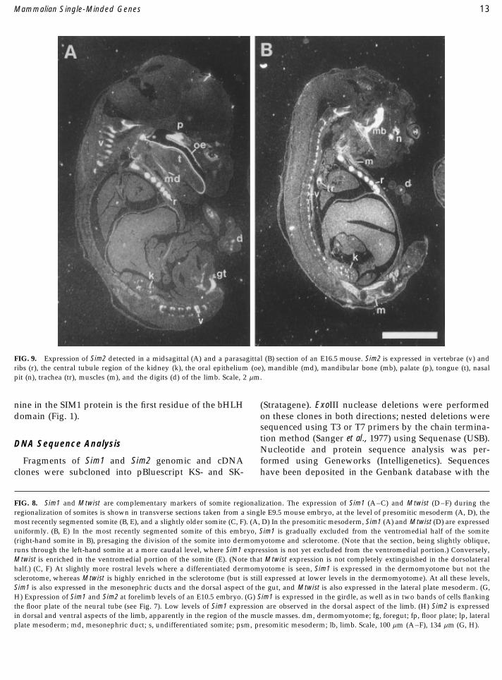
correspond approximately to the dorsal and ventral mus- have shown that the mouse genome contains at leastcles masses (Fig. 8H). At later stages (E12.5 and E16.5), two genes that are highly related to sim. The extensiveSim1 is also found in a layer of cells immediately beneath homology between the three genes suggests that theythe epidermis in the dorsal half of the embryo (data not are indeed evolutionary homologs. The high homologyshown); these cells are presumed to be dermal cells, within the bHLH domains of all three proteins stronglywhich would be consistent with the earlier expression of suggests that they may have similar DNA binding andSim1 in dermatome. At E10.5, high levels of Sim2 expres- dimerization properties.sion in the mesenchymal region of all the branchial The three SIM proteins also share a 250-amino-acid-arches were also detected (data not shown). This is con- long PAS domain. This domain is conserved among thesistent with its later expression in the tongue, mandibular Drosophila SIM and PER proteins and the mammalianbones, and trachea (Table 1). Sim2 is also expressed in ARNT, AHR, and HIF-1 proteins (Nambu et al., 1991,the vertebrae, ribs, and a subset of muscles between E12.5 Wang et al., 1995). The PAS domain of PER can homodi-and E16.5 (the latest time point examined). merize and can also form a heterodimer with the PAS
Expression in derivatives of the mesonephros and meta- domain of SIM (Huang et al., 1993). Since PER does notnephros. Sim1 is also expressed at high levels in the meso- contain a discernible bHLH domain, it has been pro-nephric ducts during their formation (Figs. 4A, 4B; 8A, 8C, posed that PER binds other PAS domain-containing pro-8E). Interestingly, Sim2 is expressed in complementary fash- teins and thereby interferes with their DNA bindingion in the mesonephric tubules, although at lower levels function, thus acting as a negative regulator of these pro-(data not shown). Expression of Sim1 and Sim2 in the meso- teins. Consistent with this possibility, the SIM PAS do-nephros persists at E10.5 and E11.5. At E12.5 and 16.5, Sim1 main when expressed alone has been shown to antago-is expressed in the cortical glomeruli (data not shown) and nize endogenous sim function in vivo (Franks and Crews,Sim2 in the central tubules of the kidney (Figs. 9A, 9B). 1994). Residues within the PAS domain that are im-
Other sites of expression. These are listed in Table 1 portant for the dimerization function of PER (Huang etand discussed below. al., 1993) are conserved in the mammalian SIM proteins,
suggesting that the PAS domains in the mammalian pro-DISCUSSION teins may indeed function in dimerization. In fact, sinceThe Mouse Genome Contains Two Sim Homologs both HLH and PAS domains are thought to mediate di-
merization, it has been proposed that the PAS domainIn Drosophila, sim is a key regulator of the developmentof cells at the midline of the central nervous system. We may serve to consolidate dimerizations initiated by HLH
AID MCN 0530 / 6p06$$$$61 03-12-96 08:56:13 mcnal AP: MCN
10 Fan et al.
(Crews et al., 1988; Lewis and Crews, 1994). Similarly inmouse, Sim1 is expressed in the brain, ventral spinalcord, and foregut, and Sim2 is expressed in the brain andin muscles. In addition, both mammalian Sim genes areexpressed in the developing mesonephros and, later, thekidney. Importantly, whereas sim is a key regulator ofCNS midline cell development in the fly, the mammalianSim genes are not expressed in floor plate cells of thespinal cord. The only expression of the mouse Sim genesin the ventral midline of the central nervous system thatwe have detected is Sim2 expression in the mammillaryarea of the diencephalon.
Sim2 Is an Early Marker of Regionalization of theEmbryonic Forebrain
Sim2 is expressed in the forebrain in a restricted pat-tern in the caudal diencephalon at early stages of neuraldevelopment (by the two-somite stage). The importanceof this expression pattern is in showing that the forebrainis already regionalized at the two-somite stage, prior toany overt morphological specialization of the diencepha-lon primordium. This observation is consistent with therecent results of Shimamura et al. (1995), defining theearliest stages of forebrain regionalization.
Sim1 Is a Marker of Regionalization of SomitesFIG. 7. Sim1 is expressed in the ventral midline region of the spinalcord. (A) A high-power view of spinal cord in a transverse section
Although Sim2 is expressed in many tissues derivedthrough the trunk region of an E10.5 embryo. Sim1 transcripts, detectedfrom somites by E12.5 (including muscle and cartilage),using a digoxygenin-labeled probe, were observed in two symmetricalit is not detected in somites. In contrast, Sim1 is detectedgroups of cells that flank the floor plate of the spinal cord (A). These
cells appear directly to abut the floor plate, which has been visualized from the earliest stages of somite development. Sim1 isin B by expression of the F-spondin gene, also detected nonisotopically first expressed uniformly in the presomitic mesodermin a nearby section of the same embryo. fp, floor plate. Scale, 10 mm. and becomes restricted to the dorsolateral compartment
of the somite, the dermamyotome, as each somite ma-tures. During the elaboration of the dermatome and myo-tome, Sim1 expression is restricted to the dermatome.domains (Huang et al., 1993). Two other known functions
of PAS domains of AHR are the binding of aromatic Interestingly, Sim1 is also detected in cells that migrateaway from the ventrolateral lip of the dermomyotome,hydrocarbon ligands and binding to heat-shock protein
90 kDa (HSP90) (Wilhelmsson et al., 1990; Burbach et al., which are known to include cells that migrate into limbswhere they become myoblasts. Mtwist, which also en-1992; Whitelaw et al., 1993). It is therefore tempting to
speculate that the PAS domains of SIM proteins also codes a bHLH-containing protein, is also expressed uni-formly in the presomitic mesoderm, but is then upregu-mediate binding to specific ligands and association with
heat shock proteins. lated in the sclerotome and down-regulated in the der-mamyotome during the maturation of the somite (Wolfet al., 1991). Myf5, also a gene encoding a bHLH protein,
General Features of Mammalian Sim Expressionis expressed in the presomitic mesoderm, albeit at lower
Patternslevels. Myf5 is subsequently detected in the dorsal–me-dial lip of the somite where the myotome precursor cellsA general feature of the expression of the mammalian
Sim genes is that some of their sites of expression roughly reside, and later in the mature myotome (Ott et al., 1991).Thus, the regionalization of the undifferentiated somiteparallel those of Drosophila sim. In Drosophila, sim is ex-
pressed in the brain, ventral nerve cord, gut, and muscles into dermatome, myotome, and sclerotome is mirrored
AID MCN 0530 / 6p06$$$$61 03-12-96 08:56:13 mcnal AP: MCN
11Mammalian Single-Minded Genes
by the restriction of these three genes encoding bHLH quenced. Each clone contained a 171-nt sequencehighly homologous to the sequence encoding theproteins to the three regions.bHLH domain of sim. Five clones contained fragmentsof one gene, designated Sim1 gene; the other two con-
Sim2 and Down Syndrome tained fragments of a second sim-related gene, desig-nated Sim2. In both genes, the 171-nt stretch homolo-Down syndrome, the most common human birth de-gous to sim is located at the extreme 5* end of thefect, arises from trisomy of chromosome 21. A region ofcoding region (see below) and, as in the sim gene, ischromosome 21 that is critical for this syndrome has beenflanked 3* by an intron.defined by analysis of cases of partial trisomy 21, leading
to the definition of the critical region around 21q22.2,duplication of which causes many of the phenotypes of
Northern Blot Analysisthe syndrome (Delabar et al., 1993). Based on chromo-somal location and sequence homology, the human sim After RNA in situ hybridization studies showed ex-homolog identified within the critical region corresponds pression of both Sim1 and Sim2 in the diencephalon andto the mouse SIM2 gene (Chen et al., 1995; Muenke et al., mesensephalon at E11.5, portions of the E11.5 head con-1995; Dahmane et al., 1995). taining diencephalon and mesencephalon were micro-
A recent study on the expression pattern of the SIM2 dissected, and total RNA was extracted (Chomczynskigene in human and rat embryos using one human SIM2 and Sacchi, 1987). Poly A/ RNA was isolated by oligo-exon as a probe (Dahmane et al., 1995) is consistent with (dT) cellulose (Sambrook et al., 1989). For Northern anal-the data presented here. The early expression of the murine ysis, 10 mg of polyA/ RNA was used (Sambrook et al.,Sim2 gene in the forebrain, craniofacial structures, the axial 1989). The blotted membrane was hybridized with a 32P-skeleton, and a subset of muscle cells (Figs. 3, 4, 6, and 9 labeled genomic fragment of Sim1 and Sim2, washed,and Table 1), is certainly consistent with a possible involve- and autoradiographed for 10 and 3 days, respectively, atment of the human SIM2 gene in causing the phenotypic 0807C in the presence of an intensifying screen. Thesecharacteristics of Down syndrome patients, which include probes detected an 8.0-kb Sim1 transcript and a 4.2-kbmental retardation, craniofacial defects, short stature, and Sim2 transcript.hypotonia. If SIM2 does indeed play a role, this wouldpresumably be a consequence of increased SIM2 expressionassociated with full or partial trisomy 21. Future studies Isolation of cDNA Clonesinvolving manipulations of Sim2 gene expression in mice
A l-uni-ZAP-XR cDNA library (Stratagene) wasshould help address this possibility.made according to manufacturer’s instructions usingE11.5 mouse head polyA/ RNA. Because the genomicprobes for Sim1 and Sim2 only contained exons forEXPERIMENTAL METHODSvery 5* sequences in the Sim1 and Sim2 cDNAs (seeabove), we first used 3* RACE (Frohman et al., 1990)Isolation of Mouse Genomic Clonesto isolate more 3* sequences. Using E11.5 mouse headcDNA as a template, we isolated a 1.2-kb Sim1 cDNAOne million recombinants of an NIH3T3 mouse l-
EMBL genomic library (gift of Dr. R. Grosschedl) were and a 0.8-kb Sim2 cDNA. These were used as probesto screen 4 1 106 recombinants from the mouse headscreened with a 32P-labeled probe for the sequences en-
coding the bHLH domain of the Drosophila sim gene. The library (Sambrook et al., 1989). Eleven independentSim2 cDNAs were isolated. One of these was se-probe was generated by PCR as described (Maisonpierre
et al., 1989) using a sim cDNA as a template (gift of Dr. quenced and found to contain the full-length Sim2 cod-ing region. The predicted initiating methionine in theS. Crews) and sequence specific primers based on the
sim sequence (Nambu et al., 1991). Filters (Hybond-N, inferred SIM2 protein is the first residue of the bHLHdomain (Fig. 1).Amersham) were hybridized at 607C in 51 SSC, 51 Den-
hardt’s, 0.5% SDS, 250 mg/ml salmon sperm DNA, and By sequential walking using overlapping Sim1 probes,the library screens also yielded five partial Sim1 cDNAs2 1 106 cpm/ml of the probe for 24 h. The filters were
washed repeatedly at 557C in 21 SSC, 1% SDS and auto- covering about 7.2 kb of Sim1 cDNA sequence. The as-signment of the translation initiation site was verifiedradiographed. Seven hybridizing clones were isolated
and digested with a variety of restriction enzymes. For by both the genomic sequences and cDNA sequencesamplified by RT–PCR across an intron junction. As ineach clone, a hybridizing fragment was identified by
Southern analysis, subcloned into pBluescript, and se- SIM2 and Drosophila SIM, the inferred initiating methio-
AID MCN 0530 / 6p06$$$$61 03-12-96 08:56:13 mcnal AP: MCN
12 Fan et al.
AID MCN 0530 / 6p06$$0530 03-12-96 08:56:13 mcnal AP: MCN
13Mammalian Single-Minded Genes
FIG. 9. Expression of Sim2 detected in a midsagittal (A) and a parasagittal (B) section of an E16.5 mouse. Sim2 is expressed in vertebrae (v) andribs (r), the central tubule region of the kidney (k), the oral epithelium (oe), mandible (md), mandibular bone (mb), palate (p), tongue (t), nasalpit (n), trachea (tr), muscles (m), and the digits (d) of the limb. Scale, 2 mm.
nine in the SIM1 protein is the first residue of the bHLH (Stratagene). ExoIII nuclease deletions were performedon these clones in both directions; nested deletions weredomain (Fig. 1).sequenced using T3 or T7 primers by the chain termina-tion method (Sanger et al., 1977) using Sequenase (USB).
DNA Sequence AnalysisNucleotide and protein sequence analysis was per-
Fragments of Sim1 and Sim2 genomic and cDNA formed using Geneworks (Intelligenetics). Sequenceshave been deposited in the Genbank database with theclones were subcloned into pBluescript KS- and SK-
FIG. 8. Sim1 and Mtwist are complementary markers of somite regionalization. The expression of Sim1 (A–C) and Mtwist (D–F) during theregionalization of somites is shown in transverse sections taken from a single E9.5 mouse embryo, at the level of presomitic mesoderm (A, D), themost recently segmented somite (B, E), and a slightly older somite (C, F). (A, D) In the presomitic mesoderm, Sim1 (A) and Mtwist (D) are expresseduniformly. (B, E) In the most recently segmented somite of this embryo, Sim1 is gradually excluded from the ventromedial half of the somite(right-hand somite in B), presaging the division of the somite into dermomyotome and sclerotome. (Note that the section, being slightly oblique,runs through the left-hand somite at a more caudal level, where Sim1 expression is not yet excluded from the ventromedial portion.) Conversely,Mtwist is enriched in the ventromedial portion of the somite (E). (Note that Mtwist expression is not completely extinguished in the dorsolateralhalf.) (C, F) At slightly more rostral levels where a differentiated dermomyotome is seen, Sim1 is expressed in the dermomyotome but not thesclerotome, whereas Mtwist is highly enriched in the sclerotome (but is still expressed at lower levels in the dermomyotome). At all these levels,Sim1 is also expressed in the mesonephric ducts and the dorsal aspect of the gut, and Mtwist is also expressed in the lateral plate mesoderm. (G,H) Expression of Sim1 and Sim2 at forelimb levels of an E10.5 embryo. (G) Sim1 is expressed in the girdle, as well as in two bands of cells flankingthe floor plate of the neural tube (see Fig. 7). Low levels of Sim1 expression are observed in the dorsal aspect of the limb. (H) Sim2 is expressedin dorsal and ventral aspects of the limb, apparently in the region of the muscle masses. dm, dermomyotome; fg, foregut; fp, floor plate; lp, lateralplate mesoderm; md, mesonephric duct; s, undifferentiated somite; psm, presomitic mesoderm; lb, limb. Scale, 100 mm (A–F), 134 mm (G, H).
AID MCN 0530 / 6p06$$$$61 03-12-96 08:56:13 mcnal AP: MCN
14 Fan et al.
following accession numbers: Sim1: U40575; Sim2: ized, and washed as described (Frohman et al., 1990).Slides were coated with photographic emulsion (K-5,U40576.Polysciences, Inc.), developed after 3–4 weeks, stainedwith hematoxylin and eosin, dehydrated, cleared with
Interspecific Mouse Backcross MappingXylene, mounted in Permount (Fisher), dried, and exam-ined by bright-field and dark-field illumination.Interspecific backcross progeny were generated by
mating (C57BL/6J1Mus Spretus)F1 females and C57BL/ For hybridization to tissue sections with digoxygenin –UTP labeled probes, the method of Schaeren-Wiemers6J males as described (Copeland and Jenkins, 1991). A
total of 205 N2 mice were used to map the Sim1 and and Gerfin-Moser (1993) was used exactly as describedwith the exception that cryosections were taken fromSim2 loci. DNA isolation, restriction enzyme digestion,
gel electrophoresis, Southern blot, and hybridization embryos fixed in 4% paraformaldehyde. Embryos wereembedded in OCT embedding compound (Tissue-Tek)were performed as described (Jenkins et al., 1982). 32P-
labeled probes were used. The Sim1 probe, a 5.6-kb and sectioned at 12 mm. Antisense RNA probes weretranscribed in the presence of digoxygenin –UTP (Boeh-EcoRI/XhoI fragment of mouse cDNA, detects a 12.0-kb
fragment in C57BL/6J DNA and fragments of 9.7 and ringer Mannheim) using T7 polymerase as recom-mended by the manufacturer.2.6 kb in M. spretus DNA following XbaI digestion. The
Sim2 probe, a 1.7-kb BamHI/XhoI fragment of mousecDNA, detects a 7.9-kb fragment in C57BL/6J and a 8.6- In Situ Hybridization to RNA in Embryo Whole-kb fragment in M. spretus DNA after BamHI digestion. MountsNeither probe contains the bHLH or the PAS domain.
The protocol for whole-mount in situ hybridizationThe presence or absence of the M. spretus-specific frag-was based on the procedure of Wilkinson and Nietoments were followed in backcross mice. The map loca-(1993), as modified by Dr. J. McMahon (personal commu-tions of several other loci used to position Sim1 and Sim2nication). The Sim1 probe contained 500bp of the 5* un-on our interspecific backcross have been previously de-translated region and intron sequences and 1.2 kb of thescribed (see Fig. 2). Recombination distances were calcu-coding sequence; the Sim2 probe contained 300 bp of 5*lated as described (Green, 1981) using the computer pro-untranslated region and intron sequences and 1 kb ofgram SPRETUS MADNESS. Gene order was determinedcoding sequence. The color reaction (NBT and BCIP,by minimizing the number of double recombinationGIBCO–BRL) was allowed to proceed for 5–24 h forevents required to explain the allele distribution patterns.Sim2, and 12–16 h for Sim1.
In Situ Hybridization to RNA in Tissue Sections
ACKNOWLEDGMENTSEmbryos from different stages were fixed in 4% para-formaldehyde in phosphate-buffered saline, embedded
We thank T. Jessell, M. Placzek, and G. Martin for helpful commentsin paraffin, and sectioned at 8-mm thickness as describedon an early version of this manuscript, D. J. Gilbert for excellent techni-(Frohman et al., 1990). RNA probes for in situ hybridiza-cal assistance, and Drs. A. Klar, A. Joyner, and F. Perrin-Schmidt for
tion were transcribed from linearized plasmids using T7 the Fspondin, En1, and Mtwist cDNAs, respectively. This work wasRNA polymerase in the presence of [35S]UTP. The Sim1 supported by grants to M.T.L. from the Lucille P. Markey Charitable
Trust and the Human Frontiers Science Program (Grant RG41/95), andprobe was an antisense transcript spanning 171 nt ofby the Howard Hughes Medical Institute. C.-M. Fan was supported bycoding and 582 nt of 5* untranslated and intron se-postdoctoral fellowships from the Damon Runyon –Walter Winchellquences; the Sim2 probe was a 450-nt antisense probeCancer Research Fund, the American Cancer Society, California Divi-
spanning 171 nt of coding and 282 nt of 5* untranslated sion, and the Howard Hughes Medical Institute. This work was alsoand intron sequences. Antisense probes containing just supported by grants to J.L.R.R. (Human Frontiers Science Program
Grant RG41/95, March of Dimes, NARSAD, the John Merck Fund,the 5* untranslated and intron sequences or just the se-Pfizer Pharmaceuticals, and NIMH RO1 MH49428-01, NIMH RO1quences encoding the bHLH domains of each gene gaveMH51561-01A1, and K02 MH01046-01) and, in part, by the Nationallower signals but identical hybridization patterns (dataCancer Institute, DHHS, under contract with ABL. M.T.L. is an Assis-
not shown). Sense probes transcribed using T3 RNA tant Investigator of the Howard Hughes Medical Institute.polymerase did not show any specific hybridization(data not shown). The En1 cDNA probe (Joyner, 1988),the Mtwist cDNA probe (Wolf et al., 1991), the Fspondin REFERENCEScDNA probe (Klar et al., 1992), and the Dlx 2 probe (Bul-fone et al., 1993) were as previously described. For each Bae, S.-C., Ogawa, E., Maruyama, M., Oka, H., Satake, M., Shigesada,
K., Jenkins, N. A., Gilbert, D. J., Copeland, N. G., and Ito, Y. (1994).probe, sections were processed, prehybridized, hybrid-
AID MCN 0530 / 6p06$$$$61 03-12-96 08:56:13 mcnal AP: MCN
15Mammalian Single-Minded Genes
PEBP2aB/mouse AML1 consists of multiple isoforms that possess Haefliger, J.-A., Bruzzone, R., Jenkins, N. A., Gilbert, D. J., Copeland,N. G., and Paul, D. L. (1992). Four novel members of the connexindifferential transactivation potential. Mol. Cell. Biol. 14: 3242 –3252.
Bulfone, A. P., Puelles, L., Porteus, M. H., Frohman, M. A., Martin, family of gap junction proteins: Molecular cloning, expression andchromosome mapping. J. Biol. Chem. 267: 2057 –2064.G. R., and Rubenstein, J. L. (1993). Spatially restricted expression of
Dlx-1, Dlx-2 (Tes-1), Gbx-2, and Wnt-3 in the embryonic day 12.5 Heyman, S., Kent, A., and Lumsden, A. (1993). Cellular morphologyand extracellular space at rhombomere boundaries in the chick em-mouse forebrain defines potential transverse and longitudinal seg-
mental boundaries. J. Neurosci. 13: 3155 –3172. bryo hindbrain. Dev. Dynamics 198: 241–253.Hoffman, E. C., Reyes, H., Chu, F. F., Sander, F., Conley, L. H., Brooks,Burbach, K. M., Poland, A., and Bradfield, C. A. (1992). Cloning of the
Ah-receptor cDNA reveals a distinctive ligand-activated transcrip- B. A., and Hankinson, O. (1991). Cloning of a factor required foractivity of the Ah (dioxin) receptor. Science 252: 954– 958.tion factor. Proc. Natl. Acad. Sci. USA 89: 8185 –8189.
Chen, H., Chrast, R., Rossier, C., Gos, A., Antonarakis, S. E., Kudoh, J., Huang, Z. J., Edery, I., and Rosbash, M. (1993). PAS is a dimerizationdomain common to Drosophila period and several transcription fac-Yamaki, A., Shindoh, N., Maeda, H., Minoshima, S., and Shimizu,
N. (1995). Single-minded and Down syndrome. Nature Genet. 10: 9– tors. Nature 364: 259–262.Jain, S., Dolwick, K. M., Schmidt, J. V., and Bradfield, C. A. (1994).10.
Chevallier, A., Kieny, M., and Mauger, A. (1977). Limb– somite relation- Potent transcription activation domains of the Ah receptor and theAh receptor nuclear translocaror map to their carboxyl termini. J.ship: Origin of the limb musculature. J. Embryol. Exp. Morph. 41: 245–
258. Biol. Chem. 269: 31518–31524.Jan, Y. N., and Jan, L. Y. (1993). HLH proteins, fly neurogenesis, andChomczynski, P., and Sacchi, N. (1987). Single-step method of RNA
isolation by acid gaunidinium thiocyanate-phenol-chloroform extrac- vertebrate myogenesis. Cell 75: 827–830.Jenkins, N. A., Copeland, N. G., Taylor, B. A., and Lee, B. K. (1982).tion. Anal. Biochem. 162: 156–159.
Christ, B., Jacob, H. J., and Jacob, M. (1977). Experimental analysis of Organization, distribution, and stability of endogenous ecotropic mu-rine leukemia virus DNA sequences in chromosomes of Mus muscu-the origin of the wing musculature in avian embryos. Anat. Embryol.
150: 171–186. lus. J. Virol. 43: 26– 36.Justice, M. J., Siracusa, L. D., Gilbert, D. J., Heisterkamp, N., Groffen,Copeland, N. G., and Jenkins, N. A. (1991). Development and applica-
tion of a molecular genetic linkage map of the mouse genome. Trends J., Chada, K., Silan, C. M., Copeland, N. G., and Jenkins, N. A. (1990).A genetic linkage map of mouse chromosome 10: Localization of 18Genet. 7: 113– 118.
Crews, S., Franks, R., Hu, S., Matthews, B., and Nambu, J. (1992). Dro- molecular markers using a single interspecific backcross. Genetics 125:855–866.sophila single-minded gene and the molecular genetics of CNS mid-
line development. J. Exp. Zool. 261: 234–244. Klar, A., Baldassare, M., and Jessell, T. M. (1992). F-spondin: A geneexpressed at high levels in the floor plate encodes a secreted proteinCrews, S., Thomas, J., and Goodman, C. (1988). The Drosophila single-
minded gene encodes a nuclear protein with sequence similarity to that promotes neural cell adhesion and neurite extension. Cell 69:95–110.the per gene product. Cell 52: 143– 151.
Dahmane, N., Charron, G., Lopes, C., Yaspo, M.-L., Maunoury, C., Lewis, J. O., and Crews, S. T. (1994). Genetic analysis of the Drosophilasingle-minded gene reveals a central nervous system influence onDecorte, L., Sinet, P.-M., Bloch, B., and Delabar, J-M. (1995). Down
syndrome critical region contains a gene homologous to Drosophila muscle development. Mech. Dev. 48: 81–91.Lindebro, M. C., Poellinger, L., and Whitelaw, M. L. (1995). Protein–sim expressed during rat and human central nervous system dvelop-
ment. Proc. Natl. Acad. Sci. USA 92: 9191 –9195. protein interaction via PAS domains: Role of the PAS domain inpositive and negative regulation of the bHLH/PAS dioxin receptor-Davis, C. A., and Joyner, A. L. (1988). Expression patterns of the homeo
box-containing genes En-1 and En-2 and the proto-oncogene int-1 Arnt transcription factor complex. EMBO J. 14: 3528– 3539.Maisonpierre, P., Belluscio, L., Squinto, S., Ip, N., Furth, M., Lindsay, R.,diverge during mouse development. Genes Dev. 2: 1736– 1744.
Delabar, J. M., Theophile, D., Rahmani, Z., Chettouch, Z., Blouin, J. L., and Yancopoulos, G. (1989). Neurotrophin-3: A neurotrophic factorrelated to NGF and BDNF. Science 247: 1446– 1451.Prieur, M., Noel, B., and Sinet, P. M. (1993). Molecular mapping of
twenty-four features of Down syndrome on chromosome 21. Eur. J. McMahon, A. P., Joyner, A. L., Bradley, A., and McMahon, J. A. (1992).The midbrain –hindbrain phenotype of Wnt-1-/Wnt-1- mice resultsHuman Genet. 1: 114–124.
Figdor, M. C., and Stern, C. D. (1993). Segmental organization of embry- from stepwise deletion of engrailed-expressing cells by 9.5 days post-coitum. Cell 69: 581–595.onic diencephalon. Nature 363: 630–634.
Franks, R. G., and Crews, S. T. (1994). Transcriptional activation do- Muenke, M., Bone, L., Mitchell, H., Hart, I., Walton, K., Hall-Johnson,K., Ippel, E., Dietz-Band, J., Karloy, K., Fan, C.-M., Tessier-Lavigne,mains of the single-minded bHLH protein are required for CNS mid-
line cell development. Mech. Dev. 45: 269–277. M., and Patterson, D. (1995). Physical mapping of the holopros-encephaly critical region in 21q22.3, exclusion of SIM2 as a candidateFrohman, M. A., Boyle, M., and Martin, G. R. (1990). Isolation of the
mouse Hox-2.9 gene: Analysis of embryonic expression suggests that gene for HPE, and mapping the SIM2 to a region of chromosome 21important for Down syndrome. Am. J. Human Genet. 57: 1074 –1079.positional information along the anterior–posterior axis is specified
by mesoderm. Development 110: 589– 607. Nambu, J. R., Lewis, J. O., Wharton, K. J., and Crews, S. T. (1991). TheDrosophila single-minded gene encodes a helix-loop-helix proteinGreen, E. L. (1981). Linkage, recombination, and mapping. In Genetics
and Probability in Animal Breeding Experiments (E. L. Green, Ed.), pp. that acts as a master regulator of CNS midline development. Cell 67:1157– 1167.77 –83. Oxford Univ. Press, New York.
Green, M. C. (1989). Catalog of mutant genes and polymorphic loci. In Olson, E. N., and Klein, W. H. (1994). bHLH factors in muscle develop-ment: Dead lines and commitments, what to leave in and what toGenetic Variants and Strains of the Laboratory Mouse (M. C. Green, Ed.),
pp. 12– 403. Oxford Univ. Press, New York. leave out. Genes Dev. 8: 1–8.Ott, M.-O., Bober, E., Lyons, G., Arnold, H., and Buckingham, M. (1991).Habets, G. G., van der Kammen, R. A., Jenkins, N. A., Gilbert, D. J.,
Copeland, N. G., Hagemeijer, A., and Collard, J. G. (1995). The inva- Early expression of the myogenic regulatory gene, Myf5, in precursorcells of skeletal muscle in the mouse embryo. Development 111: 1097–sion-inducing TIAM-1 gene maps to human chromosome 21q22 and
mouse chromosome 16. Cytogenet. Cell Genet. 70: 48–51. 1107.
AID MCN 0530 / 6p06$$$$61 03-12-96 08:56:13 mcnal AP: MCN
16 Fan et al.
Porteus, M. H., Bulfone, A., Ciaranello, R. D., and Rubenstein, J. L. R. J. L. R. (1995). Longitudinal organization of the anterior neural plateand neural tube. Development, 121: 3923– 3933.(1991). Isolation and Characterization of a Novel cDNA Encoding
Wang, G. L., Jiang, B. H., Rue, E. A., and Semenza, G. L. (1995). Hyp-a Homeodomain that is Developmentally Regulated in the Ventraloxia-inducible factor 1 is a basic helix–loop helix –PAS heterodimerForebrain. Neuron 7: 221– 229.regulated by cellular O2 tension. Proc. Natl. Acad. Sci. USA 92: 5510–Porteus, M. H., Bulfone, A., Liu, J. K., Puelles, L., Lo, L. C., and Ru-5514.benstein, J. L. R. (1994). DLX-2, MASH-1, MAP-2 expression and
Weintraub, H., Davis, R., Tapscott, S., Thayer, M., Krause, M., Benezra,bromodeoxyuridine incorporation define molecularly distinct cellR., Blackwell, T. K., Turner, D., Rupp, R., Hollenberg, S., Zhuang, Y.,populations in the embryonic mouse forebrain. J. Neurosci. 14: 6370–and Lassar, A. (1991). The myoD gene family: Nodal point during6383.specification of the muscle cell lineage. Science 248: 761–765.Puelles, L., and Rubenstein, J. L. R. (1993). Expression patterns of ho-
Whitelaw, M., Pongratz, I., Wilhelmsson, A., Gustafsson, J. A., andmeobox and other putative regulatory genes in the embryonic mousePoellinger, L. (1993). Ligand-dependent recruitment of the Arnt co-forebrain suggest a neuromeric organization. Trends Neurosci. 16:regulator determines DNA recognition by the dioxin receptor. Mol.472– 479.Cell. Biol. 13: 2504– 2514.Rao, V. N., Papas, T. S., and Reddy, E. S. (1987). erg, a human ets-related
Whitelaw, M. L., Gottlicher, M., Gustafsson, J.-A., and Poellinger, L.gene on chromosome 21: Alternative splicing, polyadenylation, and(1993). Definition of a novel ligand binding domain of a nucleartranslation. Science 237: 635– 639.bHLH receptor: Co-localization of ligand and HSP90 binding activi-Reisz-Porszasz, S., Probst, M. R., Fukunaga, B. N., and Hankinson, O.ties within the regulable inactivation domain of the dioxin receptor.(1994). Identification of functional domains of the arcryl hydrocarbonEMBO J. 12: 4169– 4179.receptor nuclear translocator protein (ARNT). Mol. Cell. Biol. 14:
Wilhelmsson, A., Cuthill, S., Denis, M., Wikstrom, A. C., Gustafsson,6075 –6086.J. A., and Poellinger, L. (1990). The specific DNA binding activity ofRobinson, G. W., Wray, S., and Mahon, K. A. (1991). Spatially restrictedthe dioxin receptor is modulated by the 90 kd heat shock protein.expression of a member of a new family of murine Distal-less homeo-EMBO J. 9: 69–76.box genes in the developing forebrain. New Biol. 3: 1183 –1194.
Wilkison, D. G., and Nieto, M. (1993). Detection of mesenger RNA byRubenstein, J. L., Martinez, S., Shimamura, K., and Puelles, L. (1994). in situ hybrydization to tissue sections and whole mounts. Methods
The embryonic vertebrate forebrain: The prosomeric model. Science Enzymol. 225: 361– 373.266: 578–580. Wolf, C., Thisse, C., Stoetzel, C., Thisse, B., Gerlinger, P., and Perrin-
Sambrook, J., Fritsch, E. F., and Maniatis, T. (1989). Molecular Cloning: Schmitt, F. (1991). The M-twist gene of Mus is expressed in subsetsA Laboratory Mannual, 2nd ed. Cold Spring Harbor Laboratory Press, of mesodermal cells and is closely related to the Xenopus X-twi andCold Spring Harbor, NY. the Drosophila twist genes. Dev. Biol. 143: 363– 373.
Sanger, F., Nicklen, S., and Coulson, A. R. (1977). DNA sequencing Yaginuma, H., Shiga, T., Homma, S., Ishihara, R., and Oppenheim,with chain-termination inhibitors. Proc. Natl. Acad. Sci. USA 74: 5463– R. W. (1990). Identification of early developing axon projections from5468. spinal interneurons in the chick embryo with a neuron specific b-
Schaeren-Wiemers, N., and Gerfin-Moser, A. (1993). A single protocol tubulin antibody: Evidence for a new ‘‘pioneer’’ pathway in the spi-to detect transcripts of various types and expression levels in neural nal cord. Development 108: 705–716.tissue and cultured cells: in situ hybridization using digoxigenin- Yamada, T., Placzek, M., Tanaka, H., Dodd, J., and Jessell, T. M. (1991).labelled cRNA probes. Histochemistry 100: 431–440. Control of cell pattern in the developing nervous system: polarizing
activity of the floor plate and notochord. Cell 64: 635–647.Shimamura, K., Hartigan, D. J., Matinez, S., Puelles, L., and Rubenstein,
Received December 15, 1995Revised December 28, 1995
Accepted January 3, 1996
AID MCN 0530 / 6p06$$$$61 03-12-96 08:56:13 mcnal AP: MCN