Diagnosis of immediate allergic reactions to beta-lactam antibiotics
Upload
independentCategory
view
0download
0
IntroductionModels of homing of memory T cells to sites of antigenchallenge incorporate an initial step of rolling adher-ence to activated vascular endothelium (1). The cuta-neous lymphocyte-associated antigen (CLA) is a fucose-containing carbohydrate that can decorate P-selectinglycoprotein ligand-1 on T cells (2). CLA is expressed
on the surface of most T cells recovered from skin, andon about 5–10% of circulating CD8+ T cells (3). E-selectin (also known as ELAM and CD62E) isexpressed by venular endothelial cells in inflamed skin,oral mucosa, and the female genital tract (4–6). An epi-tope related, although not identical (7), to CLA caninteract with E-selectin, leading to the tethering androlling of CLA-expressing cells in vitro (2). This inter-action has been hypothesized to direct lymphocyte traf-ficking to skin in vivo. However, the role of CLA-asso-ciated E-selectin ligands in the trafficking ofvirus-specific CD8+ T cells to cutaneous sites of infec-tion has not previously been investigated.
Herpes simplex virus type 2 (HSV-2) causes intermit-tent lytic infection of skin and genital mucosa, with orwithout lesion formation and symptoms. Replicatingvirus is mostly limited to the epidermis or mucosalsquamous keratinocytes (8, 9). During symptomaticrecurrent infection, antigen-specific CD4+ T cells andNK cells infiltrate the subjacent dermis by day 2 oflesion formation, while CD8+ T cell infiltration, localvirus-specific cytotoxic activity, and viral clearance typ-ically occur a few days later (10). HSV-specific CD8+
The Journal of Clinical Investigation | August 2002 | Volume 110 | Number 4 537
Expression of cutaneous lymphocyte-associated antigen by CD8+ T cells specific for a skin-tropic virus
David M. Koelle,1,2,3,4,5 Zhi Liu,2 Christopher M. McClurkan,2 Max S. Topp,1,5
Stanley R. Riddell,1,5 Eric G. Pamer,6 Andrew S. Johnson,2 Anna Wald,1,7
and Lawrence Corey1,2,5
1Department of Medicine, 2Department of Laboratory Medicine, and3Department of Pathobiology, University of Washington, Seattle, Washington, USA4Virginia Mason Research Center, Seattle, Washington, USA5Fred Hutchinson Cancer Research Center, Seattle, Washington, USA6Memorial Sloan Kettering Cancer Center, New York, New York, USA7Department of Epidemiology, University of Washington, Seattle, Washington, USA
Virus-specific CD8+ T cells traffic to infected tissues to promote clearance of infection. We used her-pes simplex virus type 2 (HSV-2) as a model system to investigate CD8+ T cell trafficking to the skinin humans. Using human leukocyte antigen (HLA) class I tetramers, we observed that HSV-specificCD8+ T cells in the peripheral blood expressed high levels of cutaneous lymphocyte-associated anti-gen (CLA). In contrast, CD8+ T cells specific for non–skin-tropic herpesviruses lacked CLA expres-sion. CLA-positive HSV-2–specific CD8+ T cells had the characteristics of central memory cells,expressing CCR7, CD62L, and CD28, and they proliferated briskly in response to antigen. CLA isrelated to a functional E-selectin ligand, and both E-selectin and CLA-positive cells were detected inHSV-2–infected skin. HSV-2–specific T cells adhered to cells transfected with E-selectin. A higher pro-portion of HSV-specific CD8+ T cells recovered from herpes lesions express CLA compared withblood, consistent with a role for CLA in skin homing. To our knowledge, this is the first report ofexpression of tissue-specific adhesion-associated molecules by virus-specific CD8+ T cells. The eval-uation of vaccines for skin and mucosal pathogens should include study of the induction of appro-priate tissue-specific homing molecules.
J. Clin. Invest. 110:537–548 (2002). doi:10.1172/JCI200215537.
Received for publication March 27, 2002, and accepted in revised formJune 11, 2002.
Address correspondence to: David M. Koelle, HarborviewMedical Center Mail Stop 359690, 325 Ninth Avenue, Seattle, Washington 98104, USA. Phone: (206) 341-5207; Fax: (206) 341-5203; E-mail: [email protected] of interest: D.M. Koelle and L. Corey are co-inventors onpatents covering the use of HSV-2 antigens as vaccines. Thepatents have been licensed to Corixa Inc. (Seattle, Washington,USA) by the University of Washington. Nonstandard abbreviations used: cutaneous lymphocyte-associated antigen (CLA); herpes simplex virus type 2 (HSV-2);human leukocyte antigen (HLA); HSV-2 virion protein (VP); HSV-2–infected cell protein 0 (ICP0); cytomegalovirus (CMV);Epstein-Barr virus (EBV); EBV-transformed B cell (EBV-LCL);Chinese hamster ovary (CHO); phytohemagglutinin (PHA); aminoacids (a.a.); phycoerythrin (PE); transporter associated with antigenprocessing (TAP).
See the related Commentary beginning on page 441.
CTL clones can be obtained at high frequency fromlesion biopsies (10, 11). Thus, we investigated whetherinteractions between the CLA-associated E-selectin lig-and and E-selectin might promote migration of HSV-2–specific CTLs to local cutaneous sites of infection.
Fluorescent human leukocyte antigen (HLA)tetramers were used to detect CD8+ cells specific forthe HSV-2 virion proteins 22 (VP22) and 13/14(VP13/14) (11) and HSV-2–infected cell protein 0(ICP0). As controls, T cells specific for cytomegalovirus(CMV) and the Epstein-Barr virus (EBV) were alsostudied. The majority of memory HSV-2–specific Tcells in the blood expressed CLA, while CMV- andEBV-specific T cells lacked CLA expression. Recurrentherpetic skin biopsies showed upregulated E-selectinexpression and contained a CLA-expressing dermalinfiltrate locally enriched in HSV-specific CD8+ T cells.These data suggest that homing receptor expressionon memory T cells may be programmed by the site oforiginal antigen encounter, promoting migration forimmune surveillance or responses to reactivation ofinfection. Additional studies indicated that circulatingCLA+ HSV-2–specific CD8+ T cells have a preservedcapacity for self-renewal and the characteristics of cen-tral memory T cells.
MethodsSubjects and specimens. Subjects were HLA typed (12).Subjects used for HSV-2 analyses were HSV-2–seropos-itive (13), HIV-1–seronegative, generally healthy, andnot taking immune-suppressive medication. For sub-jects with a clinical history of genital herpes, the firstclinical episode had occurred at least 6 months prior tophlebotomy. No subject was experiencing a sympto-matic recurrence of genital herpes or receiving antivi-ral therapy at the time of phlebotomy. HSV-2–seropos-itive subjects filled out a questionnaire concerningtheir history of genital herpes. Some subjects had HSVcultures of multiple genital and perirectal sites on adaily basis for more than 50 consecutive days to deter-mine their rates of HSV shedding, as described (14).PBMCs were cryopreserved after Ficoll-Hypaque cen-trifugation. For CD62L, flow cytometry used freshlyisolated PBMCs (15). For two subjects, biopsies of peri-anal HSV-2 culture–positive lesions and normal fore-arm skin were performed (10). Portions were frozen inOCT (Sakura Finetek, Torrance, California, USA). Sub-jects used for EBV and CMV analyses were healthy labpersonnel known to be seropositive for these agentsand to have appropriate HLA types. Protocols wereapproved by the institutional review board of the Uni-versity of Washington and were conducted accordingto Declaration of Helsinki principles.
Cells and viruses. EBV-transformed B cells (EBV-LCLs)were cultured as described (11). Chinese hamster ovary(CHO) and CHO-E cells, stably transfected withhuman E-selectin cDNA, were maintained as described(16). PBMCs were restimulated with peptide, IL-2, andIL-7 in T cell medium (11) exactly as described (11, 17),
or alternatively in T cell medium with 1.6 µg/ml phy-tohemagglutinin (PHA-P; Remel Inc., Lenexa, Kansas,USA) and 64 units/ml human natural IL-2 (HemagenDiagnostics Inc., Columbia, Maryland, USA) beginningon day 3. To test tetramer B7-RPR, peptide-stimulatedcells were stained 12 days after stimulation withtetramer and FITC-conjugated anti-CD8α cloneMHCD0801 (Caltag Laboratories Inc., Burlingame,California, USA) and sorted using the FACSVantagesystem (Becton, Dickinson and Co., San Jose, Califor-nia, USA). Cells were then rested overnight in T cellmedium with 50 U/ml IL-2 (Chiron Corp., Emeryville,California, USA), cloned at 1 cell/well, expanded asdescribed (11), and tested for cytotoxicity (see below).Skin-derived lymphocytes were expanded from biopsiesin T cell medium with PHA-P and human natural IL-2beginning on day 3 as described (10) for 10–14 days.HSV-2 strain 333 (18) and HSV-1 strain E115 (19) weregrown and titrated in Vero cells (20).
HSV-2 CD8+ T cell epitopes. Methodology and defini-tion of the HLA B7–restricted epitope VP22 aminoacids (a.a.) 49–57 have been described (11). Identicalmethods (11) were used to define the epitope recog-nized by genital herpes lesion–derived CD8+ T cellclone 5491.2000.81. HLA restriction was assigned bytransfection/infection of Cos-7 cells with HLA B7cDNA/HSV-2, coculture with 5491.2000.81, and meas-urement of IFN-γ secretion by ELISA (11). To defineHSV-2 antigens, a library of Sau3a I–digested HSV-2strain HG52 (21) DNA was cotransfected with HLA B7cDNA into our previously described Cos-7 expression-cloning system (11). This method uses IFN-γ secretionto detect T cell activation. Positive library pools weredecoded to yield single antigenic HSV-2 fragments (11).The positive library “hit” encoded a.a. 306–825 of ICP0(21). Epitope localization was done by C-terminal trun-cation analysis with nested PCR–generated fragmentsoriginating at a.a. 306. Transfection/IFN-γ readout wasused to narrow the epitope to a.a. 708–778. Algorithms(22, 23) predicted HLA B7 binding by a.a. 743–751.Peptides were synthesized as described (24).
Lymphocyte assays. Four-hour, triplicate cytotoxicityassays used 51Cr release and an effector/target ratio of20:1 (11) unless otherwise indicated. Target infectionused a multiplicity of 10 for 18 hours; peptides wereloaded for 90 minutes at 37°C. After infection orloading, targets were washed. Spontaneous releasewas less than 25%. To measure adhesion, CHO orCHO-E cells were plated at 2 × 10⁶ per 60-mm-diam-eter dish. The next day, PBMCs (2 × 10⁶ unstimulatedor 1 × 10⁶ after stimulation with peptide for 8 days)were added. Dishes were rotated at 50 rpm for 1 hourat 37°C. Unbound cells and cells from a PBS washwere pooled to form the unbound fraction. Cellsbound to the CHO or CHO-E cells were collected withchilled PBS, 4 mM EDTA, and vigorous pipetting.Microscopy confirmed the removal of lymphocytes.Fractions were washed and processed for flow cytom-etry. Results are expressed as the proportion of
538 The Journal of Clinical Investigation | August 2002 | Volume 110 | Number 4
CD8-high cells that stain with tetramer in the boundfraction divided by the proportion of similar cells inthe unbound fraction.
Tetramers. Phycoerythrin-labeled (PE-labeled)tetramers from the National Institutes of Allergy andInfectious Diseases core facility (Emory University,Atlanta, Georgia, USA) were B7-RPR-PE (HLA B7 withHSV-2 VP22 a.a. 49–57), B7-APA-PE (HLA B7 withHSV-2 ICP0 a.a. 743–751), and A2-GLA-PE (HL A A2-GLA, with HSV-2 VP13/14 a.a. 551–559), which waspreviously described (11). Tetramer A2-NLV-PE (HLAA2 with CMV pp65 a.a. 595–603) was producedaccording to published methods (25). Briefly, HLA A2heavy chain and β2-microglobulin were produced sep-arately in E. coli. The heavy chain was truncated to con-tain the extracellular domain. A substrate sequence forBirA biotinylation was added at the C terminus. HLA
complexes were folded using 30 mg of heavy chain, 25mg of β2-microglobulin, and 10 mg of peptide.Biotinylation used BirA at 5 mg/ml, 0.5 mM biotin,and 5 mM ATP for 16 hours at room temperature.Biotinylated complexes were purified by HPLC, andtetramers were assembled by mixing biotinylated com-plexes with streptavidin-phycoerythrin at a 4:1 molarratio. Tetramers A2-CLG-PE (HLA A201 with EBVLMP2 a.a. 426–434 (26), and A2-VLE-PE (HLA withCMV IE-1 a.a. 316–324 ) (27), were produced similarlyby ProImmune Ltd. (Oxford, United Kingdom).Tetramer A2-GLC-PE (HLA A201 with EBV BMLF-1a.a. 280–288) has been described (26).
Flow cytometry. For detection of HSV- or EBV-specificT cells, 1 × 10⁶ to 5 × 10⁶ cryopreserved, thawed PBMCs,or approximately 2 × 10⁵ cultured PBMCs, were stainedwith 1 µg phycoerythrin-labeled tetramer in 50 µl T cell
The Journal of Clinical Investigation | August 2002 | Volume 110 | Number 4 539
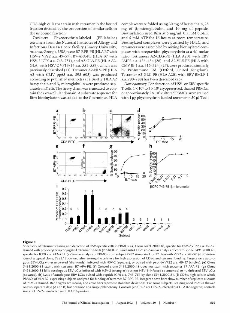
Figure 1Specificity of tetramer staining and detection of HSV-specific cells in PBMCs. (a) Clone 5491.2000.48, specific for HSV-2 VP22 a.a. 49–57,stained with phycoerythrin-conjugated tetramer B7-RPR (B7-RPR–PE) and anti-CD8α. (b) Similar analysis of control clone 5491.2000.48,specific for ICP0 a.a. 743–751. (c) Similar analysis of PBMCs from subject 7282 stimulated for 12 days with VP22 a.a. 49–57. (d) Cytotox-icity of a typical clone, 7282.12, derived after sorting the cells in c for high expression of CD8α and tetramer binding. Targets were autolo-gous EBV-LCLs either untreated (diamonds), infected with HSV-2 (squares), or pulsed with peptide VP22 a.a. 49–57 (circles). (e) Clone5491.2000.81 stains with tetramer B7-APA-PE. (f) Control clone 5491.2000.48 does not stain with tetramer B7-APA-PE. (g) Clone5491.2000.81 kills autologous EBV-LCLs infected with HSV-2 (triangles) but not HSV-1–infected (diamonds) or –uninfected EBV-LCLs(squares). (h) Lysis of autologous EBV-LCLs pulsed with peptide ICP0 a.a. 743–751 by clone 5941.2000.81. (i) CD8α-high cells in wholePBMCs of HLA B7–expressing subjects analyzed for binding of tetramer B7-RPR-PE. Integers above bars show number of replicate aliquotsof PBMCs stained. Bar heights are means, and error bars represent standard deviations. For some subjects, staining used PBMCs thawedon two separate days (A and B) but obtained at a single phlebotomy. Controls (con) 1–3 are HSV-2–infected but HLA B7-negative; controls4–6 are HSV-2–uninfected and HLA B7-positive.

medium at 20°C for 1 hour. Twenty microliters ofanti–CD8α-Cychrome or 20 µl anti–CD8α-PerCP and20 µl FITC-labeled anti-CLA monoclonal antibodyHECA-452 or FITC-labeled anti-CD62L or FITC-labeled anti-CD28 (Pharmingen, San Diego, California,USA) were added for a 30-minute incubation at 4°C,followed by washes and fixation. For CCR7, tetramerwas followed by 2 µg anti-CCR7 clone 2H4 (Pharmin-gen), washes, and then FITC-labeled goat anti-mouse(Southern Biotechnology Associates Inc., Birmingham,Alabama, USA). For CMV-specific T cells, 5 × 10⁵
PBMCs were incubated with 10 µg/ml tetramer in 20 µlPBS with 20% FCS for 20 minutes at 37°C. Cells werewashed, incubated at 4°C for 30 minutes with 20 µlanti–CD8-PerCP (Becton, Dickinson and Co.) andanti–CLA-FITC, washed, and fixed. Cells were analyzedwith a FACScan or FACSCalibur (Becton, Dickinsonand Co.) and WinMDI 2.8 software (http://facs.scripps.edu/software.html). CD8+ T cells were lym-
phocytes (forward/side scatter) staining intensely withanti-CD8α. Tetramer binding was expressed as the per-centage of CD8α-high cells with bright (usually greaterthan 100 fluorescence units) tetramer staining. CLApositivity was defined from the FL1/cell number his-togram for all lymphocytes at the junction between neg-ative cells and a “tail” of FL1-brighter events, typically at101.0 to 101.1 fluorescence units. Two-color analyses usedFITC-conjugated anti-CD8α (Caltag Laboratories Inc.)after the tetramer. To document expression of E-selectin, CHO and CHO-E cells were stained with 10µg/ml anti-CD62E (Becton, Dickinson and Co.) or isotype control at 4°C for 30 minutes, washed, stainedwith 2 µl phycoerythrin-labeled goat anti-mouse (Bio-meda, Hayward, California, USA) for 30 minutes at 4°C,washed, and fixed.
Immunohistochemistry. Frozen 4-µm sections were ace-tone-fixed, quenched in 4:1 methanol/hydrogen per-oxide, and stained as described (28). Briefly, E-selectinwas detected with anti-CD62E (see above) followed byisotype-specific peroxidase-conjugated secondary anti-body and an ABC peroxidase kit with 3,3′ diaminoben-zidine substrate (Vector Laboratories Inc., Burlingame,California, USA). CLA was visualized using biotin-con-jugated monoclonal antibody HECA-452 at 1:200(Pharmingen), followed by anti-biotin monoclonalantibody MB-9100 at 1:200 (Vector Laboratories Inc.),and detection as above. To control for nonspecificbinding, staining was performed with isotype-matchedprimary antibodies specific for irrelevant antigens. Sec-tions were counterstained with Mayer’s hematoxylin.
Statistics. Expression of surface antigens was comparedbetween tetramer-staining and -nonstaining CD8+ lym-phocytes by the two-tailed Mann-Whitney test.
ResultsDetection of HSV-2–specific CD8+ T cells in PBMCs. Wehypothesized that HSV-2–specific CD8+ T cells in theblood would express a characteristic pattern of cell-sur-face molecules involved in cell trafficking. To examine
540 The Journal of Clinical Investigation | August 2002 | Volume 110 | Number 4
Table 1Characteristics of the HSV-2–infected subjects
Subject Duration of Recurrences/yearB Shedding rateC
HSV-2 infectionA
10521 11 Not avail. 10.510292 0.5 Not avail. Not avail.4196 17 Not avail. 26.110026 29 0 Not avail.5491 26 10 Not avail.6376 UnknownD 0 010433 16 10 1.45101 17 12 31.77282 8 2.5 18.5
AYears between the first clinical episode of a syndrome consistent with genitalherpes and phlebotomy for this study, rounded off to nearest whole yearexcept for subject 10292. BDerived from subject self-report about the numberof episodes of genital ulceration in the 6 months prior to enrollment. CPer-centage of days during which any anogenital anatomic site was positive forHSV-2 by culture during more than 50 consecutive days of sampling. DSubjectis HSV-2–seropositive but has no history consistent with genital herpes.
Table 2CLA expression by CMV- and EBV-specific CD8+ T cells in PBMCs
CLA expression by CD8+ cellsSubject Virus Tetramer Tetramer+A Tetramer+ Tetramer–
1 CMV A2-NVP 0.70 4.1 12.92 CMV A2-NVP 6.6 5.1 6.03 CMV A2-NVP 0.51 5.6 2.74 CMV A2-NVP 0.33 2.9 3.45 CMV A2-VLE 3.7 2.1 2.56 CMV A2-VLE 0.47 17.5 11.17 CMV A2-VLE 2.3 11.1 9.58 CMV A2-VLE 0.98 10.4 7.89 EBV A2-GLC 0.11 1.2 2.710 EBV A2-GLC 0.15 4.4 1.611 EBV A2-CLG 0.57 7.2 6.212 EBV A2-CLG 0.74 10.5 15.8
APercentage of CD8α-high cells that stain with the indicated tetramer.

this, we first developed and validated tools to identifyHSV-specific T cells. An HLA B7 tetramer, B7-RPR, wasfolded with peptide VP22 a.a. 49–57 from the HSV-2UL49 open reading frame (21). This tetramer specifical-ly stained the HLA B7–restricted T cell clone5491.2000.48 (11), isolated from a cutaneous HSV-2lesion (Figure 1, a and b). To confirm that this tetramerbound HSV-2–specific CTLs, PBMCs from an HSV-2–infected, B7 subject were stimulated with VP22 a.a.49–57, sorted on the basis of tetramer binding and CD8+
expression, and cloned by limiting dilution. Resultantclones had specific cytotoxicity (Figure 1, c and d).
To obtain an additional marker of the HSV-2–specif-ic CD8+ response, we determined the fine specificity
of CD8+ clone 5491.2000.81, also recovered from anHSV-2 skin lesion. The epitope was found to be a.a.743–751 of the immediate early viral protein ICP0 (Fig-ure 1, g and h). An HLA B7 tetramer, B7-APA, was con-structed and specifically bound clone 5491.2000.81(Figure 1, e and f).
We then examined the frequency of CD8+ T cells forthese HSV-2 epitopes in PBMCs from HSV-2–seropos-itive, HLA B7-expressing adults with symptomaticgenital herpes of 0.5–29 years’ duration (Table 1). Sixof 11 subjects had VP22 a.a. 49–57 specific CD8+ cellsin their PBMCs. From 0.11% to 0.60% of CD8α-highlymphocytes stained with tetramer B7-RPR (Figure1i). Control PBMCs from control HSV-2–infected,
The Journal of Clinical Investigation | August 2002 | Volume 110 | Number 4 541
Figure 2CLA expression by circulating CD8+ lymphocytes specific for human herpesviruses. Subject ID numbers (Table 1) are included for HSV-2analyses. (a) Tetramer B7-RPR-PE and anti-CD8α staining of lymphocytes in PBMCs from HSV-2–infected, B7 subjects. The percentagesof CD8α-high lymphocytes staining with the tetramers are given in Figure 1i. Quadrants show criteria for CD8α-high cells and tetramerbinding. (b) Expression of CLA by CD8α-high lymphocytes staining with tetramer B7-RPR-PE. The percentage of CLA-positive cells is indi-cated. (c) Expression of CLA by CD8α-high lymphocytes that do not stain with tetramer B7-RPR. (d) Staining of PBMCs from an HLA A2subjects with anti-CD8α and tetramers A2-GLA specific for HSV-2 (panel 1), A2-VLE-PE or A2-NVP-PE specific for CMV (panels 2 and 3),or A2-CLG-PE or A2-GLC-PE specific for EBV (panels 4 and 5). For subject 10433, the gates for tetramer-high and tetramer-low CD8+ cellsare shown. The proportion of CD8α-high cells staining with tetramer and the quadrant criteria for CD8α-high cells and tetramer bindingare shown. The CMV and EBV data correspond to lines 5, 1, 10, and 12 of Table 2. (e) Expression of CLA by CD8α-high lymphocytes thatstain with herpesvirus tetramers. (f) Expression of CLA by CD8α-high lymphocytes not staining with the indicated tetramers. Percentagesof CLA-positive cells are indicated.

HLA B7–negative persons and HSV-uninfected, HLAB7–positive persons had less than 0.01% tetramer-pos-itive CD8+ cells.
CLA expression by circulating virus-specific memory CD8+
cells. It is not known whether cells destined to traffic tothe skin express CLA in the circulation prior to extrava-sation. It has previously been hypothesized that dermalT cells could acquire CLA after entering the skin (29).We compared the expression of CLA by circulating cellsspecific for the skin-tropic virus HSV-2 and thenon–skin-tropic viruses EBV and CMV.
CLA was expressed by 52.6–80.3% of circulatingCD8α-high cells that stained with tetramer B7-RPR
(66.0 ± 10.4, mean ± SD) (Figure2, a–c). Only 2.0–14.8% oftetramer-negative CD8+ cellsfrom these same subjectsexpressed CLA (6.0 ± 4.3, mean ±SD). CLA was expressed by 29.4%of circulating CD8α-high cellsspecific for an epitope in HSV-2protein VP13/14, using tetramerA2-GLA-PE (11), compared with1.5% of tetramer-negative CD8+
cells (Figure 2, d–f). Overall, forHSV-2, expression of CLA bytetramer-positive CD8+ cells washigher than expression of CLAby tetramer-negative CD8+ cells(P = 0.006). In this small study,we did not observe any associa-tion between the proportion ofVP22-specific CD8+ T cells thatexpressed CLA (Figure 2, d–f)and the severity of HSV-2 infec-tion (Table 1).
We examined CLA expressionby HLA A2–restricted CD8+ cellsspecific for either CMV or EBV.For each virus, we studied twoindependent epitopes. Expres-sion of CLA by EBV- and CMV-specific CD8+ cells was low andgenerally similar to that oftetramer-negative CD8+ cells
(examples in Figure 2). A similar pattern was noted foreach subject and each epitope (Table 2 summarizes theentire data set). For CMV, the mean ± SD values for theexpression of CLA by virus-specific and bystander CD8+
cells were 7.5% ± 5.1% and 7.7% ± 3.6%, respectively. ForEBV, CLA was expressed by 5.2% ± 3.1% of EBV-specificcells and 5.4% ± 4.2% of other CD8+ cells, respectively.For CMV and EBV analyses combined, there was no dif-ference in CLA expression between tetramer-positiveand tetramer-negative CD8+ cells (P > 0.99).
Proliferative capacity and phenotype of circulating HSV-2–spe-cific CD8+ cells. The above data indicate that HSV-2–spe-cific memory CD8+ T cells express the skin-associated
542 The Journal of Clinical Investigation | August 2002 | Volume 110 | Number 4
Figure 3Expression of CLA by in vitro restimulated herpesvirus-specific CD8+ T cells. For HSV-2–spe-cific T cells, unique subject ID numbers are indicated below the HSV-2 antigens. (a) PBMCsfrom an HLA B7-bearing, HSV-2–infected (panel 1) or two different HLA A2-bearing subjects(panels 2 and 3) stimulated for 13 days with HSV-2 VP22 a.a. 49–57, HSV-2 ICP0 a.a.743–751, or HSV-2 VP 13/14 a.a. 551–559, respectively. Subjects are described in Table 1.PBMC from two HLA A2-bearing subjects were stimulated with EBV BMLF-1 a.a. 280–288(panels 4 and 5). Dot plots display binding of the relevant tetramers and anti-CD8α. Quad-rant lines are cutoffs for CD8α-high cells and tetramer binding. The percentages of CD8α-highcells that are tetramer-positive are indicated. (b) Expression of CLA by CD8α-high lympho-cytes that stain with HSV-2 or EBV tetramers. (c) Expression of CLA by CD8α-high lympho-cytes that do not stain with tetramer. Percentages of CLA-positive cells are indicated.
Table 3Phenotype of CD8α-high cells in PBMCs analyzed by binding of tetramer B7-RPR, which identifies cells specific for HSV-2 VP22 a.a. 49–57
CD28 CD62L CCR7Subject B7-RPR+ B7-RPR– B7-RPR+ B7-RPR– B7-RPR+ B7-RPR–
10521 92.6 55.2 82.6 49.7 75.9 31.24196 84.6 43 74.2 28.5 46.3 22.910026 95 80.4 35.5 49.7 88.7 645491 95.7 30.6 79.5 44.6 53.4 32.76376 98.6 46.7 53 47 50.1 43.210292 80.9 76.4 ND ND ND NDMean ± SD 91.2 ± 6.9 55.4 ± 19.5 65.0 ± 20.1 43.9 ± 8.9 62.9 ± 18.5 38.8 ± 15.8
ND, not done.

adhesion molecule CLA while still in the circulation. Wenext investigated whether these cells had the property ofself-renewal in response to antigen. Circulating cells spe-cific for VP22 a.a. 49–57, VP13/14 a.a. 551–559, or ICP0a.a. 743–751 were able to expand briskly in vitro inresponse to one restimulation with specific HSV-2 pep-tide (Figure 3a). The proportions of VP22- and VP13/14-specific cells that expressed CLA were similar before andafter their peptide-driven expansion (Figure 2, b and e,and Figure 3b). We again noted that the proportion ofVP13/14-specific CD8+ T cells expressing CLA was some-what lower than the proportion of VP22-specific T cells.This comparison could not be made for ICP0, as the cellswere not abundant enough to identify in unmanipulat-ed PBMCs. The same peptide restimulation protocol didnot induce CLA expression by EBV-specific cells. Theseresults are consistent with a model in which lineages ofCLA-expressing and CLA-negative HSV-2–specific cellscan proliferate in vitro, although shifts in phenotype dur-ing the expansion of initially CLA-expressing and CLA-negative cells, or shorter-term fluctuations during pro-gression through the cell cycle, cannot be ruled out.
It has been reported that circulating CD8+ cells canbe divided into central memory cells expressingCD62L and CCR7 that can traffic to lymph nodes,and effector memory cells lacking CD62L and CCR7
but expressing cytolytic molecules. Effector memorycells may have reduced replicative potential (30–33).Circulating VP22-specific cells were greater than 50%CD62L+ in four of the five subjects studied (Table 3and Figure 4). CCR7 expression varied from 46% to89%. VP22-specific cells were also greater than 80%CD28+ from each donor, correlating with their abili-ty to expand in vitro (Table 3). Each of these markerswas more highly expressed by VP22-specific cells thanby CD8α-high lymphocytes with other specificities(Table 3). Comparison between tetramer-stainingand nontetramer-staining CD8+ groups reached sta-tistical significance (P = 0.009) for CD28 expression,but not for CCR7 or CD62L for these small groups.These results are consistent with most HSV-2 VP22a.a. 49–57–specific CD8+ T cells having the centralmemory phenotype. The concept of central memorycan be extended to include CD8+ T cells that haveacquired the ability to selectively home to sites ofantigenic challenge.
CLA and CLA ligand expression by T cells infiltrating geni-tal HSV-2 lesions. To explore the possible role of CLA-associated E-selectin ligand in the migration of HSV-specific CD8+ T cells to herpetic lesions, we obtainedskin biopsy tissue from an HLA B7-expressing person.Because too few cells were available from skin biopsiesfor direct analysis, we expanded skin-infiltrating cellswith PHA-P and IL-2, treatment that provides a fairlyuniform replication stimulus to most T cells and NKcells. HSV-2–specific T cells were locally enrichedamong cells expanded from an HSV-2 culture–positivelesion obtained on the third day of symptoms com-pared with cells expanded from normal skin and cellsin unmanipulated PBMCs. The percentage of CD8α-high cells from an HSV-2 biopsy specific for VP22 a.a.49–57 (Figure 5g) was 6.4%, compared with 0.1% fornormal skin and 0.21% from blood obtained the day ofbiopsy (not shown), representing a 60-fold local enrich-ment at the site of infection. The percentage of lesion-infiltrating CD8α-high cells specific for HSV-2 ICP0a.a. 743–751 was 2.3%, while the level in normal skinwas 0.06% and the level in blood was below the limit ofdetection (not shown). Circulating cells with this speci-ficity were detectable after peptide restimulation (Fig-
The Journal of Clinical Investigation | August 2002 | Volume 110 | Number 4 543
Figure 4Expression of cell-surface antigens by circulating HSV-2–specific CD8+
T cells. (a) PBMCs from donor 4196 were gated for lymphocyte sizeand scatter, high CD8α expression, and binding of tetramer B7-RPR,which is specific for VP22 a.a. 49–57. Expression of CD28, CD62L,and CCR7 is displayed in the indicated histograms. (b) Similar datafor CD8α-high lymphocytes, which did not bind tetramer B7-RPR,from the same donor.
Table 4Binding of virus-specific CD8+ lymphocytes to E-selectin
Subject Stimulation Virus Tetramer InputA CHO-E enrichmentB CHO enrichmentB
5491 NoneC HSV-2 B7-RPR 0.14 10.1 1.14196 NoneC HSV-2 B7-RPR 0.26 5.6 0.76376 NoneC HSV-2 B7-RPR 0.19 13.2 0.85491 VP22 a.a. 49–57 HSV-2 B7-RPR 23.6 9.5 1.15491 ICP0 a.a. 743–751 HSV-2 B7-APA 12.4 8.3 1.3
Cells were analyzed by flow cytometry before or after 1 hour of incubation with either CHO cells expressing E-selectin or control CHO cells. APercentage ofCD8α-high cells that stain with tetramer in PBMC. BThe percentages of CD8α-high cells that stain with the indicated tetramers were measured separately inthe fractions of cells that either bound to, or did not bind to, CHO-E or CHO cell monolayers. Each number listed is the quotient of the teramer-positive per-centage in the bound fraction divided by the tetramer-positive percentage in the unbound fraction. CUnmanipulated PBMCs were thawed, washed, and usedfor binding experiments. Phlebotomy for subject 4196 was performed on a different date from specimens used for Figure 1 and Figure 2.

ure 3a). Similar results for both T cell specificities wereobtained from a biopsy of a recurrent HSV-2 lesionobtained 2 months after the first; again, both localenrichment and almost universal (>90%) CLA expres-sion was noted. To rule out nonspecific induction ofCLA expression during replication of skin-derived Tcells in vitro, PBMCs from four donors were expandedfor 11 days with the culture conditions used for skinbiopsies. CLA was expressed by 4.5% ± 2.3% of CD8α-high cells, similar to fresh PBMCs.
We compared CLA expression by HSV-2–specific Tcells derived from different sites. HSV-2–specific cellsin PBMCs displayed a broad distribution of CLAexpression, including some CLA– cells (Figure 2, b ande). HSV-specific cells from the herpetic lesion were uni-formly CLA+ (Figure 5h). The tetramer-negative CD8+
lymphocytes from these cultures also displayed a high-er level (∼30% total expression) of CLA expression thandid similar cells from PBMCs (Figure 2, c and f).
Immunohistologic examination of an HSV-2 cul-ture–positive buttock lesion from subject 5491,obtained on day 3 of symptoms of recurrent genitalherpes, showed that about 30% of small dermal
mononuclear cells stained withanti-CLA antibody. E-selectin wasstrongly expressed in a dermalvenular pattern (Figure 5). In nor-mal skin, E-selectin stainingshowed a less intense venular pat-tern, while CLA+ cells were rarelyobserved. The presence of CLAand E-selectin in HSV-2–infectedskin suggests that a CLA-associat-ed E-selectin ligand and E-selectinmay participate in leukocyte traf-ficking to recurrent HSV-2 lesions.
Binding of CLA-expressing, HSV-spe-cific cells to E-selectin. We deter-mined whether CLA expression bycirculating HSV-2–specific CD8+
T cells was associated with func-tional binding to E-selectin.PBMCs from three HSV-2–infect-ed, HLA B7 subjects were incubat-ed with E-selectin–expressingCHO-E cells, which uniformlyexpressed E-selectin, or controlCHO cells, which lacked expres-sion (not shown). Measurement ofthe proportion of CD8α-high cellsthat were tetramer B7-RPR+ in thebound and unbound fractionsindicated that T cells specific forHSV-2 VP22 a.a. 49–57 wereenriched about tenfold by adher-ence to E-selectin (Table 4). HSV-specific CD8+ T cell lines generat-ed in vitro by restimulation withthe HSV-2 peptide (Figure 4) were
also tested. Again, HSV-2–specific cells detected withfluorescent HLA tetramers were selectively bound byCHO-E cells but not by control CHO cells.
DiscussionThis is the first description of the selective expressionof a putative tissue-specific homing molecule by cir-culating microbe-specific CD8+ T cells. The cell-sur-face expression and functional data in this report areconsistent with a role for a CLA-associated E-selectinligand in the trafficking of circulating HSV-2–specif-ic memory CD8+ T cells to skin during recurrent gen-ital herpes. Because many patients with genital her-pes have lesions on keratinized epithelial surfaces ofthe external genitalia, perineum, back, or legs (34),CLA expression by HSV-2–specific T cells is anatom-ically appropriate.
In common with HSV-2, EBV and CMV undergointermittent reactivations and are episodically shed ininfectious form by most immunocompetent, infectedindividuals (35, 36). In contrast to HSV-2, neither pri-mary nor recurrent infection with EBV or CMV is asso-ciated with cutaneous infection. The most common
544 The Journal of Clinical Investigation | August 2002 | Volume 110 | Number 4
Figure 5CLA and CLA-ligand expression in skin and by lesion-derived cells. (a) Frozen section of HSV-2 lesion from subject 5491 stained with anti–E-selectin and hematoxylin. Epidermis orientedto the top and original magnification ×200 for all panels. (b) Normal skin from subject 5491stained with anti–E-selectin. (c) HSV-2 lesion stained with anti-CLA. (d) Normal skin stainedwith anti-CLA. (e) HSV-2 lesion stained with hematoxylin and eosin. (f) Normal skin stainedwith hematoxylin and eosin. (g) Lymphocytes expanded for 11 days from the biopsy stainedwith HSV-2–specific tetramers B7-RPR-PE (left) or B7-APA-PE (right) and anti-CD8α. Quad-rant lines are cutoffs for CD8α-high cells and tetramer binding. The percentages of CD8α-highcells that are tetramer-positive are indicated. (h) Expression of CLA by CD8α-high, tetramer-binding cells for tetramer B7-RPR-PE (left) or B7-APA-PE (right). (i) Expression of CLA byCD8α-high, tetramer nonbinding cells. Percentages of CLA-positive cells are indicated.

site of EBV shedding is the oropharynx, while the mostcommon sites of CMV shedding are the uterine cervix,the urinary tract, and the oropharynx. Reactivations ofEBV and CMV in immunocompetent persons are usu-ally asymptomatic (35, 36). We did not study the pres-ence or absence of EBV or CMV reactivation at mucos-al or skin sites in our subjects. Reactivations of EBVand CMV are intermittent, brief, and anatomicallyunpredictable, complicating the assessment of the pos-sible influence of reactivation status on homing recep-tor expression at the time of phlebotomy.
We found that expression by CD8+ T cells specific forEBV and CMV was similar to the background level of5–10% (37) observed for circulating CD8+ lymphocytes.The possibility that expression levels of CLA mightchange during reactivations could be addressed bycombining intensive virologic monitoring with flowcytometry. Study of additional subjects and epitopes,and T cells specific for a variety of skin-tropic andnon–skin-tropic pathogens, will help to determine towhat extent CLA expression is tightly associated withinfection in the skin. Given the anatomic loci of EBVand CMV infection, measurement of the expression ofCD103 (αEβ7 integrin), a putative homing molecule formusocal epithelium (4), by T cells specific for theseviruses would also be of interest.
HSV-specific CD8+ T cells are functionally importantin containing HSV-2 infection. Levels of CD8+ CTLscorrelate inversely with the severity of HSV-2 in mencoinfected with HIV and HSV-2 (38), and correlate tem-porally with the local clearance of HSV-2 in lesions (8,10). CD8+ CTLs are also important in the control ofganglionic infection, maintenance of neuronal latency,and in protection against infectious challenge inmurine models (39–42). HSV evades CD8+ T cells byinhibiting the transporter associated with antigen pro-cessing (TAP) and degrading host mRNA (43). Thetetramer-based measurements in this report revealhigher levels of circulating HSV-2–specific CD8+ T cellsthan were previously observed with limiting dilutionCTL assays (38). In particular, high levels of VP22-spe-cific CD8+ T cells were detected. VP22 may be recog-nized efficiently due to its delivery into the class I anti-gen processing pathway before TAP inhibition canoccur. Our previous data show that virion input VP22can be processed and that endogenous synthesis is notrequired (11). VP22 also has efficient intercellularspread and has CTL adjuvant activity (44, 45) althoughit is not known if this is related to its antigenicity.
In the present report, we studied HSV-2–specific Tcells before and after trafficking from the circulationto HSV-2–infected skin, extending previous researchconcerning the role of CLA in pathogenesis of autoim-mune and atopic disorders. MelanA-specific CD8+ Tcells in PBMCs from subjects with vitiligo express high-er levels of CLA than do similar cells from normal sub-jects (46). In atopic subjects, proliferative responses toallergy-associated antigens are enriched among CLA+
CD4+ T cells (47). Few reports have examined homing
receptor expression by circulating virus-specific T cells.Circulating memory rotavirus-specific CD4+ cells pref-erentially express the adhesion molecule α4β7 integrin(48). Our data indicate that memory CD8+ T cells spe-cific for the skin-tropic herpesvirus HSV-2 express CLAprior to leaving the circulation.
E-selectin is expressed at low basal levels in nonin-flamed skin, and is increased in diverse inflammatoryskin conditions. We observed apparent upregulation ofE-selectin in HSV-2–infected tissue. This is not sur-prising, since IFN-γ, IL-1β, and TNF-α, which areupregulated in HSV lesions (49–51), cooperate toincrease E-selectin expression by endothelial cells (52).Additional work is required to document the magni-tude and time course of upregulation. Lymphocytesinfiltrating the dermis commonly express CLA (35,53–55). The influx of HSV-2–specific CD4+ cells andNK cells into recurrent HSV-2 lesions precedes theinflow of HSV-2–specific CD8+ T cells (10), and addi-tional work is required to define the molecules involvedin the trafficking of early responder cells. The propor-tion of HSV-2–specific CD8+ T cells that express CLAappears to be higher in the skin than in the blood (Fig-ure 5). The approximately 50–80% of circulating HSV-2–specific cells that express CLA (Figure 2) may prefer-entially migrate to skin, or the local microenvironmentmay further promote CLA expression.
Our finding that circulating HSV-2–specific memo-ry cells express CLA implies that expression of this anti-gen is upregulated during the priming of naive HSV-2–specific CD8+ T cells or at a subsequent stage of condi-tioning. The control of CLA expression is incomplete-ly understood. Expression of α(1,3)-fucosyltransferaseVII is a probable key regulator of CLA expression,although control over other glycosyltransferases (56)and the primary polypeptide backbone, P-selectin gly-coprotein ligand-1 (2), may also be important. In vivo,CLA is expressed by cells coexpressing CD45RA andCD45RO in skin-draining lymph nodes, consistentwith upregulation during the priming of naive T cells(57). In a murine model, molecules associated withskin-homing are upregulated during cutaneous prim-ing (58). It is rational to hypothesize that inflammato-ry cytokines and local antigen-presenting cells couldinfluence CLA expression during priming.
In this small cross-sectional study, the proportion ofHSV-2 VP22-specific CD8+ T cells that expressed CLAwas relatively constant in our set of six subjects (Figure2). We recently studied two more HLA B7–bearing sub-jects (not included in Table 1) who are HSV-2 seropos-itive but have no clinical history of genital herpes.Staining of unmanipulated PBMCs with tetramer B7-RPR showed that 54.5% and 62.1% of tetramer-high,CD8α-high cells expressed CLA, similar to the six sub-jects shown in Figure 2. We do not know whether CLAexpression is influenced by recurrences of HSV-2 infec-tion. While the number of subjects that we studied withfrequently recurrent versus asymptomatic HSV-2 infec-tion were too small for statistical comparisons, we
The Journal of Clinical Investigation | August 2002 | Volume 110 | Number 4 545

observed no obvious segregation of CLA expression byHSV-2–specific CD8+ T cells by the clinical or virologic(shedding) severity of HSV-2 infection (Table 1 and Fig-ure 2). Similarly, we do not yet know whether CLAexpression by HSV-2–specific cells in the peripheryfluctuates temporally in association with symptomaticor asymptomatic recurrences of HSV-2.
Epitope-specific heterogeneity in CLA expression byHSV-2–specific CD8+ T cells cannot be excluded withthe available data. CLA expression by VP13/14-specif-ic cells was somewhat lower than for VP22-specificcells, both before and after peptide restimulation, butwas still clearly above background levels for tetramer-negative cells. In sorting experiments using unmanip-ulated PBMCs, based on CLA and CD8 expression, wehave successfully enriched HSV-2–specific CTLs fromseveral subjects at the bulk and clonal levels. Expres-sion cloning to determine fine specificity, using themethods described in this report, has yielded five addi-tional novel epitopes recognized by HLA class I–restricted CD8+ CTLs (Koelle et al., unpublished obser-vations). CLA does appear to be expressed above back-ground levels by CD8+ CTLs specific for a diverse setof HSV-2 epitopes.
TGF-β and IL-12 (57, 59, 60) upregulate CLA expres-sion in vitro, while IL-4 may downregulate CLA (56).Each of these cytokines is upregulated at the proteinand/or mRNA levels in HSV-associated lesions (50, 61).Secretion of TGF-β and IL-12 in response to HSV infec-tion has been demonstrated in vitro (51, 62–64). Drain-ing lymph nodes in animals display expression of IL-12,TGF-β, IL-4, and other cytokines (65–67). While acytokine milieu appropriate for CLA induction is asso-ciated with HSV infection, the key factor or factors con-trolling CLA expression by CD8+ T cells responsive toHSV-2 remain to be determined.
Memory cells that acquire tissue-specific homingmolecules may be specialized for extravasation tosites of antigen challenge. They may also retain acapacity for self-renewal and recirculation throughlymphoid tissue. We found that most circulatingVP22-specific CD8+ T cells expressed CD28 (Figure4), an important molecule for the delivery of costim-ulatory signals to memory T cells. HSV-2–reactive Tcells specific for each of three different viral epitopeswere able to proliferate in vitro in response to pep-tide, sometimes to very high levels (Figure 3). We alsofound that the majority, but not all, circulatingVP22-specific CD8+ T cells expressed CD62L andCCR7, adhesion and chemokine receptor molecules,respectively, associated with recirculation to lym-phoid tissue and a substantial capacity for self-renew-al (68). As CD62L ligands are expressed in skin (69)as well as lymph nodes, it is possible that CD62L onHSV-specific T cells may participate in homing tothis site. There are conflicting data on the expressionof CCR7 by CLA+ cells in the circulation, with pub-lished findings concentrating on CD4+ cells (33, 70).Our findings for CCR7 are similar to those noted for
CD8+ T cells specific for some EBV epitopes, but con-trast with findings for CMV- and HIV-specific CD8+
T cells (71–73). Taken together, our data indicate thatvirus-specific CD8+ T cells with the central memoryphenotype may also be capable of trafficking to sitesof antigenic challenge.
Rolling adhesion to the vessel wall is only the initialstage of lymphocyte trafficking into tissue. Both inte-grin maturation and chemotaxis have been associatedwith chemokines and their receptors. Several candidatechemokine receptors have been implicated in homingto skin, including CCR4 and CCR10 (74–76). Most ofthe published data have concerned CD4+ T cells. Futureflow cytometric and functional studies will examinethe expression of chemokine receptors by HSV-2–spe-cific CD8+ T cells.
In summary, subjects with recurrent, symptomaticHSV-2 infection have readily detectable circulatingVP22-specific CD8+ T cells that express CLA. CLA istightly associated with functional E-selectin bindingactivity, which is an anatomically appropriate proper-ty for HSV-2–specific T cells. HSV-specific CD8+ T cellsin PBMCs expressed functional E-selectin bindingactivity. Neither CLA expression nor E-selectin bindingwas noted among the responses to CMV or EBV, twonon–skin-tropic herpesviruses. We propose that vac-cines and immunotherapies for HSV (77–79) shouldnot only elicit specific T cells but also guide these Tcells to express appropriate homing molecules. Morebroadly, preventative and therapeutic T cell–basedtreatments may be optimized if the identity, mecha-nisms of action, and control mechanisms for homingmolecules can be understood and manipulated.
AcknowledgmentsThe authors would like to thank Rhoda Ashley for per-forming HSV serology, the clinicians and staff of theVirology Research Clinic, University of Washington, forassisting with specimen collection, Stacy Selke for datamanagement, John Harlan for the CHO-E cell line, theNational Institutes of Allergy and Infectious Diseasescore tetramer facility at Emory University, and HermanEinsele, who identified normal donors with responsesto epitopes in EBV LMP2 and CMV IE-1. We also thankthe research subjects who made this work possible.This work was supported by NIH grants AI-30731 andAI-50132, the Cancer Research Institute, and DeutscheForschungsgemeinschaft grant To 208/1-1.
1. Campbell, J.J., and Butcher, E.C. 2000. Chemokines in tissue-specific andmicroenvironment-specific lymphocyte homing. Curr. Opin. Immunol.12:336–341.
2. Fuhlbrigge, R.C., Kieffer, J.D., Armerding, D., and Kupper, T.S. 1997.Cutaneous lymphocyte antigen is a specialized form of PSGL-1expressed on skin-homing T cells. Nature. 389:978–981.
3. Picker, L.J., et al. 1990. Differential expression of homing-associatedadhesion molecules by T cell subsets in man. J. Immunol. 145:3247–3255.
4. Johansson, E.L., Rudin, A., Wassen, L., and Holmgren, J. 1999. Distribu-tion of lymphocytes and adhesion molecules in human cervix and vagi-na. Immunology. 96:272–277.
5. Handschel, J., et al. 1999. Irradiation induces increase of adhesion mol-ecules and accumulation of β2-integrin-expressing cells in humans. Int.J. Radiat. Oncol. Biol. Phys. 45:475–481.
546 The Journal of Clinical Investigation | August 2002 | Volume 110 | Number 4

6. Berg, E.L., et al. 1991. The cutaneous lymphocyte antigen is a skinhoming receptor for the vascular lectin endothelial cell-leukocyteadhesion molecule 1. J. Exp. Med. 174:1461–1466.
7. Wagers, A.J., Stoolman, L.M., Craig, R., Knibbs, R.N., and Kansas, G.S.1998. An sLex-deficient variant of HL60 cells exhibits high levels ofadhesion to vascular selectins: further evidence that HECA-452 andCSLEX1 monoclonal antibody epitopes are not essential for highavidity binding to vascular selectin. J. Immunol. 160:5122–5129.
8. Cunningham, A.L., Turner, R.R., Miller, A.C., Para, M.F., and Merig-an, T.C. 1985. Evolution of recurrent herpes simplex lesions: animmunohistologic study. J. Clin. Invest. 75:226–233.
9. Kiviat, N.B., et al. 1990. Histopathology of endocervical infectioncaused by Chlamydia trachomatis, herpes simplex virus, Trichomonasvaginalis, and Neisseria gonorrhoeae. Hum. Pathol. 21:831–837.
10. Koelle, D.M., et al. 1998. Clearance of HSV-2 from recurrent genitallesions correlates with infiltration of HSV-specific cytotoxic T lym-phocytes. J. Clin. Invest. 101:1500–1508.
11. Koelle, D.M., et al. 2001. CD8 CTL from genital herpes simplexlesions: recognition of viral tegument and immediate early proteinsand lysis of infected cutaneous cells. J. Immunol. 166:4049–4058.
12. Malhotra, U., et al. 2001. Role for HLA class II molecules in HIV-1 sup-pression and cellular immunity following antiretroviral treatment. J. Clin. Invest. 107:505–517.
13. Ashley, R.A., Militoni, J., Lee, F., Nahmias, A., and Corey, L. 1988.Comparison of Western blot (immunoblot)and glycoprotein G-spe-cific immunoblot for detecting antibodies to herpes simplex types 1and 2 in human sera. J. Clin. Microbiol. 26:662–667.
14. Wald, A., et al. 2000. Reactivation of genital herpes simplex virus type2 infection in asymptomatic seropositive persons. N. Engl. J. Med.342:844–850.
15. Reimann, K.A., Chernoff, M., Wilkening, C.L., Nickerson, C.E., andLanday, A.L. 2000. Preservation of lymphocyte immunophenotypeand proliferative responses in cryopreserved peripheral bloodmononuclear cells from human immunodeficiency virus type 1-infected donors: implications for multicenter trials. The ACTGImmunology Advanced Technology Laboratories. Clin. Diagn. Lab.Immunol. 7:352–359.
16. Carlos, T., et al. 1991. Human monocytes bind to two cytokine-induced adhesive ligands on cultured human endothelial cells:endothelial-leukocyte adhesion molecule-1 and vascular adhesionmolecule-1. Blood. 77:2266–2271.
17. Lalvani, A., et al. 1997. Optimization of a peptide-based protocolemploying IL-7 for in vitro restimulation of human cytotoxic T lym-phocyte precursors. J. Immunol. Methods. 15:65–77.
18. Kit, S., Kit, M., Qavi, H., Trkula, D., and Otsuka, H. 1983. Nucleotidesequence of the herpes simplex virus type 2 (HSV-2) thymidine kinasegene and predicted amino acid sequence of thymidine kinase polypep-tide and its comparison with the HSV-1 thymidine kinase gene.Biochim. Biophys. Acta. 741:158–170.
19. Spruance, S.L., and Chow, F.S. 1980. Pathogenesis of herpes simplexvirus in cultures of epidermal cells from subjects with frequent recur-rences. J. Infect. Dis. 142:671–675.
20. Schmidt, N.J. 1989. Cell culture procedures for diagnostic virology. InDiagnostic procedures for viral, rickettsial and chlamydial infections. 6th edi-tion. N.J. Schmidt and R.W. Emmons, editors. American PublicHealth Association Inc. Washington, D.C., USA. 51–100.
21. Dolan, A., Jamieson, F.E., Cunningham, C., Barnett, B.C., andMcGeoch, D.J. 1998. The genome sequence of herpes simplex virustype 2. J. Virol. 72:2010–2021.
22. Rammensee, H., Bachmann, J., Emmerich, N.P., Bachor, O.A., and Ste-vanovic, S. 1999. SYFPEITHI: database for MHC ligands and peptidemotifs. Immunogenetics. 50:213–219.
23. Parker, K.C., Bednarek, M.A., and Coligan, J.E. 1994. Scheme for rank-ing potential HLA-A2 binding peptides based on independent bind-ing of individual peptide side-chains. J. Immunol. 152:163–168.
24. Koelle, D.M., et al. 2000. Tegument-specific, virus-reactive CD4 T-cellslocalize to the cornea in herpes simplex virus interstitial keratitis inhumans. J. Virol. 74:10930–10938.
25. Altman, J.D., et al. 1996. Phenotypic analysis of antigen-specific Tlymphocytes. Science. 274:94–96.
26. Marshall, N.A., et al. 2000. Rapid reconstitution of Epstein-Barr virus-specific T lymphocytes following allogeneic stem cell transplantation.Blood. 96:2814–2821.
27. Khan, N., Cobbold, M., Keenan, R., and Moss, P.A. 2002. Comparativeanalysis of CD8+ T cell responses against human cytomegalovirusproteins pp65 and immediate early 1 shows similarities in precursorfrequency, oligoclonality, and phenotype. J. Infect. Dis. 185:1025–1034.
28. Brodie, S.J., et al. 1999. Pediatric AIDS-associated lymphocytic inter-stitial pneumonia and pulmonary arterio-occlusive disease: role ofVCAM-1/VLA-4 adhesion pathway and human herpesviruses. Amer. J.Pathol. 154:1453–1464.
29. Pober, J.S., Kluger, M.S., and Schechner, J.S. 2001. Human endothelial
cell presentation of antigen and the homing of memory/effector Tcells to skin. Ann. NY Acad. Sci. 941:12–25.
30. Champagne, P., et al. 2001. Skewed maturation of memory HIV-specif-ic CD8 T lymphocytes. Nature. 410:106–111.
31. Appay, V., et al. 2000. HIV-specific CD8+ T cells produce antiviralcytokines but are impaired in cytolytic function. J. Exp. Med. 192:63–75.
32. Geginat, J., Sallusto, F., and Lanzavecchia, A. 2001. Cytokine-driven pro-liferation and differentiation of human naive, central memory, and effec-tor memory CD4(+) T cells. J. Exp. Med. 194:1711–1719.
33. Sallusto, F., et al. 1999. Switch in chemokine receptor expression uponTCR stimulation reveals novel homing potential for recently activatedT cells. Eur. J. Immunol. 29:2037–2045.
34. Corey, L., and Wald, A. 1999. Genital herpes. In Sexually transmitted dis-eases. 3rd edition. K.K. Holmes, et al., editors. McGraw-Hill Inc., NewYork, New York, USA. 285–312.
35. Rickinson, A.B., and Keiff, E. 2001. Epstein-Barr virus. In Fields virology.4th edition. D.M. Knipe and P.M. Howley, editors. Lippincott, Williamsand Wilkins, Philadelphia, Pennsylvania, USA. 2575–2627.
36. Pass, R.F. 2001. Cytomegalovirus. In Fields virology. 4th edition. D.M.Knipe and P.M. Howley, editors. Lippincott, Williams and Wilkins,Philadelphia, Pennsylvania, USA. 2675–2705.
37. Mortarini, R., et al. 2000. Peripheral burst of tumor-specific cytotoxic Tlymphocytes and infiltration of metastatic lesions by memory CD8+ Tcells in melanoma patients receiving interleukin 12. Cancer Res.60:3559–3568.
38. Posavad, C.M., Koelle, D.M., and Corey, L.C. 1996. High frequency ofCD8+ cytotoxic T-lymphocyte precursors specific for herpes simplexviruses in persons with genital herpes. J. Virol. 70:8165–8168.
39. Simmons, A., and Tscharke, D.C. 1992. Anti-CD8 impairs clearance ofherpes simplex virus from the nervous system: implications for the fateof virally infected neurons. J. Exp. Med. 175:1337–1344.
40. Pereira, R.A., Tscharke, D.C., and Simmons, A. 1994. Upregulation ofclass I major histocompatibility complex gene expression in primary sen-sory neurons, satellite cells, and Schwann cells in mice in response toacute but not latent herpes simplex virus infection in vivo. J. Exp. Med.180:841–850.
41. Pereira, R.A., and Simmons, A. 1999. Cell surface expression of H2 anti-gens on primary sensory neurons in response to acute but not latent her-pes simplex virus infection in vivo. J. Virol. 73:6484–6489.
42. Blaney, J.E., et al. 1998. Immunization with a single major histocompat-ibility class I-restricted cytotoxic T-lymphocyte recognition epitope ofherpes simplex virus type 2 confers protective immunity. J. Virol.72:9567–9574.
43. Tigges, M.A., Leng, S., Johnson, D.C, and Burke, R.L. 1996. Human her-pes simplex (HSV)-specific CD8+ CTL clones recognize HSV-2–infectedfibroblasts after treatment with IFN-gamma or when virion host shut-off functions are disabled. J. Immunol. 156:3901–3910.
44. Cheng, W.F., et al. 2001. Enhancement of sindbis virus self-replicatingRNA vaccine potency by linkage of herpes simplex virus type 1 VP22 pro-tein to antigen. J. Virol. 75:2368–2376.
45. Hung, C.-F., et al. 2001. Improving vaccine potency through intercellu-lar spreading and enhanced MHC class I presentation of antigen. J. Immunol. 166:5733–5740.
46. Ogg, G.S., Dunbar, P.R., Romero, P., Chen J.-L., and Cerundolo, V. 1998.High frequency of skin-homing melanocyte-specific cytotoxic T lym-phocytes in autoimmune vitiligo. J. Exp. Med. 188:1203–1208.
47. Santamaria-Babi, L.F., et al. 1995. Circulating allergen-reactive T cellsfrom patients with atopic dermatitis and allergic contact dermatitisexpress the skin-selective homing receptor, the cutaneous lymphocyte-associated antigen. J. Exp. Med. 181:1935–1940.
48. Rott, L.S., et al. 1997. Expression of mucosal homing receptor α4β7 bycirculating CD4+ cells with memory for intestinal rotavirus. J. Clin. Invest.100:1204–1208.
49. Torseth, J.W., and Merigan, T.C. 1986. Significance of local gamma inter-feron in recurrent herpes simplex infection. J. Infect. Dis. 153:979–983.
50. Van Voorhis, W.C., et al. 1996. Primary and secondary syphillis lesionscontain mRNA for Th1 cytokines and activated cytolytic T cells. J. Infect.Dis. 173:491–495.
51. Mikloska, Z., et al. 1998. In vivo production of cytokines and beta (C-C)chemokines in human recurrent herpes simplex lesions—do herpes sim-plex virus-infected keratinocytes contribute to their production? J. Infect.Dis. 177:827–838.
52. Pober, J.S. 1999. Immunobiology of human vascular endothelium.Immunol. Res. 19:225–232.
53. Lu, D., et al. 2001. The T-cell chemokine receptor CXCR3 is expressedhighly in low-grade mycosis fungoides. Am. J. Clin. Pathol. 115:413–421.
54. van den Wijngaard, R., et al. 2000. Local immune response in skin of gen-eralized vitiligo patients. Destruction of melanocytes is associated withthe prominent presence of CLA+ T cells at the perilesional site. Lab.Invest. 80:1299–1309.
55. Walton, L.J., Thornhill, M.H., Macey, M.G., and Farthing, P.M. 1997.Cutaneous lymphocyte associated antigen (CLA) and alpha E beta 7
The Journal of Clinical Investigation | August 2002 | Volume 110 | Number 4 547

integrins are expressed by mononuclear cells in skin and oral lichenplanus. J. Oral. Pathol. Med. 26:402–407.
56. Nakayama, F., et al. 2000. Expression of cutaneous lymphocyte-associ-ated antigen regulated by a set of glycosyltransferases in human T cells:involvement of alpha1,3-fucosyl transferase VII and beta1,4-galactosyl-transferase I. J. Invest. Derm. 115:299–305.
57. Picker, L.J., et al. 1993. Control of lymphocyte recirculation in man. II.Differential regulation of the cutaneous lymphocyte-associated antigen,a tissue-selective homing receptor for skin-homing T cells. J. Immunol.150:1122–1136.
58. Campbell, D.J., and Butcher, E.C. 2002. Rapid acquisition of tissue-spe-cific homing phenotypes by CD4+ T cells activated in cutaneous ormucosal lymphoid tissues. J. Exp. Med. 195:135–141.
59. Akdis, M., Klunker, S., Schliz, M., Blaser, K., and Akdis, C.A. 2000.Expression of cutaneous lymphocyte-associated antigen on humanCD4(+) and CD8(+) Th2 cells. Eur. J. Immunol. 30:3533–3541.
60. Knibbs, K.N., et al. 1998. Alpha(1,3)-fucosyltransferase VII-dependentsynthesis of P- and E-selectin ligands on cultured T lymphoblasts. J. Immunol. 161:6305–6315.
61. Kokuba, H., Aurelian, L., and Burnett, J. 1999. Herpes simplex virus asso-ciated erythema multiforme (HAEM) is mechanistically distinct fromdrug-induced erythema multiforme: interferon-gamma is expressed inHAEM lesions and tumor necrosis factor-alpha in drug-induced erythe-ma multiforme lesions. J. Invest. Derm. 113:808–815.
62. Malmgaard, L., Paludan, S.R., Mogensen, S.C., and Ellerman-Eriksen, S.2000. Herpes simplex virus type 2 induces secretion of IL-12 bymacrophages through a mechanism involving NF-kappaB. J. Gen. Virol.81:3011–3020.
63. Mikloska, Z., et al. 2000. Monophosphosphoryl lipid A and QS21increase CD8 T lymphocyte cytotoxicity to herpes simplex virus-2 infect-ed cell proteins 4 and 27 through IFN-gamma and IL-12 production. J. Immunol. 164:5167–5176.
64. Mendez-Samperio, P., Hernandez, M., and Ayala, H.E. 2000. Inductionof transforming growth factor-beta 1 production in human cells by her-pes simplex virus. J. Interferon Cytokine Res. 20:273–280.
65. Hu, M., Dutt, J., Arrunategui-Correa, V., Baltatzis, S., and Foster, C.S.1999. Cytokine mRNA in BALB/c mouse corneas infected with herpessimplex virus. Eye. 13:309–313.
66. Babu, J.S., Kanangat, S., and Rouse, B.T. 1995. T cell cytokine mRNAexpression during the course of the immunopathologic ocular diseaseherpetic stromal keratitis. J. Immunol. 154:4822–4829.
67. Kanangat, S., Thomas, J., Gangappa, S., Babu, J.S., and Rouse, B.T. 1996.Herpes simplex virus type 1-mediated up-regulation of IL-12 (p40)mRNA expression. Implications in immunopathogenesis and protec-tion. J. Immunol. 156:1110–1116.
68. Sallusto, F., Lenig, D., Forster, R., Lipp, M., and Lanzavecchia, A. 1999.Two subsets of memory T lymphocytes with distinct homing potentialsand effector functions. Nature. 401:708–712.
69. Lechleitner, S., Kuntsfeld, R., Messeritsch-Fanta, C., Wolff, K., and Pet-zelbauer, P. 1999. Peripheral lymph node addressins are expressed onskin endothelial cells. J. Invest. Derm. 113:410–414.
70. Campbell, J.J., et al. 2001. CCR7 expression and memory T cell diversityin humans. J. Immunol. 166:877–884.
71. Chen, G., et al. 2001. CD8 T cells specific for immunodeficiency virus,Esptein-Barr virus, and cytomegalovirus lack molecules for homing tolymphoid sites of infection. Blood. 98:156–164.
72. Faint, J.M., et al. 2001. Memory T cells constitute a subset of the humanCD8+ CD45RA+ pool with distinct phenotypic and migratory charac-teristics. J. Immunol. 167:212–220.
73. Hislop, A.D., et al. 2001. EBV-specific CD8+ T cell memory: relationshipsbetween epitope specificity, cell phenotype, and immediate effector func-tion. J. Immunol. 167:2019–2029.
74. Reiss, Y., Proudfoot, A.E., Power, C.A., Campbell, J.J., and Butcher, E.C.2001. CC chemokine receptor (CCR) 4 and the CCR10 ligand cutaneousT cell-attracting chemokine (CTACK) in lymphocyte trafficking toinflamed skin. J. Exp. Med. 194:1541–1547.
75. Homey, B., et al. 2002. CCL27-CCR10 interactions regulate T cell-medi-ated skin inflammation. Nat. Med. 8:157–165.
76. Campbell, J.J., et al. 1999. The chemokine receptor CCR4 in vascularrecognition by cutaneous but not intestinal memory T cells. Nature.400:776–780.
77. Stanberry, L.R., et al. 2000. Prospects for control of herpes simplex virusdisease through immunization. Clin. Infect. Dis. 30:549–566.
78. Spruance, S.L. Gender-specific efficacy of a prophylactic SBAS4-adju-vanted gD2 subunit vaccine against genital herpes disease (GHD):results of two clinical efficacy trials. Smithkline Beecham (SB) HerpesVaccine Efficacy Study Group. Paper presented at: 40th InterscienceConference on Antimicrobial Agents and Chemotherapy; September17–20, 2000; Toronto, Ontario, Canada. L6 (Abstr.).
79. Corey, L., et al. 1999. Two double-blind, placebo-controlled trials of a vac-cine containing recombinant gD2 and gB2 antigens in MF59 adjuvantfor the prevention of genital HSV-2 acquisition. JAMA. 282:331–340.
548 The Journal of Clinical Investigation | August 2002 | Volume 110 | Number 4
Copyright © 2022 FDOKUMEN