Reactive Oxygen Species and Nitric Oxide in Cutaneous Leishmaniasis
11
Hindawi Publishing Corporation Journal of Parasitology Research Volume 2012, Article ID 203818, 11 pages doi:10.1155/2012/203818 Review Article Reactive Oxygen Species and Nitric Oxide in Cutaneous Leishmaniasis Maria F´ atima Horta, 1 B´ arbara Pinheiro Mendes, 1 Eric Henrique Roma, 1 F´ atima Soares Motta Noronha, 2 Juan Pereira Macˆ edo, 1 Luciana Souza Oliveira, 1 Myrian Morato Duarte, 2 and Leda Quercia Vieira 1, 3 1 Departamento de Bioqu´ ımica e Imunologia, Instituto de Ciˆ encias Biol´ ogicas, Universidade Federal de Minas Gerais, 31270-901 Belo Horizonte, MG, Brazil 2 Departamento de Microbiologia, Instituto de Ciˆ encias Biol´ ogicas, Universidade Federal de Minas Gerais, 31270-901 Belo Horizonte, MG, Brazil 3 N´ ucleo de Pesquisas em Ciˆ encias Biol´ ogicas (NUPEB), Instituto de Ciˆ encias Biol´ ogicas e Exatas, Universidade Federal de Ouro Preto, Morro do Cruzeiro, 35400-000 Ouro Preto, MG, Brazil Correspondence should be addressed to Leda Quercia Vieira, [email protected] Received 16 August 2011; Revised 18 November 2011; Accepted 22 November 2011 Academic Editor: Dario Zamboni Copyright © 2012 Maria F´ atima Horta et al. This is an open access article distributed under the Creative Commons Attribution License, which permits unrestricted use, distribution, and reproduction in any medium, provided the original work is properly cited. Cutaneous leishmaniasis affects millions of people around the world. Several species of Leishmania infect mouse strains, and murine models closely reproduce the cutaneous lesions caused by the parasite in humans. Mouse models have enabled studies on the pathogenesis and effector mechanisms of host resistance to infection. Here, we review the role of nitric oxide (NO), reactive oxygen species (ROS), and peroxynitrite (ONOO − ) in the control of parasites by macrophages, which are both the host cells and the effector cells. We also discuss the role of neutrophil-derived oxygen and nitrogen reactive species during infection with Leishmania. We emphasize the role of these cells in the outcome of leishmaniasis early after infection, before the adaptive T h -cell immune response. 1. Introduction More than 20 Leishmania species cause leishmaniasis in people with different genetic backgrounds and general states of health. Further, the diversity of clinical manifestations, epidemiology, and immunopathology makes leishmaniasis a complex disease to study. Clinical manifestations include ulcerative skin lesions, destructive mucosal inflammation, and disseminated visceral infection (kala azar). Morbidity includes disfigurement and disability. However, some fea- tures are shared by all forms of infection by these protozoan parasites: parasitism is persistent, tissue macrophages are the main parasitized cell, and the host immune response defines the outcome of the disease [1]. Cutaneous leishmaniasis is caused by several species of the genus Leishmania, including L. major, L. tropica, L. aethiopica, L. mexicana, L. braziliensis, L. guyanensis, L. panamensis, L. peruviana, and L. amazonensis. The Leish- mania genus is divided in two subgenera, Leishmania and Viannia. In the subgenus Leishmania, L. amazonensis, L. mexicana (complex L. mexicana), and L. major (complex L. major) are by far the most studied species that cause cutaneous leishmaniasis. The subgenus Viannia comprises two important species that cause cutaneous leishmaniasis, L. guyanensis (complex L. guyanensis) and L. braziliensis (complex L. braziliensis)[2, 3]. The promastigote stage of the parasite lives in the gut of sandflies (Phlebotomus in the Old World and Lutzomyia in the New World) [4]. In the insect gut, Leishmania promastigotes develop into metacyclic (infective) forms and enter the vertebrate host when female sandflies take a blood meal. In the vertebrate host, phagocytic cells ingest the metacyclic promastigotes that, inside the phagolysosome, differentiate into the amastigote form and replicate. The
-
Upload
independent -
Category
Documents
-
view
2 -
download
0
Transcript of Reactive Oxygen Species and Nitric Oxide in Cutaneous Leishmaniasis

Hindawi Publishing CorporationJournal of Parasitology ResearchVolume 2012, Article ID 203818, 11 pagesdoi:10.1155/2012/203818
Review Article
Reactive Oxygen Species and Nitric Oxide inCutaneous Leishmaniasis
Maria Fatima Horta,1 Barbara Pinheiro Mendes,1 Eric Henrique Roma,1
Fatima Soares Motta Noronha,2 Juan Pereira Macedo,1 Luciana Souza Oliveira,1
Myrian Morato Duarte,2 and Leda Quercia Vieira1, 3
1 Departamento de Bioquımica e Imunologia, Instituto de Ciencias Biologicas, Universidade Federal de Minas Gerais,31270-901 Belo Horizonte, MG, Brazil
2 Departamento de Microbiologia, Instituto de Ciencias Biologicas, Universidade Federal de Minas Gerais,31270-901 Belo Horizonte, MG, Brazil
3 Nucleo de Pesquisas em Ciencias Biologicas (NUPEB), Instituto de Ciencias Biologicas e Exatas, Universidade Federal deOuro Preto, Morro do Cruzeiro, 35400-000 Ouro Preto, MG, Brazil
Correspondence should be addressed to Leda Quercia Vieira, [email protected]
Received 16 August 2011; Revised 18 November 2011; Accepted 22 November 2011
Academic Editor: Dario Zamboni
Copyright © 2012 Maria Fatima Horta et al. This is an open access article distributed under the Creative Commons AttributionLicense, which permits unrestricted use, distribution, and reproduction in any medium, provided the original work is properlycited.
Cutaneous leishmaniasis affects millions of people around the world. Several species of Leishmania infect mouse strains, andmurine models closely reproduce the cutaneous lesions caused by the parasite in humans. Mouse models have enabled studies onthe pathogenesis and effector mechanisms of host resistance to infection. Here, we review the role of nitric oxide (NO), reactiveoxygen species (ROS), and peroxynitrite (ONOO−) in the control of parasites by macrophages, which are both the host cellsand the effector cells. We also discuss the role of neutrophil-derived oxygen and nitrogen reactive species during infection withLeishmania. We emphasize the role of these cells in the outcome of leishmaniasis early after infection, before the adaptive Th-cellimmune response.
1. Introduction
More than 20 Leishmania species cause leishmaniasis inpeople with different genetic backgrounds and general statesof health. Further, the diversity of clinical manifestations,epidemiology, and immunopathology makes leishmaniasisa complex disease to study. Clinical manifestations includeulcerative skin lesions, destructive mucosal inflammation,and disseminated visceral infection (kala azar). Morbidityincludes disfigurement and disability. However, some fea-tures are shared by all forms of infection by these protozoanparasites: parasitism is persistent, tissue macrophages are themain parasitized cell, and the host immune response definesthe outcome of the disease [1].
Cutaneous leishmaniasis is caused by several speciesof the genus Leishmania, including L. major, L. tropica,L. aethiopica, L. mexicana, L. braziliensis, L. guyanensis, L.
panamensis, L. peruviana, and L. amazonensis. The Leish-mania genus is divided in two subgenera, Leishmania andViannia. In the subgenus Leishmania, L. amazonensis, L.mexicana (complex L. mexicana), and L. major (complexL. major) are by far the most studied species that causecutaneous leishmaniasis. The subgenus Viannia comprisestwo important species that cause cutaneous leishmaniasis,L. guyanensis (complex L. guyanensis) and L. braziliensis(complex L. braziliensis) [2, 3].
The promastigote stage of the parasite lives in the gutof sandflies (Phlebotomus in the Old World and Lutzomyiain the New World) [4]. In the insect gut, Leishmaniapromastigotes develop into metacyclic (infective) forms andenter the vertebrate host when female sandflies take a bloodmeal. In the vertebrate host, phagocytic cells ingest themetacyclic promastigotes that, inside the phagolysosome,differentiate into the amastigote form and replicate. The

2 Journal of Parasitology Research
amastigotes rupture the macrophage and proceed to infectother macrophages in the tissue, and, if unchecked by theimmune system, they will replicate indefinitely. The parasitesrely on macrophages for successful replication, although theycan also be taken up by neutrophils [5, 6] and dendriticcells [7]. Leishmania do not enter cells actively; thus, theyare macrophage obligatory parasites, and the mechanismof entrance is accepted to be phagocytosis [7]. The exit ofparasites from the macrophage is less clear. It is becomingapparent that the release of intracellular pathogens is notsimply a consequence of a physical or metabolic burdenimposed on the host cell, but rather of particular exitstrategies governed by the microorganisms (reviewed in [8]).In Leishmania, parasite-derived pore-forming cytolysins,which we call leishporin, may be involved [8–13]. The lifecycle of Leishmania is complete when sandflies feed oninfected hosts, ingesting infected cells.
Although the immune response induced by infectionwith Leishmania has been the subject of many investiga-tions, the mechanisms that underlie host resistance andpathogenesis in leishmaniasis are not entirely understood.During the late 80s and early 90s, the discovery of twodistinct subpopulations of CD4+ T helper cells based on theircytokine production, Th1 and Th2 [14], finally explainedresistance and susceptibility to L. major in the murine model.The resistance of C57BL/6 and the susceptibility of BALB/cmice were shown to be the result of the development of aTh1 or Th2 response, respectively. IFN-γ produced by Th1cells induces the expression of inducible nitric oxide synthase(iNOS or NOS2) by macrophages. This enzyme catalyzesthe oxidation of the guanidino nitrogen of l-arginine toproduce nitric oxide (NO), which kills the parasite. Incontrast, the Th2 response not only activates macrophagesto produce arginase (by the action of IL-4, IL-13, and IL-10), which competes with iNOS for the same substrate,but also inhibits the ability to produce NO [15–19]. Forsome time, Th1 cells and NO were thought to be the soleprotagonists of mouse resistance to leishmaniasis, until otherreports (referred below) showed that the polarization of theresponse to Th1 or to Th2 does not explain host resistanceor susceptibility to all species of Leishmania and does notoccur in all host/parasite combinations. Hence, infectionwith L. amazonensis is an example of the still controversialnature of protective immunity in mice. The disease causedin C57BL/6 mice by L. amazonensis, for instance, appearsto depend on Th1 cells [20], and lesions in C3HeB/FeJmice do not heal after induction of a Th1 response duringchronic infection [21]. However, Th1 cells help mice controlL. amazonensis infection established by promastigotes, butnot by amastigotes [22], and a Th1 response elicited by L.major confers resistance in C3HeB/FeJ and C57BL/6 miceto L. amazonensis challenge [23, 24]. Likewise, the lack ofresistance of C57BL/10 to L. amazonensis infection [25]and of BALB/c to L. mexicana [26] does not correlate withthe presence of a typical Th2 response, suggesting thatsusceptibility to these species of Leishmania is due to afailure to mount a Th1 response, rather than the presenceof a Th2 response. Conversely, the resistance of BALB/c toL. braziliensis appears to be due to the absence of a Th2
response rather than to the presence of a Th1 response[27]. The inconsistency of the pattern protection/Th1 andpathogenesis/Th2 to all species of Leishmania was recentlyreviewed [28].
Indeed, except for a few references [29–31], innateimmunity has largely been overlooked with respect tothe mechanism of host resistance to Leishmania infection.Dendritic cells, macrophages, and neutrophils, along withtheir early-produced cytokines and reactive nitrogen andoxygen species, have not been spotlighted as effector cellsduring the initial stages of infection. Even the leishmanicidalcompetence of macrophages has mostly been described asa T-cell-dependent event, even though inducers of NO areavailable very early after infection, namely, type 1 inter-ferons (IFN-α and IFN-β) and type 2 (IFN-γ) interferons.While IFNs-α and -β have been shown to be secreted bymacrophages [32], IFN-γ is produced by NK cells [16, 30, 33,34] and possibly by γ/δ T cells [35], NKT cells [35], or evenmacrophages [36, 37], although the latter is still controversial[38]. More recently, however, innate immunity effector cellshave been suggested to be coparticipants in the maintenanceor elimination of the parasites, acting in the early stages ofinfection in the absence of a Th-cell response.
In this paper, we highlight the participation of bothNO and reactive oxygen species (ROS) in the resistanceand pathogenesis of cutaneous leishmaniasis. We firstaddress the fate of promastigotes in the initial phase ofthe infection, discussing the role of these leishmanicidalmolecules in eliminating part of the parasite burden whilethe adaptive response is still absent (innate immunity). Wealso discuss the role of these molecules at later phases of thedisease, when Th cells are available (adaptive immunity). Inboth circumstances, we emphasize the differences amongthe various Leishmania species and mouse strains. Themechanisms that Leishmania utilize to evade killing by NOand ROS have been the subject of a recent review and willnot be discussed here [39].
2. ROS and NO
Neutrophils and macrophages produce ROS in response tophagocytosis and ligands of pattern recognition receptors(PRRs). The patterns recognized by PRRs can be either ofpathogenic origin (pathogen-associated molecular patterns(PAMPs)) or induced by danger patterns (damage-associatedmolecular patterns (DAMPs)) that signal tissue damage,which are generally hidden from PRRs, such as ATP [40–42]. Moreover, endothelial activation can also induce ROSproduction by neutrophils [43]. In response to these signals,nicotinamide adenine dinucleotide phosphate- (NADPH-)dependent phagocyte oxidase (Nox2, also known as phoxor gp91phox) is assembled, and superoxide is produced frommolecular oxygen [44, 45]. Superoxide may be dismutatedinto hydrogen peroxide, which can, in turn, generatehydroxyl radicals and other ROS. Macrophages produce ROSin higher quantities than neutrophils [43, 46, 47].
NO is also produced by neutrophils and macrophagesin response to IFN-γ and a second signal provided by a

Journal of Parasitology Research 3
PAMP ligand or TNF-α. iNOS expression is induced bythese signals. iNOS promotes the oxidation of the guanidinonitrogen of l-arginine, resulting in the production of NOand citrulline [47].
In activated macrophages, superoxide and NO are pro-duced in nearly equimolar quantities and generate perox-ynitrite (ONOO−), a free radical that is also highly toxic topathogens [48].
3. First Encounters—The Neutrophils
As early as 30 seconds after exposure of C57BL/6 mice toL. major through the bite of infected sandflies or needleinoculation of promastigotes, the injected area is infiltratedby neutrophils, which has been elegantly visualized by two-photon intravital microscopy [49]. Recruited neutrophilsreadily phagocytose promastigotes, which remain viable,although it is not known to what extent parasites are takenup or survive. In fact, it has been reported that duringthe first 24 h, most parasites are localized extracellularlyand can be taken up later by macrophages [49]. The abovereport showed that parasites taken up by the early neutrophilmigration are kept alive inside these cells and do not sufferfrom oxidative stress. However, another study showed thatat later time points, neutrophils might play a role in parasiteattrition [50], and, within 2 days, parasites inside neutrophilsshow a wide variation in their morphology from healthyto completely destroyed forms [50]. Killing of intracellularparasites has been identified by severe signs of damage, suchas aggregated cytoplasm and extended vacuolization or com-plete lysis [50], indicating that neutrophils can act as parasitekillers within the first few days of infection. Neutrophilsact through an array of microbicidal mechanisms, of whichthe ability to produce NO [51] and ROS [52] are the moststudied in leishmaniasis. Indeed, L. major has been shownto induce NO production by mouse neutrophils in vitro [53]and to stimulate the respiratory burst in mouse [54], rabbit[55], and human [56] neutrophils. Another study, however,showed that L. major failed to induce a respiratory burstin human neutrophils, and L. major-containing phagosomesdid not colocalize with granules involved in superoxideproduction [57]. However, work by Peters et al. [49] has veryeloquently shown that there is no oxidative stress within thefirst hours of infection.
Inflammatory neutrophils harvested from BALB/c micefour hours after i.p. infection with L. major harbor moreparasites than C57BL/6 cells, which, in turn, produceconsiderably higher amounts of NO than BALB/c in responseto L. major and IFN-γ [53]. In agreement with these data,we have shown that neutrophils from uninfected C57BL/6mice express much more iNOS and produce more NOthan cells from BALB/c mice when stimulated with IFN-γ in vitro, indicating that the ability of these cells to beactivated to produce NO is inherent to each strain. Thesedata suggest that NO produced by neutrophils may help tocontrol infection with L. major in very early disease stages.In vitro, however, iNOS expression and NO production canbe inhibited in neutrophils from both mouse strains by live,
but not dead, promastigotes of L. major (our unpublishedresults).
In BALB/c mice, an iron-induced oxidative burst appearsto prevent the growth of L. major, protecting the animalsfrom developing the typical large lesions. This oxidativeburst has mainly been attributed to neutrophils [58, 59].However, C57BL/6 resistance and BALB/c susceptibilityinversely correlate with the ability of their neutrophils togenerate ROS since BALB/c neutrophils produce more ROSthan C57BL/6 neutrophils when stimulated with phorbolmyristate acetate (PMA). L. major has also been shown toinhibit a PMA-induced respiratory burst in neutrophils fromboth strains of mice (our unpublished results).
Interestingly, the rapid recruitment of neutrophils to L.major-induced lesions was previously reported to follow dif-ferent kinetics in susceptible BALB/c and resistant C57BL/6mice, which might account for these opposite outcomes. Insusceptible mice, almost 100% of the initial cellular infiltrateis composed of neutrophils, half of which is replaced bymononuclear phagocytes in 2-3 days. Neutrophils comprisethe other half of the cellular infiltrate for at least 12 days afterinfection. In contrast, in resistant mice, only about 60% ofthe initial cellular infiltrate is composed of neutrophils, andthe number of these cells drastically decreases to only 1-2% atlater time points. In resistant mice, mononuclear phagocytespredominate at later time points, comprising more than70–80% of the cells [49]. Notably, infection with L. majoralso results in the differentiation of distinct neutrophilpopulations in BALB/c and C57BL/6 mice. The parasiteinduces CD49d expression in BALB/c, but not in C57BL/6,neutrophils. The levels of Toll-like receptor (TLR) 2, TLR7,and TLR9 mRNA are significantly higher in C57BL/6 cellsthan in BALB/c cells. Moreover, C57BL/6, but not BALB/c,neutrophils secrete biologically active IL-12p70 and IL-10. BALB/c neutrophils instead transcribe and secrete highlevels of IL-12p40, which forms homodimers with inhibitoryactivity. In C57BL/6 mice, neutrophils may constitute oneof the earliest sources of IL-12, while in BALB/c mice,secretion of IL-12p40 may contribute to impaired early IL-12 signaling [53]. Furthermore, C57BL/6 neutrophils werefound to release 2-3-fold more elastase than BALB/c cells,which contributes to parasite killing through activation ofTLR4 [60]. These distinct neutrophil phenotypes may thusinfluence both the early resistance or susceptibility andthe development of an L. major-specific immune response.The role of these different populations of neutrophils onresistance to parasites through reactive nitrogen and oxygenspecies production deserves further investigation.
Recently, the interaction of neutrophils and macrophageshas been investigated in vitro (reviewed in [5]). Deadneutrophils from C57BL/6 mice can activate infectedmacrophages to kill L. major. In this system, activation ismediated by the induction of TNF-α by neutrophil elastase,but NO is not involved in parasite killing. Rather, superoxideis partially responsible for parasite killing, as evidenced bythe partial inhibition of this effect when catalase was added tothis in vitro system [60, 61]. The same results were obtainedwith dead human neutrophils and L. amazonensis-infectedhuman macrophages [62]. In another study, live murine
4 Journal of Parasitology Research
neutrophils induced killing of L. braziliensis, but not L.major, by infected macrophages. Superoxide production wasdetected in this system, and killing of parasites was inhibitedby N-acetylcysteine, a superoxide scavenger. Killing of L.braziliensis by macrophages cocultured with live neutrophilswas also independent of NO [63]. Neutrophil-inducedkilling of L. amazonensis by macrophages from resistant andsusceptible mouse strains was also described and is mediatedby neutrophil elastase, TNF-α, and platelet-activating factor(PAF), but not by NO or reactive oxygen species [64].
In response to pathogens, neutrophils may release theso-called neutrophil extracellular traps (NETs), which arefibrous nets composed of decondensed chromatin, his-tones, and granule antimicrobial proteins that trap andkill microbes extracellularly [65, 66]. NETs extruded byhuman neutrophils cultured in vitro were shown to kill L.amazonensis, L. major, and L. chagasi. These NETs werefound in lesions from patients. Killing of parasites wasfound to be mediated mainly by histones [67]. Importantly,NET formation is defective in patients suffering fromchronic granulomatous disease, who lack Nox2 activity[68]. In fact, reactive oxygen species are required to ini-tiate NETs. Oxidative stress ruptures neutrophil elastaseand mieloperoxidase-containing granules, and neutrophilelastase binds to chromatin and cleaves histones, a reactionthat is further enhanced by mieloperoxidase, independentof its enzymatic activity. This enzyme promotes chromatindecondensation, which culminates in NET release due tocellular rupture [69]. The molecular mechanism linking ROSproduction to chromatin decondensation and binding toantimicrobial proteins is still unknown.
Although several in vivo studies have addressed the role ofneutrophils during infection with L. major, their function inresistance to the parasite is not totally understood and is stilla subject of debate. Due to the heterogeneous models used tostudy the role of neutrophils in experimental leishmaniasis,it is still unknown whether these cells have a protective orpathogenic role. Like other immune responses in murinemodels, the neutrophil function appears to depend on thespecies and even the strain of Leishmania and the geneticbackground of mice used as host (thoroughly reviewed in[70]). Hence, even less clear is the in vivo role of reactiveoxygen and nitrogen species from neutrophils in Leishmaniaresistance or pathology caused by the parasites. However,in vitro evidence suggests that ROS from neutrophils areinvolved in killing of the parasite, suggesting that ROS maybe important for resistance to parasites early in infection.
4. Latecomers—The Macrophages
Like neutrophils, macrophages are microbicidal cells thatare able to produce NO and ROS [47]. Paradoxically, thesecells are also the long-term host cell for Leishmania. Inexperimental leishmaniasis, macrophages are as crucial forparasite survival as for its elimination [71]. The role playedby these cells depends on the type of activation and thevulnerability of the parasite to the effector mechanisms.
The mechanism by which macrophages are responsiblefor resistance to Leishmania was first characterized by invitro experiments using murine macrophages infected withL. major. In this model, killing of parasites is dependent onthe activation of macrophages by IFN-γ and a second signalthat triggers TNF-α. This signal is given by amastigotes, pro-mastigotes, or parasite-derived glycoinositolphospholipids(GIPLs) and lipophosphoglycan (LPG), but not by killed cellsor cellular lysates. Once these two signals are present, iNOSis induced and NO is produced [72–74]. The clear role ofNO in killing L. major was established by pharmacologicalinhibition of the production of NO in vitro and by theobservation of a higher susceptibility of iNOS knockoutmice to infections with L. major [16, 74–76]. It was furtherconfirmed by the inability of macrophages from iNOSknockout mice to be activated and kill L. major by IFN-γ[77]. Hence, NO clearly has a crucial role in killing of L.major by IFN-γ-activated macrophages.
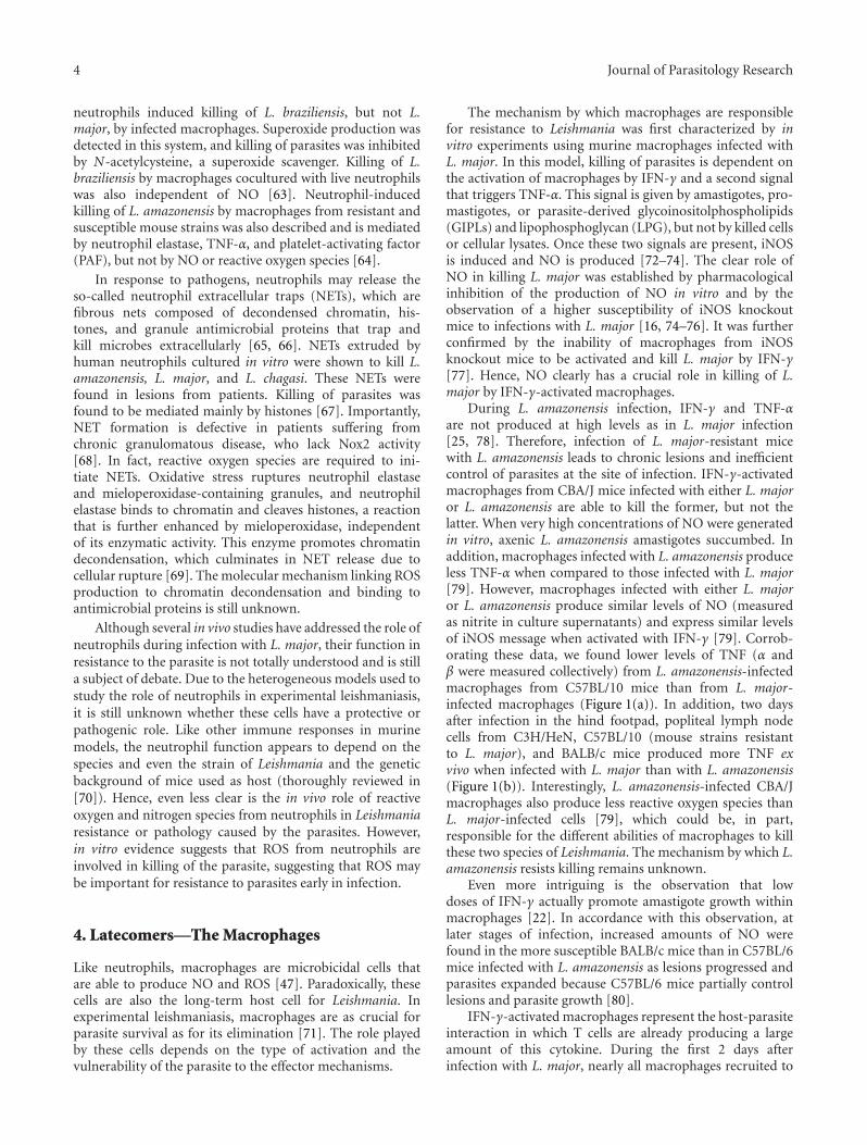
During L. amazonensis infection, IFN-γ and TNF-αare not produced at high levels as in L. major infection[25, 78]. Therefore, infection of L. major-resistant micewith L. amazonensis leads to chronic lesions and inefficientcontrol of parasites at the site of infection. IFN-γ-activatedmacrophages from CBA/J mice infected with either L. majoror L. amazonensis are able to kill the former, but not thelatter. When very high concentrations of NO were generatedin vitro, axenic L. amazonensis amastigotes succumbed. Inaddition, macrophages infected with L. amazonensis produceless TNF-α when compared to those infected with L. major[79]. However, macrophages infected with either L. majoror L. amazonensis produce similar levels of NO (measuredas nitrite in culture supernatants) and express similar levelsof iNOS message when activated with IFN-γ [79]. Corrob-orating these data, we found lower levels of TNF (α andβ were measured collectively) from L. amazonensis-infectedmacrophages from C57BL/10 mice than from L. major-infected macrophages (Figure 1(a)). In addition, two daysafter infection in the hind footpad, popliteal lymph nodecells from C3H/HeN, C57BL/10 (mouse strains resistantto L. major), and BALB/c mice produced more TNF exvivo when infected with L. major than with L. amazonensis(Figure 1(b)). Interestingly, L. amazonensis-infected CBA/Jmacrophages also produce less reactive oxygen species thanL. major-infected cells [79], which could be, in part,responsible for the different abilities of macrophages to killthese two species of Leishmania. The mechanism by which L.amazonensis resists killing remains unknown.
Even more intriguing is the observation that lowdoses of IFN-γ actually promote amastigote growth withinmacrophages [22]. In accordance with this observation, atlater stages of infection, increased amounts of NO werefound in the more susceptible BALB/c mice than in C57BL/6mice infected with L. amazonensis as lesions progressed andparasites expanded because C57BL/6 mice partially controllesions and parasite growth [80].
IFN-γ-activated macrophages represent the host-parasiteinteraction in which T cells are already producing a largeamount of this cytokine. During the first 2 days afterinfection with L. major, nearly all macrophages recruited to

Journal of Parasitology Research 5
0 1 5 10 20 40
0
10
20
30
40
50
Number of parasites per macrophage
TN
F(p
g/m
L)
(a)
L. majorL. amazonensis
0
10
20
30
40
50
TN
F(p
g/m
L)
C3H
/HeN
C57
BL/
10
BA
LB/c
(b)
Figure 1: Infection with L. major induces more TNF than infection with L. amazonensis. (a) TNF production by inflammatory macrophagesfrom C57BL/10, mice infected in vitro with L. major or L. amazonensis. (b) Production of TNF ex vivo by lymph node cells from C3H/HeN,C57BL/10 and BALB/c mice infected with L. major or L. amazonensis, 2 days after infection. A biological assay that does not distinguishbetween TNF-α or TNF-β was used in these experiments. These are representative experiments of more than five performed experiments (L.Q. Vieira and P. Scott, unpublished).
the site of infection contain phagocytosed parasites, bothin C57BL/6 and in BALB/c mice. However, the percentageof cells (mostly neutrophils and mononuclear phagocytes)containing intact parasites in BALB/c mice is higher thanthat in C57BL/6 cells (mostly mononuclear cells), and theelimination of parasites from the site of infection is higherin resistant mice [50]. This suggests that parasites may alsobe killed by tissue mononuclear cells well before the onset ofa T-cell response. Whether this killing is mediated by reactiveoxygen and nitrogen species remains unknown.
Isolated macrophages from C57BL/6 mice produce moreNO than macrophages from susceptible strains when stimu-lated with IFN-γ [81–84], TNF-α [81, 85], or LPS [83, 85–89]. This is an interesting but poorly explored aspect ofthe murine models of resistance/susceptibility to microbialinfections, which is clearly independent of the developmentof an adaptive Th1 or Th2 response. Mills et al. [90]systematically tested this observation and generalized it toother strains of mice. They showed that macrophages fromstrains that are typical Th1 responders (termed M-1) ortypical Th2 responders (termed M-2) differ qualitatively intheir ability to be activated, as measured by their argininemetabolic programs. M-2 macrophages from BALB/c mice(prototypes of Th2 responders) stimulated with a particularconcentration of LPS not only produce little or no NO, butincrease arginine metabolism to ornithine. In contrast, M-1 cells from C57BL/6 mice (prototypes of Th1 responders)generate a strong NO and citrulline response and appear todecrease their production of ornithine.
We investigated the molecular basis of the differentialproduction of NO by macrophages from mice with resistantor susceptible phenotypes to L. major by in vitro stimulationwith IFN-γ and LPS. We have shown that M-1 macrophages
show a remarkably strong expression of the enzyme iNOSupon stimulation when compared to M-2 cells [84]. Theaccumulation of iNOS mRNA is also higher in M-1 cells.Interestingly, however, we found that the accumulation ofthe iNOS protein is more dramatic than the accumulationof iNOS mRNA. The accumulation of both iNOS mRNAand protein is not a consequence of a higher stability ofthe molecule. The data showed that iNOS gene expressionis differentially regulated in M-1 and M-2 macrophages andsuggested that it is transcribed and translated at differentrates in these two types of cells [84]. Recent results fromour group indicate that the higher iNOS expression in M-1macrophages may be multifactorial and may be regulated byhigher levels of TNF-α, IL-12, and IFN-β (unpublished data).
The intrinsic differential sensitivity to IFN-γ and LPSof M-1 or M-2 cells has led to two important observationsregarding the in vivo infection.
(1) Small amounts of IFN-γ (from NK, NKT, or γ/δ Tcells) or other pathogen-derived inducers may induce M-1,but not M-2 cells, to kill the pathogen through NO, beforeT cells differentiate into the IFN-γ-Th1 subpopulation.In fact, larger numbers of L. major are found in iNOS-deficient macrophages than in wild-type macrophages 72hours after infection, indicating that some NO is producedby macrophages that have not been activated with IFN-γ andthat NO, even if not detectable, exerts some control of par-asite growth [75, 77]. Further evidence of a NO-dependentTh-cell-independent mechanism was obtained when restinghuman macrophages were infected with NO-susceptible andNO-resistant L. amazonensis and L. braziliensis isolates andselected in vitro with increasing concentrations of NaNO2:NO-resistant parasites grew better in resting macrophagesthan the NO-susceptible isolates [91].

6 Journal of Parasitology Research
(2) Activated M-1 and M-2 cells can distinctly affectsubsequent production of Th1-dominant or Th2-dominant cytokines (IFN-γ or TGF-β1, resp.), positioningmacrophages as key performers in directing the Th1 or Th2outcome. M-1 and M-2 macrophages differentially influencethe Th lymphocyte response, and how macrophages arestimulated determines the route that Th responses willtake [90]. These observations indicate that macrophagesmay contribute to the outcome of an immune responsethrough mechanisms other than by acting as establishedNO-producing cells and that their role in determining theresistant/susceptible phenotype in mice may be significant.M-1 macrophages not only can mount an early (innate)resistance, but also can consolidate the status of resistance byfavoring a Th1 adaptive response.
In addition to NO, ROS are considered to be amajor macrophage effector mechanism induced by IFN-γ to control infections. Upon bacteria or other pathogenengulfment by a phagocytic cell, ROS are rapidly producedby NADPH oxidase, an enzymatic complex comprised ofmembrane bound (p22phox and gp91phox) and cytosolic(p40phox, p47phox, p67phox, and Rac-1/2) proteins [45, 92],which may be assembled after TLR stimulation by bacterialproducts via MyD88-dependent p38 MAPK activation [93].
Macrophages [54, 76] and neutrophils [54] produce ROSin response to Leishmania in vitro. Killing of L. major by IFN-γ-activated macrophages is dependent on NO production,but not on the production of superoxide or peroxynitrite[76]. Lesions in Nox2 knockout mice [94] (Nox2 mice aregenetically deficient in the NADPH-dependent phagocyteoxidase. These mice were originally described as a model forchronic granulomatous disease and are more susceptible tobacterial infection, and neither neutrophils nor macrophagespresent respiratory burst oxidase activity [94].) infected withL. major are similar to those in wild-type C57BL6 mice. Nox2knockout mice control L. major at the site of infection at earlytime points, but display an unexpected reactivation of L.major infection after long periods of observation (more than200 days of infection). Further, they show deficient controlof parasite replication in draining lymph nodes and spleens,suggesting that Nox2 is important for the control of L. majorin vivo at later times of infection by preventing visceralization[54]. The participation of ROS in killing of L. amazonensisby mouse [95, 96] or human [97] macrophages has beenreported. Our preliminary data suggest that macrophagesfrom Nox2 knockout mice behave similarly to macrophagesfrom wild-type mice when infected with L. amazonensis.Moreover, similar to infection with L. major, Nox2 knockoutmice control parasites at the site of infection as well aswild-type mice (Figure 2). Surprisingly, at earlier times ofinfection, lesions are larger in Nox2 knockout mice, and, atlater times of infection, they become smaller than in wild-type mice (Figure 2(a)). This indicates that the differencesin Ros activity on macrophage behavior at different stagesof infection may be due to differences in the inflammatoryinfiltrate. The contradictions between the in vitro evidencefor a role for ROS in resistance to L. amazonensis and in vivodata remain to be explained.
Although BALB/c mice are the prototype model ofsusceptibility to most species of Leishmania (such as L. majorand L. amazonensis), L. braziliensis [27, 98] and L. guyanensis[99] do not cause large skin lesions in this mouse strain. Ourstudies using L. guyanensis have shown that BALB/c micedevelop minor or no lesions, do not enable parasite repli-cation, and do not die of the infection. In addition, L. guya-nensis [99] and L. braziliensis [100], unlike L. amazonensis,fail to survive within nonactivated peritoneal macrophagesin vitro. In vitro infection of BALB/c macrophages with L.guyanensis does not activate the production of NO; instead,it activates a respiratory burst that is exceptionally higherthan that activated by infection with L. amazonensis. We havefurther shown that the production of ROS is responsible forthe elimination of L. guyanensis by macrophages. We havealso shown that L. guyanensis amastigotes die inside BALB/cmacrophages through an apoptosis-like process mediated byparasite-induced ROS [99]. These findings demonstrate animportant killing mechanism of L. guyanensis amastigotes.ROS are probably involved in resistance to infection withthis species because mice that are unable to activate therespiratory burst by the regular administration of apocynin,an inhibitor of NADPH oxidase, do not control the infectionas in untreated animals (our preliminary results). Together,our results suggest that the elimination of L. guyanensis invivo may occur in early infection due to ROS production,before the development of an adaptive Th response.
There is evidence that peroxynitrite (ONOO−) is notinvolved in the killing of L. major [54, 76], but the role ofthis important oxidant has not been thoroughly explored.In contrast, the production of nitric oxide and ONOO− hasbeen shown during infection with L. amazonensis in BALB/c(more susceptible to infection) and C57BL/6 mice (moreresistant to infection). The production of nitric oxide in vivowas detected as the nitrosyl hemoglobin complex by electronparamagnetic resonance analysis of nitrosyl hemoglobin inblood drawn from mice and in infected footpads at severaltime points, and ONOO− formation was inferred fromimmunodetection of nitrotyrosine [101, 102]. C57BL/6 micepresented higher levels of nitrosyl complexes than BALB/cmice at 6 weeks of infection, at which point lesions becamechronic in this partially resistant mouse strain. Nitrosylcomplexes increased in BALB/c mice, which was dependenton lesion size. iNOS and nitrotyrosine-containing complexescolocalize in lesion macrophages from both mouse strains,and the most probable agent of protein nitration is ONOO−
[102]. Peroxynitrite killed L. amazonensis axenic amastigotesin vitro more efficiently than nitric oxide [102]. The authorsproposed that in the susceptible mouse strain, ONOO− isinvolved in tissue damage. It is possible that the delayedproduction of ONOO− impairs the capacity of BALB/c miceto control L. amazonensis. Treatment of C57BL/6 mice withTempol, a stable cyclic nitroxide radical that protects cellsfrom damage due to oxidative stress, promoted larger lesions,parasite growth, and lower levels of nitric oxide products andnitrotyrosine [103]. Albeit transient, this effect of Tempolprovides further evidence that ONOO− is involved in thecontrol of L. amazonensis in vivo.

Journal of Parasitology Research 7
2 3 4 5 6 7 8 9 10
Weeks of infection
0
0.5
1
1.5
2
2.5
Lesi
on(m
m)
∗
∗
∗∗
∗∗ ∗
WTPhox−/−
(a)
Footpad Lymph node Footpad Lymph node
Footpad Lymph nodeFootpad Lymph node
0
2
4
6
8
0
2
4
6
8
0
2
4
6
8
0
2
4
6
8
WT WT
4 weeks
8 weeks
6 weeks
10 weeks
Phox−/−Phox−/−
−log
tit
er
−log
tit
er−l
og t
iter
−log
tit
er
(b)
Figure 2: Course of infection with L. amazonensis in wild-type C57BL/6 and Nox2 knockout mice (a) and parasite quantitation using alimiting dilution analysis (b). ∗indicates statistical difference by Student’s t test, P < 0.05 (E. H. Roma and J. P. Macedo, unpublished).

8 Journal of Parasitology Research
5. Concluding Remarks
The role of reactive oxygen and nitrogen species in killingof Leishmania has been the subject of many studies, butthere is still much that is not understood. The followingquestions remain: why do some species of parasites resistoxidative stress? Why do cells that can kill parasites withreactive species harbor live parasites? Is there some attritionwhen parasites enter neutrophils and macrophages? Whatis the role of peroxinitrite? What is the reason for thedifferences in the oxidative responses among different speciesof parasites? What is the role of reactive oxygen and nitrogenspecies in the inflammatory response? Collective efforts tofully comprehend the mechanisms that produce disease uponinfection with Leishmania and the strategies hosts employ toavoid them have been made. However, leishmaniasis persistswithout safe treatments or effective vaccines. Perhaps therecent attention paid to components of the innate immunesystem might help to unravel this complex parasite-hostrelationship.
Acknowledgments
L. Q. Vieira is a member of INCT de Processos Redoxem Biomedicina-Redoxoma (CNPq/FAPESP/ CAPES573530/2008-4). The authors’ results were supportedby grants from Conselho Nacional de DesenvolvimentoCientıfico e Tecnologico (CNPq), Coordenacao deAperfeicoamento de Pessoal de Nıvel Superior (CAPES), andFundacao de Amparo a Pesquisa do Estado de Minas Gerais(FAPEMIG). The authors are grateful to FUNDEP for expertadministration of grants. M. F. Horta and L. Q. Vieira areCNPq research fellows.
References
[1] H. W. Murray, J. D. Berman, C. R. Davies, and N. G. Saravia,“Advances in leishmaniasis,” Lancet, vol. 366, no. 9496, pp.1561–1577, 2005.
[2] L. Kedzierski, Y. Zhu, and E. Handman, “Leishmania vac-cines: progress and problems,” Parasitology, vol. 133, no. 2,pp. S87–S112, 2006.
[3] A. L. Banuls, M. Hide, and F. Prugnolle, “Leishmania andthe Leishmaniases: a parasite genetic update and advancesin taxonomy, epidemiology and pathogenicity in humans,”Advances in Parasitology, vol. 64, pp. 1–109, 2007.
[4] R. Killick-Kendrick, “The life-cycle of Leishmania in thesandfly with special reference to the form infective tothe vertebrate host,” Annales de Parasitologie Humaine etComparee, vol. 65, no. 1, supplement 1, pp. 37–42, 1990.
[5] A. A. Filardy, D. R. Pires, and G. A. Dosreis, “Macrophagesand neutrophils cooperate in immune responses to Leishma-nia infection,” Cellular and Molecular Life Sciences, vol. 68,no. 11, pp. 1863–1870, 2011.
[6] N. C. Peters and D. L. Sacks, “The impact of vector-mediatedneutrophil recruitment on cutaneous leishmaniasis,” CellularMicrobiology, vol. 11, no. 9, pp. 1290–1296, 2009.
[7] D. Sacks and N. Noben-Trauth, “The immunology ofsusceptibility and resistance to Leishmania major in mice,”Nature Reviews Immunology, vol. 2, no. 11, pp. 845–858,2002.
[8] M. F. Horta, “Pore-forming proteins in pathogenic protozoanparasites,” Trends in Microbiology, vol. 5, no. 9, pp. 363–366,1997.
[9] F. S. M. Noronha, F. J. Ramalho-Pinto, and M. F. Horta,“Cytolytic activity in the genus Leishmania: involvement of aputative pore-forming protein,” Infection and Immunity, vol.64, no. 10, pp. 3975–3982, 1996.
[10] F. S. M. Noronha, J. S. Cruz, P. S. L. Beirao, and M. F. Horta,“Macrophage damage by Leishmania amazonensis cytolysin:evidence of pore formation on cell membrane,” Infection andImmunity, vol. 68, no. 8, pp. 4578–4584, 2000.
[11] F. R. Almeida-Campos and M. F. Horta, “Proteolytic acti-vation of leishporin: evidence that Leishmania amazonensisand Leishmania guyanensis have distinct inactive forms,”Molecular and Biochemical Parasitology, vol. 111, no. 2, pp.363–375, 2000.
[12] T. Castro-Gomes, F. R. Almeida-Campos, C. E. Calzavara-Silva, R. A. da Silva, F. Frezard, and M. F. Horta, “Membranebinding requirements for the cytolytic activity of Leishmaniaamazonensis leishporin,” FEBS Letters, vol. 583, no. 19, pp.3209–3214, 2009.
[13] F. S. Noronha, F. J. Ramalho-Pinto, and M. F. Horta,“Identification of a putative pore-forming hemolysin activeat acid pH in Leishmania amazonensis,” Brazilian Journal ofMedical and Biological Research, vol. 27, no. 2, pp. 477–482,1994.
[14] T. R. Mosmann, H. Cherwinski, and M. W. Bond, “Twotypes of murine helper T cell clone. I. Definition accordingto profiles of lymphokine activities and secreted proteins,”Journal of Immunology, vol. 136, no. 7, pp. 2348–2357, 1986.
[15] I. M. Corraliza, G. Soler, K. Eichmann, and M. Modolell,“Arginase induction by suppressors of nitric oxide synthesis(IL-4, IL-10 and PGE2) in murine bone-marrow-derivedmacrophages,” Biochemical and Biophysical Research Commu-nications, vol. 206, no. 2, pp. 667–673, 1995.
[16] F. Y. Liew, S. Millott, C. Parkinson, R. M. J. Palmer, andS. Moncada, “Macrophage killing of Leishmania parasite invivo is mediated by nitric oxide from L-arginine,” Journal ofImmunology, vol. 144, no. 12, pp. 4794–4797, 1990.
[17] R. M. Locksley, F. P. Heinzel, M. D. Sadick, B. J. Holaday, andK. D. Gardner, “Murine cutaneous leishmaniasis: suscepti-bility correlates with differential expansion of helper T-cellsubsets,” Annales de l’Institut Pasteur. Immunology, vol. 138,no. 5, pp. 744–749, 1987.
[18] P. Scott, P. Natovitz, R. L. Coffman, E. Pearce, and A. Sher,“Immunoregulation of cutaneous leishmaniasis. T cell linesthat transfer protective immunity or exacerbation belong todifferent T helper subsets and respond to distinct parasiteantigens,” Journal of Experimental Medicine, vol. 168, no. 5,pp. 1675–1684, 1988.
[19] S. J. Green, M. S. Meltzer, J. B. Hibbs, and C. A. Nacy, “Acti-vated macrophages destroy intracellular Leishmania majoramastigotes by an L-arginine-dependent killing mechanism,”Journal of Immunology, vol. 144, no. 1, pp. 278–283, 1990.
[20] L. Soong, C. H. Chang, J. Sun et al., “Role of CD4+ Tcells in pathogenesis associated with Leishmania amazonensisinfection,” Journal of Immunology, vol. 158, no. 11, pp. 5374–5383, 1997.
[21] Y. F. Vanloubbeeck, A. E. Ramer, F. Jie, and D. E.Jones, “CD4+ Th1 cells induced by dendritic cell-basedimmunotherapy in mice chronically infected with Leish-mania amazonensis do not promote healing,” Infection andImmunity, vol. 72, no. 8, pp. 4455–4463, 2004.

Journal of Parasitology Research 9
[22] H. Qi, J. Ji, N. Wanasen, and L. Soong, “Enhanced repli-cation of Leishmania amazonensis amastigotes in gammainterferon-stimulated murine macrophages: implications forthe pathogenesis of cutaneous leishmaniasis,” Infection andImmunity, vol. 72, no. 2, pp. 988–995, 2004.
[23] Y. Vanloubbeeck and D. E. Jones, “Protection of C3HeB/FeJmice against Leishmania amazonensis challenge after previ-ous Leishmania major infection,” American Journal of TropicalMedicine and Hygiene, vol. 71, no. 4, pp. 407–411, 2004.
[24] C. Z. Gonzalez-Lombana, H. C. Santiago, J. P. Macedo et al.,“Early infection with Leishmania major restrains pathogenicresponse to Leishmania amazonensis and parasite growth,”Acta Tropica, vol. 106, no. 1, pp. 27–38, 2008.
[25] L. C. C. Afonso and P. Scott, “Immune responses associatedwith susceptibility of C57BL/10 mice to Leishmania amazo-nensis,” Infection and Immunity, vol. 61, no. 7, pp. 2952–2959,1993.
[26] O. Guevara-Mendoza, C. Une, P. Franceschi Carreira, and A.Orn, “Experimental infection of Balb/c mice with Leishmaniapanamensis and Leishmania mexicana induction of earlyIFN-γ but not IL-4 is associated with the development ofcutaneous lesions,” Scandinavian Journal of Immunology, vol.46, no. 1, pp. 35–40, 1997.
[27] G. K. DeKrey, H. C. Lima, and R. G. Titus, “Analysis ofthe immune responses of mice to infection with Leishmaniabraziliensis,” Infection and Immunity, vol. 66, no. 2, pp. 827–829, 1998.
[28] D. Mahon-Pratt and J. Alexander, “Does the Leishmaniamajor paradigm of pathogenesis and protection hold forNew World cutaneous leishmaniases or the visceral disease?”Immunological Reviews, vol. 201, pp. 206–224, 2004.
[29] J. L. Stafford, N. F. Neumann, and M. Belosevic,“Macrophage-mediated innate host defense againstprotozoan parasites,” Critical Reviews in Microbiology,vol. 28, no. 3, pp. 187–248, 2002.
[30] T. M. Scharton and P. Scott, “Natural killer cells are a sourceof interferon γ that drives differentiation of CD4+ T cellsubsets and induces early resistance to Leishmania major inmice,” Journal of Experimental Medicine, vol. 178, no. 2, pp.567–577, 1993.
[31] R. Birnbaum and N. Craft, “Innate immunity and Leishma-nia vaccination strategies,” Dermatologic Clinics, vol. 29, no.1, pp. 89–102, 2011.
[32] A. Diefenbach, H. Schindler, N. Donhauser et al., “Type 1interferon (IFNα/β) and type 2 nitric oxide synthase regulatethe innate immune response to a protozoan parasite,”Immunity, vol. 8, no. 1, pp. 77–87, 1998.
[33] G. J. Bancroft, R. D. Schreiber, and G. C. Bosma, “A Tcell-independent mechanism of macrophage activation byinterferon-γ,” Journal of Immunology, vol. 139, no. 4, pp.1104–1107, 1987.
[34] K. N. Dileepan, K. M. Simpson, and D. J. Stechschulte, “Mod-ulation of macrophage superoxide-induced cytochrome creduction by mast cells,” Journal of Laboratory and ClinicalMedicine, vol. 113, no. 5, pp. 577–585, 1989.
[35] J. Hao, X. Wu, S. Xia et al., “Current progress in γδ T-cellbiology,” Cellular and Molecular Immunology, vol. 7, no. 6,pp. 409–413, 2010.
[36] M. Munder, M. Mallo, K. Eichmann, and M. Modolell,“Murine macrophages secrete interferon γ upon combinedstimulation with interleukin (IL)-12 and IL-18: a novelpathway of autocrine macrophage activation,” Journal ofExperimental Medicine, vol. 187, no. 12, pp. 2103–2108, 1998.
[37] P. Puddu, M. Carollo, I. Pietraforte et al., “IL-2 inducesexpression and secretion of IFN-γ in murine peritonealmacrophages,” Journal of Leukocyte Biology, vol. 78, no. 3, pp.686–695, 2005.
[38] S. Fujii, S. Motohashi, K. Shimizu, T. Nakayama, Y. Yoshiga,and M. Taniguchi, “Adjuvant activity mediated by iNKTcells,” Seminars in Immunology, vol. 22, no. 2, pp. 97–102,2010.
[39] T. van Assche, M. Deschacht, R. A.I. Da Luz, L. Maes, and P.Cos, “Leishmania-macrophage interactions: insights into theredox biology,” Free Radical Biology and Medicine, vol. 51, no.2, pp. 337–351, 2011.
[40] E. Ogier-Denis, S. B. Ogier-Denis, and A. Vandewalle,“NOX enzymes and Toll-like receptor signaling,” Seminars inImmunopathology, vol. 30, no. 3, pp. 291–300, 2008.
[41] G. Y. Chen and G. Nunez, “Sterile inflammation: sensing andreacting to damage,” Nature Reviews Immunology, vol. 10, no.12, pp. 826–837, 2010.
[42] S. Carta, P. Castellani, L. Delfino, S. Tassi, R. Vene, and A.Rubartelli, “DAMPs and inflammatory processes: the role ofredox in the different outcomes,” Journal of Leukocyte Biology,vol. 86, no. 3, pp. 549–555, 2009.
[43] W. M. Nauseef, “How human neutrophils kill and degrademicrobes: an integrated view,” Immunological Reviews, vol.219, no. 1, pp. 88–102, 2007.
[44] B. M. Babior, “NADPH oxidase: an update,” Blood, vol. 93,no. 5, pp. 1464–1476, 1999.
[45] A. Mizrahi, Y. Berdichevsky, Y. Ugolev et al., “Assembly ofthe phagocyte NADPH oxidase complex: chimeric constructsderived from the cytosolic components as tools for exploringstructure-function relationships,” Journal of Leukocyte Biol-ogy, vol. 79, no. 5, pp. 881–895, 2006.
[46] M. B. Hampton, A. J. Kettle, and C. C. Winterbourn, “Insidethe neutrophil phagosome: oxidants, myeloperoxidase, andbacterial killing,” Blood, vol. 92, no. 9, pp. 3007–3017, 1998.
[47] C. Nathan and M. U. Shiloh, “Reactive oxygen and nitro-gen intermediates in the relationship between mammalianhosts and microbial pathogens,” Proceedings of the NationalAcademy of Sciences of the United States of America, vol. 97,no. 16, pp. 8841–8848, 2000.
[48] R. Radi, G. Peluffo, M. N. Alvarez, M. Naviliat, and A. Cayota,“Unraveling peroxynitrite formation in biological systems,”Free Radical Biology and Medicine, vol. 30, no. 5, pp. 463–488,2001.
[49] N. C. Peters, J. G. Egen, N. Secundino et al., “In vivo imagingreveals an essential role for neutrophils in leishmaniasistransmitted by sand flies,” Science, vol. 321, no. 5891, pp.970–974, 2008.
[50] W. J. Beil, G. Meinardus-Hager, D. C. Neugebauer, and C.Sorg, “Differences in the onset of the inflammatory responseto cutaneous leishmaniasis in resistant and susceptible mice,”Journal of Leukocyte Biology, vol. 52, no. 2, pp. 135–142, 1992.
[51] A. K. Nussler and T. R. Billiar, “Inflammation, immunoreg-ulation, and inducible nitric oxide synthase,” Journal ofLeukocyte Biology, vol. 54, no. 2, pp. 171–178, 1993.
[52] A. W. Segal, “How neutrophils kill microbes,” Annual Reviewof Immunology, vol. 23, pp. 197–223, 2005.
[53] M. Charmoy, R. Megnekou, C. Allenbach et al., “Leishmaniamajor induces distinct neutrophil phenotypes in mice thatare resistant or susceptible to infection,” Journal of LeukocyteBiology, vol. 82, no. 2, pp. 288–299, 2007.
[54] M. Blos, U. Schleicher, F. J. S. Rocha, U. Meißner,M. Rollinghoff, and C. Bogdan, “Organ-specific andstage-dependent control of Leishmania major infection by

10 Journal of Parasitology Research
inducible nitric oxide synthase and phagocyte NADPHoxidase,” European Journal of Immunology, vol. 33, no. 5, pp.1224–1234, 2003.
[55] D. J. Mallinson, J. M. Lackie, and G. H. Coombs, “Theoxidative response of rabbit peritoneal neutrophils to leish-manias and other trypanosomatids,” International Journal forParasitology, vol. 19, no. 6, pp. 639–645, 1989.
[56] H. Laufs, K. Muller, J. Fleischer et al., “Intracellular survivalof Leishmania major in neutrophil granulocytes after uptakein the absence of heat-labile serum factors,” Infection andImmunity, vol. 70, no. 2, pp. 826–835, 2002.
[57] F. Mollinedo, H. Janssen, J. De La Iglesia-Vicente, J. A.Villa-Pulgarin, and J. Calafat, “Selective fusion of azurophilicgranules with Leishmania-containing phagosomes in humanneutrophils,” Journal of Biological Chemistry, vol. 285, no. 45,pp. 34528–34536, 2010.
[58] S. Bisti, G. Konidou, J. Boelaert, M. Lebastard, and K.Soteriadou, “The prevention of the growth of Leishmaniamajor progeny in BALB/c iron-loaded mice: a processcoupled to increased oxidative burst, the amplitude andduration of which depend on initial parasite developmentalstage and dose,” Microbes and Infection, vol. 8, no. 6, pp.1464–1472, 2006.
[59] S. Bisti and K. Soteriadou, “Is the reactive oxygen species-dependent-NF-κB activation observed in iron-loadedBALB/c mice a key process preventing growth of Leishmaniamajor progeny and tissue-damage?” Microbes and Infection,vol. 8, no. 6, pp. 1473–1482, 2006.
[60] F. L. Ribeiro-Gomes, M. C. A. Moniz-de-Souza, M. S.Alexandre-Moreira et al., “Neutrophils activate macrophagesfor intracellular killing of Leishmania major through recruit-ment of TLR4 by neutrophil elastase,” Journal of Immunology,vol. 179, no. 6, pp. 3988–3994, 2007.
[61] F. L. Ribeiro-Gomes, A. C. Otero, N. A. Gomes et al.,“Macrophage Interactions with Neutrophils Regulate Leish-mania major Infection,” Journal of Immunology, vol. 172, no.7, pp. 4454–4462, 2004.
[62] L. Afonso, V. M. Borges, H. Cruz et al., “Interactions withapoptotic but not with necrotic neutrophils increase parasiteburden in human macrophages infected with Leishmaniaamazonensis,” Journal of Leukocyte Biology, vol. 84, no. 2, pp.389–396, 2008.
[63] F. O. Novais, R. C. Santiago, A. Bafica et al., “Neutrophils andmacrophages cooperate in host resistance against Leishmaniabraziliensis infection,” Journal of Immunology, vol. 183, no.12, pp. 8088–8098, 2009.
[64] E. V. de Souza Carmo, S. Katz, and C. L. Barbieri,“Neutrophils reduce the parasite burden in Leishmania(Leishmania) amazonensis macrophages,” PLoS ONE, vol. 5,no. 11, Article ID e13815, 2010.
[65] V. Brinkmann, U. Reichard, C. Goosmann et al., “Neutrophilextracellular traps kill bacteria,” Science, vol. 303, no. 5663,pp. 1532–1535, 2004.
[66] V. Papayannopoulos and A. Zychlinsky, “NETs: a newstrategy for using old weapons,” Trends in Immunology, vol.30, no. 11, pp. 513–521, 2009.
[67] A. B. Guimaraes-Costa, M. T. C. Nascimento, G. S. Fromentet al., “Leishmania amazonensis promastigotes induce and arekilled by neutrophil extracellular traps,” Proceedings of theNational Academy of Sciences of the United States of America,vol. 106, no. 16, pp. 6748–6753, 2009.
[68] T. A. Fuchs, U. Abed, C. Goosmann et al., “Novel cell deathprogram leads to neutrophil extracellular traps,” Journal ofCell Biology, vol. 176, no. 2, pp. 231–241, 2007.
[69] V. Papayannopoulos, K. D. Metzler, A. Hakkim, and A. Zych-linsky, “Neutrophil elastase and myeloperoxidase regulate theformation of neutrophil extracellular traps,” Journal of CellBiology, vol. 191, no. 3, pp. 677–691, 2010.
[70] U. Ritter, F. Frischknecht, and G. van Zandbergen, “Areneutrophils important host cells for Leishmania parasites?”Trends in Parasitology, vol. 25, no. 11, pp. 505–510, 2009.
[71] C. Bogdan and M. Rollinghoff, “The immune response toLeishmania: mechanisms of parasite control and evasion,”International Journal for Parasitology, vol. 28, no. 1, pp. 121–134, 1998.
[72] A. Piani, T. Ilg, A. G. Elefanty, J. Curtis, and E. Hand-man, “Leishmania major proteophosphoglycan is expressedby amastigotes and has an immunomodulatory effect onmacrophage function,” Microbes and Infection, vol. 1, no. 8,pp. 589–599, 1999.
[73] G. Kavoosi, S. K. Ardestani, A. Kariminia, and Z. Tavakoli,“Production of nitric oxide by murine macrophages inducedby lipophosphoglycan of Leishmania major,” The KoreanJournal of Parasitology, vol. 44, no. 1, pp. 35–41, 2006.
[74] S. J. Green, R. M. Crawford, J. T. Hockmeyer, M. S. Meltzer,and C. A. Nacy, “Leishmania major amastigotes initiate the L-arginine-dependent killing mechanism in IFN-γ-stimulatedmacrophages by induction of tumor necrosis factor-α1,”Journal of Immunology, vol. 145, no. 12, pp. 4290–4297, 1990.
[75] X. Q. Wei, I. G. Charles, A. Smith et al., “Altered immuneresponses in mice lacking inducible nitric oxide synthase,”Nature, vol. 375, no. 6530, pp. 408–411, 1995.
[76] J. Assreuy, F. Q. Cunha, M. Epperlein et al., “Production ofnitric oxide and superoxide by activated macrophages andkilling of Leishmania major,” European Journal of Immunol-ogy, vol. 24, no. 3, pp. 672–676, 1994.
[77] X. Q. Wei, B. P. Leung, W. Niedbala et al., “Altered immuneresponses and susceptibility to Leishmania major and Staphy-lococcus aureus infection in IL-18-deficient mice,” Journal ofImmunology, vol. 163, no. 5, pp. 2821–2828, 1999.
[78] J. Ji, J. Sun, and L. Soong, “Impaired expression of inflam-matory cytokines and chemokines at early stages of infectionwith Leishmania amazonensis,” Infection and Immunity, vol.71, no. 8, pp. 4278–4288, 2003.
[79] I. N. Gomes, A. F. Calabrich, R. S. Tavares, J. Wietzerbin, L.A. Rodrigues De Freitas, and P. S. Tavares Veras, “Differentialproperties of CBA/J mononuclear phagocytes recovered froman inflammatory site and probed with two different speciesof Leishmania,” Microbes and Infection, vol. 5, no. 4, pp. 251–260, 2003.
[80] S. Giorgio, E. Linares, H. Ischiropoulos, F. J. Von Zuben,A. Yamada, and O. Augusto, “In vivo formation of elec-tron paramagnetic resonance-detectable nitric oxide and ofnitrotyrosine is not impaired during murine leishmaniasis,”Infection and Immunity, vol. 66, no. 2, pp. 807–814, 1998.
[81] F. Y. Liew, Y. Li, D. Moss, C. Parkinson, M. V. Rogers,and S. Moncada, “Resistance to Leishmania major infectioncorrelates with the induction of nitric oxide synthase inmurine macrophages,” European Journal of Immunology, vol.21, no. 12, pp. 3009–3014, 1991.
[82] S. Stenger, H. Thuring, M. Rollinghoff, and C. Bogdan,“Tissue expression of inducible nitric oxide synthase isclosely associated with resistance to Leishmania major,”Journal of Experimental Medicine, vol. 180, no. 3, pp. 783–793, 1994.
[83] Z. Zidek, D. Frankova, and M. Boubelik, “Genetic variationin in-vitro cytokine-induced production of nitric oxide by

Journal of Parasitology Research 11
murine peritoneal macrophages,” Pharmacogenetics, vol. 10,no. 6, pp. 493–501, 2000.
[84] J. L. Santos, A. A. Andrade, A. A. M. Dias et al., “Dif-ferential sensitivity of C57BL/6 (M-1) and BALB/c (M-2)macrophages to the stimuli of IFN-γ/LPS for the productionof NO: correlation with iNOS mRNA and protein expres-sion,” Journal of Interferon and Cytokine Research, vol. 26, no.9, pp. 682–688, 2006.
[85] J. Lima-Santos, I. S. Jardim, S. M. Teixeira, and M. F. Horta,“Diffferential induction of nitric oxide synthase in C57BL/6and BALB/c macrophages by IFN-gamma and TNF-alpha orLPS,” Abstracts of the Brazilian Society of Immunolgy Meeting,vol. 24, p. 122, 1999.
[86] K. N. Dileepan, J. C. Page, Y. Li, and D. J. Stechschulte,“Direct activation of murine peritoneal macrophages fornitric oxide production and tumor cell killing by interferon-γ,” Journal of Interferon and Cytokine Research, vol. 15, no. 5,pp. 387–394, 1995.
[87] I. P. Oswald, S. Afroun, D. Bray, J. F. Petit, and G. Lemaire,“Low response of BALB/c macrophages to priming andactivating signals,” Journal of Leukocyte Biology, vol. 52, no.3, pp. 315–322, 1992.
[88] I. S. Jardim, J. L. Santos, and M. F. Horta, “Differentialproduction of nitric oxide by murine macrophages fromLeishmania resistant and susceptible mice strains,” Memoriasdo Instituto Oswaldo Cruz, vol. 94, supplement, p. 197, 1999.
[89] I. S. Jardim, J. L. Santos, M. F. Horta, and F. J. Ramalho-Pinto, “Inhibition of the production of nitric oxide impairscytotoxicity of macrophages to Leishmania amazonensis,”Memorias do Instituto Oswaldo Cruz, vol. 92, supplement, p.217, 2011.
[90] C. D. Mills, K. Kincaid, J. M. Alt, M. J. Heilman, and A. M.Hill, “M-1/M-2 macrophages and the Th1/Th2 paradigm,”Journal of Immunology, vol. 164, no. 12, pp. 6166–6173, 2000.
[91] A. Giudice, I. Camada, P. T. G. Leopoldo et al., “Resistanceof Leishmania (Leishmania) amazonensis and Leishmania(Viannia) braziliensis to nitric oxide correlates with diseaseseverity in Tegumentary Leishmaniasis,” BMC InfectiousDiseases, vol. 7, article no. 7, 2007.
[92] B. M. Babior, “NADPH oxidase,” Current Opinion inImmunology, vol. 16, no. 1, pp. 42–47, 2004.
[93] S. Laroux, X. Romero, L. Wetzler, P. Engel, and C. Terhorst,“Cutting edge: MyD88 controls phagocyte NADPH oxidasefunction and killing of gram-negative bacteria,” Journal ofImmunology, vol. 175, no. 9, pp. 5596–5600, 2005.
[94] J. D. Pollock, D. A. Williams, M. A. C. Gifford et al.,“Mouse model of X-linked chronic granulomatous disease,an inherited defect in phagocyte superoxide production,”Nature Genetics, vol. 9, no. 2, pp. 202–209, 1995.
[95] A. Degrossoli, W. W. Arrais-Silva, M. C. Colhone, F. R.Gadelha, P. P. Joazeiro, and S. Giorgio, “The influence of lowoxygen on macrophage response to Leishmania infection,”Scandinavian Journal of Immunology, vol. 74, no. 2, pp. 165–175, 2011.
[96] R. M. Mukbel, C. Patten Jr., K. Gibson, M. Ghosh, C.Petersen, and D. E. Jones, “Macrophage killing of Leishmaniaamazonensis amastigotes requires both nitric oxide andsuperoxide,” American Journal of Tropical Medicine andHygiene, vol. 76, no. 4, pp. 669–675, 2007.
[97] R. Khouri, A. Bafica, M. D. P. P. Silva et al., “IFN-βimpairs superoxide-dependent parasite killing in humanmacrophages: evidence for a deleterious role of SOD1 incutaneous leishmaniasis,” Journal of Immunology, vol. 182,no. 4, pp. 2525–2531, 2009.
[98] R. A. Neal and C. Hale, “A comparative study of susceptibilityof inbred and outbred mouse strains compared with ham-sters to infection with New World cutaneous leishmaniases,”Parasitology, vol. 87, no. 1, pp. 7–13, 1983.
[99] J. Sousa-Franco, E. Araujo-Mendes, I. Silva-Jardim etal., “Infection-induced respiratory burst in BALB/cmacrophages kills Leishmania guyanensis amastigotesthrough apoptosis: possible involvement in resistance tocutaneous leishmaniasis,” Microbes and Infection, vol. 8, no.2, pp. 390–400, 2006.
[100] P. Scott and A. Sher, “A spectrum in the susceptibilityof Leishmanial strains to intracellular killing by murinemacrophages,” Journal of Immunology, vol. 136, no. 4, pp.1461–1466, 1986.
[101] S. Giorgio, E. Linares, M. D. L. Capurro, A. G. De Bianchi,and O. Augusto, “Formation of nitrosyl hemoglobin andnitrotyrosine during murine leishmaniasis,” Photochemistryand Photobiology, vol. 63, no. 6, pp. 750–754, 1996.
[102] E. Linares, S. Giorgio, R. A. Mortara, C. X. C. Santos,A. T. Yamada, and O. Augusto, “Role of peroxynitrite inmacrophage microbicidal mechanisms in vivo revealed byprotein nitration and hydroxylation,” Free Radical Biologyand Medicine, vol. 30, no. 11, pp. 1234–1242, 2001.
[103] E. Linares, S. Giorgio, and O. Augusto, “Inhibition of in vivoleishmanicidal mechanisms by tempol: nitric oxide down-regulation and oxidant scavenging,” Free Radical Biology andMedicine, vol. 44, no. 8, pp. 1668–1676, 2008.