Double blind, randomized controlled trial, to evaluate the effectiveness of a controlled nitric...
11
BioMed Central Page 1 of 11 (page number not for citation purposes) Trials Open Access Study protocol Double blind, randomized controlled trial, to evaluate the effectiveness of a controlled nitric oxide releasing patch versus meglumine antimoniate in the treatment of cutaneous leishmaniasis [NCT00317629] Sandra Y Silva 1 , Ligia C Rueda 1 , Marcos López 2 , Iván D Vélez 3 , Christian F Rueda-Clausen 1 , Daniel J Smith 2 , Gerardo Muñoz 4 , Hernando Mosquera 1 , Federico A Silva 1,4 , Adriana Buitrago 1 , Holger Díaz 5 and Patricio López-Jaramillo* 1,6 Address: 1 VILANO Group. Research Institute, Fundación Cardiovascular de Colombia (FCV), Floridablanca, Santander, Colombia, 2 Department of Chemistry, University of Akron, Akron, Ohio, USA, 3 Program for the Study and Control of Tropical Diseases, PECET, Universidad de Antioquia, Medellín, Antioquia, Colombia, 4 Universidad Industrial de Santander, Bucaramanga, Santander, Colombia, 5 Secretaría de Salud de Santander, Colombia and 6 Facultad de Medicina, Universidad de Santander, Bucaramanga, Santander, Colombia Email: Sandra Y Silva - [email protected]; Ligia C Rueda - [email protected]; Marcos López - [email protected]; Iván D Vélez - [email protected]; Christian F Rueda-Clausen - [email protected]; Daniel J Smith - [email protected]; Gerardo Muñoz - [email protected]; Hernando Mosquera - [email protected]; Federico A Silva - [email protected]; Adriana Buitrago - [email protected]; Holger Díaz - [email protected]; Patricio López-Jaramillo* - [email protected] * Corresponding author Abstract Background: Cutaneous Leishmaniasis is a worldwide disease, endemic in 88 countries, that has shown an increasing incidence over the last two decades. So far, pentavalent antimony compounds have been considered the treatment of choice, with a percentage of cure of about 85%. However, the high efficacy of these drugs is counteracted by their many disadvantages and adverse events. Previous studies have shown nitric oxide to be a potential alternative treatment when administered topically with no serious adverse events. However, due to the unstable nitric oxide release, the topical donors needed to be applied frequently, making the adherence to the treatment difficult. The electrospinning technique has allowed the production of a multilayer transdermal patch that produces a continuous and stable nitric oxide release. The main objective of this study is to evaluate this novel nitric oxide topical donor for the treatment of cutaneous leishmaniasis. Methods and design: A double-blind, randomized, double-masked, placebo-controlled clinical trial, including 620 patients from endemic areas for Leishmaniasis in Colombia was designed to investigate whether this patch is as effective as meglumine antimoniate for the treatment of cutaneous leishmaniasis but with less adverse events. Subjects with ulcers characteristic of cutaneous leishmaniasis will be medically evaluated and laboratory tests and parasitological confirmation performed. After checking the inclusion/exclusion criteria, the patients will be randomly assigned to one of two groups. During 20 days Group 1 will receive simultaneously meglumine antimoniate and placebo of nitric oxide patches while Group 2 will receive placebo of meglumine antimoniate and active nitric oxide patches. During the treatment visits, the medications Published: 15 May 2006 Trials 2006, 7:14 doi:10.1186/1745-6215-7-14 Received: 27 April 2006 Accepted: 15 May 2006 This article is available from: http://www.trialsjournal.com/content/7/1/14 © 2006 Silva et al; licensee BioMed Central Ltd. This is an Open Access article distributed under the terms of the Creative Commons Attribution License (http://creativecommons.org/licenses/by/2.0 ), which permits unrestricted use, distribution, and reproduction in any medium, provided the original work is properly cited.
-
Upload
independent -
Category
Documents
-
view
1 -
download
0
Transcript of Double blind, randomized controlled trial, to evaluate the effectiveness of a controlled nitric...

BioMed CentralTrials
ss
Open AcceStudy protocolDouble blind, randomized controlled trial, to evaluate the effectiveness of a controlled nitric oxide releasing patch versus meglumine antimoniate in the treatment of cutaneous leishmaniasis [NCT00317629]Sandra Y Silva1, Ligia C Rueda1, Marcos López2, Iván D Vélez3, Christian F Rueda-Clausen1, Daniel J Smith2, Gerardo Muñoz4, Hernando Mosquera1, Federico A Silva1,4, Adriana Buitrago1, Holger Díaz5 and Patricio López-Jaramillo*1,6Address: 1VILANO Group. Research Institute, Fundación Cardiovascular de Colombia (FCV), Floridablanca, Santander, Colombia, 2Department of Chemistry, University of Akron, Akron, Ohio, USA, 3Program for the Study and Control of Tropical Diseases, PECET, Universidad de Antioquia, Medellín, Antioquia, Colombia, 4Universidad Industrial de Santander, Bucaramanga, Santander, Colombia, 5Secretaría de Salud de Santander, Colombia and 6Facultad de Medicina, Universidad de Santander, Bucaramanga, Santander, Colombia
Email: Sandra Y Silva - [email protected]; Ligia C Rueda - [email protected]; Marcos López - [email protected]; Iván D Vélez - [email protected]; Christian F Rueda-Clausen - [email protected]; Daniel J Smith - [email protected]; Gerardo Muñoz - [email protected]; Hernando Mosquera - [email protected]; Federico A Silva - [email protected]; Adriana Buitrago - [email protected]; Holger Díaz - [email protected]; Patricio López-Jaramillo* - [email protected]
* Corresponding author
AbstractBackground: Cutaneous Leishmaniasis is a worldwide disease, endemic in 88 countries, that hasshown an increasing incidence over the last two decades. So far, pentavalent antimony compoundshave been considered the treatment of choice, with a percentage of cure of about 85%. However,the high efficacy of these drugs is counteracted by their many disadvantages and adverse events.Previous studies have shown nitric oxide to be a potential alternative treatment when administeredtopically with no serious adverse events. However, due to the unstable nitric oxide release, thetopical donors needed to be applied frequently, making the adherence to the treatment difficult.The electrospinning technique has allowed the production of a multilayer transdermal patch thatproduces a continuous and stable nitric oxide release. The main objective of this study is to evaluatethis novel nitric oxide topical donor for the treatment of cutaneous leishmaniasis.
Methods and design: A double-blind, randomized, double-masked, placebo-controlled clinicaltrial, including 620 patients from endemic areas for Leishmaniasis in Colombia was designed toinvestigate whether this patch is as effective as meglumine antimoniate for the treatment ofcutaneous leishmaniasis but with less adverse events. Subjects with ulcers characteristic ofcutaneous leishmaniasis will be medically evaluated and laboratory tests and parasitologicalconfirmation performed. After checking the inclusion/exclusion criteria, the patients will berandomly assigned to one of two groups. During 20 days Group 1 will receive simultaneouslymeglumine antimoniate and placebo of nitric oxide patches while Group 2 will receive placebo ofmeglumine antimoniate and active nitric oxide patches. During the treatment visits, the medications
Published: 15 May 2006
Trials 2006, 7:14 doi:10.1186/1745-6215-7-14
Received: 27 April 2006Accepted: 15 May 2006
This article is available from: http://www.trialsjournal.com/content/7/1/14
© 2006 Silva et al; licensee BioMed Central Ltd.This is an Open Access article distributed under the terms of the Creative Commons Attribution License (http://creativecommons.org/licenses/by/2.0), which permits unrestricted use, distribution, and reproduction in any medium, provided the original work is properly cited.
Page 1 of 11(page number not for citation purposes)

Trials 2006, 7:14 http://www.trialsjournal.com/content/7/1/14
will be daily administered and the presence of adverse events assessed. During the follow-up, theresearch group will visit the patients at days 21, 45, 90 and 180. The healing process of the ulcer,the health of the participants, recidivisms and/or reinfection will also be assessed. The evolution ofthe ulcers will be photographically registered. In case that the effectiveness of the patches isdemonstrated, a novel and safe therapeutic alternative for one of the most important public healthproblems in many countries will be available to patients.
BackgroundCutaneous Leishmaniasis (CL) is a worldwide disease thatis endemic in 88 countries [1]. It is estimated that 1.5 mil-lion people suffer from CL annually and that more than350 million are at risk of contracting the infection [2-4].In America, 60,000 new cases of CL are reported annually[5], being endemic in 20 of its 22 countries and in 2islands of the Caribbean [2]. Currently, CL has affectedmore than 500 U.S. Army soldiers serving in Iraq [6]. Inthe Andean region, the incidence of Leishmaniasis hasbeen increasing dramatically over the last two decades;reaching more than 14,000 cases per year from 1996–98[7]. In Colombia 6,500 cases have been reported [8]. Theincrease in the reported cases of CL in Colombia has beenrelated to factors such as migration, deforestation, themultiplication of illicit plantations, the armed politicalconflict and the behavioral changes of the vector. Themain strains of Leishmania in Colombia are L. panamensis,L. brazilensis, L. infantum and L. guyanensis, which are dis-tributed throughout the entire national territory, predom-inantly in the rural areas [9]. CL is caused by intracellularprotozoan parasites of the genus Leishmania [1] and istransmitted to humans through the bite of a small per-centage of the species of phlebotomus and lutzomyiasandflies classified to date [9]. In the digestive system ofthe sandflies, this dimorphic parasite presents an extracel-lular flagellated form called a promastigote, which uponits release in the host blood, is phagocyted by the macro-phage, losing its flagella and turning into an amastigote[10]. Dogs, rodents and didelphidae are the natural hostsof the parasite while man is an incidental host [11]. Thiszoonosis has suffered an interesting urbanization phe-nomenon, changing from an eminent rural entity affect-ing mainly men of an active age, to a disease that isaffecting all people, especially children [8,12]. The charac-teristic lesions of this disease are ulcers that heal sponta-neously over a period of three months to a year,depending on the isolate, and that leave a flat, atrophicand depigmented scar [13-15]. The CL, especially the oneproduced by L. brazilensis can evolve into mucocutaneousLeishmaniasis (MCL), which has a worse prognosis owingto the deforming character of its lesions [16]. The sponta-neous cure of these lesions allows for the acquisition ofpartial resistance to reinfection, which could explain thehigher pathogenicity observed in the children and youngadult population [12]. Previous studies have shown a
higher incidence of CL and a poor response to treatmentin the children population [17]. The program of epidemi-ological vigilance in Colombia requires that the probablecases of CL (identified by ulcer features and by thepatient's origin) be confirmed by microscopic directexamination of a secession sample obtained from theulcer, if these are negative, by biopsy of the wound. Onceconfirmed, the cases must be notified to the Local HealthSecretary using clinical-epidemiological records. Thisinstitution, in charge of the epidemiological vigilance,studies the sources of transmission and distributes themedication to the people affected. Currently, variousaspects are considered when treating CL, among which,the risk of developing MCL, the grade, localization,number, size, evolution and persistence of the lesions, arethe most important [18]. For more than 60 years, the pen-tavalent antimony compounds: sodium stibogluconate,(Pentostan®, produced by Glaxo-Wellcome) and meglu-mine antimoniate, (Glucantime®, produced by Sanofi-Aventis) have been considered the treatments of choicefor this disease [19]. Studies made in Colombia reporteda percentage of cure of 85%, using meglumine antimoni-ate [20,21]. Despite the efficacy of these drugs is high, theypresent many disadvantages such as parenteral adminis-tration, and, reversible secondary effects such as nausea,vomiting, muscular and abdominal pain, cardiac prob-lems, a rise in the concentration of hepatic aminotrans-ferases, and chemical pancreatitis [22,23]. Additionally,the adherence to the treatment is affected by its duration(several weeks) and its availability by the restriction in itsdistribution. Therapeutic alternatives of second line havebeen proposed; amphotericin B and pentamidine havebeen used with excellent results, nevertheless their highcost, little availability, the necessity to hospitalize thepatients for their administration and the severity of theirsecondary effects have limited their use [23,24]. In the lastdecade new treatments for CL have been developed, usingoral agents such as mefloquine, itraconazole, miltefosine,paromomycin, ketoconazole, allopurinol and dapsone,however, they have not shown enough evidence of theireffectiveness [19,21,25,26]. In an effort to develop a topi-cal treatment for CL, paromomycin has been used in dif-ferent preparations. However, healing rates achieved withthis medication have not been higher than conventionaltreatments, even when compared with placebo [27,28]. Inseveral studies, in vitro and in vivo, it has been demon-
Page 2 of 11(page number not for citation purposes)

Trials 2006, 7:14 http://www.trialsjournal.com/content/7/1/14
strated that nitric oxide (NO) is effective to eliminate var-ious strains of Leishmania in its amastigote form [29-35].The production of NO from the oxidation of L-argininecaused by the inducible nitric oxide synthase (iNOS) con-stitutes one of the most important defense mechanisms ofthe macrophages [36], in which two oxidative forms ofdefense against Leishmania have been identified. Duringthe first phase of infection, in response to the phagocyto-sis process, some promastigotes are eliminated due to therelease of the superoxide ion, a process which is catalyzedby the NADPH oxidase [29]. Those promastigotes thatsurvive this defense mechanism evolve into amastigotes,activating the production of IL-12 in the macrophages andpromoting the presentation of the antigens of Leishmania[29] to the T helpers 1 lymphocytes that enhance the cyto-toxic activities of the macrophages toward the intracellu-lar parasites via the interferon gamma (INFγ) and thetumor necrosis factor alpha (TNFα) by promoting theproduction of NO catalyzed by iNOS [31-33]. A recentstudy shows a higher activity of iNOS in the macrophagesof subjects infected with CL, suggesting a vital role of NOin the immunological activity against Leishmania [34]. InStudies with rodents resistant to Leishmania infection(C57BL/6), where L. major, L. chagasi or L. donovani wereinoculated, the application of iNOS inhibitors like NG-monomethyl-L-arginine (L-NMMA) caused a higher rateof survival and virulence of the parasites in macrophages[33,35,37,38]. After inoculating L. major in mice with thegenetic susceptibility to develop infections with Leishma-nia (BALB/C), no activity of iNOS was observed. However,the application of IL-12, was able to control the infectionby activating iNOS [31]. In humans, several clinical trialshave been realized with topical treatments containing NOdonors [39,40]. In Ecuador, our group developed andtested a NO generating topical cream with S-nitroso-N-acetylpenicillamine (SNAP), evidencing a beneficial effectin the management of this type of ulcers with no reportsof any serious adverse event.
Nevertheless, due to the unstable nitric oxide release, thecream had to be applied frequently (4 times a day) mak-ing the adherence to the treatment difficult [39]. In Syria,another group used potassium nitrate acidified with sali-cylic acid and ascorbic acid for the topical treatment of L.tropica [40]. In vitro, this NO generating mixture destroyedthe amastigotes and promastigotes of Leishmania; how-ever, in vivo, the study of 40 patients presented inconsist-ent results, reducing the size of the ulcer in 28% of thesubjects and healing only 12%. The discrepancy in theseresults is believed to be due to the technique used toobtain the NO. The acidification of nitrite produces aninstant blast of NO, but its release is not maintained overa long period of time[40]. The difficulty of controlling theliberation of NO has created the necessity of looking fornew techniques to regulate its release. The nanofiber pol-
ymers produced by the electrospinning technique havebeen studied in order to guarantee the constant release ofpharmaceuticals on the lesion. In the electrospinningprocess, a high voltage is used to create an electricallycharged jet of polymer solution, which dries and solidifiesto leave behind a dry polymer fiber [41]. As this jet travelsthrough the air, the solvent evaporates leaving behind acharged fiber that can be electrically deflected and col-lected on a metal screen [42,43]. Fibers with a variety ofcross sectional shapes and sizes are produced from differ-ent polymers. With this technique, the encapsulation orentrapment of several pharmaceuticals, enzymes and pro-teins has been successful. In a previous study, nanofiberpatches were successfully used as releasing vehicles of tet-racycline hydrochloride. The release of tetracycline wasconstant for a period of 5 days [41,43]. Using the samemodel, a multilayer transdermal patch has been pro-duced, in which nitrite is bound to an ion exchange resin(DOWEX) and electrospun into a polyurethane nanofib-ers layer. A solution containing Waterlock® superabsorb-ent and polyurethane is electrospun on top of the nitrite-DOWEX layer. The ascorbic acid entrapped in the poly-urethane solution is electrospun onto a third layer, withanother layer of Waterlock® superabsorbent and poly-urethane as the fourth and final one. Upon hydration, thisNitric Oxide Releasing Patch (NOP) produces a stablerelease of 3.5 µmol of NO during 12 hrs [41,44,45]. In apilot study, developed in Landazuri, Santander, Colom-bia, a placebo-controlled clinical trial was conducted with35 patients who presented 68 ulcers produced by L. pana-mensis. Using the NOP, a 65% improvement was observedin the treated ulcers, with only a 25% improvement in theplacebo group (p = 0.001). In this pilot study the uniqueadverse event described was pruritus in the area where thepatch was applied (unpublished data). Taking intoaccount the wide distribution of CL, the changes in itsform of transmission and the difficulty related with theavailability of medication, this study proposes to investi-gate whether the NO donor transdermal patch, producedby electrospinning is, at least, as effective as the meglu-mine antimoniate for the treatment of CL, with lessadverse events and a lower cost, constituting therefore aneffective therapeutic alternative. In case that the effective-ness of the NOP is demonstrated in this study, a novel andsafe therapeutic alternative of easy access and higheradherence for one of the most important public healthproblems in our country will be made available.
ObjectivesGeneral objectiveTo evaluate the effectiveness and safety of NOP in thetreatment of CL compared with meglumine antimoniate(Glucantime®).
Page 3 of 11(page number not for citation purposes)

Trials 2006, 7:14 http://www.trialsjournal.com/content/7/1/14
Specific objectives1. To evaluate the healing rate of CL ulcers using a NOPcompared with the plan of treatment with meglumineantimoniate recommended by the health ministry.
2. To identify adverse events associated with the applica-tion of NOP and compare them with the ones producedby the treatment with meglumine antimoniate.
3. To identify and compare the recidivisms that may occurwith both NOP and meglumine antimoniate.
4. To advance in the search of a therapeutic alternative forCL in Colombia.
DesignDouble blind, randomized, double-masked, placebo-con-trolled clinical trial, comparing nitric oxide releasingpatches with meglumine antimoniate.
Sample sizeThe sample was calculated according to the arccosine for-mula using a power of 80% and a type 1 error of 0.05%.Assigning a successful rate of 85% for meglumine antimo-niate and 75% for NOP, 558 patients will be needed. Afteradjustment for a loss rate of 5%, the total of patients thatmust be recruited is 620 (310 patients per treatmentgroup).
PopulationThe population will be composed from two regions ofColombia. The first one is an endemic zone in Santander,Colombia, located between the Magdalena Valley and theEast Mountain Range, which includes the municipalitiesof Landazuri, El Carmen, San Vicente, El Playon and Rion-egro. The second region is an endemic zone in NorthTolima, which includes the municipalities of Chaparral,San Antonio, Libano, Falan, Palocabildo and Mariquita.
Selection of the patientsInclusion criteria1. Men and women between 18 and 50 years old
2. Cutaneous ulcers of more than two weeks of evolution
3. Positive parasitological diagnosis for CL
4. Patients that voluntarily accept to participate in thestudy and sign the informed consent.
5. Disposition to attend all the visits punctually (initial,treatment and follow-up)
6. Acceptation of not using any other treatment for CLwhile in the study
Exclusion criteria1. Pregnant women
2. Presence of any condition or disease that compromisesthe patient immunologically (ie. diabetes, cancer, etc.) or,any other, that, based on the judgment of the researcher,could alter the course of CL.
3. Diffuse CL or more than five active lesions.
4. Mucocutaneous leishmaniasis (no lesion must belocated less than 2 cm from the nasal, uro-genital, and/oranal mucous membranes or from the edge of the lips).
5. Visceral leishmaniasis.
6. Complete or incomplete treatment with antimonycompounds in the last three months.
7. Patients with history of hepatic, renal or cardiovasculardisease.
8. Mentally or neurologically disabled patients that areconsidered not fit to approve their participation in thestudy.
Study developmentLogistic phaseThis phase will last 4 months and will include the follow-ing activities:
1. Training of the personnel that will participate in thestudy.
2. Acquisition of the materials required for the develop-ment of the project.
3. Elaboration of flyers, promotional and educative mate-rial, procedures manual and case report forms (CRFs).
4. Treatment randomization.
The treatment randomization will be realized by the epi-demiologist of the Cardiovascular Foundation of Colom-bia. This randomization will be done in blocks in order tokeep the size of the treatment groups similar, to avoidlong sequences of the same treatment and to balancewhen possible some of the bias inherent to the simplerandomization process. Additionally, this randomizationin blocks will facilitate the execution of interim analyses.
Recruitment phaseThis phase will take 22 months. In the selected municipal-ities, the health personnel that work in hospitals andhealth centers will receive training regarding the disease
Page 4 of 11(page number not for citation purposes)

Trials 2006, 7:14 http://www.trialsjournal.com/content/7/1/14
and the study methodology. Simultaneously an epidemi-ological focus study will be done with the leaders of thecommunity and the health personnel to identify the geo-graphic and demographic conditions with the purpose ofdeveloping strategies for the recruitment of possible casesof leishmaniasis. Subsequently, the subjects that presentactive ulcers with more than 2 weeks of evolution, with orwithout parasitological confirmation of CL, will beinvited by the health promoters to attend the screeningvisit (Table 1).
Screening visitDuring this visit a complete clinical history will be elabo-rated and data about antecedents of leishmaniasisobtained (administered treatment, localization, numberof lesions, etc). A full medical evaluation will be realizedbased on universally accepted techniques. The inclusion/
exclusion criteria will be applied and the selected candi-dates informed about the study after which they will signan informed consent. For those subjects with ulcers ofmore than two weeks of evolution without parasitologicaldiagnosis a direct test will be performed by the parasitol-ogist. If the first direct test is negative, it will be repeatedup to three times; after which, in case of negative results,a biopsy will be performed. In case leishmaniasis is notconfirmed the patient will be remitted to the originalhealth center.
Initial visitThe included patients will be randomly assigned in one oftwo groups. A culture in Novy-Nicolle-McNeal (NNN)medium will be taken for strain identification and a bloodsample withdrawn from the antecubital vein for hepaticenzymes, creatinine, and pancreatic amylase determina-
Table 1: Schedule of activities
PROTOCOL ACTIVITIES
Screening Visit
Initial Visit Treatment visits
Follow-up visits
Visit 1 Visit 2 Visit 3 Visit 4
SCHEDULE OF VISITS
0 1–20 21+3 45 ± 7 90 ± 14 180 ± 14
Clinical History X X X X X XPhysical
ExaminationX X X X X X
Inclusion/Exclusion criteria
X
Informed Consent
X
Direct test XSampling of
culturesX
Laboratory tests
X X
Randomization XTreatment
administrationX X
Clinical assessment of
the lesion
X X X X X X X
Measurement of ulcers
X X X X X
Photographic registration
X X X X X
Education of patients
X X X X X X X
Handing over of diary
X
Evaluation of effectiveness
X X X X
Documentation of adverse
events
X X X X X X X
Recording of concomitant medications
X X X X X X X
Page 5 of 11(page number not for citation purposes)

Trials 2006, 7:14 http://www.trialsjournal.com/content/7/1/14
tion. During this visit, a complete medical evaluation willbe performed, the ulcer will be measured and a picturewill be taken. Before taking the picture, a graduated rulewill be placed next to the ulcer along with a sticker markedwith the identification code of the participant, and thedate of the visit. The first NOP (active or placebo) will beapplied covering the lesion and the first shot of meglu-mine antimoniate (active or placebo) administered. Thepatients will receive information on how not to damagethe patches and how to identify and report adverse events.To help the patients to do so, they will receive a diary toregister them. The two groups randomly composed will bedivided as follows:
Group 1: During 20 days this group will receive simulta-neously intramuscular (IM) meglumine antimoniate(Glucantime® 20 mg/kg/day with a maximum dose of 3ampoules per day); and an NOP placebo.
Group 2: During 20 days this group will receive simulta-neously placebo of IM meglumine antimoniate (5–15 cc/day) and an active NOP.
Treatment visitsThe patients will visit the health center daily during 20days to receive both the NOP (placebo or active) and meg-lumine antimoniate (placebo or active). Daily, the sub-jects and their adherence to the treatment will be assessedand the data collected. If any adverse event is detected, thepatient will be referred immediately to the medical staffthat will make an evaluation and report it to the adverseevent committee that will take the final decision in eachcase.
Follow-up visitsThis phase will last 10 months. During the follow-up, thepatients will be seen by the research group in 4 opportu-nities. The first visit will take place the day after the end ofthe treatment (day 21) in which new blood samples willbe taken for biochemical determinations. The second,third and fourth visits will be realized on day 45, 90 and180 respectively. During these visits the healing process ofthe ulcer, the presence of recidivisms and/or reinfectionand the health of the participants will be assessed. Theevolution of the ulcers will be photographically registered.
The maximum and minimum diameters of the ulcer willbe measured using a graduated ruler, and the indurationusing the ballpoint pen technique. The ulcer and indura-tion areas will be calculated separately, and then regis-tered in the CRF. The evaluation of the clinical responsewill be based on the ulcer showing the least improvement.
1. Complete clinical response: Complete reepithelizationof the ulcer and disappearance of the induration.
2. Clinical Improvement: Reduction of more than 50% ofthe ulcer and the induration areas in relation to the lastclinical evaluation.
3. Absence of clinical response: Increase or reduction ofless than 50% of the ulcer and the induration areas in rela-tion to the last clinical evaluation.
4. Therapeutic failure:
a. Increase in the size of the ulcer by more than 50% inrelation to the last clinical evaluation
b. Presence of the ulcer three months after the beginningof the treatment.
c. Reactivation: Appearance of a lesion on the edge or inthe center of the scar, with positive parasitological diagno-sis, after a period of complete reepithelization.
d. Affection of the mucous membranes: Presence of affec-tions of the mucous membranes during the treatment, atthe end of it or in the follow-up visits.
5. Reinfection: Activation of an ulcer in an area differentfrom the original lesion.
For subjects whose treatment will have been considered afailure the code will be broken and they will be remittedto their original health center to look for other therapeuticapproach.
ProceduresPhysical examinationA complete physical examination will be realized and vitalsigns will be measured.
Blood samples withdrawnBlood samples will be withdrawn from the antecubitalvein to perform the following biochemical analyses:
1. Creatinine by spectrophotometry
2. Alanine transaminase (ALT) by spectrophotometry
3. Aspartate transaminase (AST) by spectrophotometry
4. Pancreatic amylase by spectrophotometry
Sampling technique for the direct test of CLA direct test is performed to confirm the presence of cellswith amastigotes in a dermis scrape [46,47]; the samplemust be, in so far as it is possible, free of blood, cellulardetritus or pus. If the patients present several lesions, thesample must be taken from the one with the shortest time
Page 6 of 11(page number not for citation purposes)

Trials 2006, 7:14 http://www.trialsjournal.com/content/7/1/14
Page 7 of 11(page number not for citation purposes)
Table 2: Toxicity grading scale for determining the severity of clinical adverse events
SYSTEMIC
Grade 1 (Mild) Grade 2 (Moderate) Grade 3* (Severe) Grade 4* (Life-threatening)
Fever: oral1 38 – 38.5°C 38.6 – 39.5°C 39.6 – 40.5°C >40.5°CHeadache1 Mild; no Rx required Transient, moderate; Rx
requiredSevere; responds to initial
narcotic RxIntractable; required repeated narcotic Rx
Allergic reaction1 Pruritus without rash Localized urticaria Generalized urticaria; angioedema
Anaphylaxis
GASTROINTESTINAL
Nausea1 Mild discomfort; maintains reasonable intake
Moderate discomfort; intake decreased
significantly; some activity limited
Severe discomfort; no significant intake; activities
limited
Minimal fluid intake
Vomiting2 1 episode in 24 hours 2 – 5 episodes in 24 hours > 6 episodes in 24 hours or needing IV fluids
Physiologic consequences requiring hospitalization or
parenteral nutritionDiarrhea2 Mild or transient; 3–4
loose stools/day or mild diarrhea lasting < 1 week
Moderate or persistent; 5–7 loose stools/day or
diarrhea lasting > 1 week.
>7 loose stools/day or bloody diarrhea; or
orthostatic hypotension or electrolyte imbalance or >2
LIV fluids required
Hypotensive shock or physiologic consequences requiring hospitalization
Oral discomfort2 Mild discomfort; no difficulty swallowing
Some limits on eating/drinking
Eating/talking very limited; unable to swallow solid
food
Unable to drink fluids; requires IV fluids
CARDIOVASCULAR
Cardiac rhythm2 ---- Asymptomatic, transient signs, no Rx required
Recurrent/persistent symptomatic Rx required
Unstable dysrythmia; hospitalization and Rx
requiredHypertension1 Transient increase > 20
mmHg; no RxRecurrent, chronic > 20
mmHg, Rx requiredRequires acute Rx; no
hospitalizationRequires hospitalization
Hypotension1 Transient orthostatic hypotension, no Rx
Symptoms corrected with oral fluids
Requires IV fluids; no hospitalization required
Requires hospitalization
MUSCULOSKELETAL
Arthralgia2(joint pain) Mild pain not interfering with function
Moderate pain, analgesics and/or pain interfering with function but not with ADL
Severe pain; pain and/or analgesics interfering with
ADL
Disabling pain
Myalgia2 Myalgia with no limitation of activity
Muscle tenderness (at other than injection 2site)
or with moderate impairment of activity
Severe muscle tenderness with marked impairment of
activity
Frank myonecrosis
RESPIRATORY
Cough3 Mild, relieved by non-prescription medication
only
Symptomatic requiring narcotic prescription
medication
Symptomatic and significantly interfering with
sleep or ADL
NA
Dyspnea3 Dyspnea on exertion, but can walk one flight of stairs
without stopping
Dyspnea on exertion, but unable to walk 1 flight of stairs or 1 city block (0.1
Km) without stopping
Dyspnea interfering with ADL
Dyspnea at rest requiring oxygen therapy; intubation/
ventilator indicated
1 = WHO Toxicity Grading Scale; 2 = DMID, NIH, Adult Toxicity Table; 3 = CTCAE v3.0; *Report Grade 3 and Grade 4 adverse events to the investigators in charge.ADL: Activities of daily livingRef: Toxicity Grading based on CTCAE v3.0 DCTD, NCI, NIH, DHHS December 12, 2003 http://ctep.cancer.gov.

Trials 2006, 7:14 http://www.trialsjournal.com/content/7/1/14
of evolution, the biggest induration area and/or the leastpurulent discharge. If the lesion has a scab it must beremoved to improve the quality of the sample. The samplecan be taken from the active edge or from the bottom ofthe ulcer. When the sample is taken from the active edgeasepsis must be realized with alcohol at 70% and hemos-tasis then performed using the first and second fingers tomake sure that there is no blood in the sample. A smallincision of about 3 mm in length and depth is made witha scalpel in the active edge parallel to the edge of the ulcer.With the sharp side of the scalpel, a scrape is done in thedermic wall of the incision to obtain tissue. The extractedmaterial is extended on two microscope slides. The sam-ple is dried at room temperature, and then fixed withmethanol and stained with Giemsa, Wright or Field.Using immersion oil the sample is observed under amicroscope with a 100× lens, the presence of amastigotesof Leishmania assessed and their structure verified(nucleus, kinetoplast and cell membrane). If the sample istaken from the center of the lesion the same hemostatictechnique must be used, the scab removed, and the bot-tom of the ulcer well cleaned using the sharp side of thescalpel to prevent the presence of cellular detritus and/orpurulent material. This procedure must be continueduntil the granulomas are seen at the bottom. With thesame technique used to process the samples from theactive edge the presence of amastigotes of Leishmania isverified.
Technique for the sampling of culturesThe sample for the culture may be obtained by suctioningthe ulcer active edge or by extracting a fragment of tissuewhich is then macerated in a phosphate-buffered salinesolution (PBS) with antibiotics (1000 UI of crystallinepenicillin per cc), before it is put in the culture medium.A tuberculin syringe with a thin needle (26G), containing0.3 cc of PBS solution with antibiotics is used in the suc-tion technique. Previous asepsis of the ulcer with alcoholat 70%, a needle is introduced into the dermis andthrough rotating movements a small amount of tissue ismacerated by the needle bevel during about a minute,after which it is suctioned into the syringe. The sample isdeposited in aseptic conditions into a NNN culturemedium and incubated at 26°C during 4 weeks [48,49].Every week a drop is extracted from the culture mediumand placed between two slides to be observed under amicroscope. In case that promastigotes are not found, thecultures are rejected as negative [46]. The strains are iden-tified by species using the monoclonal antibodies devel-oped by Dr. Diane Mc Mahon Pratt [50, 51]
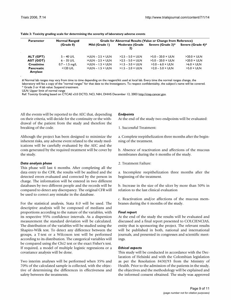
Evaluation and management of adverse eventsDuring the treatment and the follow-up visits, the patientswill be asked about adverse events. Each adverse event willbe classified by the physician as serious or non-seri-
ous(Table 2 and Table 3). A serious adverse event shouldmeet one or more of the following criteria:
1. Death
2. Life-threatening (i.e., immediate risk of death)
3. In-patient hospitalization or prolongation of existinghospitalization
4. Persistent or significant disability/incapacity
The presence of a serious adverse event that puts thepatient's life at risk and/or requires immediate medical orsurgical procedure will call for the discontinuation of thetreatment and the initiation of the pertinent medical man-agement of the patients. The investigator will notify theAdverse Event Committee (AEC) of the FCV of any seriousadverse event within 24 hours of learning about it.
A non-serious adverse event will be classified as follows:
1. Mild: The patients are aware of their symptoms and/orsigns, but those are tolerable. They do not require medicalintervention or specific treatment.
2. Moderate: Patients present troubles that interfere withtheir daily activities. They require medical intervention orspecific treatment.
3. Severe: The patients are unable to work or to attendtheir daily activities. They require medical intervention orspecific treatment.
The possible relationship between the adverse events andthe tested medication will be classified by the investigatoron the basis of his/her clinical judgment and the follow-ing definitions:
1. Definitely related: Event can be fully explained by theadministration of the tested medication.
2. Probably related: Event is most likely to be explained bythe administration of the tested medication rather thanother medications or by the patient's clinical state.
3. Possibly related: Event may be explained by the admin-istration of the tested medication or other medications orby the patient's clinical state.
4. Not related: Event is most likely to be explained by thepatient's clinical state or other medications, rather thanthe tested one.
Page 8 of 11(page number not for citation purposes)
Trials 2006, 7:14 http://www.trialsjournal.com/content/7/1/14
All the events will be reported to the AEC that, dependingon their criteria, will decide for the continuity or the with-drawal of the patient from the study and therefore thebreaking of the code.
Although the project has been designed to minimize theinherent risks, any adverse event related to the study med-ications will be carefully evaluated by the AEC and thecosts generated by the required treatment will be cover bythe study.
Data analysis phaseThis phase will last 6 months. After completing all thedata entry to the CFR, the results will be audited and thedetected errors evaluated and corrected by the person incharge. The information will be entered in two differentdatabases by two different people and the records will becompared to detect any discrepancy. The original CFR willbe used to correct any mistake in the database.
For the statistical analysis, Stata 8.0 will be used. Thedescriptive analysis will be composed of medians andproportions according to the nature of the variables, withits respective 95% confidence intervals. As a dispersionmeasurement the standard deviation will be calculated.The distribution of the variables will be studied using theShapiro-Wilk test. To detect any difference between thegroups, a T-test or a Wilcoxon test will be performedaccording to its distribution. The categorical variables willbe compared using the Chi2 test or the exact Fisher's test.If required, a model of multiple logistic regressions or acovariance analysis will be done.
Two interim analyses will be performed when 35% and70% of the calculated sample is collected, with the objec-tive of determining the differences in effectiveness andsafety between the treatments.
EndpointsAt the end of the study two endpoints will be evaluated:
1. Successful Treatment:
a. Complete reepithelization three months after the begin-ning of the treatment.
b. Absence of reactivation and affections of the mucousmembranes during the 6 months of the study.
2. Treatment Failure:
a. Incomplete reepithelization three months after thebeginning of the treatment.
b. Increase in the size of the ulcer by more than 50% inrelation to the last clinical evaluation
c. Reactivation and/or affections of the mucous mem-branes during the 6 months of the study.
Final reportAt the end of the study the results will be evaluated anddiscussed and a final report presented to COLCIENCIAS,entity that is sponsoring the project. The relevant resultswill be published in both, national and internationaljournals, and presented in congresses and scientific meet-ings.
Ethical aspectsThis study will be conducted in accordance with the Dec-laration of Helsinki and with the Colombian legislationas per the Resolution 8430/93 from the Ministry ofHealth. Prior to the admission of the patients in the study,the objectives and the methodology will be explained andthe informed consent obtained. The study was approved
Table 3: Toxicity grading scale for determining the severity of laboratory adverse events
Parameter Normal Range# Grade for Abnormal Results (Value or Change from Reference)(Grade 0) Mild (Grade 1) Moderate (Grade
2)Severe (Grade 3)* Severe (Grade 4)*
ALT (GPT) 5 – 40 U/L >ULN – 2.5 × ULN >2.5 – 5.0 × ULN >5.0 – 20.0 × ULN >20.0 × ULNAST (GOT) 6 – 35 U/L >ULN – 2.5 × ULN >2.5 – 5.0 × ULN >5.0 – 20.0 × ULN >20.0 × ULNCreatinine 0.7 – 1.5 mg/L >ULN – 1.5 × ULN >1.5 – 3.0 × ULN >3.0 – 6.0 × ULN >6.0 × ULNPancreatic Amylase
<120 U/L >ULN – 1.5 × ULN >1.5 – 2.0 × ULN >2.0 – 5.0 × ULN >5.0 × ULN
# Normal lab ranges may vary from time to time depending on the reagent/kit used at local lab. Every time the normal ranges change, the laboratory will fax a copy of the "normal ranges" for that date to the Investigators. To respect confidentiality, the subject's name will be covered.* Grade 3 or 4 lab value: Suspend treatment.ULN: Upper limit of normal rangeRef: Toxicity Grading based on CTCAE v3.0 DCTD, NCI, NIH, DHHS December 12, 2003 http://ctep.cancer.gov.
Page 9 of 11(page number not for citation purposes)

Trials 2006, 7:14 http://www.trialsjournal.com/content/7/1/14
by the Research Ethic Committee of the CardiovascularFoundation of Colombia (Act# 105/January 28/2005).The right to confidentiality of the patients will be main-tained in all the phases of the study.
Competing interestsThe author(s) declare that they have no competing inter-ests.
Authors' contributionsPLJ has made substantial contributions to the conceptionand design of the study, will be responsible for the overalladministration and direction of the project, the analysisand interpretation of data and will give the final approvalof the version to be published. DS, who participated in thedesign of the project, will be responsible for the overalladministration and direction of the study, and the pro-duction and analysis of the NO releasing electrospunnanofiber patches. IDV has made substantial contribu-tions to the conception and design of the study and to thedrafting of the manuscript. He will also be responsible foranalyzing and interpreting the final data.
ML was in charge of the development of the NOPs todetermine their optimum dosage. He was also involved inthe drafting of the manuscript and will participate in theanalysis and interpretation of data. HM and HD wereinvolved in the drafting of the manuscript and the plan-ning of the clinical trial. He will also be responsible foroverseeing its logistics and will interpret the final data.GM will be responsible for the parasitological analyses,biopsies, and the processing of the patients' samples. Hewill also participate in the acquisition, analysis and inter-pretation of data. FS-S, contributed to the conception anddesign of the study, performed sample size calculationand randomization and will analyze the data obtainedfrom the clinical trial. SYS made substantial contributionsto the conception and design of the study and wasinvolved in drafting the manuscript. LCR contributed tothe design of the study, was involved in drafting the man-uscript, will be responsible for the recruitment and fol-low-up of the patients enrolled, and will also participatein the analysis and interpretation of data. CFR-C contrib-uted to the design of the study, was involved in draftingthe manuscript, will be responsible for the recruitmentand follow-up of the patients enrolled, and will also par-ticipate in the analysis and interpretation of data. AB par-ticipated in the design of the study, will contribute to theacquisition of data, and will be responsible for overseeingthe application of the protocol by monitoring the clinicaltrial to ensure compliance with the cGCP (current GoodClinical Practices). All authors read and approved thefinal manuscript.
AcknowledgementsThis study is supported by grants from the Colombian Institute for the development of science and technology "Francisco Jose Caldas" (COL-CIENCIAS), Grant No. 6566-04-18090 and funds from the Research Insti-tute from the Cardiovascular Foundation of Colombia (FCV).
We would like to express our gratitude to Jean Noel Guillemot for review-ing the English style.
References1. S S, R S: Recent Advance in the diagnosis of Leishmaniasis. J
Postgrad Med 2003, 49:55-60.2. P D: Leishmaniasis: Public health aspects and control. Clin
Dermatol 1996, 14:417-413.3. Mandell: Principles and practice of infection diseases. 5th edition. Edited
by: Elsevier . Churchill Livingstone, Inc.; 2000. 4. BL H: Leishmaniasis. Lancet 1999, 354:1191-1199.5. RW A, P D, P R: Estimation of population at risk of infection
and number of cases of Leishmaniasis. Parasitology Today 1992,7:73-107.
6. Update: Cutaneous leishmaniasis in U.S. military personnel--Southwest/Central Asia, 2002-2004. MMWR Morb Mortal WklyRep 2004, 53:264-265.
7. CR D, R R, D CL, D F, R B, N R: The epidemiology and controlof Leishmaniasis in Andean countries. Cad Saude Publica 2000,16:925-950.
8. LA A, J U, D S, F R, ID V: Presence of American cutaneousLeishmaniasis vectors surrounding the city of Medellín,Colombia. Mem Inst Oswaldo Cruz 2002, 97:641-642.
9. NG S, I S, AF H, C S, L V, C O: Epidemiologic, genetic, and clin-ical associations among phenotypically distinct populationsof Leishmania (Viannia) in Colombia. Am J Trop Med Hyg 1998,59:86-94.
10. W M, K M: Cutaneous Leishmaniasis: Recognition and treat-ment. Am Fam Physician 2004, 69:1455-1460.
11. L R, E H, S F: Leishmaniasis. BMJ 2000, 321:801-804.12. G M, C D: Leishmania panamensis transmission in the domes-
tic environment: the results of a prospective epidemiologicalsurvey in Santander, Colombia. Biomedica 2006, In press:.
13. NC H: Cutaneous Leishmaniasis: an overview. J Postgrad Med2003, 49:50-54.
14. NC H, MJ T, JA H: Cutaneous Leishmaniasis in British troopsfrom Belize. Br J Dermatol 1993, 128:63-68.
15. BL H, BA A, TR N: The natural history of cutaneous Leishma-niasis in Guatemala. J Infect Dis 1992, 165:518-527.
16. LE O, MC C, MT O: Mucosal Leishmaniasis due to Leishmania(viannia)panamensis in Colombia: clinical characteristics.Am J Trop Med Hyg 1998, 59:49-52.
17. R P, LE O, LF G, MT O: Treatment failure in children in a ran-domized clinical trial with 10 and 20 days of meglumine anti-monate for cutaneous Leishmaniasis due toLeishmaniaviannia species. Am J Trop Med Hyg 2001, 64:187-193.
18. BL H, JD B: Recommendations for treating Leishmaniasis withsodium stibogluconate (Pentostam) and review of pertinentclinical studies. Am J Trop Med Hyg 1992, 46:296-306.
19. J B, P D, E S, B B, C H: Treatment of cutaneous Leishmaniasisamong travelers. J Antimicrob Chemother 2004, 53:158-166.
20. J S, P F, R H, J B: Topical paromomycin/methylbenzethoniumchloride plus parenteral meglumine antimotiate as treat-ment for american cutaneos leishmaniasis: ControlledStudy. Clin Infect Disease 1998, 26:56-58.
21. EP H, SP A, DL M, JA P, ID V: Lack of efficacy of mefloquine inthe treatment of new world cutaneous leishmaniasis incolombia. Am J Trop Med Hyg 1998, 59:889-892.
22. JD B: Leishmaniasis: clinical, diagnostic, and chemotherapeu-tic developments in the last 10 years. Clin Infect Dis 1997,24:684-703.
23. MC F, MJ GM, F A, BR V, A C, AI A: Tratamiento de cortaduración de la Leishmaniasis visceral con anfotericina Bliposómica en pacientes inmunocompetentes. An Pediatr (Barc)2003, 59:535-540.
24. RA S, J M, I M, J W, D N: Out-patient parenteral antimicrobialtherapy-a viable option for the management of cutaneousLeishmaniasis. QJM 1999, 92:659-667.
Page 10 of 11(page number not for citation purposes)
http://www.ncbi.nlm.nih.gov/entrez/query.fcgi?cmd=Retrieve&db=PubMed&dopt=Abstract&list_uids=8889319
http://www.ncbi.nlm.nih.gov/entrez/query.fcgi?cmd=Retrieve&db=PubMed&dopt=Abstract&list_uids=9684634
http://www.ncbi.nlm.nih.gov/entrez/query.fcgi?cmd=Retrieve&db=PubMed&dopt=Abstract&list_uids=9684634
http://www.ncbi.nlm.nih.gov/entrez/query.fcgi?cmd=Retrieve&db=PubMed&dopt=Abstract&list_uids=9684634
http://www.ncbi.nlm.nih.gov/entrez/query.fcgi?cmd=Retrieve&db=PubMed&dopt=Abstract&list_uids=8381299
http://www.ncbi.nlm.nih.gov/entrez/query.fcgi?cmd=Retrieve&db=PubMed&dopt=Abstract&list_uids=8381299
http://www.ncbi.nlm.nih.gov/entrez/query.fcgi?cmd=Retrieve&db=PubMed&dopt=Abstract&list_uids=1538157
http://www.ncbi.nlm.nih.gov/entrez/query.fcgi?cmd=Retrieve&db=PubMed&dopt=Abstract&list_uids=1538157
http://www.ncbi.nlm.nih.gov/entrez/query.fcgi?cmd=Retrieve&db=PubMed&dopt=Abstract&list_uids=9684627
http://www.ncbi.nlm.nih.gov/entrez/query.fcgi?cmd=Retrieve&db=PubMed&dopt=Abstract&list_uids=9684627
http://www.ncbi.nlm.nih.gov/entrez/query.fcgi?cmd=Retrieve&db=PubMed&dopt=Abstract&list_uids=1313656
http://www.ncbi.nlm.nih.gov/entrez/query.fcgi?cmd=Retrieve&db=PubMed&dopt=Abstract&list_uids=1313656
http://www.ncbi.nlm.nih.gov/entrez/query.fcgi?cmd=Retrieve&db=PubMed&dopt=Abstract&list_uids=1313656
http://www.ncbi.nlm.nih.gov/entrez/query.fcgi?cmd=Retrieve&db=PubMed&dopt=Abstract&list_uids=9886195
http://www.ncbi.nlm.nih.gov/entrez/query.fcgi?cmd=Retrieve&db=PubMed&dopt=Abstract&list_uids=9886195
http://www.ncbi.nlm.nih.gov/entrez/query.fcgi?cmd=Retrieve&db=PubMed&dopt=Abstract&list_uids=9886195
http://www.ncbi.nlm.nih.gov/entrez/query.fcgi?cmd=Retrieve&db=PubMed&dopt=Abstract&list_uids=9145744

Trials 2006, 7:14 http://www.trialsjournal.com/content/7/1/14
Publish with BioMed Central and every scientist can read your work free of charge
"BioMed Central will be the most significant development for disseminating the results of biomedical research in our lifetime."
Sir Paul Nurse, Cancer Research UK
Your research papers will be:
available free of charge to the entire biomedical community
peer reviewed and published immediately upon acceptance
cited in PubMed and archived on PubMed Central
yours — you keep the copyright
Submit your manuscript here:http://www.biomedcentral.com/info/publishing_adv.asp
BioMedcentral
25. J S, M G, J B, P O: Limited efficacy of injectable aminosidine assingle-agent therapy for Colombian cutaneous Leishmania-sis. Trans R Soc Trop Med Hyg 1994, 88:695-698.
26. J S, BA A, J T, N R, JC V, A D, P G, M A, JD B, K J, J E, H S: Miltefosinefor new world cutaneous leishmaniasis. Clin Infect Dis 2004,38:1266-1272.
27. J S, J T, P G, M A, RS N, JR P, JD B, CK E, M G: Treatment of cuta-neous Leishmaniasis with a topical antileishmanial drug(WR279396): phase 2 pilot study. J Trop Med Hyg 2002,66:147-152.
28. A A, T J, JA W, RL G, M N, P O: A randomized, placebo-control-led trial of a two-week regimen of aminosidine (paromomy-cin) ointment for treatment of cutaneous Leishmaniasis inIran. Am J Trop Med Hyg 1995, 53:648-651.
29. MA M, SE MG, KR G, M C, SL N, KA A, CJ B, N Y, BE B, ME W:Inducible resistance to oxidant stress in the protozoan Leish-maniachagasi. J Biol Chem 2000, 275:33883-33889.
30. M K: Regulator and effector functions of T-cell subsets inhuman Leishmania infections. APMIS 1997, 68:1-33.
31. A D, H S, M R, WM Y, C B: Requirement for type 2 NO synthasefor IL-12 signaling in innate immunity. Science 1999,284:951-955.
32. NL D, M F, E F, R R, IB M, FJ T: Nitric oxide and cellular immu-nity in experimental cutaneous Leishmaniasis. Clin Exp Derma-tol 2003, 28:288-263.
33. K G, TL G, ML MC, MA M, S J, BE B, ME W: Oxidative Responsesof Human and Murine Macrophages During Phagocytosis ofLeishmania chagasi. J of Immunol 2001, 167:893-901.
34. M Q, I B, N D, M R, C B: Expression of inducible nitric oxide syn-thase in skin lesions of patients with american cutaneousLeishmaniasis. Infect Immun 2002, 70:4638-4642.
35. FY L, S M, C P, RM P, S M: Macrophage killing of Leishmania par-asite in vivo is mediated by nitric oxide from L-arginine. JImmunol 1990, 144:4794-4797.
36. Moncada S, Higgs A: The L-arginine-nitric oxide pathway. NEngl J Med 1993, 329:2002-2012.
37. I V, PA B, V RM, M A, O S, P D, D M, MD M: Interleukin-10 andinterleukin-4 inhibit intracellular killing of Leishmania infan-tum and Leishmania major by human macrophages bydecreasing nitric oxide generation. Eur J Immunol 1997,27:860-865.
38. L P, CA OD, FY L: Glycoinositolphospholipids of Leishmaniamajor inhibit nitric oxide synthesis and reduce leishmani-cidal activity in murine macrophages. Eur J Immunol 1995,25:745-750.
39. P LJ, C R, J R, E T, R SI, JV E, S M: Treatment of cutaneous Leish-maniasis with nitric-oxide donor. Lancet 1998, 351:1176-1177.
40. RN D, V Y, SL C, P K, N B: A topical nitric oxide-generatingtherapy for cutaneous Leishmaniasis. Trans R Soc Trop Med Hyg2000, 94:319-322.
41. J D, DH R: Electrospinning process and applications of elec-trospun fibers. J of Electrostatics 1995, 35:151-160.
42. Kenawy ER, J L, JR W, GL B, JA M, DG S, GE W: Electrospinningof poly(ethylene-co-vinyl alcohol) fibers. Biomaterials 2003,24:907-913.
43. Kenawy ER, GL B, K M, J L, DG S, EH S, GE W: Release of tetracy-cline hydrochloride from electrospun poly(ethylene-co-viny-lacetate), poly(lactic acid), and a blend. J Control Release 2002,81:57-64.
44. LM H, W K, DH R, DJ S: Production of superabsorbent electro-spun nanofibers. Polymeric Materials Science and Engineering 2004,90:508.
45. D S: Stabilization and ionic triggering of nitric oxide release.PCT Int Appl WO 2004-US23867 2004.
46. Vega-Lopez F: Diagnosis of cutaneous leishmaniasis. Curr OpinInfect Dis 2003, 16:97-101.
47. Foreyt WJ: Diagnostic parasitology. Vet Clin North Am Small AnimPract 1989, 19:979-1000.
48. Kar K: Serodiagnosis of leishmaniasis. Crit Rev Microbiol 1995,21:123-152.
49. Grimaldi G, McMahon-Pratt D: Monoclonal antibodies for theidentification of New World Leishmania species. Mem InstOswaldo Cruz 1996, 91:37-42.
Page 11 of 11(page number not for citation purposes)
http://www.ncbi.nlm.nih.gov/entrez/query.fcgi?cmd=Retrieve&db=PubMed&dopt=Abstract&list_uids=7886777
http://www.ncbi.nlm.nih.gov/entrez/query.fcgi?cmd=Retrieve&db=PubMed&dopt=Abstract&list_uids=7886777
http://www.ncbi.nlm.nih.gov/entrez/query.fcgi?cmd=Retrieve&db=PubMed&dopt=Abstract&list_uids=7886777
http://www.ncbi.nlm.nih.gov/entrez/query.fcgi?cmd=Retrieve&db=PubMed&dopt=Abstract&list_uids=8561269
http://www.ncbi.nlm.nih.gov/entrez/query.fcgi?cmd=Retrieve&db=PubMed&dopt=Abstract&list_uids=8561269
http://www.ncbi.nlm.nih.gov/entrez/query.fcgi?cmd=Retrieve&db=PubMed&dopt=Abstract&list_uids=8561269
http://www.ncbi.nlm.nih.gov/entrez/query.fcgi?cmd=Retrieve&db=PubMed&dopt=Abstract&list_uids=2351828
http://www.ncbi.nlm.nih.gov/entrez/query.fcgi?cmd=Retrieve&db=PubMed&dopt=Abstract&list_uids=2351828
http://www.ncbi.nlm.nih.gov/entrez/query.fcgi?cmd=Retrieve&db=PubMed&dopt=Abstract&list_uids=7504210
http://www.ncbi.nlm.nih.gov/entrez/query.fcgi?cmd=Retrieve&db=PubMed&dopt=Abstract&list_uids=9130636
http://www.ncbi.nlm.nih.gov/entrez/query.fcgi?cmd=Retrieve&db=PubMed&dopt=Abstract&list_uids=9130636
http://www.ncbi.nlm.nih.gov/entrez/query.fcgi?cmd=Retrieve&db=PubMed&dopt=Abstract&list_uids=9130636
http://www.ncbi.nlm.nih.gov/entrez/query.fcgi?cmd=Retrieve&db=PubMed&dopt=Abstract&list_uids=7705404
http://www.ncbi.nlm.nih.gov/entrez/query.fcgi?cmd=Retrieve&db=PubMed&dopt=Abstract&list_uids=7705404
http://www.ncbi.nlm.nih.gov/entrez/query.fcgi?cmd=Retrieve&db=PubMed&dopt=Abstract&list_uids=7705404
http://www.ncbi.nlm.nih.gov/entrez/query.fcgi?cmd=Retrieve&db=PubMed&dopt=Abstract&list_uids=9643692
http://www.ncbi.nlm.nih.gov/entrez/query.fcgi?cmd=Retrieve&db=PubMed&dopt=Abstract&list_uids=9643692
http://www.ncbi.nlm.nih.gov/entrez/query.fcgi?cmd=Retrieve&db=PubMed&dopt=Abstract&list_uids=2678716
http://www.ncbi.nlm.nih.gov/entrez/query.fcgi?cmd=Retrieve&db=PubMed&dopt=Abstract&list_uids=7639932
http://www.ncbi.nlm.nih.gov/entrez/query.fcgi?cmd=Retrieve&db=PubMed&dopt=Abstract&list_uids=8734946