FIRST REPORT ON OTOTOXICITY OF MEGLUMINE ANTIMONIATE
96
I ISSN 0036-4665 ISSN 1678-9946 on line Established: 1959. The year 2014 is the 56 th anniversary of continuous publication UNIVERSIDADE DE SÃO PAULO - BRAZIL FACULDADE DE MEDICINA Instituto de Medicina Tropical de São Paulo Director: Prof. Dr. Paulo C. Cotrim EDITOR-IN-CHIEF EMERITUS EDITORS Prof. Dr. Thales F. de Brito Prof. Dr. Luis Rey (Founding Editor) Associate Editors: Prof. Dr. Pedro Paulo Chieffi Prof. Dr. Carlos da Silva Lacaz Prof. Dr. Thelma S. Okay EDITORIAL BOARD Alan L. de Melo (Belo Horizonte, MG) Alberto Duarte (S. Paulo, SP) Angela Restrepo M. (Medellin, Colombia) Anna Sara S. Levin (S. Paulo, SP) Antonio A. Barone (S. Paulo, SP) Antonio Carlos Nicodemo (S. Paulo, SP) Antonio Sesso (S. Paulo, SP) Antonio W. Ferreira (S. Paulo, SP) Barnett L. Cline (New Orleans, USA) Carlos F. S. Amaral (Belo Horizonte, MG) Celso Granato (S. Paulo, SP) Cesar A. Cuba Cuba (Brasília, DF) César Naquira V. (Lima, Peru) Clarisse M. Machado (S. Paulo, SP) Claudio S. Pannuti (S. Paulo, SP) Dalton L. F. Alves (Belo Horizonte, MG) Eridan Coutinho (Recife, PE) Ernesto Hofer (Rio de Janeiro, RJ) Euclides A. Castilho (S. Paulo, SP) Eufrosina S. Umezawa (S. Paulo, SP) Expedito J. A. Luna (S. Paulo, SP) Fan Hui Wen (S. Paulo, SP) Fernando A. Corrêa (S. Paulo, SP) Fernando Montero-Gei (San José, Costa Rica) Flair J. Carrilho (S. Paulo, SP) Gil Benard (S. Paulo, SP) Gioconda San-Blas (Caracas, Venezuela) Govinda Visvesvara (Atlanta, USA) Heitor F. Andrade Jr. (S. Paulo, SP) Hiro Goto (S. Paulo, SP) Ises A. Abrahamsohn (S. Paulo, SP) João Carlos Pinto Dias (Belo Horizonte, MG) João Renato Rebello Pinho (Sao Paulo, SP) José Eduardo Levi (S. Paulo, SP) José M. R. Zeitune (Campinas, SP) Julia Maria Costa-Cruz (Uberlândia, MG) Julio Litvoc (S. Paulo, SP) Luiz Carlos Severo (P. Alegre, RS) Luiz T. M. Figueiredo (Rib. Preto, SP) Lygia B. Iversson (S. Paulo, SP) Marcello Fabiano de Franco (S. Paulo, SP) Marcos Boulos (S. Paulo, SP) M. A. Shikanai-Yasuda (S. Paulo, SP) Maria I. S. Duarte (S. Paulo, SP) Maria L. Higuchi (S. Paulo, SP) Mario Mariano (S. Paulo, SP) Mirian N. Sotto (S. Paulo, SP) Moisés Goldbaum (S. Paulo, SP) Moysés Mincis (S. Paulo, SP) Moysés Sadigursky (Salvador, BA) Myrthes T. Barros (S. Paulo, SP) Nilma Cintra Leal (Recife, PE) Paulo C. Cotrim (São Paulo, SP) Paulo M. Z. Coelho (Belo Horizonte, MG) Regina Abdulkader (S. Paulo, SP) Ricardo Negroni (B. Aires, Argentina) Robert H. Gilman (Baltimore, USA) Roberto Martinez (Rib. Preto, SP) Ronaldo Cesar B. Gryschek (S. Paulo, SP) Semíramis Guimarães F. Viana (Botucatu, SP) Silvio Alencar Marques (Botucatu, SP) Tsutomu Takeuchi (Tokyo, Japan) Venâncio A. F. Alves (S. Paulo, SP) Vicente Amato Neto (S. Paulo, SP) Zilton A. Andrade (Salvador, BA) Executive Board - Librarians: Maria do Carmo Berthe Rosa; Sonia Pedrozo Gomes; Maria Ângela de Castro Fígaro Pinca; Carlos José Quinteiro The Revista do Instituto de Medicina Tropical de São Paulo is abstracted and/or indexed in: Index Medicus, Biological Abstracts, EMBASE/Excerpta Medica, Hepatology/Rapid Literature Review, Tropical Diseases Bulletin, Referativnyi Zhurnal: All-Russian Institute of Scientific and Technical Information (VINITI), Periódica - Índice de Revistas Latinoamericanas en Ciencias, Helminthological Abstracts, Protozoological Abstracts, Review of Medical and Veterinary Mycology, PubMed, PubMed Central (PMC), UnCover, HealthGate, OVID, LILACS, MEDLINE, New Jour, ExtraMED, Free Medical Journals, ISI (Institute for Scientific Information), BIOSIS Previews, Scopus, Science Citation Index Expanded (SciSearch), Journal Citation Reports/Science Edition, Current Contents®/Clinical Medicine and Index Copernicus. ON LINE ACCESS - http://www.imt.usp.br/portal/ - FREE PDF ACCESS TO ALL PAST ISSUES, from 1959 on (Financial support by “Alves de Queiroz Family Fund for Research). http://www.scielo.br/rimtsp - FULL TEXT, SINCE 1984. E-mail: [email protected] Reprints may be obtained from Pro Quest Inf. and Learning, 300 North Zeeb Road, Ann Arbor, Michigan 48106-1346 - USA. The Revista do Instituto de Medicina Tropical de São Paulo is supported by: Fundação de Amparo à Pesquisa do Estado de São Paulo (FAPESP), Conselho Nacional de Desenvolvimento Científico e Tecnológico (CNPq), Universidade de São Paulo and Coordenação de Aperfeiçoamento de Pessoal de Nível Superior (CAPES). This issue was financed by: CNPq Proc. 405008/2013-9. Desktop Publishing by: Hermano - e-mail: [email protected]. Phone: 55.11.5571-8937. - Printed by: Elyon Indústria Gráfica, Phone: 55.11.3783-6527. English Revision: [email protected]
Transcript of FIRST REPORT ON OTOTOXICITY OF MEGLUMINE ANTIMONIATE

I
ISSN 0036-4665ISSN 1678-9946 on line
Established: 1959.
The year 2014 is the 56th anniversary
of continuous publication
UNIVERSIDADE DE SÃO PAULO - BRAZILFACULDADE DE MEDICINA
Instituto de Medicina Tropical de São PauloDirector: Prof. Dr. Paulo C. Cotrim
EDITOR-IN-CHIEF EMERITUS EDITORS Prof. Dr. Thales F. de Brito Prof. Dr. Luis Rey (Founding Editor)Associate Editors: Prof. Dr. Pedro Paulo Chieffi Prof. Dr. Carlos da Silva Lacaz Prof. Dr. Thelma S. Okay
EDITORIAL BOARDAlan L. de Melo (Belo Horizonte, MG) Alberto Duarte (S. Paulo, SP) Angela Restrepo M. (Medellin, Colombia) Anna Sara S. Levin (S. Paulo, SP)Antonio A. Barone (S. Paulo, SP)Antonio Carlos Nicodemo (S. Paulo, SP) Antonio Sesso (S. Paulo, SP) Antonio W. Ferreira (S. Paulo, SP) Barnett L. Cline (New Orleans, USA) Carlos F. S. Amaral (Belo Horizonte, MG) Celso Granato (S. Paulo, SP) Cesar A. Cuba Cuba (Brasília, DF) César Naquira V. (Lima, Peru) Clarisse M. Machado (S. Paulo, SP) Claudio S. Pannuti (S. Paulo, SP) Dalton L. F. Alves (Belo Horizonte, MG) Eridan Coutinho (Recife, PE) Ernesto Hofer (Rio de Janeiro, RJ) Euclides A. Castilho (S. Paulo, SP)Eufrosina S. Umezawa (S. Paulo, SP) Expedito J. A. Luna (S. Paulo, SP)Fan Hui Wen (S. Paulo, SP)
Fernando A. Corrêa (S. Paulo, SP) Fernando Montero-Gei (San José, Costa Rica) Flair J. Carrilho (S. Paulo, SP)Gil Benard (S. Paulo, SP)Gioconda San-Blas (Caracas, Venezuela)Govinda Visvesvara (Atlanta, USA) Heitor F. Andrade Jr. (S. Paulo, SP) Hiro Goto (S. Paulo, SP)Ises A. Abrahamsohn (S. Paulo, SP) João Carlos Pinto Dias (Belo Horizonte, MG) João Renato Rebello Pinho (Sao Paulo, SP) José Eduardo Levi (S. Paulo, SP)José M. R. Zeitune (Campinas, SP) Julia Maria Costa-Cruz (Uberlândia, MG)Julio Litvoc (S. Paulo, SP) Luiz Carlos Severo (P. Alegre, RS) Luiz T. M. Figueiredo (Rib. Preto, SP) Lygia B. Iversson (S. Paulo, SP) Marcello Fabiano de Franco (S. Paulo, SP)Marcos Boulos (S. Paulo, SP)M. A. Shikanai-Yasuda (S. Paulo, SP)Maria I. S. Duarte (S. Paulo, SP)
Maria L. Higuchi (S. Paulo, SP)Mario Mariano (S. Paulo, SP)Mirian N. Sotto (S. Paulo, SP)Moisés Goldbaum (S. Paulo, SP)Moysés Mincis (S. Paulo, SP)Moysés Sadigursky (Salvador, BA)Myrthes T. Barros (S. Paulo, SP)Nilma Cintra Leal (Recife, PE)Paulo C. Cotrim (São Paulo, SP)Paulo M. Z. Coelho (Belo Horizonte, MG)Regina Abdulkader (S. Paulo, SP)Ricardo Negroni (B. Aires, Argentina)Robert H. Gilman (Baltimore, USA)Roberto Martinez (Rib. Preto, SP)Ronaldo Cesar B. Gryschek (S. Paulo, SP)Semíramis Guimarães F. Viana (Botucatu, SP)Silvio Alencar Marques (Botucatu, SP)Tsutomu Takeuchi (Tokyo, Japan)Venâncio A. F. Alves (S. Paulo, SP)Vicente Amato Neto (S. Paulo, SP)Zilton A. Andrade (Salvador, BA)
Executive Board - Librarians: Maria do Carmo Berthe Rosa; Sonia Pedrozo Gomes; Maria Ângela de Castro Fígaro Pinca; Carlos José Quinteiro
The Revista do Instituto de Medicina Tropical de São Paulo is abstracted and/or indexed in: Index Medicus, Biological Abstracts, EMBASE/Excerpta Medica, Hepatology/Rapid Literature Review, Tropical Diseases Bulletin, Referativnyi Zhurnal: All-Russian Institute of Scientific and Technical Information (VINITI), Periódica - Índice de Revistas Latinoamericanas en Ciencias, Helminthological Abstracts, Protozoological Abstracts, Review of Medical and Veterinary Mycology, PubMed, PubMed Central (PMC), UnCover, HealthGate, OVID, LILACS, MEDLINE, New Jour, ExtraMED, Free Medical Journals, ISI (Institute for Scientific Information), BIOSIS Previews, Scopus, Science Citation Index Expanded (SciSearch), Journal Citation Reports/Science Edition, Current Contents®/Clinical Medicine and Index Copernicus.
ON LINE ACCESS - http://www.imt.usp.br/portal/ - FREE PDF ACCESS TO ALL PAST ISSUES, from 1959 on (Financial support by “Alves de Queiroz Family Fund for Research).
http://www.scielo.br/rimtsp - FULL TEXT, SINCE 1984. E-mail: [email protected]
Reprints may be obtained from Pro Quest Inf. and Learning, 300 North Zeeb Road, Ann Arbor, Michigan 48106-1346 - USA.
The Revista do Instituto de Medicina Tropical de São Paulo is supported by: Fundação de Amparo à Pesquisa do Estado de São Paulo (FAPESP), Conselho Nacional de Desenvolvimento Científico e Tecnológico (CNPq), Universidade de São Paulo and Coordenação de Aperfeiçoamento de Pessoal de Nível Superior (CAPES).
This issue was financed by: CNPq Proc. 405008/2013-9.
Desktop Publishing by: Hermano - e-mail: [email protected]. Phone: 55.11.5571-8937. - Printed by: Elyon Indústria Gráfica, Phone: 55.11.3783-6527. English Revision: [email protected]

II
The purpose of the “Revista do Instituto de Medicina Tropical de São Paulo” (Journal of the São Paulo Institute of Tropical Medicine) is to publish the results of researches which contri-bute significantly to knowledge of all transmissible diseases.
REVISTA DO INSTITUTO DE MEDICINA TROPICAL DE SÃO PAULO(JOURNAL OF THE S. PAULO INSTITUTE OF TROPICAL MEDICINE).
São Paulo, SP-Brasil, 1959 -v. ilust. 28 cm
1959-2013, 1-551973-2002 (supl. 1-12)2003 (supl. 13 - on-line only)2005-2012 (supl. 14-18)2014, 56 (1-5)
ISSN 0036-4665ISSN 1678-9946 on line

III
Rev. Inst. Med. Trop. Sao Paulo Vol. 56 No. 5 P. 369-460 September-October, 2014
ISSN 0036-4665ISSN 1678-9946 on line
ADDRESSINSTITUTO DE MEDICINA TROPICAL DE SÃO PAULO
Av. Dr. Enéas de Carvalho Aguiar, 47005403-000 São Paulo, SP - Brazil
Phone/Fax: 55.11.3062.2174; 55.11.3061-7005e-mail: [email protected]
SUBSCRIPTIONSFOREIGN COUNTRIESOne year (six issues) ........ U$ 200.00Single issue ...................... U$ 50.00
CONTENTS
ENTOMOLOGY369 Laboratory evaluation of the development of Aedes aegypti in two
seasons: influence of different places and different densities T.F. LOPES, M.M. HOLCMAN, G.L. BARBOSA, M.F. DOMINGOS & R.M.O.V. BARREIROS
LEISHMANIASIS375 Montenegro skin test and age of skin lesion as predictors of treatment
failure in cutaneous leishmaniasis L.F. ANTONIO, A. FAGUNDES, R.V.C. OLIVEIRA, P.G. PINTO, S.J. BEDOYA-PACHECO, É.C.F. VASCONCELLOS, M.C. VALETE-ROSALINO, M.R. LYRA, S.R.L. PASSOS, M.I.F. PIMENTEL & A.O. SCHUBACH
381 South American collaboration in scientific publications on leish-maniasis: bibliometric analysis in SCOPUS (2000-2011) C. HUAMANÍ, F. ROMANÍ, G. GONZÁLEZ-ALCAIDE, M.O. MEJIA, J.M. RAMOS, M. ESPINOZA & C. CABEZAS
391 Detection of Leishmania (Viannia) in Nyssomyia neivai and Nys-somyia whitmani by Multiplex Polymerase Chain Reaction, in Southern Brazil H.C. NEITZKE-ABREU, K.R. REINHOLD-CASTRO, M.S. VENAZZI, R.B.L. SCODRO, A.C. DIAS, T.G.V. SILVEIRA, U. TEODORO & M.V.C. LONARDONI
BACTERIOLOGY397 Differentiation between Nocardia spp. and Mycobacterium spp.:
critical aspects for bacteriological diagnosis E.C.M. MURICY, R.A. LEMES, S. BOMBARDA, L. FERRAZOLI & E. CHIMARA
MALARIA403 Anopheles (Kerteszia) cruzii (Diptera: Culicidae) in peridomicili-
ary area during asymptomatic malaria transmission in the Atlantic Forest: molecular identification of blood-meal sources indicates humans as primary intermediate hosts K. KIRCHGATTER, R.M. TUBAKI, R.S. MALAFRONTE, I.C. ALVES, G.F.M.C. LIMA, L.O. GUIMARÃES, R.A. ZAMPAULO & G. WUNDERLICH
LEPTOSPIROSIS411 High sensitive PCR method for detection of pathogenic Leptospira
spp. in paraffin-embedded tissues A.A. NODA, I. RODRÍGUEZ, Y. RODRÍGUEZ, A. GOVÍN, C. FERNÁNDEZ & A.M. OBREGÓN
YELLOW FEVER417 Yellow fever prevention strategies awareness among HIV-infected
patients in Sao Paulo, Brazil V.I. AVELINO-SILVA, H.S. FRANCELINO & E.G. KALLÁS
MOLLUSCICIDES421 Effects of single, binary and tertiary combinations with Jatropha
gossypifolia and other plant-derived molluscicides on reproduction and survival of the snail Lymnaea acuminata R.P. YADAV & A. SINGH
Impact Factor: 0.9075-year Impact Factor: 1.213
MICROBIOLOGY427 Detection of virulence genes in environmental strains of Vibrio
cholerae from estuaries in Northeastern Brazil F.G.R. MENEZES, S.S. NEVES, O.V. SOUSA, C.M.V.M. VILA-NOVA, R. MAGGIONI, G.N.D. THEOPHILO, E. HOFER & R.H.S.F. VIEIRA
TOXOPLASMOSIS433 Toxoplasmosis-related knowledge among pregnant and postpartum
women attended in public health units in Niterói, Rio de Janeiro, Brazil P.R. MILLAR, F.L. MOURA, O.M.P. BASTOS, D.P.B.G. MATTOS, A.B.M. FONSECA, A.P. SUDRÉ, D. LELES & M.R.R. AMENDOEIRA
CASE REPORT439 First report on ototoxicity of meglumine antimoniate
C.M. VALETE-ROSALINO, M.H. ARAUJO-MELO, D.C.O. BEZERRA, R.O. BARCELOS, V. MELO-FERREIRA, T.S.S. TORRACA, A.C.C. MARTINS, J.S. MOREIRA, M.C.M. VARGAS, F.P.B. BRAGA, M.M. SALGUEIRO, M.N. SAHEKI & A.O. SCHUBACH
443 Interferon beta-1a treatment in HTLV-1-associated myelopathy/tropical spastic paraparesis: a case report G.M.C. VIANA, M.A.C.N. SILVA, V.L. SOUZA, N.B.S. LOPES, D.L.F. SILVA & M.D.S.B. NASCIMENTO
BRIEF COMMUNICATION447 False-negative dengue cases in Roraima, Brazil: an approach regar-
ding the high number of negative results by NS1 Ag kits P.O.A. ACOSTA, F. GRANJA, C.A. MENESES, I.A.S. NASCIMENTO, D.D. SOUSA, W.P. LIMA JÚNIOR & F.G. NAVECA
451 Prevalence of Entamoeba histolytica/Entamoeba dispar in the city of Campina Grande, Northeastern Brazil M.T.N. SILVA, J.V. SANTANA, G. BRAGAGNOLI, A.M.N. MARINHO & E. MALAGUEÑO
455 Prevalence of Calodium hepaticum (syn. Capillaria hepatica) in Rattus norvegicus in the urban area of Rio de Janeiro, Brazil R.O. SIMÕES, J.L. LUQUE, M.J. FARO, E. MOTTA & A. MALDONADO JR.
LETTER TO THE EDITOR458 Analogies in medicine: slapped cheek appearance
J.S. ANDRADE-FILHO.
CORRESPONDENCE AND AUTHORS REPLY459 Trombocytopenic purpura and dengue
B. JOOB & V. WIWANITKIT Authors reply
F.F. AMÂNCIO, M.A.PEREIRA, F.C.M. IANI, L. D’ANUNCIAÇÃO, J.L.C. ALMEIDA, J.A.S. SOARES, M.L. FERRAZ, T.C. VALE, J.R.LAMBERTUCCI & M. CARNEIRO

IV
ENDEREÇOINSTITUTO DE MEDICINA TROPICAL DE SÃO PAULO
Av. Dr. Enéas de Carvalho Aguiar, 47005403-000 São Paulo, SP - Brasil
Fone/Fax: 55.11.3062.2174; 55.11.3061-7005e-mail: [email protected]
Rev. Inst. Med. Trop. Sao Paulo Vol. 56 No. 5 P. 369-460 Setembro-Outubro, 2014
ISSN 0036-4665ISSN 1678-9946 on line
ENTOMOLOGIA369 A avaliação do desenvolvimento do Aedes aegypti em duas estações
do ano: influência de diferentes locais e densidades T.F. LOPES, M.M. HOLCMAN, G.L. BARBOSA, M.F. DOMINGOS & R.M.O.V. BARREIROS
LEISHMANIOSE375 Intensidade da intradermorreação de Montenegro e tempo de evo-
lução da lesão como preditores de falha na resposta terapêutica da leishmaniose cutânea L.F. ANTONIO, A. FAGUNDES, R.V.C. OLIVEIRA, P.G. PINTO, S.J. BEDOYA-PACHECO, É.C.F. VASCONCELLOS, M.C. VALETE-ROSALINO, M.R. LYRA, S.R.L. PASSOS, M.I.F. PIMENTEL & A.O. SCHUBACH
381 Colaboración Sudamericana en publicaciones científicas sobre leishmaniasis: análisis bibliométrico en SCOPUS (2000-2011) C. HUAMANÍ, F. ROMANÍ, G. GONZÁLEZ-ALCAIDE, M.O. MEJIA, J.M. RAMOS, M. ESPINOZA & C. CABEZAS
391 Detecção de Leishmania (Viannia) em Nyssomyia neivai e Nys-somyia whitmani por Multiplex Reação em Cadeia da Polimerase, no sul do Brasil H.C. NEITZKE-ABREU, K.R. REINHOLD-CASTRO, M.S. VENAZZI, R.B.L. SCODRO, A.C. DIAS, T.G.V. SILVEIRA, U. TEODORO & M.V.C. LONARDONI
BACTERIOLOGIA397 Diferenciação de Nocardia spp. e Mycobacterium spp.: aspectos
críticos para o diagnóstico bacteriológico E.C.M. MURICY, R.A. LEMES, S. BOMBARDA, L. FERRAZOLI & E. CHIMARA
MALARIA403 Anopheles (Kerteszia) cruzii (Diptera: Culicidae) em área peri-
domiciliar durante transmissão de malária assintomática na Mata Atlântica: identificação molecular das fontes de repasto sanguíneo indica humanos como hospedeiros intermediários primários K. KIRCHGATTER, R.M. TUBAKI, R.S. MALAFRONTE, I.C. ALVES, G.F.M.C. LIMA, L.O. GUIMARÃES, R.A. ZAMPAULO & G. WUNDERLICH
LEPTOSPIROSE411 PCR altamente sensible para la detección de Leptospira spp. pató-
genas en tejidos embebidos en parafina A.A. NODA, I. RODRÍGUEZ, Y. RODRÍGUEZ, A. GOVÍN, C. FERNÁNDEZ & A.M. OBREGÓN
FEBRE AMARELA417 Nível de conhecimento sobre estratégias de prevenção contra febre
amarela entre pessoas que vivem com HIV em São Paulo, Brasil V.I. AVELINO-SILVA, H.S. FRANCELINO & E.G. KALLÁS
MOLUSCICIDAS421 Efeitos de combinações unitárias, binárias e terciárias de Jatropha
gossypifolia e outros muluscicidas derivados de plantas na repro-dução e sobrevivência do caramujo Lymnaea acuminata R.P. YADAV & A. SINGH
Impact Factor: 0.9075-year Impact Factor: 1.213
MICROBIOLOGIA427 Detecção de genes de virulência em estirpes de Vibrio cholerae
isolados de estuários no Nordeste do Brasil F.G.R. MENEZES, S.S. NEVES, O.V. SOUSA, C.M.V.M. VILA-NOVA, R. MAGGIONI, G.N.D. THEOPHILO, E. HOFER & R.H.S.F. VIEIRA
TOXOPLASMOSE433 Conhecimento sobre toxoplasmose entre gestantes e puérperas
atendidas na rede pública de saúde do município de Niterói, Rio de Janeiro, Brasil P.R. MILLAR, F.L. MOURA, O.M.P. BASTOS, D.P.B.G. MATTOS, A.B.M. FONSECA, A.P. SUDRÉ, D. LELES & M.R.R. AMENDOEIRA.
RELATO DE CASO439 Primeiro relato de ototoxicidade pelo antimoniato de meglumina
C.M. VALETE-ROSALINO, M.H. ARAUJO-MELO, D.C.O. BEZERRA, R.O. BARCELOS, V. MELO-FERREIRA, T.S.S. TORRACA, A.C.C. MARTINS, J.S. MOREIRA, M.C.M. VARGAS, F.P.B. BRAGA, M.M. SALGUEIRO, M.N. SAHEKI & A.O. SCHUBACH
443 Tratamento com interferon beta-1a em mielopatia associada ao HTLV-1/paraparesia espástica tropical: relato de caso G.M.C. VIANA, M.A.C.N. SILVA, V.L. SOUZA, N.B.S. LOPES, D.L.F. SILVA & M.D.S.B. NASCIMENTO
COMUNICAÇÃO BREVE447 Casos de dengue falso-negativo em Roraima, Brasil: abordagem
acerca do alto número de testes Antígeno NS1 negativos P.O.A. ACOSTA, F. GRANJA, C.A. MENESES, I.A.S. NASCIMENTO, D.D. SOUSA, W.P. LIMA JÚNIOR & F.G. NAVECA
451 Prevalência de Entamoeba histolytica/Entamoeba dispar na cidade de Campina Grande, Nordeste do Brasil M.T.N. SILVA, J.V. SANTANA, G. BRAGAGNOLI, A.M.N. MARINHO & E. MALAGUEÑO
455 Prevalência de Calodium hepaticum (sin. Capillaria hepatica) em Rattus norvegicus em área urbana do Rio de Janeiro, Brasil R.O. SIMÕES, J.L. LUQUE, M.J. FARO, E. MOTTA & A. MALDONADO JR.
CARTA AO EDITOR458 Analogies in medicine: slapped cheek appearance
J.S. ANDRADE-FILHO.
CORRESPONDÊNCIA E RESPOSTA DOS AUTORES459 Trombocytopenic purpura and dengue
B. JOOB & V. WIWANITKIT Authors reply
F.F. AMÂNCIO, M.A.PEREIRA, F.C.M. IANI, L. D’ANUNCIAÇÃO, J.L.C. ALMEIDA, J.A.S. SOARES, M.L. FERRAZ, T.C. VALE, J.R.LAMBERTUCCI & M. CARNEIRO
CONTEÚDO

Rev. Inst. Med. Trop. Sao Paulo56(5):369-374, September-October, 2014doi: 10.1590/S0036-46652014000500001
Superintendência de Controle de Endemias (SUCEN), Secretaria de Estado da Saúde São Paulo. R. Paula Souza166, Luz, 01027-000 São Paulo, SP, Brazil.Correspondence to: Marcia Moreira Holcman. E-mail: [email protected]
LABORATORY EVALUATION OF THE DEVELOPMENT OF Aedes aegypti IN TWO SEASONS: INFLUENCE OF DIFFERENT PLACES AND DIFFERENT DENSITIES
Tatiana Forte LOPES, Marcia Moreira HOLCMAN, Gerson Laurindo BARBOSA, Maria de Fatima DOMINGOS & Rosa Maria Oliveira Veiga BARREIROS
SUMMARY
Aedes aegypti is an important vector in Brazil being the main vector of the dengue-fever. This paper employs survival curves to describe the time in days from larvae to adult forms of Aedes aegypti raised, individually and collectively, and compares it during winter and spring when positioned inside and outside a laboratory. The study was conducted in São Vicente, a coastal city in Southeastern Brazil. The lowest water temperature in winter and in spring was 20 ºC and the highest was 26 ºC in spring. Higher and more stable temperatures were measured in the intra compared to the peri in both seasons. Consequently, larvae positioned in the intra resulted in the lowest median time to develop in the individual and collective experiment (nine and ten days, respectively). At least 25% of the larvae positioned in the intra in the individual experiment in the spring took only seven days to reach adulthood. Sex ratios and the median time development by sex did not show significant differences. These results indicate that efforts to control Aedes aegypti must be continuous and directed mainly to prevent the intra-domiciliary sites that can be infested in a week in order to reduce the human-vector contact.
KEYWORDS: Aedes aegypti; Development time; Temperature; Survival analysis.
INTRODUCTION
Aedes (Stegomya) aegypti (L.) is one of the most important vectors in Brazil, as the main vector of urban yellow fever and dengue fever. The World Health Organization estimates that 50 to 100 million people are infected annually and 2.5 billion people are living in places at risk of acquiring dengue in the World28. In 2011, according to the Pan American Health Organization, 64% of the fatal cases of dengue in South America were from Brazil21. At the present time, dengue fever is the most important infectious disease in Brazil25.
The Aedes aegypti is usually found between latitudes 45º N and 35º S, and less frequent at altitudes of more than 1,000 meters due to low temperatures in these places14. Its distribution is directly associated to human activities because of the availability of a great variety of artificial containers used by modern society that are usually employed as breeding sites. These containers are responsible for the maintenance of great populations of the vector and consequently favor the interaction between man and vector16,24.
In Brazil, this species was introduced with the colonization and the first outbreak of yellow fever occurred in 1685 in Recife-PE15. In 1958, after several campaigns undertaken to eradicate the vector, the species was considered as eliminated. After that, the vector alternated periods of elimination and reintroduction and is currently present in all regions of Brazil4.
Temperature is one of the main ecological factors associated with its development and feeding habits13,23. BESERRA et al. (2006) studying the life cycle of the Aedes aegypti at four different temperatures reported that temperatures between 21 ºC and 29 ºC were the most favorable for its development and FARNESI et al. (2009) observed that low (16 ºC) or very high (36 ºC) temperatures act as a limiting factor regarding development and population growth. According to YANG et al. (2009), studying the effects of temperature on the Aedes aegypti population concluded that the temperature range from 15 ºC to 30 ºC was suitable for survival of adult mosquitoes while the range from 15 ºC to 35 ºC was optimal for the aquatic stages. Likewise, daily temperature differences can affect their competence to transmit the virus in infected mosquitoes6,7,18. So, the knowledge of the effects of the temperature in the life cycle of the Aedes aegypti is an important issue to consider in the surveillance and control programs2. This paper aims to describe the duration of development of the Aedes aegypti during winter and spring and compare it when positioned in places inside and outside buildings.
MATERIAL AND METHODS
It is an experimental study conducted in the Laboratory of the Regional Service of the Superintendencia de Controle de Endemias/Sucen located in the coastal city of São Vicente in the São Paulo State situated in Southeastern Brazil at 23º57’47” latitude South and 46º23’31” longitude West. The city has approximately 332,000 inhabitants, characterized by tropical rainy climate with no dry season and an average rainfall in the

LOPES, T.F.; HOLCMAN, M.M.; BARBOSA, G.L.; DOMINGOS, M.F. & BARREIROS, R.M.O.V. - Laboratory evaluation of the development of Aedes aegypti in two seasons: influence of different places and different densities. Rev. Inst. Med. Trop. Sao Paulo, 56(5): 369-74, 2014.
370
driest month exceeding 60 mm(8). The Aedes aegypti was first reported in 1995 in São Vicente and the municipality has been reporting dengue cases since 199711.
The study was conducted in 2008 in two seasons of the year, the first in winter (starting at 15/08/2008 and finishing at 05/09/2008)) and the second in the spring (starting at 29/11/2008 and finishing at 22/12/2008). The strain of the Aedes aegypti employed was originated from field collection in the city of Santos, second generation (F2). The eggs were placed in strips of filter paper inside white plastic bowls containing 400 mL of water and monitored daily to verify hatching larvae. The recent hatched larvae were reared in two different densities, individually (only one larva) into glass jars of 15 mL containing 5 mL of water and collectively (50 larvae) in plastic bowls containing 250 mL of water. In each season the individual experiment consisted of observing a group of 60 jars, 30 placed inside the laboratory (intra) and the other 30 placed outside (peri) in a place protected from the rain. In the same way, the collective experiment consisted of the observation of four plastic bowls each containing 50 larvae, two placed intra and the other two placed peri. These four treatments were held in order to simulate some of the possible conditions of the life cycle of the vector in urban areas in two seasons of the year.
The larvae were fed with a mixture of 0.05 g of macerated fish food diluted with 400 mL of water from the public water supply. Every two days, 5 mL and 150 mL of the mixture was added in the individual observation jars and in the bowls of the collective observations, respectively.
Each individual glass jar was monitored daily, in order to identify the presence of exuviae that denotes the change to the pupa stage. When the pupa stage was achieved, the glasses were covered with a punctured plastic bag to allow the transition to adult form. Equally, the collective experiment was monitored daily and when individuals pupated they were transferred to a glass recipient of 500 mL containing 200 mL of water, these glasses were sealed with a plastic punctured bag to allow the transition to adult form.
The water temperature was registered daily in the intra and peri places. The length of time in days elapsed from newly hatched larvae to the adult form and the sex of the specimens were registered for all the treatments.
The Kruskal-Wallis one way analysis of variance by ranks test was used to compare the medians of the water temperature in the four treatments. The multiple comparison tests using a significance level adjusted to the number of possible comparisons and the differences of the average ranks were used to identify the paired differences among the four groups22. The chi-square test was used to compare proportions. The Kaplan-Meier estimates were used to analyze time in days from newly hatched larvae to adult form by type of experiment (individual and collective), stratified by place (intra and peri) and season of the year (winter and spring) and these survival curves were compared using the log-rank test9. All statistical tests were performed using the value of α = 0.05 significance levels and all tests with p-value less than 0.05 were considered as significant.
RESULTS
Of the 400 newly hatched larvae in the collective experiment, 360
(90%) reached adulthood. In the winter, this proportion was smaller (84%; 168/200) (chi-square = 16.00; DF = 1; p < 0.00001) than in the spring (96%; 192/200). In the winter, the losses were greater in the peri (29%; 71/100) than in the intra (3%; 97/100) (chi-square = 25.15; DF = 1; p < 0.0001). In the spring, the losses by place (5% in the peri and 3% in the intra) were equal (chi-square = 0.52; DF = 1; p = 0.470). In the individual experiment, from the 120 glasses observed, 115 (95.8%) reached adulthood and no significant differences were observed between the losses by season or by place.
The smallest temperature (20 ºC) was measured in the winter in the peri and the highest (26 ºC) was measured in the spring in the intra. In general higher temperatures were measured in the intra compared to the peri and the variation of temperature was greater in the peri (Fig. 1). There was a statistically significant difference between, at least, one of the four treatments (chi-square = 21.467; DF = 3; p = 0.0001). The paired comparisons between the temperatures measured in Winter/peri and Spring/intra treatments and between Spring/peri and Spring/intra treatments have been statistically significant.
In general, the median time from larvae to adult was smaller in the individual experiment. Furthermore, lower median times of development were achieved in spring in the intra in both individual (nine days) and collective (ten days) experiments compared to the other treatments (p < 0.001). The minimum time of development (seven days) was achieved by 25% of the larvae in spring in the individual treatment placed intra (Fig. 2).
Overall the proportion of males emerged (56.63%; 269/475) was greater than females (z = 2.86; p = 0.04). Considering each treatment most of the sex ratios obtained showed no significant difference, except for spring in the collective experiment (chi-square = 4.26; DF = 1; p = 0.0391) (Table 1).
The median time from larvae to adult development in the males was smaller than the females except from the individual density in the spring/peri treatment (chi-square = 6.3; DF = 1; p = 0.0122). The estimated curves differed significantly between sexes according to the log-rank test
Fig. 1 - Box Plot of water temperature by season and place, São Vicente, SP, 2008.

LOPES, T.F.; HOLCMAN, M.M.; BARBOSA, G.L.; DOMINGOS, M.F. & BARREIROS, R.M.O.V. - Laboratory evaluation of the development of Aedes aegypti in two seasons: influence of different places and different densities. Rev. Inst. Med. Trop. Sao Paulo, 56(5): 369-74, 2014.
371
in the individual experiment in the winter/peri treatment (chi-square = 26.0; DF = 1; p = 0.0150) and in the spring/peri treatments (chi-square = 6.28; DF = 1; p = 0.0122). In the collective experiment the estimated curves differed significantly in the winter/intra treatment (chi-square = 26.0; DF = 1; p < 0.0001) and in the spring/intra treatment (chi-square = 4.3; DF = 1; p = 0.0383) (Fig. 3).
DISCUSSION
The aim of this study was to simulate the natural conditions of development of the Aedes aegypti vector in the urban environment in a laboratory in a tropical climate municipality. The smallest period (seven days) of development was achieved by larvae reared individually inside the laboratory and the largest time (22 days) was accomplished by the larvae reared collectively outside the laboratory.
The feasibility of the eggs and period of development in this study was similar to other experiments conducted at temperatures above 20 ºC2,5,10,16. The specimens raised in the intra where the temperature was higher than the peri places resulted in a shorter period of development of the larvae. These results agree with other studies2,3,5,10,26, in which the temperature was inversely proportional to the period of development.
Some of the larvae placed Intra in the spring simulating the possibility of finding the vector inside households in a protected place had the lowest time to develop (seven days), indicating the risk of infestation in only a few days. These outcomes are similar to the results obtained by TUN-LIN et al. (2000) in two Australian cities where the containers classified as exposed were more likely to increase the duration of the period of development. On the other hand, another study performed in two neighborhoods in the city of Rio de Janeiro, reported controversial results, at one site the pupae productivity was greater in containers placed in the sunlight, while in the other site shaded areas had more productivity19.
The individual experiment resulted in lower periods of development, suggesting the existence of density dependence between larvae. Similar results were found in other experiments where larvae in isolated areas pupated earlier, and experiments with higher larval densities resulted in higher mortality or vectors with smaller wings1,17,27.
The proportion of males was higher than females although without significance. In most of the treatments, the same results were achieved by
Fig. 2 - Kaplan-Meier survival plot for Aedes aegypti development by type of experiment,
season and local, São Vicente, SP, 2008.
Table 1 Number and proportion of Aedes aegypti by type of experiment, season, location and sex, São Vicente-SP, 2008
Type of expe-riment
Season LocalFemale Male Total
p*N % N % N %
Individual
winterPeri 11 37.9 18 62.1 29 100.0 0.351
Intra 15 50.0 15 50.0 30 100.0
Total 26 44.1 33 55.9 59 100.0
springPeri 7 25.9 20 74.1 27 100.0 0.672
Intra 9 31.0 20 69.0 29 100.0
Total 16 28.6 40 71.4 56 100.0
Collective
winterPeri 42 59.2 29 40.8 71 100.0 0.560
Intra 53 54.6 44 45.4 97 100.0
Total 95 56.5 73 43.5 168 100.0
springPeri 41 43.2 54 56.8 95 100.0 0.039
Intra 28 28.9 69 71.1 97 100.0
Total 69 35.9 123 64.1 192 100.0
*p value of the chi-square test with one degree of freedom.

LOPES, T.F.; HOLCMAN, M.M.; BARBOSA, G.L.; DOMINGOS, M.F. & BARREIROS, R.M.O.V. - Laboratory evaluation of the development of Aedes aegypti in two seasons: influence of different places and different densities. Rev. Inst. Med. Trop. Sao Paulo, 56(5): 369-74, 2014.
372
Fig. 4 - Kaplan-Meier survival plot for Aedes aegypti development of the collective experiment by season, local and sex, São Vicente, SP, 2008.
Fig. 3 - Kaplan-Meier survival plot for Aedes aegypti development of the individual experiment by season, local and sex, São Vicente, SP, 2008.

LOPES, T.F.; HOLCMAN, M.M.; BARBOSA, G.L.; DOMINGOS, M.F. & BARREIROS, R.M.O.V. - Laboratory evaluation of the development of Aedes aegypti in two seasons: influence of different places and different densities. Rev. Inst. Med. Trop. Sao Paulo, 56(5): 369-74, 2014.
373
other studies where the temperature did not affect the sex proportion5,10,20. In general, the period of development of males was shorter than females during the treatments. This result is similar to that reported by another study where the male development was shorter compared to females1.
In this study, other factors that could modify the conditions of development of Aedes aegypti such as accumulated precipitation and evaporation of water were not measured, representing a possible constraint of the results.
The study was performed in a location where the median temperature remained higher than 20 ºC during the experiment, suiting the appropriate temperature required for vector development29, even so, the period of development of the Aedes aegypti was influenced by the variation of temperature in the experiment. Other studies highlighted the influence of the pattern of variability and amplitude of the temperature in vector competence and vector survival6,18. Even though the relationship among vector productivity and dengue are not direct since it does not account for other factors related to the disease, like the virus circulation or the host immune system, the amount of vectors and their adaptation capacity play an important role in the transmission dynamics25. So these results indicate that the efforts to control Aedes aegypti inside homes must be continuous to prevent infestation and reduce the human-vector contact to achieve a dengue infection control.
RESUMO
A avaliação do desenvolvimento do Aedes aegypti em duas estações do ano: influência de diferentes locais e densidades
Foram utilizadas curvas de sobrevida para analisar o tempo de desenvolvimento do Aedes aegypti, principal vetor da dengue no Brasil. Foram comparadas as curvas de sobrevida dos vetores criados individualmente e coletivamente quando posicionados dentro e fora do laboratório no inverno e na primavera. O estudo foi realizado em São Vicente, cidade costeira do sudeste do Brasil. A temperatura mínima da água atingiu 20 ºC no inverno e na primavera, e a máxima 26 ºC na primavera. As temperaturas mais elevadas e estáveis foram medidas dentro do laboratório em comparação com as medidas fora em ambas as estações. Consequentemente as larvas posicionadas dentro apresentaram menor tempo mediano de desenvolvimento no experimento individual e coletivo (nove e dez dias, respectivamente). Pelo menos 25% das larvas criadas individualmente dentro do laboratório levaram apenas sete dias para atingir a forma adulta. As proporções macho/fêmea e o tempo de desenvolvimento por sexo não diferiu significativamente. Estes resultados indicam que as medidas para controlar o Aedes aegypti e reduzir o contato humano com o vetor devem ser contínuas e dirigidas, principalmente para os locais dentro dos domicílios uma vez que o ciclo do vetor pode durar apenas uma semana nestes locais.
REFERENCES
1. Agnew P, Hide M, Sidobre C, Michalakis Y. A minimalist approach to the effects of density-dependent competition on insect life-history traits. Ecol Entomol. 2002;27:396-402.
2. Beserra EB, Castro FP Jr, Santos JW, Santos TS, Fernandes CRM. Biologia e exigências térmicas de Aedes aegypti (L.) (Diptera: Culicidae) provenientes de quatro regiões bioclimáticas da Paraíba. Neotrop Entomol. 2006;35:853-60.
3. Beserra EB, Castro FP Jr. Biologia comparada de populações de Aedes (Stegomyia) aegypti (L.) (Diptera Culicidae) da Paraiba. Neotrop Entomol. 2008;37:81-5.
4. Braga IA, Valle D. Aedes Aegypti: histórico do controle no Brasil. Epidemiol Serv Saúde. 2007;16:113-8.
5. Calado DC, Silva MAN. Avaliação da influência da temperatura sobre o desenvolvimento de Aedes albopictus. Rev Saúde Pública. 2002;36:173-9.
6. Carrington LB, Armijos MV, Lambrechts L, Scott TW. Fluctuations at a low mean temperature accelerate dengue virus transmission by Aedes aegypti. PLOS Negl Trop Dis. 2013;7:e2190.
7. Carrington LB, Armijos MV, Lambrechts L, Barker CM, Scott TW. Effects of fluctuating daily temperatures at critical thermal extremes on Aedes aegypti life-history traits. PLOS One. 2013;8:e58824.
8. CEPAGRI. Centro de Pesquisas Metereológicas e Climáticas Aplicadas à Agricultura. Informações sobre o clima. [cited 2013 Jan 15]. Available from: http://www.cpa.unicamp.br/outras-informacoes/clima_muni_572.html
9. Colosimo EA, Giolo SR. Análise de sobrevivência aplicada. São Paulo: Edgard Blucher; 2006.
10. Costa FS, Silva JJ, Souza CM, Mendes J. Dinâmica populacional de Aedes Aegypti em área urbana de alta incidência de dengue. Rev Soc Bras Med Trop. 2008;41:309-12.
11. CVE. Centro de Vigilância Epidemiológica. Dengue dados estatísticos. São Paulo: CVE Secretaria de Estado da Saúde do Estado de São Paulo; 2008. [cited 2013 Jan 28] Available from: http://www.cve.saude.sp.gov.br/htm/zoo/Den_dir06.htm
12. Farnesi LC, Martins AJ, Valle D, Rezende GL. Embryonic development of Aedes aegypti (Diptera:Culicidae): influence of different constant temperatures. Mem Inst Oswaldo Cruz. 2009;104:124-6.
13. Focks DA, Haile DG, Daniels E, Mount GA. Dynamic life table model for Aedes aegypti (Diptera:Culicidae): analysis of the literature and model development. J Med Entomol. 1993;30:1003-17.
14. Forattini OP. Entomologia médica. São Paulo: Edusp; 1965. v. 2.
15. Franco O. Historia da febre amarela no Brasil. Rio de Janeiro: Ministério da Saúde/Departamento Nacional de Endemias Rurais; 1969.
16. Gadelha DP, Toda AT. Biologia e comportamento do Aedes aegypti. Rev Bras Malariol Doenças Trop. 1985;37:29-36.
17. Gama RA, Alves KC, Martins RF, Eiras AE, Resende MC. Efeito da densidade larval no tamanho de adultos de Aedes Aegyti criados em condições de laboratório. Rev Soc Bras Med Trop. 2005;38:64-6.
18. Lambrechts L, Paaijmans KP, Fansiri T, Carrington LB, Kramer LD, Thomas MB, et al. Impact of daily temperature fluctuations on dengue virus transmission by Aedes aegypti. Proc Natl Acad Sci USA. 2011;108:7460-5.
19. Maciel-de-Freitas R, Marques WAM, Peres RC, Cunha SP, Lourenço de Oliveira R. Variation in Aedes aegypti (Diptera: Culicidae) container productivity in a slum and a suburban district of Rio de Janeiro during dry and wet seasons. Mem Inst Oswaldo Cruz. 2007;102:489-96.
20. Monteiro LCC, Souza JRB, Albuquerque CM. Eclosion rate, development and survivorship of Aedes albopictus (Skuse) (Diptera:Culicidae) under different water temperatures. Neotrop Entomol. 2007;36:966-71.
21. Pan American Health Organization. Epidemiological alert: update on dengue situation in the Americas. [cited 2014 March 18]. Available from: http://www2.paho.org/hq/dmdocuments/2011/epi_alerts_March_18_2011_dengue_update1.pdf
22. Siegel S, Castellan NJ Jr. Nonparametric statisticss for the behavioral sciences. 2nd ed. New York: McGraw-Hill Book Company; 1988.

LOPES, T.F.; HOLCMAN, M.M.; BARBOSA, G.L.; DOMINGOS, M.F. & BARREIROS, R.M.O.V. - Laboratory evaluation of the development of Aedes aegypti in two seasons: influence of different places and different densities. Rev. Inst. Med. Trop. Sao Paulo, 56(5): 369-74, 2014.
374
23. Silveira Neto S, Nakano O, Barbin D, Villa Nova N. Manual de ecologia dos insetos. São Paulo: Agronômica Ceres; 1976.
24. Tauil PL. Aspectos críticos do controle do dengue no Brasil. Cad Saúde Pública. 2002;18:867-71.
25. Teixeira MG, Costa MCN, Barreto F, Barreto ML. Dengue: twenty-five years since reemergence in Brazil. Cad Saúde Pública. 2009;25(Suppl 1):S7-S18.
26. Tun-Lin W, Burkot TR, Kay BH. Effects of temperature and larval diet on development rates and survival of the dengue vector Aedes aegypti in north Queensland, Australia. Med Vet Entomol. 2000;14:31-7.
27. Walch RK, Facchinelli L, Ramsey JM, Bond JG, Gould F. Assessing the impact of density on field population of Aedes aegypti. J Vector Ecol. 2011;36: 300-7.
28. World Health Organization. Dengue and severe dengue. 2014. Fact sheet nº 117. [cited 2014 March 18]. Available from: http://www.who.int/mediacentre/factsheets/fs117/en/
29. Yang HM, Macoris MLG, Galvani KC, Andrighetti MTM, Wanderley DMV. Assessing the effects of temperature on the population of Aedes aegypti, the vector of dengue. Epidemiol Infect. 2009;137:1188-202.
Received: 13 May 2013Accepted: 18 March 2014

Rev. Inst. Med. Trop. Sao Paulo56(5):375-380, September-October, 2014doi: 10.1590/S0036-46652014000500002
(1) Laboratório de Vigilância em Leishmanioses (Lab VigiLeish), Instituto de Pesquisa Clínica Evandro Chagas (IPEC)/Fiocruz, Brazil.(2) Laboratório de Epidemiologia Clínica (Lab. EpiClin), Instituto de Pesquisa Clínica Evandro Chagas (IPEC)/Fiocruz, Brazil.(3) Otorhinolaryngology and Ophthalmology Department, Federal University of Rio de Janeiro, Rio de Janeiro, RJ, Brazil.(4) Researcher Level 1D of the National Council of Scientific and Technologic Development (CNPq).Correspondence to: Liliane de Fátima Antonio. Laboratório de Vigilância em Leishmanioses (Lab VigiLeish), Instituto de Pesquisa Clínica Evandro Chagas (IPEC)/Fiocruz, Brasil. Tel.:
+55.21.80228384; +55.21.38659541. Av. Brasil 4365, Manguinhos, 21040-360 Rio de Janeiro, Brasil. E-mail: [email protected], [email protected]
MONTENEGRO SKIN TEST AND AGE OF SKIN LESION AS PREDICTORS OF TREATMENT FAILURE IN CUTANEOUS LEISHMANIASIS
Liliane de Fátima ANTONIO(1), Aline FAGUNDES(1), Raquel Vasconcellos Carvalhaes OLIVEIRA(2), Priscila Garcia PINTO(1), Sandro Javier BEDOYA-PACHECO(1), Érica de Camargo Ferreira e VASCONCELLOS(1), Maria Cláudia VALETE-ROSALINO(1,3),
Marcelo Rosandiski LYRA(1), Sônia Regina Lambert PASSOS(2), Maria Inês Fernandes PIMENTEL(1) & Armando de Oliveira SCHUBACH(1,4)
SUMMARY
A case-control study was conducted to examine the association among the Montenegro skin test (MST), age of skin lesion and therapeutic response in patients with cutaneous leishmaniasis (CL) treated at Evandro Chagas National Institute of Infectious Diseases (INI), Oswaldo Cruz Foundation (FIOCRUZ), Rio de Janeiro, Brazil. For each treatment failure (case), two controls showing skin lesion healing following treatment, paired by sex and age, were randomly selected. All patients were treated with 5 mg Sb5+/kg/day of intramuscular meglumine antimoniate (Sb5+) for 30 successive days. Patients with CL were approximately five times more likely to fail when lesions were less than two months old at the first appointment. Patients with treatment failure showed less intense MST reactions than patients progressing to clinical cure. For each 10 mm of increase in MST response, there was a 26% reduction in the chance of treatment failure. An early treatment - defined as a treatment applied for skin lesions, which starts when they are less than two months old at the first appointment -, as well as a poor cellular immune response, reflected by lower reactivity in MST, were associated with treatment failure in cutaneous leishmaniasis.
KEYWORDS: Skin tests; Cutaneous leishmaniasis; Treatment failure.
INTRODUCTION
American tegumentary leishmaniasis (ATL) is an anthropozoonosis widely spread throughout Brazil. There were 21,866 new cases in 20105,8, with two main forms: cutaneous leishmaniasis (CL) and mucosal leishmaniasis. The localized form of CL, presenting as single or multiple ulcers, is the most typical one5.
Culture of biopsy specimens of suspected lesions and the direct smear of material obtained from the lesion are the main confirmatory tests. Diagnosis may also be established by detection of parasites in histological specimens and by the polymerase chain reaction13. Due to high cost and technical complexity of tests, culture parasite and molecular detection of Leishmania in Brazil are restricted to the reference centers for this disease5,13. The Montenegro skin test (MST), with a sensitivity rate of 86.4% up to 100%6,15,23,29, is the main diagnostic test in primary care. In the presence of a suspicious cutaneous lesion, MST supports the diagnosis of Leishmania infection. Skin reactions to MST ≥ 5 mm are considered positive and < 5 mm are considered negative5. Patients with negative MST and diagnostic confirmation by other tests are more prone to relapse22. MST is a marker of cellular immune response10, evaluating type IV hypersensitivity response mediated by T cells28. However, it may be influenced by clinical presentation18 and by disease duration7,15.
The first choice for CL drug treatment in Brazil is meglumine antimoniate (Sb5+)5. The Brazilian Ministry of Health (2010) recommends 10 - 20 mg Sb5+/kg/day for 20 days. However, clinical studies in Rio de Janeiro suggest that 5 mg Sb5+/kg/day for 30 days may be easier to deliver, allowing effective treatment of the disease, with lower toxicity and cost19-21.
Therapeutic failure is defined by DEPS et al. (2000) as lack of clinical response during and after the treatment, with stabilization or worsening of the ulcerated lesion12. In individuals treated with meglumine antimoniate with 10-20 mg Sb5+/kg/day, therapeutic failure varies from 46% to 75%12,25,31,32. Factors related to therapeutic failure include: three or more cutaneous lesions, prior treatment to leishmaniasis, weight exceeding 68 kg and irregular treatment27.
In this study, the association among MST, age of cutaneous lesion and therapeutic response in patients with CL was evaluated.
MATERIALS AND METHODS
1. Study design: This was an observational retrospective case-control study, with localized CL patients who attended the Leishmaniasis

ANTONIO, L.F.; FAGUNDES, A.; OLIVEIRA, R.V.C.; PINTO, P.G.; BEDOYA-PACHECO, S.J.; VASCONCELLOS, E.C.F.; VALETE-ROSALINO, M.C.; LYRA, M.R.; PASSOS, S.R.L.; PIMENTEL, M.I.F. & SCHUBACH, A.O. - Montenegro skin test and age of skin lesion as predictors of treatment failure in cutaneous leishmaniasis. Rev. Inst. Med. Trop. Sao Paulo, 56(5): 375-80, 2014.
376
Surveillance Laboratory (Lab Vigileish) at Evandro Chagas National Institute of Infectious Diseases (INI), Oswaldo Cruz Foundation (FIOCRUZ), Rio de Janeiro, Brazil, between January 1989 and December 2009.
2. Inclusion criteria: Patients with CL diagnosis made by parasitological criteria (culture of Leishmania in appropriate media, parasites seen at histological exam or direct smear and/or imprint of a biopsy specimen; or Leishmania DNA detection by polymerase chain reaction) and/or epidemiological (patients living or travelling to endemic areas) and/or clinical (suggestive cutaneous lesions) and/or immunological criteria (Leishmania serology – indirect immunofluorescence reaction and/or immunoenzymatic assay) were included. They were treated with intramuscular meglumine antimoniate 5 mg Sb5+/kg/day for 30 consecutive days29.
3. Exclusion criteria: Patients with other clinical forms of CL, without register of MST in millimeters, lack of post-treatment follow-up for at least two years, and/or those presenting relapse after an initial response (temporary healing) were excluded.
4. Selection of cases and controls: Therapeutic failures, defined as the non-occurrence of progression to definitive scar formation after the initial treatment, were considered as cases.
Patients with permanent clinical cure, defined as the epithelization of the ulcerated lesions with progressive disappearance of crusts, scaling, infiltration and erythema, with a final establishment of an atrophic, hypertrophic or imperceptible scar, as well as the lack of mucosal lesion after the initial treatment, were considered as controls. Controls were randomly selected among the patients with clinical cure and were matched by sex and age range to the cases in the proportion of 2:1. Matching was performed according to the following age ranges: 5-9; 10-14; 15-19; 20-24; 25-29; 30-34; 35-39; 40-45; 46-50; 51-55 years.
5. Data analyses: Description of the data was made using simple frequencies of categorical variables (most probable area of infection; circumstances of exposure; results of MST, parasitological, serological and histopathological exams; co-morbidities; number of lesions; site of lesions; and age of lesions), and with average and minimum/maximum of continuous variables (number of lesions; MST in millimeters; duration of disease before treatment; and time until the occurrence of therapeutic failure).
The McNemar test was used for comparison of categorical variables between cases and controls, and the Wilcoxon test was used for continuous variables, when normality hypothesis was rejected by the Kolmogorov-Smirnov test.
Simple models of binary conditional logistic regression were used to evaluate the effect of each variable in the occurrence of therapeutic failure (age of cutaneous lesion, number of lesions, site of lesions, co-morbidities, MST in millimeters, and positivity of serology, histopathology and culture). After that, a multiple model was performed to therapeutic failure, adjusted by the same characteristics. The likelihood ratio test was used to indicate the best adjusted model. Odds ratio (OR) and its respective confidence interval (CI) was used as a measure of the association among the variables in the regression models.
MST in millimeters was used and, due to the lack of normal distribution, it was performed using its logarithm. Consequently, the application of anti-logarithm was used to the analysis of the results.
p-values < 0.05 indicated significant association in the performed tests.
Data were analyzed with the Statistical Package for the Social Sciences (SPSS-WIN) version 16.0 and STATA 10.0 softwares.
RESULTS
From 800 ATL patients followed-up in the studied period, 634 (79.25%) presented CL. Four hundred and seven individuals were excluded from the study (Fig. 1).
From 227 eligible patients, 32 (14.1%) evidenced therapeutic failure, and 64 matched controls were selected, with 96 patients being included in the study. Studied population included 59.4% men, with age varying between five and 55 years. For 89.6%, the probable place of infection was Rio de Janeiro State and 64.6% were from rural areas. Ninety seven per cent of the studied patients lived and/or had previously been in endemic areas for the disease. All patients with negative MST resided in endemic areas. Three of the subjects had no data concerning the place of residence or travels to endemic areas, and all of them showed strong MST reaction.
Main exposure motive in 93.8% of the patients was domicile. Co-morbidities were observed in 34.4% of the patients, the most frequent being anemia, arterial hypertension, thyroid disease, atopic disease, smoking and tuberculosis. Data regarding HIV-serology, use of drugs or other diseases that could compromise immunity were not found in the studied records.
Culture, serology and histopathology for leishmaniasis were positive in 78.1%, 62.5% and 63.5%, respectively. Among the case group, all patients had parasitological confirmed diagnosis. In the control group, 60 patients had parasitological confirmation, two had epidemiological, clinical and serological diagnosis, and two had epidemiological and clinical diagnosis. As a prerequisite for inclusion in the control group, all patients had had a good response to antimonial treatment. All of the four control subjects who had no parasitological confirmed diagnosis underwent mycological examination with negative results; all of them had a positive MST; and two had chronic granulomatous infiltrate in histopathology of the cutaneous lesion, suggestive of leishmaniasis. No significant difference among these variables and the occurrence of therapeutic failure was found.
Average time to detection of therapeutic failure after the end of the treatment was 93 days, varying from 18 to 316 days. In the control group, the average in days until definitive scar formation was 194 days and varied from 51 to 418 days.
There was a significant difference in cases regarding the age of the skin lesion prior to the diagnosis. Cases with lesions that were less than two months old at the first appointment were 71.9%, compared to 35.9% for controls (p < 0.001).
MST reactivity in millimeters also exhibited significant difference

ANTONIO, L.F.; FAGUNDES, A.; OLIVEIRA, R.V.C.; PINTO, P.G.; BEDOYA-PACHECO, S.J.; VASCONCELLOS, E.C.F.; VALETE-ROSALINO, M.C.; LYRA, M.R.; PASSOS, S.R.L.; PIMENTEL, M.I.F. & SCHUBACH, A.O. - Montenegro skin test and age of skin lesion as predictors of treatment failure in cutaneous leishmaniasis. Rev. Inst. Med. Trop. Sao Paulo, 56(5): 375-80, 2014.
377
between cases and controls (p = 0.027). In cases, MST varied from 0 to 60 mm, with median of 13.5 mm; and in controls, it varied from 0 to 50 mm, with median of 17.5 mm. However, significant difference between groups was not observed when comparing MST using 5 mm as a cut-off (positive or negative) (p = 0.289).
Age of skin lesion and the LogMST were significant regarding the occurrence of therapeutic failure in the simple model of conditional logistic regression, p < 0.001 and p = 0.026, respectively. Other variables (number of lesions, site of the lesion, co-morbidities, serology, histopathology and culture) were not statistically significant in the simple regression model (Table 1).
The likelihood ratio test indicated a final multiple logistic model constituted by the LogMST in millimeters (adjusted OR: 0.26 / CI: [0.09 - 0.77] / p = 0.015) and by the age of the skin lesion (adjusted OR: 6.33 / CI: [2.52 - 15.90] / p < 0.001), and there was no significant interaction between these two variables. Patients with skin lesions less than two months old had 5.33 more chances of therapeutic failure than those with skin lesions more than two months old. For each 10 millimeters of increase in MST, there was a decrease of 26% in the chances of occurrence of failure.
Fig. 1 - Selection of cases and controls after analysis of the Data Bank of Lab Vigileish, INI/FIOCRUZ, 1989 to 2009, Rio de Janeiro, Brazil.
Table 1Odds ratio (non-adjusted) of the conditional simple logistic regression model
Variables ORa 95% CI p-value
Evolution time before treatment 5.64 [2.35 - 13.53] < 0.001
LogMSTb 0.32 [0.12 - 0.87] 0.026
Number of lesions 1.19 [0.53 - 2.68] 0.679
Site of the lesion 1.15 [0.56 - 2.37] 0.712
Co-morbidities 0.60 [0.25 - 1.47] 0.268
Serology positivity 1.76 [0.55 - 5.64] 0.345
Histopathology positivity 1.75 [0.70 - 4.34] 0.229
Culture positivity 1.23 [0.26 - 5.69] 0.793
aodds ratio; bLog skin test Montenegro results in millimiters.

ANTONIO, L.F.; FAGUNDES, A.; OLIVEIRA, R.V.C.; PINTO, P.G.; BEDOYA-PACHECO, S.J.; VASCONCELLOS, E.C.F.; VALETE-ROSALINO, M.C.; LYRA, M.R.; PASSOS, S.R.L.; PIMENTEL, M.I.F. & SCHUBACH, A.O. - Montenegro skin test and age of skin lesion as predictors of treatment failure in cutaneous leishmaniasis. Rev. Inst. Med. Trop. Sao Paulo, 56(5): 375-80, 2014.
378
DISCUSSION
This study evaluated the association between therapeutic response and clinical and laboratory parameters, defined as age of skin lesion and MST.
An inverse association between the intensity of MST reaction and the occurrence of therapeutic failure was observed, with strong reactors to MST being less susceptible to the occurrence of treatment failure.
There was a significantly higher chance of development of therapeutic failure in patients with lesions less than two months old at the first appointment.
The percentage of therapeutic failure (14%) was lower than reported in previous studies with meglumine antimoniate treatment (46% to 75%)12,27,29,33. Low failure rates found in this study support the effectiveness of low-dose treatment (5 mg Sb5+/kg/day) previously reported in studies at INI/FIOCRUZ19,29.
The fact that the majority of the studied population acquired the infection at their domicile is consistent with the peri-urban epidemiological profile of the disease in Rio de Janeiro6. Most of the subjects were from the state of Rio de Janeiro, where Leishmania (Viannia) braziliensis is responsible for almost all cases11.
MST has been used since the 1920s, when it was developed by MONTENEGRO17. The importance of the standardization of the technique (application and reading) of MST and of the used antigen for its performance has been highlighted by several authors. The test must be performed by a trained technician, in order to assure comparable results and to avoid common causes of variation in the execution and interpretation of cutaneous tests, as follows: the amount of injected antigen; the site and deepness of the injection; the physiological status of the patient; the used vehicle; and the observer performing the measure3,16,35. Patients seen at Lab Vigileish, who performed MST in the institution, are always submitted to a standard protocol, ensuring the quality of the assistance in the test.
The safety and quality of the used antigen was ensured with the use of the antigen delivered by the Brazilian Ministry of Health, produced on a large scale within the standards of good practices1. Although the used strain is Leishmania (Leishmania) amazonensis, it is produced to be used throughout the country, regardless of the species of Leishmania causing disease.
The main difficulty found in the study was the incomplete patients’ records. The use of electronic records, however, turned easier the process of data collection.
Study population of referral centers have characteristics which may differ from the general population, and this is a possible selection bias in this study. An assessment of all patients with poor response to treatment followed-up during the study, in addition to the matching between groups, was the strategy chosen to decrease biases. However, internal validity of the study was guaranteed by homogeneity in the distribution of the groups.
Previous studies demonstrated higher rates of therapeutic failure in patients treated before the development of the cutaneous lesion; in those ones, treatment did not prevent the occurrence of the lesion14,34.
Reactivity to MST reflects the development of specific Th1 cellular immune response to the Leishmania parasite28. The development of adaptive immune response against leishmaniasis is the basis for the resistance to infection4. Usually, MST is used as an indicator of protection in vaccine evaluation studies against Leishmania9,10.
MST indicates the establishment of a cellular immune response against Leishmania antigens2,4,23. Low percentage of negative response to MST in this study confirms the high sensitivity of the test7,17,25,30. The proportion of patients with negative MST in the case group was twice that of the control group. The lack of statistical significance in MST analysis when classified in positive or negative may be related to the low proportion of negative tests27. PASSOS et al. suggested that a negative response to MST may function as a predictor of post-treatment relapse22.
A defect of the host cellular immune response associated with the persistence of the parasite is a hypothesis to the occurrence of therapeutic failure or relapse5,26,32. A negative response to MST after two months of disease evolution may be related to a defect of the host immune response. However, patients presenting definitive cure with negative MST indicate that other factors may also be related to the occurrence of cure or therapeutic failure.
Additional studies with the standard dose of meglumine antimoniate for CL (10 - 20 mg Sb5+/kg/day during 20 continuous days) and with different populations in other locations should be performed to confirm these results.
CONCLUSION
Early treatment, reflected by an evolution time of the lesion of less than two months at the first appointment, and a poor cellular immune response, reflected by a less intense MST, both demonstrated to contribute to the occurrence of treatment failure in cutaneous leishmaniasis in CL patients with intramuscular meglumine antimoniate in the dose of 5 mg Sb5+/kg/day for 30 consecutive days. Clinicians should be aware of the possibility of therapeutic failure in the follow-up of negative MST patients and those who undergo treatment before the skin lesion is two months old.
FUNDING
This work received support from INI/FIOCRUZ and the National Council of Scientific and Technicological Development - Brazil (CNPq), which did not participate in the design, execution, or report.
CONFLICTS OF INTEREST
None declared.
ETHICS CONSIDERATIONS
This study follows National Health Council guidelines and was approved by the Ethics Committee on Human Research (CEP) of INI/FIOCRUZ, number CAAE 0016.0.009.000-02.

ANTONIO, L.F.; FAGUNDES, A.; OLIVEIRA, R.V.C.; PINTO, P.G.; BEDOYA-PACHECO, S.J.; VASCONCELLOS, E.C.F.; VALETE-ROSALINO, M.C.; LYRA, M.R.; PASSOS, S.R.L.; PIMENTEL, M.I.F. & SCHUBACH, A.O. - Montenegro skin test and age of skin lesion as predictors of treatment failure in cutaneous leishmaniasis. Rev. Inst. Med. Trop. Sao Paulo, 56(5): 375-80, 2014.
379
RESUMO
Intensidade da intradermorreação de Montenegro e tempo de evolução da lesão como preditores de falha na resposta terapêutica
da leishmaniose cutânea
Conduzimos estudo caso-controle que verificou a associação entre a intradermorreação de Montenegro (IDRM), o tempo de evolução da lesão e a resposta terapêutica em pacientes com leishmaniose cutânea (LC) atendidos no Instituto de Infectologia Evandro Chagas (INI), Fundação Oswaldo Cruz (Fiocruz), Rio de Janeiro, Brasil. Para cada caso com má resposta à terapêutica foram selecionados aleatoriamente dois controles que evoluíram com cicatrização das lesões após o tratamento, pareados por sexo e idade. Todos os pacientes realizaram tratamento com antimoniato de meglumina (Sb5+) IM, na dose de 5 mg Sb5+/kg/dia, continuamente, por 30 dias. Pacientes com LC apresentaram aproximadamente cinco vezes mais chance de falhar quando as lesões apresentavam menos de dois meses de evolução no primeiro dia de atendimento. Pacientes com falha terapêutica apresentaram reações de IDRM menos intensas que pacientes que evoluíram para a cura clínica. A cada 10 milímetros de aumento na resposta à IDRM, houve uma redução de 26% na chance de ocorrência de falha. O tratamento precoce, traduzido pelo tempo de evolução da lesão menor que dois meses no primeiro dia de atendimento, e resposta de imunidade celular deficiente, traduzida por IDRM menos intensa, demonstraram contribuir para a ocorrência de falha terapêutica na leishmaniose cutânea.
ACKNOWLEDGMENTS
To the staff of LabVigiLeish - IPEC/FIOCRUZ-RJ for help and contribution.
REFERENCES
1. Antígeno de Montenegro. Bula do antígeno. Paraná: Centro de Produção e Pesquisa de Imunobiológicos - CPPI; 2008. [cited 2011 Oct 17]; Available from: http://www.saude.pr.gov.br/arquivos/File/CPPI/bulas/montenegro.pdf
2. Bacellar O, Lessa H, Schriefer A, Machado P, Ribeiro de Jesus A, Dutra W, et al. Up-regulation of Th1-type responses in mucosal leishmaniasis patients. Infect Immun. 2002;70:6734-40.
3. Bearman JE, Kleinman H, Glyer VV, Lacroix OM. A study of variability in tuberculin test reading. Am Rev Respir Dis. 1964;90:913-9.
4. Ben Salah A, Louzir H, Chlif S, Mokni M, Zaatour A, Raouene M, et al. The predictive validity of naturally acquired delayed-type hypersensitivity to leishmanin in resistance to Leishmania major-associated cutaneous leishmaniasis. J Infect Dis. 2005;192:1981-7.
5. Brasil. Ministério da Saúde. Manual de vigilância da leishmaniose tegumentar cutânea. 3a ed. Brasília: Ministério da Saúde; 2010.
6. Bustamante MCFS, Pereira MJS, Schubach AO, Fonseca AH. Epidemiological profile of cutaneous leishmaniasis in an endemic region in the State of Rio de Janeiro, Brazil. Rev Bras Parasitol Vet. 2009;18:34-40.
7. Curti MCM, Silveira TGV, Arraes SMAA, Bertolini DA, Zanzarini PD, Venazzi EAS, et al. Epidemiological and clinical characteristics of cutaneous leishmaniasis and their relationship with the laboratory data, south of Brazil. Braz J Infect Dis. 2011;15:12-6.
8. DATASUS. Leishmaniose tegumentar americana: casos confirmados notificados no sistema de informação de agravos de notificação. Brasília: Ministério da Saúde/SINAN; 2011. [cited 2011 Oct 17]. Available from: http://dtr2004.saude.gov.br/sinanweb/tabnet/dh?sinan/lta/bases/ltabr.def
9. De Luca PM, Mayrink W, Alves CR, Coutinho SG, Oliveira MP, Bertho AL, et al. Evaluation of the stability and immunogenicity of autoclaved and nonautoclaved preparations of a vaccine against American tegumentary leishmaniasis. Vaccine. 1999;17:1179-85.
10. De Luca PM, Mayrink W, Pinto JA, Coutinho SG, Santiago MA, Toledo VP, et al. A randomized double-blind placebo-controlled trial to evaluate the immunogenicity of a candidate vaccine against American tegumentary leishmaniasis. Acta Trop. 2001;80:251-60.
11. De Oliveira-Neto MP, Mattos MS, Perez MA, Da-Cruz AM, Fernandes O, Moreira J, et al. American tegumentary leishmaniasis (ATL) in Rio de Janeiro State, Brazil: main clinical and epidemiologic characteristics. Int J Dermatol. 2000;39:506-14.
12. Deps PD, Viana MC, Falqueto A, Dietze R. Avaliacão comparativa da eficácia e toxicidade do antimoniato de N-metil-glucamina e do estibogluconato de sódio BP88© no tratamento da leishmaniose cutânea localizada. Rev Soc Bras Med Trop. 2000;33:535-43.
13. Gontijo B, Carvalho MLR. Leishmaniose tegumentar americana. Rev Soc Bras Med Trop. 2003;36:71-80.
14. Machado P, Araújo C, Da Silva AT, Almeida RP, D’Oliveira Jr A, Bittencourt A, et al. Failure of early treatment of cutaneous leishmaniasis in preventing the development of an ulcer. Clin Infect Dis. 2002;34:E69-73.
15. Manzur A, Bari Au. Sensitivity of leishmanin skin test in patients of acute cutaneous leishmaniasis. Dermatol Online J. 2006;12:2.
16. Melo MN, Mayrink W, da Costa CA, Magalhaes PA, Dias M, Williams P, et al. Padronização do antígeno de Montenegro. Rev Inst Med Trop Sao Paulo. 1977;19:161-4.
17. Montenegro J. A cútis-reação na leishmaniose. Ann Fac Med Univ Sao Paulo. 1926;1:323-30.
18. Nogueira MF, Goto H, Sotto MN, Cucé LC. Cytokine profile in Montenegro skin test of patients with localized cutaneous and mucocutaneous leishmaniasis. Rev Inst Med Trop Sao Paulo. 2008;50:333-7.
19. Oliveira LF, Schubach AO, Martins MM, Passos SL, Oliveira RV, Marzochi MC, et al. Systematic review of the adverse effects of cutaneous leishmaniasis treatment in the New World. Acta Trop. 2011;118:87-96.
20. Oliveira-Neto MP, Schubach AO, Mattos M, Gonçalves-Costa SC, Pirmez C. A low-dose antimony treatment in 159 patients with American cutaneous leishmaniasis: extensive follow-up studies (up to 10 years). Am J Trop Med Hyg. 1997a;57:651-5.
21. Oliveira-Neto MP, Schubach AO, Mattos M, Gonçalves-Costa SC, Pirmez C. Treatment of American cutaneous leishmaniasis: a comparison between low dosage (5 mg/kg/day) and high dosage (20 mg/kg/day) antimony regimens. Pathol Biol (Paris). 1997b;45:496-9.
22. Passos VM, Barreto SM, Romanha AJ, Krettli AU, Volpini AC, Lima e Costa MF. American cutaneous leishmaniasis: use of a skin test as a predictor of relapse after treatment. Bull World Health Organ. 2000;78:968-74.
23. Pessoa SB, Pestana BR. A intradermo-reação de Montenegro nas campanhas sanitárias contra a leishmaniose. Arq Hyg Saúde Publ. 1940;5:125-37.
24. Pirmez C, Yamamura M, Uyemura K, Paes-Oliveira M, Conceição-Silva F, Modlin RL. Cytokine patterns in the pathogenesis of human leishmaniasis. J Clin Invest. 1993;91:1390-5.
ANTONIO, L.F.; FAGUNDES, A.; OLIVEIRA, R.V.C.; PINTO, P.G.; BEDOYA-PACHECO, S.J.; VASCONCELLOS, E.C.F.; VALETE-ROSALINO, M.C.; LYRA, M.R.; PASSOS, S.R.L.; PIMENTEL, M.I.F. & SCHUBACH, A.O. - Montenegro skin test and age of skin lesion as predictors of treatment failure in cutaneous leishmaniasis. Rev. Inst. Med. Trop. Sao Paulo, 56(5): 375-80, 2014.
380
25. Reis LC, Brito MEF, Almeida EL, Félix SM, Medeiros ACR, Silva CJ, et al. Clinical, epidemiological and laboratory aspects of patients with American cutaneous leishmaniasis in the State of Pernambuco. Rev Soc Bras Med Trop. 2008;41:439-43.
26. Reis LC, Brito MEF, Souza MA, Medeiros ACR, Silva CJ, Luna CF, et al. Cellular immune response profile in patients with American tegumentary leishmaniasis prior and post chemotherapy treatment. J Clin Lab Anal. 2009;23:63-9.
27. Rodrigues AM, Hueb M, Santos TARR, Fontes CJF. Fatores associados ao insucesso do tratamento da leishmaniose cutânea com antimoniato de meglumina. Rev Soc Bras Med Trop. 2006;39:139-45.
28. Sassi A, Louzir H, Ben Salah A, Mokni M, Ben Osman A, Dellagi K. Leishmanin skin test lymphoproliferative responses and cytokine production after symptomatic or asymptomatic Leishmania major infection in Tunisia. Clin Exp Immunol. 1999;116:127-32.
29. Schubach AO, Marzochi KBF, Moreira JS, Schubach TMP, Araújo ML, Vale ACF, et al. Retrospective study of 151 patients with cutaneous leishmaniasis treated with meglumine antimoniate. Rev Soc Bras Med Trop. 2005;38:213-7.
30. Silva AF. Avaliação do teste intradérmico de Montenegro em populações militares do Brasil: positividade e resposta inespecífica. [dissertação]. Rio de Janeiro: Fundação Oswaldo Cruz, Instituto Oswaldo Cruz; 1999.
31. Silva AF. A reação intradérmica de Montenegro na clínica e na epidemiologia da leishmaniose tegumentar. [tese]. Rio de Janeiro: Fundação Oswaldo Cruz, Instituto Nacional de Controle de Qualidade em Saúde; 2007.
32. Stefanidou MP, Antoniou M, Koutsopoulos AV, Neofytou YT, Krasagakis K, Krüger-Krasagakis S, et al. A rare case of leishmaniasis recidiva cutis evolving for 31 years caused by Leishmania tropica. Int J Dermatol. 2008;47:588-9.
33. Teixeira AC, Paes MG, Guerra JO, Prata A, Silva-Vergara ML. Failure of both azithromycin and antimony to treat cutaneous leishmaniasis in Manaus, AM, Brazil. Rev Inst Med Trop Sao Paulo. 2008;50:157-60.
34. Unger A, O’Neal S, Machado PRL, Guimarães LH, Morgan DJ, Schriefer A, et al. Association of treatment of American cutaneous leishmaniasis prior to ulcer development with high rate of failure in northeastern Brazil. Am J Trop Med Hyg. 2009;80:574-9.
35. Weigle KA, Valderrama L, Arias AL, Santrich C, Saravia NG. Leishmanin skin test standardization and evaluation of safety, dose, storage, longevity of reaction and sensitization. Am J Trop Med Hyg. 1991;44:260-71.
Received: 3 September 2013Accepted: 21 February 2014

Rev. Inst. Med. Trop. Sao Paulo56(5):381-390, September-October, 2014doi: 10.1590/S0036-46652014000500003
(1) Instituto Nacional de Salud. Lima, Peru.(2) Departamento de Historia de la Ciencia y Documentación, Universitat de València. Valencia, Spain.(3) Facultad de Medicina Humana “San Fernando”, Universidad Nacional Mayor de San Marcos, Lima, Peru.(4) Department of Internal Medicine, Hospital General Universitario de Alicante. Alicante, Spain.Correspondence to: Charles Huamaní. E-mail: [email protected]
SOUTH AMERICAN COLLABORATION IN SCIENTIFIC PUBLICATIONS ON LEISHMANIASIS: BIBLIOMETRIC ANALYSIS IN SCOPUS (2000-2011)
Charles HUAMANÍ(1), Franco ROMANÍ(1), Gregorio GONZÁLEZ-ALCAIDE(2), Miluska O. MEJIA(3), José Manuel RAMOS(4), Manuel ESPINOZA(1) & César CABEZAS(1)
SUMMARY
Objectives: Evaluate the production and the research collaborative network on Leishmaniasis in South America. Methods: A bibliometric research was carried out using SCOPUS database. The analysis unit was original research articles published from 2000 to 2011, that dealt with leishmaniasis and that included at least one South American author. The following items were obtained for each article: journal name, language, year of publication, number of authors, institutions, countries, and others variables. Results: 3,174 articles were published, 2,272 of them were original articles. 1,160 different institutional signatures, 58 different countries and 398 scientific journals were identified. Brazil was the country with more articles (60.7%) and Oswaldo Cruz Foundation (FIOCRUZ) had 18% of Brazilian production, which is the South American nucleus of the major scientific network in Leishmaniasis. Conclusions: South American scientific production on Leishmaniasis published in journals indexed in SCOPUS is focused on Brazilian research activity. It is necessary to strengthen the collaboration networks. The first step is to identify the institutions with higher production, in order to perform collaborative research according to the priorities of each country.
KEYWORDS: Leishmania; Neglected diseases; South America; Biomedical research; Community networks; Analysis; Bibliometric.
INTRODUCTION
Leishmaniasis is considered a “neglected tropical disease” by the World Health Organization19, with a worldwide distribution affecting mainly tropical and developing countries. It is characterized by a variety of clinical conditions that include the visceral and tegumentary forms, the latter of which has two: cutaneous and mucocutaneous8,17. These are the most frequent forms in Latin America, where 66,941 cases of cutaneous leishmaniasis were reported per year, between 2004 and 2008, mostly in Brazil, Colombia and Peru2,6.
Though leishmaniasis is a public health problem in many Latin American countries, the production of scientific literature on this disease is concentrated in Brazil, in collaboration with institutes in the United States, United Kingdom and France1,16. This is despite many South American countries having included leishmaniasis among their national research priorities, in order to ensure the best possible use of their resources to develop policies, standards, scientific knowledge, and health technology for its control, prevention and treatment4,15. Within this context, identifying the institutions that presently contribute to the development of leishmaniasis research is necessary, particularly within the South American sector.
The first step for increasing and improving scientific production
from a broad range of Latin American countries is to identify the research groups with the highest levels of production and identify their collaborative networks.
The intention is to identify and characterize collaborative research networks and foster the exchange and transfer of technology and knowledge. This is particularly pressing in the wake of the formation of the Union of South American Nations (UNASUR; www.unasurg.org) and the UNASUR Network of National Institutes of Health (RINS), whose objectives are to strengthen the exchange of knowledge and develop cooperation programs. For these reasons, the aim of this study is to describe the characteristics of scientific collaborations, researching leishmaniasis in South American countries between 2000 and 2011, as shown by scientific publications, and to identify the primary collaborative networks at the regional level. Results will comprise an up-to-date reference of the institutions investigating leishmaniasis in the region.
METHODOLOGY
Design and study population: The bibliometric study that was conducted searched for and extracted publications in SCOPUS from 2000 to 2011 which dealt with leishmaniasis and included at least one South American author. The SCOPUS database was selected because it included all MEDLINE journals and contained the largest collection
HUAMANÍ, C.; ROMANÍ, F.; GONZÁLEZ-ALCAIDE, G.; MEJIA, M.O.; RAMOS, J.M.; ESPINOZA, M. & CABEZAS, C. - South American collaboration in scientific publications on leishmaniasis: bibliometric analysis in SCOPUS (2000-2011). Rev. Inst. Med. Trop. Sao Paulo, 56(5): 381-90, 2014.
382
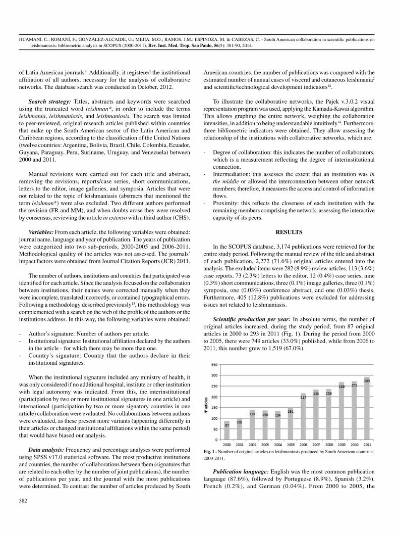
of Latin American journals5. Additionally, it registered the institutional affiliation of all authors, necessary for the analysis of collaborative networks. The database search was conducted in October, 2012.
Search strategy: Titles, abstracts and keywords were searched using the truncated word leishman*, in order to include the terms leishmania, leishmaniasis, and leishmaniosis. The search was limited to peer-reviewed, original research articles published within countries that make up the South American sector of the Latin American and Caribbean regions, according to the classification of the United Nations (twelve countries: Argentina, Bolivia, Brazil, Chile, Colombia, Ecuador, Guyana, Paraguay, Peru, Suriname, Uruguay, and Venezuela) between 2000 and 2011.
Manual revisions were carried out for each title and abstract, removing the revisions, reports/case series, short communications, letters to the editor, image galleries, and symposia. Articles that were not related to the topic of leishmaniasis (abstracts that mentioned the term leishman*) were also excluded. Two different authors performed the revision (FR and MM), and when doubts arose they were resolved by consensus, reviewing the article in extenso with a third author (CHS).
Variables: From each article, the following variables were obtained: journal name, language and year of publication. The years of publication were categorized into two sub-periods, 2000-2005 and 2006-2011. Methodological quality of the articles was not assessed. The journals’ impact factors were obtained from Journal Citation Reports (JCR) 2011.
The number of authors, institutions and countries that participated was identified for each article. Since the analysis focused on the collaboration between institutions, their names were corrected manually when they were incomplete, translated incorrectly, or contained typographical errors. Following a methodology described previously13, this methodology was complemented with a search on the web of the profile of the authors or the institutions address. In this way, the following variables were obtained:
- Author’s signature: Number of authors per article.- Institutional signature: Institutional affiliation declared by the authors
in the article - for which there may be more than one.- Country’s signature: Country that the authors declare in their
institutional signatures.
When the institutional signature included any ministry of health, it was only considered if no additional hospital, institute or other institution with legal autonomy was indicated. From this, the interinstitutional (participation by two or more institutional signatures in one article) and international (participation by two or more signatory countries in one article) collaboration were evaluated. No collaborations between authors were evaluated, as these present more variants (appearing differently in their articles or changed institutional affiliations within the same period) that would have biased our analysis.
Data analysis: Frequency and percentage analyses were performed using SPSS v17.0 statistical software. The most productive institutions and countries, the number of collaborations between them (signatures that are related to each other by the number of joint publications), the number of publications per year, and the journal with the most publications were determined. To contrast the number of articles produced by South
American countries, the number of publications was compared with the estimated number of annual cases of visceral and cutaneous leishmania2 and scientific/technological development indicators18.
To illustrate the collaborative networks, the Pajek v.3.0.2 visual representation program was used, applying the Kamada-Kawai algorithm. This allows graphing the entire network, weighing the collaboration intensities, in addition to being understandable intuitively14. Furthermore, three bibliometric indicators were obtained. They allow assessing the relationship of the institutions with collaborative networks, which are:
- Degree of collaboration: this indicates the number of collaborators, which is a measurement reflecting the degree of interinstitutional connection.
- Intermediation: this assesses the extent that an institution was in the middle or allowed the interconnection between other network members; therefore, it measures the access and control of information flows.
- Proximity: this reflects the closeness of each institution with the remaining members comprising the network, assessing the interactive capacity of its peers.
RESULTS
In the SCOPUS database, 3,174 publications were retrieved for the entire study period. Following the manual review of the title and abstract of each publication, 2,272 (71.6%) original articles entered into the analysis. The excluded items were 282 (8.9%) review articles, 113 (3.6%) case reports, 73 (2.3%) letters to the editor, 12 (0.4%) case series, nine (0.3%) short communications, three (0.1%) image galleries, three (0.1%) symposia, one (0.03%) conference abstract, and one (0.03%) thesis. Furthermore, 405 (12.8%) publications were excluded for addressing issues not related to leishmaniasis.
Scientific production per year: In absolute terms, the number of original articles increased, during the study period, from 87 original articles in 2000 to 293 in 2011 (Fig. 1). During the period from 2000 to 2005, there were 749 articles (33.0%) published, while from 2006 to 2011, this number grew to 1,519 (67.0%).
Publication language: English was the most common publication language (87.6%), followed by Portuguese (8.9%), Spanish (3.2%), French (0.2%), and German (0.04%). From 2000 to 2005, the
Fig. 1 - Number of original articles on leishmaniasis produced by South American countries,
2000-2011.

HUAMANÍ, C.; ROMANÍ, F.; GONZÁLEZ-ALCAIDE, G.; MEJIA, M.O.; RAMOS, J.M.; ESPINOZA, M. & CABEZAS, C. - South American collaboration in scientific publications on leishmaniasis: bibliometric analysis in SCOPUS (2000-2011). Rev. Inst. Med. Trop. Sao Paulo, 56(5): 381-90, 2014.
383
predominant language used in original leishmaniasis articles was English (92.3%), which decreased to 85.3% during the period from 2006 to 2011. In contrast, Portuguese was used in 5.5% of the articles from 2000 to 2005, and then for 2006-2011, its use grew to 10.7%. As for Spanish, 2.0% of the total articles from 2000 to 2005 appeared in said language, while for 2006-2011, the number grew to 3.8%. In absolute terms, the number of articles in Portuguese increased from 41 (2000-2005) to 162 (2006-2011), while the number of articles in English increased from 691 (2000-2005) to 1296 (2006-2011).
Journals of publication: The total number of articles appeared in 398 scientific journals. Eight of them accounted for 32.3% of the articles on leishmaniasis. One-half of the South American scientific production was published in 21 journals, while the other half was scattered throughout 379 journals. The number of journals publishing only one article on leishmaniasis was 198. Forty-six journals contained 65.5% of the publications. The two journals publishing the most articles on leishmaniasis were Brazilian; Memórias do Instituto Oswaldo Cruz and Revista da Sociedade Brasileira de Medicina Tropical which together accounted for 11.2% of the articles throughout the entire study period. The journals with the highest impact factor were Clinical Infectious Diseases (9,154), Journal of Immunology (5,788), and the Journal of Antimicrobial Chemotherapy (5,068) (Table 1).
Number of authors and institutions: The median number of authors per article was six with an interquartile range from five to eight. The trend was five authors. There were 10 authors or fewer in 91.5% of the articles, 0.4% of them had only one, 54.2% listed from two to six authors, and 45.4% had seven or more. One article listed 101 authors. The median number of authors per article remained constant in all years.
Regarding the number of institutional affiliations per article, the median and trend was two. The interquartile range was from two to three, i.e., 50% of the articles had two or three affiliated institutions: 32.3% cited two different institutional affiliations and 22.8% cited three. Just under a quarter of the articles listed a single institution, while 20.7% listed four institutions or more. One article listed 24 collaborating institutions. The median number of institutions remained constant in all years.
Production per country: The identified institutions represented 58 different countries. Twenty countries accounted for 97.7% of the signatures. Brazil was the country with the most signatures (60.7%), followed by the United States (7.1%), Venezuela (4.9%), and Colombia (4.4%). Fifteen countries registered a single institutional signature. Five countries registered two institutional signatures, another five had three signatures and three of the countries registered four institutional signatures. Two countries had five institutions represented, three countries registered six institutions, three countries had ten, and two countries registered twelve signatures. Table 2 compares the production by South American countries with the annual number of leishmaniasis cases and other scientific/technological development indicators.
Production per institution: The 2,272 original articles had 1,160 different institutional signatures. Most institutions with higher quantities come from Brazil and include the Oswaldo Cruz Foundation (FIOCRUZ), with 11% of South American production and 18% of Brazilian production. It is followed by the University of São Paulo, the Federal University of Minas Gerais, the Federal University of Rio de Janeiro, and
the Federal University of Bahia (Table 3). The top four institutions also have the highest degree of collaboration, working with more than 100 different institutions, in addition to the highest values of intermediation and proximity to other network members.
Among the institutions with fewer than 100 articles, but with high values for degree of collaboration, intermediation and proximity are the Cayetano Heredia University, the London School of Hygiene and Tropical Medicine, and the University of Antioquia. Table 4 shows the non-South American countries and their institutions with the highest quantities of signatures for original articles on leishmaniasis produced by South American countries.
Collaborative networks: One large collaborative network was identified (Fig. 2) along with five smaller ones (Fig. 3). The largest network is centered on Brazilian institutions, whose production center is FIOCRUZ. This institution collaborates intensely with the five most productive institutions and collaborates independently with several institutions primarily in Brazil, the USA, and the United Kingdom. The largest network has two extensions, the first of which derives from the collaboration between the Federal University of Rio de Janeiro with the Venezuelan Institute for Scientific Research, which then integrates other Venezuelan institutions into the network. The second extension begins at the Evandro Chagas Institute of Brazil, followed by the London School of Hygiene and Tropical Medicine of the United Kingdom, and concludes with the Cayetano Heredia University of Peru (UPCH), which then integrates European institutions that are primarily Belgian. Beyond the Venezuelan and Peruvian collaboration in these large networks, no collaboration with other South American institutions is evident.
The five smaller collaborative networks are constituted by i) four Japanese universities, the Catholic University of Santiago de Guayaquil, and the Ministry of Public Health of Ecuador; ii) exclusively Argentinean institutions; iii) Venezuelan institutions for Scientific Research with the University of Wurzburg (Germany); iv) Colombian institutions (Del Valle University and International Center of Training and medical research-CIDEIM) with Yale University; and v) two Colombian universities.
DISCUSSION
Overall, collaborations between South American institutions have been reported in other studies12,13 as being intense and including all countries in the region. However, in the case of collaborations for leishmaniasis research, (with the exception of the collaboration between Brazil and Venezuela, and to a lesser extent with Peru), South American countries collaborate less intensively. Leishmaniasis research has characteristics that differentiate it from other diseases. Individuals affected by the disease are usually rural, low-income, and there is low incidence in European countries and the United States, where the predominant clinical form is visceral leishmaniasis17. Because of these characteristics, leishmaniasis is not considered a research priority in these countries, which reduces the potential for collaborative research with South American countries. Furthermore, pharmaceutical industry investment is low, thus new drugs with fewer side effects and lower toxicity are not being developed. These characteristics, among others, have led leishmaniasis to be characterized as a “forgotten disease” (also known as “neglected tropical disease”)11.

HUAMANÍ, C.; ROMANÍ, F.; GONZÁLEZ-ALCAIDE, G.; MEJIA, M.O.; RAMOS, J.M.; ESPINOZA, M. & CABEZAS, C. - South American collaboration in scientific publications on leishmaniasis: bibliometric analysis in SCOPUS (2000-2011). Rev. Inst. Med. Trop. Sao Paulo, 56(5): 381-90, 2014.
384
Table 1Scientific journals containing two-thirds of the scientific articles published on leishmaniasis produced by South American countries, 2000-2011
N° Journal n % Country Impact Factor (2011)a
1 Memorias do Instituto Oswaldo Cruz 146 6.4 Brazil 2.147
2 Revista da Sociedade Brasileira de Medicina Tropical 109 4.8 Brazil 0.681
3 American Journal of Tropical Medicine and Hygiene 102 4.5 USA 2.592
4 Experimental Parasitology 83 3.7 USA 2.122
5 Veterinary Parasitology 78 3.4 Netherlands 2.579
6 Parasitology Research 77 3.4 Germany 2.149
7 Acta Tropica 73 3.2 Netherlands 2.722
8 Transactions of the Royal Society of Tropical Medicine and Hygiene 65 2.9 Netherlands 2.022
9 Vaccine 45 2.0 Netherlands 3.766
10 Molecular and Biochemical Parasitology 44 1.9 Netherlands 2.551
11 Cadernos de Saude Publica 37 1.6 Brazil 0.889
12 Antimicrobial Agents and Chemotherapy 34 1.5 USA 4.841
13 Revista do Instituto de Medicina Tropical de Sao Paulo 34 1.5 Brazil 1
14 Parasitology 33 1.5 UK 2.961
15 Infection and Immunity 29 1.3 USA 4.165
16 Journal of Medical Entomology 28 1.2 USA 1.762
17 PLoS Neglected Tropical Diseases 27 1.2 USA 4.716
18 Journal of Infectious Diseases 26 1.1 USA 6.41
19 Parasite Immunology 23 1.0 UK 2.601
20 Veterinary Immunology and Immunopathology 22 1.0 Netherlands 2.076
21 International Journal for Parasitology 20 0.9 UK 3.393
22 Brazilian Journal of Medical and Biological Research 19 0.8 Brazil 1.129
23 Journal of Parasitology 19 0.8 USA 1.405
24 Neotropical Entomology 19 0.8 Brazil 0.603
25 Microbes and Infection 18 0.8 Italy 3.101
26 Revista Brasileira de Parasitologia Veterinaria 18 0.8 Brazil 0.712
27 PLoS ONE 17 0.7 USA 4.716
28 Journal of Clinical Microbiology 16 0.7 USA 4.153
29 Journal of Ethnopharmacology 16 0.7 Ireland 3.014
30 Bioorganic and Medicinal Chemistry 15 0.7 UK 2.921
31 European Journal of Medicinal Chemistry 15 0.7 Italia 3.346
32 Annals of Tropical Medicine and Parasitology 14 0.6 UK 1.429
33 Arquivo Brasileiro de Medicina Veterinaria e Zootecnia 14 0.6 Brazil 0.291
34 Journal of Immunology 13 0.6 USA 5.788
35 Parasitology International 13 0.6 Ireland 2.132
36 Tropical Medicine and International Health 13 0.6 UK 2.795
37 Anais Brasileiros de Dermatologia 12 0.5 Brazil 0.554
38 Infection 12 0.5 Germany 2.659
39 Journal of Venomous Animals and Toxins Including Tropical Diseases 12 0.5 Brazil 0.429
40 Revista de Saude Publica 12 0.5 Brazil 1.328
41 BMC Infectious Diseases 11 0.5 UK 3.118
42 Clinical Infectious Diseases 11 0.5 USA 9.154

HUAMANÍ, C.; ROMANÍ, F.; GONZÁLEZ-ALCAIDE, G.; MEJIA, M.O.; RAMOS, J.M.; ESPINOZA, M. & CABEZAS, C. - South American collaboration in scientific publications on leishmaniasis: bibliometric analysis in SCOPUS (2000-2011). Rev. Inst. Med. Trop. Sao Paulo, 56(5): 381-90, 2014.
385
Table 1Scientific journals containing two-thirds of the scientific articles published on leishmaniasis produced by South American countries, 2000-2011 (cont.)
Table 3Bibliometric indicators of the institutions with the largest number of signatures in original articles on leishmaniasis produced by
South American countries in SCOPUS, 2000-2011
Order Institution Country Papers (%) DegreeInterm. x 100
Proximity x 100
1 Oswaldo Cruz Foundation/Fiocruz Brazil 652 (11.1) 315 33.1 53.7
2 Universidade de São Paulo Brazil 322 (5.5) 222 18.1 48.3
3 Universidade Federal de Minas Gerais Brazil 266 (4.5) 148 7.8 44.1
4 Universidade Federal do Rio de Janeiro Brazil 196 (3.4) 122 7.5 44.7
5 Universidade Federal da Bahia Brazil 126 (2.2) 98 2.9 42.0
6 Universidade Estadual Paulista Brazil 102 (1.7) 64 3.4 40.6
7 Instituto Evandro Chagas Brazil 98 (1.7) 74 1.8 41.7
8 Universidad Central de Venezuela Venezuela 89 (1.5) 80 6.1 40.4
9 Universidade Federal de Ouro Preto Brazil 82 (1.4) 58 1.1 40.0
10 Universidade Estadual de Maringá Brazil 76 (1.3) 35 1.6 39.9
11 Universidade Federal de São Paulo Brazil 72 (1.2) 59 1.7 42.5
12 Universidad Peruana Cayetano Heredia Peru 65 (1.1) 97 6.4 42.2
13 Universidad de Antioquia Colombia 61 (1.0) 68 5.5 41.7
Table 2 Comparison of the production by South American countries with the number of annual cases of leishmaniasis and
other indicators of scientific/technological development
N° Country Papers published CL cases/yeara VL cases/yeara RDE b (million dollars) RDE/GDP %b
1 Brazil 3552 26008 3481 20,237.6 1.07
2 Venezuela 285 2480 40 ND ND
3 Colombia 256 17420 60 600.6 0.16
4 Argentina 218 261 8 2,658.7 0.51
5 Peru 124 6405 - 238.1 0.15
6 Bolivia 44 2647 - ND 0.26
7 Ecuador 35 1724 - 145.9 0.15
8 Paraguay 19 431 48 20.1 0.20
CL. Cutaneous Leishmania, VL: Visceral Leishmania, RDE: Research and Development Expenditure, GDP: Gross domestic product. a Source: Alvar J, Velez ID, Bern C, et al. Leishmaniasis worldwide and global estimates of its incidence. PloS one. 2012;7(5):e35671(Alvar et al. 2012). b UNESCO (2010) UNESCO Science Report. The Current Status of Science around the World.
N° Journal n % Country Impact Factor (2011)a
43 Journal of Antimicrobial Chemotherapy 11 0.5 UK 5.068
44 Biomedica 10 0.4 Colombia 0.545
45 Brazilian Journal of Pharmacognosy 10 0.4 Brazil 0.261
46 Phytomedicine 10 0.4 Germany 3.268aImpact Factor by JCR 2011.

HUAMANÍ, C.; ROMANÍ, F.; GONZÁLEZ-ALCAIDE, G.; MEJIA, M.O.; RAMOS, J.M.; ESPINOZA, M. & CABEZAS, C. - South American collaboration in scientific publications on leishmaniasis: bibliometric analysis in SCOPUS (2000-2011). Rev. Inst. Med. Trop. Sao Paulo, 56(5): 381-90, 2014.
386
Order Institution Country Papers (%) DegreeInterm. x 100
Proximity x 100
14 Universidade Federal do Piauí Brazil 55 (0.9) 46 1.0 40.1
15 Universidade Estadual de Campinas Brazil 53 (0.9) 33 1.1 40.1
16 Universidade de Brasília Brazil 52 (0.9) 55 2.7 42.3
17 Centro Internacional de Entrenamiento e Investigaciones Médicas Colombia 51 (0.9) 50 3.1 39.8
18 Universidad de Los Andes Colombia 44 (0.8) 36 2.4 35.3
19 Universidade Federal do Maranhão Brazil 43 (0.7) 45 1.0 39.8
20 Universidade Federal do Rio Grande do Norte Brazil 41 (0.7) 50 1.3 40.0
21 Universidade Federal do Ceará, Fortaleza Brazil 40 (0.7) 29 0.7 38.1
22 London School of Hygiene and Tropical Medicine U.K. 38 (0.7) 93 4.3 43.9
23 Universidade Federal de Mato Grosso do Sul Brazil 38 (0.7) 28 0.8 37.5
24 Universidade Federal Fluminense Brazil 37 (0.6) 25 0.6 38.5
25 Universidade Federal de Pernambuco Brazil 35 (0.6) 41 2.0 39.8
26 Instituto Venezolano de Investigaciones Científicas Venezuela 32 (0.6) 32 2.3 38.7
27 Administración Nacional de Laboratorios e Institutos de Salud Argentina 30 (0.5) 50 3.6 39.3
28 National Institutes of Health USA 29 (0.5) 52 3.1 41.8
29 Universidad de Carabobo Venezuela 27 (0.5) 39 1.5 39.3
30 Instituto Adolfo Lutz Brazil 26 (0.4) 19 0.3 37.2
31 Kochi University, Kohasu Japan 26 (0.4) 34 1.2 31.2
32 University of California USA 26 (0.4) 39 1.2 41.1
33 University of Glasgow U.K. 25 (0.4) 42 0.9 41.1
34 Institut de Recherche pour le Développement France 24 (0.4) 44 2.6 39.1
35 Universidad Nacional de Colombia Colombia 24 (0.4) 29 2.0 38.3
36 Universidade Federal de Juiz de Fora Brazil 24 (0.4) 12 0.0 37.7
37 Universidade Federal do Espírito Santo Brazil 24 (0.4) 23 0.7 38.3
38 Universidade Federal do Triângulo Mineiro Brazil 23 (0.4) 23 0.7 38.1
39 Consejo Nacional de Investigaciones Científicas y Técnicas Argentina 22 (0.4) 59 2.7 37.4
40 University of Cambridge, Cambridge U.K. 22 (0.4) 42 1.1 41.7
41 Universidad Autónoma de Madrid Spain 21 (0.4) 26 1.0 39.5
42 Universidade Federal do Paraná Brazil 21 (0.4) 29 1.4 38.9
43 Universidade do Estado do Rio de Janeiro Brazil 21 (0.4) 21 0.4 38.9
44 University of Washington USA 21 (0.4) 69 2.0 42.5
45 Escola Bahiana de Medicina e Saúde Pública Brazil 20 (0.3) 21 0.1 37.5
46 Universidad de Buenos Aires Argentina 20 (0.3) 25 0.7 32.0
47 Universidade Federal do Pará Brazil 20 (0.3) 24 0.2 38.6
48 University of Iowa USA 19 (0.3) 34 0.4 39.5
49 University of Texas USA 19 (0.3) 35 3.0 41.5
Table 3Bibliometric indicators of the institutions with the largest number of signatures in original articles on leishmaniasis produced by
South American countries in SCOPUS, 2000-2011 (cont.)

HUAMANÍ, C.; ROMANÍ, F.; GONZÁLEZ-ALCAIDE, G.; MEJIA, M.O.; RAMOS, J.M.; ESPINOZA, M. & CABEZAS, C. - South American collaboration in scientific publications on leishmaniasis: bibliometric analysis in SCOPUS (2000-2011). Rev. Inst. Med. Trop. Sao Paulo, 56(5): 381-90, 2014.
387
Table 4Non-South American countries with the largest number of signatures in original articles on leishmaniasis produced by South American countries, 2000-2011
Country Frequency % Institutions with more productiona
USA 415 7.1 NIH (29), Univ. California (26), Univ. Washington (21)
UK 206 3.5 London School of Hygiene and Tropical Medicine (38), Univ. Glasgow (25), Univ. Cambridge (22)
France 111 1.9 Institut de Recherche pour le Développement (24), Univ. Toulouse (9), Univ. Paris-Sud (7)
Spain 111 1.9 Univ. Autónoma de Madrid (21), Instituto de Parasitología y Biomedicina Lopez-Neyra (14), Insti-tuto de Salud Carlos III (11)
Japan 75 1.3 Univ. Kochi (26), Univ. of the Ryukyus (17), Univ. Yamaguchi (11)
Germany 68 1.2 Univ. Wurzburg (14), Charité Univ. Med (4)
Belgium 52 0.9 Institute of Tropical Medicine (16), Institut Prince Leopold (15), Univ. Antwerp (7)
Canada 50 0.9 Univ. MacGill (9), Univ. Laval (7), Centre de Recherche en Infectiologie (5)
Switzerland 46 0.8 Word Health Organization (12), Univ. Geneva (11), Swiss Tropical and Public Health Institute (6)
Italy 20 0.3 Univ. degli Studi di Bari (6) b
Netherland 15 0.3 Royal Tropical Institute (7) b
Mexico 14 0.2 Centro de Investigación Científica de Yucatán (3)
a Only three institutions with the largest number of articles. b Other institutions have only one article.
Fig. 2 - Primary South American collaborative network for the production of research articles on leishmaniasis.

HUAMANÍ, C.; ROMANÍ, F.; GONZÁLEZ-ALCAIDE, G.; MEJIA, M.O.; RAMOS, J.M.; ESPINOZA, M. & CABEZAS, C. - South American collaboration in scientific publications on leishmaniasis: bibliometric analysis in SCOPUS (2000-2011). Rev. Inst. Med. Trop. Sao Paulo, 56(5): 381-90, 2014.
388
The forgotten disease condition for leishmaniasis is exemplified in Peru, for example, by the very active collaborations that Cayetano Heredia University has with more than ten North American institutions in cancer research and with more than 20 institutions in clinical medicine research13, while the collaboration intensity with North American institutions is lower. Although leishmaniasis research is a specific area of development and can be carried out by fewer institutions, this does not mean that they are to be isolated from other foreign collaborators and institutions. On the contrary, as there are few national institutions researching it, collaborative work is needed (especially with foreign institutions), in order to increase their production.
Peruvian-Belgian collaboration, specifically that between the UPCH and the Institute of Tropical Medicine (Amberes, Belgium), has been described in its constitution, importance, and products9, by which the network described in our study reflects the results of such collaboration, which mainly comprises exchanges of researchers following training grants10. Although studies showing the products of the collaboration between South American institutions with other countries have not been found yet, the Peruvian example (Peruvian-Belgian collaboration) may explain the results of the specific cooperation between Ecuador and Japan.
Brazil leads in the South American context, participating in more than 50% of the South American scientific production12, and is the country with most cases in the region. Its investment in development, science and technology, along with its research policies, has allowed it to address its own health problems and require less collaboration to maintain and increase its scientific production. In the area of neglected diseases, it leads South American research7 in Chagas disease following collaboration patterns similar to those described in our study. There are three factors that make Brazil the country with the highest production on leishmaniasis and, perhaps, also with other neglected diseases: i) the
high number of new cases of cutaneous and visceral leishmaniasis2; ii) increased public spending in research & development (the most in South America)18; and iii) an active network of interinstitutional collaboration, something which increases research productivity3.
Globally, India leads scientific production on leishmaniasis. Following it are Brazil, Spain, the USA, and France1,16, however, the clinical form studied most is the visceral variety, which may explain why the remaining South American countries, which have a lower incidence of these clinical forms2, have less European or North American collaboration. The collaboration between these countries is focused in priority topics for European countries and the USA, and not in complete response to the research agendas of the South American countries. Theses agendas in several cases include neglected infectious diseases but those not prioritize leishmaniasis as a specific cause of disease burden. In this context, participation by UNASUR could play an integrating role in the promotion of research efforts within South America. The RINS-UNASUR 2011-2015 five-year plan establishes the coordination of projects through the creation of thematic networks composed of staff members from UNASUR member countries with recognized expertise in the field of study on both national institutes of health, as well as university levels in these countries. One of the main proposed thematic networks is research on neglected infectious diseases. This initiative could focus research efforts on leishmaniasis, which, like dengue fever, is widespread throughout the region. It would also strengthen the leishmaniasis collaborative network on a regional level. The results of this study could serve as a baseline diagnosis to be used for comparison purposes, once the specified measures envisaged in the five-year plan materialize.
As for the distribution of the published articles, our sample appeared in almost 400 different journals. However, 32.3% of them are concentrated in only eight journals. Another research has reported
Fig. 3 - Smaller South American collaborative networks for the production of research articles on leishmaniasis.

HUAMANÍ, C.; ROMANÍ, F.; GONZÁLEZ-ALCAIDE, G.; MEJIA, M.O.; RAMOS, J.M.; ESPINOZA, M. & CABEZAS, C. - South American collaboration in scientific publications on leishmaniasis: bibliometric analysis in SCOPUS (2000-2011). Rev. Inst. Med. Trop. Sao Paulo, 56(5): 381-90, 2014.
389
the top eight journals accounting for 21% and 24% of the articles on leishmaniasis16. Seventy percent of the articles are distributed throughout 390 different journals. This nucleus of eight journals is comprised by two that are Brazilian (and are those that published the most) while the remaining six are not South American. Both Brazilian journals are institutional - Memórias do Instituto Oswaldo Cruz and the Revista da Sociedade Brasileira de Medicina Tropical belong to a public research institute and a medical society, respectively. They publish research on tropical diseases, preventive medicine, public health, and infectious diseases. Furthermore, both journals are free, which improves their visibility within the South American scientific community. If these results are compared with another study that explores worldwide production over a similar period of time (2000-2009), we can find some differences that can be explained by the geographic limits in our study. In that study, the journals with the highest publication of articles on leishmaniasis were Molecular and Biochemical Parasitology, Infection and Immunity, Experimental Parasitology, The American Journal of Tropical Medicine and Hygiene, and Parasitology Research. Although it is true that these journals are found among those with the most publications in our analysis, the order according to contribution is clearly different.
This study has some limitations. Our sample does not represent all the South American collaboration or actions taken to address the problems associated with leishmaniasis, as this may be reflected differently from a scientific publication (patents, clinical guidelines, among others)11. For example, the scientific production described does not represent the region’s entire production, only research appearing in journals with international visibility. Therefore, it is possible that articles have been published about leishmaniasis in local journals that are not indexed by SCOPUS or MEDLINE. However, we felt that not including them would not affect our analysis of collaborative networks given that the production in local journals is less and because that research is conducted with less international collaboration. The IF was obtained from 2011 by JCR. This IF does not represent the whole period, but it is necessary to understand the current importance of the journals. Finally, it is possible that networks under construction are not evident in our network, since they still have to publish enough articles together. These will be revealed in subsequent studies, with our study serving as a baseline.
In conclusion, South American scientific production on leishmaniasis published in journals indexed in SCOPUS is focused on Brazilian activity. This activity integrates several institutions that collaborate primarily with FIOCRUZ and integrate Venezuelan and Peruvian institutions, but with little foreign participation, especially from South American countries. Networks producing less are those integrated by Ecuadorian-Japanese collaboration, or Venezuelan, Colombian or Argentinean institutions. It is possible that the South American collaborative networks will integrate the research factors from several countries as part of RINS-UNASUR soon, so we recommend that institutions interested in conducting or increasing their scientific production on leishmaniasis promote collaboration with the institutions nearest the center of these networks, i.e., those that demonstrate the highest values in collaboration and intermediation. All this considering that leishmaniasis is more than a neglected disease, it is a disease of forgotten or excluded populations, and finding solutions for its treatment (new and more effective drugs), prevention and control will only be possible by the attention paid and initiative shown by affected countries, providing the necessary funding and favoring collaborative work on a common problem.
SPONSORSHIPS
Self-funded.
RESUMEN
Colaboración Sudamericana en publicaciones científicas sobre leishmaniasis: análisis bibliométrico en SCOPUS (2000-2011)
Objetivos: Evaluar la producción y la red de colaboración de investigación sobre leishmaniasis en Sudamérica. Métodos: Se realizó un estudio bibliométrico usando la base de datos SCOPUS. La unidad de análisis fueron los artículos originales de investigación publicados desde el año 2000 hasta 2011, realizados sobre leishmaniasis y que incluyeron al menos un autor sudamericano. Se obtuvieron para cada artículo: nombre de la revista, idioma, año de publicación, número de autores, instituciones, países y otras variables. Resultados: 3174 artículos fueron publicados, 2.272 de ellos eran artículos originales. Se identificaron 1.160 firmas institucionales diferentes, 58 países y 398 revistas científicas. Brasil fue el país con más artículos (60,7%) y la Fundación Oswaldo Cruz (FIOCRUZ) tuvo 18% de la producción brasileña, convirtiéndose en el núcleo Sudamericano de la mayor red científica de Leishmaniasis. Conclusiones: La producción científica de Sudamérica sobre Leishmaniasis publicada en revistas indexadas en SCOPUS se centra en la actividad de la investigación brasileña. Es necesario fortalecer las redes de colaboración, el primer paso para ello es la identificación de las instituciones con mayor producción con el fin de llevar a cabo la investigación en colaboración de acuerdo con las prioridades de cada país.
ACKNOWLEDGMENTS
I wish to thank Dr Kim Hoffman for her recommendations to the final version.
REFERENCES
1. Al-Mutawakel K, Scutaru C, Shami A, Sakr M, Groneberg D, Quarcoo D. Scientometric analysis of the world-wide research efforts concerning leishmaniasis. Parasit Vectors. 2010;3:14.
2. Alvar J, Vélez ID, Bern C, Herrero M, Desjeux P, Cano J, et al. Leishmaniasis worldwide and global estimates of its incidence. PLoS One. 2012;7:e35671.
3. Bales ME, Johnson SB, Keeling JW, Carley KM, Kunkel F, Merrill JA. Evolution of coauthorship in public health services and systems research. Am J Prev Med. 2011;41:112-7.
4. Caballero P, Yagui M, Espinoza M, Castilla T, Granados A, Velasquez A, et al. Prioridades regionales y nacionales de investigación en salud, Perú 2010-2014: un processo con enfoque participativo y descentralista. Rev Peru Med Exp Salud Publica. 2010;27:398-411.
5. Chinchilla-Rodriguez Z, Benavent-Perez M, Moya-Anegon F, Miguel S. International collaboration in medical research in Latin America and the Caribbean (2003-2007). J Am Soc Inf Sci Technol. 2012;63:2223-38.
6. Desjeux P. Leishmaniasis: current situation and new perspectives. Comp Immunol Microbiol Infect Dis. 2004;27:305-18.
7. Gonzalez-Alcaide G, Park J, Huamani C, Gascón J, Ramos JM. Scientific authorships and collaboration network analysis on Chagas disease: papers indexed in PubMed (1940-2009). Rev Inst Med Trop Sao Paulo. 2012;54:219-28.

HUAMANÍ, C.; ROMANÍ, F.; GONZÁLEZ-ALCAIDE, G.; MEJIA, M.O.; RAMOS, J.M.; ESPINOZA, M. & CABEZAS, C. - South American collaboration in scientific publications on leishmaniasis: bibliometric analysis in SCOPUS (2000-2011). Rev. Inst. Med. Trop. Sao Paulo, 56(5): 381-90, 2014.
390
8. Goto H, Lindoso JA. Current diagnosis and treatment of cutaneous and mucocutaneous leishmaniasis. Expert Rev Anti Infect Ther. 2010; 8:419-33.
9. Gotuzzo E, Gonzalez E, Verdonck K. Formación de investigadores en el contexto de proyectos colaborativos: experiencias en el Instituto de Medicina Tropical “Alexander von Humbolt”, Universidad Peruana Cayetano Heredia. Rev Peru Med Exp Salud Publica. 2010;27:419-27.
10. Guerra H. La beca de retorno de la Universidad Peruana Cayetano Heredia. Rev Peru Med Exp Salud Publica. 2010;27:428-31.
11. Holveck JC, Ehrenberg JP, Ault SK, Rojas R, Vasquez J, Cerqueira MT, et al. Prevention, control, and elimination of neglected diseases in the Americas: pathways to integrated, inter-programmatic, inter-sectoral action for health and development. BMC Public Health. 2007;7:6.
12. Huamani C, Gonzalez AG, Curioso WH, Pacheco-Romero J. Redes de colaboración y producción científica sudamericana en medicina clínica, ISI Current Contents 2000-2009. Rev Med Chil. 2012;140:466-75.
13. Huamani C, Mayta-Tristan P. Producción científica peruana en medicina y redes de colaboración, análisis del Science Citation Index 2000-2009. Rev Peru Med Exp Salud Publica. 2010;27:315-25.
14. Kamada T, Kawai S. An algorithm for drawing general undirected graphs. Inf Process Lett. 1989;31:7.
15. Pacheco Santos LM, Moura EC, Barradas Barata R de C, Serruya SJ, da Motta ML, Silva Elias FT, et al. Fulfillment of the Brazilian agenda of priorities in health research. Health Res Policy Syst. 2011;9:35.
16. Ramos JM, Gonzalez-Alcaide G, Bolanos-Pizarro M. Bibliometric analysis of leishmaniasis research in Medline (1945-2010). Parasit Vectors. 2013;6:55.
17. Reithinger R, Dujardin J, Louzir H, Pirmez C, Alexander B, Brooker S. Cutaneous leishmaniasis. Lancet Infect Dis. 2007;7:581-96.
18. UNESCO. The Current status of science around the World. Paris: UNESCO Science Report; 2010.
19. WHO. Neglected tropical diseases. Geneva: WHO; 2013. [cited 2013 Mar 19]. Available from: http://www.who.int/neglected_diseases/en/
Received: 3 October 2013Accepted: 25 February 2014

Rev. Inst. Med. Trop. Sao Paulo56(5):391-395, September-October, 2014doi: 10.1590/S0036-46652014000500004
(1) Programa de Pós Graduação em Ciências da Saúde, Universidade Estadual de Maringá, Maringá, Paraná, Brazil.(2) Departamento de Medicina, Universidade Estadual de Maringá, Maringá, Paraná, Brazil.(3) Departamento de Análises Clínicas e Biomedicina, Universidade Estadual de Maringá, Maringá, Paraná, Brasil.Correspondence to: Maria Valdrinez Campana Lonardoni, Av. Colombo 5790, 87020-900 Maringá, Paraná, Brasil. Phone: 55 44 3011 4878. Fax: 55 44 3011 4860. E-mail:
DETECTION OF Leishmania (Viannia) IN Nyssomyia neivai AND Nyssomyia whitmani BY MULTIPLEX POLYMERASE CHAIN REACTION, IN SOUTHERN BRAZIL
Herintha Coeto NEITZKE-ABREU(1), Kárin Rosi REINHOLD-CASTRO(1), Mateus Sabaini VENAZZI(2), Regiane Bertin de Lima SCODRO(3), Alessandra de Cassia DIAS(1), Thaís Gomes Verzignassi SILVEIRA(1,3), Ueslei TEODORO(1) & Maria Valdrinez Campana LONARDONI(1,3)
SUMMARY
Sandflies transmit pathogens of leishmaniasis. The natural infection of sandflies by Leishmania (Viannia) was assessed in municipalities, in the state of Paraná, in Southern Brazil. Sandflies were collected with Falcão and Shannon traps. After dissection in search of flagellates in digestive tubes and identification of the species, female sandflies were submitted to the Multiplex Polymerase Chain Reaction (multiplex PCR) for detection of the fragment of the kDNA of Leishmania (Viannia) and the fragment from the IVS6 cacophony gene region of the phlebotomine insects. The analysis was performed in pools containing seven to 12 guts from females of the same species. A total of 510 female sandflies were analyzed, including nine Migonemyia migonei, 17 Pintomyia fischeri, 216 Nyssomyia neivai, and 268 Nyssomyia whitmani. Although none of the females was found naturally infected by flagellates through dissection, the fragment of DNA from Leishmania (Viannia) was shown by multiplex PCR in one sample of Ny. neivai (0.46%) and three samples of Ny. whitmani (1.12%). It was concluded that Ny. neivai and Ny. whitmani are susceptible to Leishmania infection, and that multiplex PCR can be used in epidemiological studies to detect the natural infection of the sandfly vector, because of its sensitivity, specificity and feasibility.
KEYWORDS: Cutaneous leishmaniasis; Sandfly; PCR; Leishmania; Nyssomyia whitmani; Nyssomyia neivai.
INTRODUCTION
Knowledge of the fauna composition, behavior, rates of natural infection of the sandfly, Leishmania species identification, and environmental characteristics of endemic areas are essential for the public-health services responsible for protecting populations that live in areas in which leishmaniasis is endemic. Leishmaniasis has worldwide propagation. In Brazil, cutaneous leishmaniasis (CL) has been reported in all states3.
Cases of CL have increased significantly since the 1980s, and are appearing over wider areas in the State of Paraná, in the South of Brazil28,29. This disease has been recorded in occupied areas for more than a century, even in urban areas, contrary to expectations that human pressure would eliminate natural foci and reduce the incidence of this endemic disease16,33,46. The organization of rural areas in the Brazilian colonial period created environmental conditions that clearly favor CL transmission28.
There are several reports of Leishmania detection in sandflies in endemic CL areas in Brazil and in the world1,32,41. However, considering the wide geographical distribution of this dermatosis in the Americas47,
knowledge of the natural infection rate of sandflies is still insufficient to estimate the risk of Leishmania infection in many endemic areas.
Given the occurrence of autochthonous CL cases in several municipalities, in the state of Paraná, natural infection rates of sandflies by Leishmania (Viannia) were investigated, in order to identify the species of Leishmania present in locations where cases of this disease had been reported.
MATERIAL AND METHODS
Sandfly collection: Sandflies were collected in the localities of Recanto Marista, Água Azul Farm and Flor de Maio Grange, in the municipalities of Doutor Camargo, Fênix and Mandaguari, respectively, in the state of Paraná, where CL cases had been reported. Sandflies were collected from January through September 2006, from 6 p.m. to 6 a.m., with Falcão light traps and a Shannon trap, installed in woods, domiciles, peridomicile and domestic-animal shelters (cattle shelter and pigsty)30. The collected insects were kept alive for further dissection and observation of the natural infection by flagellates.
Dissection and identification of sandflies: The dissection was carried

NEITZKE-ABREU, H.C.; REINHOLD-CASTRO, K.R.; VENAZZI, M.S.; SCODRO, R.B.L.; DIAS, A.C.; SILVEIRA, T.G.V.; TEODORO, U. & LONARDONI, M.V.C. - Detection of Leishmania (Viannia) in Nyssomyia neivai and Nyssomyia whitmani by Multiplex Polymerase Chain Reaction, in Southern Brazil. Rev. Inst. Med. Trop. Sao Paulo, 56(5): 391-5, 2014.
392
out under a stereoscope; the legs and wings were removed and the dissection was carried out by making two incisions in the distal portion of the abdomen and, with zigzag movements, the digestive tubes were removed and examined under an optical microscope (400 x) in the search for flagellates and the identification of the species of the sandfly30. The nomenclature of the species follows GALATI12.
After dissection and identification30 of species, digestive tubes were stored at -18 °C in tubes containing 150 µL STE buffer (0.1 M NaCl, 10 mM Tris-base; Na
2EDTA-2H
2O 1 mM, pH 8.0), each containing seven
to 12 guts from females of the same species.
Extraction of DNA: The samples were macerated, and DNA was extracted with a solution of guanidine isothiocyanate and phenol, and hydrated in 20 µL of ultra-pure H
2O31. For each group of 22 samples
submitted for DNA extraction, one negative control (male sandflies) and one positive control [male sandflies plus 104 L. (V.) braziliensis promastigotes] were used.
Multiplex PCR: Two pairs of primers were used: MP3H (5´-GAA CGG GGT TTC TGT ATG C-3´) and MP1L (5´-TAC TCC CCG ACA TGC CTC TG-3´)18 to amplify a fragment of 70-bp from the mini circle region of the kinetoplast (kDNA) of the Leishmania (Viannia), and 5Llcac (5’-TGG CCG AAC ATA ATG TTA G-3’) and 3Llcac (5’-CCA CGA ACA AGT TCA ACA TC-3’)17 to amplify a fragment of 220-bp from the IVS6 cacophony gene region of the phlebotomine insects.
The PCR reaction mixture (final volume 25 µL) was composed of 0.5 µM of each of the primers (Invitrogen Life Technologies, São Paulo, Brazil), 0.2 mM dNTP (Invitrogen, Carlsbad, CA, USA), 1U Platinum Taq DNA Polymerase, (Invitrogen, Carlsbad, CA, USA), 1.5 mM MgCl
2,
1X enzyme buffer, and 2 µL DNA template. The amplification was carried out in a PC Thermocycler (Biometra, Germany) at 94 °C for seven min to activate the enzyme, followed by 30 cycles, each divided into three stages, of denaturation (1.5 min at 95 °C), annealing (1.5 min at 57 °C), and polymerization (two min at 72 °C). After this, the extension was
continued for a further 10 min at 72 °C, and the tubes were then kept at 4 °C until analysis31. The amplification products were submitted to electrophoresis in 2% of agarose gel (Invitrogen, Paisley, Scotland, UK) stained with 0.1 µg/mL ethidium bromide, at 10-15 V/cm. The presence of bands was observed in a transilluminator (Macro VueTM UV-20, Hoefer). For every five samples, one positive control [reaction mixture plus L. (V.) braziliensis DNA] and one negative control (reaction mixture plus water) were added.
RESULTS
In total, 510 (52 pools) female sandflies were analyzed by dissection and multiplex PCR, including nine Migonemyia migonei (one pool), 17 Pintomyia fischeri (two pools), 216 Nyssomyia neivai (22 pools), and 268 Nyssomyia whitmani (27 pools) (Table 1). A total of 244 female sandflies were collected at Recanto Marista, 107 at Água Azul Farm, and 159 at Flor de Maio Grange (Table 1).
All 52 sandfly pools contained the 220-bp fragment from the IVS6 cacophony gene region of the sandflies, and four pools (7.7%) showed the 70-bp fragment from the mini circle kDNA of Leishmania (Viannia) (Fig. 1). The minimal infection rate of Ny. neivai was 0.46% (1/216), and of Ny. whitmani was 1.12% (3/268). At Recanto Marista one Ny. neivai pool with Leishmania infection was detected on the porch of a domicile; at Água Azul Farm, one Ny. whitmani pool was detected in the peridomicile (near the woods ); and at Flor de Maio Grange, two Ny. whitmani pools were detected, one on the porch of a domicile and another from a domestic-animal shelter. The minimal infection rate of Recanto Marista was 0.41% (1/244), of Flor de Maio Grange was 1.26% (2/159), and at Água Azul Farm was 0.93% (1/107).
DISCUSSION
Mi. migonei, Pi. fischeri, Ny. neivai, and Ny. whitmani are frequently found in Brazil1,2,4,31,34,35,43,46. Both species detected with Leishmania infection are widely propagated in Brazil4,5,6,9,20,21 and in the state of
Table 1 Sandflies collected in Recanto Marista, Água Azul Farm, and Flor de Maio Grange, in Southern Brazil, from January to September, 2006
Specimens/Localities (Municipalities)Recanto Marista
(Doutor Camargo)Água Azul Farm
(Fênix)Flor de Maio Grange
(Mandaguari)Total
Migonemyia migoneiNa 9 0 0 9
Positive pools / Poolsb 0/1 0/0 0/0 0/1
Nyssomyia whitmaniNa 19 97 152 268
Positive pools / Poolsb 0/2 1/10 2/15 3/27
Nyssomyia neivaiNa 216 0 0 216
Positive pools / Poolsb 1/22 0/0 0/0 1/22
Pintomyia fischeriNa 0 10 7 17
Positive pools / Poolsb 0/0 0/1 0/1 0/2
TotalNa 244 107 159 510
Positive pools / Poolsb 1/25 1/11 2/16 4/52aNumber of Specimens; bNumber of positive pools/number of pools composed. Each pool contained seven to 12 guts from females of the same species.

NEITZKE-ABREU, H.C.; REINHOLD-CASTRO, K.R.; VENAZZI, M.S.; SCODRO, R.B.L.; DIAS, A.C.; SILVEIRA, T.G.V.; TEODORO, U. & LONARDONI, M.V.C. - Detection of Leishmania (Viannia) in Nyssomyia neivai and Nyssomyia whitmani by Multiplex Polymerase Chain Reaction, in Southern Brazil. Rev. Inst. Med. Trop. Sao Paulo, 56(5): 391-5, 2014.
393
Paraná, where either Ny. whitmani or Ny. neivai predominate, depending on environmental characteristics26,42,43,46.
Although no flagellates were detected in the dissected sandfly digestive tubes, DNA from Leishmania (Viannia) was found in 0.78% (4/510), indicating that at least one infected sandfly was present in each positive pool. The infected specimens were collected at sites near a riparian forest, which is inhabited by small wild mammals (e.g. rodents, armadillo) that are possible natural reservoirs of Leishmania. These locations show favorable environment for the formation of sandfly-breeding sites. Furthermore, the domestic animals are highly attractive blood sources for female sandflies37,44,45. Natural infection by Leishmania has been recorded in Pi. fischeri23,36,38, Mi. migonei2,7,34, Ny. neivai8,22,31,41,47, and Ny. whitmani1,4,11,19, by dissection or PCR, in several localities in many of the Brazilian states.
Natural infection rates of sandflies by Leishmania vary widely: 15.68% in Venezuela by PCR13, 0.83% and 7.14%4, and 18.2%23, in Brazil, by PCR4, and 1.4% and 2.6%, in Peru, by dissection15. Studies on natural infection rates have revealed that PCR is more sensitive and specific than dissection showing the presence of Leishmania in sandflies10,27,39,40. This method has often been used in areas where sandfly infection rates are low27,31, due to its sensitivity (it is able to detect the presence of a single parasite), specificity (independently of the number, location and stage of flagellates in the digestive tubes of the vector)25,27,34, rapidity, and ease of performance enabling it to be used in leishmaniasis epidemiological surveillance24. The dissection of sandflies for the detection of flagellates in the digestive tubes needs confirmation by Leishmania in vitro, cultivation or inoculation in laboratory animals27 while molecular methods allow the identification of Leishmania species, isolated in cultures from patients or reservoirs, as well from sandflies25,27.
Several primers have been used14,27,34, however, the primers used in this study have been tested successfully in diagnosis and detection of naturally infected sandflies, besides having good sensitivity [8 fg/µL of Leishmania (Viannia)]31.
The advantage of employing the multiplex PCR technique is that, in addition to the primers for detection of Leishmania (Viannia), the pair of primers used for internal control can assess the presence of probable interference from digestive contents of insects, which can inhibit the detection of Leishmania31,34.
The results show the susceptibility of sandflies to Leishmania strains. The minimum infection rates in Ny. neivai (0.46%, 1/216) and Ny. whitmani (1.12%, 3/268) are low, and might explain the low CL endemicity in the municipalities in question. Multiplex PCR, because of its sensitivity, specificity and feasibility, can be used in epidemiological studies to detect the natural infection of the sandfly vector.
RESUMO
Detecção de Leishmania (Viannia) em Nyssomyia neivai e Nyssomyia whitmani por Multiplex Reação em Cadeia da
Polimerase, no sul do Brasil
Flebotomíneos transmitem os patógenos das leishmanioses. Foi avaliada a infecção natural de flebotomíneos por Leishmania (Viannia) em municípios do Estado do Paraná, sul do Brasil. Os flebotomíneos foram coletados com armadilhas de Falcão e Shannon. Após dissecação para pesquisa de flagelados no tubo digestório e identificação das espécies, as fêmeas de flebotomíneos foram submetidas a Multiplex Reação em Cadeia da Polimerase (multiplex PCR) para a detecção do fragmento do kDNA de Leishmania (Viannia) e do fragmento do gene IVS6 da cacofonia de flebotomíneos. A análise foi realizada em pools contendo sete a 12 tubos digestórios de fêmeas da mesma espécie. Um total de 510 fêmeas foram analisadas, incluindo nove Migonemyia migonei, 17 Pintomyia fischeri, 216 Nyssomyia neivai e 268 Nyssomyia whitmani. Embora nenhuma fêmea tenha sido encontrada naturalmente infectada com flagelados pela dissecação, o fragmento de DNA de Leishmania (Viannia) foi mostrado por multiplex PCR em uma amostra de Ny. neivai (0,46%) e três amostras de Ny. whitmani (1,12%). Conclui-se que Ny. neivai e Ny. whitmani são suscetíveis à infecção por Leishmania, e que multiplex PCR, devido à sua sensibilidade, especificidade e viabilidade, pode ser utilizada em estudos epidemiológicos para a detecção da infecção natural do inseto vetor.
ACKNOWLEDGEMENTS
The authors would like to thank Colégio Marista, Água Azul Farm, and Flor de Maio Grange, for the permission granted to conduct the assessments, and for logistical support. Conselho Nacional de Desenvolvimento Científico e Tecnológico (CNPq Process 410550/2006-0), Coordenação de Aperfeiçoamento de Pessoal de Nível Superior (CAPES), and Fundação Araucária for their financial support.
REFERENCES
1. Azevedo ACR, Rangel EF, Costa EM, David J, Vasconcelos AW, Lopes UG. Natural infection of Lutzomyia (Nyssomyia) whitmani (Antunes & Coutinho, 1939) by Leishmania of the braziliensis complex in Baturité, Ceará State, northeast Brazil. Mem Inst Oswaldo Cruz. 1990;85:251.
2. Azevedo ACR, Rangel EF, Queiroz RG. Lutzomyia migonei (França, 1920) naturally infected with peripylarian flagellates in Baturité, a focus of cutanous leishmaniasis in Ceará State, Brazil. Mem Inst Oswaldo Cruz. 1990;85:479.
Fig 1 - Multiplex PCR in 2% of agarose gel showing fragments of 70-bp and 220-bp. The
fragment of 70-bp of the kDNA mini circle region of subgenus Leishmania (Viannia) were
amplified with MP3H and MP1L primers. The fragment of 220-bp of the IVS6 cacophony
gene region of the phlebotomine insects were amplified with 5Llcac and 3Llcac primers. Lane
1, Ny. whitmani DNA sample collected in Fênix; lanes 2, 3 and 5, Ny. whitmani DNA samples
collected in Mandaguari; lane 4, Ny. neivai DNA sample collected in Doutor Camargo; lane
6, positive control [DNA from L. (V.) braziliensis promastigotes]; lane 7, negative control
(all reagents without DNA). M, 100-bp molecular marker (Invitrogen Life Technologies,
São Paulo, Brazil).

NEITZKE-ABREU, H.C.; REINHOLD-CASTRO, K.R.; VENAZZI, M.S.; SCODRO, R.B.L.; DIAS, A.C.; SILVEIRA, T.G.V.; TEODORO, U. & LONARDONI, M.V.C. - Detection of Leishmania (Viannia) in Nyssomyia neivai and Nyssomyia whitmani by Multiplex Polymerase Chain Reaction, in Southern Brazil. Rev. Inst. Med. Trop. Sao Paulo, 56(5): 391-5, 2014.
394
3. Brasil, Ministério da Saúde. Manual de vigilância da leishmaniose tegumentar americana. Available from: http://goo.gl/mPcbz.
4. Carvalho GM, Andrade-Filho JD, Falcão AL, Rocha-Lima AC, Gontijo CM. Naturally infected Lutzomyia sand flies in a Leishmania-endemic area of Brazil. Vector Borne Zoonotic Dis. 2008;8:407-14.
5. Carvalho MSL, Bredt A, Meneghin ERS, Oliveira C. Flebotomíneos (Diptera: Psychodidae) em áreas de ocorrência de leishmaniose tegumentar americana no Distrito Federal, Brasil, 2006 a 2008. Epidemiol Serv Saúde. 2010;19:227-37.
6. Carvalho SMS, Santos PRB, Lanza H, Brandão-Filho SP. Diversidade de flebotomíneos no município de Ilhéus, Bahia. Epidemiol Serv Saúde. 2010;19:239-44.
7. Carvalho MR, Valença HF, Silva FJ, Pita-Pereira D, Araújo-Pereira T, Britto C, et al. Natural Leishmania infantum infection in Migonemyia migonei (França, 1920) (Diptera: Psychodidae: Phlebotominae) the putative vector of visceral leishmaniasis in Pernambuco State, Brazil. Acta Trop. 2010;116:108-10.
8. Córdoba-Lanús E, De Grosso ML, Piñero JE, Valladares B, Salomón OD. Natural infection of Lutzomyia neivai with Leishmania spp. in northwestern Argentina. Acta Trop. 2006;98:1-5.
9. Costa SM, Cechinel M, Bandeira V, Zannuncio JC, Lainson R, Rangel EF. Lutzomyia (Nyssomyia) whitmani s.l. (Antunes & Coutinho, 1939) (Diptera: Psychodidae: Phlebotominae): geographical distribution and the epidemiology of American cutaneous leishmaniasis in Brazil: mini-review. Mem Inst Oswaldo Cruz. 2007;102:149-53.
10. Dinesh DS, Kar SK, Kishore K, Palit A, Verma N, Gupta AK, et al. Screening sandflies for natural infection with Leishmania donovani, using a non-radioactive probe based on the total DNA of the parasite. Ann Trop Med Parasitol. 2000;94:447-51.
11. Galati EAB, Nunes VLB, Dorval MEC, Oshiro ET, Cristaldo G, Espíndola MA, et al. Estudo dos flebotomíneos (Diptera, Pychodidae) em área de leishmaniose tegumentar, no Estado de Mato Grosso do Sul, Brasil. Rev Saúde Pública. 1996;30:115-28.
12. Galati EAB. Morfologia e taxonomia. In: Rangel EF, Lainson R, editores. Flebotomíneos do Brasil. Rio de Janeiro: Fiocruz; 2003. p. 23-51.
13. Jorquera A, González R, Marchán-Marcano E, Oviedo M, Matos M. Multiplex-PCR for detection of natural Leishmania infection in Lutzomyia spp. captured in an endemic region for cutaneous leishmaniasis in state of Sucre, Venezuela. Mem Inst Oswaldo Cruz. 2005;100:45-8.
14. Kato H, Uezato H, Katakura K, Calvopiña M, Marco JD, Barroso PA, et al. Detection and identification of Leishmania species within naturally infected sand flies in the Andean areas of Ecuador by a polymerase chain reaction. Am J Trop Med Hyg. 2005;72:87-93.
15. Kato H, Gomez EA, Cáceres AG, Vargas F, Mimori T, Yamamoto K, et al. Natural infections of man-biting sand flies by Leishmania and Trypanosoma species in the northern Peruvian Andes. Vector Borne Zoonotic Dis. 2011;11:515-21.
16. Lima AP, Minelli L, Teodoro U, Comunello E. Tegumentary leishmaniasis distribution by satellite remote sensing imagery, in Paraná State, Brazil. An Bras Dermatol. 2002;77:681-92.
17. Lins RM, Oliveira SG, Souza NA, Queiroz RG, Justiniano SC, Ward RD, et al. Molecular evolution of the cacophony IVS6 region in sandflies. Insect Mol Biol. 2002;11:117-22.
18. Lopez M, Inga R, Cangalaya M, Echevarria J, Llanos-Cuentas A, Orrego C, et al. Diagnosis of Leishmania using the polymerase chain reaction: a simplified procedure for field work. Am J Trop Med Hyg. 1993;49:348-56.
19. Luz E, Membrive N, Castro EA, Dereure J, Pratlong F, Dedet JA, et al. Lutzomyia whitmani (Diptera: Psychodidae) as vector of Leishmania (V.) braziliensis in Paraná State, southern Brazil. Ann Trop Med Parasitol. 2000;94:623-31.
20. Marcondes CB, Lozovei AL, Vilela JH. Distribuição geográfica de flebotomíneos do complexo Lutzomyia intermedia (Lutz & Neiva, 1912) (Diptera, Psychodidae). Rev Soc Bras Med Trop. 1998;31:51-8.
21. Marcondes CB, Conceição MBE, Portes MGT, Simão BP. Phlebotomine sandflies in a focus of dermal leishmaniasis in the eastern region of the Brazilian state of Santa Catarina - preliminary results (Diptera: Psychodidae). Rev Soc Bras Med Trop. 2005;38:353-5.
22. Marcondes CB, Bittencourt IA, Stoco PH, Eger I, Grisard EC, Steindel M. Natural infection of Nyssomyia neivai (Pinto, 1926) (Diptera: Psychodidae, Phlebotominae) by Leishmania (Viannia) spp. in Brazil. Trans R Soc Trop Med Hyg. 2009;103:1093-7.
23. Margonari C, Soares RP, Andrade-Filho JD, Xavier DC, Saraiva L, Fonseca AL, et al. Phlebotomine sand flies (Diptera: Psychodidae) and Leishmania infection in Gafanhoto Park, Divinópolis, Brazil. J Med Entomol. 2010;47:1212-9.
24. Martín-Sánchez J, Gállego M, Barón S, Castillejo S, Morillas-Marques F. Pool screen PCR for estimating the prevalence of Leishmania infantum infection in sandflies (Diptera: Nematocera, Phlebotomidae). Trans R Soc Trop Med Hyg. 2006;100:527-32.
25. Medeiros ACR, Rodrigues SS, Roselino AMF. Comparison of the specificity of PCR and the histopathological detection of Leishmania for the diagnosis of American cutaneous leishmaniasis. Braz J Med Biol Res. 2002;35:421-4.
26. Membrive NA, Rodrigues G, Membrive U, Monteiro WM, Neitzke HC, Lonardoni MVC, et al. Flebotomíneos de municípios do norte do estado do Paraná, sul do Brasil. Entomol Vectores. 2004;11:673-80.
27. Michalsky ÉM, Fortes-Dias CL, Pimenta PFP, Secundino NFC, Dias ES. Assessment of PCR in the detection of Leishmania spp in experimentally infected individual phlebotomine sandflies (Diptera: Psychodidae: Phlebotominae). Rev Inst Med Trop Sao Paulo. 2002;44:255-9.
28. Monteiro WM, Neitzke HC, Lonardoni MVC, Silveira TGV, Ferreira MEMC, Teodoro U. Distribuição geográfica e características epidemiológicas da leishmaniose tegumentar americana em áreas de colonização antiga do Estado do Paraná, Sul do Brasil. Cad Saúde Pública. 2008;24:1291-303.
29. Monteiro WM, Neitzke HC, Silveira TGV, Lonardoni MVC, Teodoro U, Ferreira MEMC. Pólos de produção de leishmaniose tegumentar americana no norte do Estado do Paraná, Brasil. Cad Saúde Pública. 2009;25:1083-92.
30. Neitzke HC, Scodro RBL, Reinhold-Castro KR, Dias-Sversutti AC, Silveira TGV, Teodoro U. Pesquisa de infecção natural de flebotomíneos por Leishmania, no Estado do Paraná. Rev Soc Bras Med Trop. 2008;41:17-22.
31. Oliveira DM, Reinhold-Castro KR, Bernal MVZ, Legriffon CMO, Lonardoni MVC, Teodoro U, et al. Natural infection of Nyssomyia neivai by Leishmania (Viannia) spp. in the state of Paraná, southern Brazil, detected by multiplex polymerase chain reaction. Vector Borne Zoonotic Dis. 2011;11:137-43.
32. Parvizi P, Ready PD. Nested PCRs and sequencing of nuclear ITS-rDNA fragments detect three Leishmania species of gerbils in sandflies from Iranian foci of zoonotic cutaneous leishmaniasis. Trop Med Int Health. 2008;13:1159-71.
33. Pessoa SB, Barreto MP. Leishmaniose tegumentar americana. Rio de Janeiro: Imprensa Nacional - São Paulo: Serviço de Parasitologia, Departamento de Medicina, Faculdade de São Paulo; 1948.
34. Pita-Pereira D, Alves CR, Souza MB, Brazil RP, Bertho AL, Figueiredo-Barbosa A, et al. Identification of naturally infected Lutzomyia intermedia and Lutzomyia migonei with Leishmania (Viannia) braziliensis in Rio de Janeiro (Brazil) revealed by a PCR multiplex non-isotopic hybridisation assay. Trans R Soc Trop Med Hyg. 2005;99:905-13.
35. Pita-Pereira D, Souza GD, Zwetsch A, Alves CR, Britto C, Rangel EF. First report of Lutzomyia (Nyssomyia) neivai (Diptera: Psychodidae: Phlebotominae) naturally infected by Leishmania (Viannia) braziliensis in a periurban area of south Brazil using a multiplex polymerase chain reaction assay. Am J Trop Med Hyg. 2009;80:593-5.

NEITZKE-ABREU, H.C.; REINHOLD-CASTRO, K.R.; VENAZZI, M.S.; SCODRO, R.B.L.; DIAS, A.C.; SILVEIRA, T.G.V.; TEODORO, U. & LONARDONI, M.V.C. - Detection of Leishmania (Viannia) in Nyssomyia neivai and Nyssomyia whitmani by Multiplex Polymerase Chain Reaction, in Southern Brazil. Rev. Inst. Med. Trop. Sao Paulo, 56(5): 391-5, 2014.
395
36. Pita-Pereira D, Souza GD, Pereira TA, Zwetsch A, Britto C, Rangel EF. Lutzomyia (Pintomyia) fischeri (Diptera: Psychodidae: Phlebotominae), a probable vector of American cutaneous leishmaniasis: detection of natural infection by Leishmania (Viannia) DNA in a specimens from the municipality of Porto Alegre (RS), Brazil, using multiplex PCR assay. Acta Trop. 2011;120:273-5.
37. Reinhold-Castro KR, Scodro RBL, Dias-Sversutti AC, Neitzke HC, Rossi RM, Kuhl JB, et al. Avaliação de medidas de controle de flebotomineos. Rev Soc Bras Med Trop. 2008;41:269-76.
38. Rocha LSO, Santos CB, Falqueto A, Grimaldi Jr G, Cupolillo E. Molecular biological identification of monoxenous trypanosomatids and Leishmania from antropophilic sand flies (Diptera: Psychodidae) in Southeast Brazil. Parasitol Res. 2010;107:465-8.
39. Rodriguez N, Aguilar CM, Barrios MA, Barker DC. Detection of Leishmania braziliensis in naturally infected individual sandflies by the polymerase chain reaction. Trans R Soc Trop Med Hyg. 1999;93:47-9.
40. Rodriguez N, Lima H, Aguilar CM, Rodriguez A, Barker DC, Convit J. Molecular epidemiology of cutaneous leishmaniasis in Venezuela. Trans R Soc Trop Med Hyg. 2002;96(Suppl 1):105-9.
41. Saraiva L, Carvalho GML, Gontijo CMF, Quaresma PF, Lima ACVMR, Falcão AL, et al. Natural infection of Lutzomyia neivai and Lutzomyia sallesi (Diptera: Psychodidae) by Leishmania infantum chagasi in Brazil. J Med Entomol. 2009;46:1159-63.
42. Silva AM, Camargo NJ, Santos DR, Massafera R, Ferreira AC, Postai C, et al. Diversidade, distribuição e abundância de flebotomíneos (Diptera: Psychodidae) no Paraná. Neotrop Entomol. 2008;37:209-25.
43. Teodoro U, Santos DR, Santos AR, Oliveira O, Poiani LP, Silva AM, et al. Preliminary information on sandflies in the north of Paraná State, Brazil. Rev Saúde Publica. 2006;40:327-30.
44. Teodoro U, Lonardoni MVC, Silveira TGV, Dias AC, Abbas M, Alberton D, et al. Luz e galinhas como fatores de atração de Nyssomyia whitmani em ambiente rural, Paraná, Brasil. Rev Saúde Publica. 2007;41:383-8.
45. Teodoro U, Santos DR, Santos AR, Oliveira O, Poiani LP, Kuhl JB, et al. Avaliação de medidas de controle de flebotomineos no norte do Estado do Paraná, Brasil. Cad Saúde Publica. 2007;23:2597-604.
46. Teodoro U, Santos DR, Silva AM, Massafera R, Imazu LE, Monteiro WM, et al. Fauna de flebotomíneos em municípios do norte pioneiro do estado do Paraná, Brasil. Rev Patol Trop. 2010;39:322-30.
47. World Health Organization, Leishmaniasis. Available from: http://goo.gl/PiM9n.
Received: 23 September 2013Accepted: 10 March 2014

LIBRARY OF THE SÃO PAULO INSTITUTE OF TROPICAL MEDICINE
Website: www.imt.usp.br/portal Address: Biblioteca do Instituto de Medicina Tropical de São Paulo da Universidade de São Paulo
Av. Dr. Enéas de Carvalho Aguiar, 470. Prédio 1 – Andar térreo.05403-000 São Paulo, SP, Brazil.
Telephone: 5511 3061-7003 - Fax: 5511 3062-2174
The Library of the São Paulo Institute of Tropical Medicine (IMTSP Library) was created on January 15, 1959 in order to serve all those who are interested in tropical diseases. To reach this objective, we select and acquire by donation and / or exchange appropriate material to be used by researchers and we maintain interchange between Institutions thorough the Journal of the São Paulo Institute of Tropical Medicine, since the Library has no funds to build its own patrimony.
The IMTSP Library has a patrimony consisting of books, theses, annals of congresses, journals, and reference works.
The collection fo journals existing in the Library can be verified through the USP – Bibliographic Database – OPAC – DEDALUS http://dedalus.usp.br:4500/ALEPH/eng/USP/USP/DEDALUS/start of the USP network.

Rev. Inst. Med. Trop. Sao Paulo56(5):397-401, September-October, 2014doi: 10.1590/S0036-46652014000500005
(1) Medical Mycology Laboratory (LIM-53), Instituto de Medicina Tropical, São Paulo, SP, Brazil. E-mail: [email protected](2) Tuberculosis and Mycobacteriosis Branch, Instituto Adolfo Lutz, São Paulo, SP, Brazil. E-mails: [email protected], [email protected], [email protected](3) Epidemiological Surveillance Center Prof. Alexandre Vranjac, São Paulo, SP, Brazil. E-mail: [email protected] Correspondence to: Erica Chimara, Nucleo de Tuberculose e Micobacterioses, Centro de Bacteriologia, Instituto Adolfo Lutz, Av. Dr. Arnaldo 351, 9. Andar, 01246-902 São Paulo, SP, Brasil.
E-mail: [email protected]
DIFFERENTIATION BETWEEN Nocardia spp. AND Mycobacterium spp.: CRITICAL ASPECTS FOR BACTERIOLOGICAL DIAGNOSIS
Edna Cleide Mendes MURICY(1), Romilda Aparecida LEMES(2), Sidney BOMBARDA(3), Lucilaine FERRAZOLI(2) & Erica CHIMARA(2)
SUMMARY
New methodologies were developed for the identification of Nocardia but the initial diagnosis still requires a fast and accurate method, mainly due to the similarity to Mycobacterium, both clinical and bacteriologically. Growth on Löwenstein-Jensen (LJ) medium, presence of acid-fast bacilli through Ziehl-Neelsen staining, and colony morphology can be confusing aspects between Nocardia and Mycobacterium. This study describes the occurrence of Nocardia spp. in a mycobacterial-reference laboratory, observing the main difficulties in differentiating Nocardia spp. from Mycobacterium spp., and correlating isolates with nocardiosis cases. Laboratory records for the period between 2008 and 2012 were analyzed, and the isolates identified as Nocardia sp. or as non-acid-fast filamentous bacilli were selected. Epidemiological and bacteriological data were analyzed as well. Thirty-three isolates identified as Nocardia sp. and 22 as non-acid-fast bacilli were selected for this study, and represented 0.12% of isolates during the study period. The presumptive identification was based on macroscopic and microscopic morphology, resistance to lysozyme and restriction profiles using the PRA-hsp65 method. Nocardia spp. can grow on media for mycobacteria isolation (LJ and BBL MGITTM) and microscopy and colony morphology are very similar to some mycobacteria species. Seventeen patients (54.8%) were reported and treated for tuberculosis, but presented signs and symptoms of nocardiosis. It was concluded that the occurrence of Nocardia sp. during the study period was 0.12%. Isolates with characteristics of filamentous bacilli, forming aerial hyphae, with colonies that may be pigmented, rough and without the BstEII digestion pattern in PRA-hsp65 method are suggestive of Nocardia spp. For a mycobacterial routine laboratory, a flow for the presumptive identification of Nocardia is essential, allowing the use of more accurate techniques for the correct identification, proper treatment and better quality of life for patients.
KEYWORDS: Nocardia; Mycobacteria; Diagnostic; Identification.
INTRODUCTION
Nocardia species are saprophyte aerobic actinomycetes, distributed in water, soil, dust, and decaying vegetation worldwide, which may occasionally cause disease1,24. Immunosuppression is an important risk factor for human nocardial disease, and pulmonary nocardiosis is the most common presentation2,14. Cutaneous disease also occurs, through which Nocardia may disseminate, mainly in AIDS patients, causing lung and central nervous system (CNS) diseases16,21. The mortality rate in patients with disseminated disease is high, and alcoholism is a risk factor in the dissemination to the CNS17.
Diagnosis is challenging as clinical and radiographic findings are not specific and microbiological identification may be difficult24. The laboratory diagnosis is based on microscopy and culture isolation, but Nocardia can be mistaken for Mycobacterium, as they not only grow in specific media for mycobacteria, but also form partially acid-fast beaded branching filaments, similar to those formed by rapidly
growing mycobacteria18. Only experts can differentiate Nocardia from Mycobacteria filamentous branches8,18. Moreover, it is difficult to identify Nocardia only by phenotypic methods because recent taxonomic changes have not been included in the existing schemes5,22. Therefore, molecular methods have been used to identify these agents. This study describes the occurrence of Nocardia spp. in a mycobacterial-reference laboratory, focusing on the main difficulties in differentiating Nocardia from Mycobacterium, and correlating isolates with nocardiosis cases.
MATERIAL AND METHODS
This study was conducted to analyze retrospectively the results from isolates sent to the Tuberculosis and Mycobacteriosis Branch of the Adolfo Lutz Institute (NTM-IAL), São Paulo, Brazil, for mycobacteria identification. NTM-IAL only receives isolates from the State of São Paulo. Records from 2008 to 2012 were analyzed and the data was stored in a Microsoft Excel spreadsheet. Isolates were classified into two groups, 1) isolates identified as Nocardia sp. and 2) isolates presumptively
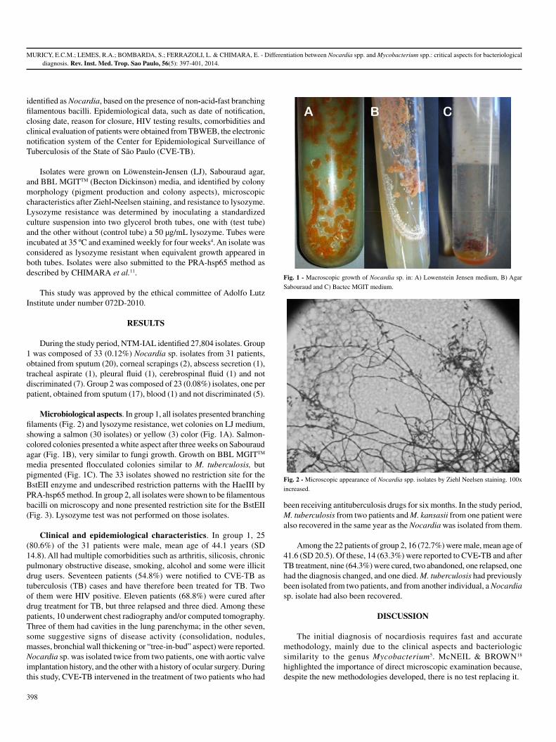
MURICY, E.C.M.; LEMES, R.A.; BOMBARDA, S.; FERRAZOLI, L. & CHIMARA, E. - Differentiation between Nocardia spp. and Mycobacterium spp.: critical aspects for bacteriological diagnosis. Rev. Inst. Med. Trop. Sao Paulo, 56(5): 397-401, 2014.
398
identified as Nocardia, based on the presence of non-acid-fast branching filamentous bacilli. Epidemiological data, such as date of notification, closing date, reason for closure, HIV testing results, comorbidities and clinical evaluation of patients were obtained from TBWEB, the electronic notification system of the Center for Epidemiological Surveillance of Tuberculosis of the State of São Paulo (CVE-TB).
Isolates were grown on Löwenstein-Jensen (LJ), Sabouraud agar, and BBL MGITTM (Becton Dickinson) media, and identified by colony morphology (pigment production and colony aspects), microscopic characteristics after Ziehl-Neelsen staining, and resistance to lysozyme. Lysozyme resistance was determined by inoculating a standardized culture suspension into two glycerol broth tubes, one with (test tube) and the other without (control tube) a 50 µg/mL lysozyme. Tubes were incubated at 35 ºC and examined weekly for four weeks4. An isolate was considered as lysozyme resistant when equivalent growth appeared in both tubes. Isolates were also submitted to the PRA-hsp65 method as described by CHIMARA et al.11.
This study was approved by the ethical committee of Adolfo Lutz Institute under number 072D-2010.
RESULTS
During the study period, NTM-IAL identified 27,804 isolates. Group 1 was composed of 33 (0.12%) Nocardia sp. isolates from 31 patients, obtained from sputum (20), corneal scrapings (2), abscess secretion (1), tracheal aspirate (1), pleural fluid (1), cerebrospinal fluid (1) and not discriminated (7). Group 2 was composed of 23 (0.08%) isolates, one per patient, obtained from sputum (17), blood (1) and not discriminated (5).
Microbiological aspects. In group 1, all isolates presented branching filaments (Fig. 2) and lysozyme resistance, wet colonies on LJ medium, showing a salmon (30 isolates) or yellow (3) color (Fig. 1A). Salmon-colored colonies presented a white aspect after three weeks on Sabouraud agar (Fig. 1B), very similar to fungi growth. Growth on BBL MGITTM media presented flocculated colonies similar to M. tuberculosis, but pigmented (Fig. 1C). The 33 isolates showed no restriction site for the BstEII enzyme and undescribed restriction patterns with the HaeIII by PRA-hsp65 method. In group 2, all isolates were shown to be filamentous bacilli on microscopy and none presented restriction site for the BstEII (Fig. 3). Lysozyme test was not performed on those isolates.
Clinical and epidemiological characteristics. In group 1, 25 (80.6%) of the 31 patients were male, mean age of 44.1 years (SD 14.8). All had multiple comorbidities such as arthritis, silicosis, chronic pulmonary obstructive disease, smoking, alcohol and some were illicit drug users. Seventeen patients (54.8%) were notified to CVE-TB as tuberculosis (TB) cases and have therefore been treated for TB. Two of them were HIV positive. Eleven patients (68.8%) were cured after drug treatment for TB, but three relapsed and three died. Among these patients, 10 underwent chest radiography and/or computed tomography. Three of them had cavities in the lung parenchyma; in the other seven, some suggestive signs of disease activity (consolidation, nodules, masses, bronchial wall thickening or “tree-in-bud” aspect) were reported. Nocardia sp. was isolated twice from two patients, one with aortic valve implantation history, and the other with a history of ocular surgery. During this study, CVE-TB intervened in the treatment of two patients who had
been receiving antituberculosis drugs for six months. In the study period, M. tuberculosis from two patients and M. kansasii from one patient were also recovered in the same year as the Nocardia was isolated from them.
Among the 22 patients of group 2, 16 (72.7%) were male, mean age of 41.6 (SD 20.5). Of these, 14 (63.3%) were reported to CVE-TB and after TB treatment, nine (64.3%) were cured, two abandoned, one relapsed, one had the diagnosis changed, and one died. M. tuberculosis had previously been isolated from two patients, and from another individual, a Nocardia sp. isolate had also been recovered.
DISCUSSION
The initial diagnosis of nocardiosis requires fast and accurate methodology, mainly due to the clinical aspects and bacteriologic similarity to the genus Mycobacterium5. McNEIL & BROWN18 highlighted the importance of direct microscopic examination because, despite the new methodologies developed, there is no test replacing it.
Fig. 1 - Macroscopic growth of Nocardia sp. in: A) Lowenstein Jensen medium, B) Agar
Sabouraud and C) Bactec MGIT medium.
Fig. 2 - Microscopic appearance of Nocardia spp. isolates by Ziehl Neelsen staining. 100x
increased.

MURICY, E.C.M.; LEMES, R.A.; BOMBARDA, S.; FERRAZOLI, L. & CHIMARA, E. - Differentiation between Nocardia spp. and Mycobacterium spp.: critical aspects for bacteriological diagnosis. Rev. Inst. Med. Trop. Sao Paulo, 56(5): 397-401, 2014.
399
In this study, 33 isolates and 17 cases of nocardiosis in a five-year period were found. Nocardia spp. isolation accounted for 0.12% of all isolates identified by NTM-IAL, from 2008 to 2011. HELAL et al.15 studied 200 sputum samples from patients diagnosed as pulmonary TB in Egypt, based on clinical symptoms, chest X-ray and direct smear test by acid-fast staining method. Four (2.0%) of these were PCR positive for 16S rRNA primer specific for Nocardia, a higher percentage than the one obtained in this study. Despite the presence of Nocardia detected by PCR, HELAL et al.15 could not obtain growth on LJ. Although the literature shows the need for special culture media17, isolates of Nocardia spp. received by NTM-IAL showed good growth on LJ medium, as well as on other media used for isolation of mycobacteria (Fig. 1). Colony morphology on LJ from many species of Nocardia may be confused with Mycobacterium species, making diagnosis difficult. Moreover, microscopic analysis should be carefully performed, because, as these two genera may present ramifications18, Nocardia may be misidentified as mycobacteria13 in the routine diagnosis of TB. The differentiation is performed by observation of aerial hyphae, produced only by species of the genus Nocardia18. Change in cell morphology of Nocardia depending on the age of the culture may also lead to misidentification. Young cultures exhibit branched-long filaments, while old cultures present bacilli and cocci originating from fragmentation of filaments, which can be considered mycobacteria bacilli for non-expert microscopists. Despite the Ziehl Neelsen method having been used for strong acid fast bacteria, such as the Mycobacterium species, and the modified Ziehl Neelsen method for weak acid fast bacteria, such as the Nocardia species, this study detected the Nocardia by the Ziehl Neelsen method used for mycobacteria. This fact shows the importance of an expertise reading, since both genera can be detected.
Many studies have shown that phenotypic methods are not accurate for Nocardia identification and the need for molecular methods is
a reality1,5,19. BAIO et al.3 identified seven isolates by 31 different phenotypic tests and analyzed profiles using six identification systems. Two isolates were identified as Nocardia sp., because a common outcome was not found using different schemes. Only two isolates were correctly identified by phenotypic identification, compared to the MLSA results. Although phenotypic identification produces misidentifications, it can improve molecular identification since some molecular targets can present high sequence similarity12.
PRA-hsp65 method13 is routinely used at NTM-IAL to amplify a 440 pb gene fragment that encodes a protein of 65 kDa for mycobacteria and actinomycetes9. As Nocardia lack the BstEII restriction site, this feature can be used in the presumptive identification of the genus. Additionally, Nocardia HaeIII patterns are different to the algorithm described by CHIMARA et al. 11.
This study showed three cases of mixed infection by different Nocardia and mycobacteria species in immunocompetent patients. A similar situation was described at the Baystate Medical Center, MA, USA, where bacteriological analysis revealed the presence of mixed cultures of N. asteroides and M. tuberculosis7 in two patients with clinical TB. The authors of this study showed the importance of bacteriological investigation in cases of treatment failure, because it may indicate that the patient harbors another agent that is not being addressed, also observed in the present study. Other cases of mixed infection by these two genera have been reported in immunocompromised patients10,20.
The finding of Nocardia should not be considered contamination and the case should be investigated23. In our study, 17 patients had some kind of comorbidity (silicosis, smoking, alcohol abuse) or immunosuppression, characterizing this group as high risk for nocardiosis23.
Mycobacteriosis and nocardiosis treatments are different, so the correct identification of the agent is essential to avoid ineffective therapy15. Initially, all notified patients were treated as TB, but for 31.2% of them, treatment was not effective. In these cases, the failure of TB treatment was evaluated and new clinical, radiological and bacteriological exams were performed to establish the cause of failure. However, Nocardia isolation was not taken into consideration and some patients were characterized as TB relapse cases.
In Brazil, TB patients to be considered cured should have, at least, two negative smears during the follow-up phase and one more after treatment of the disease. In this survey, all patients with prior treatment for TB had this criterion6. Pulmonary nocardiosis should be considered as differential diagnosis when TB fails to respond with standard therapy, or if the patient’s condition worsens despite adequate therapy.
The isolates of group 2 were only presumptively identified as Nocardia sp., a limitation of the study, and possibly they are Nocardia or even another actinomycete. An alternative to solve the identification can be sequencing the 16SrRNA region. BHARADWAJ et al.5, by sequencing organisms with ambiguous phenotypes, was able to identify different actinomycetes recovered from clinically significant specimens.
Therefore , pat ients with a his tory of comorbidi t ies , immunosuppression, whose isolates present rough, pigmented colonies of filamentous aerial hyphae forming bacilli, resistant to lysozyme and
Fig. 3 - Molecular pattern of Nocardia spp. isolates digested by BstEII restriction enzyme.
M: 50 bp molecular size marker.

MURICY, E.C.M.; LEMES, R.A.; BOMBARDA, S.; FERRAZOLI, L. & CHIMARA, E. - Differentiation between Nocardia spp. and Mycobacterium spp.: critical aspects for bacteriological diagnosis. Rev. Inst. Med. Trop. Sao Paulo, 56(5): 397-401, 2014.
400
without the restriction site for BstEII, are suggestive of having Nocardia spp. infection. In a mycobacterial-routine laboratory, an algorithm for genera and species identification of isolates suggestive of Nocardia spp. is essential for the correct diagnosis, which assists in providing adequate and early treatment for patients.
RESUMO
Diferenciação de Nocardia spp. e Mycobacterium spp.: aspectos críticos para o diagnóstico bacteriológico
Novas metodologias têm sido desenvolvidas para a identificação de Nocardia spp. mas o diagnóstico inicial ainda necessita de método rápido e preciso, principalmente devido à similaridade com o gênero Mycobacterium, clínica e bacteriologicamente. O crescimento em meio de Löwenstein Jensen (LJ), a presença de bacilos corados pela coloração de Ziehl Neelsen e colônias com características diferentes podem ser fatores de confusão entre nocardias e micobactérias. Este estudo descreve a ocorrência de Nocardia spp. em laboratório de referência em micobacteriologia, observando-se as principais dificuldades em diferenciar Nocardia spp. e Mycobacterium spp., correlacionando isolados com casos de nocardiose. Os registros laboratoriais dos anos 2008 a 2012 foram analisados e os isolados identificados como Nocardia sp. ou como bacilos não álcool - ácido resistentes (NBAAR) foram selecionados. Os dados epidemiológicos e bacteriológicos foram analisados. Trinta e três isolados identificados como Nocardia sp. e 22 como NBAAR foram selecionados para este estudo, perfazendo 0,12% do total de isolados identificados no período estudado. A identificação presuntiva foi baseada na morfologia macroscópica e microscópica, resistência à lisozima e perfis de restrição pelo método PRA-hsp65. Nocardia spp. pode crescer em meios de isolamento para micobactérias (LJ e BBL MGITTM) e microscopia de morfologia e as colônias são muito semelhantes a algumas espécies de micobactérias. Dezessete pacientes (54,8%) foram notificados e tratados para tuberculose, mas apresentaram sinais e sintomas para nocardiose. Concluimos que a ocorrência de Nocardia sp. no período estudado foi de 0,12%. Os isolados com características de bacilos filamentosos, formadores de hifas aéreas, com colônias que podem ter pigmento, rugosas e que não possuem padrão de digestão para BstEII no método PRA-hsp65 são sugestivos de Nocardia spp. Para um laboratório de rotina de Micobactérias, um fluxo de identificação presuntiva para Nocardia spp. é essencial para permitir que esses isolados sejam identificados com técnicas mais precisas, para que seja oferecido o tratamento adequado e qualidade de vida aos pacientes.
ACKNOWLEDGMENTS
We thank Angela Pires Brandão for her excellent critical comments and suggestions to improve the manuscript.
This work was supported by grant number 2010/19122-0, of the São Paulo Research Foundation (FAPESP), Brazil.
REFERENCES
1. Almeida LA, Araujo R. Highlights on molecular identification of closely related species. Infect Genet Evol. 2013;13:67-75.
2. Alnaum HM, Elhassan MM, Mustafa FY, Hamid ME. Prevalence of Nocardia species among HIV-positive patients with suspected tuberculosis. Trop Doct. 2011;41:224-6.
3. Baio PVP, Ramos JN, Santos LS, Soriano MF, Ladeira EM, Souza MC, et al. Molecular identification of Nocardia isolates from clinical samples and an overview of human nocardiosis in Brazil. PLoS Negl Trop Dis. 2013;7:e2573.
4. Berd D. Laboratory identification of clinically important aerobic actinomycetes. Appl Microbiol. 1973;25:665-81.
5. Bharadwaj R, Swaminathan S, Salimnia H, Fairfax M, Frey A, Chandrasekar PH. Clinical impact of the use of 16S rRNA sequencing method for the identification of “difficult-to-identify” bacteria in immunocompromised hosts. Transpl Infect Dis. 2012;14:206-12.
6. Brasil. Ministério da Saúde. Manual de recomendações para o controle da tuberculose no Brasil. Brasília: Ministério da Saúde/Secretaria de Vigilância em Saúde/Departamento de Vigilância Epidemiológica; 2011. p. 284.
7. Brown RB, Sands M, Ryczak M. Community-acquired pneumonia caused by mixed aerobic bacteria. Chest. 1986;90:810-4.
8. Brown-Elliott BA, Brown JM, Conville PS, Wallace RJ Jr. Clinical and laboratory features of the Nocardia spp. based on current molecular taxonomy. Clin Microbiol Rev. 2006;19:259-82.
9. Chang PL, Hsieh WS, Chiang CL, Tuohy MJ, Hall GS, Procop GW, et al. The hsp65 gene patterns of less common Mycobacterium and Nocardia spp. by polymerase chain reaction-restriction fragment length polymorphism analysis with capillary electrophoresis. Diagn Microbiol Infect Dis. 2007;58:315-23.
10. Chaudhury RC, Aher AR, Rastogi V, Prabhu TK. A case of mixed pulmonary infection by nocardia and Mycobacterium tuberculosis. Indian J Pathol Microbiol. 2009;52:294-5.
11. Chimara E, Ferrazoli L, Ueky SY, Martins MC, Durham AM, Arbeit RD, et al. Reliable identification of mycobacterial species by PCR-restriction enzyme analysis (PRA)-hsp65 in a reference laboratory and elaboration of a sequence-based extended algorithm of PRA-hsp65 patterns. BMC Microbiol. 2008;8:48.
12. Conville PS, Witebsky FG. Multiple copies of the 16S rRNA gene in Nocardia nova isolates and implications for sequence-based identification procedures. J Clin Microbiol. 2005;43:2881-5.
13. De S, Desikan P. Pulmonary nocardiosis mimicking relapse of tuberculosis. BMJ Case Rep. 2009;2009.
14. Garcia-Bellmunt L, Sibila O, Solanes I, Sanchez-Reus F, Plaza V. Pulmonary nocardiosis in patients with COPD: characteristics and prognostic factors. Arch Bronconeumol. 2012;48:280-5.
15. Helal ZH, Khan MI, Ashour MSE, Eissa SA. Detection and characterization of Nocardia from patients diagnosed as tuberculosis in Egypt. Int J Biomed Sci. 2008;3:179-84.
16. Jones N, Khoosal M, Louw M, Karstaedt A. Nocardial infection as a complication of HIV in South Africa. J Infect. 2000;41:232-9.
17. Martínez Tomás R, Menéndez Villanueva R, Reyes Calzada S, Santos Durantez M, Vallés Tarazona JM, Modesto Alapont M, et al. Pulmonary nocardiosis: risk factors and outcomes. Respirology. 2007;12:394-400.
18. McNeil MM, Brown JM. The medically important aerobic actinomycetes: epidemiology and microbiology. Clin Microbiol Rev. 1994;7:357-417.
19. Minero MV, Marín M, Cercenado E, Rabadán PM, Bouza E, Muñoz P. Nocardiosis at the turn of the century. Medicine (Baltimore). 2009;88:250-61.
20. Rasheed MU, Belay G. Nocardiosis in HIV seropositive clinically suspected pulmonary tuberculosis patients. Trop Doct. 2008;38:34-5.

MURICY, E.C.M.; LEMES, R.A.; BOMBARDA, S.; FERRAZOLI, L. & CHIMARA, E. - Differentiation between Nocardia spp. and Mycobacterium spp.: critical aspects for bacteriological diagnosis. Rev. Inst. Med. Trop. Sao Paulo, 56(5): 397-401, 2014.
401
21. Uttamchandani RB, Daikos GL, Reyes RR, Fischl MA, Dickinson GM, Yamaguchi E, et al. Nocardiosis in 30 patients with advanced human immunodeficiency virus infection: clinical features and outcome. Clin Infect Dis. 1994;18:348-53.
22. Wauters G, Avesani V, Charlier J, Janssens M, Vaneechoutte M, Delmee M. Distribution of Nocardia species in clinical samples and their routine rapid identification in the laboratory. J Clin Microbiol. 2005;43:2624-8.
23. Wilson JW. Nocardiosis: updates and clinical overview. Mayo Clin Proc. 2012;87:403-7.
24. Yildiz O, Doganay M. Actinomycoses and Nocardia pulmonary infections. Curr Opin Pulm Med. 2006;12:228-34.
Received: 23 July 2013Accepted: 7 March 2014

Revista do Instituto de Medicina Tropical de São Pauloon line.
Publications from 1984 to the present data are now available on:http://www.scielo.br/rimtsp
PAST ISSUES FROM 1959 ON (PDF)www.imt.usp.br/portal/
SciELO – The Scientific Electronic Library OnLine - SciELO is an electronic virtual covering a selected collection of Brazilian scientific journals. The library is an integral part of a project being developed by FAPESP – Fundação de Amparo à Pesquisa do Estado de São Paulo, in partnership with BIREME – the Latin American and Caribbean Center on Health Sciences Information. SciELO interface provides access to its serials collection via an alphabetic list of titles or a subject index or a search by word of serial titles, publisher names, city of publication and subject. The interface also provides access to the full text of articles via author index or subject index or a search form on article elements such as author names, words from title, subject and words from full text.
FAPESP/BIREME Project on Scientific Electronic PublicationsLatin American and Caribbean Center on Health Sciences Information
Rua Botucatu 862 – 04023-901 São Paulo, SP – BrazilTel. (011) 5576-9863

Rev. Inst. Med. Trop. Sao Paulo56(5):403-409, September-October, 2014doi: 10.1590/S0036-46652014000500006
(1) Núcleo de Estudos em Malária, Superintendência de Controle de Endemias/Instituto de Medicina Tropical de São Paulo, Universidade de São Paulo, Av. Dr. Enéas de Carvalho Aguiar 470, 05403-000 São Paulo, SP, Brazil.
(2) Laboratório de Entomologia Médica, Superintendência de Controle de Endemias, São Paulo, SP, Brazil.(3) Laboratório de Protozoologia, Instituto de Medicina Tropical de São Paulo, Universidade de São Paulo, São Paulo, SP, Brazil. (4) Departamento de Doenças Parasitárias e Infecciosas, Faculdade de Medicina, Universidade de São Paulo, São Paulo, SP, Brazil.(5) Laboratórios de Investigação Médica, LIM49, Hospital das Clínicas, São Paulo, SP, Brazil.(6) Departamento de Parasitologia, Instituto de Ciências Biomédicas II, Universidade de São Paulo, São Paulo, SP, Brazil.Correspondence to: Karin Kirchgatter. Núcleo de Estudos em Malária, Superintendência de Controle de Endemias/Instituto de Medicina Tropical de São Paulo, Universidade de São Paulo,
Av. Dr. Enéas de Carvalho Aguiar 470, 05403-000 São Paulo, SP, Brasil. E-mail: [email protected]
Anopheles (Kerteszia) cruzii (DIPTERA: CULICIDAE) IN PERIDOMICILIARY AREA DURING ASYMPTOMATIC MALARIA TRANSMISSION IN THE ATLANTIC FOREST: MOLECULAR IDENTIFICATION OF BLOOD-MEAL
SOURCES INDICATES HUMANS AS PRIMARY INTERMEDIATE HOSTS
Karin KIRCHGATTER(1), Rosa Maria TUBAKI(2), Rosely dos Santos MALAFRONTE(3,4), Isabel Cristina ALVES(5), Giselle Fernandes Maciel de Castro LIMA(1), Lilian de Oliveira GUIMARÃES(1), Robson de Almeida ZAMPAULO(2) & Gerhard WUNDERLICH(6)
SUMMARY
Anopheles (Kerteszia) cruzii has been implicated as the primary vector of human and simian malarias out of the Brazilian Amazon and specifically in the Atlantic Forest regions. The presence of asymptomatic human cases, parasite-positive wild monkeys and the similarity between the parasites infecting them support the discussion whether these infections can be considered as a zoonosis. Although many aspects of the biology of An. cruzii have already been addressed, studies conducted during outbreaks of malaria transmission, aiming at the analysis of blood feeding and infectivity, are missing in the Atlantic Forest. This study was conducted in the location of Palestina, Juquitiba, where annually the majority of autochthonous human cases are notified in the Atlantic Forest of the state of São Paulo. Peridomiciliary sites were selected for collection of mosquitoes in a perimeter of up to 100 m around the residences of human malaria cases. The mosquitoes were analyzed with the purpose of molecular identification of blood-meal sources and to examine the prevalence of Plasmodium. A total of 13,441 females of An. (Ker.) cruzii were collected. The minimum infection rate was calculated at 0.03% and 0.01%, respectively, for P. vivax and P. malariae and only human blood was detected in the blood-fed mosquitoes analyzed. This data reinforce the hypothesis that asymptomatic human carriers are the main source of anopheline infection in the peridomiciliary area, making the probability of zoonotic transmission less likely to happen.
KEYWORDS: Asymptomatic malaria; Atlantic forest; Anopheles (Kerteszia) cruzii; Plasmodium malariae; Plasmodium vivax.
INTRODUCTION
Currently, the Amazon Region concentrates 99.8% of the malaria cases described in Brazil, with approximately 306,000 cases registered in 200928. Malaria outside the Amazon region is situated mainly in Atlantic forest regions, due to the presence of bromeliads where Anopheles mosquitoes of the subgenus Kerteszia use the axils as larval habitat28. An. (Ker.) cruzii and An. (Ker.) bellator are considered malaria vectors, and the first is the primary vector of human and simian malaria in these regions6,7,10,12. Accordingly, since the 1960’s there is a discussion whether these infections, termed “forest-malaria” or “bromeliad-malaria”, can be considered as a zoonosis, where monkeys possibly act as reservoirs8.
Some data support the hypothesis that malaria can be a zoonosis in the Atlantic Forest. In relation to the vector, synanthropy of An. cruzii has been demonstrated in Atlantic Forest areas of the state of São Paulo16,20. These studies have shown that females may fly to the anthropic environment to feed on blood mainly in the domestic and peridomestic
areas and then return to the natural environment20. Moreover, a high vertical mobility, with distribution from ground level to tree tops, has been shown in An. cruzii36. In relation to the hosts, simian malaria was shown to frequently occur in the forested coastal mountains of the Southeastern region, where 35% of the examined monkeys were positive for P. brasilianum or P. simian and the identification of natural accidental human infection due to P. simium9. The parasitological prevalence of P. vivax and P. malariae in wild monkeys from the Atlantic Forest has also been described in more recent studies11,39. Finally, the simian malaria parasites P. brasilianum and P. simium are genetically indistinguishable from those responsible for human malaria in the Atlantic forest, P. malariae and P. vivax, respectively13,25.
On the other hand, it is also important to note that human cases detected in the Atlantic forest are generally asymptomatic, showing only subpatent levels of parasites. These individuals figure as appropriate reservoir hosts because they are not routinely identified as parasite carriers by typical malaria control programs which monitor only patent malaria cases.

KIRCHGATTER, K.; TUBAKI, R.M.; MALAFRONTE, R.S.; ALVES, I.C.; LIMA, G.F.M.C.; GUIMARÃES, L.O.; ZAMPAULO, R.A. & WUNDERLICH, G. - Anopheles (Kerteszia) cruzii (Diptera: Culicidae) in peridomiciliary area during asymptomatic malaria transmission in the Atlantic Forest: molecular identification of blood-meal sources indicates humans as primary intermediate hosts. Rev. Inst. Med. Trop. Sao Paulo, 56(5): 403-9, 2014.
404
Despite the fact that many aspects of the biology of An. cruzii have been elucidated26, studies conducted in settings of asymptomatic malaria transmission, aiming at the analysis of vector blood feeding and infectivity, are missing for the Atlantic Forest. Using this approach, this study tried to screen individual mosquitoes for the presence of non-human blood, mainly monkey blood, in order to support or reject the hypothesis of zoonotic transmission of malaria in this region. The city of Juquitiba was chosen to investigate these aspects, as thirteen cases, almost one third of all autochthonous cases in the Atlantic Forest of the state of São Paulo, were notified there in 20075. The location of Palestina was specifically chosen for being responsible for the identification of the largest number of cases in the region and because dwellings are in the vicinity of woods, where monkeys are frequently seen. The mosquitoes were analyzed with the purposes of (i) molecular identification of blood-meal sources; (ii) to examine the prevalence of Plasmodium; (iii) to collect information on seasonality; and (iv) to evaluate methods of Kerteszia sampling, with the final aim of contributing to the design of new strategies for asymptomatic malaria prevention and control in Atlantic Forest.
MATERIAL AND METHODS
Study Area: The city of Juquitiba is located in the metropolitan area of São Paulo, state of São Paulo, Brazil, and covers an area of 521.6 km2, with a population of 28,961 inhabitants, of whom 22.6% live in the rural area32. Juquitiba has an ecotourism economic activity that comprises activities such as canoeing, camping and fishing, due to the mountainous landscape and rivers. Concerning exposure to malaria vectors in peridomiciliary habitats, the local population consists mainly of employees who look after the properties and live near the cottages, as well as people who come from the city of São Paulo to spend the weekend at holiday cottages for recreation. The climate in this region is humid and subtropical24, with the coldest mean temperature below 18 °C in a dry winter (June-August) and the warmest month above 22 °C in the wet summer season (December-February). The annual rainfall is
about 1300 mm and the average altitude is 685 m. In 2006 and 2007, 16 and 13 mainly asymptomatic malaria cases were reported in Juquitiba, respectively5.
Mosquito sampling and handling: Adult female mosquitoes were collected from January 2006 to September 2007 in the wet and dry seasons at eight sites in the peridomiciliary environment (Fig. 1). The collection sites have the same landscape features; they are rural human settlements with anthropic modifications and variable distances from patches of forest, allowing frequent contact of man-forest and man-mosquitoes. The dwellings are scattered along Palestina Road, usually located near the forest, and the anthropic environment is constituted by extensive agricultural activities. The collection sites are connected by Embratel Road, Palestina Road and Olaria Road (Fig. 1B).
Mosquitoes were collected from January 2006 to September 2007 by: (i) Nasci aspirator27; (ii) Shannon trap34; and (iii) UV-CDC (Centers for Disease Control) light traps baited with CO
235. These methods were
used from nine to 12 am, seven to nine pm and six pm to seven am, respectively, during wet (January to March) and dry (July to September) seasons, for three consecutive days per month, totaling 36 days.
Collections with Nasci aspirator were made to supply representative samples of blood-engorged resting females33. The Nasci aspirators were used in the early morning, in the proximity of bromeliads (Kerteszia breeding sites), to collect resting females at four peridomiciliary sites where malaria cases were identified. One collector went towards the canopy trees to aspirate near the bromeliads, for intervals of 15 minutes, to complete the three-hour period.
Collections with CDC light traps and Shannon traps were used to sample Plasmodium infected females. Ten CDC light traps with UV light baited with CO
2 were distributed on site 4, specifically in
the peridomiciliary outskirts of the cottage. This site was chosen
Fig. 1 - A. Location of the County (Municipality) of Juquitiba, São Paulo State, Brazil. B. Sites of collection at the domiciliary environment in Juquitiba County. Modified from Google Earth
and Wikimedia Commons.

KIRCHGATTER, K.; TUBAKI, R.M.; MALAFRONTE, R.S.; ALVES, I.C.; LIMA, G.F.M.C.; GUIMARÃES, L.O.; ZAMPAULO, R.A. & WUNDERLICH, G. - Anopheles (Kerteszia) cruzii (Diptera: Culicidae) in peridomiciliary area during asymptomatic malaria transmission in the Atlantic Forest: molecular identification of blood-meal sources indicates humans as primary intermediate hosts. Rev. Inst. Med. Trop. Sao Paulo, 56(5): 403-9, 2014.
405
because the first malaria case detected in the parasitological survey done in January 2005 lives in this cottage. The complete data of the parasitological survey will be published elsewhere. Site 4 is a remnant of the forest in a steep relief with higher altitudes situated 40 m away from the cottage. The peridomiciliary area corresponds to open man-made fields with isolated trees without bromeliads. Samples were collected at site 4 to verify the abundance of Kerteszia subgenus in a high forest cover in a steeper area in the proximity of the dwelling. The traps were distributed in an altitudinal range of 711-778 m (above sea level) with 10 m of distance between each other. Altitudinal positions of mosquito traps were measured using portable GPS. The seasonal abundance of An. cruzii with the overall collections at site 4 (Fig. 1B) was estimated by using the William’s mean21, and the altitudinal distribution of An. cruzii abundances was compared in dry and wet seasons by the non-parametric Friedman test.
A comparison of the effectiveness of CDC light traps and the Shannon traps at sampling An. cruzii was carried out in the peridomiciliary habitats of the sites 1, 2 and 6, where collections were conducted simultaneously. The collections were compared by standardizing their number to obtain the monthly Williams geometric mean. The comparison of the anopheline abundances was verified by Wilcoxon’s matched-pairs test.
Adult mosquitoes were killed with chloroform steam and transported to the laboratory, where their specimens were identified on chilled tables with a stereomicroscope and using identification keys18,40. All mosquitoes with fresh or visible blood remnants were individually placed in 1.5-mL microcentrifuge tubes, sealed with parafilm, labeled according to species, collection site and stored at -20 °C.
Mosquito gDNA extraction and PCR amplification of Plasmodium SSU fragment: DNA from each mosquito was extracted as described31, and the assay to detect Plasmodium infection was performed in pools of ten mosquitoes. The pools were separated by species, day of capture and type of trap. PCR amplification was carried out according to a full-nested protocol38, which uses oligonucleotides in conserved sequences, in the small subunit (SSU) ribosomal RNA of human Plasmodium species in a first reaction. The second amplification was carried out with specific primers for three human Plasmodium species circulating in Brazil (P. falciparum, P. vivax and P. malariae). PCR products were electrophoresed in 2% agarose gels, stained with ethidium bromide and visualized under UV-light. The minimum infection rate (MIR) was calculated as the ratio of the number of positive pools to the total number of mosquitoes tested37.
gDNA extraction from blood-fed mosquitoes and blood meal identification: Genomic DNA of blood fed mosquitoes was obtained using PureLinkTM Genomic DNA Purification Kit (Invitrogen). PCR was used to amplify host DNA from the mosquito blood meal using primers L14841 and H1514923 or B1 and B630 designed to amplify, respectively, fragments with ~300 bp and ~1 kb of the mitochondrial cytb gene from a wide array of animals, including mammals, birds, amphibians, reptiles and fishes. This methodology was successfully used to identify the blood meal sources in mosquitoes from São Paulo Zoo2.
Amplified fragments were purified from gels and sequenced directly using the corresponding flanking primers. Sequences were identified by comparison to the GenBank DNA sequence database (www.ncbi.nlm.nih.gov/blast/Blast.cgi). Positive identification and host species assignment
were made when exact or nearly exact matches (> 98%) were obtained. Human cytb fragments show less than 86% of sequence identity with those from different primate species normally described in the Atlantic Forest of the state of São Paulo (Callithrix jacchus, Alouatta guariba and Cebus apella) (data not shown).
RESULTS
Anopheline abundance, distribution and seasonal variation: A total of 13,462 Anopheles females were collected, including 13,441 of An. (Ker.) cruzii, 13 of An. (Nys.) evansae, six of An. (Nys.) lutzii, one of An.(Nys.) galvaoi and one of An. (Ker.) bellator. Since the study of An. (Ker.) cruzii is the objective of this work, and this species corresponded to 99.8% of the collected individuals, only these mosquitoes were used for infection rate determination and blood meal identification. From the 13,441 of An. (Ker.) cruzii collected, 55.2% were obtained in CDC, 42% in Shannon traps and 2.8% in Nasci aspirators (Table 1).
To verify the abundance of An. cruzii in a high forest cover in a steeper area in the proximity of the dwelling samples were collected at site 4 using CO
2-baited UV-CDC traps. Most mosquitoes were captured
in the wet season (January-March), as shown in Table 2. The comparison made in the varying altitudinal positions of CDCs showed statistical support (Fr = 45.3, p < 0.0001), meaning that there was a statistically
Table 1Females of An. (Ker.) cruzii collected by different methods, at eight
peridomiciliary collection sites, in Juquitiba, State of São Paulo, from January 2006 through September 2007
SitePosition
according GPS#Malaria case*
MethodMosquitoes
collected
124o00’01.2”S 47o06'12.9"W
19/05/2005CDC 1
Shannon 51
224o00’46.8”S 47o05’57.2”W
19/05/2005
CDC 304
Aspirator 361
Shannon 1759
324o00’03.8”S 47o06'09.0"W
02/06/2005CDC 143
Aspirator 1
424o01’31.7”S 47o05'44.5"W
08/12/2005 CDC 5925
524o01’46.6”S 47o05'40.4"W
02/06/2005 CDC 996
624o01’20.8”S 47o05'18.4"W
--CDC 57
Shannon 3374
724o01’10.1”S 47o05'08.4"W
-- Shannon 349
823o59’28.7”S 47o05'33.7"W
--Aspirator 9
Shannon 111
Total 13441
# Global Positioning System. *Date (DD/MM/YY) of the last malaria case detected by thick blood smear before the beginning of the entomological survey.

KIRCHGATTER, K.; TUBAKI, R.M.; MALAFRONTE, R.S.; ALVES, I.C.; LIMA, G.F.M.C.; GUIMARÃES, L.O.; ZAMPAULO, R.A. & WUNDERLICH, G. - Anopheles (Kerteszia) cruzii (Diptera: Culicidae) in peridomiciliary area during asymptomatic malaria transmission in the Atlantic Forest: molecular identification of blood-meal sources indicates humans as primary intermediate hosts. Rev. Inst. Med. Trop. Sao Paulo, 56(5): 403-9, 2014.
406
significant difference between the number of females collected according to the season.
The efficacy of CDC and Shannon traps as collection methods was compared by standardizing the number of collections and collectors, and also evaluated in the sites, where collections were conducted simultaneously (sites 1, 2 and 6). The Williams mean of An. (Ker.) cruzii collected with Shannon traps suggested higher values than those obtained by CDC-CO
2 traps (Fig. 2). The Wilcoxon test (W = 20.0, p =
0.19) showed no significant differences in the numbers of captured An. (Ker.) cruzii collected by the two methods in the peridomiciliary sites.
Infection rate: Considering the large number of An. (Ker.) cruzii females obtained, only 67.5% of the total (9,072 mosquitoes from 13,441 females) was tested for Plasmodium infection (1,119 and 7,953,
collected in 2006 and 2007, respectively). From mosquitoes collected in 2006, two pools, both from January 16th, were positive for P. vivax: one with females sampled from site 4 and another one with females obtained from site 5. From those collected in 2007, one pool from site 4, also from January 16th, was positive for P. vivax, and one pool sampled from site 6 on January 17th, was positive for P. malariae. All samples were collected with CDCs. The minimum infection rate (MIR) was 0.03% and 0.01% for P. vivax and P. malariae, respectively.
Blood meal identification: All engorged mosquitoes collected by CDC and Nasci aspirator (10 and 16 mosquitoes, respectively), in both years, were tested for blood meal identification (Table 3). Blood-meal sources were successfully identified by DNA sequencing of the cytb fragment in 73% of engorged mosquitoes analyzed. In a total of 27% of engorged mosquitoes, no DNA from blood was detected, since the amplification of cytb gene was not obtained. All detected blood meals were identified as human-derived. In a more detailed analysis carried out using the PCR fragment sequence obtained from a mosquito (collected at site 2 in 2006 using Nasci aspirator), 730 bp were submitted to BLASTN to GenBank and only two substitutions were found in relation to a cytb sequence from Homo sapiens haplotype I [GenBank:EU091245].
DISCUSSION
This study reports molecular identification of blood-meal sources, seasonal abundance and malaria infectivity in Anopheles (Kerteszia) cruzii (Diptera: Culicidae) from a small focus of malaria transmission in a non-endemic area in the Atlantic Forest, São Paulo, Brazil. To the authors’ knowledge, this is the first report using a molecular methodology based on cytochrome b sequences to identify the blood-meal source carried out with mosquitoes collected during malaria transmission in Brazil.
During the sampling period, a good amount of mosquitoes were obtained, with the vast majority (97.2%) collected by CDC and Shannon traps. Although an excessive number of Kerteszia females were collected
Table 2Seasonal activity of An. cruzii (William’ s mean) collected with CO
2 baited UV-
CDC, at different altitudes at site 4, Juquitiba, during summer (Jan-Mar) and winter (Jul-Sep), 2006-2007, São Paulo
Position (m)
Altitude (m)
Jan Feb Mar Jul Aug Set
10 711 2.1 1.0 1.6 0.1 0.0 0.3
20 722 3.1 0.6 1.5 0.2 0.1 0.6
30 729 2.6 0.9 0.6 0.3 0.1 0.2
40 742 3.7 1.9 0.9 0.0 0.1 0.4
50 740 4.3 1.0 2.5 0.5 0.2 0.5
60 741 4.6 1.2 3.4 0.2 0.2 0.6
70 755 4.3 2.1 2.0 0.4 0.2 1.2
80 773 2.0 1.6 2.4 0.0 0.1 0.4
90 779 4.1 2.1 2.1 0.2 0.1 0.8
100 799 2.3 0.7 1.6 0.3 0.0 0.0
Fig. 2 - Comparison of collections with CDC and Shannon traps in the peridomiciliary sites
1, 2 and 6, in Juquitiba, São Paulo. The values were shown as William’s mean of An. (Ker.)
cruzii collected in the wet (Jan-Mar) and dry (Jul-Sep) seasons in 2006 and 2007.
Table 3Number of blood-fed mosquitoes analyzed for blood meal identification,
according to site, method and year of collection
Collection site
Blood meal source
Method2006 2007
TotalHuman ND Human
2CDC
Aspirator-
02- -
01 03
01 05
3CDC
Aspirator-
01- -
- -
- 01
4 CDC - 05 02 07
5 CDC - - 01 01
6 CDC - - 01 01
8 Aspirator 08 02 - 10
Total 11 07 08 26
ND = not determined.

KIRCHGATTER, K.; TUBAKI, R.M.; MALAFRONTE, R.S.; ALVES, I.C.; LIMA, G.F.M.C.; GUIMARÃES, L.O.; ZAMPAULO, R.A. & WUNDERLICH, G. - Anopheles (Kerteszia) cruzii (Diptera: Culicidae) in peridomiciliary area during asymptomatic malaria transmission in the Atlantic Forest: molecular identification of blood-meal sources indicates humans as primary intermediate hosts. Rev. Inst. Med. Trop. Sao Paulo, 56(5): 403-9, 2014.
407
at the three sites in the wet season of 2007 with the Shannon trap, the Wilcoxon test results showed no significant difference between CDC and Shannon traps. CDC collection frequencies compensated negative collections with Shannon traps in the dry season. Negative collections must have occurred due to unidentified specific environmental conditions.
Human malaria cases in the Atlantic Rain Forest, as well as infected mosquitoes, were described by several authors3,4,12,31, and the finding of natural infections of P. vivax and P. malariae in anophelines from this study match those detected by these authors. Also, the herein used molecular identification method is able to identify one naturally infected mosquito in a pool of ten mosquitoes and has been used in many other studies3,12. Here, the minimum infection rate (MIR) was 0.03% and 0.01% for P. vivax and P. malariae, respectively. Similar rates were found by using ELISA methodology in anophelines collected in the same city1 or by using PCR-based techniques, in Parelheiros, a locality at a distance of 70 km from Juquitiba and also located in Atlantic Forest regions12. Altogether, these rates are well around ten times lower than in endemic regions in the Amazon19. It is important to note that the positive mosquitoes were collected with CDCs on January 16th, 2006, at sites 4 and 5, and on February 17th, 2006, one additional malaria case was detected at site 5. In 2007, the positive mosquitoes were also collected in January, at sites 4 and 6. Perhaps not coincidentally, in February and March, five malaria cases were detected in the region by thick blood smear. The complete data of the epidemiological investigation will be published elsewhere.
Studies of feeding preferences of An. cruzii in the state of São Paulo using precipitin tests showed the presence of equine blood and rodent blood, but mainly human blood among the engorged mosquitoes14,15. However, the methods used in these studies did not allow the differentiation of human blood and simian blood. Here, a molecular approach capable of discriminating, at the species level, the source of the blood meal was used. Notably, mosquitoes were collected during the transmission season, in an area where malaria can be considered a zoonosis. Notwithstanding, only human blood was identified in the mosquitoes. Importantly, although the blood meal source was not identified for some of the mosquitoes examined, this result was also found in previous studies, using another14 or the same methodology2. This probably occurs due to the fact that the blood begins to be digested soon after ingestion by the mosquito and, as a consequence, the DNA is being degraded. Consequently, the longer the post-ingestion period, the lower the possibility of DNA detection29. Ultimately, it is important to note that the methodology used here for blood meal identification is one of the most widely used all over the world and is able to reliably detect blood from all vertebrates with the same performance22.
In relation to the collection method used to obtain engorged females, very few mosquitoes were captured with the Nasci aspirator, although this was properly used. Females of the Kerteszia subgenus are deliberately exophilic, resting in natural outdoor shelters, but it was assumed that the mosquitoes were rare or flew away with the proximity of the collectors. On the other hand, using CDC traps, other studies also showed a lower probability of collecting engorged mosquitoes, since they collect females that are actively host-seeking33. Thus, the analysis of the results showed that the methods for collecting mosquitoes employed here are reliable tools to sample Kerteszia females and showed regularity in accordance with seasonal periods of Kerteszia activity. However, it would be desirable
to improve and standardize the collection methods to enable comparisons of results of different studies.
The participation of An. cruzii as a vector of Plasmodium in Atlantic Forest regions is recognized in Brazil, as well as the strong dependence of this species on the forest17. The predominance of this vector in the peridomiciliary area was confirmed, as well as the capacity of transmitting P. malariae and P. vivax. However, only human blood was recorded in the engorged mosquitoes. Given the significant sampling reached herein, the complete absence of detection of monkey blood does not favor the hypothesis of considering malaria cases as a zoonosis, and the occurrence of asymptomatic infections can possibly explain the maintenance of the circulation of parasites in humans, in the peridomiciliary habitat. Complementary studies testing the infection rates of monkeys in the study area may elucidate what real risk these animals play in terms of apparently very rarely occurring interspecies transmission of malaria.
CONCLUSION
In the present study, the predominance of An. (Ker.) cruzii was confirmed in the peridomiciliary area, as well as the capacity of transmitting P. malariae and P. vivax and the first molecular study of identification of the blood-meal source carried out with mosquitoes collected during asymptomatic malaria transmission in the Brazilian Atlantic Forest was reported. As only human blood was identified in the engorged mosquitoes, this data reinforce the hypothesis that asymptomatic human carriers are the main source of anopheline infection in the peridomiciliary area.
RESUMO
Anopheles (Kerteszia) cruzii (Diptera: Culicidae) em área peridomiciliar durante transmissão de malária assintomática na
Mata Atlântica: identificação molecular das fontes de repasto sanguíneo indica humanos como hospedeiros intermediários
primários
Anopheles (Kerteszia) cruzii é o vetor primário das malárias humana e simiana fora da Amazônia Brasileira e especificamente nas regiões de Mata Atlântica. A presença de casos humanos assintomáticos, macacos silvestres positivos para Plasmodium e a similaridade entre os parasitas que os infectam suportam a discussão se essas infecções podem ser consideradas como zoonoses. Embora muitos aspectos da biologia de An. cruzii já tenham sido abordados, estudos conduzidos durante surtos de transmissão de malária, visando a análise de repasto sanguíneo e infectividade, são ausentes na Mata Atlântica. Este estudo foi conduzido na localidade de Palestina, Juquitiba, Mata Atlântica do Estado de São Paulo, onde anualmente a maioria dos casos humanos autóctones é notificada. Locais em peridomicílio foram selecionados para coleta de mosquitos em um perímetro de até 100 m em torno das residências de casos humanos de malária e da floresta circundante. Os mosquitos foram analisados com o objetivo de identificação molecular das fontes de repasto sanguíneo e para examinar a prevalência de Plasmodium. Um total de 13.441 fêmeas de An. (Ker.) cruzii foi coletado. A taxa de infecção mínima foi calculada a 0,03% e 0,01%, respectivamente, para P. vivax e P. malariae e somente sangue humano foi detectado nos mosquitos analisados que se alimentaram com sangue. Nossos dados reforçam a hipótese de que

KIRCHGATTER, K.; TUBAKI, R.M.; MALAFRONTE, R.S.; ALVES, I.C.; LIMA, G.F.M.C.; GUIMARÃES, L.O.; ZAMPAULO, R.A. & WUNDERLICH, G. - Anopheles (Kerteszia) cruzii (Diptera: Culicidae) in peridomiciliary area during asymptomatic malaria transmission in the Atlantic Forest: molecular identification of blood-meal sources indicates humans as primary intermediate hosts. Rev. Inst. Med. Trop. Sao Paulo, 56(5): 403-9, 2014.
408
os portadores humanos assintomáticos são a principal fonte de infecção para os anofelinos na área do peridomicílio, tornando a transmissão zoonótica improvável.
ACKNOWLEDGEMENTS
This research was funded by FAPESP (03/06420-0).
REFERENCES
1. Branquinho MS, Marrelli MT, Curado I, Natal D, Barata JM, Tubaki R, et al. Infecção do Anopheles (Kerteszia) cruzii por Plasmodium vivax e Plasmodium vivax variante VK247 nos municípios de São Vicente e Juquitiba, São Paulo. Rev Panam Salud Publica. 1997;2:189-93.
2. Bueno MG, Lopez RPG, Menezes RMT, Costa-Nascimento MJ, Lima GFMC, Araújo RAS, et al. Identification of Plasmodium relictum causing mortality in penguins (Spheniscus magellanicus) from São Paulo Zoo, Brazil. Vet Parasitol. 2010;173:123-7.
3. Cerutti C Jr, Boulos M, Coutinho AF, Hatab MC, Falqueto A, Rezende HR, et al. Epidemiologic aspects of the malaria transmission cycle in an area of very low incidence in Brazil. Malar J. 2007;6:33.
4. Curado I, Malafronte RS, Duarte AMC, Kirchgatter K, Branquinho MS, Galati EAB. Malaria epidemiology in low-endemicity areas of the Atlantic Forest in the Vale do Ribeira, São Paulo, Brazil. Acta Trop. 2006;100:54-62.
5. CVE (Centro de Vigilância Epidemiológica). Situação epidemiológica da malária no Estado de São Paulo, 2007. Bol Epidemiol Paul. 2008;5(56):24-5. Available from: f tp : / / f tp . cve . saude . sp .gov.b r /doc_ tec /ou t ros /bo l_bepa5608 .pdf
6. Deane LM, Deane MP, Ferreira Neto J. Studies on transmission of simian malaria and on a natural infection of man with Plasmodium simium in Brazil. Bull World Health Organ. 1966;35:805-8.
7. Deane LM, Ferreira Neto JA, Deane SP, Silveira IP. Anopheles (Kerteszia) cruzii, a natural vector of the monkey malaria parasites, Plasmodium simium and Plasmodium brasilianum. Trans R Soc Trop Med Hyg. 1970;64:647.
8. Deane LM, Ferreira-Neto JA, Lima MM. The vertical dispersion of Anopheles (Kerteszia) cruzii in a forest in southern Brazil suggests that human cases of malaria of simian origin might be expected. Mem Inst Oswaldo Cruz. 1984;79:461-3.
9. Deane LM. Simian malaria in Brazil. Mem Inst Oswaldo Cruz. 1992;87(Suppl 3):1-20.
10. Deane LM. Studies on simian malaria in Brazil. Bull World Health Organ. 1964;31:752-3.
11. Duarte AM, Malafronte RS, Cerutti C Jr, Curado I, de Paiva BR, Maeda AY, et al. Natural Plasmodium infections in Brazilian wild monkeys: reservoirs for human infections? Acta Trop. 2008;107:179-85.
12. Duarte AM, Pereira DM, de Paula MB, Fernandes A, Urbinatti PR, Ribeiro AF, et al. Natural infection in anopheline species and its implications for autochthonous malaria in the Atlantic Forest in Brazil. Parasit Vectors. 2013;6:58.
13. Escalante AA, Barrio E, Ayala FJ. Evolutionary origin of human and primate malarias: evidence from the circumsporozoite protein gene. Mol Biol Evol. 1995;12:616-26.
14. Forattini OP, Gomes AC, Natal D, Kakitani I, Marucci D. Preferências alimentares de mosquitos Culicidae no Vale do Ribeira, São Paulo, Brasil. Rev Saude Publica. 1987;21:171-87.
15. Forattini OP, Gomes AC, Natal D, Kakitani I, Marucci D. Preferências alimentares e domiciliação de mosquito Culicidae no Vale do Ribeira, São Paulo, Brasil, com especial referência a Aedes scapularis e Culex (Melanoconion). Rev Saude Publica. 1989;23:9-19.
16. Forattini OP, Kakitani I, dos Santos RC, Kobayashi KM, Ueno HM, Fernández Z. Potencial sinantrópico de mosquitos Kerteszia e Culex (Diptera:Culicidae) no Sudeste do Brasil. Rev Saude Publica. 2000;34:565-9.
17. Forattini OP, Kakitani I, Massad E, Marucci D. Studies on mosquitoes (Diptera:Culicidae) and anthropic environment. 11- Biting activity and blood-seeking parity of Anopheles (Kerteszia) cruzii in South-Eastern Brazil. Rev Saude Publica. 1996;30:107-14.
18. Forattini OP. Culicidologia médica. Identificação, biologia, epidemiologia. São Paulo: Edusp; 2002.v. 2.
19. Gil LH, Tada MS, Katsuragawa TH, Ribolla PE, da Silva LH. Urban and suburban malaria in Rondônia (Brazilian Western Amazon). II. Perennial transmissions with high anopheline densities are associated with human environmental changes. Mem Inst Oswaldo Cruz. 2007;102:271-6.
20. Guimarães AE, Gentile C, Lopes CM, Mello RP. Ecology of mosquitoes (Diptera: Culicidae) in areas of Serra do Mar State Park, State of São Paulo, Brazil. II. Habitat distribution. Mem Inst Oswaldo Cruz. 2000;95:17-28.
21. Haddow AJ. Studies on the biting habits and medical importance of East African mosquitoes in the genus Aedes. I- Subgenera Aedimorphus, Banksinella and Dunnius. Bull Entomol Res. 1960;50:759-79.
22. Kent RJ. Molecular methods for arthropod bloodmeal identification and applications to ecological and vector-borne disease studies. Mol Ecol Resour. 2009;9:4-18.
23. Kocher TD, Thomas WK, Meyer A, Edwards SV, Pääbo S, Villablanca FX, et al. Dynamics of mitochondrial DNA evolution in animals: amplification and sequencing with conserved primers. Proc Natl Acad Sci USA. 1989;86:6196-200.
24. Köppen W. Climatologia: con un estudio de los climas de la tierra. México: Fondo de Cultura Economica; 1948.
25. Lim CS, Tazi L, Ayala FJ. Plasmodium vivax: recent world expansion and genetic identity to Plasmodium simium. Proc Natl Acad Sci USA. 2005;102:15523-8.
26. Marrelli MT, Malafronte RS, Sallum MA, Natal D. Kerteszia subgenus of Anopheles associated with the Brazilian Atlantic rainforest: current knowledge and future challenges. Malar J. 2007;6:127.
27. Nasci RS. A lightweight battery-powered aspirator for collecting resting mosquitoes in the field. Mosq News. 1981;41:808-11.
28. Oliveira-Ferreira J, Lacerda MV, Brasil P, Ladislau JL, Tauil PL, Daniel-Ribeiro CT. Malaria in Brazil: an overview. Malar J. 2010;9:115.
29. Oshaghi MA, Chavshin AR, Vatandoost H, Yaaghoobi F, Mohtarami F, Noorjah N. Effects of post-ingestion and physical conditions on PCR amplification of host blood meal DNA in mosquitoes. Exp Parasitol. 2006;112:232-6.
30. Pereira SL, Baker AJ. Vicariant speciation of curassows (Aves, Cracidae): a hypothesis based on mitochondrial DNA phylogeny. Auk. 2004;121:682-94.
31. Rezende HR, Soares RM, Cerutti C Jr, Alves IC, Natal D, Urbinatti PR, et al. Entomological characterization and natural infection of anophelines in an area of the Atlantic Forest with autochthonous malaria cases in mountainous region of Espírito Santo State, Brazil. Neotrop Entomol. 2009;38:272-80.
32. SEADE (Fundação Sistema Estadual de Análise de Dados). Perfil Municipal de Juquitiba. 2012. [cited 2012 Oct 04]. Available from: http://www.seade.gov.br/produtos/perfil/perfil.php
33. Service MW. Mosquito ecology field sampling methods. 2nd ed. Essex: Elsevier Science Publishers; 1993.
34. Shannon RC. Methods for collecting and feeding mosquitoes in jungle yellow fever studies. Am J Trop Med Hyg. 1939;(s1-19):131-40.

KIRCHGATTER, K.; TUBAKI, R.M.; MALAFRONTE, R.S.; ALVES, I.C.; LIMA, G.F.M.C.; GUIMARÃES, L.O.; ZAMPAULO, R.A. & WUNDERLICH, G. - Anopheles (Kerteszia) cruzii (Diptera: Culicidae) in peridomiciliary area during asymptomatic malaria transmission in the Atlantic Forest: molecular identification of blood-meal sources indicates humans as primary intermediate hosts. Rev. Inst. Med. Trop. Sao Paulo, 56(5): 403-9, 2014.
409
35. Sudia WD, Chamberlain RW. Battery-operated light trap, an improved model. Mosq News. 1962;22:126-9.
36. Ueno HM, Forattini OP, Kakitani I. Distribuição vertical e sazonal de Anopheles (Kerteszia) em Ilha Comprida, SP, Brasil. Rev Saude Publica. 2007;41:269-75.
37. Walter SD, Hildreth SW, Beaty BJ. Estimation of infection rates in populations of organisms using pools of variable size. Am J Epidemiol. 1980;112:124-8.
38. Win TT, Lin K, Mizuno S, Zhou M, Liu Q, Ferreira MU, et al. Wide distribution of Plasmodium ovale in Myanmar. Trop Med Int Health. 2002;7:231-9.
39. Yamasaki T, Duarte AM, Curado I, Summa ME, Neves DV, Wunderlich G, et al. Detection of etiological agents of malaria in howler monkeys from Atlantic Forests, rescued in regions of São Paulo city, Brazil. J Med Primatol. 2011;40:392-400.
40. Zavortink TJ. Mosquito studies (Diptera, Culicidae). XXIX. A review of the subgenus Kerteszia of Anopheles. Contrib Am Entomol Inst. 1973;9:1-54.
Received: 17 January 2014Accepted: 17 April 2014

LIBRARY OF THE SÃO PAULO INSTITUTE OF TROPICAL MEDICINE
Website: www.imt.usp.br/portal Address: Biblioteca do Instituto de Medicina Tropical de São Paulo da Universidade de São Paulo
Av. Dr. Enéas de Carvalho Aguiar, 470. Prédio 1 – Andar térreo.05403-000 São Paulo, SP, Brazil.
Telephone: 5511 3061-7003 - Fax: 5511 3062-2174
The Library of the São Paulo Institute of Tropical Medicine (IMTSP Library) was created on January 15, 1959 in order to serve all those who are interested in tropical diseases. To reach this objective, we select and acquire by donation and / or exchange appropriate material to be used by researchers and we maintain interchange between Institutions thorough the Journal of the São Paulo Institute of Tropical Medicine, since the Library has no funds to build its own patrimony.
The IMTSP Library has a patrimony consisting of books, theses, annals of congresses, journals, and reference works.
The collection fo journals existing in the Library can be verified through the USP – Bibliographic Database – OPAC – DEDALUS http://dedalus.usp.br:4500/ALEPH/eng/USP/USP/DEDALUS/start of the USP network.

Rev. Inst. Med. Trop. Sao Paulo56(5):411-415, September-October, 2014doi: 10.1590/S0036-46652014000500007
(1) Department of Bacteriology-Mycology. Institute of Tropical Medicine “Pedro Kourí”. Havana. Cuba. Tel.: 255 3530. E-mail: [email protected](2) Department of Pathology. Institute of Tropical Medicine “Pedro Kourí”. Havana. Cuba.Correspondence to: Angel Alberto NODA, Instituto de Medicina Tropical “Pedro Kourí”. Ave Novia del Mediodía, Km 6 ½, La Lisa, La Habana, 11400, Cuba. Tel.: 255 3530. E-mail:
HIGH SENSITIVE PCR METHOD FOR DETECTION OF PATHOGENIC Leptospira spp. IN PARAFFIN-EMBEDDED TISSUES
Angel Alberto NODA(1), Islay RODRÍGUEZ(1), Yaindrys RODRÍGUEZ(1), Anamays GOVÍN(2), Carmen FERNÁNDEZ(1) & Ana Margarita OBREGÓN(1)
SUMMARY
This study describes the development and application of a new PCR assay for the specific detection of pathogenic leptospires and its comparison with a previously reported PCR protocol. New primers were designed for PCR optimization and evaluation in artificially-infected paraffin-embedded tissues. PCR was then applied to post-mortem, paraffin-embedded samples, followed by amplicon sequencing. The PCR was more efficient than the reported protocol, allowing the amplification of expected DNA fragment from the artificially infected samples and from 44% of the post-mortem samples. The sequences of PCR amplicons from different patients showed >99% homology with pathogenic leptospires DNA sequences. The applicability of a highly sensitive and specific tool to screen histological specimens for the detection of pathogenic Leptospira spp. would facilitate a better assessment of the prevalence and epidemiology of leptospirosis, which constitutes a health problem in many countries.
KEYWORDS: Leptospira; PCR; LipL32; Pathology; Diagnosis.
INTRODUCTION
Leptospirosis is one of the most important zoonotic diseases worldwide. The disease varies from subclinical infection to a severe illness with multi-organ involvement. Because of the variety of clinical symptoms, leptospirosis is often misdiagnosed as influenza, hepatic disease and hemorrhagic fever with renal syndrome or dengue fever, leading to an underestimation of prevalence6. The tropical climate, which provides periods of intense rainfall resulting in floods, combined with a dense population living in poverty and poor sanitation conditions are key features which favor the maintenance and spread of leptospirosis7,10.
The direct demonstration of pathogenic Leptospira spp. as causative agent is an invaluable tool for the diagnosis of human leptospirosis; however, the current bacteriological methods for the detection of leptospires in clinical specimens such as blood, cerebrospinal fluid, urine and post mortem tissue have some disadvantages for routine use. Despite substantial recent improvements there is no current method that is sufficiently sensitive and specific11, mainly in samples from deceased individuals where the diagnosis is based on presumptive stain assays.
The polymerase chain reaction (PCR) is the most commonly used method for detecting leptospires in clinical samples16. However, post-mortem diagnosis of leptospirosis in paraffin-embedded tissue is much less widely used and PCR based methods have low sensitivity for these types of samples3.
The most frequently analyzed material in pathology laboratories is paraffin-embedded tissue; indeed, biological samples preserved in this way constitute a valuable source of information for retrospective analysis17. Due to DNA fragmentation during sample processing for paraffin blocks, the PCR must be highly sensitive and specific; therefore, few reliable PCR methods exist currently for the detection of pathogen leptospires. The development of more efficient detection systems would therefore be of benefit in the confirmatory diagnosis and the understanding of the epidemiology of leptospirosis in a susceptible population.
MATERIALS AND METHODS
Primers amplifying a 115bp PCR product from 23S ribosomal DNA were published previously18. Partial sequences of genes encoding 23S rRNA of pathogenic leptospires were obtained from GenBank (http://www.ncbi.nlm.nih.gov/) and aligned using Clustal software (www.ebi.ac.uk/clustalw/). The specificity of primers was verified by the software BLAST (http://blast.ncbi.nlm.gov/Blast.cgi). Dimer formation was predicted with OPERON (http://www.operon.com/technical/toolkit.aspx). Other parameters such as melting and annealing temperatures, primer length, GC content and 3´ extreme stability were evaluated using BioEdit software (www.bioedit.com/).
In order to design a new set of primers and using Bioedit software, we aligned sequences corresponding to the gene encoding the major

NODA, A.A.; RODRÍGUEZ, I.; RODRÍGUEZ, Y.; GOVÍN, A.; FERNÁNDEZ, C. & OBREGÓN, A.M. - High sensitive PCR method for detection of pathogenic Leptospira spp. in paraffin-embedded tissues. Rev. Inst. Med. Trop. Sao Paulo, 56(5): 411-5, 2014.
412
lipoprotein in pathogenic Leptospira spp., lipL32, which is found only in pathogenic strains of Leptospira.
Leptospira borgpetersenii serovar Castellonis strain Castellon 3 was subcultured in EMJH medium for seven days. When cell density had reached approximately 108cell/mL, the DNA was extracted using High Pure PCR Template Preparation Kit (ROCHE, Switzerland) for genomic DNA. The quality of extracted DNA was checked by spectrophotometry and by agarose gel electrophoresis.
The PCR was performed in a 25 µL volumes containing variable concentrations of each primer, 0.125 U of Taq polymerase (QIAGEN, Germany), PCR buffer 1X (dNTP 0.02 mM, MgCl
2 0.25 mM, KCl
0.025M, Tris HCl 0.025M, bovine serum albumin 0.1 mg/mL), 5 µL of template DNA and made up to 25 µL with double-distilled water.
The reaction was performed using a Mastercycler personal Eppendorf thermal cycler for 40 cycles. The annealing temperature was chosen from a temperature gradient (53, 55, 57, 59, 61 and 63 oC) with 0.5 µM of primer concentration and then, different primers concentrations were used: 0.5; 0.2 and 0.1 µM. The cycling process involved five steps as follows: 1) 94 oC for five min, 2) 94 oC for one min, 3) optimal annealing temperature for 30 sec, 4) extension at 72 oC for one min, and a final cooling step of 4 oC. A total of 20 µL of the PCR products was analyzed on a 2% agarose gel and visualized with ethidium bromide staining under ultraviolet light.
Serial and ten-fold dilutions of serovar Castellonis DNA were made from 7x10⁷geq/reaction to 0.7geq/reaction for estimating the PCR detection limit. In addition, DNA extracted from related strains (L. interrogans serovar Copenhageni (M20), L. interrogans serovar Icterohaemorrhagiae (RGA), L. interrogans serovar Pomona (Pomona), L. interrogans serovar Canicola (Hond Utrecht IV), L. interrogans serovar Hardjo (Hardjoprajitno), L. interrogans serovar Pyrogenes (Salinem), L. kirschneri serovar Gryppotiphosa (Moskva V), L. interrogans serovar Autumnalis (Akiyami A), L. kirschneri serovar Cynopteri (3522 C)) and unrelated species (L. biflexa serovar Patoc (Patoc I), Borrelia burgdorferi sensu stricto B31, B. garinii NE83, B. afzelii NE17, Mycoplasma genitalium ATCC 33530, Haemophilus influenzae type b ATCC 49629, Streptococcus pneumoniae ATCC 49619, S. agalactiae ATCC 13813, Pseudomonas aeuruginosa ATCC 27853, Escherichia coli ATCC 35218, and E. coli ATCC 25922), were used for the specificity assay. Results from the PCR sensitivity and specificity tests were compared with 23S-based PCR (23S PCR) results, according to the WOO et al. (1997) procedure18.
The new PCR method (LipL32 PCR) in paraffin-embedded tissues was compared with the 23S PCR. First, paraffin-embedded human tissues negative for leptospirosis by Haematoxilin-Eosin and Warthin-Starry stains were evaluated. Fragments of liver, lung and kidney samples were cut into small pieces (approximately 5 g) and inoculated with 100 µL of a bacterial suspension (approximately 107cell/mL) and other bacterial species as described above using a syringe. Two pieces of inoculated and non-inoculated (negative control) tissues were immediately paraffin-embedded; finally they were used directly for DNA extraction.
Fifty seven paraffin-embedded organ samples (liver, kidney, lung, brain, heart or spleen) from 24 dead patients with suspicion of leptospirosis, received at the National Reference Laboratory of
Leptospires (IPK) during 2012, were tested firstly by a PCR method detecting sample inhibition and this amplified a 268 bp of the gene encoding human β-globin15; after, the non-inhibited samples were subjected to the application of both PCR methods, LipL32 PCR and 23S PCR.
The DNA extraction was performed with Chelex-100 as described by DE ARMAS et al. (2006)5 with minor modifications. Briefly, 10 µm fragments of paraffin-embedded tissue (cut previously using microtome) were thinly cut with scalpel and put into a 1.5 mL tube containing 200 µL of 5% Chelex-100 solution in Tris-EDTA 1X; then it was vortexed and heated to 95 oC during 15 min, followed by a 13 000 xg spin for 10 min. Finally, 5 µL of supernatant containing the DNA was used as template for PCR assays.
Ten PCR amplified products from different patients were gel-purified with the QIAEX II (QIAGEN) commercial kit following the manufacturer’s instructions. Sequencing of the gel-purified amplicons was performed using the GenomeLabTM Dye Terminator Cycle Sequencing with Quick Start Kit (Beckman Coulter®; USA).
This study was conducted in compliance with the Declaration of Helsinki and it was approved by the Institutional Ethical Committee.
RESULTS
The complete in silico analysis of 23S PCR primers predicted the formation of primer dimers, but the main deficiency was the recognition by BLAST, with 100% homology, of non-leptospiral bacterial strains; mainly E. coli, Streptococcus suis, S. agalactiae, Yersinia enterocolitica, Pseudomonas aeruginosa, P. mendocina, P. basiacearum, Bacillus thuringiensis, B. amyloliquefaciens, Aeromonas veronii, Vibrio cholerae, Brucella melitensis, Clostridium botulinum, C. lentocellum, Leishmania mexicana, Trichomonas vaginalis and Candida parapsilosis. This unexpected result led us to design a new set of primers that fulfilled the requirement for PCR specificity; they define a 146 bp region of DNA unique to pathogenic Leptospira spp. The new primers designated pfLp32-1 and pfLp32-2 have the sequences 5´-TAGAATCAAGATCCCAAATCCTCC-3´ and 5´-CCAACAGATGCAACGAAAGATCC-3´, respectively.
The annealing temperature gradient assay showed that the best amplification in terms of quantity of PCR product was observed at 59 oC, and it was selected for all further studies. A primer concentration of 0.5 µM showed the clearest and most prominent PCR product in agarose gel electrophoresis; this was used, for all subsequent experiments. The detection limit assay of LipL32 PCR showed amplification down to a dilution corresponding to 7geq/reaction (Fig. 1). In addition, all the leptospiral strains except for the saprophytic serovar Patoc gave rise to the expected DNA fragment. On the other hand, no bands were observed when the non-leptospiral strains were used for the LipL32 PCR. In contrast, the 23S PCR resulted in non-specific amplification in some cases with non-leptospiral strains as Haemophilus influenzae and Streptococcus pneumoniae, although weak signals were observed in both cases.
When we applied the LipL32 PCR to detect pathogenic Leptospira spp. in fresh tissues that had been inoculated with bacterial suspensions and immediately embedded in paraffin, all of the artificially infected

NODA, A.A.; RODRÍGUEZ, I.; RODRÍGUEZ, Y.; GOVÍN, A.; FERNÁNDEZ, C. & OBREGÓN, A.M. - High sensitive PCR method for detection of pathogenic Leptospira spp. in paraffin-embedded tissues. Rev. Inst. Med. Trop. Sao Paulo, 56(5): 411-5, 2014.
413
samples showed the expected amplicon by gel electrophoresis, while no product was observed from the organs artificially infected with non-leptospiral strains. However, with 23S PCR three samples infected with Leptospira were negative and the others were positive but with a lower intensity band; no product was generated from the non-leptospiral strains.
Just two samples from liver and kidney revealed PCR inhibition by human β-globin PCR; these were not used in further studies. Forty four percent of samples (24/55) from 12 deceased patients with suspected leptospirosis were positive by the LipL32 PCR, while no amplification was observed when 23S PCR was used (Fig. 2). The organs showing the highest intensity of PCR product were kidney (10/18), liver (7/16) and lung (6/9), but with no statistically significant differences between them (p > 0.05). In 83% (10/12) of the patients it was noted there was positivity in more than one organ.
All sequenced DNA fragments showed > 99% sequence identity to the pathogenic Leptospira spp. lipL32 gene fragment in GenBank, confirming the amplification specificity of the pfLp32-1 and pfLp32-2 primers.
DISCUSSION
In this study, we designed and validated a PCR strategy for the detection of pathogenic leptospires in paraffin-embedded organs. PCR analysis of such specimens is hindered by fastidious specimen due to the possible DNA fragmentation produced during the paraffin fixing process and by the low burden of leptospires, particularly when the analysis is performed on thin paraffin sections and with samples contaminated by other microorganisms19. In these cases, it is necessary to have a highly sensitive and specific PCR method, capable of amplifying a small DNA region.
The primers reported for use in the 23S PCR are not suitable for a highly specific PCR due to the fact that they theoretically recognized, with 100% match, a wide group of microorganisms not belonging to the genus Leptospira. This lack of specificity presents a major problem, since several species of microorganisms could be present in organ fragments and unexpected amplification can appear. In contrast, the LipL32 PCR was highly specific for pathogenic leptospires. According to results obtained with respect to specificity assay for 23S PCR, the in silico results for non-leptospiral strains (except to Haemophilus influenzae and Streptococcus pneumoniae where weak and unspecific bands appeared) were different from the empirical results. This could be explained by genetic differences among the experimentally used bacterial strains and those whose DNA sequences appear in GenBank database. Besides, it is necessary to have the same recognizing pattern by both primers to target DNA, in order to assure a specific amplification.
What was remarkable in the sensitivity assay with LipL32 primers (Fig. 1, panel A) was the fact that free primers start to appear when the template concentration is lower and less product is amplified. However, Panel B did not show any band of free primers, which suggested that the 23S primer concentration was insufficient for optimal amplification, and this could have affected the PCR sensitivity. But given the unsatisfactory in silico evaluation performance of primers, we decided not to optimize parameters in the PCR.
Successful performance evaluation of LipL32 PCR in artificially infected tissues demonstrated the usefulness of this assay when tissues were used as matrix. In addition, during the artificial infection procedure, 107cell/mL was used in order to ensure efficiency of the process, in spite of the low leptospiral burden probably founded in post-mortem samples.
The successful PCR amplification with paraffin-embedded samples supports for the first time the use of Chelex-100 for the DNA extraction from leptospires in this kind of sample. Chelex-100 has an increased affinity for heavy metal cations such as Ca2+, Mn2+ and Mg2+. It is known that the divalent metals can introduce DNA damage at a high temperature (e.g. 100 oC), and in the case of Mg2+, it is necessary for nuclease activity. Therefore, the use of Chelex-100 inactivates the nucleases and removes the divalent metals and some other PCR inhibitors1. The use of an internal control has allowed the demonstration of the high performance of Chelex-100 extraction method in these samples, with only a 3.5% of inhibited reactions.
The positive LipL32 PCR for the paraffin-embedded tissues and the identification of the amplicon as a lipL32 gene fragment of L. interrogans confirmed the etiological cause of death for the patients
Fig. 1 - PCR amplification from Leptospira borgpetersenii serovar Castellonis strain Castellon
3 DNA dilutions. (A) Using primers pfLp32-1 and pfLp32-2, (B) Using primers of 23S PCR.
Lane 1: Molecular weight pattern (100bp PROMEGA); lane 2: DNA 7x10⁷ geq/reaction;
lane 3: DNA 7x10⁶ geq/reaction; lane 4: DNA 7x10⁵ geq/reaction; lane 5: DNA 7x10⁴ geq/
reaction; lane 6: DNA 7x10³ geq/reaction; lane 7: DNA 7x10² geq/reaction; lane 8: DNA
7geq/reaction; lane 9: DNA 0.7 geq/reaction.
Fig. 2 - PCR amplification showing some positive paraffin-embedded tissue samples from
deceased with suspect of leptospirosis. Lane 1: Molecular weight pattern (100bp PROMEGA);
lanes 2 to 14: samples; lane 15: negative control (H2O ultrapure); lane 16: positive control
(DNA of L. borgpetersenii serovar Castellonis strain Castellon 3).

NODA, A.A.; RODRÍGUEZ, I.; RODRÍGUEZ, Y.; GOVÍN, A.; FERNÁNDEZ, C. & OBREGÓN, A.M. - High sensitive PCR method for detection of pathogenic Leptospira spp. in paraffin-embedded tissues. Rev. Inst. Med. Trop. Sao Paulo, 56(5): 411-5, 2014.
414
studied. The deficiency of the 23S PCR for the detection of DNA of pathogenic leptospires was also demonstrated; however, D’ANDREA et al. (2012) validated this assay in paraffin-embedded tissue with encouraging results4. Hence, a highly sensitive and specific PCR constitutes a valuable alternative for the leptospirosis confirmation in fatal human cases, in which a positive serological result is absent, because of either the lack of a serum sample or a significant level of antibodies.
Leptospirosis causes damage to capillary endothelial cells as the underlying cause of clinical manifestations, such as renal tubular dysfunction, liver disease, myocarditis and pulmonary hemorrhage9. The fact of finding higher positivity rates in kidney, liver and lung samples was consistent with this notion. A similar observation was reported in a recent molecular study using fresh tissues12. The positivity found in the lung samples supports the results reported by other authors on the potentially fatal damage to this organ during leptospiral infection8,13,14. The positivity was found in more than one organ per patient which suggests multi-organ infection. Unfortunately, clinical-epidemiological information from the deceased was incomplete; thus we could not make any inference of the founded positivity with multiorgan failure.
Paraffin-embedded tissue blocks are routinely used for histopathological examination and are also useful for specific pathogen detection by PCR. Paraffin-embedded tissue is stable at ambient temperature for an extended period of time and relatively easy to transport compared to fresh tissue, which has to be processed or frozen immediately2. In addition, archival material is an invaluable source for retrospective molecular and clinical investigation; nevertheless, fresh tissue samples are the specimens of relevance for prevalence and epidemiological studies for leptospirosis, consequently this kind of sample would be also evaluated using the proposed PCR.
As a final point, the detection of DNA from paraffin-embedded specimens by PCR is important for the retrospective study of different infectious diseases, including their epidemiology and risk assessment. This is particularly important for leptospirosis, due to the variety of clinical symptoms and signs, potentially leading to misdiagnosis and underestimation of disease prevalence worldwide.
RESUMEN
PCR altamente sensible para la detección de Leptospira spp. patógenas en tejidos embebidos en parafina
El presente estudio describe el desarrollo y aplicación de un nuevo ensayo de PCR para la detección específica de leptospiras patógenas y su comparación con un protocolo reportado previamente. Se diseñaron nuevos cebadores para la optimización y evaluación de la PCR en tejidos embebidos en parafina infectados artificialmente. La PCR se aplicó además a muestras de tejidos embebidos en parafina y se realizó la secuenciación del amplicón resultante. La PCR diseñada fue más eficiente que el protocolo reportado, permitiendo la amplificación del fragmento de ADN esperado en las muestras infectadas artificialmente y del 44% de las muestras post mortem. Se secuenciaron 10 amplicones provenientes de pacientes diferentes. La aplicabilidad de una herramienta altamente sensible y específica en la búsqueda de leptospiras patógenas en especímenes histopatológicos podría facilitar una mejor valoración
de la prevalencia y la epidemiología de la leptospirosis, la que constituye un problema de salud en disímiles países.
CONFLICT OF INTEREST
The authors declare that they have no conflict of interest.
ACKNOWLEDGEMENTS
We would like to acknowledge Prof. Virginia Capó, for her attention and suggestions, to Prof. Ben Adler and Prof. James Matsunaga, for the critical revision of the manuscript.
REFERENCES
1. Aygan A. Nucleic acid extraction from clinical specimens for PCR applications. Turk J Biol. 2006;30:107-20.
2. Behrhof W, Springer E, Bräuninger W, Kirkpatrick CJ, Weber A. PCR testing for Treponema pallidum in paraffin-embedded skin biopsy specimens: test design and impact on the diagnosis of syphilis. J Clin Pathol. 2008;61:390-5.
3. Brown PD, Carrington DG, Gravekamp C, van de Kemp H, Edwards CN, Jones SR, et al. Direct detection of leptospiral material in human postmortem samples. Res Microbiol. 2003;154:581-6.
4. D’Andrea A, Zamora Y, Alduina R, Monteverde V, Fernández C, Vitale M. Comparison of two PCR methods for detection of Leptospira interrogans in formalin-fixed and paraffin-embedded tissues. Mem Inst Oswaldo Cruz. 2012;107:85-8.
5. De Armas Y, Capó V, González E, Mederos L, Díaz R. DNA extraction from paraffin embedded tissues by Chelex-100. Rev Esp Patol. 2006;39:171-4.
6. Evangelista KV, Coburn J. Leptospira as an emerging pathogen: a review of its biology, pathogenesis and host immune responses. Future Microbiol. 2010;5:1413-25.
7. Fonseca CA, Freitas VLT, Romero EC, Spinosa C, Arroyo Sanches MC, Da Silva MV, et al. Polymerase chain reaction in comparison with serological tests for early diagnosis of human leptospirosis. Trop Med Int Health. 2006;11:1699-707.
8. Gouveia EL, Metcalfe J, de Carvalho AL, Aires TS, Villasboas-Bisneto JC, Queirroz A, et al. Leptospirosis associated Severe Pulmonary Hemorrhagic Syndrome, Salvador, Brazil. Emerg Infect Dis. 2008;14:505-8.
9. Hill MK, Sanders CV. Leptospiral pneumonia. Semin Respir Infect. 1997;12:44-9.
10. Ko AI, Galvão RM, Ribeiro Dourado CM, Johnson WD Jr, Riley LW. Urban epidemic of severe leptospirosis in Brazil. Salvador Lystospirosis Study Group. Lancet. 1999;354(9181):820-5.
11. Rodríguez I, Rodríguez I, Fernández C, Rodríguez JE, Cantillo J. Detection of leptospires from infected urine and tissue samples in vitro by modified Fontana silver stain. J Bras Patol Med Lab. 2013a;49:43-9.
12. Rodríguez Y, Rodríguez I, Zamora Y, Rodríguez JE, Valdés Y, Echevarria E, et al. Detección de ADN de leptospiras en tejidos frescos de fallecidos en Cuba, 2008-2011. Rev Cubana Med Trop. 2013b;65:211-22.
13. Santos Pérez LA, Bilbao González K, Segredo Molina Y, Cartaya Irastorga JM. Leptospirosis grave asociada con hemorragia pulmonar. Presentación de 3 pacientes pediátricos UCIP. Hospital Universitario “José Luis Miranda”. Rev Panam Infectol. 2008;10:36-42.
14. Seijo A, Coto H, San Juan J, Videla J, Deodato B, Cernigoi B, et al. Lethal leptospiral pulmonary hemorrhage: an emerging disease in Buenos Aires, Argentina. Emerg Infect Dis. 2002;8:1004-5.

NODA, A.A.; RODRÍGUEZ, I.; RODRÍGUEZ, Y.; GOVÍN, A.; FERNÁNDEZ, C. & OBREGÓN, A.M. - High sensitive PCR method for detection of pathogenic Leptospira spp. in paraffin-embedded tissues. Rev. Inst. Med. Trop. Sao Paulo, 56(5): 411-5, 2014.
415
15. Talmaci R, Traeger-Synodinos J, Kanavakis E, Coriu D, Colita D, Gavrila L. Scanning of beta-globin gene for identification of beta-thalassemia mutation in Romanian population. J Cell Mol Med. 2004;8:232-40.
16. Toyokawa T, Ohnishi M, Koizumi N. Diagnosis of acute leptospirosis. Expert Rev Anti Infect Ther. 2011;9:111-21.
17. Wiedorn KH, Olert J, Stacy R, Goldmann T, Kühl H, Matthus J. HOPE a new fixing technique enables preservation and extraction of high molecular weight DNA and RNA of >20 kb from paraffin-embedded tissues. Herpes-Glutamic acid buffer mediated organic solvent Protection Effect. Pathol Res Pract. 2002;198:735-40.
18. Woo TH, Smythe LD, Symonds ML, Norris MA, Dohnt MF, Patel BK. Rapid distinction between Leptospira interrogans and Leptospira biflexa by PCR amplification of 23S ribosomal DNA. FEMS Microbiol Lett. 1997;150:9-18.
19. Wu L, Patten N, Yamashiro CT, Chui B. Extraction and amplification of DNA from formalin-fixed, paraffin-embedded tissues. Appl Immunohistochem Mol Morphol. 2002;10:269-74.
Received: 26 August 2013Accepted: 12 February 2014

Revista do Instituto de Medicina Tropical de São Pauloon line.
Publications from 1984 to the present data are now available on:http://www.scielo.br/rimtsp
PAST ISSUES FROM 1959 ON (PDF)www.imt.usp.br/portal/
SciELO – The Scientific Electronic Library OnLine - SciELO is an electronic virtual covering a selected collection of Brazilian scientific journals. The library is an integral part of a project being developed by FAPESP – Fundação de Amparo à Pesquisa do Estado de São Paulo, in partnership with BIREME – the Latin American and Caribbean Center on Health Sciences Information. SciELO interface provides access to its serials collection via an alphabetic list of titles or a subject index or a search by word of serial titles, publisher names, city of publication and subject. The interface also provides access to the full text of articles via author index or subject index or a search form on article elements such as author names, words from title, subject and words from full text.
FAPESP/BIREME Project on Scientific Electronic PublicationsLatin American and Caribbean Center on Health Sciences Information
Rua Botucatu 862 – 04023-901 São Paulo, SP – BrazilTel. (011) 5576-9863

Rev. Inst. Med. Trop. Sao Paulo56(5):417-420, September-October, 2014doi: 10.1590/S0036-46652014000500008
(1) Department of Infectious Diseases, University of São Paulo Medical School, São Paulo, SP, Brazil.(2) Discipline of Clinical Immunology and Allergy, University of Sao Paulo Medical School, São Paulo, SP, Brazil.Correspondence to: Vivian Iida Avelino-Silva. E-mail:[email protected]
YELLOW FEVER PREVENTION STRATEGIES AWARENESS AMONG HIV-INFECTED PATIENTS IN SÃO PAULO, BRAZIL
Vivian Iida AVELINO-SILVA(1), Hilario Sousa FRANCELINO(2) & Esper Georges KALLÁS(1,2)
SUMMARY
Introduction: Vaccination is the main preventive strategy against Yellow Fever (YF), which is a public health concern in Brazil. However, HIV-infected patients might have insufficient knowledge regarding YF, YF prevention, and vaccines in general. Methods: In this questionnaire-based study, data from 158 HIV-infected individuals were addressed in three distinct outpatient clinics in São Paulo. Information was collected on demographic and clinical characteristics, as well as patients´ knowledge of vaccines, YF and YF preventive strategies. In addition, individual YF vaccine recommendations and vaccine status were investigated. Results: Although most participants adequately ascertain the vaccine as the main prevention strategy against YF, few participants were aware of the severity and lack of specific treatment for YF. Discrepancy in YF vaccine (patients who should have taken the vaccine, but did not) was observed in 18.8% of participants. Conclusion: YF is an important and preventable public health concern, and these results demonstrate that more information is necessary for the HIV-infected population.
KEYWORDS: HIV; AIDS; Yellow Fever; Vaccine; Questionnaire.
INTRODUCTION
Yellow fever (YF) is a public health concern in several Latin American and African countries. Vaccination is the main preventive strategy for this highly lethal disease, for which a specific treatment is lacking6.
In Brazil, the areas with recommendation for YF vaccine have been subjected to extension during the last decades after new YF epidemics and epizooties were documented in regions previously considered as out of risk1-2. Nevertheless, the population’s awareness on the disease, as well as the prevention strategies, is probably far from ideal.
HIV-infected patients are supported by the Brazilian public health system, for inpatient and outpatient care, antiretroviral therapy, and vaccines. A very small percentage of HIV-infected patients attend private medical offices. Despite regular access for treatment and health information during their clinical follow up, there is a hypothesis that HIV-infected patients have insufficient knowledge regarding YF, YF preventive strategies, and vaccines in general.
To address this issue, a self-completion questionnaire was applied to 158 HIV-infected patients regularly monitored in three outpatient clinics in São Paulo, Brazil.
METHODS
A cross sectional study was performed from March to November 2013, and included HIV-infected patients regularly monitored in three outpatient clinics in São Paulo, Brazil: one large public outpatient clinic (Group 1), one HIV clinical research cohort (Group 2), and one private clinic (Group 3). All participants provided an informed consent, and participants’ identities were kept confidential. The study was approved by the Ethics Committee of the University of São Paulo Medical School.
Self-completion questionnaires were applied in the clinic’s waiting area, either before or immediately after the medical appointment. Data were collected regarding demographic and clinical characteristics, last season’s Influenza vaccination history, and patients´ knowledge of vaccines, YF and YF preventive strategies. In addition, individual YF vaccine status was investigated. All data were collected in the form of closed questions.
The demographic and clinical characteristics referred by the three groups of patients were compared by Pearson’s chi-square test or by Fisher’s two-sided exact test. The age between the groups was compared using the Kruskal-Wallis-test. The Bonferroni correction was calculated for each pairwise comparison performed. The 95% confidence intervals for the percentage of binomial variables were calculated using the exact binomial method. All statistical tests used a significance level of 0.05

AVELINO-SILVA, V.I.; FRANCELINO, H.S. & KALLÁS, E.G. - Yellow fever prevention strategies awareness among HIV-infected patients in Sao Paulo, Brazil. Rev. Inst. Med. Trop. Sao Paulo, 56(5): 417-20, 2014.
418
and were performed using STATA 13.0 (StataCorp. College Station, TX: StataCorp LP.).
RESULTS
The study enrolled 91 participants in Group 1, 37 participants in Group 2 and 30 participants in Group 3, for an overall population of 158 HIV-infected patients.
Most participants were male (77.9%), with a median age of 44 years. Group 1 was significantly older than Groups 2 and 3, while Group 3 had a higher proportion of white ethnicity when compared to Group 1, and a higher proportion of graduate participants when compared to Groups 1 and 2.
According to the age distribution, chronic conditions other than HIV were found more frequently in Group 1.
Most participants in the three groups reported that children, healthy adults, adults with chronic conditions and elderly are populations to whom vaccines are important. A significantly smaller proportion of participants in Group 3 reported receiving a medical recommendation for vaccines during follow up, when compared to Group 1. In addition, fewer patients in Group 3 reported receiving the flu vaccine in the last season when compared to Group 1.
Regarding YF knowledge, although most participants adequately identify mosquitoes as YF transmission agents and vaccine as the main prevention strategy, in all three groups few participants are aware of the severity and lack of specific treatment for YF.
A higher proportion of participants in Group 3 reported traveling to or living in YF endemic areas, and accordingly Group 3 had a higher proportion of vaccinated participants. Discrepancy in YF vaccine (patients who should have taken the vaccine, but did not) was observed in 18.8% of participants (CI 95% = 13.0-25.9).
Table 1Demographic characteristics of participants and survey results
Total (N=158)
Group 1 (N=91)
Group 2 (N=37)
Group 3 (N=30)
Age (median, p25-75) 44 (36-52) 49 36 37
Male (% (N)) 77.8 (123) 73.6 (67) 83.8 (31) 83.3 (25)
Ethnicity (% (N)) White Black Mulatto Other/not reported
60.8 (96) 8.9 (14) 27.9 (44) 2.5 (4)
53.8 (49) 13.2 (12) 29.7 (27) 3.3 (3)
59.5 (22) 5.4 (2)
35.1 (13) -
83.3 (25)
- 13.3 (4) 3.3 (1)
Education level (% (N)) Elementary Middle school High school College/Graduate Not informed
3.2 (5)
12.7 (20) 33.5 (53)
50 (79; 42-58) 0.6 (1)
4.4 (4)
19.8 (18) 37.4 (34) 37.4 (34) 1.1 (1)
-
5.4 (2) 43.2 (16) 51.4 (19)
-
3.3 (1)
- 10 (3)
86.7 (26) -
Chronic conditions other than HIV (% (N)) 52.9 (83) 65.9 (60) 33.3 (12) 36.7 (11)
To which groups are vaccine important? (% (N)) Children Healthy adults Adults c. conditions Elderly All the above
88.0 (139) 82.9 (131) 73.4 (116) 82.9 (131) 63.3 (100)
90.1 (82) 80.2 (73) 68.1 (62) 80.2 (73) 59.3 (54)
78.4 (29) 89.2 (33) 75.7 (28) 81.1 (30) 64.9 (24)
93.3 (28) 83.3 (25) 86.7 (26) 93.3 (28) 73.3 (22)
Personal physician recommended vaccines during follow up (% (N)) 81.6 (129) 89.0 (81) 83.8 (31) 56.7 (17)
Received last season’s Flu vaccine 81.0 (128) 89.0 (81) 81.1 (30) 56.7 (17)
Knowledge in YF (% (N)) Correctly identifies mosquitoes as transmission route Correctly identifies YF as potentially severe disease, with no specific treatment Correctly identifies vaccine as YF prevention strategy Travels to YF endemic areas Past YF vaccine Travels to YF endemic areas but never received YF vaccine
82.2 (129) 14.6 (23)
86.0 (135) 44.6 (70) 27.4 (43) 18.8 (29)
76.9 (70) 15.4 (14)
81.3 (74) 28.6 (26) 15.4 (14) 19.3 (17)
86.1 (31) 11.1 (4)
94.4 (34) 66.7 (24)
25 (9) 22.2 (8)
93.3 (28) 16.7 (5)
90 (27)
66.7 (20) 66.7 (20) 13.3 (4)

AVELINO-SILVA, V.I.; FRANCELINO, H.S. & KALLÁS, E.G. - Yellow fever prevention strategies awareness among HIV-infected patients in Sao Paulo, Brazil. Rev. Inst. Med. Trop. Sao Paulo, 56(5): 417-20, 2014.
419
DISCUSSION
Brazilian state policies define selected regions in which the vaccine is recommended, either for those living or traveling to these locations. In the last decades, massive vaccination campaigns have followed the expansions in the YF vaccine recommendation area1,2. Nevertheless, this study found a large percentage of HIV-infected patients who are unaware of YF severity and lack of specific treatment. Furthermore, a significant proportion of participants should have been vaccinated, but remains unprotected.
Although several countries keep border vigilance systems for YF vaccine adequacy, vigilance for persons already inside the country’s frontiers are frequently lacking. Thus, non-immune individuals may enter the vaccine recommendation regions, posing a significant individual risk, as well as a risk for YF urbanization5.
Several previous studies have addressed knowledge, attitudes and practices (KAP) of travelers towards travel-related infectious diseases in airport surveys, also demonstrating important drawbacks in travelers’ KAP, which emphasize the need for better knowledge among travelers regarding preventable diseases3,8,9.
Although income was not investigated in this survey, it is likely due to better financial conditions that more patients in Group 3 reported traveling to YF risk areas and, accordingly, more participants in this Group had been vaccinated against YF, with only 13% reporting traveling to endemic areas without previous vaccination. Group 3 had less patients vaccinated against Influenza in the last season, and a smaller proportion of patients in Group 3 had received recommendation for vaccines during follow up. One possible explanation is related to the existence of a vaccination unit inside the clinic attended by Groups 1 and 2, but not by Group 3.
A general notion, possibly more consistent among individuals with a lower level of education, states that vaccines are beneficial only in extreme ages - young children and the elderly - and unnecessary in adults. This hypothesis has been tested in this study. These results may be biased due to the fact that this population of HIV-infected patients is different to the average adult population, either due to a higher level of education, or to the information delivered by the health care providers during clinical follow up. In addition, adult HIV-infected patients have specific vaccine recommendations, which might also increase awareness regarding vaccines in this population.
Risk perception is a complex issue, and previous studies have addressed the importance of effective communication on the risk of preventable diseases, as opposed to the risk of vaccination and other social, cultural and economic factors that may influence vaccination decisions4,7.
Anti-vaccination movements might also have an influence on vaccine compliance, but they do not seem to bring a particular impact on YF vaccine. On the other hand, reports on severe adverse events of the YF vaccine, and more specifically the YF vaccine-associated viscerotropic disease, have been described with increasing frequency. Even though the increasing frequency may be, at least, partially attributed to surveillance, the fear of such adverse events might have diminished vaccine uptake by travelers and residents in risky areas, or even health care providers’
recommendations for the vaccine; however, this hypothesis needs further investigation.
It is believed that these results are essential for this group of patients. First, they point out the main deficiencies in the knowledge of the HIV-infected population, and provide hints for education strategies. Second, they are indicators of the quality of HIV care in this setting. This is increasingly important in the Highly Active Antiretroviral Therapy era, when opportunistic conditions become rarer, and focus has changed to prevention strategies and primary care management. Third, the presence of unvaccinated participants who should have taken the vaccine in this group points to a deficiency in vaccination strategies.
This study has some limitations. The sample size was not very large and not randomly attained. Furthermore, selected outpatient clinics may not adequately represent the overall HIV-infected population. However, the results revealed important information on YF and YF vaccine awareness that may guide future actions to spread the knowledge of these issues and other preventable infectious diseases with immunogens, especially to those travelling to infectious disease-endemic areas.
In conclusion, YF is an important and preventable public health concern, and these results demonstrate that more information is necessary for the HIV-infected population.
RESUMO
Nível de conhecimento sobre estratégias de prevenção contra febre amarela entre pessoas que vivem com HIV em São Paulo, Brasil
A vacinação é a principal forma de prevenção contra a Febre Amarela (FA), doença de importância em saúde pública no Brasil. Entretanto, pessoas que vivem com HIV possivelmente possuem conhecimentos insuficientes a respeito da FA, suas formas de prevenção e também sobre vacinas de modo geral. Métodos: Neste estudo baseado em questionários de autopreenchimento, avaliamos dados de 158 pacientes infectados por HIV atendidos em três diferentes serviços ambulatoriais do Município de São Paulo. Foram coletados dados demográficos, clínicos, e dados relacionados ao grau de conhecimento a respeito de vacinas, da FA e de suas formas de prevenção. Além disso, avaliamos individualmente a indicação e antecedente de vacinação contra FA. Resultados: Embora a maioria dos participantes tenha identificado corretamente que a vacina é a principal forma de prevenção da FA, poucos tinham conhecimento a respeito da gravidade clínica e ausência de tratamento específico da doença. Discrepância na vacinação (caracterizada quando o participante deveria ter recebido a vacina, mas não a recebeu) foi observada em 18,8% dos casos. Conclusão: A FA é importante agravo em saúde pública, passível de prevenção, e nossos resultados demonstram que são necessárias mais ações de educação voltadas à população de pessoas que vivem com HIV.
ACKNOWLEDGEMENTS
The authors would like to thank Karina Takesaki Miyaji, Ana Marli Sartori and Marta Heloisa Lopes for reviewing the original survey and providing useful suggestions. The authors would also like to thank Tamara Newman Lobato Sousa, Jessica Fernandes Ramos and Fabiana Siroma for helping the application of the questionnaires. This study was supported by CAPES (Ministério da Educação - Brazil).

AVELINO-SILVA, V.I.; FRANCELINO, H.S. & KALLÁS, E.G. - Yellow fever prevention strategies awareness among HIV-infected patients in Sao Paulo, Brazil. Rev. Inst. Med. Trop. Sao Paulo, 56(5): 417-20, 2014.
420
AUTHORS´ CONTRIBUTION
VIAS devised the study, participated in the collection of clinical data, study design and manuscript writing; HSF participated in the collection of clinical data and manuscript revision; EGK coordinated the study and helped to draft the manuscript. All of the authors read and approved the final manuscript.
REFERENCES
1. Brasil. Ministério da Saúde. Secretaria de Vigilância em Saúde. Emergências em saúde pública de importância nacional (ESPIN) de febre amarela silvestre em São Paulo e no Rio Grande do Sul e a situação epidemiológica atual no Brasil (2008/2009). Brasília: Ministério da Saúde; 2009.
2. Brasil. Ministério da Saúde. Secretaria de Vigilância em Saúde. Situação da febre amarela silvestre no Brasil, 2007 e 2008. Brasília: Ministério da Saúde; 2008.
3. Hamer DH, Connor BA. Travel health knowledge, attitudes and practices among United States travelers. J Travel Med. 2004;11:23-6.
4. Larson H, Brocard Paterson P, Erondu N. The globalization of risk and risk perception: why we need a new model of risk communication for vaccines. Drug Saf. 2012;35:1053-9.
5. Lima JTF. Risco de urbanização da febre amarela no Brasil. Cad Saúde Pública. 1985;1:377-84.
6. Monath TP. Yellow fever: an update. Lancet Infect Dis. 2001;1:11-20.
7. Reyna VF. Risk perception and communication in vaccination decisions: a fuzzy-trace theory approach. Vaccine. 2012;30:3790-7.
8. Toovey S, Jamieson A, Holloway M. Travelers’ knowledge, attitudes and practices on the prevention of infectious diseases: results from a study at Johannesburg International Airport. J Travel Med. 2004;11:16-22.
9. Van Herck K, Van Damme P, Castelli F, Zuckerman J, Nothdurft H, Dahlgren AL, et al. Knowledge, attitudes and practices in travel-related infectious diseases: the European airport survey. J Travel Med. 2004;11:3-8.
Received: 12 November 2013Accepted: 14 March 2014

Rev. Inst. Med. Trop. Sao Paulo56(5):421-426, September-October, 2014doi: 10.1590/S0036-46652014000500009
Natural Products Laboratory, Department of Zoology, DDU, Gorakhpur University, Gorakhpur-273009 (U.P.), INDIA. E-mails: [email protected] Phone: 09450436049, [email protected] Phone no. 0551-2201171 (o) 2202127 (R).
Correspondence to: Dr. Ajay Singh, Phone no. 0551-2201171 (o) 2202127 (R ), E-mail: [email protected]
EFFECTS OF SINGLE, BINARY AND TERTIARY COMBINATIONS WITH Jatropha gossypifolia AND OTHER PLANT-DERIVED MOLLUSCICIDES ON REPRODUCTION AND SURVIVAL OF THE SNAIL
Lymnaea acuminata
Ram P. YADAV & Ajay SINGH
SUMMARY
The effect of sub-lethal doses (40% and 80% of LC50
/24h) of plant derived molluscicides of singly, binary (1:1) and tertiary (1:1:1) combinations of the Rutin, Ellagic acid, Betulin and taraxerol with J. gossypifolia latex, leaf and stem bark powder extracts and their active component on the reproduction of freshwater snail Lymnaea acuminata have been studied. It was observed that the J. gossypifolia latex, stem bark, individual leaf and their combinations with other plant derived active molluscicidal components caused a significant reduction in fecundity, hatchability and survival of young snails. It is believed that sub-lethal exposure of these molluscicides on snail reproduction is a complex process involving more than one factor in reducing the reproductive capacity.
KEYWORDS: Lymnaea; Fascioliasis; Jatropha gossypifolia; Molluscicides
INTRODUCTION
The freshwater snail Lymnaea acuminata is the intermediate host of the liver flukes Fasciola hepatica and Fasciola gigantica, which cause endemic fascioliasis in cattle and livestock, in eastern Uttar Pradesh, India1,13,26,28,38,39. This snail breeds all year-round and lays eggs on the lower surface of the aquatic plants, which are used by poor people as fodder for cattle and livestock.
One way to tackle the problem of fascioliasis is to delink the life cycle of the flukes by destroying the carrier snails1,9,14,25,26,. Molluscan pests can be destroyed by the use of molluscicide18,33,37. This can be achieved with the aid of synthetic products or, alternatively, with molluscicide from plant sources17,25,26,27. Molluscicides of plant origin have gained more importance, because they are ecologically sound and culturally more acceptable than synthetic ones.
The aim of the present study is to evaluate the effects of the above noted plant-derived molluscicides on the reproduction (fecundity, egg viability and survival of young snails) of Lymnaea acuminata in single, binary and tertiary combinations with different active compounds, such as rutin of Croton tiglium, ellagic acid of Euphorbia hirta, taraxerol of Codiaeum variegatum and betulin of Euphorbia lathyrus and Jatropha gossypifolia latex, stem bark and leaf powder.
MATERIALS AND METHODS
Leaf, stem bark and latex of Jatropha gossypifolia were collected
locally from their natural habitat and identified by the Botany Department, University of Gorakhpur (U.P) India. The leaf and stem bark of Jatropha gossypifolia were kept in an incubator at 37 0C for 48 hours. Dried pieces of leaf and stem bark were pulverized in a grinder. The latex of the Jatropha gossypifolia was drained into glass tubes by cutting their stem apices; this latex was lyophilized at -40 0C and lyophilized powder was stored for further use. The freeze-dried powder was mixed with an appropriate volume of distilled water to obtain the desired concentrations. The sub-lethal doses (40% and 80% LC
50/24h) using
Jatropha gossypifolia latex, stem bark and leaf, in a single, binary and tertiary combination with rutin, ellagic acid, taraxerol and betulin for the reproduction studied41.
Rutin (C27
H30
O16
) (EC NO-205-814-1), Ellagic acid (C14
O6O
8)
(4,4,5,5,6,6-Hexahydroxydiphenic acid, 2,6,2,6-dilactone) (EC NO-207-508-3), Betulin (C
30H
50O
2) (Lup-20(2a)-ene-3β-28-diol) (EC
NO-207-475-6) supplied by Sigma Chemical Co. P.O. Box 14508 St. Louis. Mo.63178 USA 314-771-5750. Rutin is obtained from the leaf of Croton tiglium23, Ellagic acid is found in the flower of Euphorbia hirta11 and Betulin is found in the stem bark of Euphorbia lathyrus24, taraxerol extracted from the stem-bark of Codiaeum variegatum40 and latex of Jatropha gossypifolia were prepared using the method of YADAV & SINGH41. Different binary (1:1) and tertiary (1:1:1) combinations were prepared with lyophilized powder of Jatropha gossypifolia with Rutin, Ellagic acid, Taraxerol and Betulin.
Fecundity Experiments: The snail was transferred from the earthen cemented ponds to laboratory conditions for fecundity, survival and

YADAV, R.P. & SINGH, A. - Effects of single, binary and tertiary combinations with Jatropha gossypifolia and other plant-derived molluscicides on reproduction and survival of the snail Lymnaea acuminata. Rev. Inst. Med. Trop. Sao Paulo, 56(5): 421-6, 2014.
422
hatchability experiments in three sets of aquaria, each of them set up and filled with 5L natural pond water which were prepared with extracts of latex, stem bark and leaf of J. gossypifolia, rutin, ellagic acid, betulin and taraxerol extracts of active compounds. The first set was treated with 40% of LC
50 (24h), the second set with 80% of LC
50 (24h) of latex, stem
bark and leaf powder of Jatropha gossypifolia singly and combination of active compound, while the third set of the aquarium contained only natural pond water without any treatment (i.e. control). Fifty snails were placed in each aquarium for the fecundity tests. Each set of aquarium contained six replicates, water temperatures were kept at 23 ± 1.5 0C during the whole experiment.
The freshwater adult snail Lymnaea acuminata (2.6 ± 0.3 cm in shell length) were collected from freshwater ponds and stored for 48 hours in glass aquaria containing dechlorinated tap water. These snails lay their eggs in the form of an elongated gelatinous capsule containing 5-180 eggs. Groups of 20 snails in 5L water were exposed to sub-lethal concentrations (40% and 80% of LC
50/24h) of single, binary (1: 1) and
tertiary combinations (1:1:1) for survival and hatchability of snails.
As it is difficult to detect the mother snails for a particular spawn, capsules containing eggs from each treated group were incubated at 30 0C, in covered petri dishes, containing the same concentration as given to adult snails. The development of embryos was observed under a binocular microscope at regular intervals up until their hatching. Percentage hatching was studied with the eggs laid after a 24 hours exposure period. Dead eggs were removed to avoid any water contamination. Survival of young snails was observed up to 72 hours after hatching.
Statistical Analysis: Student’s ‘t’ test was applied to determine the significant (p < 0.05) differences between treated and controlled animals. Analysis of variance was applied to determine the significant differences observed in the fecundity caused by the different combinations. A product moment correlation coefficient was applied in between exposure time and fecundity/survival of snails29.
RESULTS
Lymnaeid snails attached spawns (egg masses), containing a large number of eggs to the back surface of Nymphaea leaf when reproducing. The spawns produced every 24 hours and the number of eggs was counted under a compound microscope. As each group spawned, they were transferred into separate Petri dishes for hatching under the same exposure conditions as above. Numbers of hatched snails were counted and their survivability was recorded up to 72 hours after the hatching.
The cumulative number of eggs decreased during the treatment of J. gossypifolia latex, stem bark and leaf, a binary combination (1:1) of J. gossypifolia latex+rutin, J. gossypifolia latex+ellagic acid, J. gossypifolia latex+taraxerol and tertiary combinations (1:1:1) J. gossypifolia latex+rutin+betulin, J. gossypifolia latex+ellagic acid+betulin and J. gossypifolia latex+taraxerol+betulin extracts than those that were controlled. Snails exposed to the above extract showed reduced oviposition at all the sub-lethal doses (40% and 80% of LC
50/24h) of
exposure periods. Decrease in the number of eggs is significantly lower when exposed to all the sub-lethal doses of the extract. All the laid eggs,
which were placed in the glass Petri dishes for hatching and survivability test, are shown in (Tables 1, 2 and 3).
Tables 1, 2 and 3 show that the treatment with the sub-lethal doses (40% and 80% of LC
50/24h) of J. gossypifolia latex, stem bark and
leaf of the binary combinations (1:1) J. gossypifolia latex+rutin, J. gossypifolia latex+ellagic acid, J. gossypifolia latex+taraxerol and tertiary (1:1:1) J. gossypifolia latex+rutin+betulin, J. gossypifolia latex+ellagic acid+betulin and J. gossypifolia latex+taraxerol+betulin caused a significant reduction in the hatchability of freshwater snails L. acuminata.
After 72h of hatching, 100% mortality was observed in the young snails of L. acuminata exposed to concentrations of 24h leaf, 48h latex, stem bark and leaf, 72h latex, stem bark and leaf, 96h latex, stem bark and leaf (Table 1). The same trend was also observed in binary (1:1) combination (Table 2) and tertiary (1:1:1) combination (Table 3). The hatching period was also observed in treated groups (9-16 days) in respect to controlled groups (11-17 days) (Tables 1, 2 and 3).
DISCUSSION
It is evident from the results that the sub-lethal exposure (40% and 80% of LC
50/24h) with J. gossypifolia latex, leaf and stem bark and their
different combinations with betulin, ellagic acid, rutin and taraxerol caused a significant reduction in survival and hatchability of snail L. acuminata. Increased egg laying was also observed in the freshwater snail Lymnaea stagnalis under fenvalerate exposure22. NOMURA et al., (1979)21 identified prostaglandins in mollusks by using a thin layer chromatography to indicate that prostaglandins may be playing a critical role in the gametogenesis of snails as well. Induction of spawning behavior by prostaglandins was observed in fish10. Prostaglandins have been shown to control estrous, ovulation and fertility20. The prostaglandin level in snails might be increased after extract exposure. Prostaglandins, rather than acting as hormones, may modulate the action of other hormones responsible for spawning behavior30. HAFS et al., (1974)12 reported that PGE
1 and PGF
2 ά also affect the release
of luteinizing hormone and follicle stimulating hormone indicating the critical role that prostaglandins play in the control of gonadotropin release. It may be possible that the extract induced the prostaglandin level in freshwater snail Lymnaea acuminata results deposition of many more eggs compared to those controlled.
VAN DEN BIGGELAAR (1971a, 1971b, 1971c)34,35,36 pointed out a relationship between the length of the cell cycle and amount of RNA synthesis in Lymnaea, and16 has expanded this topic to rRNA synthesis. KIDDER (1976b)16 suggested that a rapid cell cycle does not permit enough time for the formation of functional nucleoli, which are probably required for rRNA synthesis. Observations show that there is no enhancement of rRNA transcription of gastrulation in this species and that the measured level of transcription would not be detectable during early cleavage when each embryo consists of only a few cells. Furthermore, the time for the initiation of rRNA synthesis for Lymnaea, Acmaea, Oncopeltus and the rabbit15 do not support a correlation between the length of the cell cycle and the onset of rRNA synthesis.
The activation of rDNA transcription would be misleading, in regard to the regulation of transcription. The answer to this question may be a low level of rRNA synthesis modulated at various stages of embryogenesis. Ribosomal RNA synthesis has been shown to continue

YADAV, R.P. & SINGH, A. - Effects of single, binary and tertiary combinations with Jatropha gossypifolia and other plant-derived molluscicides on reproduction and survival of the snail Lymnaea acuminata. Rev. Inst. Med. Trop. Sao Paulo, 56(5): 421-6, 2014.
423
until the trochophore stage in Spisula8 and Mulinia15, and until the veliger stage in llyanassa7. The first major increase in RNA accumulation that is attributable to the synthesis of rRNA occurs only after three days of development6. Work on the incorporation of P32 into alkali stable materials of the Lymnaea eggs suggests that RNA synthesis begins in the uncleaved egg and continues during cleavage5,31,32.
The amount of protein synthesis is very low in the unfertilized egg, which increases three to fourfold after fertilization. An increase in permeability to amino acids occurred about 50 min after fertilization and the relative rate of protein synthesis continued to increase with a concomitant increase in polyribosomes throughout early development4. Many freshwater gastropods, typified by Lymnaea and other pond snails,
undergo direct development, that is, they do not have a free living larval stage but develop in an egg capsule containing a peri-vitellin albuminous fluid. In review of protein synthesis in pulmonates, it was19 pointed out that the capsule fluid of Lymnaea is pinocytotically ingested by the embryo and presumably serves as a nutrient after the consumption of the egg yolk by the end of the gastrula. Using those points and a parallel increase of embryonic proteins with a decrease in capsular food protein that begins during the second day of development, it can be observed that reduced hatchability of L. acuminata exposed to different plant-derived molluscicides and their binary combinations is due to interference with embryonic growth and development of the snails. In the treated group, egg masses swelled and turned somewhat viscous. The color of the egg capsules in the control group was dark cream, but changed into white
Table 1Effects of sub-lethal exposure (40% and 80% of 24h LC
50) of the extracts of latex, stem bark and leaf on hatchability and survival of young
snail Lymnaea acuminata at different time intervals
Plant part testedConcentrations
(mg/L)%Hatchability
(hatching period in days)Survival 24h after
hatching (%)Survival 48h after
hatching (%)Survival 72h after
hatching (%)
24 h
Latex
Control 100 (11-17) 100 100 100
40% of 24h LC50
(4.12) 362.33±1.086 (11-16) 312.33±1.763 272.50±0.375 86.50±0.736
80% of 24h LC50
(8.24) 332.66±0.926 (12-17) 265.48±0.136 208.50±1.012 71.83±0.524
Stem bark40% of 24h LC
50 (5.06) 286.66±1.808 (11-16) 232.19±0.215 197.79±0.285 -
80% of 24h LC50
(10.12) 271.33±0.968 (12-17) 225.66±0.366 192.64±0.223 -
Leaf40% of 24h LC
50 (9.84) 284.16±1.183 (11-16) 258.58±0.075 196.07±0.014 -
80% of 24h LC50
(19.68) 265.76±0.354 (12-17) 212.60±0.093 172.44±0.057 -
48 h
Latex40% of 24h LC
50 (3.44) 265.33±0.926 (11-16) 212.48±5.248 185.73±0.019 -
80% of 24h LC50
(6.88) 254.16±0.956 (12-17) 211.12±0.330 181.70±0.012 -
Stem bark40% of 24h LC
50 (4.15) 118.33±0.881 (11-16) 78.09±0.312 60.34±0.063 -
80% of 24h LC50
(8.30) 113.33±0.785 (12-17) 89.26±0.127 57.00±0.750 -
Leaf40% of 24h LC
50 (5.34) 125.16±1.564 (11-16) 101.37±3.307 82.60±0.106 -
80% of 24h LC50
(10.68) 120.33±0.543 (12-17) 84.23±0.177 73.40±0.105 -
72 h
Latex40% of 24h LC
50 (2.66) 131.50±2.874 (11-16) 84.16±5.960 76.27±0.105 -
80% of 24h LC50
(5.32) 122.10±0.108 (12-17) 82.22±0.401 71.00±0.223 -
Stem bark40% of 24h LC
50 (3.51) 366.83±0.870 (11-16) 227.43±0.613 150.40±0.018 -
80% of 24h LC50
(7.02) 365.16±1.485 (12-17) 220.83±0.870 143.50±0.885 -
Leaf40% of 24h LC
50 (4.66) 343.00±0.802 (11-16) 277.83±0.010 168.07±0.112 -
80% of 24h LC50
(9.32) 333.50±1.089 (12-17) 266.00±0.567 156.33±0.543 -
96 h
Latex40% of 24h LC
50 (2.26) 328.00±1.022 (11-16) 291.92±0.010 157.44±0.164 -
80% of 24h LC50
(4.52) 323.00±1.289 (12-17) 277.78±0.017 143.16±0.660 -
Stem bark40% of 24h LC
50 (3.25) 315.00±1.500 (11-16) 192.15±0.055 163.80±0.072 -
80% of 24h LC50
(6.50) 287.83±1.868 (12-17) 204.35±0.568 153.83±0.870 -
Leaf40% of 24h LC
50 (4.22) 312.50±2.117 (11-16) 225.00±0.968 184.37±0.020 -
80% of 24h LC50
(8.44) 293.66±1.491 (12-17) 212.33±0.463 149.76±0.085 -
Each value is mean ±SE of six replicates; Each replicate represents the eggs laid by a group of 20 snails; Significant (p < 0.05) when Student’s t test was applied to treated and control groups. -; No fecundity, hatchability or survival was observed.

YADAV, R.P. & SINGH, A. - Effects of single, binary and tertiary combinations with Jatropha gossypifolia and other plant-derived molluscicides on reproduction and survival of the snail Lymnaea acuminata. Rev. Inst. Med. Trop. Sao Paulo, 56(5): 421-6, 2014.
424
in the treated samples3. Time dependent reduction in the survival of newly hatched snails, even after transfer to fresh water, indicates that chemicals received either from the mother snail or the eggs are lethal to young newly hatched snails. It seems that the thin and fragile shell of newly hatched snails in the treated groups is due to decalcification, as observed in cobaltous sulfate-treated Planorbis exustus and thiourea treated Lymnaea acuminata2,26.
These combinations are effective in killing the snails as well as making them sterile since the resulting combinations also kill the eggs in most of the treatments. The use of these natural products in combination with single, binary and tertiary active compounds would have an added advantage in their use against aquatic snails, as they would cause only short-term environmental toxicity, if any.
RESUMO
Efeitos de combinações unitárias, binárias e terciárias de Jatropha gossypifolia e outros muluscicidas derivados de plantas na
reprodução e sobrevivência do caramujo Lymnaea acuminata
O efeito de doses sub-letais (40% e 80% de LC50
/24h) de moluscicidas derivados de plantas com combinações unitárias, binárias (1:1) e terciárias (1:1:1) de Rutin, ácido Elágico, Betulin e taraxerol com látex da J. gossypifolia, folhas e extrato em pó de casca de caule e seus componentes ativos foram estudados na reprodução do caramujo de água fresca Lymnaea acuminata. Foi observado que o látex da J. gossypifolia, casca do caule, folhas individualmente e suas combinações com componentes moluscicidas ativos derivados de outras plantas causaram
Table 2Effects of sub-lethal exposure (40% and 80% of 24h LC
50) of the extracts of J. gossypifolia latex+rutin, J. gossypifolia latex+ellagic acids and J. gossypifolia
latex+taraxerol in binary combinations (1:1) hatchability and survival of young snail Lymnaea acuminata at different time intervals
Plant part testedConcentrations
(mg/L)%Hatchability
(hatching period in days)Survival 24h after
hatching (%)Survival 48h after
hatching (%)Survival 72h after
hatching (%)
24 h
J. gossypifolia latex+Rutin
Control 100 (11-17) 100 100 100
40% of 24h LC50
(0.54) 135.83±2.666 (11-16) 80.13±0.082 67.91±10.124 35.83±0.528
80% of 24h LC50
(1.08) 116.83±1.511 (12-17) 80.61±0.080 65.42±0.094 32.84±0.870
J. gossypifolia latex+Ellagic acids
40% of 24h LC50
(2.02) 147.50±3.352 (11-16) 103.25±0.059 84.07±0.011 -
80% of 24h LC50
(4.04) 111.16±1.825 (12-17) 87.16±0.013 54.46±0.110 -
J. gossypifolia latex+taraxerol
40% of 24h LC50
(4.00) 107.00±1.360 (11-16) 96.30±0.061 73.83±0.076 -
80% of 24h LC50
(8.00) 95.83±0.958 (12-17) 79.53±0.071 48.87±0.059 -
48 h
J. gossypifolia latex+Rutin
40% of 24h LC50
(0.42) 276.33±2.096 (11-16) 149.21±0.060 107.76±0.108 -
80% of 24h LC50
(0.84) 136.83±1.217 (12-17) 88.93±0.107 77.99±0.094 -
J. gossypifolia latex+Ellagic acids
40% of 24h LC50
(1.70) 290.83±2.362 (11-16) 189.03±0.245 113.42±0.210 -
80% of 24h LC50
(3.40) 275.33±2.096 (12-17) 176.16±0.660 112.88±0.017 -
J. gossypifolia latex+taraxerol
40% of 24h LC50
(3.66) 262.66±0.732 (11-16) 196.99±0.660 128.70±0.034 -
80% of 24h LC50
(7.32) 217.66±2.190 (12-17) 187.18±0.127 128.41±0.095 -
72 h
J. gossypifolia latex+Rutin
40% of 24h LC50
(0.34) 144.83±0.440 (11-16) 141.33±0.675 113.16±0.660 -
80% of 24h LC50
(0.68) 134.00±0.401 (12-17) 129.33±0.231 87.33±0.834 -
J. gossypifolia latex+Ellagic acids
40% of 24h LC50
(1.42) 283.00±0.750 (11-16) 276.35±0.675 123.32±0.968 -
80% of 24h LC50
(2.84) 273.33±0.968 (12-17) 262.33±0.366 113.50±0.838 -
J. gossypifolia latex+taraxerol
40% of 24h LC50
(3.16) 252.50±0.838 (11-16) 247.16±0.772 209.50±0.839 -
80% of 24h LC50
(6.32) 247.50±0.680 (12-17) 234.33±0.675 208.16±1.038 -
96 h
J. gossypifolia latex+Rutin
40% of 24h LC50
(0.29) 137.00±0.694 (11-16) 129.33±1.086 95.67±0.732 -
80% of 24h LC50
(0.58) 134.00±0.401 (12-17) 128.50±0.470 82.83±0.772 -
J. gossypifolia latex+Ellagic acids
40% of 24h LC50
(1.13) 215.33±0.675 (11-16) 213.00±1.022 206.84±1.678 -
80% of 24h LC50
(2.26) 211.16±0.660 (12-17) 188.50±0.470 186.33±1.408 -
J. gossypifolia latex+taraxerol
40% of 24h LC50
(2.79) 244.66±0.926 (11-16) 236.84±1.312 207.17±1.589 -
80% of 24h LC50
(5.58) 236.67±0.612 (12-17) 210.83±0.870 182.51±0.49 -
Details as given in Table 1.

YADAV, R.P. & SINGH, A. - Effects of single, binary and tertiary combinations with Jatropha gossypifolia and other plant-derived molluscicides on reproduction and survival of the snail Lymnaea acuminata. Rev. Inst. Med. Trop. Sao Paulo, 56(5): 421-6, 2014.
425
redução significante na fecundidade, incubação e sobrevivência dos caramujos jovens. Acredita-se que a exposição sub-letal destes moluscicidas sobre a reprodução dos caramujos é processo complexo envolvendo mais de um fator na redução da capacidade reprodutiva.
REFERENCES
1. Agarwal RA, Singh DK. Harmful gastropods and their control. Acta Hydrochim Hydrobiol. 1988;16:113-38.
2. Bhide M. Effect of thiourea on the development of Lymnaea stagenalis. Symp Man Bio Dev Environ. 1986;46:42.
3. Bhide M. Effect of nuvan, methyl parathion and thimet on the mortality, behaviour and reproduction performance of freshwater mollusk, Lymnaea stagnalis. J Environ Biol. 1998;19:325-32.
4. Bell E, Reeder R. The effect of fertilization on protein synthesis in the egg of the surf clam Spisula solidissima. Biochem Biophys Acta. 1976;142:500-11.
5. Brahmachary RL, Palchoudhury SR. Further investigations on transcription and translation in Lymnaea embryos. Can J Biochem. 1971;49:926-32.
6. Collier JR. Nucleic acid synthesis in the normal and lobeless embryo of IIyanassa obsoleta. Exp Cell Res. 1975;95:254-62.
7. Collier JR. Nucleic acid chemistry of the IIyanassa embryo. Am Zool. 1976;16:483-500.
8. Firtel RA, Monroy A. Polysomes and RNA synthesis during early development of the surf clam Spisula solidissima. Dev Biol. 1970;21:87-104.
9. Godan D. Pest slugs and snails: biology and control. Berlin: Springer Verlag; 1983.
Table 3Effects of sub-lethal exposure (40% and 80% of 24h LC
50) to the powder J. gossypifolia latex+rutin+betulin, J. gossypifolia latex+ellagic acids+betulin and
J. gossypifolia latex+taraxerol+betulin in combinations (1:1:1) hatchability and survival of young freshwater snail Lymnaea acuminata at different time intervals
Plant part testedConcentrations
(mg/L)%Hatchability (hatch-
ing period in days)Survival 24h after
hatching (%)Survival 48h after
hatching (%)Survival 72h after
hatching (%)
24 h
J. gossypifolia latex+Rutin+Betulin
Control 100 (11-17) 100 100 100
40% of 24h LC50
(2.44) 123.66±0.463 (11-16) 78.16±0.524 62.50±0.789 21.34±0.544
80% of 24h LC50
(4.89) 113.66±0.366 (12-17) 78.00±0.401 61.34±0.675 16.85±0.440
J. gossypifolia latex+Ellagic acids+Betulin
40% of 24h LC50
(0.68) 135.66±0.881 (11-16) 98.50±0.245 82.66±0.732 -
80% of 24h LC50
(1.36) 107.66±0.463 (12-17) 88.66±0.366 52.16±0.524 -
J. gossypifolia latex+taraxerol+Betulin
40% of 24h LC50
(1.29) 106.00±1.236 (11-16) 98.00±0.567 69.00±0.568 -
80% of 24h LC50
(2.59) 83.00±0.337 (12-17) 78.00±0.401 41.66±0.366 -
48 h
J. gossypifolia latex+Rutin+Betulin
40% of 24h LC50
(2.23) 182.51±0.680 (11-16) 146.83±0.440 97.00±0.694 -
80% of 24h LC50
(4.47) 86.50±0.618 (12-17) 68.50±0.245 52.50±0.839 -
J. gossypifolia latex+Ellagic acids+Betulin
40% of 24h LC50
(0.58) 201.52±0.470 (11-16) 182.83±0.772 108.51±0.838 -
80% of 24h LC50
(1.16) 197.16±0.822 (12-17) 173.16±0.772 108.32±0.612 -
J. gossypifolia latex+taraxerol+Betulin
40% of 24h LC50
(1.19) 196.35±0.785 (11-16) 192.00±0.401 124.34±0.543 -
80% of 24h LC50
(2.39) 123.50±0.838 (12-17) 101.67±0.926 91.50±0.470 -
72 h
J. gossypifolia latex+Rutin+Betulin
40% of 24h LC50
(2.11) 122.00±0.401 (11-16) 118.16±0.596 109.16±0.524 -
80% of 24h LC50
(4.22) 106.00±0.694 (12-17) 96.00±0.491 87.17±0.661 -
J. gossypifolia latex+Ellagic acids+Betulin
40% of 24h LC50
(0.50) 210.34±0.881 (11-16) 172.66±0.366 115.00±0.401 -
80% of 24h LC50
(1.00) 196.66±0.834 (12-17) 166.16±0.524 104.16±0.822 -
J. gossypifolia latex+taraxerol+Betulin
40% of 24h LC50
(1.07) 241.83±0.598 (11-16) 216.50±0.838 196.33±0.463 -
80% of 24h LC50
(2.15) 207.17±0.524 (12-17) 192.33±0.675 189.30±0.543 -
96 h
J. gossypifolia latex+Rutin+Betulin
40% of 24h LC50
(2.00) 114.16±0.524 (11-16) 101.66±0.926 83.83±0.870 -
80% of 24h LC50
(4.00) 103.66±0.732 (12-17) 95.50±0.736 83.67±0.881 -
J. gossypifolia latex+Ellagic acids+Betulin
40% of 24h LC50
(0.41) 196.18±0.524 (11-16) 168.83±0.772 112.16±0.524 -
80% of 24h LC50
(0.82) 163.66±0.881 (12-17) 107.00±0.634 96.16±0.660 -
J. gossypifolia latex+taraxerol+Betulin
40% of 24h LC50
(0.96) 167.16±0.660 (11-16) 119.33±0.885 95.50±0.051 -
80% of 24h LC50
(1.92) 196.17±0.524 (12-17) 115.33±0.968 82.16±0.052 -
Details as given in Table 1.

YADAV, R.P. & SINGH, A. - Effects of single, binary and tertiary combinations with Jatropha gossypifolia and other plant-derived molluscicides on reproduction and survival of the snail Lymnaea acuminata. Rev. Inst. Med. Trop. Sao Paulo, 56(5): 421-6, 2014.
426
10. Goets FW, Theofan G. In vitro stimulation of germinal vesicle breakdown and ovulation of yellow perch (Perca flavescens) oocytes. Effects of 17-hydroxyl-20 β-dihydroprogesterone and prostaglandins. Gen Comp Endocrinol. 1979;37:273-85.
11. Gupta DR, Garg SK. A chemical examination of Euphorbia hirta Linn. Bull Chem Soc Jpn. 1966;39:2532-4.
12. Hafs HD, Louis TM, Stellflug JN. Increased sperm numbers in the deferent duct after prostaglandin F
2ά in rabbits. Proc Soc Exp Biol Med. 1974;145:1120-4.
13. Hyman LH. The invertebrate. In: Mollusca I. New York: Mc Graw Hill; 1970. v. VI.
14. Katz N. Possibilidade de controle da esquistossomose. J Bras Med. 1986; 50:85-8.
15. Kidder G. The ribosomal RNA cistrons in clam gametes. Dev Biol. 1976a;49:132-42.
16. Kidder G. RNA synthesis and the ribosomal cistrons in early molluscan development. Am Zool. 1976b;16:501-20.
17. Marston A, Hostettmann K. Plant molluscicides. Phytochemistry. 1985;24:639-52.
18. McCullough FS. Snail control in relation to a strategy for reduction of morbidity due to schistosomiasis. Trop Med Parasitol. 1986;37:181-4.
19. Morrill JB, Rubin RW, Grandi M. Protein synthesis and differentiation during pulmonate development. Am Zool. 1976;16:547-61.
20. Moore NW, Eppleston J. The control of oestrus, ovulation and fertility in relation to artificial insemination in the Angora goat. Aust J Agric Res. 1979;30:965-72.
21. Nomura T, Ogata H, Lucas A. Mise en evidence de prostaglandines chez des mollusques bivalves. Rev Int Oceanogr Med. 1979;53-54:87-94.
22. Presing M. Influence of an insecticide, K-Othrine, on the reproduction and mortality of the pond snail (Lymnaea stagnalis L.). Arch Environ Contam Toxicol. 1993;25:387-93.
23. Rastogi RP, Mehrotra BN. Compendium Indian medicinal plants. New Delhi: PID/ICMR; 1990a. v. 1, p. 131.
24. Rastogi RP, Mehrotra BN. Compendium Indian medicinal plants. New Delhi: PID/ICMR; 1990b. v. 1, p. 183.
25. Schall VT, Vasconcellos MC, Souza CP, Baptista DF. The molluscicidal activity of crown of Christ (Euphorbia splendens var hislopii) latex on snails acting as intermediate hosts of Schistosoma mansoni and Schstosoma haematobium. Am J Trop Med Hyg. 1998;58:7-10.
26. Sherbet GV, Lakshmi MS. Inhibition of development of Planorbis exustus by cobaltous sulphate and chloramphenicol. Naturwissenchaften. 1964;51:119.
27. Singh A, Singh DK, Mishra TN, Agarwal RA. Molluscicides of plant origin. Biol Agric Hortic. 1996;13:205-52.
28. Singh O, Agarwal RA. Toxicity of certain pesticides to two economic species of snails in northern India. J Econ Entomol. 1981;74:568-71.
29. Sokal RR, Rohlf FJ. Introduction of biostatistics. San Franscisco: Freeman WH & Co.; 1973.
30. Stryer L. Hormone action: prostaglandins are modulators of hormone action. In: Biochemistry. San Francisco: Freeman WH & Co.; 1975. p. 821-2.
31. Tapaswi PK. RNA synthesis during “oogenesis to the onset of fertilization” in Lymnaea (Mollusc). Z Naturforschung B. 1972;27:581-2.
32. Tapaswi PK. Further investigation on transcription during oogenesis and immediately after activation by sperm in Lymnaea (Mollusca) eggs. Acta Embryol Exp (Palermo). 1974;2:191-7.
33. Thomas JD. Schistosomiasis and the control of molluscan hosts of human schistosomes with particular reference to possible self-regulatory mechanisms. Adv Parasitol. 1973;11:307-94.
34. Van den Biggelaar JAM. RNA synthesis during cleavage of the Lymnaea egg. Exp Cell Res. 1971a;67:207-10.
35. Van den Biggelaar JAM. Timming of the phases of the cell cycle with tritiated thymidine and feulgen cytophotometry during the period of synchronous division in Lymnaea. J Embryol Exp Morphol. 1971b;26:353-66.
36. Van den Biggelaar JAM. Timming of the phases of the cell cycle during the period of asynchronous division up to the 49-cell stage in Lymnaea. J Embryol Exp Morphol. 1971c;26:367-91.
37. World Health Organization. Snail control and prevention of bilharziasis. Geneva: WHO; 1965. (Monog. Ser. No. 5).
38. Yadav RP, Singh A. Environmentally safe molluscicides from two common euphorbiales. Iberus. 2001;19:65-73.
39. Yadav RP, Singh A. Toxic effects of latex of Croton tiglium on Lymnaea acuminata and Channa punctatus. Iberus. 2002;20:33-45.
40. Yadav RP, Tiwari S, Singh A. Toxic effect of taraxerol extracted Codiaeum variegatum stem bark on target vector snail Lymnaea acuminata and non-target fish. Iberus. 2005;23:1-13.
41. Yadav RP, Singh A. Toxic effects of Jatropha gossypifolia and its binary and tertiary combinations with other plant molluscicides in natural ponds. Iberus. 2006;24:47-54.
Received: 31 July 2013Accepted: 3 February 2014

Rev. Inst. Med. Trop. Sao Paulo56(5):427-432, September-October, 2014doi: 10.1590/S0036-46652014000500010
(1) Federal University of Ceará, Department of Fisheries Engineering, Campus Universitário do Pici, UFC, Av. Mister Hull s/n, Antônio Bezerra, 60455-460 Fortaleza, Ceará, Brazil. E-mails: [email protected] (F.G.R.M), [email protected] (S.S.NEVES).
(2) Federal University of Ceará, Marine Sciences Institute (LABOMAR), Av. da Abolição 3207, Meireles, 60165-081 Fortaleza, Ceará, Brazil. E-mails: [email protected] (O.V.SOUSA), [email protected] (C.M.V.M.VILA-NOVA), [email protected] (R.MAGGIONI), [email protected] (R.H.S.F.VIEIRA)
(3) The Oswaldo Cruz Institute (FIOCRUZ), Av. Brasil 4365, Manguinhos, 21040-360, Rio de Janeiro, RJ, Brazil. E-mails: [email protected] (G.N.D.THEOPLHILO), [email protected] (E. HOFER).
Correspondence to: Francisca Gleire Rodrigues de Menezes, Av. da Abolição 3207, Meireles, 60165-081 Fortaleza, CE, Brasil. Tel.: +55 85 3366 7027, Fax: +55 85 3366 7002. E-mail: [email protected]
DETECTION OF VIRULENCE GENES IN ENVIRONMENTAL STRAINS OF Vibrio cholerae FROM ESTUARIES IN NORTHEASTERN BRAZIL
Francisca Gleire Rodrigues de MENEZES(1), Soraya da Silva NEVES(1), Oscarina Viana de SOUSA(2), Candida Machado Vieira Maia VILA-NOVA(2), Rodrigo MAGGIONI(2), Grace Nazareth Diogo THEOPHILO(3), Ernesto HOFER(3) & Regine Helena Silva dos Fernandes VIEIRA(2)
SUMMARY
The objectives of this study were to detect the presence of Vibrio cholerae in tropical estuaries (Northeastern Brazil) and to search for virulence factors in the environmental isolates. Water and sediment samples were inoculated onto a vibrio-selective medium (TCBS), and colonies with morphological resemblance to V. cholerae were isolated. The cultures were identified phenotypically using a dichotomous key based on biochemical characteristics. The total DNA extracted was amplified by PCR to detect ompW and by multiplex PCR to detect the virulence genes ctx, tcp, zot and rfbO1. The results of the phenotypic and genotypic identification were compared. Nine strains of V. cholerae were identified phenotypically, five of which were confirmed by detection of the species-specific gene ompW. The dichotomous key was efficient at differentiating environmental strains of V. cholerae. Strains of V. cholerae were found in all four estuaries, but none possessed virulence genes.
KEYWORDS: Cholera; Estuaries; Pathogenicity; Genes.
INTRODUCTION
The genus Vibrio (family: Vibrionaceae) comprises 104 species6, some of which are pathogenic to humans. One of the best known of these is the Gram-negative species Vibrio cholerae, which is capable of inducing cholera, an acute intestinal infection, when ingested through contaminated water and food16.
In the early 1960s, Colwell demonstrated the importance of aquatic environments to the ecology and epidemiology of V. cholerae35, contrary to the earlier notion that the organism could only be transmitted by a human source and that water served merely to carry it from host to host.
V. cholerae is widely distributed in estuarine, marine and freshwater environments and has been associated with outbreaks of endemic, epidemic and pandemic proportions11,12,23.
Over 200 serogroups of V. cholerae are known, but only O1 and O139 have been implicated in epidemic cholera12,36, although local sporadic outbreaks of diarrhea associated with non-O1 and non-O139 strains have been documented. Nevertheless, despite the extensive research done over the past years on the ecology, pathogenicity and epidemiological behavior of the species, many questions remain unanswered9,32.
In Brazil, cholera reemerged in 1991, after a century of absence.
Between 1992 and 2005, the highest incidences of V. cholerae in samples of water from aquatic ecosystems and foods were registered in the northeastern region4, especially in the state of Pernambuco, making that state one of the most strongly impacted by cholera8. In the state of Ceará, outbreaks were reported between December 1991 and September 199315.
The pathogenesis of cholera is complex and involves the synergy of a number of genes, such as ctx, tcp, zot and rfbO110,12. The presence of these genes may be used as an indicator of virulence, although the cholera toxin is considered the most important epidemic marker.
The purpose of the present study was to isolate and test environmental strains of Vibrio cholerae from estuaries in northeastern Brazil, due to the presence of virulence markers.
MATERIAL AND METHODS
Sampling locations: Sixty-four samples of water (n = 32) and sediments (n = 32) were collected in the estuaries of the rivers Pacoti, Choró, Pirangi and Jaguaribe (east of Fortaleza, Ceará, northeastern Brazil). Sample collections took place on a monthly basis, between January and April 2009. Two points in each river were chosen: one close to and other far away from the river mouth. The coordinates of the sampling locations were registered by GPS (Garmin III Plus) (Fig. 1): Pacoti 1 03°49'16.6"S and 038°24'11.7"W, Pacoti 2 03°48'52.4"S and

MENEZES, F.G.R.; NEVES, S.S.; SOUSA, O.V.; VILA-NOVA, C.M.V.M.; MAGGIONI, R.; THEOPHILO, G.N.D.; HOFER, E. & VIEIRA, R.H.S.F. - Detection of virulence genes in environmental strains of Vibrio cholerae from estuaries in Northeastern Brazil. Rev. Inst. Med. Trop. Sao Paulo, 56(5): 427-32, 2014.
428
038°24'38.1"W, Choró 1 04°06'07.2"S and 038°09'01.8"W, Choró 2 04°06'13.2"S and 038°09'13.8"W, Pirangi 1 04°23'11.6"S and 037°50'18.4"W, Pirangi 2 04°24'03.8"S and 037°51'00.3"W, Jaguaribe 1 04°25'28.7"S and 037°46'22.5"W, Jaguaribe 2 04°27'39.9"S and 037°47'39.5"W.
Collection of samples: Water samples were collected from a depth of 50 cm and stored in 1-L sterilized amber vials. Core-surface sediment samples were collected using a soil sampler. The samples were transported in isothermal boxes to the Laboratory of Seafood and Environmental Microbiology (LABOMAR/UFC) for immediate analysis. The temperature of the water was registered (thermometer, Incoterm) at the time of sampling.
Isolation of Vibrio cholerae from environmental samples
Water and sediment samples: The sampled water was used to make serial decimal dilutions (from 10-1 to 10-4) in alkaline peptone water (APW) at pH 7.5-8.5. In order to prepare the sediment for analysis, 25-g aliquots were homogenized in 225 mL APW for 30 minutes (10-1). Based on this first dilution, subsequent serial decimal dilutions (from 10-2 to 10-4) were prepared using the same diluent.
One hundred microliters of each dilution was inoculated onto Thiosulfate Citrate Bile salts sucrose (TCBS) agar plates. Inoculated plates were incubated for 18 hours at 37 ºC. Colonies with morphological
resemblance to V. cholerae (2-3 mm diameter, smooth, yellow, slightly flattened with opaque centers and translucent borders) were reseeded in tryptic soy agar (TSA) for purification19.
Identification of strains of Vibrio cholerae and detection of pathogenic potential
Phenotypic identification: Following purification, the cultures were submitted to biochemical identification using the dichotomous key of NOGUEROLA & BLANCH24. The standard strains V. cholerae O1 Classic 569B and V. cholerae non-O1 IOC 15.177 (supplied by the microbe bank of the Oswaldo Cruz Institute, Rio de Janeiro, Brazil) were used as positive controls. Commercially available antibiotic disks were used to test the susceptibility patterns. Antimicrobial classes used in panel screens included: gentamicin, streptomycin, sulfazotrim, tetracycline, ciprofloxacin, nalidixic acid, penicillin, ampicillin, ceftriaxone, ceftriaxone, aztreonam, cephalothin, chloramphenicol, florfenicol and oxytetracycline. This assay was carried out according to the CLSI3 guidelines.
Genotypic identification
Extraction of chromosomal DNA: Strains of V. cholerae were inoculated in APW + 1% sodium chloride and incubated at 35 °C for 24 hours. Subsequently, 1.0-mL aliquots were submitted to DNA extraction using a commercially available kit (DNeasy Blood & Tissue Kit, Qiagen)12.
Fig. 1 - Map of the four estuaries Pacoti, Choró, Pirangi and Jaguaribe (Ceará, Brazil) from where water and sediment were sampled between January and April 2009.

MENEZES, F.G.R.; NEVES, S.S.; SOUSA, O.V.; VILA-NOVA, C.M.V.M.; MAGGIONI, R.; THEOPHILO, G.N.D.; HOFER, E. & VIEIRA, R.H.S.F. - Detection of virulence genes in environmental strains of Vibrio cholerae from estuaries in Northeastern Brazil. Rev. Inst. Med. Trop. Sao Paulo, 56(5): 427-32, 2014.
429
Target genes: The identification of V. cholerae was confirmed with the primer for the gene ompW, while virulence was evaluated with the primers for the genes ctxAB (cholera toxin), tcp (toxin co-regulated pilus), rfbO1 (serogroup O1) and zot (zonula occludens toxin)12. The primers were supplied by Croma BioTechnologies (Brazil) (Table 1). The standard strains V. cholerae O1 Classic 569B and V. cholerae non-O1 IOC 15.177 (supplied by the microbe bank of the Oswaldo Cruz Institute, Rio de Janeiro, Brazil) were used as positive controls.
PCR: Control strains were used in all amplifications. The total DNA extracted was amplified by PCR to detect ompW (V. cholerae-specific gene) and by multiplex PCR to detect the virulence genes ctxAB (cholera toxin), tcp (toxin co-regulated pilus), rfbO1 (serogroup O1 identification gene) and zot (zonula occludens toxin) using a thermocycler (Techne) (Table 2).
Visualization of extraction products and amplicons: The DNA extraction products and amplicons were submitted to electrophoresis in 1% agarose gel and Gel Red (GelRed Nucleic Acid Gel stain) and viewed under UV light with a Spectroline transilluminator. The runs lasted 60 minutes each and were performed with 7x14 cm agarose gel at 120V and 500mA. The gels were photo-documented with a digital camera (Kodak EDAS290). A 1000-bp DNA ladder (Sigma) was used as molecular size standard.
RESULTS AND DISCUSSION
Overall, 212 strains of Vibrio spp. were isolated, 98 of which from water samples and 114 from sediment samples. Nine strains were phenotypically identified as V. cholerae, five of which from water samples and four from sediment samples. There was no resistance to
Table 1Primers and thermocycler conditions used in the molecular study of V. cholerae strains isolated from water and sediments collected in four estuaries in Ceará, Brazil,
between January and April 2009
Technique Genes Primer sequence (5’- 3’)Thermocycling
conditionsAmplicons
(bp)f Source
PCR ompW a F: 5’ –cac caa gaa ggt gac ttt att gtg- 3’ R:5’ – ggt ttg tcg aat tag ctt cac c - 3’.
94°C/10 min. 30 cycles
(94°C/1 min., 59°C/1min., 72°C/ 2 min.)
72°C/10 min.
304
GOEL et al., (2007)
Multiplex PCR
ctxAB b F:5’ –gcc ggg ttg tgg gaa tgc tcc aag - 3’ R:5’ – gcc ata cta att gcg gca atc gca tg - 3’
536
tcp c F:5’ – cgt tgg cgg tca gtc ttg- 3’ R:5’ – cgg gct ttc ttc ttg ttc g - 3’
805
rfbO1 d F:5’ – tct atg tgc tgc gat tgg tg - 3’ R:5’ – ccc cga aaa cct aat gtg ag - 3’;
638
zot e F:5’- tcg ctt aac gat ggc gcg ttt t - 3’ R:5’– aac ccc gtt tca ctt cta ccc a - 3’
947
ompW a = V. cholerae-specific gene; ctxAB b = cholera toxin; tcp c = toxin coregulated pilus; rfbO1 d = serogroup O1 identification gene; zot e = zonula occludens toxin; (bp)f = base pair.
Table 2Composition and concentrations used in the reactions of the molecular study of V. cholerae strains isolated from water and sediments collected in four estuaries in
Ceará, Brazil, between January and April 2009
Reagents of the reaction*
PCR Multiplex PCR
Species-specific gene Virulence genes
ompW ctxAB tcp rfbO1 zot
Buffer 10X 20 mM Tris pH 8.4, 50 mM KCl 20 mM Tris pH 8.4, 50 mM KCl
dNTP’s (2.5 mM) 0.25 µM 0.25 µM
Primer F (10 µM) 0.4 µM 0.4 µM 0.4 µM 0.4 µM 0.4 µM
Primer R (10 µM) 0.4 µM 0.4 µM 0.4 µM 0.4 µM 0.4 µM
MgCl2 (50 mM) 1.5 mM 1.5 mM
Taq polymerase (500 U) 4 U 4 U
Sample 20-35 ng** 20-35 ng**
Final reaction volume 25 µL 25 µL
* = water q.s. was added to each reaction; ** = sample concentration varied from 20 to 25 ng.

MENEZES, F.G.R.; NEVES, S.S.; SOUSA, O.V.; VILA-NOVA, C.M.V.M.; MAGGIONI, R.; THEOPHILO, G.N.D.; HOFER, E. & VIEIRA, R.H.S.F. - Detection of virulence genes in environmental strains of Vibrio cholerae from estuaries in Northeastern Brazil. Rev. Inst. Med. Trop. Sao Paulo, 56(5): 427-32, 2014.
430
the antimicrobial drugs tested in the nine Vibrio cholera strains found.
On PCR, only five percent (55%) of the strains phenotypically identified were confirmed to be V. cholerae (Fig. 2). Phenotypic identification of Vibrio is often ambiguous due to intra-species biochemical variation16, whereas genotypic identification yields a 100% match for V. cholerae using an ompW-specific primer31.
According to GOEL et al.10,12, OmpW acts as an internal control for V. cholerae, confirming the biochemical identification of suspected strains. This was recently demonstrated by JAIN et al.17 and by IZUMIYA et al.16 in studies based on clinical and environmental samples.
In the present study, the greatest number of V. cholerae strains (40%) came from the Pirangi estuary, followed by Jaguaribe, Choró and Pacoti (20% each). The relatively small number of V. cholerae isolated (n = 5) matches the findings of SOUSA et al.33, who identified only eight strains of V. cholerae among 80 vibrio strains isolated from water and sediment samples collected in the same estuaries.
Figure 3 shows the electrophoretic profile of strains identified as Vibrio cholerae using primers for the virulence genes ctxAB (536 pb), tcp (805 pb), rfbO1 (638 pb) and zot (947 pb) on multiplex PCR. None of the isolates tested positive for these genes.
The absence on PCR of virulence genes in these environmental samples is supported by the findings of GONÇALVES et al.13: none of their 31 strains of V. cholerae isolated from zooplankton, from Baía de São Marcos (São Luis, Maranhão, Brazil), was positive for ctxA, zot or ace.
In an analysis of water collected by LEAL et al.20 from four rivers in Pernambuco (northeastern Brazil), strains of non-O1 and non-O139 V. cholerae tested negative for ctxA, zot and ace, and only one strain was positive for tcpA. In contrast, THEOPHILO et al.34 assessed the distribution of virulence markers in Brazilian clinical and environmental strains of non-O1 and non-O139 V. cholerae isolated between 1991 and 2000 and found one of two environmental samples to be positive for ctx, while zot, ace and tcp were detected in clinical and environmental samples.
Nevertheless, according to SINGH et al.32, environmental strains of V. cholerae do not carry the toxin TCP, which is responsible for one of
the factors of pathogenicity in toxigenic strains belonging to serogroups O1 and O139. REEN & BOYD28 have shown the predominant association between ctx and tcp genes and epidemic isolates of V. cholerae O1 and O139 serogroups.
The absence of virulence genes in environmental strains of V. cholerae is explained by DRYSELIUS et al.5, HEIDELBERG et al.14 and RASMUSSEN et al.27. According to these authors, all vibrios possess two chromosomes; in the case of V. cholerae, a large chromosome of 2.96Mb and a small chromosome of 1.07Mb. SCHOONILK & YILDIZ30 have shown that the large chromosome contains most of the genes required for growth and pathogenicity, while the small chromosome encodes a number of essential metabolic components and regulatory pathways. The duplicity of genetic elements can generate false results in DNA extraction-based gene detection studies. Thus, knowledge of the chromosomal location of target genes can help minimize false-negative results by optimizing the extraction and amplification protocols. However, according to NORIEGA-OROZCO et al.25, genotype studies have shown that V. cholerae, V. vulnificus and V. alginolyticus do not express virulence factors in natural environments.
The strains of non-O1 and non-O139 V. cholerae isolated from the water and sediments collected in four different estuaries for this study presented no virulence markers. However, there is evidence that, even in the absence of these virulence genes, non-O1 and non-O139 V. cholerae can cause diarrhea similar to cholera18, but do not generate epidemics. Many cases of diarrhoeal diseases related to non-O1 and non-O139 V. cholerae were reported2,7,21,26. In addition, environmental strains of V. cholerae may cause outbreaks in the future1,29, possibly triggered by environmental changes, or by lateral or horizontal transference of virulence genes mediated by phages, pathogenicity is lets and/or other mobile genetic elements encoding cholera toxin20,34, as previously reported for marine populations of V. cholerae and V. parahaemolyticus22.
CONCLUSION
The dichotomous key of NOGUEROLA & BLANCH24 was efficient at differentiating environmental strains of V. cholerae isolated from
P1 = standard strain V. cholerae O1 classical 569B, P2 = standard strain V. cholerae non-O1
IOC 15.177, *M = marker, **B = negative control (white).
Fig. 2 - Electrophoretic profile of nine strains identified genotypically as Vibrio cholerae
using primers for the species-specific gene OmpW (304 pb) on PCR.
P1 = standard strain V. cholerae O1 classical 569B, P2 = standard strain V. cholerae non-O1
IOC 15.177, *M = marker, **B = negative control (white).
Fig. 3 - Electrophoretic profile of strains identified as Vibrio cholerae (Vc) using primers
for the virulence genes ctxAB (536 pb), tcp (805 pb), rfbO1 (638 pb) and zot (947 pb) on
multiplex PCR.

MENEZES, F.G.R.; NEVES, S.S.; SOUSA, O.V.; VILA-NOVA, C.M.V.M.; MAGGIONI, R.; THEOPHILO, G.N.D.; HOFER, E. & VIEIRA, R.H.S.F. - Detection of virulence genes in environmental strains of Vibrio cholerae from estuaries in Northeastern Brazil. Rev. Inst. Med. Trop. Sao Paulo, 56(5): 427-32, 2014.
431
four estuaries, confirming the importance of aquatic ecosystems in the dissemination, evolution and, in some cases, transmission of this pathogen to humans.
Although these strains possessed no virulence genes capable of causing cholera, environmental strains can evolve into epidemic lineages through contact with toxigenic strains.
RESUMO
Detecção de genes de virulência em estirpes de Vibrio cholerae isolados de estuários no Nordeste do Brasil
O objetivo deste estudo foi detectar a presença potencial virulência de Vibrio cholerae isolado de estuários do Nordeste do Brasil. Amostras de água e sedimento foram coletadas e inoculadas sobre meio seletivo para víbrios (TCBS) e colônias com características morfológicas de V. cholerae foram isoladas. A identificação fenotípica seguiu chave dicotômica baseada em caraterísticas bioquímicas. Foram empregadas as técnicas de amplificação da polimerase em cadeia (PCR) utilizando o gene ompW e a de multiplex PCR para detecção de genes de virulência (ctx, tcp, zot e rfbO1). Os resultados da identificação das diferentes abordagens foram comparados. Nove cepas de V. cholerae foram identificadas fenotipicamente e cinco confirmadas através da detecção do gene ompW. A chave dicotômica utilizada foi eficiente para a confirmação da espécie. Os quatro estuários analisados apresentaram estirpes de V. cholerae, e nenhuma das cepas isoladas apresentaram genes de virulência.
ACKNOWLEDGMENTS
The authors would like to thank the government research promotion agencies FUNCAP and CAPES for financial support.
REFERENCES
1. Cariri FAMO, Costa APR, Melo CC, Theophilo GND, Hofer E, Melo Neto OP, et al. Characterization of potentially virulent non-O1/non-O139 Vibrio cholerae strains isolated from human patients. Clin Microbiol Infect. 2010;16:62-7.
2. Chatterjee S, Ghosh K, Raychoudhuri A, Chowdhury G, Bhattacharya MK, Mukhopadhyay AK, et al. Incidence, virulence factors, and clonality among clinical strains of non-O1, non-O139 Vibrio cholerae isolates from hospitalized diarrheal patients in Kolkata, India. J Clin Microbiol. 2009;47:1087-95.
3. Clinical and Laboratory Standards Institute. Performance standards for antimicrobial susceptibility testing; twenty-third informational supplement. Wayne: CLSI; 2013. (Document M100-S23).
4. Colaço W, Silva Filho SV, Rodrigues DP, Hofer E. Vibrio cholerae O1 em amostras de ambientes aquáticos e de alimentos analisados no Estado de Pernambuco, Brasil. Cad Saúde Pública. 1998;14:465-71.
5. Dryselius R, Kurokawa K, Iida T. Vibrionaceae, a versatile bacterial family with evolutionarily conserved variability. Res Microbiol. 2007;158:479-86.
6. DSMZ. Leibniz-Institu Deutsche Sammlung von Mikroorganismen und Zellkulturen GmbH. Bacterial diversity. [cited 2012 Jan. 24]. Available from: http://www.dsmz.de/bacterial-diversity.html
7. Dutta D, Chowdhury G, Pazhani GP, Guin S, Dutta S, Ghosh S, et al. Vibrio cholerae non-O1, non-O139 serogroups and cholera-like diarrhea, Kolkata, India. Emerg Infect Dis. 2013;19:464-7.
8. Filizola LRS, Figueirôa ACTA, Araújo MCMD, Cavalcanti VO, Lima CM, Hofer E. Significância de anticorpos vibriocidas circulantes em área pós-epidêmica de diarréia, São Bento do Uma, Estado de Pernambuco. Rev Soc Bras Med Trop. 2007;40:686-9.
9. Fraga SG, Pichel M, Costagliola M, Cecilia M, Jurquiza V, Peressutti S, et al. Environment and virulence factors of Vibrio cholerae strains isolated in Argentina. J Appl Microbiol. 2007;103:2448-56.
10. Goel AK, Jain M, Kumar P, Jiang SC. Molecular characterization of Vibrio cholerae outbreak strains with altered El Tor biotype from southern India. World J Microbiol Biotechnol. 2010;26:281-7.
11. Goel AK, Jiang SC. Genetic determinants of virulence, antibiogram and altered biotype among the Vibrio cholerae O1 isolates from different cholera outbreaks in India. Infect Gent Evol. 2010;10:815-9.
12. Goel AK, Ponmariappan S, Kambol DV, Singh L. Single multiplex polymerase chain reaction for environmental surveillance of toxigenic-pathogenic O1 and non-O1 Vibrio cholerae. Folia Microbiol. 2007;52:81-5.
13. Gonçalves EGR, Leal NC, Hofer E. Estudo molecular de Vibrio cholerae não-O1 isolado de zooplâncton da Baía de São Marcos/São Luis - MA, Brasil. Rev Soc Bras Med Trop. 2004;37:324-8.
14. Heidelberg JFJA, Eisen WC, Nelson WC, Clayton RA, Gwinn ML, Dodson RJ, et al. DNA sequence of both chromosomes of the cholera pathogen Vibrio cholerae. Nature. 2000;406:477-83.
15. Hofer E, Reis EMF, Quintaes BR, Rodrigues DP, Feitosa IS, Angelo MRF, et al. Vibrio cholerae resistant to 2,4-diamino-6, 7-diisopropylpteridine (O/129) isolated from patients with enteritis in Ceará, Brazil. J Health Popul Nutr. 2001;19:39-42.
16. Izumiya H, Matsumoto K, Yahiro S, Lee J, Morita M, Yamamoto S, et al. Multiplex PCR assay for identification of three major pathogenic Vibrio spp., Vibrio cholerae, Vibrio parahaemolyticus, and Vibrio vulnificus. Mol Cell Probes. 2011;25:174-6.
17. Jain M, Goel AK, Bhattacharya P, Ghatole M, Kamboj DV. Multidrug resistant Vibrio cholerae O1 El Tor carrying classical ctxB allele involved in a cholera outbreak in South Western India. Acta Trop. 2011;117:152-6.
18. Jagadeeshan S, Kumar P, Abraham WP, Thomas S. Multiresistant Vibrio cholerae non-O1/non-O139 from waters in South India: resistance patterns and virulence-associated gene profiles. J Basic Microbiol. 2009;49:538-44.
19. Kaysner CA, Depaola A. Vibrio. In: Bacteriological analytical manual on line. Rockville: U.S. Food and Drug Administration; 2004. Chapter 9. [cited 2012 Jan 13]. Available from: < http://www.cfsan.fda.gov/~ebam/bam-9.html >.
20. Leal NC, Figueiroa ACTA, Cavalcanti VO, Silva SC, Leal-Balbino TC, Almeida AMP, et al. Characterization of Vibrio cholerae isolated from the aquatic basins of the State of Pernambuco, Brazil. Trans R Soc Trop Med Hyg. 2008;102:272-6.
21. Lukinmaa S, Mattila K, Lehtinen V, Hakkinen M, Koskela M, Siitonen A. Territorial waters of the Baltic Sea as a source of infections caused by Vibrio cholerae non-O1, non-O139: report of 3 hospitalized cases. Diagn Microbiol Infect Dis. 2006;54:1-6.
22. Masini L, De Grandis G, Principi F, Mengarelli C, Ottaviani D. Research and characterization of pathogenic vibrios from bathing water along the Conero Riviera (Central Italy). Water Res. 2007;41:4031-40.
23. Nandi B, Nandy R, Mukhopadhyay S, Nair GB, Shimada T, Ghose AC. Rapid method for species-specific identification of Vibrio cholerae using primers targeted to the gene of outer membrane protein OmpW. J Clin Microbiol. 2000;38:4145-51.
24. Noguerola I, Blanch AR. Identification of Vibrio spp. with a set of dichotomous keys. J Appl Microbiol. 2008;105:175-85.

MENEZES, F.G.R.; NEVES, S.S.; SOUSA, O.V.; VILA-NOVA, C.M.V.M.; MAGGIONI, R.; THEOPHILO, G.N.D.; HOFER, E. & VIEIRA, R.H.S.F. - Detection of virulence genes in environmental strains of Vibrio cholerae from estuaries in Northeastern Brazil. Rev. Inst. Med. Trop. Sao Paulo, 56(5): 427-32, 2014.
432
25. Noriega-Orozco L, Acedo-Félix E, Higuera-Ciapara I, Jiménez-Flores R, Cano T. Pathogenic and non pathogenic Vibrio species in aquaculture shrimp ponds. Rev Latinoam Microbiol. 2007;49:60-7.
26. Ottaviani D, Leoni F, Rocchegiani E, Santarelli S, Masini L, Trani VD, et al. Prevalence and virulence properties of non-O1 non-O139 Vibrio cholerae strains from seafood and clinical samples collected in Italy. Int J Food Microbiol. 2009;132:47-53.
27. Rasmussen T, Jensen RB, Skovgaard O. The two chromosomes of Vibrio cholerae are initiated at different time points in the cell cycle. EMBO J. 2007;26:3124-31.
28. Reen FJ, Boyd EF. Molecular typing of epidemic and nonepidemic Vibrio cholerae isolates and differentiation of V. cholerae and V.mimicus isolates by PCR-single-strand conformation polymorphism analysis. J Appl Microbiol. 2005;98:544-55.
29. Rivera ING, Chun J, Huq A, Sack RB, Colwell RR. Genotypes associated with virulence in environmetal isolates of Vibrio cholerae. Appl Environ Microbiol. 2001;67:2421-9.
30. Schoolnik GK, Yildiz FH. The complete genome sequence of Vibrio cholerae: a tale of two chromosomes and of two lifestyles. Genome Biol. 2000;1:REVIEWS 1016.
31. Sharma A, Chaturvedi AN. Prevalence of virulence genes (ctxA, stn, OmpW and tcpA) among non-O1 Vibrio cholerae isolated from fresh water environment. Int J Hyg Environ Health. 2006;209:521-6.
32. Singh DV, Matte MH, Matte GR, Jiang S, Sabeena F, Shukla BN, et al. Molecular analysis of Vibrio cholerae O1, O139, non-O1, and non-O139 strains: clonal relationships between clinical and environmental isolates. Appl Environ Microbiol. 2001;67:910-21.
33. Sousa OV, Macrae A, Menezes FGR, Gomes NCM, Vieira RHSF, Mendonça-Hagler LCS. The impact of shrimp farming effluent on bacterial communities in mangrove waters, Ceará, Brazil. Mar Pollut Bull. 2006;52:1725-34.
34. Theophilo GND, Rodrigues DP, Leal NC, Hofer E. Distribution of virulence markers in clinical and environmental Vibrio cholerae non-O1/non-O139 strains isolated in Brazil from 1991 to 2000. Rev Inst Med Trop Sao Paulo. 2006;48:65-70.
35. Vezzulli L, Pruzzo C, Huq A, Colwell RR. Environmental reservoirs of Vibrio cholerae and their role in cholera. Environ Microbiol Rep. 2012;2:27-33.
36. Xu J, Zhang J, Lu X, Liang W, Zhang L, Kan B. O antigen is the receptor of Vibrio cholerae serogroup O1 El Tor typing phage VP4. J Bacteriol. 2013;195:798-806.
Received: 8 March 2013Accepted: 25 February 2014

Rev. Inst. Med. Trop. Sao Paulo56(5):433-438, September-October, 2014doi: 10.1590/S0036-46652014000500011
(1) Departamento de Microbiologia e Parasitologia da Universidade Federal Fluminense, Niterói, Rio de Janeiro, Brazil.(2) Laboratório de Toxoplasmose, LabTOXO, Instituto Oswaldo Cruz -Fiocruz, Rio de Janeiro/RJ, Brazil.(3) Departamento de Estatística da Universidade Federal Fluminense, Niterói, Rio de Janeiro, Brazil.Correspondence to: Dra. Patricia Riddell Millar, Departamento de Microbiologia e Parasitologia, Instituto Biomédico, Universidade Federal Fluminense. R. Professor Hernani Melo 101, São
Domingos, 24.210-130 Niterói, RJ, Brasil. Phone: +55 21 2629-2424 or +55 21 2629-2425. Email: [email protected]
TOXOPLASMOSIS-RELATED KNOWLEDGE AMONG PREGNANT AND POSTPARTUM WOMEN ATTENDED IN PUBLIC HEALTH UNITS IN NITERÓI, RIO DE JANEIRO, BRAZIL
Patricia Riddell MILLAR(1,2), Fernanda Loureiro de MOURA(1,2), Otílio Machado Pereira BASTOS(1), Danuza Pinheiro Bastos Garcia de MATTOS(1), Ana Beatriz Monteiro FONSECA(3), Adriana Pittella SUDRÉ(1), Daniela LELES(1) & Maria Regina Reis Amendoeira(2)
SUMMARY
The present study conducted a toxoplasmosis-related knowledge level survey with 400 pregnant and puerperal women attended in public health units in the municipality of Niterói, Rio de Janeiro. Only 111 (27.8%) women claimed to know about the disease. Most of them (n = 289; 72.2%) had never heard about toxoplasmosis nor knew how to prevent the infection by Toxoplasma gondii. A significant difference (p = 0.013) regarding the presence of anti-T. gondii IgG was observed between women who claimed to know about the disease and those who had never heard about it. These results highlight the importance of a systematic serological screening process for toxoplasmosis, as well as the importance of primary prevention by accurate information during prenatal care, an important Public Health action to be implemented.
KEYWORDS: Toxoplasmosis; Pregnant women; Knowledge; Primary prevention.
INTRODUCTION
Toxoplasmosis is a parasitic infection caused by protozoan Toxoplasma gondii (T. gondii). T. gondii is a facultative heteroxenous parasite, intracellular obligatory, found in different species of vertebrates, including humans11,27. In felids, its definitive hosts, the parasite reproduces asexually and sexually, while in its intermediate hosts (birds and mammals), the parasite only reproduces asexually.
Transmission varies according to several factors, such as the environment, nutrition, and cultural habits12. Susceptible humans can be infected by T. gondii through ingestion of bradyzoites in raw or undercooked meat and other animal products6,25; contact with feces of domestic felids or soil contaminated with sporulated oocysts10,15; ingestion of sporulated oocysts contaminating water7,3, fruits and vegetables2,34; and also by tachyzoites transmitted through placenta, blood transfusion, and organ transplant22,27.
T. gondii can cause a congenital infection when women are infected during pregnancy35, or less commonly when chronic infections become disseminated due to immune-compromising32 or reinfections16,20,. Studies reported that most infected women are asymptomatic28. If congenital toxoplasmosis is not treated, severe or fatal sequelae can occur21. Fetal transmission rates depend on three concomitant factors: maternal parasitemia, gestational age at infection, and maternal immune response to T. gondii37.
The most frequently observed clinical signs of congenital toxoplasmosis are premature birth, low birth weight, retinocoroiditis, squint, jaundice, cerebral calcifications, hydrocephalus, and hepatosplenomegaly5.
There are very few reports of prenatal screening in Brazil14. Moreover, most of the studies were based on selected sampling, because there is no national screening of women or children for toxoplasmosis in the country. Studies on pregnant women reported anti-T.gondii IgG occurrence ranging from 31%, in Rio Grande do Sul17, to 91.6%, in Mato Grosso do Sul13 (two different states in Brazil). The risks of fetal infection are directly associated with disease prevalence in the population, and with the number of women in fertile age not infected and susceptible to infection during pregnancy23. Thus, the importance of preventive actions should be emphasized, as well as the early diagnosis and treatment, to prevent or attenuate fetal infection4,9,18,24,31,36.
A previous survey of pregnant and postpartum women in the same population in Niterói29 showed that a significant number (41.5%) of susceptible women can acquire Toxoplasma gondii during pregnancy. These susceptible patients are at risk of transmitting the parasite to their offspring through vertical transmission.
Congenital toxoplasmosis prevention can be performed at primary, secondary, and tertiary levels1. Primary prevention is characterized by educational programs and Public Health19. Secondary prevention

MILLAR, P.R.; MOURA, F.L.; BASTOS, O.M.P.; MATTOS, D.P.B.G.; FONSECA, A.B.M.; SUDRÉ, A.P.; LELES, D. & AMENDOEIRA, M.R.R. - Toxoplasmosis-related knowledge among pregnant and postpartum women attended in public health units in Niterói, Rio de Janeiro, Brazil. Rev. Inst. Med. Trop. Sao Paulo, 56(5): 433-8, 2014.
434
consists of serological screening procedures during prenatal care to detect and treat acute infections by T. gondii, thus reducing the sequelae caused by congenital toxoplasmosis19. Tertiary prevention is focused on the newborn, in which newborns with subclinical congenital toxoplasmosis are treated in order to prevent further complications, especially ocular and risks of reactivation26.
The present study surveyed pregnant and puerperal women who attended public health units in the municipality of Niterói, Rio de Janeiro, in order to assess their toxoplasmosis-related knowledge level. Questions in the survey focused on infection pathways and disease prevention.
MATERIAL AND METHODS
A cross-sectional survey was given to pregnant (n = 276) and puerperal (n = 124) women from September 2010 to September 2011. All women signed the Free and Informed Consent term, according to resolution No. 196/96 of the National Health Council. The women were interviewed using a survey made up of multiple choice questions regarding transmission, symptoms, prevention, vertical transmission, diagnosis, and treatment of Toxoplasma gondii. Personal information (age and educational level) of the pregnant women were also collected. After the interview, a brochure with information about toxoplasmosis and prevention of this zoonosis was given to each woman. All women had already been subjected to serological tests for toxoplasmosis in a previous study29, where IgG and IgM anti-T. gondii antibodies were analyzed using an indirect fluorescent antibody test (IFAT) with a cutoff point of 1:16; and ELISA (BioKit®, Barcelona, Spain), in which readings at or above the cutoff value were considered positive.
Participants were selected by convenience sampling. Postpartum women between the first and tenth day postpartum and pregnant women at any gestational age were eligible. On the other hand, women who did not agree to sign the consent statement were excluded from the analysis.
The questionnaires guided the face-to-face interviews conducted by the researchers, who provided guidance about the proposed research and read the consent form out loud, which the interviewees then signed. The questions were answered orally by the interviewees and recorded by the researchers, in order to facilitate the understanding of the subject matter. As the participant’s name was written on the questionnaire, the risk of interviewing the same person twice was eliminated.
This study was conducted in the municipality of Niterói, located in Rio de Janeiro State. This municipality is considered of medium size (133 km2 of territorial extension) with approximately 489,562 inhabitants, and an urbanization index of 100%. The urban growth setting is unequal, and neighborhoods of high and medium classes coexist alongside slums, where households exhibit poor health and sanitation conditions, with a low-income population.
Data were collected at a maternity and prenatal ambulatory of the Antônio Pedro University Hospital of the Fluminense Federal University; at the Viradouro, Souza Soares, Palácio and Preventório I and II units of the “Programa Médico de Família” (Family Doctor Program); and at the “Policlínica Comunitária Sérgio Arouca” (Sérgio
Arouca Community Polyclinic) of the Municipal Health Foundation of Niterói.
The Antônio Pedro University Hospital is a referral hospital for high-risk pregnancy, and receives patients living in Niterói, as well as in other neighboring cities, such as São Gonçalo, Maricá and Itaboraí. Units of the “Programa Médico de Família” as well as the “Policlínica Comunitária Sérgio Arouca” provide prenatal care for low-risk patients who live in specified regions of Niterói. Hence, each unit has its own area of activity and number of registered families. The people who use these services are characterized mostly by low educational level, financial difficulties, and lack of adequate sanitary conditions.
Statistical analyses were performed using the Chi-square test and the exact Fisher test. A p value below 0.05 was considered significant. Data were analyzed using the software SPSS 10.0 (Statistical Package for the Social Sciences).
The present study was approved by the Ethics Committee for Research on Human Beings of the Fluminense Federal University and Antônio Pedro University Hospital (CAAE number 0059.0.258.000-10), in accordance with Resolution 196/96 of the National Health Council.
RESULTS
Out of the 400 completed questionnaires, 276 (69%) were answered by pregnant and 124 (31%) by postpartum women. Regarding the knowledge about toxoplasmosis, only 111 (27.8%) women claimed to know about the disease. Most of them (n = 289; 72.2%) had never heard about toxoplasmosis nor knew how to prevent the infection by Toxoplasma gondii. The presence of IgG anti-T.gondii was significantly different between patients who knew about toxoplasmosis and those who did not (Table 1). There was no significant difference between the knowledge about toxoplasmosis and the location where data were collected (Table 2).
From all women who claimed to know about toxoplasmosis, 36.9% (41/111) correctly associated T. gondii transmission with the
Table 1Distribution of pregnant and postpartum women referred to public health units of Niterói-RJ, Brazil, according to toxoplasmosis knowledge and presence of
anti-Toxoplasma gondii IgG antibodies, 2010-2011
VariablesReagents
n (%)Not reagents
n (%) Total n (%)
p
Know about toxoplasmosis
Yes 54 (48.6) 57 (51.4) 111 (100.0) 0.013
No 180 (62.3) 109 (37.7) 289 (100.0)
Total 234 (58.5) 166 (41.5) 400 (100.0)
IgG: Immunoglobilin G.

MILLAR, P.R.; MOURA, F.L.; BASTOS, O.M.P.; MATTOS, D.P.B.G.; FONSECA, A.B.M.; SUDRÉ, A.P.; LELES, D. & AMENDOEIRA, M.R.R. - Toxoplasmosis-related knowledge among pregnant and postpartum women attended in public health units in Niterói, Rio de Janeiro, Brazil. Rev. Inst. Med. Trop. Sao Paulo, 56(5): 433-8, 2014.
435
consumption of raw and/or undercooked meat, 25.2% (28/111) with the consumption of vegetables, and 95.5% (106/111) with contact to feline feces. On the other hand, 26.1% (29/111) erroneously answered that transmission also occurs by pigeon’s feces, and 36.9% (41/111) that it also occurs by other animals’ feces, such as dogs, rabbits, rats and birds. It is noteworthy that 14.4% (16/111) of the women that knew about toxoplasmosis did not know that this disease could be vertically transmitted.
Regarding outcome, 83.8% of the women did not know the symptoms of toxoplasmosis. Among the women who claimed to know some of the symptoms, fetal malformations and visual impairment were the most cited ones (11.5% [n = 46] and 11% [n = 44], respectively). Fever, headache and muscular pain were also mentioned.
The lack of knowledge about how to prevent the disease was
observed in 85% (n = 340) of the interviews, even in those women who claimed to know about toxoplasmosis (Table 3). Relationship between educational level and the knowledge about toxoplasmosis is demonstrated in Table 4.
DISCUSSION
The present study demonstrated that 72.2% (n = 289) of the interviewed women had never heard about toxoplasmosis, especially how to prevent it. This condition puts these women in a high risk group, since the lack of information could allow the exposition of pregnant women to T. gondii, leading to an acute infection and consequently raising the risks of congenital transmission and even fetal death.
Table 2Distribution of pregnant and postpartum women referred to public health units
of Niterói-RJ, Brazil, according to toxoplasmosis knowledge and sampling places
VariablesKnow n (%)
Do not know n (%)
Total n (%)
p
HUAP* 89 (29.7) 211 (70.3) 300 (100.0)
PCSA 17 (25.4) 50 (74.6) 67 (100.0) 0.187
PMF 5 (15.2) 28 (84.8) 33 (100.0)
Total 111 (27.8) 289 (72.2) 400 (100.0)
*Reference category; HUAP: Hospital Universitário Antônio Pedro; PCSA: Policlínica Comunitária Sérgio Arouca; PMF: Programa Médico de Família.
Table 3Description of knowledge about toxoplasmosis prevention from pregnant and
postpartum women referred to public health units of Niterói-RJ, Brazil
Variables n
(400)%
Forms of prevention
Wash raw foods with clean water.
Wash hands before and after handling food,
soil and cat litter box.
Not ingest raw or undercooked meat.
Use gloves when handling soil or cat litter box.
Feed cats with cat food or well cooked meat.
When pregnant women get infected, she
should look for medical assistance to avoid
transmission to the baby.
Do not know.
48
53
50
40
24
44
340
12.0
13.3
12.5
10.0
6.0
11.0
85.0
Table 4Distribution of pregnant and postpartum women referred to public health units of Niterói-RJ, Brazil, according to toxoplasmosis
knowledge and schooling, 2010-2011
SchoolingKnow n (%)
Do not know n (%)
Total n (%)
p
Incomplete fundamental education* 14 (11.9) 104 (88.1) 118 (100.0) -
Complete fundamental education 07 (20.0) 28 (80.0) 35 (100.0) 0.170
Incomplete medium education 15 (19.5) 62 (80.5) 77 (100.0) 0.105
Complete medium education 50 (36.2) 88 (63.7) 138 (100.0) 0.000
Incomplete higher education 12 (70.6) 05 (29.4) 17 (100.0) 0.000
Complete higher education 13 (86.7) 02 (13.3) 15 (100.0) 0.000
Total 111 (27.8) 289 (72.2) 400 (100.0)
* Reference category.

MILLAR, P.R.; MOURA, F.L.; BASTOS, O.M.P.; MATTOS, D.P.B.G.; FONSECA, A.B.M.; SUDRÉ, A.P.; LELES, D. & AMENDOEIRA, M.R.R. - Toxoplasmosis-related knowledge among pregnant and postpartum women attended in public health units in Niterói, Rio de Janeiro, Brazil. Rev. Inst. Med. Trop. Sao Paulo, 56(5): 433-8, 2014.
436
Amongst women who did not know about toxoplasmosis, 180 (62.3%) were reactive.
Previous studies have already reported the unawareness of pregnant and postpartum women about this zoonosis in other regions of Brazil. In Mato Grosso23, 78% of the participant women did not know about toxoplasmosis. In Rio Grande do Sul8 and Minas Gerais18, 64.9% and 93% of the pregnant women had never heard about this disease, respectively. In Paraná4, only 16.2% of the pregnant women received information regarding toxoplasmosis prevention during prenatal care. In a study conducted in the United States22, 61% of the pregnant women knew that T. gondii is eliminated in the feces of infected cats, however, only 30% knew that the protozoa can be transmitted by raw or undercooked meat, and 29% knew that it can be transmitted by practicing gardening without gloves. In the present study, 95.5% (106/111) of the women who claimed to know about toxoplasmosis were aware that transmission can occur by infected cat’s feces, 36.9% (41/111) knew that transmission can occur by raw or undercooked meat and 25.2% (28/111) by poorly washed vegetables.
The amount of people who knew the risk factors associated with T. gondii infection was very low in the studied population. Consequently, hygienic and dietary habits that could avoid the infection during pregnancy are not adopted, putting the fetus’s health at risk. Moreover, there are many misconceptions regarding T. gondii transmission produced by the local press or schools that could be clarified by well-informed health agents. For example, it is possible for pregnant women to have cats in their homes, provided that hygienic habits are kept and there is no direct contact with cat’s feces. An additional misunderstanding was that toxoplasmosis could be transmitted by dog’s or pigeon’s feces.
Another important finding of this study is that 83.8% of the participating women did not know the symptoms of toxoplasmosis. Although toxoplasmosis in pregnancy is asymptomatic in most cases, this is a very critical point, because recognizing the symptoms (when they are present) can be helpful for an early diagnosis during pregnancy. Moreover, these symptoms are severe in children and can cause permanent sequelae, such as ocular lesion or even death.
According to Table 4, educational level and the percentage of women who claimed to know the disease are directly related. In this regard, educational level is a protection factor against T. gondii infection.
The present results reflect the situation observed in the prenatal care of public health institutes of Niterói, which is similar to what is found in other Brazilian cities. Primary prevention actions to avoid exposure to T. gondii and other infectious agents are not performed during prenatal care, such as meeting groups to exchange information and experiences, as well as orientation by a health agent. Information during prenatal care contributed to a 63% reduction in T. gondii first infection during pregnancy19. A study in Canada30 highlighted the importance of education to prevent toxoplasmosis during prenatal care.
In Londrina, Paraná, after the implementation of a congenital toxoplasmosis control program, it was observed a 63% reduction in the number of infected pregnant women and a 42% reduction in the
number of children forwarded to reference services. The definition of the protocols resulted in standardized assistance procedures and greater safety for decision-making by the doctors24.
Another study in 2006 with pregnant teenagers in Ceará also highlighted the importance of preventive actions for toxoplasmosis during pregnancy9. In order to evaluate the toxoplasmosis-related knowledge of doctors and nurses, a study was conducted in Juiz de Fora, Minas Gerais. Only 44% of the answers were correct, and most of the incorrect answers were related to prevention and diagnosis. These results demonstrated the lack of interest and information update by health agents33.
Health agents need to be well-informed and capable to give correct information to the patients; otherwise, all care will be compromised. The high rate of unawareness about toxoplasmosis (72.2%), observed in the present study, indirectly reflects the assistance quality provided by the health agents to the patients during prenatal care. These findings become more important with the results obtained by MOURA et al. (2013)29 in the same region, which observed that 41.5% (166/400) of pregnant and postpartum women were susceptible to T. gondii infection. In Niterói, there is no program to control congenital toxoplasmosis. This situation can make the health agents insecure to correctly inform their patients, since they will have to get information by themselves, i.e., without the help of specialized professionals.
Primary prevention actions against congenital toxoplasmosis during prenatal care is a national need, since the results observed in the city where the study was conducted are similar to those observed in other Brazilian cities.
The lack of knowledge about the disease observed in most of the interviewed population indicates that a large number of women are susceptible to T.gondii infection during pregnancy. These susceptible women can transmit the protozoa to their offspring, leading to severe sequelae and even death. These results highlight the importance of serological screening programs, as well as primary prevention by providing correct information during prenatal care. Guidelines for women who intend to get pregnant are also important in reducing the occurrence of congenital toxoplasmosis.
RESUMO
Conhecimento sobre toxoplasmose entre gestantes e puérperas atendidas na rede pública de saúde do município de Niterói, Rio
de Janeiro, Brasil
Este estudo teve por objetivo realizar um levantamento do conhecimento de 400 gestantes e puérperas atendidas na rede pública de saúde do município de Niterói, Estado do Rio de Janeiro sobre a toxoplasmose. Apenas 111 (27,8%) participantes relataram conhecer a doença. A maioria 289 (72,2%) nunca ouviu falar sobre a toxoplasmose, principalmente sobre as formas de prevenção da infecção pelo Toxoplasma gondii. Foi encontrada diferença significativa entre as pacientes que disseram conhecer a toxoplasmose e aquelas que relataram desconhecer a doença com relação à presença de anticorpos IgG anti- T.gondii (p = 0,013). Estes resultados permitem ressaltar a importância do rastreamento sorológico sistemático na infecção

MILLAR, P.R.; MOURA, F.L.; BASTOS, O.M.P.; MATTOS, D.P.B.G.; FONSECA, A.B.M.; SUDRÉ, A.P.; LELES, D. & AMENDOEIRA, M.R.R. - Toxoplasmosis-related knowledge among pregnant and postpartum women attended in public health units in Niterói, Rio de Janeiro, Brazil. Rev. Inst. Med. Trop. Sao Paulo, 56(5): 433-8, 2014.
437
toxoplásmica, bem como a prevenção primária por meio de informações corretas durante o pré-natal, sendo esta uma importante medida de saúde pública a ser implementada.
ACKNOWLEDGEMENTS
CAPES, Laboratório de Toxoplasmose (LABToxo) of Instituto Oswaldo Cruz (IOC/Fiocruz), Instituto de Pesquisas Clínicas Evandro Chagas, Fiocruz and Universidade Federal Fluminense.
REFERENCES
1. Ambroise-Thomas P. Toxoplasmose congénitale: les différentes stratégies préventives. Arch Pediatr. 2003;10(Suppl 1):12-4.
2. Avelino MM, Campos D Jr, Parada JB, Castro AM. Risk factors for Toxoplasma gondii infection in women of childbearing age. Braz J Infect Dis. 2004;8:164-74.
3. Bahia-Oliveira LMG, Jones JL, Azevedo-Silva J, Alves CCF, Oréfice F, Addiss DG. Highly endemic, waterborne toxoplasmosis in north Rio de Janeiro State, Brazil. Emerg Infect Dis. 2003;9:55-62.
4. Branco BHM, de Araújo SM, Falavigna-Guilherme AL. Prevenção primária da toxoplasmose: conhecimento e atitudes de profissionais de saúde e gestantes do serviço público de Maringá, estado do Paraná. Scienc Med. 2012;22:185-90.
5. Brasil. Ministério da Saúde. Manual técnico pré-natal e puerpério: atenção qualificada e humanizada. Brasília: Departamento de Ações Programáticas e Estratégicas/Ministério da Saúde; 2005 (Série A. Normas e manuais técnicos, Série Direitos sexuais e direitos reprodutivos - caderno n° 5). ISBN 85-334-0885-4.
6. Bonametti AM, Passos J do N, da Silva EM, Bortoliero AL. Surto de toxoplasmose aguda transmitida através de ingestão de carne crua de gado ovino. Rev Soc Bras Med Trop. 1997;30:21-5.
7. Bowie WR, King AS, Werker DH, Isaac-Renton JL, Bell A, Eng SB, et al. Outbreak of toxoplasmosis associated with municipal drinking water. The BC Toxoplasma Investigation Team. Lancet. 1997;350(9072):173-7.
8. Cademartori BG, Farias NAR, Brod CS. Soroprevalência e fatores de risco à infecção por Toxoplasma gondii em gestantes de Pelotas, Sul do Brasil. Rev Panam Infect. 2008;10:30-5.
9. Costa FF, Gondim AP, de Lima MB, Braga JU, Vieira LJ, Araújo MA. Preventive behavior for toxoplasmosis in pregnant adolescentes in the state of Ceara, Brazil. BMC Public Health. 2012;12:73.
10. Coutinho SG, Lobo R, Dutra G. Isolation of Toxoplasma from soil during an outbreak of toxoplasmosis in a rural area in Brazil. J Parasitol. 1982;68:866-8.
11. Coutinho SG, Garcia AP, Amendoeira MRR, Assumpção MR, Albano N. Detection of newborn infants at risk for congenital toxoplasmosis in Rio de Janeiro, Brazil. Rev Inst Med Trop Sao Paulo. 1983;25:25-30.
12. Demar M, Azenberg D, Maubon D, Djossou F, Panchoe D, Punwasi W, et al. Fatal outbreak of human toxoplasmosis along the Maroni River: epidemiological, clinical, and parasitological aspects. Clin Infect Dis. 2007;45:e88-95.
13. Detanico L, Basso RMC. Toxoplasmose: perfil sorológico de mulheres em idade fértil e gestantes. Rev Bras Anál Clín. 2006;38:15-8.
14. Dubey JP, Lago EG, Gennari SM, Su C, Jones JL. Toxoplasmosis in humans and animals in Brazil: high prevalence, high burden of disease, and epidemiology. Parasitology. 2012;139:1375-424.
15. Dumètre A, Dardé ML. How to detect Toxoplasma gondii oocysts in environmental samples? FEMS Microbiol Rev. 2003;27:651-61.
16. Elbez-Rubinstein A, Ajzenberg D, Dardé ML, Cohen R, Dumètre A, Yera H, et al. Congenital toxoplasmosis and reinfection during pregnancy: case report, strain characterization, experimental model of reinfection, and review. J Infect Dis. 2009;199:280-5.
17. Figueiró- Filho EA, Lopes AHA, Senefonte FRA, Souza Júnior VG, Botelho CA, et al. Toxoplasmose aguda: estudo da frequência, taxa de transmissão vertical e relação entre os testes diagnósticos materno-fetais em gestantes em Estado da Região Centro-Oeste do Brasil. Rev Bras Ginecol Obstet. 2005; 27:442-9.
18. Fonseca AL, Silva RA, Fux B, Madureira AP, Sousa FF, Margonari C. Epidemiologic aspects of toxoplasmosis and evaluation of its seroprevalence in pregnant women. Rev Soc Bras Med Trop. 2012;45:357-64.
19. Foulon W. Congenital toxoplasmosis: is screening desirable? Scand J Infect Dis. 1992;84(Suppl):11-7.
20. Gavinet MF, Robert F, Firtion G, Delouvrier E, Hennequin C, Mawrin JR, et al. Congenital toxoplasmosis due to maternal reinfection during pregnancy. J Clin Microbiol. 1997;35:1276-7.
21. Jones JL, Lopez A, Wilson M, Schulkin J, Gibbs R. Congenital toxoplasmosis: a review. Obstet Gynecol Surv. 2001;56:296-305.
22. Jones J, Lopez A, Wilson M. Congenital toxoplasmosis. Am Fam Physician. 2003;67:2131-8.
23. Leão PRD, Meirelles Filho J, Medeiros SF. Toxoplasmose: soroprevalência em puérperas atendidas pelo Sistema Único de Saúde. Rev Bras Ginecol Obstet. 2004;26:627-32.
24. Lopes-Mori FMR, Mitsuka-Breganó R, Capobiango JD, Inoue IT, Reiche EMV, Morimoto HK, et al. Programas de controle da toxoplasmose congênita. Rev Assoc Med Bras. 2011;57:594-9.
25. Magaldi C, Elkis H, Pattoli D, de Queiróz JC, Coscina AL, Ferreira JM. Surto de toxoplasmose em um seminário de Bragança Paulista (Estado de São Paulo): aspectos clínicos, sorológicos e epidemiológicos. Rev Saude Publica. 1967;1:141-71.
26. Magorzata P, Petersen E, Szczapa J. Prevalence of congenital Toxoplasma gondii infection among newborns from the Pozna Region of Poland: validation of a new combined enzyme immunoassay for Toxoplasma gondii specific immunoglobulin A and immunoglobulin M antibodies. J Clin Microbiol. 2001;39:1912-6.
27. Montoya JG, Liesenfeld O. Toxoplasmosis. Lancet. 2004;363(9425):1965-76.
28. Montoya JG, Rosso F. Diagnosis and management of toxoplasmosis. Clin Perinatol. 2005;32:705-26.
29. Moura FL, Amendoeira MRR, Bastos OMP, Mattos DPBG de, Fonseca ABM, Nicolau JL, et al. Prevalence and risk factors for Toxoplasma gondii infection among pregnant and postpartum women attended at public healthcare facilities in the City of Niterói, State of Rio de Janeiro, Brazil. Rev Soc Bras Med Trop. 2013;46:200-7.
30. Paquet C, Yudin MH. Toxoplasmosis in pregnancy: prevention, screening, and treatment. J Obstet Gynaecol Can. 2013;35:78-9.
31. Pawlowski ZS, Gromadecka-Sutkiewicz M, Skommer J, Paul M, Rokossowski H, Suchocka E, et al. Impact of health education on knowledge and prevention behavior for congenital toxoplasmosis: the experience in Poznan, Poland. Health Educ Res. 2001;16:493-502.
32. Remington JS, McLeod R, Wilson CB, Desmonts G. Toxoplasmosis. In: Remington JS, Klein JO, editors. Infectious diseases of the fetus and newborn infant. 7th ed. Pennsylvania: Elsevier; 2010. p. 918-1041.
33. Silva LB, Oliveira RVC, Silva MP, Bueno WF, Amendoeira MRR, Neves ES. Knowledge of toxoplasmosis among doctors and nurses who provide prenatal care in an endemic region. Infect Dis Obstet Gynecol. 2011;2011:750484. doi: 10.1155/2011/750484.

MILLAR, P.R.; MOURA, F.L.; BASTOS, O.M.P.; MATTOS, D.P.B.G.; FONSECA, A.B.M.; SUDRÉ, A.P.; LELES, D. & AMENDOEIRA, M.R.R. - Toxoplasmosis-related knowledge among pregnant and postpartum women attended in public health units in Niterói, Rio de Janeiro, Brazil. Rev. Inst. Med. Trop. Sao Paulo, 56(5): 433-8, 2014.
438
34. Souza WJ, Coutinho SG, Lopes CW, dos Santos CS, Neves NM, Cruz AM. Epidemiological aspects of toxoplasmosis in schoolchildren residing in localities with urban or rural characteristics within the city of Rio de Janeiro, Brazil. Mem Inst Oswaldo Cruz. 1987;82:475-82.
35. SYROCOT (Systematic Review on Congenital Toxoplasmosis) study group, Thiebaut RS, Leproust S, Chêne G, Glibert R. Effectiveness of prenatal treatment for congenital toxoplamosis: a meta-analysis of individual patients data. Lancet. 2007;369(9556):115-22.
36. Wallon M, Peyron F, Cornu C, Vinault S, Abrahamowicz M, Kopp CB, et al. Congenital toxoplasma infection: monthly prenatal screening decreases transmission rate and improves clinical outcome at age 3 years. Clin Infect Dis. 2013;56:1223-31.
37. Wilson CB, Remington JS. Toxoplasmosis. In: Feigin RD, Cherry JD. Textbook of pediatric infectious diseases. 3 ed. Philadelphia: Saunders; 1992. p. 2057-69.
Received: 11 September 2013Accepted: 20 February 2014

Rev. Inst. Med. Trop. Sao Paulo56(5):439-442, September-October, 2014doi: 10.1590/S0036-46652014000500012
(1) Instituto de Pesquisa Cínica Evandro Chagas, Fundação Oswaldo Cruz (IPEC-FIOCRUZ), Rio de Janeiro, RJ, Brazil. (2) Departamento de Otorrino e Oftalmologia, Universidade Federal do Rio de Janeiro (UFRJ), Rio de Janeiro, RJ, Brazil.(3) Universidade Federal do Estado do Rio de Janeiro (UNIRIO), Rio de Janeiro, RJ, Brazil.(4) Fundação de Amparo à Pesquisa do Estado do Rio de Janeiro (FAPERJ), Bolsista de iniciação científi ca, Rio de Janeiro, RJ, Brazil. (5) Conselho Nacional de Desenvolvimento Científi co e Tecnológico (CNPq).Correspondence to: Cláudia Maria Valete-Rosalino, Av. Brasil 4365, 21040-360 Rio de Janeiro, RJ, Brasil. Tel: 55+21 38659541. E-mail: [email protected] ocruz.br
CASE REPORT
FIRST REPORT ON OTOTOXICITY OF MEGLUMINE ANTIMONIATE
Cláudia Maria VALETE-ROSALINO(1,2), Maria Helena ARAUJO-MELO(1,3), Débora Cristina de Oliveira BEZERRA(2), Renata Oliveira de BARCELOS(2), Vanessa de MELO-FERREIRA(1,2,4), Tânia Salgado de Sousa TORRACA(1,2), Ana Cristina da Costa MARTINS(1),
João Soares MOREIRA(1), Mirian Catherine Melgares VARGAS(1), Frederico Pereira Bom BRAGA(1), Mariza de Matos SALGUEIRO(1), Maurício Naoto SAHEKI(1) & Armando Oliveira SCHUBACH(1,5)
SUMMARY
Introduction: Pentavalent antimonials are the fi rst drug of choice in the treatment of tegumentary leishmaniasis. Data on ototoxicity related with such drugs is scarcely available in literature, leading us to develop a study on cochleovestibular functions. Case Report: A case of a tegumentary leishmaniasis patient, a 78-year-old man who presented a substantial increase in auditory threshold with tinnitus and severe rotatory dizziness during the treatment with meglumine antimoniate, is reported. These symptoms worsened in two weeks after treatment was interrupted. Conclusion: Dizziness and tinnitus had already been related to meglumine antimoniate. However, this is the fi rst well documented case of cochlear-vestibular toxicity related to meglumine antimoniate.
KEYWORDS: Mucocutaneous Leishmaniasis; Drug Toxicity; Meglumine antimoniate; Hearing loss; Tinnitus; Dizziness.
INTRODUCTION
Pentavalent antimonials are still the fi rst drug of choice in the treatment of tegumentary leishmaniasis (TL), although they can cause adverse reactions such as electrocardiographic, liver, pancreas and kidney function alterations4. Myalgia, arthralgia and fever are common and vestibular alterations such as dizziness are occasionally observed2,12. However, we could not fi nd any well documented report on cochlear and vestibular toxicity caused by meglumine antimoniate.
The present case report of ototoxicity of meglumine antimoniate (MA) was diagnosed during an ongoing longitudinal study in patients with TL treated at IPEC-FIOCRUZ, Rio de Janeiro, Brazil. All patients were submitted to vestibular evaluation (Vectoelectronystagmography - VENG) and audiologic evaluation (conventional pure tone audiometry, distortion product otoacoustic emissions - DPOAE and tympanogram) before, during (every ten days) and up to three months after treatment discontinuation or normalization of alterations. During the monitoring process, patients were submitted to physical examination, blood biochemistry, complete blood count and ECG. The normal values of the laboratory tests were: glucose (70-110 mg/dL); urea (15-38 mg/dL); creatinine (0.6-1.3 mg/dL); amylase (25-115 mg/dL); lypase (114-286 mg/dL); (aspartate transaminase (AST) (15-37 U/L); alanine transaminase (ALT) (12-78 U/L); alkaline phosphatase (ALP) (50-136 U/L); gamma glutamyl transpeptidase (GGT) 15-85 U/L.
Ototoxicity was considered the onset of dizziness and/or hearing loss (with a unilateral or bilateral audiometric threshold increase equal to or greater than 20 dB, at one or more frequencies, or a 10dB increase at two adjacent frequencies) during treatment with meglumine antimoniate1,6.
This study was submitted to and approved by the Research on Human Beings Ethics Committee of the Evandro Chagas Research Institute of the Oswaldo Cruz Foundation (IPEC- FIOCRUZ), under the number 0055.0.009.000-07. The patient agreed to participate in the study and signed a free informed consent form.
CASE REPORT
A seventy-eight year-old man, without previous skin lesions or skin scars consistent with tegumentary leishmaniasis (TL), evolved during the last year with volume increase in the nasal pyramid to the right side, nasal obstruction, epistaxis to manipulation of nasal cavities, crusting and occasional cacosmia. He also had 117 mg/dL glucose; 48 mg/dL urea; 1.5 mg/dL creatinine; 78 mg/dL amylase; 22 U/L AST; 30 U/L ALT; 74 U/L ALP. Systemic arterial hypertension was treated with 25 mg/day hydrochlorothiazide 20 mg/day nifedipine, 2 mg/day doxazosin mesylate, besides 50 mg SOS sodium diclofenac, due to chronic muscle pain. He reported bilateral hearing loss for the last seven years, but he denied tinnitus, dizziness, noise exposure or family history of auditory and/or vestibular symptoms. Otoscopy was normal and the endoscopic
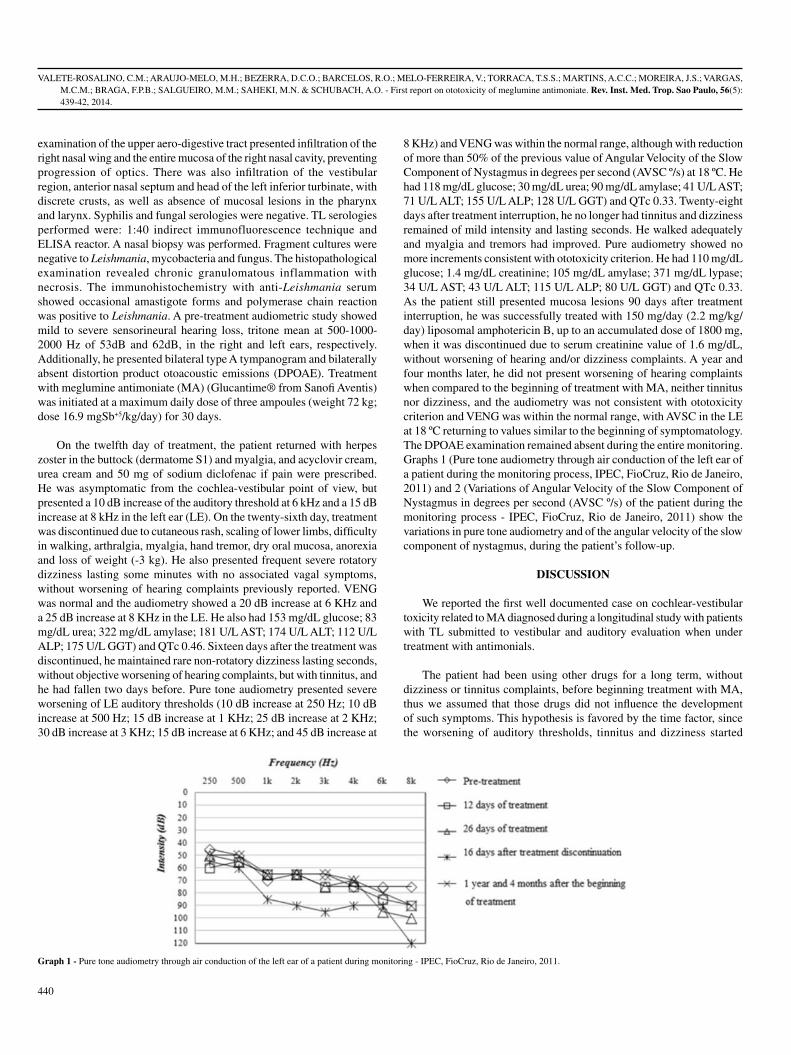
VALETE-ROSALINO, C.M.; ARAUJO-MELO, M.H.; BEZERRA, D.C.O.; BARCELOS, R.O.; MELO-FERREIRA, V.; TORRACA, T.S.S.; MARTINS, A.C.C.; MOREIRA, J.S.; VARGAS, M.C.M.; BRAGA, F.P.B.; SALGUEIRO, M.M.; SAHEKI, M.N. & SCHUBACH, A.O. - First report on ototoxicity of meglumine antimoniate. Rev. Inst. Med. Trop. Sao Paulo, 56(5): 439-42, 2014.
440
examination of the upper aero-digestive tract presented infiltration of the right nasal wing and the entire mucosa of the right nasal cavity, preventing progression of optics. There was also infiltration of the vestibular region, anterior nasal septum and head of the left inferior turbinate, with discrete crusts, as well as absence of mucosal lesions in the pharynx and larynx. Syphilis and fungal serologies were negative. TL serologies performed were: 1:40 indirect immunofluorescence technique and ELISA reactor. A nasal biopsy was performed. Fragment cultures were negative to Leishmania, mycobacteria and fungus. The histopathological examination revealed chronic granulomatous inflammation with necrosis. The immunohistochemistry with anti-Leishmania serum showed occasional amastigote forms and polymerase chain reaction was positive to Leishmania. A pre-treatment audiometric study showed mild to severe sensorineural hearing loss, tritone mean at 500-1000-2000 Hz of 53dB and 62dB, in the right and left ears, respectively. Additionally, he presented bilateral type A tympanogram and bilaterally absent distortion product otoacoustic emissions (DPOAE). Treatment with meglumine antimoniate (MA) (Glucantime® from Sanofi Aventis) was initiated at a maximum daily dose of three ampoules (weight 72 kg; dose 16.9 mgSb+5/kg/day) for 30 days.
On the twelfth day of treatment, the patient returned with herpes zoster in the buttock (dermatome S1) and myalgia, and acyclovir cream, urea cream and 50 mg of sodium diclofenac if pain were prescribed. He was asymptomatic from the cochlea-vestibular point of view, but presented a 10 dB increase of the auditory threshold at 6 kHz and a 15 dB increase at 8 kHz in the left ear (LE). On the twenty-sixth day, treatment was discontinued due to cutaneous rash, scaling of lower limbs, difficulty in walking, arthralgia, myalgia, hand tremor, dry oral mucosa, anorexia and loss of weight (-3 kg). He also presented frequent severe rotatory dizziness lasting some minutes with no associated vagal symptoms, without worsening of hearing complaints previously reported. VENG was normal and the audiometry showed a 20 dB increase at 6 KHz and a 25 dB increase at 8 KHz in the LE. He also had 153 mg/dL glucose; 83 mg/dL urea; 322 mg/dL amylase; 181 U/L AST; 174 U/L ALT; 112 U/L ALP; 175 U/L GGT) and QTc 0.46. Sixteen days after the treatment was discontinued, he maintained rare non-rotatory dizziness lasting seconds, without objective worsening of hearing complaints, but with tinnitus, and he had fallen two days before. Pure tone audiometry presented severe worsening of LE auditory thresholds (10 dB increase at 250 Hz; 10 dB increase at 500 Hz; 15 dB increase at 1 KHz; 25 dB increase at 2 KHz; 30 dB increase at 3 KHz; 15 dB increase at 6 KHz; and 45 dB increase at
8 KHz) and VENG was within the normal range, although with reduction of more than 50% of the previous value of Angular Velocity of the Slow Component of Nystagmus in degrees per second (AVSC º/s) at 18 ºC. He had 118 mg/dL glucose; 30 mg/dL urea; 90 mg/dL amylase; 41 U/L AST; 71 U/L ALT; 155 U/L ALP; 128 U/L GGT) and QTc 0.33. Twenty-eight days after treatment interruption, he no longer had tinnitus and dizziness remained of mild intensity and lasting seconds. He walked adequately and myalgia and tremors had improved. Pure audiometry showed no more increments consistent with ototoxicity criterion. He had 110 mg/dL glucose; 1.4 mg/dL creatinine; 105 mg/dL amylase; 371 mg/dL lypase; 34 U/L AST; 43 U/L ALT; 115 U/L ALP; 80 U/L GGT) and QTc 0.33. As the patient still presented mucosa lesions 90 days after treatment interruption, he was successfully treated with 150 mg/day (2.2 mg/kg/day) liposomal amphotericin B, up to an accumulated dose of 1800 mg, when it was discontinued due to serum creatinine value of 1.6 mg/dL, without worsening of hearing and/or dizziness complaints. A year and four months later, he did not present worsening of hearing complaints when compared to the beginning of treatment with MA, neither tinnitus nor dizziness, and the audiometry was not consistent with ototoxicity criterion and VENG was within the normal range, with AVSC in the LE at 18 ºC returning to values similar to the beginning of symptomatology. The DPOAE examination remained absent during the entire monitoring. Graphs 1 (Pure tone audiometry through air conduction of the left ear of a patient during the monitoring process, IPEC, FioCruz, Rio de Janeiro, 2011) and 2 (Variations of Angular Velocity of the Slow Component of Nystagmus in degrees per second (AVSC º/s) of the patient during the monitoring process - IPEC, FioCruz, Rio de Janeiro, 2011) show the variations in pure tone audiometry and of the angular velocity of the slow component of nystagmus, during the patient’s follow-up.
DISCUSSION
We reported the first well documented case on cochlear-vestibular toxicity related to MA diagnosed during a longitudinal study with patients with TL submitted to vestibular and auditory evaluation when under treatment with antimonials.
The patient had been using other drugs for a long term, without dizziness or tinnitus complaints, before beginning treatment with MA, thus we assumed that those drugs did not influence the development of such symptoms. This hypothesis is favored by the time factor, since the worsening of auditory thresholds, tinnitus and dizziness started
Graph 1 - Pure tone audiometry through air conduction of the left ear of a patient during monitoring - IPEC, FioCruz, Rio de Janeiro, 2011.

VALETE-ROSALINO, C.M.; ARAUJO-MELO, M.H.; BEZERRA, D.C.O.; BARCELOS, R.O.; MELO-FERREIRA, V.; TORRACA, T.S.S.; MARTINS, A.C.C.; MOREIRA, J.S.; VARGAS, M.C.M.; BRAGA, F.P.B.; SALGUEIRO, M.M.; SAHEKI, M.N. & SCHUBACH, A.O. - First report on ototoxicity of meglumine antimoniate. Rev. Inst. Med. Trop. Sao Paulo, 56(5): 439-42, 2014.
441
after initiating MA treatment, following a reasonable time after drug administration, when the levels of this drug were well established in body fluids and tissues7, and it was confirmed because signs and symptoms improved when the treatment was discontinued, thus showing that a relationship between these adverse effects and MA is likely to be true. Although infection has not responded to treatment with MA, we do not believe that the outcome of the infection may have had an influence on the MA ototoxicity, since the disease itself would not be capable of causing the sensorineural changes found in this patient. The success of treatment with pentavalent antimonials depends on the peak value reached in serum or the maintenance of a minimum inhibitory concentration in serum, during most part of the time3. The finding that pentavalent antimonial is rapidly excreted in the urine, resulting in sub therapeutic serum levels in a few hours, led to the conclusion that the risk of cumulative toxicity is low and that intermittent therapeutic schemes are pharmacologically inconsistent3,7. However, in vivo evidences of meglumine antimoniate conversion from Sb5+ into Sb3+ and the accumulation of Sb3+ in the tissues suggest that Sb3+ production could be responsible for the sustained activity of the drug, both therapeutic and toxic. Just a small conversion of the Sb5+ drug seems to occur during the phase of rapid excretion, however, it is followed by a phase of slow excretion of a high proportion of Sb3+
with a half-life of more than 50 days7. This pharmacokinetic behavior could explain the worsening of the auditory thresholds and AVSC at 18 ºC alterations up to 28 days after discontinuing the treatment. Additionally, the patient had mucosal leishmaniasis which requires a treatment of 30 days with MA. Therefore, ototoxicity could not have been so intense, if he had cutaneous leishmaniasis which requires a shorter treatment time up to 21 days.
However, the use of other ototoxic drugs may lead to more frequent cochlear-vestibular toxicity which, in this particular case, could have been enhanced by the use of doxazosin mesylate and hydrochlorothiazide, both potentially ototoxic13. Non-steroidal anti-inflammatory drugs (NSAIDs) are related to reversible ototoxicity, with 1% to 3% incidence17. They can induce vestibular disorders, less frequent than auditory disorders, such as tinnitus and hearing loss. The effect is more metabolic than structural, usually reversible, and proportional to the plasma concentration of the toxic drug. However, the patient had been using this drug for the last
seventeen years, sporadically, without reporting any audio vestibular symptom5,14,17.
Our study failed in not performing high frequency audiometry, since cochleotoxicity can occur, initially or solely, at the high frequency series from 9 to 20 kHz. Thus, the use of audiometry at conventional frequencies only, could have hindered an earlier and/or bilateral alteration in this case study1. The role of effective monitoring of the cochlear function in early detection of ototoxicity of DPOAE has been reported, through drop of their responses18. In this patient, this assessment was hampered because the DPOAE were already absent at the beginning of treatment. However, the evolution of the reported case was documented through serial examinations16.
The occurrence of subclinical ototoxicity of some drugs1 diagnosed by audiometry control has been described. To our knowledge, this is the first controlled case study of ototoxicity related to MA. This explains the fact that we could not find in literature references of hearing loss caused by this drug.
The most frequently reported adverse effects of pentavalent antimonials were musculoskeletal pain, gastrointestinal disturbances, mild to moderate headache, electrocardiographic QTc interval prolongation, mild to moderate increase in liver and pancreatic enzymes and significant suppression of mean lymphocyte and platelet counts11,19. However, cochlear-vestibular toxicity is not frequently reported. Tinnitus and dizziness had already been reported during ML treatment with tartar emetic9. Considering a recent report stating that Sb3+ levels represent more than 30% of total Sb concentrations in Glucantime®, we can suppose that Glucantime® may not be so different from trivalent antimonial tartar emetic and that Sb3+ may account for its ototoxicity15. The patient studied in the present case was a senior, male, and presented renal, hepatic and pancreatic toxicity. Dizziness associated to the use of meglumine antimoniate has already been reported regarding females, aged 60 years or more and related to pancreatic toxicity2. Because of its toxicity, meglumine antimoniate should be used with caution in patients older than 60 years8.
As for vestibular toxicity, the most common finding is hyporeflex or
Graph 2 - Variations of Angular Velocity of the Slow Component of Nystagmus in degrees per second (AVSC °/s) of the patient during monitoring - IPEC, FioCruz, Rio de Janeiro, 2011.
RE - Right Ear; LE - Left Ear.

VALETE-ROSALINO, C.M.; ARAUJO-MELO, M.H.; BEZERRA, D.C.O.; BARCELOS, R.O.; MELO-FERREIRA, V.; TORRACA, T.S.S.; MARTINS, A.C.C.; MOREIRA, J.S.; VARGAS, M.C.M.; BRAGA, F.P.B.; SALGUEIRO, M.M.; SAHEKI, M.N. & SCHUBACH, A.O. - First report on ototoxicity of meglumine antimoniate. Rev. Inst. Med. Trop. Sao Paulo, 56(5): 439-42, 2014.
442
simply normal reflex associated to vestibular complaints of the patient. Besides, it has been described that a reduction of 50% or more of the vestibular system, in at least one ear, indicates vestibular toxicity10. Although all the examinations of this case presented AVSC at 18 ºC within normal reflex values, it is evident that a reduction of more than 50% of this value occurred from the beginning of symptoms to 16 days after treatment, and that, a year later, the patient being asymptomatic, there was a return to values similar to the beginning of symptoms. Unfortunately, we do not have values before treatment to compare. Apparently tinnitus and dizziness are not related, since time of maximum tinnitus alteration did not correspond to that of maximum dizziness.
At the end of the ongoing longitudinal study, we intend to clarify some aspects related to the ototoxicity of meglumine antimoniate, such as dose dependence, relation with treatment duration, reversibility or not, as well as the associated risk factors.
RESUMO
Primeiro relato de ototoxicidade pelo antimoniato de meglumina
Introdução: Antimoniais pentavalentes são os fármacos de primeira escolha no tratamento da leishmaniose tegumentar. Dados de ototoxicidade relacionados a tais fármacos são escassos na literatura, o que nos levou a desenvolver um estudo de funções cócleo-vestibulares. Relato de caso: Relatamos caso de paciente masculino de 78 anos com leishmaniose tegumentar, que apresentou aumento significativo dos limiares auditivos com zumbido e tontura rotatória grave durante o tratamento com antimoniato de meglumina. Os sintomas pioraram até duas semanas após a interrupção do tratamento. Conclusão: Tontura e zumbido já tinham sido associados ao antimoniato de meglumina. Entretanto, este é o primeiro caso bem documentado de toxicidade cócleo-vestibular relacionado ao antimoniato de meglumina.
ACKNOWLEDGMENTS
The authors would like to thank Maria Inês Fernandes Pimentel and Érica Camargo de Vasconcellos.
This study had the financial support of FAPERJ and CNPQ to buy the audio-vestibulometry equipment and in the form of scholarships of scientific initiation and PQ CNPq and FAPERJ Our State Scientist, master, doctors etc.
The authors state no conflict of interest.
Cláudia Maria Valete-Rosalino has full access to all the data in the study and takes responsibility for their integrity.
REFERENCES
1. American Speech-Language-Hearing Association. Guidelines: audiologic management of individuals receiving cochleotoxic drug therapy. Practice Policy. ASHA. 1994;36:11-9. doi: 10.1044/policy.GL1994-00003
2. Araujo-Melo MH, Meneses AM, Schubach AO, Moreira JS, Conceição-Silva F, Salgueiro MM, et al. Risk factors associated to dizziness during treatment for mucosal leishmaniasis with meglumine antimoniate: 16-year retrospective study of cases from Rio de Janeiro, Brazil. J Laryngol Otol. 2010;124:1056-60.
3. Chulay JD, Fleckenstein G, Smith DH. Pharmacokinetics of antimony during treatment of visceral leishmaniasis with sodium stibogluconate or meglumine antimoniate. Trans R Soc Trop Med Hyg. 1988;82:69-72.
4. de Camargo Ferreira e Vasconcellos E, Schubach AO, Valete-Rosalino CM, Coutinho RS, Conceição-Silva F, Salgueiro MM, et al. American tegumentary leishmaniasis in older adults: 44 cases treated with an intermittent low-dose antimonial schedule in Rio de Janeiro, Brazil. J Am Geriatr Soc. 2010;58:614-6.
5. Dukes MNG, Aronson JK. Meyler´s side effects of drugs. 14th ed. Amsterdan: Elsevier; 2000.
6. Lima MLLT. Tratamento para tuberculose com estreptomicina: perfil auditivo e vestibular. [dissertação]. Recife: Centro de Pesquisas Aggeu Magalhães, FIOCRUZ; 2003.
7. Miekeley N, Mortari SR, Schubach AO. Monitoring of total antimony and its species by ICP-MS and on-line ion chromatography in biological samples from patients treated for leishmaniasis. Anal Bioanal Chem. 2002;372:495-502.
8. Mosimann V, Neumayr A, Hartz C, Blum JA. Cutaneous leishmaniasis in Switzerland: first experience with species-specific treatment. Infection. 2013;41:1177-82.
9. Nery-Guimarães F. Estudo de um foco de leishmaniose muco-cutânea na Baixada Fluminense (Estado do Rio de Janeiro). Mem Inst Oswaldo Cruz. 1955;53:1-11.
10. Oliveira JAA. Ototoxicidade. In: Costa SS, Cruz OLM, Oliveira JAA. Otorrinolaringologia: princípios e prática. Porto Alegre: Artes Médicas, 1994. p. 217.
11. Oliveira LF, Schubach AO, Martins MM, Passos SL, Oliveira RV, Marzochi MC, et al. Systematic review of the adverse effects of cutaneous leishmaniasis treatment in the New World. Acta Trop. 2011;118:87-96.
12. Passos VM, Barreto SM, Romanha AJ, Krettli AU, Volpini AC, Gontijo CM, et al. Cutaneous leishmaniasis in the metropolitan region of Belo Horizonte: clinical, laboratorial, therapeutic and prognosis features (1989-1995). Rev Soc Bras Med Trop. 2001;34:5-12.
13. Picon PD, Della Giustina ML, Rizzon CFC, Bassanesi SL, Zanardo AP, Michalczuk MT, et al. Resultado do tratamento da tuberculose com estreptomicina, isoniazida e etambutol (esquema SHM). J Bras Pneumol. 2002;28:187-92.
14. Rascol O, Hain TC, Brefel C, Benazet M, Clanet M, Montastruc J. Antivertigo medications and drug-induced vertigo. A pharmacological review. Drugs. 1995;50:777-91.
15. Salaün P, Frézard F. Unexpectedly high levels of antimony (III) in the pentavalent antimonial drug glucantime: insights from a new voltammetric approach. Anal Bioanal Chem. 2013;405:5201-14.
16. Santos CF, Valete CM, Martins AG, Ferreira, NGM, Tomita S. Clinical aspects of ototoxicity of aminiglicosideos. Acta ORL. 2000;19:160-4.
17. Seligmann H, Podoshin L, Ben-David J, Fradis M, Goldsher M. Drug-induced tinnitus and other hearing disorders. Drug Saf. 1996;14:198-212.
18. Vallejo JC, Silva MN, Oliveira JAA, Carneiro JJ, Rocha LSO, Figueiredo JFC, et al. Detecção precoce de ototoxicidade usando emissões otoacústicas produtivas de distorção. Braz J Otorhinolaryngol. 2001;67:845-51.
19. Wise ES, Armstrong MS, Watson J, Lockwood DN. Monitoring toxicity associated with parenteral sodium stibogluconate in the day-case management of returned travellers with New World cutaneous leishmaniasis. Plos Negl Trop Dis. 2012:6(6) e1688.
Received: 7 August 2013Accepted: 27 February 2014

Rev. Inst. Med. Trop. Sao Paulo56(5):443-445, September-October, 2014doi: 10.1590/S0036-46652014000500013
(1) Departamento de Patologia, Universidade Federal do Maranhão, São Luis, MA, Brazil. E-mails: [email protected], [email protected](2) Curso de Medicina, Universidade Federal do Maranhão, São Luis, MA, Brazil. E-mails: [email protected], [email protected], [email protected], diego@
hotmail.comCorrespondence to: Graça Maria de Castro Viana, Praça Madre Deus 1, Madre Deus, São Luis, MA, Brasil. E–mail: [email protected]
INTERFERON BETA-1A TREATMENT IN HTLV-1-ASSOCIATED MYELOPATHY/TROPICAL SPASTIC PARAPARESIS: A CASE REPORT
Graça Maria de Castro VIANA(1), Marcos Antonio Custódio Neto da SILVA(2), Victor Lima SOUZA(2), Natália Barbosa da Silva LOPES(2), Diego Luz Felipe da SILVA(2) & Maria do Desterro Soares Brandão NASCIMENTO(1)
SUMMARY
Here a young patient (< 21 years of age) with a history of infective dermatitis is described. The patient was diagnosed with myelopathy associated with HTLV-1/tropical spastic paraparesis and treated with interferon beta-1a. The disease was clinically established as HTLV-1-associated myelopathy/tropical spastic paraparesis (HAM/TSP), and laboratory tests confirmed the presence of antibodies to HTLV-1 in the cerebrospinal fluid (CSF). Mumps, cytomegalovirus, Epstein-Barr virus, schistosomiasis, herpes virus 1 and 2, rubella, measles, varicella-zoster toxoplasmosis, hepatitis, HIV, and syphilis were excluded by serology. The patient was diagnosed with neurogenic bladder and presented with nocturia, urinary urgency, paresthesia of the lower left limb, a marked reduction of muscle strength in the lower limbs, and a slight reduction in upper limb strength. During the fourth week of treatment with interferon beta-1a, urinary urgency and paresthesia disappeared and clinical motor skills improved.
KEYWORDS: HAM/TSP; HTLV-1; Interferon beta-1a; Treatment.
INTRODUCTION
The human T-lymphotropic virus (HTLV) is a retrovirus belonging to the Retroviridae family. HTLV can infect cells of the human immune system and has tropism for T-lymphocytes14,15 (T-cells). Approximately 10-20 million people worldwide are infected with HTLV-1, which is considered a global epidemic. HTLV-1 is associated with a wide spectrum of manifestations including tropical spastic paraparesis, a disease characterized by the slow and progressive development of myelopathy, as well as leukemia/lymphoma in adult T-cells6. HTLV-1 is endemic in many regions of the world, more common in adult women, and usually insidious, but may be sudden. There is a high prevalence of HTLV-1 in southern Japan, the Caribbean, Africa, South America, Papua New Guinea, the Middle East, Australia, and Southeast Italy11. In Brazil, HTLV-1 is found in several states, but is more prevalent in certain regions, such as in the Northeast, particularly Maranhão, where the prevalence is 10 per 1000 blood donors3.
HTLV-1-associated myelopathy/tropical spastic paraparesis (HAM/TSP) is a demyelinating disease with inflammatory changes in the central nervous system. HTLV-1 is predominantly transmitted via blood; however, vertical and sexual transmission is also possible12. HAM/TSP is characterized by neurological manifestations, such as chronic spastic paraparesis, impaired gait, and weakness in the lower limbs. Other symptoms include sphincter signs and symptoms such as
bladder disorders (urinary urge incontinence and nocturia) and sensory symptoms including paresthesia, hyperreflexia of the lower limbs, and the presence of the Babinski signal often with clonus (signs of pyramidal release). Laboratory diagnosis is made by the identification of HTLV-1/2 antibodies in the blood and cerebrospinal fluid (CSF). Results are confirmed by Western blot or detection of proviral DNA in the blood or CSF12.
In this study, a patient with HAM/TSP treated with interferon beta-1a was presented, with a good outcome.
CASE REPORT
A 21-year-old widowed female patient, with a history of infective dermatitis in childhood reported, decreased muscle strength in the upper and lower limbs, particularly on the right side. The patient was seen as a referral to the rehabilitation department. She was diagnosed with HTLV-1-associated myelopathy/tropical spastic paraparesis, confirmed by positive levels of HTLV in serum (ELISA: 3.15, Cut-Off: 0.18) and the CSF (ELISA: 2.57, Cut-Off: 0.18). Additionally, anti-HTLV confirmation was made using Western blot analyses.
The referral hospital also identified antibodies in the CSF and excluded mumps, cytomegalovirus, Epstein-Barr virus, schistosomiasis, herpes virus 1 and 2, rubella, measles, toxoplasmosis, and varicella-zoster

VIANA, G.M.C.; SILVA, M.A.C.N.; SOUZA, V.L.; LOPES, N.B.S.; SILVA, D.L.F. & NASCIMENTO, M.D.S.B. - Interferon beta-1a treatment in HTLV-1-associated myelopathy/tropical spastic paraparesis: a case report. Rev. Inst. Med. Trop. Sao Paulo, 56(5): 443-5, 2014.
444
virus. Serology for hepatitis, syphilis and HIV were also negative. Protein electrophoresis showed albumin values of 15.6 mg/dL (normal range: 5.6-45.6 mg/dL), beta-globulin values of 5.7 mg/dL (normal range: 0.8-10.8 mg/dL), and gamma-globulin values of 7.2 mg/dL (normal range: 0.3-7.2 mg/dL). The complete blood count (CBC) was 3.09 million red blood cells (RBCs)/mm3, 9.4 g/dL hemoglobin, hematocrit 29%, lymphocytes 41%, erythrocyte sedimentation rate (ESR) of 40 mm in the first hour, and 192,000 platelets.
A differential diagnosis for demyelinating disease and lupus was performed. Lupus and demyelinating disease were ruled out by clinical presentation and laboratory tests for lupus (anticoagulant, anticardiolipin IgG and IgM, and anti-DNA antibodies, all negatives). The serology of the patient’s mother could not be assessed, because she refused testing. The patient’s husband was also not tested, because he died before the onset of her clinical manifestations. However, the patient has a daughter (aged two years) who was seropositive for HTLV-1.
One year prior to diagnosis of HTLV-1, the patient was diagnosed with neurogenic bladder, with clinical nocturia (~four times per night), urinary urgency, severe loss of muscle strength in the lower limbs, positive Babinski reflex and paresthesia in the left leg. The patient had also started walking with support on the right side. The patient could walk 10 m in 12 s.
Interferon beta-1a treatment (3,000,000 IU) was started three times a week for 10 weeks. Clinical evaluations were performed after four weeks of therapy. Improvement was first observed in sensory manifestations followed by motor evaluation. The patient showed improvements in muscle strength and nocturia (reduced to once per night) with the disappearance of urinary urgency and paresthesia. The patient’s gait became more agile and quick, and she could walk 10 m in 10 s. Two years after completing treatment, the patient still reports improved clinical sensory and motor skills.
DISCUSSION
HTLV-1/2 infection is endemic in Brazil4 where 5% of people are seropositive. There is a high incidence in Maranhão, where 10 per 1000 blood donors are seropositive3. HTLV-1/2 is more frequent among women10 in the fourth and fifth decade of life5, contrary to the age of the patient in this study and the data provided by CARVALHO et al. (2009)2.
Several promising therapies have been considered and evaluated for HAM/TSP, although none have been approved for use in patients yet.
The treatment of HTLV-1-associated myelopathy/tropical spastic paraparesis can be divided into specific and symptomatic cases. Specific immunosuppressive drugs (corticosteroids) and/or immunomodulators (interferon alpha and beta-1) are used for treatment. These drugs work to inhibit T-lymphocyte activation, which modulates leukocyte migration and minimizes the production of inflammatory cytokines. There are drugs for symptomatic treatment that can control pain, neurogenic bladder, constipation, and spasticity1.
Studies using interferon alpha to treat patients with HAM/TSP have shown better clinical outcomes and reduced proviral loads8,13,16.
Interferon beta-1a has been used to treat relapsing-remitting multiple sclerosis and has been shown to be beneficial in several trials7,10. In HAM/TSP, only two studies evaluated interferon beta-1a treatment. One study used a dose of 60 mcg twice per week, which reduced tax-CD8+ cells, but did not reduce the proviral DNA load9. The other study used a lower dose of 30 mcg every 15 days and noted good outcomes4.
In this study, it was chosen to treat with interferon beta-1a instead of interferon alpha, because the latter has a daily dosage schedule rather than the simple weekly interferon beta-1a dosage.
Treatment with interferon beta-1a (3,000,000 IU) three times a week for 10 weeks improved the urinary and motor symptoms of the patient. Although interferon beta-1a may not be the standard treatment for HTLV-1-associated myelopathy/tropical spastic paraparesis, in this case it proved to be very promising. This underscores the need for more clinical studies using interferon beta-1a.
RESUMO
Tratamento com interferon beta-1a em mielopatia associada ao HTLV-1/paraparesia espástica tropical: relato de caso
Descreve-se caso de mielopatia associada ao HTLV-1/paraparesia espástica tropical tratada com interferon beta-1a em paciente jovem de 21 anos e com história de dermatite infecciosa na infância. Foi estabelecida clinicamente paraparesia espástica tropical (HAM/TSP), confirmada laboratorialmente pela presença de anticorpos para HTLV-1 no LCR e excluídas caxumba, citomegalovirus, Epstein-Barr, esquistossomose, herpes virus 1 e 2, rubéola, sarampo, toxoplasmose varicela-zoster, hepatite, HIV e sífilis por sorologias. Foi diagnosticada bexiga neurogênica, com quadro clínico de nictúria, urgência urinária, parestesia no membro inferior esquerdo e discreta redução de força muscular nos membros superiores, mais acentuada nos membros inferiores. Na 4ª semana de tratamento com interferon beta-1a houve desaparecimento da urgência urinária e da parestesia e melhora da clínica motora.
REFERENCES
1. Araújo A, Lima MA, Silva MTT. Avanços e perspectivas na terapêutica das doenças associadas à infecção por HTLV. In: Proietti ABFC, editor. Cadernos Hemominas HTLV. 4ª ed. Belo Horizonte: Fundação Hemominas; 2006. p. 228-35.
2. Carvalho AGJ, Galvão-Phileto AV, Lima NS, Jesus RS, Galvão-Castro B, Lima MGL. Frequency of mental disturbances in HTLV-1 patients in the state of Bahia, Brazil. Braz J Infect Dis. 2009;13:5-8.
3. Catalan-Soares B, Carneiro-Proietti ABF, Proetti FA, Interdisciplinary HTLV Research Group. Heterogeneous geographic distribution of human T-cell lymphotropic viruses I and II (HTLV I/II): serological screening prevalence rates in blood donors from large urban areas in Brazil. Cad Saúde Pública. 2005;21:926-31
4. Costa DT, Sundberg M, Passos L, Muniz AL, Santos S. Interferon beta-1a improves urinary symptoms, reduces proviral load, and modifies the immune response in a patient with HAM/TSP. Case Rep Neurol Med. 2012;2012:1-3.
5. Gaspar-Sobrinho FP, Souza-Machado A, Santos SB, Orge G, Lessa HA, Cruz AA, et al. Clinical and immunological features of patients with atopy and concomitant HLTV-1 infection. Braz J Med Biol Res. 2010;43:1167-72.
6. Gonçalves CCA, Ribeiro LCP, Sá CAM, Puccioni-Sohler M. Diagnóstico laboratorial da mielopatia associada ao HTLV-I: métodos para análise do líquido cefalorraquidiano. J Bras Patol Med Lab. 2009;45:99-110.

VIANA, G.M.C.; SILVA, M.A.C.N.; SOUZA, V.L.; LOPES, N.B.S.; SILVA, D.L.F. & NASCIMENTO, M.D.S.B. - Interferon beta-1a treatment in HTLV-1-associated myelopathy/tropical spastic paraparesis: a case report. Rev. Inst. Med. Trop. Sao Paulo, 56(5): 443-5, 2014.
445
7. Jacobs LD, Cookfair DL, Rudick RA, Herndon RM, Richert JR, Salazar AM, et al. Intramuscular interferon beta-1a for disease progression in relapsing multiple sclerosis. The Multiple Sclerosis Collaborative Research Group (MSCRG). Ann Neurol. 1996;39:285-94.
8. Kuroda Y, Kurohara K, Fujiyama F, Takashima H, Endo C, Matsui M, et al. Systemic interferon-alpha in the treatment of HTLV-I-associated myelopathy. Acta Neurol Scand. 1992;86:82-6.
9. Oh U, Yamano Y, Mora CA, Ohayon J, Bagnato F, Butman JA, et al. Interferon-β1 a therapy in human T-lymphotropic virus type I-associated neurologic disease. Ann Neurol. 2005;57:526-34.
10. Randomised double-blind placebo-controlled study of interferon beta-1a in relapsing/remitting multiple sclerosis. PRISMS (Prevention of Relapses and Disability by Interferon beta-1a Subcutaneously in Multiple Sclerosis) Study Group. Lancet. 1998;352(9139):1498-504.
11. Rêgo A, Feitosa F, Cavalcante D, Paraná R. VHC e HTLV-I: aspectos clínicos e epidemiológicos da co-infecção. Rev Cienc Méd Biol (Salvador). 2003;2:230-9.
12. Ribas JGR, Melo GCN. Mielopatia associada ao vírus linfotrópico humano de células T do tipo 1 (HTLV-1). Rev Soc Bras Med Trop. 2002;35:377-84.
13. Saito M, Nakagawa M, Kaseda S, Matsuzaki T, Jonosono M, Eiraku N, et al. Decreased human T lymphotropic virus type I (HTLV-I) provirus load and alteration in T cell phenotype after interferon-alpha therapy for HTLV-I associated myelopathy/tropical spastic paraparesis. J Infect Dis. 2004;189:29-40.
14. Santos FLN, Lima FWM. Epidemiologia, fisiopatogenia e diagnóstico laboratorial da infecção pelo HTLV-I. J Bras Patol Med Lab. 2005;41:105-16.
15. Santos NSO, Wigg MD, Oliveira DP. Viroses multissistêmicas. In: Santos NSO, Romanos MTV, Wigg MD, editores. Introdução à virologia humana. 2a ed. Rio de Janeiro: Guanabara Koogan; 2008. p. 290-330.
16. Yamasaki K, Kira J, Koyanagi Y, Kawano Y, Miyano-Kurosaki N, Nakamura M, et al. Long term, high dose interferon-alpha treatment in HTLV-I-associated myelopathy/tropical spastic paraparesis: a combined clinical, virological and immunological study. J Neurol Sci. 1997;147:135-44.
Received: 8 August 2013Accepted: 18 February 2014
LIBRARY OF THE SÃO PAULO INSTITUTE OF TROPICAL MEDICINE
Website: www.imt.usp.br/portal Address: Biblioteca do Instituto de Medicina Tropical de São Paulo da Universidade de São Paulo
Av. Dr. Enéas de Carvalho Aguiar, 470. Prédio 1 – Andar térreo.05403-000 São Paulo, SP, Brazil.
Telephone: 5511 3061-7003 - Fax: 5511 3062-2174
The Library of the São Paulo Institute of Tropical Medicine (IMTSP Library) was created on January 15, 1959 in order to serve all those who are interested in tropical diseases. To reach this objective, we select and acquire by donation and / or exchange appropriate material to be used by researchers and we maintain interchange between Institutions thorough the Journal of the São Paulo Institute of Tropical Medicine, since the Library has no funds to build its own patrimony.
The IMTSP Library has a patrimony consisting of books, theses, annals of congresses, journals, and reference works.
The collection fo journals existing in the Library can be verified through the USP – Bibliographic Database – OPAC – DEDALUS http://dedalus.usp.br:4500/ALEPH/eng/USP/USP/DEDALUS/start of the USP network.

Rev. Inst. Med. Trop. Sao Paulo56(5):447-450, September-October, 2014doi: 10.1590/S0036-46652014000500014
(1) Laboratório de Biologia Molecular, Centro de Estudos da Biodiversidade, Universidade Federal de Roraima, Boa Vista, RR, Brasil.(2) Laboratório Central de Roraima, Boa Vista, RR, Brasil.(3) Instituto Leônidas e Maria Deane, Fiocruz, Manaus, AM, Brasil.Correspondence to: Pablo O. A. Acosta, UFRR, Laboratório de Biologia Molecular/CBio/UFRR. Av. Ene Garcez 2413, 69304-000 Boa Vista, RR, Brasil. E-mail: [email protected]
BRIEF COMMUNICATION
FALSE-NEGATIVE DENGUE CASES IN RORAIMA, BRAZIL: AN APPROACH REGARDING THE HIGH NUMBER OF NEGATIVE RESULTS BY NS1 AG KITS
Pablo O.A. ACOSTA(1), Fabiana GRANJA(1), Cátia A. MENESES(2), Ismael A.S. NASCIMENTO(1), Débora D. SOUSA(1), Wilson P. LIMA JÚNIOR(1) & Felipe Gomes NAVECA(3)
SUMMARY
Serum samples from 150 NS1-negative (Platelia ELISA) patients presumptively diagnosed with dengue were analyzed by the TaqMan probed real-time reverse transcription PCR (TaqMan qRT-PCR) method. The qRT-PCR positive samples were tested for serotype by semi-nested RT-PCR and a qualitative immunochromatographic assay for IgG and IgM. Molecular detection methods showed 33 (22%) positive samples out of 150 NS1-antigen negative samples. Of these, 72% were collected up to day 2 after the onset of symptoms, when diagnostic sensitivity of NS1-antigen test assays is significantly enhanced. Most of the cases were not characterized as secondary infection. Twenty-eight samples were successfully serotyped, 75% of which for DENV-4, 14% for DENV-2, 7% for DENV-3 and 4% for DENV-1. These findings reaffirm the hyperendemic situation of the state of Roraima and suggest a lower sensitivity of the NS1 test, mainly when DENV-4 is the predominant serotype. Health care providers should therefore be aware of samples tested negative by NS1 antigen assays, especially when clinical symptoms and other laboratory data results show evidence of dengue infection.
KEYWORDS: Dengue; Serotypes; Diagnosis; NS1 antigen; qRT-PCR.
Dengue is currently regarded as the most important mosquito-borne viral disease, in terms of both geographical distribution and number of cases reported annually. Recent studies have estimated a risk population of 3.97 billion people, and of the major vector-borne diseases the cases of dengue have been prevailing over those of malaria in the Americas, Asia and much of Africa5,6.
The etiological agent, the Dengue Virus (DENV), belongs to the genus Flavivirus of the family Flaviviridae, and the main mosquito vector to transmit the disease to humans is the predominantly urban species Aedes aegypti11.
The disease has a wide clinical spectrum - ranging from a flu-like illness to severe dengue - and, therefore, it is not possible to determine the clinical manifestations that can ensue at the time of dengue infection. Although, there is no specific treatment for dengue, early diagnosis may guide appropriate clinical management and, thereby, prevent severe complications and/or death24.
The state of Roraima, located in the Brazilian Amazon region, has shown a high incidence of infection over the past decade1 and is
currently recognized as a hyperendemic area, as well as a port-of-entry for serotypes and genotypes of dengue viruses into Brazil. DENV-4 reemerged in Roraima in 20102,19, 28 years after it was last detected in the state, and now all four dengue serotypes are circulating1. In recent years, the state has seen an increase in severe forms of the disease, probably due to secondary infections and/or higher virulence of circulating strains1. The World Health Organization (WHO) currently recommends early laboratory tests for the diagnosis of the dengue virus infection using NS1 antigen detection in human serum24, and this is mostly adopted in Roraima. The highly immunogenic non-structural glycoprotein NS1 contains approximately 353 amino acids (46 kDa) and is more conserved among the flaviviruses. Although, NS1 does not form part of the virus structure, it is required for viral RNA replication. It is also displayed on the surface of infected cells and is secreted into the bloodstream, during the acute phase of the disease. This latter characteristic makes it sensitive enough to diagnose a patient on the very first days of infection4,17.
In 2012, 998 serum samples from presumptive diagnosis of dengue were tested with the use of the Bio-Rad Platelia™ Dengue NS1 antigen capture kit at the Central Public Health Laboratory in Roraima (LACEN/RR). Of these samples, 778 (78%) tested negative, i.e. they were ruled

ACOSTA, P.O.A.; GRANJA, F.; MENESES, C.A.; NASCIMENTO, I.A.S.; SOUSA, D.D.; LIMA JÚNIOR, W.P. & NAVECA, F.G. - False-negative dengue cases in Roraima, Brazil: an approach regarding the high number of negative results by NS1 Ag kits. Rev. Inst. Med. Trop. Sao Paulo, 56(5): 447-50, 2014.
448
out for dengue infection through laboratory diagnosis. Based on this, two questions arise: have these patients been infected by other arboviruses? Does the dengue NS1 antigen test give rise to false-negative results?
To find answers to these questions, it was first decided to assess the possibility of false-negative results for the samples ruled out by the dengue virus NS1 antigen detection. In order to carry out this evaluation, the most sensitive TaqMan - MGB probed real-time quantitative reverse transcription PCR (TaqMan qRT-PCR) technique was applied.
Of the 778 cases initially discarded in 2012 as not being dengue, 150 samples were randomly selected from patients who, in addition to fever, had in their clinical records one or more common signs of dengue such as arthralgia, headache, retro-orbital pain, chills, myalgia, and rash. The average time of sample collection was 1-5 days after the onset of the symptoms.
Viral RNA was extracted directly from serum by the Axygen® AxyPrep™ Body Fluid Viral DNA/RNA Miniprep kit, and subsequently subjected to the qRT-PCR assay (GURUKUMAR et al., 2009)12. This technique is able to detect the genome of any of the four dengue serotypes, but unable to identify specific serotype DENV infection. Fluorescence reading was performed on a StepOne™ Real-Time PCR system. All reactions were performed under the same conditions with the use of ROX as a passive reference dye for normalization of data and VIC as reporter. Samples with Ct < 39 were considered to be dengue positive and underwent serotyping by semi-nested RT-PCR, as described by LANCIOTTI et al. (1992)15 and an immunochromatographic assay that allows qualitative and differential detection for IgG and IgM antibodies (Dengue Test Bioeasy)8. IgG positive samples, collected until the 4th day from the onset of symptoms, were characterized as secondary infection7.
Of the 150 samples tested, 33 (22%) were positive by qRT-PCR. The samples with the false-negative NS1 antigen were collected between 1-4 days after the onset of symptoms (Table 1) within this period, according to the literature and the manufacturer’s instructions, dengue NS1 antigen tests are highly sensitive3,20.
Studies from different countries indicate a low sensitivity on the NS1 test in secondary infection9,14,16,18.23, and as Roraima is a hyperendemic State to dengue, at first, it was considered that the main cause for this
high number of false negative samples was due to secondary infection. Nevertheless, only eight (28%), of the 30 analyzed samples, showed positive IgG (Table 1).
In 37% of the IgG positive samples IgM antibodies were also detected. FELIX et al., on a dengue outbreak in 2010 in Santos-Brazil, found 64.2% IgM positive among a total of 260 samples with secondary infection10.
All four dengue virus serotypes were reported among the positive cases (Fig.1), and these findings are sufficient to classify the state of Roraima as a hyperendemic area. The most common serotype DENV-4 accounted for 75% of the tested serum samples. These data are consistent with that of the Roraima State Epidemiology and Surveillance Branch for this period, which shows that a further 90% of the samples sent for viral identification in 2012 were DENV-421. According to the manufacturer’s instructions the sensitivity of the assay does not differentiate between dengue serotypes20, however, a study conducted in the state of Sergipe, in Northeastern Brazil, found 48.7% false negative, among 119 samples tested for the NS1 antigen. All the negative samples were identified as DENV-4, and authors point to a low sensitivity of the Platelia dengue NS1Ag test in regions where this serotype is predominant22. The DENV-4 strains tested in this study belong to genotype II, the same one that is circulating in the state of Roraima19, and considering that the Bio-Rad kit uses a monoclonal antibody, it was suggested that this antibody is likely to have low-affinity to epitopes of NS1 from the DENV-4 genotype II, however, there are studies that did not find any difference in the NS1 amino acid sequence in DENV-2 false negative and positive samples for the NS1 Ag detection10.
After interrelating the NS1 test sensitivity to dengue serotypes, GUZMAN et al.’s study13 evaluated the four dengue serotypes in samples from Southeast Asia and Latin America, which described the lower sensitivities by the Platelia Dengue NS1 Ag kit in DENV-2. Studies performed in Brazil, before DENV-4 reemerged, found the lowest sensitivity to serotypes DENV-2 and DENV-310,18.
The analysis presented here reinforces the importance of increasing the awareness of healthcare providers regarding the fact that patients tested negative by the NS1 antigen detection may be infected with the dengue virus, especially when the clinical symptoms and other laboratory data indicate such infection.
Table 1Results description of used samples in this study
150 samples negatives to NS1-Ag and tested by qRT-PCR Serology results for IgM and IgG
qRT-PCR Negatives qRT-PCR PositivesIgM a, b IgG a, b
Positivesd Negatives Positives Negatives
117 (78%) 33 (22%) 03 (10%) 27 (90%) 08 (27%) 22 (73%)
Beginning of symptomsa Identified serotypesa, c
1st day 2nd day 3rd day 4th day DENV-1 DENV-2 DENV-3 DENV-4
10 (30%) 14 (43%) 7 (21%) 2 (6%) 1 (4%) 4 (14%) 2 (7%) 21 (75%)aSymptom onset results, serology and identified serotypes refer to samples positive by qRT-PCR; b 03 samples tested by qRT-PCR were insufficient for serology; c Five samples positive by qRT-PCR could not be detected by semi-nested RT-PCR; d All samples positives to IgM were also positive to IgG.

ACOSTA, P.O.A.; GRANJA, F.; MENESES, C.A.; NASCIMENTO, I.A.S.; SOUSA, D.D.; LIMA JÚNIOR, W.P. & NAVECA, F.G. - False-negative dengue cases in Roraima, Brazil: an approach regarding the high number of negative results by NS1 Ag kits. Rev. Inst. Med. Trop. Sao Paulo, 56(5): 447-50, 2014.
449
RESUMO
Casos de dengue falso-negativo em Roraima, Brasil: abordagem acerca do alto número de testes Antígeno NS1 negativos
Amostras séricas de 150 pacientes, com diagnóstico presuntivo de dengue e resultado negativo para dengue por ELISA-NS1-Antígeno do kit Platelia™ (NS1-Ag), foram analisadas pela técnica de TaqMan Transcrição Reversa seguida da Reação em Cadeia da Polimerase em Tempo Real (qRT-PCR). As amostras positivas por qRT-PCR, foram submetidas a identificação dos sorotipos por RT-Hemi nested-PCR e a ensaio imunocromatográfico para detecção qualitativa dos anticorpos IgG e IgM. A técnica molecular apresentou como resultado 33 (22%) amostras positivas entre as 150 negativas pela detecção do NS1-Ag, destas o 72% foram coletadas até o segundo dia de início dos sintomas da doença, período de maior sensibilidade para pesquisas de NS1-Ag. A maioria dos casos não evidenciou infecção secundária. Dessas amostras, 28 foram satisfatoriamente sorotipadas sendo 75% de DENV-4, 14% de DENV-2, 7% de DENV-3 e 4% de DENV-1. Os resultados reafirmam a situação hiperendêmica do Estado de Roraima e sugerem baixa sensibilidade do NS1 test, especialmente quando o sorotipo predominante é DENV-4. Sugerimos assim, que a comunidade médica deve ser alertada no sentido de ser cautelosa com resultados de NS1-Ag negativo, principalmente quando sintomas clínicos e outros resultados laboratoriais sejam indicativos de provável infecção por dengue.
ACKNOWLEDGMENTS
The authors would like to thank the Federal University of Roraima. This work was supported by grants from Instituto Leônidas e Maria Deane and Santander Universidades.
REFERENCES
1. Acosta POA, Cordeiro JS, Granja F, Siqueira TCS, Brito FEG, Freitas AG, et al. Dengue in the northernmost part of Brazil from 1999 to 2011: characterization of circulating DENV strains. Dengue Bull. 2012;36:50-63.
2. Acosta POA, Maito RM, Granja F, Cordeiro JS, Siqueira T, Cardoso MN, et al. Dengue virus serotype 4, Roraima State, Brazil. Emerg Infect Dis. 2011;17:1979-80.
3. Alcon S, Talarmin A, Debruyne M, Falconar A, Deubel V, Flamand M. Enzyme-linked immunosorbent assay specific to dengue virus type 1 nonstructural protein NS1 reveals circulation of the antigen in the blood during acute phase of disease in patients experiencing primary or secondary infections. J Clin Microbiol. 2002;40:376-81.
4. Avirutnan P, Punyadee N, Noisakran S, Komoltri C, Thiemmeca S, Auethavornanan K, et al. Vascular leakage in severe dengue virus infections: a potential role for the nonstructural viral protein NS1 and complement. J Infect Dis. 2006;193:1078-88.
5. Bhatt S, Gething PW, Brady OJ, Messina JP, Farlow AW, Moyes CL, et al. The global distribution and burden of dengue. Nature. 2013;496(7446):504-7.
6. Brady OJ, Gething PW, Bhatt S, Messina JP, Brownstein JS, Hoen AG, et al. Refining the global spatial limits of dengue virus transmission by evidence-based consensus. PLoS Negl Trop Dis. 2012;6:e1760.
7. de Souza VAUF, Tateno AF, Oliveira RR, Domingues RB, Araújo ES, Kuster GW, et al. Sensitivity and specificity of three ELISA-based assays for discriminating primary from secondary acute dengue virus infection. J Clin Virol. 2007;39:230-3.
8. Dengue IgG/IgM test Bioeasy. Available from: http://trisul.com.br/arquivos/dengue_(bula).pdf
9. Duong V, Ly S, Lorn Try P, Tuiskunen A, Ong S, Chroeung N, et al. Clinical and virological factors influencing the performance of a NS1 antigen-capture assay and potential use as a marker of dengue disease severity. PLoS Negl Trop Dis. 2011;5:e1244.
10. Felix AC, Romano CM, Centrone CC, Rodrigues CL, Villas-Boas L, Araujo ES, et al. Low sensitivity of NS1 protein tests evidenced during a dengue type 2 virus outbreak in Santos, Brazil, in 2010. Clin Vaccine Immunol. 2012;19:1972-6.
11. Gubler DJ. Epidemic dengue and dengue hemorrhagic fever: a global public health problem in the 21st Century. In: Scheld WM, Armstrong D, Hughes JM, editors. Emerging Infections 1. Washington: ASM Press; 1998. p. 1-14.
12. Gurukumar KR, Priyadarshini D, Patil JA, Bhagat A, Singh PS, Shah PS, et al. Development of real time PCR for detection and quantitation of dengue viruses. Virol J. 2009;6:10:1-8.
13. Guzman MG, Jaenisch T, Gaczkowski R, Ty Hang VT, Sekaran SD, Kroeger A, et al. Multi-country evaluation of the sensitivity and specificity of two commercially-available NS1 ELISA assays for dengue diagnosis. PLoS Negl Trop Dis. 2010:4:e811.
14. Hang VT, Nguyet NM, Trung DT, Tricou V, Yoksan S, Dung NM, et al. Diagnostic accuracy of NS1 ELISA and lateral flow rapid tests for dengue sensivity, specificity and relationship to viraemia and antibody responses. PLoS Negl Trop Dis. 2009;3:e360.
15. Lanciotti RS, Calisher CH, Gubler DJ, Chang G-J, Vorndam V. Rapid detection and typing of dengue viruses from clinical samples by using reverse transcriptase-polymerase chain reaction. J Clin Microbiol. 1992;30:545-51.
16. Lapphra K, Sangcharaswichai A, Chokephaibulkit K, Tiengrim S, Piriyakarnsakul W, Chakorn T, et al. Evaluation of an NS1 antigen detection for diagnosis of acute dengue infection in patients with acute febrile illness. Diagn Microbiol Infect Dis. 2008;60:387-91.
17. Libraty DH, Young PR, Pickering D, Endy TP, Kalayanarooj S, Green S, et al. High circulating levels of the dengue virus nonstructural protein NS1 early in dengue illness correlate with the development of dengue hemorrhagic fever. J Infect Dis. 2002;186:1165-8.
Fig 1 - Agarose gel analysis of Hemi nested RT-PCR products from Dengue virus. Lane 1:
100pb ladder; Lane 2: positive control DENV-1 (482pb); Lane 3: positive control DENV-
2 (119pb); Lane 4: positive control DENV-3 (290pb); Lane 5: positive control DENV-4
(392pb); Lane 6: sample patient A05 (482pb); Lane 7: sample patient A16 (119pb); Lane 8:
sample patient A63 (290pb); Lane 9: sample patient A44 (392pb); Lane 10 negative control.

ACOSTA, P.O.A.; GRANJA, F.; MENESES, C.A.; NASCIMENTO, I.A.S.; SOUSA, D.D.; LIMA JÚNIOR, W.P. & NAVECA, F.G. - False-negative dengue cases in Roraima, Brazil: an approach regarding the high number of negative results by NS1 Ag kits. Rev. Inst. Med. Trop. Sao Paulo, 56(5): 447-50, 2014.
450
18. Lima MRQ, Nogueira RMR, Schatzmayr HG, Santos FB. Comparison of three commercially available dengue NS1 antigen capture assays for acute diagnosis of dengue in Brazil. PLos Negl Trop Dis. 2010:4:e738.
19. Naveca FG, Souza VC, Silva GAV, Maito RM, Granja F, Siqueira T, et al. Complete genome sequence of a dengue virus serotype 4 strain isolated in Roraima, Brazil. J Virol. 2012;86:1897.
20. PlateliaTM Dengue NS1 Ag (Bio-Rad): detecção qualitativa ou semi-quantitativa do antígeno NS1 do vírus da dengue no soro ou plasma humano, pelo método imunoenzimático. Available from: https://www.bio-rad.com/webroot/web/pdf/inserts/CDG/pt/72830_12-2008.pdf
21. Roraima. Secretaria Estadual de Saúde. Departamento Vigilância Epidemiológica. Relatório: casos de dengue notificados no ano 2012. Sistema de Informação de Agravos de Notificação (SINAN). Boa Vista: SINAN; 2012.
22. Sea VRF, Cruz ACR, Gurgel RQ, Nunes BTD, Silva EVP, Dolabella SS, et al. Underreporting of dengue-4 in Brazil due to low sensitivity of the NS1 Ag test in routine control programs. PLoS One. 2013;8:e64056.
23. Sekaran SD, Ew CL, Subramaniam G, Kanthesh BM. Sensitivity of dengue virus NS1 detection in primary and secondary infections. Afr J Microbiol Res. 2009;3:105-110.
24. World Health Organization. Dengue: guidelines for diagnosis, treatment, prevention and control. Geneva: World Health Organization; 2009. Available from: http://whqlibdoc.who.int/publications/2009/9789241547871 _eng. pdf
Received: 28 October 2013Accepted: 13 March 2014

Rev. Inst. Med. Trop. Sao Paulo56(5):451-454, September-October, 2014doi: 10.1590/S0036-46652014000500015
(1) Centro de Ciências Biológicas e da Saúde, Universidade Federal de Campina Grande, Paraíba, Brazil.(2) Laboratório de Imunopatologia Keizo Asami (LIKA), Universidade Federal de Pernambuco, Recife, Brazil. Correspondence to: Elizabeth Malagueño, LIKA-Universidade Federal de Pernambuco, Recife, PE, Brasil. Comendador Bento Aguiar 84, Ilha do Retiro, 50720-705 Recife, PE, Brasil.
E-mail:[email protected]
BRIEF COMMUNICATION
PREVALENCE OF Entamoeba histolytica/Entamoeba dispar IN THE CITY OF CAMPINA GRANDE, IN NORTHEASTERN BRAZIL
Maria Teresa Nascimento SILVA(1), José Valfrido SANTANA(2), Gérson BRAGAGNOLI(1), Alexandre Magno da Nóbrega MARINHO(1) & Elizabeth MALAGUEÑO(2)
SUMMARY
There is a clear need to perform epidemiological studies to find the true prevalence of Entamoeba histolytica around the world. The evaluation of this prevalence has been hindered by the existence of two different species which are morphologically identical, but genetically different, namely E. histolytica, which causes amebiasis, and E. dispar, which is non-pathogenic. In Brazil, the E. dispar has been detected in communities in the Southeastern (SE) and Northeastern (NE) regions with poor sanitation. However, individuals infected with E. histolytica have been identified in other regions. There is an absence of reports on the prevalence of these parasites in the state of Paraíba, which also has areas with poor sanitary conditions where a high prevalence of the E. histolytica/E. dispar complex has been detected in children from urban slums. The present study evaluated the prevalence of E. histolytica and E. dispar in 1,195 asymptomatic children between two and 10 years of age, living in a sprawling urban slum in Campina Grande, in the state of Paraíba, in Northeastern Brazil. These children were examined and their feces samples were analyzed microscopically. A total of 553 children tested positive for the E. histolytica/E. dispar complex, and 456 of the positive samples were tested with the E. histolytica II® ELISA kit. All 456 samples were negative for the presence of the adhesin E. histolytica specific antigen. The evidence suggests that in this community E. histolytica is absent and E. dispar is the dominant species.
KEYWORDS: Entamoeba histolytica; Entamoeba dispar; Prevalence; Adhesin detection immunoassay.
INTRODUCTION
Until 1993, the studies on the prevalence of amoebiasis showed that approximately 10% of the world’s population was infected with Entamoeba histolytica. However, only 1% of these infections developed into an invasive form of the disease, with a mortality factor of about 100,000 cases per year26,27. Entamoeba histolytica was redescribed and separated into two species: Entamoeba dispar, non-pathogenic to humans; and Entamoeba histolytica, pathogenic and the cause of invasive intestinal and extra intestinal disease8,16. These species are morphologically identical, but new methods for parasite detection based on enzymes, lectin antigens, or DNA sequencing have been extremely helpful in determining the most current data1,11,18,21,27. These studies show that E. dispar infection is more frequent than E. histolytica infection in most of the endemic regions2,12,13,14,15,20. An accurate worldwide distribution of these species has not yet been determined. It is important to obtain complete epidemiological profiles, particularly in case of pediatric patients, to avoid unnecessary antiamoebic pharmacotherapy. The Pan American Health Organization does not recommend treatment to asymptomatic individuals presenting cysts of E. histolytica/E. dispar in stools without the specific identification of E. histolytica26.
The prevalence of E. histolytica infection is variable among different geographic regions in Brazil due to differences in sanitation and socioeconomic conditions. For example, the city of Belém, in the state of Pará, in Northern Brazil, presents a high frequency of hepatic amoebiasis cases (29.35%)19,22. In neighboring regions, such as in the city of Manaus, Amazonas State, the E. histolytica prevalence is 6.8%, whereas in the Northeast city of Fortaleza, Ceará, the prevalence is 14.9%3,5. On the other hand, in the city of Maceió, Alagoas, and in several cities, in Pernambuco (in Northeastern Brazil), several parasites are endemic due to poor sanitation. However, no autochthonous cases of E. histolytica are present in spite of the great prevalence of non-pathogenic amoebas9,10,17. Campina Grande in the state of Paraíba is located in the same Northeastern region of Brazil and displays the same sanitary and socioeconomic conditions; however, the distribution of Entamoeba species is unknown, particularly in children. The current study was designed to determine the prevalence of E. histolytica in an urban slum of this city.
METHODS
The population chosen for this study was the community of Pedregal, in the city of Campina Grande. The E. histolytica/E. dispar complex was

SILVA, M.T.N.; SANTANA, J.V.; BRAGAGNOLI, G.; MARINHO, A.M.N. & MALAGUEÑO, E. - Prevalence of Entamoeba histolytica/Entamoeba dispar in the city of Campina Grande, in Northeastern Brazil. Rev. Inst. Med. Trop. Sao Paulo, 56(5): 451-4, 2014.
452
detected through microscopic examinations in a previous survey23 of this community (in 89.9% of the children). Campina Grande is a city located at 7°13’50’’S and 35°52’52’’W, at 552 meters above sea level, with temperatures varying between 15 °C and 30 °C, and a relative humidity of 82%. The city’s average pluvial index is approximately 810 mm/year24. It has a population of around 400,000 inhabitants. Pedregal has 9,267 inhabitants, of which 16.62% are under the age of 10. This community lacks proper waste disposal, sewage, and drainage systems. Most of the sewage is discharged in a central channel that overflows during the rainy season, flooding major access routes to school and homes used by the whole community. The study was conducted from January to November 2007, when researchers and a Community Health Worker visited children between two and 10 years of age in their homes. The children were clinically examined for amoebiasis symptoms: dysentery, diarrhea, cramping abdominal pain, rectal tenesmus, vomiting and hepatomegaly18.
The samples of feces were recovered in containers supplied during the visit. Every child that brought their stools became part of the study. The study was approved by the Ethics Committee in Human Research of the Federal University of Campina Grande on May 22nd, 2006, and authorized by the Secretary of Municipal Health in Campina Grande (protocol # 20060314-001, 05/30/2006). Detailed explanations of the protocols to be used in the study were provided to the parents or guardians who voluntarily signed the form of consent. The 1,195 fecal samples collected from children between two and 10 years of age were separated into two aliquots: one aliquot was submitted to the Ritchie Formalin-Ether concentration method, and the pellets examined for the presence of erythrocytes, E. histolytica/E. dispar complex cysts and trophozoites, with 0.9% saline and Lugol’s iodine solutions. The second aliquot was stored without preservatives at -20 °C for the ELISA Entamoeba differentiation test.
The E. histolytica II® (TechLab, USA) ELISA kit was used for the detection of E. histolytica specific adhesin antigen in 456 stool samples. This test was performed on thawed samples randomly selected from the population that had been identified as positive for the E. histolytica/E. dispar complex by microscopic examination; the test was performed strictly according to the manufacturer’s instructions.
RESULTS
Light microscope examination: Analysis of 1,195 stool samples demonstrated that 314 (26.3%) were free of any kind of intestinal parasite and 553 (46.3%) were positive for the presence of four-nuclei amoeba. Giardia lamblia (229 samples, 19.2%) and Entamoeba coli (369, 30.9%) were the next most prevalent protozoa observed in the stool samples. Ascaris lumbricoides (314, 26.3%) and Trichuris trichiura (198, 16.6%) were the most prevalent helminths; Ancylostoma sp (6, 0.5%), Strongyloides stercoralis (5, 0.4%), Hymenolepis sp (12, 1%), and Taenia sp (4, 0.3%) were also observed in the samples. Associations of parasites were observed among 492 (88.9%) children infected with the E. histolytica/E. dispar complex. Forty-five children showed unformed feces. Six of them showed the presence of numerous bacteria, moderate leucocytes and mucus in the feces. No erythrocytes were observed. The remainder 39 unformed feces were concomitantly parasitized by the E. histolytica/E. dispar complex and by G. lamblia and/or helminths, mostly Ascaris lumbricoides. Infected children were pharmacologically treated according to the parasite infection detected. The symptoms
evaluated - diarrhea, abdominal pain, vomiting and anorexia - were common to all children, but nothing was identified as gastrointestinal amoebic infection. No rare symptoms, such as mucous sanguineous evacuation, rectal tenesmus, cramping abdominal pain or hepatomegaly were observed.
Entamoeba differentiation test: 456 (82.5%) positive stool samples (showing the presence of E. histolytica/E. dispar complex cysts through microscopy) were submitted to the E. histolytica differentiation test. None of these samples were reactive to the antibody against the specific E. histolytica adhesin antigen.
DISCUSSION
Considering the sanitary conditions of endemic regions, the Brazilian Ministry of Health recommends the treatment of every child resulting positive for amoebas to avoid spreading of this parasite6. The accurate identification of the amoeba species found in stools and the true determination of E. histolytica prevalence in endemic regions become necessary, once studies have shown the neurotoxicity of metronidazole - the first line antibiotic drug to treat amoeba infections4.
In the previous study conducted in 200523, a prevalence of 89.9% for the E. histolytica/E. dispar complex was determined in children living in this community, through microscopic examination of stool samples. These children were treated with metronidazole23. In this study, analyses of the feces of 1,195 children under the age of 10 were performed, revealing that 46.3% of the population remains infected. All of them were asymptomatic for amoebiasis as observed through clinical examination. The presence of parasites in 73.7% of the population under study, demonstrates that the poor hygienic conditions, important risk factor of fecal-oral transmission, were not improved in the community. The socio-economic and environmental conditions were the same. In this context, the observed decrease in E. histolytica/E. dispar complex prevalence is probably due to treatments conducted previously with metronidazole.
Only one stool sample was collected once the community declined to provide more samples, even after extensive explanations on the benefits of the examination of multiple samples for confirmatory diagnosis. Ritchie’s concentration method has showed limitations, associated with the preservation of trophozoites. However, this technique allowed us to observe four nuclei-amoeba cysts. The microscopic examination was performed by two experienced parasitologists to minimize the reported low sensitivity of the methodology used7.
No specific E. histolytica adhesin antigens were detected in the 456 cyst positive stool samples for the E. histolytica/E. dispar complex. Based on the high specificity and sensitivity of the ELISA test used11, it could be assumed that in this population, the high prevalence of Entamoeba cysts (46.3% of the samples studied) does not represent cases of E. histolytica infection. Although precise identification of the E. dispar species requires the use of molecular methods, this evidence suggests that in this community E. dispar is the dominant species. The absence of E. histolytica in the universe under study is in accordance with reports from Pernambuco9, Maceió in Alagoas10, and São Leopoldo in Rio Grande do Sul25, using the same ELISA method for E. histolytica adhesin; and with results from Salvador in Bahia21, and the state of Pernambuco17using PCR. The analyzed sample (1,195 children) represents 77.57% of the

SILVA, M.T.N.; SANTANA, J.V.; BRAGAGNOLI, G.; MARINHO, A.M.N. & MALAGUEÑO, E. - Prevalence of Entamoeba histolytica/Entamoeba dispar in the city of Campina Grande, in Northeastern Brazil. Rev. Inst. Med. Trop. Sao Paulo, 56(5): 451-4, 2014.
453
child population in this urban slum (1,540 children); therefore, the results obtained are representative for this community. Most of the reports for the prevalence of E. histolytica were obtained from cities situated in coastal regions. The city of Campina Grande is in the “agreste” region, inland and in between humid Atlantic Forest and the semiarid region of the Northeast. Nevertheless, the sanitary conditions and risk factors23,24
in this city are similar to other regions such as Fortaleza5, Manaus3, and Belém22, displaying a high prevalence of E. histolytica. There are no records of the prevalence of E. histolytica in the states of Paraíba, Rio Grande do Norte and Maranhão. Hence, it is highly recommended that a national epidemiological survey be performed to determine with accuracy the prevalence of E. histolytica in the various regions of the country, and assist in reviewing the adopted strategies for medicating asymptomatic cyst carriers.
RESUMO
Prevalência de Entamoeba histolytica/Entamoeba dispar na cidade de Campina Grande, Nordeste do Brasil
A prevalência mundial de Entamoeba histolytica não está bem estabelecida. Este fato deve-se à complicação derivada da existência de duas espécies morfologicamente idênticas, mas geneticamente diferentes: a E. histolytica que causa amebíases e a E. dispar descrita como não patogênica. No Brasil, em comunidades com precárias condições sanitárias e endêmicas para várias parasitoses, localizadas nas regiões Sudeste (SE) e Nordeste (NE), somente E. dispar tem sido encontrada, porém outras regiões, apresentam indivíduos infectados por E. histolytica. Na região agreste do Estado da Paraíba (NE) que apresenta as mesmas precárias condições sanitárias, não tem sido reportada prevalência específica destes parasitos, embora fosse encontrada alta prevalência do complexo E. dispar/E. histolytica em crianças em favela urbana. O presente estudo foi realizado em favela da cidade de Campina Grande, Estado da Paraíba, onde 1.195 crianças de dois a 10 anos sem sintomatologia foram examinadas. Amostras de fezes destas crianças foram analisadas microscopicamente, encontrando-se 553 positivas para o complexo E. dispar/E. histolytica. Do total de amostras positivas, 456 foram submetidas à pesquisa do antígeno especifico para E. histolytica pelo teste ELISA E. histolytica II®,obtendo-se resultado negativo para a presença do antígeno adesina específico de E. histolytica, em todas as amostras testadas. Os resultados sugerem que nesta comunidade não há infecção por E. histolytica, e que E. dispar é a espécie dominante na região.
REFERENCES
1. Aca IS, Kobayashi S, Carvalho Júnior LB, Tateno S, Takeushi T. Prevalence and pathogenicity of Entamoeba histolytica in three different regions of Pernambuco, Northeast Brazil. Rev Inst Med Trop Sao Paulo. 1994;36:519-24.
2. Beltramino JC, Sosa H, Gamba N, Busquets N, Navarro L, Virgolini S. et al. Sobrediagnóstico de amebiasis en niños con disentería. Arch Argent Pediatr. 2009;107:510-4.
3. Benetton ML, Gonçalves AV, Meneghini ME, Silva EF, Carneiro M. Risk factors for infection by the Entamoeba histolytica/E.dispar complex: an epidemiological study conducted in outpatient clinics in the city of Manaus, Amazon region, Brazil. Trans R Soc Trop Med Hyg. 2005;99:532-40.
4. Bottenberg MM, Hegge KA, Eastman DK, Kumar R. Metronidazole-induced encephalopathy: a case report and review of the literature. J Clin Pharmacol. 2011;51:112-6.
5. Braga LL, Gomes ML, Silva MW, Paiva C, Sales A, Mann BJ. Entamoeba histolytica and Entamoeba dispar infections as detected by monoclonal antibody in an urban slum in Fortaleza, Northeastern Brazil. Rev Soc Bras Med Trop. 2001;34:467-71.
6. Brasil. Ministério da Saúde. Secretaria de Vigilância em Saúde. Amebíase doenças infecciosas e parasitárias: guia de bolso. 3ª ed. Brasília: Ministério da Saúde; 2004. vol. I. (Série textos básicos de saúde).
7. Chacín-Bonilla L. Diagnóstico microscópico de la amibiasis: método obsoleto pero necesario en el mundo en desarrollo. Invest Clin. 2011;52:291-4.
8. Diamond LS, Clark CG. A redescription of Entamoeba histolytica Schaudinn, 1903 (Emended Walker, 1911) separating it from Entamoeba dispar Brumpt, 1925. J Eukaryot Microbiol. 1993;40:340-4.
9. Dourado A, Maciel A, Aca IS. Ocorrência de Entamoeba histolytica/Entamoeba dispar em pacientes ambulatoriais de Recife, PE. Rev Soc Bras Med Trop. 2006;39:388-99.
10. Duarte IAC. Prevalência da Entamoeba histolytica em alunos de escolas públicas da cidade de Maceió. [tese]. Recife: Universidade Federal de Pernambuco, Medicina Tropical; 2006.
11. Fotedar R, Stark D, Beebe N, Marriott D, Ellis J, Harkness J. Laboratory diagnostic techniques for Entamoeba species. Clin Microbiol Rev. 2007;20:511-32.
12. Guzmán C, López MC, Reyes P, Gómez J, Corredor A, Agudelo CA. Diferenciación de Entamoeba histolytica y Entamoeba dispar en muestras de materia fecal por detección de adhesina de E. histolytica mediante ELISA. Biomédica. 2001;21:167-71.
13. Haque R, Ali IM, Petri WA Jr. Prevalence and immune response to Entamoeba histolytica infection in preschool children in Bangladesh. Am J Trop Med Hyg. 1999;60:1031-4.
14. Leiva B, Lebbad M, Winiecka-Krusnell J, Altamirano I, Tellez A, Linder E. Overdiagnosis of Entamoeba histolytica and Entamoeba dispar in Nicaragua: a microscopic, triage parasite panel and PCR study. Arch Med Res. 2006;37:529-34.
15. Nesbitt R, Mosha FW, Katki HA, Ashraf M, Assenga C, Lee CM. Amebiasis and comparison of microscopy to ELISA technique in detection of Entamoeba histolytica and Entamoeba dispar. J Natl Med Assoc. 2004;96:671-7.
16. Petithory JC, Brumpt LC, Poujade F. Entamoeba histolytica (Schaudinn 1903) and Entamoeba dispar (E. Brumpt 1925) are 2 different species. Bull Soc Pathol Exot. 1994;87:231-7.
17. Pinheiro SMB, Carneiro RM, Aca IS, Irmão JI, Morais MA Jr, Coimbra MRM, et al. Determination of the prevalence of Entamoeba histolytica and E. dispar in the Pernambuco State of northeastern Brazil by a polymerase chain reaction. Am J Trop Med Hyg. 2004;70:221-4.
18. Redondo RB, Méndez LM, Baer G. Entamoeba histolytica and Entamoeba dispar. Differentiation by enzyme-linked immunosorbent assay (ELISA) and its clinical correlation in pediatric patients. Parasitol Latinoam. 2006;61:37-42.
19. Salles JM, Moraes LA, Salles MC. Hepatic amebiasis. Braz J Infect Dis. 2003;7:96-110.
20. Samie A, Obi LC, Bessong PO, Stroup S, Houpt E, Guerrant RL. Prevalence and species distribution of E. histolytica and E. dispar in the Venda region, Limpopo, South Africa. Am J Trop Med Hyg. 2006;75:565-71.
21. Santos FLN, Gonçalves MS, Soares NM. Validation and utilization of PCR for differential diagnosis and prevalence determination of Entamoeba histolytica/Entamoeba dispar in Salvador City, Brazil. Braz J Infect Dis. 2011;15:119-25.
22. Silva MC, Monteiro PCS, Araujo VB, Silva VJ, Povoa MM. Determinação da infecção por Entamoeba histolytica em residentes da área metropolitana de Belém, Pará, Brasil, utilizando ensaio imunoenzimático (ELISA) para detecção de antígenos. Cad Saúde Pública. 2005;21:969-73.

SILVA, M.T.N.; SANTANA, J.V.; BRAGAGNOLI, G.; MARINHO, A.M.N. & MALAGUEÑO, E. - Prevalence of Entamoeba histolytica/Entamoeba dispar in the city of Campina Grande, in Northeastern Brazil. Rev. Inst. Med. Trop. Sao Paulo, 56(5): 451-4, 2014.
454
23. Silva MTN, Pontes A, Aragão P, Andrade J, Tavares-Neto J. Prevalência de parasitas intestinais em crianças com baixos indicadores socioeconômicos em Campina Grande (Paraíba). Rev Bahiana Saúde Pública. 2005;29:121-5.
24. Silva MTN, Souza MV, Bragagnoli G, Pereira TGR, Malagueño E. Atopic dermatitis and ascariasis in children aged 2 to 10 years. J. Pediatr (Rio J). 2010;86:53-8.
25. Tomé JBS, Tavares RG. Diferenciação entre Entamoeba histolytica e Entamoeba dispar por meio de ensaio imunoenzimático para pesquisa de antígenos em amostras fecais. Rev Inst. Adolfo Lutz. 2007;66:305-7.
26. WHO/PAHO/UNESCO report. A consultation with experts on amoebiasis. Mexico City, Mexico 28-29 January. Epidemiol Bull. 1997;18(1):13-4.
27. Ximénez C, Morán P, Rojas L, Valadez A, Gomez A. Reassessment of the epidemiology of amebiasis: state of the art. Infect Genet Evol. 2009;9:1023-32.
Received: 7 January 2012Accepted: 19 March 2014

Rev. Inst. Med. Trop. Sao Paulo56(5):455-457, September-October, 2014doi: 10.1590/S0036-46652014000500016
(1) Post-Graduate Program in Veterinary Science, Universidade Federal Rural do Rio de Janeiro (UFRRJ), BR 465, Km 7, 23890-000 Seropédica, RJ, Brazil. Phone: 55 21 2682-1617.(2) Departament of Animal Parasitology (UFRRJ).(3) Laboratório de Biologia e Parasitologia de Mamíferos Silvestres Reservatórios, Oswaldo Cruz Foundation (FIOCRUZ), RJ, Brazil. (4) Laboratório de Patologia (FIOCRUZ).Correspondence to: Raquel de Oliveira Simões. E-mail: [email protected]
BRIEF COMMUNICATION
PREVALENCE OF Calodium hepaticum (SYN. Capillaria hepatica) IN Rattus norvegicus IN THE URBAN AREA OF RIO DE JANEIRO, BRAZIL
Raquel O. SIMÕES(1), José L. LUQUE(2), Marta Júlia FARO(3), Ester MOTTA(4) & Arnaldo MALDONADO JR.(3)
SUMMARY
The nematode Calodium hepaticum (syn. Capillaria hepatica) is a zoonotic helminth found mainly infecting rats. It was studied the prevalence of C. hepaticum infection in Rattus norvegicus in an urban area of Rio de Janeiro (Brazil), with low urban planning and sanitation. The presence of C. hepaticum was identified through visible yellowish-white lesions in liver tissue and histological analyses. The total prevalence of infection was 45%, with no significant differences between sex and age. The presence of infected rodents near the peridomestic area poses substantial risk to human health.
KEYWORDS: Rattus norvegicus; Calodium hepaticum; Prevalence; Rio de Janeiro.
The zoonotic nematode Calodium hepaticum (Brancroft, 1893) Moravec, 1982 (syn. Capillaria hepatica, Tricocephalus hepaticus, Hepaticola hepatica) has global propagation and mainly infects rodents and other mammals, including humans3,9,10. Synanthropic rats of the genus Rattus are considered the most important hosts and reservoirs of this parasite, due to the high prevalence and low pathogenicity of the infection10.
Adult worms colonize the hepatic parenchyma of the host, where the eggs are released. For successful parasite transmission, host death is necessary, after which the infected tissue needs either to be consumed by a predator and released into the feces (spurious infection) or decomposed in the environment, allowing the eggs to become embryonated and infective. In this stage, humans can be infected ingesting the eggs in water, soil or contaminated fruits and vegetables8. The disseminator animals ingest the eggs, which pass through the gastrointestinal tract and are dispersed in the environment11.
In Brazil, this parasite has been reported infecting humans and various species of domestic and wild mammals2,12,13,14,15,18,20. In synanthropic rodents, C. hepaticum has been recorded in the states of São Paulo, Bahia and Pará5,7,13. In Rio de Janeiro, a previous survey has been performed to monitor Rattus norvegicus for detection of zoonotic parasitic diseases in a densely populated urban region19. The objective of this study was to determine the prevalence of C. hepaticum infection in R. norvegicus, in an urban area with low urban planning and sanitation.
In 2011, seventy-four specimens of R. norvegicus were captured in
the city of São Gonçalo (22°48’26.7”S, 43°00’49.1”W), state of Rio de Janeiro, using Tomahawk® traps (Model 201; 40.6 x 12.7 x 12.7 cm). Rodent collection permits were obtained from the committee on animal research ethics (CEUA no. LW 24/10) of Oswaldo Cruz Foundation (Fiocruz). Body weight was used as a proxy for host age, according to WEBSTER & MACDONALD (1995)21. Prevalence was measured as per BUSH et al.1 followed by a 95% confidence interval (CI). Chi-squared analyses were used to test significant differences in the prevalence of parasites between the sex and age of the rodents16. Macroscopic examination of the liver was conducted to screen for the presence of C. hepaticum, detected by the presence of yellowish-white lesions caused by adult worms and eggs. These findings were then confirmed by histology. Collected liver samples were fixed in Milloning and routinely processed for histological examination, then embedded in paraffin and sliced into 5-µm sections. The material was stained with hematoxylin-eosin (HE). The histological slides were observed under a Zeiss Observer Z1 light microscope, and images were acquired using a Zeiss Axio Cam HRc camera. The images were processed using Axio Vision Rel. 4.7 software. Two randomly chosen sections of each lesion were included for histological examination.
The overall prevalence of infection was 45% IC 33.8-55.9 (33/74); 52% IC 33.7-66.5 (22/42) in males and 34% IC 20.3-51.7 (11/32) in females. Among the rat ages, the prevalence rates were: 20% IC 4.5-52.1 (2/10) in juveniles (< 100g), 40% IC 19.7-64.3 (6/15) in sub-adults (100-200g) and 51% IC 37.6-68.2 (25/49) in adults (> 200g). There was no significant difference between sex and age (p > 0.05). The histological analysis of the infected livers revealed a granulomatous tissue reaction

SIMÕES, R.O.; LUQUE, J.L.; FARO, M.J.; MOTTA, E. & MALDONADO JR., A. - Prevalence of Calodium hepaticum (syn. Capillaria hepatica) in Rattus norvegicus in the urban area of Rio de Janeiro, Brazil. Rev. Inst. Med. Trop. Sao Paulo, 56(5): 455-7, 2014.
456
with different stages of fibrocellular tissue remodeling. Some granulomas with central necrosis were observed, and they contained intact eggs, with surrounding intense granulomatous inflammatory infiltration (monocytes and macrophages) and congestion in the infiltration area (Fig. 1A). Pronounced septal fibrosis remodeling of the parenchyma was also observed (Fig. 1B).
This is the first report on infection by C. hepaticum in R. norvegicus in Rio de Janeiro. The prevalence in this study is high and similar to that found in the states of São Paulo (59%)5, Bahia (56%)8 and Pará (42%)13. The short life cycle of rats causes rapid release of large numbers of eggs in the environment. Moreover, the high rat birth rate provides a sufficient number of hosts to maintain the parasite cycle6, contributing towards the high prevalence in this host.
The lack of influence of sex and age on the prevalent infection is in accordance with other studies of urban areas4,9. This probably occurs because of cannibalism, predation, presence of vector vertebrates and invertebrates, soil texture and social behavior, which are important factors in the propagation and maintenance of C. hepaticum infection4. Moreover, the study area has large presence of dogs and cats in the streets (both stray and those allowed to wander freely by their owners). These animals often prey on infected rats, perhaps contributing towards the dissemination of eggs and serving as a source of infection to other animals and humans.
The histopathological findings in the liver were according to the observations of other studies in naturally infected rats4,9,13. The most characteristic finding in rats infected with C. hepaticum is septal fibrosis of the liver, which is characterized by formation of long and thin fibrous septa along the acinar zone III, the connection of central veins between them and, later, the development of portal spaces, forming bridges17.
From these results, it is possible to conclude that the infected rodents are a potential source of parasite transmission to domestic animals in
peridomestic areas, with substantial risk to human health, mainly to children18.
RESUMO
Prevalência de Calodium hepaticum (sin. Capillaria hepatica) em Rattus norvegicus em área urbana do Rio de Janeiro, Brasil
O nematóide Calodium hepaticum (sin. Capillaria hepatica) é um helminto zoonótico encontrado infectando principalmente ratos. A prevalência da infecção de C. hepaticum em Rattus norvegicus foi investigada em área urbana do Rio de Janeiro (Brasil) com baixo planejamento e saneamento. A presença de C. hepaticum foi identificada através da presença de lesões macroscópicas caracterizadas por manchas extensas de coloração branco-amarelada difusa por toda superfície do tecido do fígado e através de análise histológica. A prevalência total da infecção foi de 45% sem diferença significativa entre o sexo e idade. A presença de roedores infectados próximos do peridomicílio representa um risco substancial para a saúde humana.
ACKNOWLEDGEMENTS
Arnaldo Maldonado Jr., José L. Luque and Raquel O. Simões received financial support from the National Council for Scientific and Technological Research (CNPq).
REFERENCES
1. Bush AO, Lafferty KD, Lotz JM, Shostak AW. Parasitology meets ecology on its own terms: Margolis et al. revisited. J Parasitol. 1997;83:575-83.
2. Camargo LMA, Camargo JSAA, Vera LJS, Barreto PTC, Tourinho EK, Souza MM. Capillariaisis (Trichurida, Trichinellidae, Capillaria hepatica) in the Brazilian Amazon: low pathogenicity, low infectivity and a novel mode of transmission. Parasit Vectors. 2010;3:11.
Fig. 1 - Histological features of the liver of Rattus norvegicus infected with Calodium hepaticum. A) Hepatic parenchyma characterized by the presence of worm eggs (eg) and reaction of the
cellular immune system of the host. B) Hepatic parenchyma with chronic infection characterized by the presence of fibrous tissue (arrows) and buildup of conjunctive tissue adjacent to the
body of the parasite filled with eggs.

SIMÕES, R.O.; LUQUE, J.L.; FARO, M.J.; MOTTA, E. & MALDONADO JR., A. - Prevalence of Calodium hepaticum (syn. Capillaria hepatica) in Rattus norvegicus in the urban area of Rio de Janeiro, Brazil. Rev. Inst. Med. Trop. Sao Paulo, 56(5): 455-7, 2014.
457
3. Carvalho-Costa FA, Silva AG, de Souza AH, Moreira CJC, Souza DL, Valverde JG, et al. Pseudoparasitism by Calodium hepaticum (syn. Capillaria hepatica; Hepaticola hepatica) in the Negro River, Brazilian Amazon. Trans R Soc Trop Med Hyg. 2009;103:1071-3.
4. Ceruti R, Sonzogni O, Origgi F, Vezzoli F, Cammarata S, Giusti AM, et al. Capillaria hepatica infection in wild brown rats (Rattus norvegicus) from the urban area of Milan, Italy. J Vet Med B Infect Dis Vet Public Health. 2001;48:235-40.
5. Chieffi PP, Dias RMDS, Mangini ACS, Grispino DMA, Pacheco MA. Capillaria hepatica (Brancroft, 1893), em murídeos capturados no município de São Paulo, SP, Brasil. Rev Inst Med Trop Sao Paulo. 1981;23:143-6.
6. Farhang-Azad A. Ecology of Capillaria hepatica (Bancroft 1893) (Nematoda). II. Egg-releasing mechanisms and transmission. J Parasitol. 1977;63:701-6.
7. Galvão VA. Capillaria hepatica, estudo da incidência em ratos de Salvador, Bahia, e dados imunopatológicos preliminares. Rev Soc Bras Med Trop. 1976;10:333-8.
8. Galvão VA. Estudos sobre Capillaria hepatica: uma avaliação do seu papel patogênicos para o homem. Mem Inst Oswaldo Cruz. 1981;76:415-33.
9. Kataranovski M, Zolotarevski L, Belij S, Mirkov I, Stosic J, Popov A, et al. First record of Calodium hepaticum and Taenia taeniaeformis liver infection in wild Norway rats (Rattus norvegicus) in Serbia. Arch Biol Sci. 2010;62:431-40.
10. Layne JN. Host and ecological relationship of the parasitic helminth Capillaria hepatica in Florida mammals. Zoologica. 1968;53:107-23.
11. Li C-D, Yang H-L, Wang Y. Capillaria hepatica in China. World J Gastroenterol. 2010;16:698-702.
12. Ilha MRS, Barros CSL. Capilariose hepática em cães e gatos: 15 casos. Cienc Rural. 2000;30:665-9.
13. Moreira VLC, Giese EG, Silva DCB, Melo FTV, Furtado AP, Maldonado A, et al. Calodium hepaticum (Nematoda: Capillariidae) in synanthropic rodents (Rattus norvegicus and Rattus rattus) in Eastern Amazonia. Rev Bras Parasitol Vet. 2013;22:265-9.
14. Piazza R, Correa MO, Fleury RN. Sôbre um caso de infestação humana por Capillaria hepatica. Rev Inst Med Trop Sao Paulo. 1963;5:37-41.
15. Quadros RM, Pilati C, Marques SMT, Mazzolli M, Benedet RC. Capillaria hepatica in Puma concolor: first report in Brazil. J Zoo Wildl Med. 2009;40:586-7.
16. Reiczigel J, Rózsa L. Quantitative Parasitology 3.0. Budapest; 2005. Available from: http://www.zoologia.hu/qp.html
17. Santos AB, Tolentino M Jr, Andrade ZA. Pathogenesis of hepatic septal fibrosis associated with Capillaria hepatica infection of rats. Rev Soc Bras Med Trop. 2001;34:503-6.
18. Sawamura R, Fernandes MI, Peres LC, Galvão LC, Goldani HA, Jorge SM, et al. Hepatic capillariasis in children: report of 3 cases in Brazil. Am J Trop Med Hyg. 1999;61:642-7.
19. Simões RO, Monteiro FA, Sanchez E, Thiengo SC, Garcia JS, Costa-Neto SF, et al. Endemic angiostrongyliasis, Rio de Janeiro, Brazil. Emerg Infect Dis. 2011;17:1331-3.
20. Soares MCP, Nunes HM, Silveira FAA, Alves MM, Souza AJS. Capillaria hepatica (Bancroft, 1893) (Nematoda) entre populações indígenas e mamíferos silvestres no noroeste do Estado do Mato Grosso, Brasil, 2000. Rev Pan-Amazônica Saúde. 2011;2:35-40.
21. Webster JP, Macdonald DW. Parasites of wild brown rats (Rattus norvegicus) on UK farms. Parasitology. 1995;111:247-55.
Received: 22 October 2013Accepted: 13 March 2014

Rev. Inst. Med. Trop. Sao Paulo56(5):458, September-October, 2014doi: 10.1590/S0036-46652014000500017
LETTER TO THE EDITOR
ANALOGIES IN MEDICINE: SLAPPED CHEEK APPEARANCE
Belo Horizonte, April 2014
Fig.1 - Child with the characteristic “slapped cheeks”
Dear Sir
The first known clinical picture of a patient with erythema infectiosum was drawn in Robert Willan’s book On Cutaneous Diseases of 1808. The disease was thought to be a mild form of rubella or measles. Anton Tschamer described a distinct illness compatible with erythema infectiosum in 1889, although he thought it was abortive rubella. It was designated as fifth disease in the early 1900s when infectious exanthems were numbered first through to sixth. The first clue to the etiologic agent came in 1975, in England. During routine screening of serum from healthy blood donors for hepatitis B surface antigen, nine samples of blood had false-positive results by counter-immunoelectrophoresis (CIE) but were negative by more sensitive techniques of hemagglutination and radioimmunoassay (RIA). Electron microscopy of these serum samples demonstrated viral particles that were designated as B19 after a specimen label from one of the blood donors. The authors postulated that this was an infectious agent because 30% of adults had the IgG antibody to the viral agent. It was later confirmed that B19 was a parvovirus2.
Erythema infectiosum or fifth disease is one of several possible manifestations of infection by erythrovirus, previously called parvovirus B19.
It starts with symptoms like a low-grade fever, headache, and flu symptoms, such as a runny or stuffy nose. These symptoms pass, and then a few days later a rash appears. The bright red rash most commonly appears in the face. Cheeks are a defining symptom of the infection in children, hence the name “slapped cheek disease”3 (Port. Aspecto de bochecha esbofeteada ou “tapa na cara”) (Fig.1). Occasionally the rash will extend over the bridge of the nose or around the mouth. In addition to red cheeks, children often develop a red, lacy rash on the rest of the body, with the upper arms, torso, and legs being the most common locations. The rash typically lasts a couple of days and may itch; some cases have been known to last for several weeks. Patients are usually no longer infectious once the rash appears.
Teenagers and adults may present a self-limited arthritis. It manifests itself as painful swelling of the joints that are similar to arthritis. Older children and adults with fifth disease may have difficulty in walking and in bending joints such as wrists, knees, ankles, fingers, and shoulders.
The disease is usually mild, but in certain risk groups it can have serious consequences:
In pregnant women, infection in the first quarter has been linked
to hydrops fetalis, causing spontaneous miscarriage.
In people with sickle-cell disease or other forms of chronic hemolytic anemia such as hereditary spherocytosis, infection can precipitate an aplastic crisis.
It should also be noted that those who are immuno-compromised (HIV/AIDS, Chemotherapy) may be at risk to complications if exposed.
Erythema infectiosum is transmitted primarily by respiratory secretions (saliva, mucus, etc.) but can also spread through
contact with infected blood. The incubation period (the time between the initial infection and the onset of symptoms) is usually between four and 21 days. Individuals with fifth disease are most infectious before the onset of symptoms. Typically, school children, day-care workers, teachers and mothers are most likely to be exposed to the virus. When symptoms are evident, there is little risk of transmission; therefore, symptomatic individuals need not be isolated.
Any age may be affected although it is most common in children aged five to fifteen years. By the time adulthood is reached about half the population will have become immune following infection at some time in their past.
Outbreaks can arise especially in nursery schools, preschools, and elementary schools1,3.
The name “fifth disease” derives from its historical classification as the fifth of the classical childhood skin rashes or exanthems. The classification is as follows: 1- Measles, 2- Scarlet fever, 3- Rubella, 4- Dukes’ disease (“childhood exanthem”), 5- Fifth disease (erythema infectiosum), 6- Roseola.
José de Souza ANDRADE-FILHO Faculdade de Ciências Médicas de Minas Gerais
Belo Horizonte, Minas Gerais, Brasil E-mail: [email protected]
REFERENCES
1. Children’s Health. [cited 2014 Apr 9]. Available from: http://www.webmd.com/children/tc/fifth-disease-topic-overview
2. Freedberg IM, editor. Fitzpatrick’s dermatology in general medicine. 5th ed. New York: McGraw-Hill; 1999. p. 2409-13.
3. Pena GP, Andrade-Filho JS. Analogies in medicine: valuable for learning, reasoning, remembering and naming. Adv Health Sci Educ Theory Pract. 2010:15:609-19.

Rev. Inst. Med. Trop. Sao Paulo56(5):459-460, September-October, 2014doi: 10.1590/S0036-46652014000500018
CORRESPONDENCE
THROMBOCYTOPENIC PURPURA AND DENGUE
June 13, 2014
Sir, the recent report on “thrombocytopenic purpura and dengue” is very interesting1. AMÂNCIO et al. concluded that “DENV-4 can also result in severe forms of the disease and lead to hemorrhagic complications and death, mainly when dengue infection is associated with coexisting conditions1”. In fact, dengue is a common arboviral disease that can be seen in several tropical countries. No doubt that this infection can co-occur with other thrombohemostatic disorders. The concomitant disorder between dengue and thrombocytopenic purpura is not common and can be problematic. This problem can induce to severe bleeding as well as atypical manifestations such as angina2. To manage those cases, adding to fluid replacement therapy, steroid therapy seems to be useful6. Nevertheless, dengue can be the cause of immune thrombocytopenic purpura3,4 and it is observed for relationship to severe cardiovascular failure and shock5. In the present case, it is needed to clarify whether the observed thrombocytopenic purpura problem is the new superimposed or old problem. Since the clinical management of the dengue case with underlying thrombocytopenic purpura is different from general cases, it is the role of the physician in charge to carefully take the patient past history on personal illness.
Beuy JOOB(1) & Viroj WIWANITKIT(2)(1)Sanitation 1 Medical Academic Center, Bangkok Thailand
(2)Visiting professor, Hainan Medical University, ChinaCorrespondence to: Beuy Joob, Sanitation 1 Medical Academic Center,
Bangkok Thailand. E-mail: [email protected]
REFERENCES
1. Amâncio FF, Pereira MA, Iani FC, D’anunciação L, Almeida JL, Soares JA, et al. Fatal outcome of infection by dengue 4 in a patient with thrombocytopenic purpura as a comorbid condition in Brazil. Rev Inst Med Trop Sao Paulo. 2014;56:267-70.
2. Campos MA, Junior Prota ML, Gomes CA, Amaral KP, Almeida DC. Ludwig’s angina after severe thrombocytopenic purpura associated with dengue fever. Rev Soc Bras Med Trop. 2014;47:116-8.
3. Imbert P, Verrot D, de Pina JJ, Debord T. Purpura thrombopenique aigu post-infectieux: manifestation atypique de la dengue. Med Trop (Mars). 1994;54:191.
4. Kumar S, Khadwal A, Verma S, Singhi SC. Immune thrombocytopenic purpura due to mixed viral infections. Indian J Pediatr. 2013;80:421-2.
5. Putintseva E. Transient remission of chronic thrombocytopenic purpura induced by dengue virus infection. Acta Haematol. 1985;73:241-3.
6. Rodríguez-Angulo EM, Sosa Muñoz J, García-Miss MR, Farfán-Ale JA, Loroño-Pino MA. Caso de púrpura trombocitopénica autoinmune y dengue. Rev Invest Clin. 1997;49:47-9.
AUTHORS REPLY
To the Editor
We would like to thank JOOB & WIWANITKIT for their comments on our article1. In fact, as a variety of conditions associated with dengue could explain the bleeding and thrombocytopenic picture described, it is difficult to ensure that the patient got worse by an exacerbation of immune thrombocytopenic purpura. That is why it makes difficult the use of steroids in such cases. As reported1, dengue associated with sepsis or just severe dengue with gastrointestinal bleeding are possible explanations for the described clinical picture. Despite anecdotal reports of favorable outcome in specific dengue cases who used steroids3,4, a thorough review showed no benefit from use of corticosteroids in dengue shock2. The steroids use during the early acute phase of dengue infection was also not associated with reduction in the development of shock7 and there was no benefit of using adjunctive corticosteroid therapy in term of changing the severity of thrombocytopenia5,6. We completely agree that the treatment of dengue should be personalized considering comorbidities and clinical history of the patients, however, as corticosteroids can potentially do harm, we believe that their use need to be better defined and limited to very specific dengue situations.

CORRESPONDENCE AND AUTHORS REPLY. Rev. Inst. Med. Trop. Sao Paulo, 56(5): 459-60, 2014.
460
We wish to thank JOOB & WIWANITKIT again for their attention and observations. We enjoyed the opportunity to discuss the case reported and we hope that future studies adequately powered, designed and controlled evaluate the possible beneficial effects of steroids on the various manifestations of dengue infection.
Frederico F. AMÂNCIO(1), Maira A. PEREIRA(2), Felipe C.M. IANI(2), Lorena D’ANUNCIAÇÃO(2), Jorge L.C. ALMEIDA(3), Janer A.S. SOARES(4), Marcela L. FERRAZ(5), Thiago C. VALE(6),
José R. LAMBERTUCCI(1) & Mariângela CARNEIRO(1,7) (1) Curso de Pós-Graduação em Ciências da Saúde: Infectologia e Medicina Tropical,
Fac. Medicina, Univ. Fed. Minas Gerais, Belo Horizonte, MG, Brazil.(2) Fundação Ezequiel Dias, Belo Horizonte, MG, Brazil.
(3) Hospital Universitário Clemente Faria, Montes Claros, MG, Brazil.(4) Univ. Est. de Montes Claros, MG, Brazil.
(5) Secret. Estado de Saúde de Minas Gerais, Belo Horizonte, MG, Brazil.(6) Fac. Med., Univ. Fed. Minas Gerais, Belo Horizonte, MG, Brazil.
(7) Dep. Parasitologia, Inst. de Ciências Biol. Univ. Fed. Minas Gerais, Belo Horizonte, MG, Brazil.Correspondence to: Dr. Frederico Figueiredo Amâncio, E-mail: [email protected]
REFERENCES
1. Amâncio FF, Pereira MA, Iani FC, D’anunciação L, Almeida JL, Soares JA, et al. Fatal outcome of infection by dengue 4 in a patient with thrombocytopenic purpura as a comorbid condition in Brazil. Rev Inst Med Trop Sao Paulo. 2014;56:267-70.
2. Panpanich R, Sornchai P, Kanjanaratanakorn K. Corticosteroids for treating dengue shock syndrome. Cochrane Database Syst Rev. 2006;(3):CD003488.
3. Premaratna R, Rodrigo KM, Anuratha A, de Alwis VK, Perera UD, de Silva HJ. Repeated dengue shock syndrome and “dengue myocarditis”’ responding dramatically to a single dose of methyl prednisolone. Int J Infect Dis. 2012;16:e565-9.
4. Rodríguez-Angulo EM, Sosa Muñoz J, García-Miss MR, Farfán-Ale JA, Loroño-Pino MA. Caso de púrpura trombocitopénica autoinmune y dengue. Rev Invest Clin. 1997;49:47-9.
5. Shashidhara KC, Murthy KA, Gowdappa HB, Bhograj A. Effect of high dose of steroid on plateletcount in acute stage of dengue fever with thrombocytopenia. J Clin Diagn Res. 2013;7:1397-400.
6. Srichaikul T, Punyagupta S, Sorakhunpipitkul L, Udomsubpayakul U. Adjunctive corticosteroid therapy in 149 grade II (non-shock) adult DHF patients: an analysis during January 2008-February 2010. J Med Assoc Thai. 2011;94:1419-23.
7. Tam DT, Ngoc TV, Tien NT, Kieu NT, Thuy TT, Thanh LT, et al. Effects of short-course oral corticosteroid therapy in early dengue infection in Vietnamese patients: a randomized, placebo-controlled trial. Clin Infect Dis. 2012;55:1216-24.