Mitochondrial gene mutation is a significant predisposing factor in aminoglycoside ototoxicity
Upload
independentCategory
view
3download
0
Apaf-1 Inhibitors Protect from Unwanted Cell Death in InVivo Models of Kidney Ischemia and ChemotherapyInduced OtotoxicityMar Orzaez1*", Monica Sancho1", Sandra Marchan2, Laura Mondragon1, Rebeca Montava1, Juan
Garcıa Valero3, Olatz Landeta3, Gorka Basanez3, Rodrigo J. Carbajo4, Antonio Pineda-Lucena4,
Jordi Bujons5, Alejandra Moure5, Angel Messeguer5, Carmen Lagunas2, Carmen Herrero2, Enrique Perez-
Paya1,6
1 Laboratory of Peptide and Protein Chemistry, Centro de Investigacion Prıncipe Felipe, Valencia, Spain, 2 Laboratorios SALVAT S.A., Esplugues de Llobregat, Barcelona,
Spain, 3 Unidad de Biofısica, CSIC, UPV/EHU, Leioa, Spain, 4 Laboratory of Structural Biochemistry, Centro de Investigacion Prıncipe Felipe, Valencia, Spain, 5 Department
of Chemical and Biomolecular Nanotechnology and Department of Biological Chemistry and Molecular Modeling, Instituto de Quımica Avanzada de Cataluna (CSIC),
Barcelona, Spain, 6 Instituto de Biomedicina de Valencia (CSIC), Valencia, Spain
Abstract
Background: Excessive apoptosis induces unwanted cell death and promotes pathological conditions. Drug discoveryefforts aimed at decreasing apoptotic damage initially targeted the inhibition of effector caspases. Although such inhibitorswere effective, safety problems led to slow pharmacological development. Therefore, apoptosis inhibition is still consideredan unmet medical need.
Methodology and Principal Findings: The interaction between Apaf-1 and the inhibitors was confirmed by NMR. Targetspecificity was evaluated in cellular models by siRNa based approaches. Cell recovery was confirmed by MTT, clonogenicityand flow cytometry assays. The efficiency of the compounds as antiapoptotic agents was tested in cellular and in vivomodels of protection upon cisplatin induced ototoxicity in a zebrafish model and from hypoxia and reperfusion kidneydamage in a rat model of hot ischemia.
Conclusions: Apaf-1 inhibitors decreased Cytc release and apoptosome-mediated activation of procaspase-9 preventing celland tissue damage in ex vivo experiments and in vivo animal models of apoptotic damage. Our results provide evidencethat Apaf-1 pharmacological inhibition has therapeutic potential for the treatment of apoptosis-related diseases.
Citation: Orzaez M, Sancho M, Marchan S, Mondragon L, Montava R, et al. (2014) Apaf-1 Inhibitors Protect from Unwanted Cell Death in In Vivo Models of KidneyIschemia and Chemotherapy Induced Ototoxicity. PLoS ONE 9(10): e110979. doi:10.1371/journal.pone.0110979
Editor: Boris Zhivotovsky, Karolinska Institutet, Sweden
Received July 2, 2014; Accepted September 18, 2014; Published October 20, 2014
Copyright: � 2014 Orzaez et al. This is an open-access article distributed under the terms of the Creative Commons Attribution License, which permitsunrestricted use, distribution, and reproduction in any medium, provided the original author and source are credited.
Data Availability: The authors confirm that all data underlying the findings are fully available without restriction. All relevant data are within the paper and itsSupporting Information files.
Funding: This work was supported by grants from the Spanish Ministry of Science and Innovation (MICINN - BIO2007-60066, SAF2008-00048, SAF30542-C01-01and SAF2010-15512), Laboratorios SALVAT, S.A., Fundacion Renal Tomas de Osma, Generalitat Valenciana Prometeo 2010/005 (partially funded with ERDF),Consolider-Ingenio 2010 (MICINN - CSD2008-00005C) and by the Generalitat Valenciana through Prometeo 2014/061. Moure was funded by a predoctoralfellowship from JAE-pre CSIC. The funders provided financial support and had a role in the data collection, analysis and manuscript preparation of cellular andin vivo model of ototoxicity. The specific roles of these authors are articulated in the ‘author contributions’ section.
Competing Interests: The authors of this manuscript have read the journal’s policy and have the following competing interests: The Laboratory of Peptide andProtein Chemistry received research support from Laboratorios SALVAT, S.A. However, this does not alter the authors’ adherence to PLOS ONE policies on sharingtheir data and materials.
* Email: [email protected]
" MO and MS are co-first authors on this work.
Introduction
The intrinsic or mitochondria mediated apoptosis pathway can
be initiated by a number of cellular stress factors that together with
the participation of members of the BCL-2 family of proteins, lead
to mitochondrial outer membrane permeabilization (MOMP) [1].
This is followed by cytochrome c (Cytc) release from mitochondria
that binds to the protein Apaf-1 (apoptotic protease-activating
factor) [2] and forms the multiprotein complex termed apopto-
some. The apoptosome recruits and activates an initiator member
of the caspase family of cysteine aspartyl proteases, procaspase-9,
that in turn activates apoptosis-effector caspases initiating there-
fore apoptotic cell death. Defects in the regulation of apoptosis are
at the root of a variety of diseases. When cells acquire resistance to
apoptosis it frequently correlates with cancer or autoimmune
diseases. In contrast, excessive apoptosis induces unwanted cell
death and promotes pathological conditions related to stroke,
ischemia-reperfusion damage and degenerative diseases [3].
Therefore, there is a medical need for treatments based on
unwanted apoptosis inhibition, but no treatment has been
approved. In this sense, drug discovery efforts initially targeted
PLOS ONE | www.plosone.org 1 October 2014 | Volume 9 | Issue 10 | e110979

the inhibition of caspase activity, particularly the executioner
caspase-3 [4]. This strategy demonstrated a promising potential in
several animal models [5,6,7,8] although caspase inhibitor-based
drugs are experimenting slow pharmacological advance due to the
reported side effects [9].
To address the goal of developing unwanted apoptosis inhibitors
new pharmacological targets of the apoptotic pathway have to be
explored. Of special interest are the protein-protein interactions
upstream of caspase activation in particular, the formation of the
apoptosome offered evidences to be considered as an interesting
target for developing anti-apoptotic therapies [10,11]. The main
constituent of the apoptosome is Apaf-1, a protein involved in
nucleotide and Cytc binding [12]. Apaf-1 is a multidomain protein
with an N-terminal caspase recruitment domain (CARD), a
central nucleotide-binding and oligomerization domain (NOD),
and a C-terminal WD40 repeats domain. We previously reported
on a first generation of small molecules that inhibit apoptosis by
interfering with the apoptosome activity [13,14]. In particular,
SVT016426 (previously named QM31 [14] is an efficient inhibitor
of apoptosis. Here we demonstrated that SVT016426 specifically
targets Apaf-1 inhibiting the activation of procaspase-9 in vitroand in cellulo-based systems. The treatment of apoptosis injured
cells with SVT016426 and its derivatives showed a decrease in
Cytc release from mitochondria and an improvement in cell
viability. We provide evidences that a single target could define a
pharmacological alternative that prevents mitochondrial damage
and caspase activation and present proof of principle for
therapeutic relevance in inhibition of unwanted apoptosis in
animal models.
Results
Apaf-1 inhibitorsSVT016426 was discovered as a result of an initial medicinal
chemistry program directed to improve a series of linear
peptidomimetics discovered as inhibitors of the activity of the
apoptosome via chemical library screening [13]. The compound
inhibits anthracyclin-induced apoptosis in a variety of transformed
human cell lines and cell death induced by doxycycline-inducible
BAX overexpression in human osteosarcoma cells [13]. These
results suggest that SVT016426 may constitute a new class of
cytoprotective agents. One of the main concerns to achieve in vivoexperiments with the SVT016426 was the low solubility of this
drug. Then, we initiated a study of the putative binding site of the
compound on the surface of Apaf-1 to obtain information for the
design of SVT016426-derivatives. A blind docking screening
targeting the reported human WD40 repeats depleted Apaf-1
(Apaf-1 1–591) structure [15] revealed potential binding sites for
SVT016426 at the CARD-NOD interface and at the reported
dATP binding site in the NOD domain [15] (Fig. 1A; Table S1
and Fig. S1 and S2 in File S1). Thus, binding of SVT016426 could
either stabilize Apaf-1 into a ‘‘locked’’ conformation, which may
hinder unpacking of the CARD-NOD interface that facilitates
nucleotide binding or directly block the nucleotide binding site.
Furthermore, we confirmed by NMR-based experiments that
there was binding to Apaf-1 and Apaf-1 1–591 (Fig. 1B). We
applied two complementary ligand-based NMR techniques that
analyze the effects of ligand binding on NMR signals: water-
LOGSY (water-ligand observed by gradient spectroscopy) [16]
and STD (saturation transfer difference) [17]. In these techniques,
an excess of ligand is mixed with the target protein (here,
SVT016426 and Apaf-1), and the exchange between the bound
and free states of the ligand modulates the NMR signal of the free
ligand. Both STD and waterLOGSY, experiments produced
positive interaction results with Apaf-1 and Apaf-1 1–591
constructs (Fig. 1B). In addition, we used a carboxyfluorescein-
labelled derivative of SVT016426 (CF-SVT016426) and fluores-
cence polarization spectroscopy to demonstrate that SVT016426
bound to recombinant Apaf-1 and to recombinant Apaf-1 1–591
(Fig. 1C). dATP decreased the affinity, suggesting that the Apaf-1
binding site for SVT016426 involves the CARD and NOD
domains. Based on the structural information we generated a
number of SVT016426-derivatives in a medicinal chemistry effort
focused on identifying compounds with similar activities but better
pharmacological properties that were amenable to the different
therapeutic applications. Then, we synthesized compounds with a
six-member ring in the central core of the molecule (core B
Fig. 1D) and different substituents, generating the compounds
listed in Fig. 1D. All of them inhibited the activation of
procaspase-9 in an in vitro reconstituted apoptosome [18] and
in a cell extract-based assay [13] (Fig. 1D; Fig. S3A and S3B in
File S1). To confirm apoptosome inhibiting activity, the caspase-9
processing was also followed by immunoblotting in cellular assays
(Fig. S4 in File S1). However, SVT compounds did not have a
direct inhibitory effect on recombinant caspase-3 and caspase-9
and did not show features of being a promiscuous aggregator in
the b-lactamase inhibition assay [19] (Fig. S3C–S3E in File S1).
The protective effect of SVT016426 requires the presenceof Apaf-1 in the cell
Excessive apoptosis is a problem associated with many diseases
and organ-stress processes that remains to be solved from the
pharmacological point of view. Current anti-apoptotic drugs such
as caspase inhibitors seem to act too late in the cell death process
and recovery results are being not as good as expected. Then, in
order to rationalize the advantages of working with Apaf-1
inhibitors, we performed a comparative study of the Apaf-1
inhibition-cell recovery potential with other apoptosis inhibitors in
wild type mouse embryonic fibroblasts (wtMEFS) (Fig. 2A) and
HeLa cells (Fig. S5A in File S1). We have included in this study
caspase-3 inhibitors, such as z-VAD-fmk and IDN-6556 [20], and
the antioxidant ebselen. Ebselen (2-phenyl-1,2-benzisoselenazol-
3[2H]-one) is a selenoorganic compound exhibiting both gluta-
thione peroxidase activity and antioxidant activity which has been
reported to inhibit apoptosis in several models [21]). We used a
cell-based model to demonstrate that after treatment with the
intrinsic apoptotic inducer cisplatin (cis-diammineplatinum(II)
dichloride, CDDP), cell death in wtMEFS was inhibited by
SVT016426 and its derivatives (Fig. 2A and Fig. S5A in File S1).
While all the compounds decreased CDDP-induced caspase-3
activity; only Apaf-1 inhibitors diminished Cytc release and cell
death, improving survival (Fig. 2A and 2C; Fig. S5A in File S1).
In order to show that all the observed effects were dependent on
Apaf-1 inhibition, we demonstrated that CDDP-induced cell death
was not inhibited by SVT016426 in Apaf-1 siRNA-based
knockdown HeLa cells; although, it did inhibit CDDP-induced
cell death in control cells transfected with random siRNA (Fig. 2B
and Fig. S5B in File S1). These results correlated with
measurements of caspase-3 activity and Cytc release from
mitochondria (Fig. 2B and 2D), suggesting that the inhibitory
capacity of SVT016426 was dependent on the levels of Apaf-1 in
the cell. Equivalent results were observed with the different
SVT016426-derivatives (Fig. S5C in File S1). We also evaluated
several intrinsic apoptosis inducers, such as etoposide, with similar
results (data not shown). Taken together, these results confirmed
that SVT016426 and its derivatives avoid apoptosis activation
through an inhibitory effect on Apaf-1 activity and that the
Apaf-1 and Prevention of Cell Death
PLOS ONE | www.plosone.org 2 October 2014 | Volume 9 | Issue 10 | e110979

Figure 1. Design of new Apaf-1 inhibitor derivatives (SVTs). (A) Human Apaf-1 1–591 structure showing the CARD (green) and NOD (lightgray) domains, as well as the best docked poses, for SVT016426 at the CARD-NOD interface (orange) and at the nucleotide binding site (cyan). Theinset shows also the interacting Apaf-1 residues (see File S1). (B) Details of the aromatic region of NMR spectra acquired with Apaf-1 and Apaf-1 1–591 in the presence of SVT016426. Under the experimental conditions, only the signals from SVT016426 are observed. From top, one dimension 1H
Apaf-1 and Prevention of Cell Death
PLOS ONE | www.plosone.org 3 October 2014 | Volume 9 | Issue 10 | e110979

cytoprotective effect of the compounds requires the presence of
Apaf-1 in the cell.
Remarkably, silencing of Apaf-1 renders cells with less
mitochondrial Cytc in the two different Apaf-1 siRNA assayed
(Fig. 2D) suggesting a role for Apaf-1 in the dynamics of Cytcrelease. Supporting this hypothesis, the addition of rApaf-1 to
isolated mitochondria inhibits cBid mediated Cytc release
(Fig. 2E). The levels of mitochondrial Cytc in the presence of
the apoptotic inductor CDDP decrease, but are recovered when
cells are treated with SVT016426 (Fig. 2D). This effect is lost
when Apaf-1 is silenced, ruling out the existence of an SVT016426
off-target responsible of this effect. Then we propose a
SVT016426 mechanism of action where the inhibition of the
apoptosome function of Apaf-1 promotes its interaction with the
mitochondria avoiding Cytc release, and thus improving cell
survival. In fact, treatment with CDDP in the presence of
SVT016426 recovers mitochondrial membrane permeability (Fig.
S6 in File S1) and allows partial clonogenic recovery on MEFS
cells (Fig. 2F). These results open the field to future studies about a
putative role of Apaf-1 in mitochondrial function.
Apaf-1 inhibitors prevent unwanted apoptosis in animalmodels of disease
CDDP-based chemotherapy has wide application to treat
different cancers although not devoid of side effects. CDDP
induced ototoxicity is one of the main dose-limiting side-effect of
anti-neoplastic treatment. In CDDP-therapy, the drug accumu-
lates in the inner ear fluids and is then taken up by otic epithelial
cells, particularly in the cochlea [22] inducing ototoxicity that can
lead to permanent hearing loss. At the molecular level, CDDP
triggers the reactive oxygen and nitrogen species production
inducing thereby apoptosis cell death of inner hair cells. The
efficacy of Apaf-1 inhibitors, SVT017686 and SVT017923, in
preventing CDDP-induced apoptosis was tested in an organ of
Corti-derived cell line (HEI-OC1). CDDP treatment induced
apoptotic cell death characterized by caspase-3 activation and
Cytc release. Both were decreased in the presence of the
compounds and this effect was translated in an improved cell
survival (Fig. 3A). Compound treatment reduced the expression of
Apaf-1 which was increased after CDDP treatment (Fig. 3B).
These results suggest that Apaf-1 inhibition has a cytoprotective
effect in HEI-OC1 cell line after CDDP-induced cell death.
Finally, in order to probe the in vivo efficacy of SVT017686, a
zebrafish model of CDDP induced ototoxicity was set up [23].
Fig. 3C shows the distribution of auditory neuromasts in 5 dpf
zebrafish, as detected by DASPEI staining. SVT017686 markedly
decreases the CDDP induced loss of neuromast, demonstrating
that Apaf-1 inhibition is an effective strategy against CDDP
induced ototoxicity.
Ischemia and reperfusion (I/R) is a pathological condition
characterized by blood restriction-dependent tissue hypoxia. In
renal transplantation, I/R is associated with pathophysiological
alterations (tubular epithelial cells undergo necrosis and apoptosis)
resulting in the destruction of renal tissue and delayed graft
function, affecting the long-term transplant outcome [24,25]. We
evaluated whether the beneficial effects observed for SVT016426
and its derivatives on chemotherapeutic-induced cell death could
be applied to hypoxia-induced apoptosis. We used cultured
porcine proximal renal tubule (LLC-PK-1) cells treated with
SVT016448 (this compound was more active than SVT016426 in
the experimental setup procedures with these cells). The cells were
exposed to hypoxia/hypercapnia (HH) conditions for 24 h and
then to normoxia for 24 h to simulate I/R. Apoptosis was reduced
in SVT016448-treated cells, as measured by increased cell viability
and a decreased level of I/R-induced caspase-3 activity (Fig. 4A).
The HH treatment led to transcriptional upregulation of Apaf-1 at
the same levels observed for Bnip3 mRNA after hypoxia-inducible
factor 1a (HIF-1a) pathway activation [26] (Fig. 4B), and both
levels were inhibited by SVT016448 treatment. If functional
problems derived from hot ischemia injury could be overcome,
kidneys from non-heart-beating donors could be used for
transplantation, which would increase the number of organs
available. To determine the impact of Apaf-1 inhibition in vivo,
we evaluated SVT016448 in a rat model of hot ischemia [27].
Immunofluorescence labeling of kidney slices for active caspase-3
showed that animals treated with intraperitoneal injections of
SVT016448 had lowered levels of apoptosis (Fig. 4C) and less
kidney lesions in blind histopathological analysis (Fig. 4D). RT-
qPCR analysis of kidney tissue showed that SVT016448 treatment
lowered the expression of Apaf-1, which was overexpressed after
hot ischemia treatment (Fig. 4E). Similar results were found when
the compound was supplied intravenously (Fig. S7 in File S1).
Thus, Apaf-1 can be defined as pharmacological target for
inhibition of unwanted apoptosis.
Discussion
Apoptosis is on the basis of numerous pathologies but there is
not an approved treatment based on its inhibition. Previous
proposals on apoptosis inhibitors faced with troubles in develop-
ment. Here, we describe a new pharmacological alternative with
two fold significance. First, we characterized SVT016426 and its
derivatives as selective inhibitors of Apaf-1, which lead to
decreased unwanted cell death in animal models of excessive
apoptosis-related pathological disorders. Second, the results
obtained when apoptotic injured cells were treated with
SVT016426 suggested a mitochondrio-protective effect that
prompted to seek for a role of Apaf-1 at Cytc release from
mitochondria.
We propose that the SVT family of Apaf-1 inhibitors binds to
Apaf-1 at the CARD-NOD interface or at the reported dATP
binding site in the NOD domain and thwarted the required
conformational change that permits a productive Apaf-1 oligo-
merization [28]. As a consequence the recruitment of procaspase-9
is not effective, inhibiting the induction of the apoptotic pathway
in cells [13]. The SVT family has not unspecific cell toxicity and
did not show any noticeable effect in cells where Apaf-1 was not
present by gene knock down by siRNA (Fig. 2). In addition we
found that long term treatment with Apaf-1 inhibitors did not
induce alterations in cell proliferation and aneuploidy (Fig. S8 in
File S1). Caspases have been considered as the effectors of cell
death during apoptosis then caspase inhibitors were therapeuti-
cally evaluated in apoptosis related diseases [4,7]. Furthermore,
due to the MOMP-dependent generation of reactive oxygen
species, different antioxidants as ebselen or free radical scavengers
as N-Acetylcysteine (NAC) have been also proposed as agents to
NMR reference spectrum of SVT016426; Saturation Transfer Difference experiment (STD) and WaterLOGSY for SVT016426 with Apaf-1 and Apaf-1 1–591. (C) CF-SVT016426 fluorescence polarization assays for Apaf-1 and Apaf-1 1–591 in the presence or absence of dATP (100 mM) (n = 3). (D) TheIC50s of SVTs were analyzed using in vitro reconstitution of the apoptosome and HEK293 cell extract-based assays (see Materials and Methods andFig. S3A and S3B in File S1).doi:10.1371/journal.pone.0110979.g001
Apaf-1 and Prevention of Cell Death
PLOS ONE | www.plosone.org 4 October 2014 | Volume 9 | Issue 10 | e110979
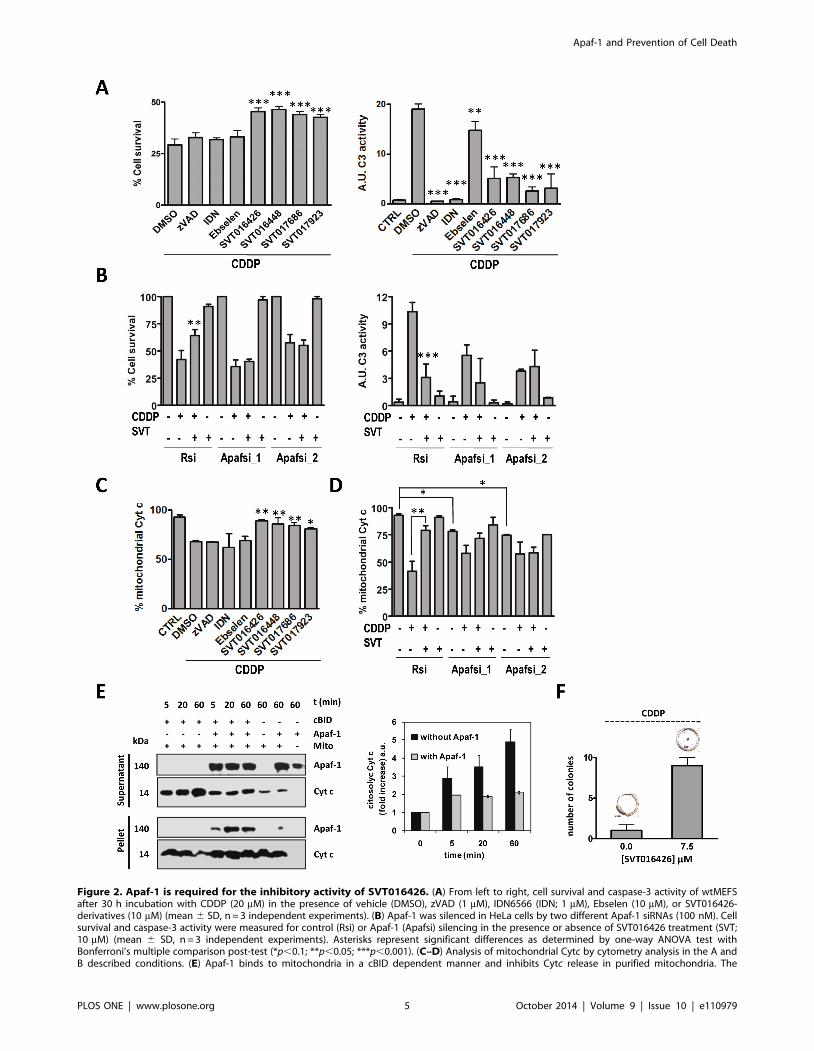
Figure 2. Apaf-1 is required for the inhibitory activity of SVT016426. (A) From left to right, cell survival and caspase-3 activity of wtMEFSafter 30 h incubation with CDDP (20 mM) in the presence of vehicle (DMSO), zVAD (1 mM), IDN6566 (IDN; 1 mM), Ebselen (10 mM), or SVT016426-derivatives (10 mM) (mean 6 SD, n = 3 independent experiments). (B) Apaf-1 was silenced in HeLa cells by two different Apaf-1 siRNAs (100 nM). Cellsurvival and caspase-3 activity were measured for control (Rsi) or Apaf-1 (Apafsi) silencing in the presence or absence of SVT016426 treatment (SVT;10 mM) (mean 6 SD, n = 3 independent experiments). Asterisks represent significant differences as determined by one-way ANOVA test withBonferroni’s multiple comparison post-test (*p,0.1; **p,0.05; ***p,0.001). (C–D) Analysis of mitochondrial Cytc by cytometry analysis in the A andB described conditions. (E) Apaf-1 binds to mitochondria in a cBID dependent manner and inhibits Cytc release in purified mitochondria. The
Apaf-1 and Prevention of Cell Death
PLOS ONE | www.plosone.org 5 October 2014 | Volume 9 | Issue 10 | e110979

palliate the noxious consequences of unwanted apoptosis [21,29].
There were problems determining the clinical benefit of NAC [30]
and although the pan-caspase inhibitor IDN-6556 was effective in
different settings [6,7,8] safety concerns were raised due to atypical
cellular infiltrate in the liver, kidney, and adrenal glands in
experimental models [9]. Here we demonstrated that ebselen,
IDN-6556 as well as the SVT compounds, showed inhibition of
caspase activity (Fig. 2B) however only SVT compounds showed
potential to prevent CDDP-, and hypoxia-induced cell death
(Fig. 2 and 4). In fact, in mammals solely caspase inhibition,
downstream of MOMP, delays and modifies the outcome, more
than prevents cell death [31]. Strikingly, in all the cell types
analyzed in our study the SVT-dependent cell death inhibition
correlated with a decrease in Cytc release from mitochondria. In
contrast, no such effect was observed in cells treated with caspase
inhibitors or antioxidants. We conclude that SVT-compounds
were inducing a pre-MOMP event probably related to Apaf-1.
Previous reports include conflicting data on the role of Apaf-1 on
Cytc release. Franklin and Robertson [32] showed that in Apaf-1
deficient Jurkat T-lymphocytes etoposide- and mitoxantrone-
induced Cytc release and loss of mitochondrial membrane
potential was impaired. In contrast, Ferraro et al. [33] observed
that Cytc release from Apaf-1-deficient proneural embryonic
telencephalic naıve Apaf-1 knockout cells, followed faster kinetics
than that of wild type cells. In addition, Potokar et al. [34]
observed that at short times after rotenone-induced apoptosis in
mitochondria treated with rApaf-1 (150 nM) and cBID (10 nM) were centrifuged and separated into pellet (mitochondria) and supernatant andimmunobloted for Cytc release and Apaf-1 incorporation (left panel). Bands were quantified by densitometry (right panel). (F) wtMEFS were treatedwith 5 mM CDDP for 6 h and incubated in the presence or absence SVT016426. After two weeks cells were fixed and stained with crystal violet andthe quantification of the number of cell colonies was performed. Data represent the mean 6 SD of three independent experiments.doi:10.1371/journal.pone.0110979.g002
Figure 3. Apaf-1 inhibitors protected against CDDP-induced ototoxicity in an in vitro and in vivo model. (A) Caspase-3 activity, Cytcrelease and cell survival, respectively from HEI-OC1 cells treated with CDDP (25 mM) in the presence or absence of compounds (10 mM). Data areexpressed as the mean 6 SD (n = 3 independent assays). Asterisks represent significant differences relative to DMSO treatment as determined by one-way ANOVA test with Bonferroni’s multiple comparison post-test (*p,0.1; **p,0.05; ***p,0.001). (B) Transcriptional regulation of Apaf-1 analyzedby RT-qPCR from HEI-OC1 cells treated with CDDP (25 mM) in the presence or absence of compounds (10 mM) (mean 6 SD; n = 3). (C) Apaf-1inhibitors protected against CDDP-induced ototoxicity in a zebrafish model. Five-day embryos (n = 10) were treated with CDDP (10 mM) for 24 h inthe presence or absence of SVT017686 (15 mM). Neuroblasts are the white dots (arrows). Scale bar, 1 mm. Hair cells were counted and cell survivalwas calculated as a percentage of the control group (not exposed to CDDP). Data bars represent mean 6 SD hair cell survival (n = 9/10 fishes).doi:10.1371/journal.pone.0110979.g003
Apaf-1 and Prevention of Cell Death
PLOS ONE | www.plosone.org 6 October 2014 | Volume 9 | Issue 10 | e110979

pituitary cells Apaf-1 redistributed to mitochondria. Our in vitroexperiments with isolated mitochondria aiming to reproduce the
initial events of mitochondria-mediated apoptosis using cBID
showed [35] that the presence of Apaf-1 on mitochondria cBID-
induced attenuated release of Cytc (Fig. 2) and thus Apaf-1 is
probably a modulator of MOMP events. The Cytc release
Figure 4. Apaf-1 inhibitors provide protection in an in vivo animal ischemic induced acute kidney injury model. (A) Cell survival andcaspase-3 activity in cell lysates from LLC-PK-1 cells measured after 24 h hypoxia/hypercapnia plus 24 h normoxia in the absence or presence of Apaf-1 inhibitor (10 mM) (mean 6 SD; n = 3). (B) Transcriptional regulation of Apaf-1 and Bnip-3 were analyzed by RT-qPCR in LLC-PK-1 cells. mRNA levelswere compared in hypoxia/hypercapnia (Hx) versus normoxia (Nx) conditions after SVT016448 treatment (mean 6 SD; n = 3). (C) Intraperitonealinjection of SVT016448 (10 mg/Kg) decreases caspase activity in a renal hot ischemia in vivo model. A representative image of theimmunofluorescence of active caspase-3 (left panel) and mean grey value quantification (right panel) (mean 6 SD, n = 5). Asterisks representsignificant differences relative to ischemia treatment as determined by one-way ANOVA test with Bonferroni’s multiple comparison post-test (*p,0.05). (D) Damaged tissue decreases in the presence of the Apaf-1 inhibitor, as determined by histopathological evaluation. (E) SVT016448 treatment-dependent decrease in Apaf-1 mRNA levels, as analyzed by RT-qPCR.doi:10.1371/journal.pone.0110979.g004
Apaf-1 and Prevention of Cell Death
PLOS ONE | www.plosone.org 7 October 2014 | Volume 9 | Issue 10 | e110979
inhibitory capability of SVT016426 was dependent on the Apaf-1
expression levels in the cell. Moreover, Apaf-1 gene silencing
renders cells with less mitochondrial Cytc (Fig. 2D). All together,
our results shift the balance to the existence of a role for Apaf-1 in
the control of Cytc release.
Nowadays there is no ideal protective agent in clinical use
against cisplatin ototoxicity. This would eliminate one of the dose-
limiting side effects of cisplatin therapy and improve the quality of
life for many patients [36]. As an example clinical trials for the
transtympanicof L-N-Acetylcysteine administration in patients
receiving cisplatin chemotherapy for head and neck cancerdid
not demonstrate a significant benefit [37]. There is a great need to
find safe and effective protective agents. In this study we show the
effectiveness of treatment with SVTs in cellular and in vivo models
of cisplatin- induced ototoxicity.
Ischemic, toxic, or obstructive renal damage triggers apoptotic
mechanisms that cause tubular cell loss and decreased renal
function [38]. Currently there are no effective pharmacological
strategies to treat acute renal failure. Here, we show the ability of
Apaf-1 inhibitors to decrease cell death in kidney ischemia cellular
and in vivo models. These results support the potential application
of Apaf-1 inhibitors to prevent acute kidney injury (AKI).
In conclusion, we characterized SVT016426 and its derivatives
as selective inhibitors of Apaf-1 that prevent cell death in different
cellular settings and show efficiency as potential apoptosis
inhibitory drugs in models of disease.
Materials and Methods
Chemical synthesisThe general method for the synthesis of perhydro-1,4-
diazepine-2,5-diones such as SVT016426 and 3-substituted 1,4-
piperazine-2,5-dione derivatives (SVT016448, SVT017686 and
SVT017923) has been previously reported [39].
Apoptosome activity assaysApoptosome was reconstituted by incubating rApaf-1 (100 nM)
with vehicle (DMSO) or SVT compounds in assay buffer (20 mM
HEPES, 10 mMKCl, 1.5 mM MgCl2, 1 mM EDTA, 1 mM
EGTA, 1 mM DTT, 0.1 mM PMSF) at 30uC for 30 min. Next,
100 mM dATP and 100 nM Cytc were added and incubated for
additional 40 min. Then, caspase-9 (100 nM) was added, and
activity was monitored by the Ac-LEHD-afc substrate (50 mM;
Enzo Life Sciences) using a Victor 2 spectrofluorimeter
(lexc = 390 nm; lem = 510 nm). A cell-free caspase activation assay
was also performed as described [13,40]. Briefly, Apaf-1 depleted
cytosolic extracts from HEK293 were incubated with rApaf-1
(100 nM) and in the presence of the different SVT-derivatives. Ac-
DEVD-afc substrate (Enzo Life Sciences) was used to measure
caspase 3 activity, using a Victor 2 spectrofluorimeter.
Fluorescence polarization spectroscopyA 60 nM solution of 59-69carboxyfluorescein-labeled
SVT016426 was titrated with concentrated protein solutions in
reaction buffer (total reaction volume was 200 ml) Fluorescence
polarization measurements were recorded in a Victor 2 Wallac
1420 Workstation spectrofluorimeter (lexc = 480 nm;
lem = 535 nm).
Computational dockingThe blind docking protocol consisted on a search for cavities in
the Apaf-1 CARD-NOD structure to identify potential ligand
binding sites, followed by a flexible docking of the ligand on to the
different sites located. Fig. S1 in File S1 shows the best scored sites
for interaction within the target structure and Table S1 in File S1
summarizes the site scores. Docking at the binding sites rendered
plausible binding poses for compound SVT016426 (Fig. S2 and
Table S1 in File S1).
Computational Methods. Molecular simulations were con-
ducted with the package Schrodinger Suite 2011 [41] through its
graphical interface Maestro [42]. The program Macromodel [43],
with its default force field OPLS 2005, a modified version of the
OPLS-AA force field [44], and GB/SA water solvation conditions
[45] were used for all energetic calculations. The coordinates of
WD40-deleted human Apaf-1 (Apaf-1 1–591, PDB 1Z6T, chain
B) were obtained from the Protein Data Bank [46] at Brookhaven
National Laboratory. The structure of the protein was prepared
using the Protein Preparation Wizard included in Maestro to
remove the unused subunits as well as the solvent molecules and
ligands, adding hydrogens, setting protonation states and mini-
mizing the energy using the OPLS force field. SiteMap [47,48,49],
was used to identify and score potential binding sites on the
protein. The score provided by SiteMap is constructed and
calibrated so that the average SiteScore for 157 investigated
submicromolar sites is 1.0. Therefore, a score$1.0 suggests a site
of particular promise, while a score#0.8 has been found to
accurately distinguish between drug-binding and non-drug-bind-
ing sites. The best scored sites were used as targets for docking of
the SVT016426 ligand with the program Glide XP [50,51,52].
The structure of SVT016426 was built within Maestro and then it
was prepared with the LigPrep application [53] included in the
software to generate ring conformers of the two stereoisomers of
the compound. Eight ring conformers for each enantiomer were
generated and used as input ligands for the docking at each target
site. In order to ensure a good conformational sampling during the
docking simulations the following settings were used: a maximum
of 5000000 poses per input structure for the initial phase of
docking, the extendend sampling protocol, and 50 poses for the
post-docking minimization. Glide XP scores were used to rank the
resulting docked poses. The interaction diagrams for the best poses
were built with the Ligand Interaction Diagram application
implemented in Maestro.
NMR experimentsNMR spectra for protein ligand interaction were recorded at
298 K with a BrukerUltrashield Plus Avance II 600 MHz
spectrometer equipped with a 5-mm cryogenically-cooled TCI
probe. An NMR sample consisted of 1 mM rApaf-1 or Apaf-1 1–
591 and 20 or 100 mM SVT017683 to yield protein:ligand ratios
of 1:20 and 1:100 for the waterLOGSY and STD experiments,
respectively. The sample buffer was 50 mM phosphate, pH 7.0,
with 50 mMNaCl; 8 K points were used for a sweep width of
9,600 Hz and a total of 1 K and 4 K scans were accumulated for
the waterLOGSY and STD experiments, respectively.
Cell culture and reagentsHuman cervix adenocarcinoma (HeLa) and proximal tubular
renal porcine LLC-PK-1 cells were obtained from ATCC
(Rockville, MD). wtMEFS, previously established in the referenced
publications [54], were kindly provided by Dr. Franceso Cecconi.
The HEI-OC1 cell line (House Ear Institute-Organ of Corti-1)
was obtained from F. Kalinec and primary human dermal
fibroblasts (HDFn) were obtained from Cascade Biologics. HeLa,
HDFn, wtMEFS cells were cultured in DMEM supplemented with
10% FBS (Invitrogen). LLC-PK-1 cell line was grown in M199
supplemented with 3% FBS. HEI-OC1 cells were cultured in
antibiotic free DMEM with 10% FBS (GIBCO) and 5 mg/ml of
gamma interferon (Sigma) at 33uC and 10% CO2 in air. All the
Apaf-1 and Prevention of Cell Death
PLOS ONE | www.plosone.org 8 October 2014 | Volume 9 | Issue 10 | e110979

rest of cell lines were maintained at 37uC in an atmosphere of 5%
carbon dioxide. Cisplatin (CDDP) and ebselen were purchased
from Sigma and pan-caspase inhibitor Z-Val-Ala-Asp(OMe)-
fluoromethylketone (zVAD-fmk) from Tocris. IDN-5665 was
synthesized by Laboratorios Salvat, S.A. LipofectamineTM 2000
(Invitrogen) was used according to the manufacturer’s instructions
to transfect HeLa cells with the control siRNA (Rsi; Cell Signaling)
and Apaf-1 siRNA (Dharmacon and Cell Signaling).
Cell-based caspase activation assayMEFS and HeLa cells were seeded in 6-well plate at a cellular
density of 16105 cells/well and 26105 cells/well, respectively,
while 36105 cells/well were grown for LLC-PK-1. Cells were pre-
treated with SVT compounds for 1 h and then cisplatin (25 mM)
was added. After 30 h cells were harvested and cytosolic extracts
were obtained as described previously [13]. Total protein (50 mg)
was mixed with assay buffer containing 20 mM Ac-DEVD-afc
substrate. Activity was measured using a Victor 2 spectrofluorim-
eter. HeLa cells were treated in the same conditions after 24 h of
siRNA transfection. LLC-PK-1 were also pre-treated with SVT
compounds for 1 h and then subjected to hypoxia/hypercapnia
(HH - 1% O2, 18% CO2) conditions for 24 h plus 24 h of re-
oxygenation before cytosolic extracts were prepared.
HEI-OC1 cells were plated in sterile 96-well microtiter plates
(BD) at cellular density of 4500 cells/well and treated with the
SVT compounds and the caspase inhibitor zVAD-fmk in a dose
response manner. Apoptosis was induced with cisplatin and cells
were incubated for 24 h. Caspase 3 activity was determined by
using Caspase-Glo 3/7 Assay Kit (Promega) according to the
manufacturer’s instruction.
Cell viability assaysCell proliferation was measured using a 3-(4,5-dimethylthiazol-
2-yl)-2,5-diphenyltetrazolium bromide (MTT) colorimetric assay.
MEFS, LLC-PK-1 and HEI-OC1 cells were grown in 96-well
plates at a cellular density of 1500 cells/well, 2500 cells/well and
4500 cells/well, respectively. After seeding, the cells were left to
adhere to the plate overnight and MEFS and HEI-OC1 were
treated with cisplatin plus SVT compounds for 24 h at 37uC. Four
hours before the end of the treatment MTT (1 mg/ml in PBS) was
added to each well and the plates were incubated for a further 4 h
at 37uC. Finally, the medium was removed and the precipitated
formazan crystals were dissolved in optical grade DMSO. Plates
were read at 570 nm on a Wallac 1420 workstation. Same
procedure was achieved in the case of LLC-PK-1 cells after 24 h
of HH plus 24 h of re-oxygenation.
Cytochrome c release assayMEFS and HeLa cells were grown in 6-well plate at the same
conditions described above. After 24 h in the presence of SVT
compounds, mitochondrial Cytc was followed using the Innocy-
teTM Flow Cytometric Cytochrome c Release kit (Calbiochem).
Same procedure was applied for HEI-OC1 cells upon 48 h of
CDDP administration in the presence or not of the SVT
compounds or the caspase inhibitor zVAD-fmk. Cells were
analysed on a Cytomics FC 500 (Beckman Coulter) flow
cytometer.
Cytochrome c release assays in purified mitochondriaPurified mitochondria were prepared from wtMEFS cells.
Briefly, cells were mechanically broken using a 2 ml glass/glass
dounce homogenizer (Kontes), using two rounds of 30 strokes.
Homogenates were cleared in MB buffer (10 mM Hepes pH 7.5,
210 mM mannitol, 70 mM sucrose, and 1 mM EDTA) at 1,500 g
and the mitochondria were spun down at 10,500 g. For Cytcrelease assays, 20 mg of purified mitochondria were resuspended
in RB buffer (125 mMKCl, 5 mM succinate and 0.5 mM EGTA)
supplemented with protease inhibitor cocktail (Roche). Proteins at
indicated concentrations were added to the samples and the
incubations were carried out at 30uC. At the indicated time points,
samples were spun down and supernatants and pellets were
analyzed by immunoblotting using anti-cytochrome c or anti-
Apaf-1 antibodies.
Clonogenicity assays1250 MEFS cells per well were seeded in 24-well plates, treated
with cisplatin 5 mM in the presence or absence of SVT016426.
After 6 h of treatment, the apoptotic stimulus was removed. Two
weeks latter cell were fixed with 2% paraformaldehyde and stained
with crystal violet.
ImmunoblottingWhole cell extracts were obtained by lysing cells in 25 mMTris-
HCl pH 7.4, 1 mM EDTA, 1 mM EGTA, and 1% SDS plus
protease and phosphatase inhibitors. Protein concentration was
determined by the BCA protein assay. Lysates were subjected to
SDS-PAGE, transferred to nitrocellulose membranes, and im-
munoblotted following standard procedures. The antibodies used
were Apaf-1 (611364 from BD), cytochrome c (556433 from BD),
capase-9 (9508 from Cell Signaling) and a-tubulin (T-8203 from
Sigma). Quantifications were performed with Image J software.
RT-qPCRTotal RNA was isolated using the RNAsy Kit (Qiagen)
according to the manufacturer’s instructions. First strand cDNA
was synthesized from 500 ng of RNA using Superscript First
Strand Synthesis System (Invitrogen). Gene products were
analyzed by qPCR using SYBR Green Mix (Roche) and specific
oligonucleotides in a Roche 480 Lightcycler. Primer sequences are
the following: F 59-CTGGCAACGGGAGATGACAATGG-39
and R 59-AGCGGAGCACACAAATGAAGAAGC-39 for Apaf-1
and F 59-CTGGACGGAGTAGCTCCAAG-39 and R 59-
GACGCTCGTGTTCCTCATGC-39 for Bnip3. Each value
was normalized to b-actin (F 59-GGACTTCGAGCAAGA-
GATGG-39 and R 59-AGCACTGTGTTGGCGTACAG-39)
and expressed as relative units (R.U.). All reactions were
performed in triplicate.
Zebra fishZebrafish (Daniorerio) embryos of the AB wild-type strain were
used. At 5 days post-fertilization (dpf) larvae were dispensed in 24
well plates in embryo medium in a tissue incubator at 28.5uC.
Cisplatin at the concentration of 10 mM and compounds were
added and incubated for 24 h. After this period of treatment, the
embryos were stained with 0.01% DASPEI (Molecular Probes),
washing and anesthetized with 0.04% of tricaine. In order to take
photomicrographs, the embryos were mounted in methylcellulose
for observation under a fluorescence microscope. Pictures
obtained were analyzed by counting the number of neuroblast
presented in the lateral line of embryos.
Renal hot ischemia assayThis study was approved by the Committee on the Ethics and
Animal Welfare of the CIPF (Permit Number: 10-0185). All efforts
were made to minimize suffering.
Apaf-1 and Prevention of Cell Death
PLOS ONE | www.plosone.org 9 October 2014 | Volume 9 | Issue 10 | e110979

Male Wistar rats (200 g) were distributed in 4 different groups
(n = 5); vehicle, ischemia plus vehicle; ischemia plus 10 or 20 mg
kg21 SVT016448. Rats were anesthetised (Pentobarbital 6 mg
ml21), sacrificed by cervical dislocation and maintained during
60 min at 37uC. Then kidneys were extracted by abdominal
incision and divided into three parts. The first slice was frozen in
liquid nitrogen, the second was fixed in formaldehyde 4% and the
third was fixed in OCT. In the intraperitoneal assay (vehicle,
methyl cellulose 0.5%, Tween80 0.1% in PBS) the treatments
were administered 60 min before the rats were sacrificed. For the
intravenous assay (vehicle, 30% of DMSO in PBS) compound
treatments were administered 6 h before the sacrifice and the
ischemia was prolonged by additional 90 min.
Pathological analysisKidneys were analyzed in the SIAL (Integral services for
Laboratory animals Barcelona, Spain). Briefly, kidneys fixed in
formaldehyde were included in paraffin and slices stained with
haematoxylin-eosin. Five levels of damage were established
between no damage and serious lesions and scored from 1 to 5.
Score 5 corresponds to severe lesions and Score 4 corresponds to
moderate damage.
ImmunohistochemistryKidney slices (8 mm) were fixed in cold acetone and blocked
with 5% goat serum, 0.3% Triton X-100 in PBS, incubated with
10 ml of caspase-3 active antibody (PE active caspase-3, BD
Pharmigen) and mounted in Mowiol with DAPI. Confocal images
of four different fields per sample were captured with a Leica
microscope. Quantification of caspase-3 activity was performed
evaluating the mean grey value of the different fields using the
ImageJ software analysis.
Supporting Information
File S1 This file contains supporting information for the
Materials and Methods section, Figures S1–S8 and Table S1.
(DOCX)
Acknowledgments
This paper was dedicated to the memory of Enrique Perez-Paya. We thank
Eliana Sirvent for technical assistance and all members of our laboratories
for discussions. We would like to thank Dr. Francesco Cecconi
(UniversitadegliStudi di Roma ‘Tor Vergata’) for providing us with MEFS.
We are grateful to Dr. Kalinec (House Ear Institute) for provide HEI-OC1
cell line. We thank the CIPF proteomics and confocal microscopy services
for technical support.
Author Contributions
Conceived and designed the experiments: MO MS SM CH GB A.
Messeguer EPP. Performed the experiments: MO MS LM RM SM JGV
OL RJC JB A. Moure. Analyzed the data: MO MS RJC JB GB A.
Messeguer CH EPP. Contributed reagents/materials/analysis tools: JB
RJC APL A. Messeguer CL CH. Contributed to the writing of the
manuscript: MO MS CL CH A. Messeguer EPP.
References
1. Wang C, Youle RJ (2009) The role of mitochondria in apoptosis. Annu Rev
Genet 43: 95–118.
2. Zou H, Henzel WJ, Liu X, Lutschg A, Wang X (1997) Apaf-1, a human protein
homologous to C. elegans CED-4, participates in cytochrome c-dependent
activation of caspase-3. Cell 90: 405–413.
3. Green DR, Kroemer G (2005) Pharmacological manipulation of cell death:
clinical applications in sight? J Clin Invest 115: 2610–2617.
4. Linton SD (2005) Caspase inhibitors: a pharmaceutical industry perspective.
Curr Top Med Chem 5: 1697–1717.
5. Yaoita H, Ogawa K, Maehara K, Maruyama Y (2000) Apoptosis in relevant
clinical situations: contribution of apoptosis in myocardial infarction. Cardiovasc
Res 45: 630–641.
6. Hotchkiss RS, Karl IE (2003) The pathophysiology and treatment of sepsis.
N Engl J Med 348: 138–150.
7. Hoglen NC, Chen LS, Fisher CD, Hirakawa BP, Groessl T, et al. (2004)
Characterization of IDN-6556 (3-[2-(2-tert-butyl-phenylaminooxalyl)-amino]-
propionylamino]-4-oxo-5-(2,3,5,6-tetrafluoro-phenoxy)-pentanoic acid): a liver-
targeted caspase inhibitor. J Pharmacol Exp Ther 309: 634–640.
8. McCall M, Toso C, Emamaullee J, Pawlick R, Edgar R, et al. (2011) The
caspase inhibitor IDN-6556 (PF3491390) improves marginal mass engraftment
after islet transplantation in mice. Surgery 150: 48–55.
9. Burgess G, Ayoub A, Leblanc B, Gropp K, Kawabata T, et al. (2008) Treatment
with the pancaspase inhibitor PF-03491390, resulted in atypical cellular
infiltrates in mouse and rat liver. Cell: iiA–ivA, 1A–1241A.
10. Mochizuki H, Hayakawa H, Migita M, Shibata M, Tanaka R, et al. (2001) An
AAV-derived Apaf-1 dominant negative inhibitor prevents MPTP toxicity as
antiapoptotic gene therapy for Parkinson’s disease. Proc Natl Acad Sci U S A 98:
10918–10923. Epub 12001 Sep 10914.
11. Gao Y, Liang W, Hu X, Zhang W, Stetler RA, et al. (2009) Neuroprotection
against hypoxic-ischemic brain injury by inhibiting the apoptotic protease
activating factor-1 pathway. Stroke 41: 166–172.
12. Srinivasula SM, Ahmad M, Fernandes-Alnemri T, Alnemri ES (1998)
Autoactivation of procaspase-9 by Apaf-1-mediated oligomerization. Mol Cell
1: 949–957.
13. Malet G, Martin AG, Orzaez M, Vicent MJ, Masip I, et al. (2006) Small
molecule inhibitors of Apaf-1-related caspase- 3/-9 activation that control
mitochondrial-dependent apoptosis. Cell Death Differ 13: 1523–1532.
14. Mondragon L, Galluzzi L, Mouhamad S, Orzaez M, Vicencio JM, et al. (2009)
A chemical inhibitor of Apaf-1 exerts mitochondrioprotective functions and
interferes with the intra-S-phase DNA damage checkpoint. Apoptosis 14: 182–
190.
15. Riedl SJ, Li W, Chao Y, Schwarzenbacher R, Shi Y (2005) Structure of the
apoptotic protease-activating factor 1 bound to ADP. Nature 434: 926–933.
16. Dalvit C, Pevarello P, Tato M, Veronesi M, Vulpetti A, et al. (2000)
Identification of compounds with binding affinity to proteins via magnetization
transfer from bulk water. J Biomol NMR 18: 65–68.
17. Meyer B, Peters T (2003) NMR spectroscopy techniques for screening and
identifying ligand binding to protein receptors. Angew Chem Int Ed Engl 42:
864–890.
18. Zou H, Li Y, Liu X, Wang X (1999) An APAF-1.cytochrome c multimeric
complex is a functional apoptosome that activates procaspase-9. J Biol Chem
274: 11549–11556.
19. Feng BY, Simeonov A, Jadhav A, Babaoglu K, Inglese J, et al. (2007) A high-
throughput screen for aggregation-based inhibition in a large compound library.
J Med Chem 50: 2385–2390.
20. Linton SD, Aja T, Armstrong RA, Bai X, Chen LS, et al. (2005) First-in-class
pan caspase inhibitor developed for the treatment of liver disease. J Med Chem
48: 6779–6782.
21. Kotamraju S, Konorev EA, Joseph J, Kalyanaraman B (2000) Doxorubicin-
induced apoptosis in endothelial cells and cardiomyocytes is ameliorated by
nitrone spin traps and ebselen. Role of reactive oxygen and nitrogen species.
J Biol Chem 275: 33585–33592.
22. Slattery EL, Warchol ME (2010) Cisplatin ototoxicity blocks sensory
regeneration in the avian inner ear. J Neurosci 30: 3473–3481.
23. Ton C, Parng C (2005) The use of zebrafish for assessing ototoxic and
otoprotective agents. Hear Res 208: 79–88.
24. Sadis C, Teske G, Stokman G, Kubjak C, Claessen N, et al. (2007) Nicotine
protects kidney from renal ischemia/reperfusion injury through the cholinergic
anti-inflammatory pathway. PLoS ONE 2: e469.
25. Eltzschig HK, Eckle T (2011) Ischemia and reperfusion-from mechanism to
translation. Nat Med 17: 1391–1401.
26. Bruick RK (2000) Expression of the gene encoding the proapoptotic Nip3
protein is induced by hypoxia. Proc Natl Acad Sci U S A 97: 9082–9087.
27. Laskowski IA, Hancock WW, Pratchke J, Wilhelm MJ, Tilney NL (2001)
Accelerated graft dysfunction in renal isografts from non-heart-beating donors.
Transplant Proc 33: 909–910.
28. Reubold TF, Wohlgemuth S, Eschenburg S (2011) Crystal structure of full-
length Apaf-1: how the death signal is relayed in the mitochondrial pathway of
apoptosis. Structure 19: 1074–1083.
29. Sarker KP, Biswas KK, Rosales JL, Yamaji K, Hashiguchi T, et al. (2003)
Ebselen inhibits NO-induced apoptosis of differentiated PC12 cells via inhibition
of ASK1-p38 MAPK-p53 and JNK signaling and activation of p44/42 MAPK
and Bcl-2. J Neurochem 87: 1345–1353.
30. Thiele H, Hildebrand L, Schirdewahn C, Eitel I, Adams V, et al. (2010) Impact
of high-dose N-acetylcysteine versus placebo on contrast-induced nephropathy
and myocardial reperfusion injury in unselected patients with ST-segment
Apaf-1 and Prevention of Cell Death
PLOS ONE | www.plosone.org 10 October 2014 | Volume 9 | Issue 10 | e110979

elevation myocardial infarction undergoing primary percutaneous coronary
intervention. The LIPSIA-N-ACC (Prospective, Single-Blind, Placebo-Con-trolled, Randomized Leipzig Immediate PercutaneouS Coronary Intervention
Acute Myocardial Infarction N-ACC) Trial. J Am Coll Cardiol 55: 2201–2209.
31. Kroemer G, Martin SJ (2005) Caspase-independent cell death. Nat Med 11:725–730.
32. Franklin EE, Robertson JD (2007) Requirement of Apaf-1 for mitochondrialevents and the cleavage or activation of all procaspases during genotoxic stress-
induced apoptosis. Biochem J 405: 115–122.
33. Ferraro E, Pesaresi MG, De Zio D, Cencioni MT, Gortat A, et al. (2011) Apaf1plays a pro-survival role by regulating centrosome morphology and function.
J Cell Sci 124: 3450–3463.34. Potokar M, Kreft M, Chowdhury HH, Vardjan N, Zorec R (2006) Subcellular
localization of Apaf-1 in apoptotic rat pituitary cells. Am J Physiol Cell Physiol290: C672–677.
35. Lovell JF, Billen LP, Bindner S, Shamas-Din A, Fradin C, et al. (2008)
Membrane binding by tBid initiates an ordered series of events culminating inmembrane permeabilization by Bax. Cell 135: 1074–1084.
36. Rybak LP, Mukherjea D, Jajoo S, Ramkumar V (2009) Cisplatin ototoxicity andprotection: clinical and experimental studies. Tohoku J Exp Med 219: 177–186.
37. Yoo J, Hamilton SJ, Angel D, Fung K, Franklin J, et al. (2014) Cisplatin
otoprotection using transtympanic L-N-acetylcysteine: A pilot randomized studyin head and neck cancer patients. Laryngoscope.
38. Ardura JA, Sanz AB, Ortiz A, Esbrit P (2013) Parathyroid hormone-relatedprotein protects renal tubuloepithelial cells from apoptosis by activating
transcription factor Runx2. Kidney Int 83: 825–834.39. Moure A, Sanclimens G, Bujons J, Masip I, Alvarez-Larena A, et al. (2011)
Chemical modulation of peptoids: synthesis and conformational studies on
partially constrained derivatives. Chemistry 17: 7927–7939.40. Fearnhead HO (2001) Cell-free systems to study apoptosis. Methods Cell Biol
66: 167–185.
41. (2011) Schrodinger Suite 2011. New York, NY: Schrodinger, LLC.
42. (2011) Maestro, version 9.2. New York, NY: Schrodinger, LLC.
43. (2011) MacroModel, version 9.9. New York, NY: Schrodinger, LLC.
44. Jorgensen WL, Maxwell DS, Tirado-Rives J (1996) Development and Testing of
the OPLS All-Atom Force Field on Conformational Energetics and Properties of
Organic Liquids. J Am Chem Soc 118: 11225–11236.
45. Still WC, Tempczyk A, Hawley RC, Hendrickson T (1990) Semianalytical
treatment of solvation for molecular mechanics and dynamics. Journal of the
American Chemical Society 112: 6127–6129.
46. Berman HM, Westbrook J, Feng Z, Gilliland G, Bhat TN, et al. (2000) The
Protein Data Bank. NucleicAcidsRes 28: 235–242.
47. (2011) SiteMap, version 2.5. New York, NY: Schrodinger, LLC.
48. Halgren T (2007) New method for fast and accurate binding-site identification
and analysis. Chem Biol Drug Des 69: 146–148.
49. Halgren TA (2009) Identifying and characterizing binding sites and assessing
druggability. J Chem Inf Model 49: 377–389.
50. Friesner RA, Banks JL, Murphy RB, Halgren TA, Klicic JJ, et al. (2004) Glide: a
new approach for rapid, accurate docking and scoring. 1. Method and
assessment of docking accuracy. J Med Chem 47: 1739–1749.
51. Friesner RA, Murphy RB, Repasky MP, Frye LL, Greenwood JR, et al. (2006)
Extra precision glide: docking and scoring incorporating a model of hydrophobic
enclosure for protein-ligand complexes. J Med Chem 49: 6177–6196.
52. Halgren TA, Murphy RB, Friesner RA, Beard HS, Frye LL, et al. (2004) Glide:
a new approach for rapid, accurate docking and scoring. 2. Enrichment factors
in database screening. J Med Chem 47: 1750–1759.
53. (2011) LigPrep, version 2.5. New York, NY: Schrodinger, LLC.
54. Cecconi F, Alvarez-Bolado G, Meyer BI, Roth KA, Gruss P (1998) Apaf1 (CED-
4 homolog) regulates programmed cell death in mammalian development. Cell
94: 727–737.
Apaf-1 and Prevention of Cell Death
PLOS ONE | www.plosone.org 11 October 2014 | Volume 9 | Issue 10 | e110979
Copyright © 2022 FDOKUMEN