Primary Ear and Hearing Care Training Resource TRAINER'S ...
Upload
independentCategory
view
2download
0
http://www.jstor.org
!"#$%&'()*+&(,"--$(%.(*(/+$0(1*&2(13+4"#5"(%.(67-8+9-"(:'9"$(;*$"4(%#(<8%8%=+5+8'(!"#$+8+3+8'>780%&?$@2()%#A(BC(B*#D(E+--+*F(6C(!*+4"-D(G*#"8(!C(,0*#AD(G%"--"(,C(H&"$$%#D(>&807&(ICH%99"&!%7&5"2(H&%5""4+#A$2(;+%-%A+5*-(!5+"#5"$D(J%-C(KLMD(I%C(NONO(?>7AC(KKD(NPPN@D(99C(NOOQNORH7S-+$0"4(S'2(:0"(T%'*-(!%5+"8'!8*S-"(UTV2(http://www.jstor.org/stable/76561>55"$$"42(WNXWPXKWWR(NW2NM
Your use of the JSTOR archive indicates your acceptance of JSTOR's Terms and Conditions of Use, available athttp://www.jstor.org/page/info/about/policies/terms.jsp. JSTOR's Terms and Conditions of Use provides, in part, that unlessyou have obtained prior permission, you may not download an entire issue of a journal or multiple copies of articles, and youmay use content in the JSTOR archive only for your personal, non-commercial use.
Please contact the publisher regarding any further use of this work. Publisher contact information may be obtained athttp://www.jstor.org/action/showPublisher?publisherCode=rsl.
Each copy of any part of a JSTOR transmission must contain the same copyright notice that appears on the screen or printedpage of such transmission.
JSTOR is a not-for-profit organization founded in 1995 to build trusted digital archives for scholarship. We work with thescholarly community to preserve their work and the materials they rely upon, and to build a common research platform thatpromotes the discovery and use of these resources. For more information about JSTOR, please contact [email protected].
Sensory hair cells of a fish ear: evidence of multiple types based on ototoxicity sensitivity HONG Y. YAN, WILLIAM M. SAIDELt,JANET S. CHANG, JOELLE C. PRESSON AND ARTHUR N. POPPER
Department of Zoology, The University of Maryland, College Park, Maryland 20742, U.S.A.
SUMMARY
Sensory hair cells from the striolar region (striolar hair cells) of the utricle and the lagena of the ear of a teleost fish Astronotus ocellatus (Cuvier) ear are sensitive to gentamicin sulphate, an ototoxic drug. In contrast, sensory hair cells from outside the striolar region (extra-striolar hair cells) are not sensitive to gentamicin. These data, combined with results from studies showing different ultrastructural features and different immunoreactivity to a calcium binding protein, S-100, lead to the suggestion that there are distinguishable types of hair cells in these endorgans. These results add to the increasing evidence that classifying the sensory hair cells of fish ears only as the traditional 'vestibular type II' may be inadequate for properly understanding structure and function of the fish ear.
1. INTRODUCTION
Two types of sensory hair cell have traditionally been distinguished in the peripheral vestibular apparatus of amniote vertebrates (Lewis et al. 1985). The type I hair cell is shaped like an amphora and is innervated by a nerve calyx which envelops all but the most distal region of the hair cell body. The type II hair cell is cylindrical in shape and is innervated basally by small nerve endings from comparatively thin fibres (see figure 9 of Wersall (1956)).
In contrast, because of reported similarities in shape, ultrastructure and innervation, all of the sensory hair cells of the fish ear have generally been characterized as being similar to the amniote type II hair cells (Wersall 1961; Lowenstein et al. 1964; Hama 1969; Nakajima & Wang 1974; Popper & Hoxter 1981). However, ciliary bundle differences (Dale 1976; Popper 1976; Platt & Popper 1981; Platt 1983), cell soma size, innervation, orientation (Wegner 1979, 1982; Jensen 1984; Mathiesen 1985) and recently even physiology (Sugihara & Furukawa 1989) have been used to suggest the presence of multiple types of sensory hair cells in the otolithic organs of fish ears.
In the past few years we have used a variety of different techniques to investigate the sensory epithelia of the inner ear of fishes, particularly the maculae of the three otolithic organs, the utricle, lagena and saccule. Results of these studies have prompted us to consider that there may be a novel kind of differ- entiation within the hair cell populations of the fish ear. First, afferent axons with diameters greater than
t Present address: Department of Poultry Science, University of Maryland, College Park, Maryland 20742, U.S.A.
4-5 upm only innervate the striolar region of the utricular and lagenar epithelia of the oscar, Astronotus ocellatus (Cuvier), and the kissing gourami, Helostoma temincki (Cuvier & Valenciennes). In the utricle and lagena, terminal arbors that stain by Prussian blue indicate that the locations of nodal-like postsynaptic membrane are restricted to the striolar region (Saidel 1988; Saidel et al. 1990a). Second, studies using immunoreactivity to a calcium-binding protein, S-100, demonstrated that sensory hair cells in the striolar region of the utricle and in the saccule of the oscar respond to the S-100 antibody, whereas cells outside of the striola (extra-striola) show little immunoreactivity to S-100 (Saidel et al. 1990 b).
Most recently we studied the effects on the ear of gentamicin sulphate, an aminoglycoside antibiotic that causes structural damage to sensory hair cells of vestibular endorgans in many mammalian species (see Hawkins 1976). In particular, type I hair cells are generally more sensitive to gentamicin than are type II hair cells, and type I cells are usually damaged earlier than type II hair cells (Wersall & Hawkins 1962; Wersall et al. 1971; Wersall 1981). Because of its selective damage to the two types of hair cells in mammalian vestibular organs, aminoglycoside anti- biotics can be used as a tool to distinguish between vestibular sensory hair cell types.
In the present study, we report the discovery of selective damage by gentamicin sulphate to one specific population of sensory hair cells of both the utricle and lagena of a fish ear. In combining these findings with our previous data on the selective immunoreactivity to S-100 and differentiation of trigger zones, the present discovery allows us to propose new criteria for
Proc. R. Soc. Lond. B (1991) 245, 133-138 133 Printed in Great Britain
10 Vol. 245. B

134 H. Y. Yan and others Hair cell types of ish ear
L
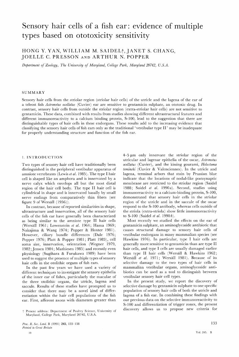
Figure 1. Scanning electron micrographs of the utricle. (a) A normal utricle of a control animal. Note the hair cells in the striolar region (S) are less densely distributed than the extra-striolar hair cells. (b) Higher magnification of the striolar area of (a) indicated by asterisk sign. (c) Selective damage of hair cells in the striola of a utricle from a fish that had received three injections of 40 mg kg-l of gentamicin sulphate. The fish was allowed to survive seven days after the last injection. (d) Higher magnification of the denuded striolar region of (c) indicated by asterisk sign. Bars indicate 50 ptm. Orientation: Rostral (R) to the right, lateral (L) to the top.
distinguishing multiple types of sensory hair cells in a fish ear.
2. MATERIALS AND METHODS
Young oscars (Astronotus ocellatus; Cichlidae) (average body weight 10 g and standard length 8 cm) obtained from a local wholesaler were used in the study. Gentamicin sulphate (GE) (Lot No. 58F-0353; Sigma Chemical Co., St Louis, Missouri) was dissolved in teleost saline (0.7 ? NaCI) and diluted to prepare four different dosages for injection (10 mg kg-1, 20 mg kg-1, 40 mg kg- and 120 mg kg-1). Ex- perimental fish were injected intramuscularly near the basal part of the dorsal fin once daily for one, two, three or four sequential days. Each experimental group had 12 fish. Control fish, four in each group, received the same volume of saline as the GE given to the experimental animals. Fish were allowed to survive from 1 to 10 days following the initial
injection. During this time the overall behaviour of the fish was monitored to look for apparent vestibular disturbances brought about by the gentamicin.
Following the selected survival time, fish were chilled in ice water and decapitated. Fish ears were excised following the procedures of Saidel et al. (1990 b) and prepared for scanning electron microscopy (SEM) (Popper & Hoxter 1984; Saidel et al. 1990 b).
3. RESULTS
In a normal (control) fish, the hair cells with relatively short ciliary bundles form a band curving inside the anterior and lateral borders of the sensory epithelia of both the utricle and the lagena. In addition, hair cells in this region are distributed in a lower population density than in the rest of the epithelia (figures l a, b and 2 a, b). This region of lower hair cell
Proc. R. Soc. Lond. B (1991)

Hair cell types offish ear H. Y. Yan and others 135
Figure 2. Scanning electron micrographs of the lagena. (a) A normal lagena of a control animal. Note the hair cells in the striolar region (S) are less densely distributed than the extra-striolar hair cells. (b) Higher magnification of the striolar area of (a) indicated by asterisk sign. (c) Selective damage of hair cells in the striola of the lagena of a fish that had received four injections of 120 mg kg-' of gentamicin sulphate. The fish was allowed to survive one day after the last injection. Note some extra-striolar hair cells are covered with membranes which might give an impression that the ciliary bundles are missing. (d) Higher magnification of the denuded striolar region of (c) indicated by asterisk sign. Bars indicate 50 gm. Orientation: Rostral (R) to the right, dorsal (D) to the top.
density and shorter ciliary bundles has been called the striola (see Lewis et al. 1985). The area outside the striola is called the extra-striola region.
In the saccule, hair cells with short ciliary bundles cover almost the entire sensory epithelia except for marginal areas (figure 3a, b). Therefore, with criteria based only on surface morphology, no specific striola region can be identified in the saccule.
There were no apparent behavioural or ultra- structural effects after two injections of gentamicin at any dosage level. However, just one day after three injections of 120 mg kg-1, fish started showing signs of losing balance control, for example, occasionally the fish was carried away by the water current (generated by aeration) for up to 10 s. However, the fish would then regain control. Under SEM, the ciliary bundles of striola hair cells of the 120 mg kg-1 fish were seen to be
fused to one another. One day after four injections at a dosage of 120 mg kg-1 fish would lie on the bottom of the aquarium against the glass wall or would be constantly carried adrift by the water current. In these fish, all the hair cells in the striola of the utricle (figure 1 c, d) and lagena (figure 2c, d) had lost their ciliary bundles. Similar damage patterns were also observed in the utricle of fish receiving lower dosages (40 mg kg-1 and 20 mg kg-1 treatments. However, it took a longer time (up to 10 days) to see the change of swimming behaviour and ciliary bundle damage.
The minimum dose required to denude the ciliary bundles of the striolar hair cells in the lagena was 40 mg kg-1, double the concentration needed to cause the same damage in the utricle. No apparent damage to ciliary bundles of all hair cells was observed for fish treated with 10 mg kg-1.
Proc. R. Soc. Lond. B (1991)

136 H. Y. Yan and others Hair cell types offish ear
Figure 3. Scanning electron micrographs of the saccule. (a) A normal saccule of a control animal. (b) Higher magnification of the area of (a) indicated by asterisk sign. (c) A saccule from a fish receiving four injections of 40 mg kg-' gentamicin sulphate. The fish was allowed to survive seven days after the last injection. There was no apparent damage to the hair cells in the saccule. (d) Higher magnification of the area of the (c) indicated by asterisk sign. Bars in (a), (b) and (c) indicate 50 ,jm. Bar in (d) indicates 25 ptm. Orientation: Rostral (R) to the right, dorsal (D) to the top.
Despite the selective damage to striolar hair cells of both the utricle and lagena, no apparent damage to the saccular hair cells of fish ear was observed with any dosages (figure 3c, d).
4. DISCUSSION
The present experiments provide findings that hair cells of a fish ear are not physiologically homogeneous. It is extremely interesting that the pattern of ototoxic destruction of ciliary bundles in the striola hair cells appears to follow a similar pattern to that for ototoxicity for mammalian type I hair cells.
Fusion of hair cell ciliary bundles is the first morphological sign of damage by gentamicin in the oscar ear. The same phenomena have been observed in vestibular (crista ampullaris) sensory hair cells of guinea-pigs following gentamicin treatment (Wersall
et al. 1971; Wersall 1981). Tachibana et al. (1985) attributed the fusion of stereocilia to the binding of gentamicin to triphosphoinositide (TPI), a second messenger in cellular signal transduction through its ability to bind with Ca2+. TPI has been demonstrated to concentrate in the stereocilia and kinocilium (Tachibana et al. 1985). The binding of gentamicin to TPI subsequently could impair calcium entry and lead to the damage of the ciliary bundle (Tachibana et al. 1985; Dulon et al. 1989).
As seen with SEM, treatment of the oscar with gentamicin sulphate induces a loss of ciliary bundles in the striolar hair cells of the utricle and the lagena. Preliminary transmission electron microscopy (TEM) analysis of gentamicin-treated fish showed a variety of damage to cellular organelles such as rupture of mito- chondrial cristae, swelling and vacuolization of the cytoplasm, and pycnosis of the nuclei (H. Y. Yan, J. S.
Proc. R. Soc. Lond. B (1991)

Hair cell types offish ear H. Y. Yan and others 137
Chang & A. N. Popper, in preparation). Details of the sequence of damage to striolar hair cell bodies as examined by light microscopy and TEM (H. Y. Yan, J. S. Chang & A. N. Popper, in preparation) will be reported elsewhere.
The damage caused by gentamicin sulphate to the ciliary bundles of striola hair cells in both the utricle and the lagena is clearly dose dependent. Higher dosages inflict damage earlier than the lower dosages. Same dosage dependent effects have been reported in vestibular sensory epithelia of guinea-pigs receiving either streptomycin sulphate or kanamycin sulphate (two other aminoglycoside ototoxic drugs) (Lindeman 1969).
Differences in sensitivity to gentamicin, when com- paring the lagena and utricle of the oscar, suggest that the striola hair cells of the utricle are the most sensitive to gentamicin, followed by the striola hair cells of the lagena. Hair cells of the saccule show little sensitivity to gentamicin. Interestingly, the same specific ototoxicant sensitivity pattern of striola hair cells is also observed in guinea-pigs receiving either of two other ototoxic drugs, streptomycin or kanamycin (Lindeman 1969). In addition, as the dosage and duration of admin- istration of aminoglycoside antibiotic is increased, the lesion could extend to the extra-striola hair cells (Hawkins 1976). In the present study, the oscar could not survive more than 10 days with four injections of high dosage (120 mg kg-1) of gentamicin. Therefore it is impossible to assess whether higher doses of genta- micin would affect all hair cells.
Although earlier studies of the hair cells of the fish ear have suggested some differences in cellular struc- ture (e.g. Wegner 1979, 1982), there has never been evidence for a simple dichotomy of hair cell types, as has been described between the type I and type II hair cells of amniotes (Lewis et al. 1985). Morphological differentiation of fish sensory hair cells has been based mainly upon the ciliary bundles (e.g. Lowenstein et al. 1964; Platt & Popper 1981). These differences have, up to now, been accepted as relating to the positional location of the sensory cells and the biomechanical characteristics of the ciliary bundles. The results of the gentamicin experiments, however, provide evidence that there are at least two different 'populations' of sensory hair cells in the lagena and utricle of teleost fishes. Furthermore, ototoxic response of striola hair cells provides an independent new measure to sup- plement our previous suggestion based upon our earlier studies of selective immunoreactivity to a calcium binding protein, S-100 (Saidel et al. 1990b) and ferric- ferrocyanide binding to nodel-like postsynaptic mem- brane (Saidel 1988; Saidel et al. 1990a).
It is of some potential importance that the cells in the lagena and utricle that are damaged by gentamicin are the same groups of cells that show immunoreactivity to S-100 (Saidel et al. 1990b). When combined, the gentamicin and S-100 results indicate that striola hair cells of both utricle and lagena possess different biochemical properties than those extra-striolar hair cells.
Despite the attempt to correlate gentamicin sen- sitivity and immunoreactivity to S-100, this does not
occur for the saccule where almost all cells are immunoreactive with S-100 (Saidel et al. 1990b) but insensitive to gentamicin. Thus, it is difficult to interpret why gentamicin affects lagena and utricular hair cells but not those of the saccule, because S-100 is immunoreactive with cells in all three endorgans. In a separate study analysing the afferent and efferent synapses on sensory hair cells of the saccular epithelium of the oscar, it was found that the synaptic profiles (the ratio of the number of afferent synapses to the number of efferent synapses) do not vary anywhere on the saccular epithelium except at the edges (Popper & Saidel 1990). These findings suggest that the majority of the hair cells of the saccule are of a unique cell type.
One hypothesis for this difference is that the correlation between S-100 immunoreactivity, of the lagena and utricle, and gentamicin sensitivity is purely coincidental. Alternatively, S-100 immunoreactive hair cells of the saccule may differ in their cellular biochemical properties from those of lagena and utricular striola hair cells. Interestingly, physiological studies of the saccule of the goldfish show that streptomycin and kanamycin, when administered extraluminally, fail to suppress microphonic potentials, although intraluminal application suppresses the microphonic activity (Matsuura et al. 1968, 1971). The same 'application route' effect is also observed in the guinea-pig when parenteral application induces mini- mal damage to the inner ear sensory epithelia, whereas massive degenerative changes can be produced by instilling streptomycin into the middle ear (Lindeman 1969). In addition, our preliminary studies suggest that the high doses of gentamicin injections (120 mg kg-1 for four days) delivered intramuscularly, as in the oscar, do not cause any behavioural or ultra- structural effects on three endorgans of the goldfish ear (C. Platt, H. Y. Yan & A. N. Popper, in preparation). In light of these results, it is possible that gentamicin fails to reach the saccular hair cells, although given by intramuscular administration it could cause damage in the utricle and the lagena.
Recently, de Groot et al. (1990) proposed a model for the uptake and intracellular trafficking of gentamicin in hair cells. They suggest that gentamicin is internal- ized by endocytotic vesicles and is transferred to the lysosomal compartment as well as to the endoplasmic reticulum and Golgi complex. Interestingly, recent TEM studies have demonstrated significant ultra- structural differences in hair cells of striolar and extra- striolar region. These differences include far more extensive endoplasmic reticulum, mitochondria, and synaptic bodies associated with striolar hair cells than with extra-striolar hair cells (J. S. Chang, H. Y. Yan & A. N. Popper, in preparation). Moreover, striolar hair cells are substantially larger than extra-striolar hair cells. In light of the de Groot et al. (1990) gentamicin trafficking model, and our preliminary findings, we suggest that the differences in the response to gentamicin when comparing extra-striolar and striolar hair cells may be correlated with metabolic processes in the different hair cell classes of the oscar ear.
In terms of selective sensitivity to gentamicin,
Proc. R. Soc. Lond. B (1991)

138 H. Y. Yan and others Hair cell types offish ear
differences in ultrastructure and selective immuno- reactivity to S-100 calcium binding protein, it is evident that striolar and extra-striolar hair cells may be considered two different types of cells. We suggest that sensory hair cells of the fish ear can no longer be considered just as type II hair cells similar to those of mammalian species. However, the respective role of both striolar and extra-striolar hair cells either in auditory or vestibular function in fish requires further investigations.
This research was supported by grants from National Institutes of Health (DC-00140; NS-25876) and the Office of Naval Research (N-00014-87-K-0604). We are grateful to Dr C. Platt for many helpful discussions during the course of the experiments. Ms Carolyn Hue offered comments on the manuscript. Mr Timothy K. Maugel provided technical assistance in electron microscopy. This work is contribution No. 59 from the Laboratory for Biological Ultrastructure, University of Maryland at College Park.
REFERENCES
Dale, T. 1976 The labyrinthine mechanoreceptor organs of the cod Gadus morhua L. (Teleostei: Gadidae). Norw. J. Zool. 24, 85-128.
De Groot, J. C. M.J., Meeuwsen, F., Ruizendaal, W. E. & Veldman, J. E. 1990 Ultrastructural localization of gentamicin in the cochlea. Hear. Res. 50, 35-42.
Dulon, D., Zajic, G., Aran, J.-M. & Schacht, J. 1989 Aminoglycoside antibiotics impair calcium entry but not viability and motility in isolated cochlear outer hair cells. J. Neurosci. Res. 24, 338-346.
Hama, K. 1969 A study on the fine structure of the saccular macula of the goldfish. Z. Zellforsch. mikrosk. Anat. 94, 155-171.
Hawkins, J. E. Jr 1976 Drug ototoxicity. In Handbook of sensory physiology (ed. S. D. Keidel & W. D. Neff), vol. 5, pp. 707-748. Berlin: Springer-Verlag.
Jensen, J. C. 1984 On the polarization and innervation of the pars inferior sensory epithelia of the herring labyrinth. Acta zool., Stockh. 65, 61-74.
Lewis, E. R., Leverenz, E. L. & Bialek, W. S. 1985 The vertebrate inner ear (248 pages). Boca Raton: CRC Press.
Lindeman, H. H. 1969 Regional differences in sensitivity of the vestibular sensory epithelia to ototoxic antibiotics. Acta otolaryngol. 67, 177-189.
Lowenstein, O., Osborne, M. P. & Wersill, J. 1964 Structure and innervation of the sensory epithelia of the labyrinth in the thornback ray (Raja clavata). Proc. R. Soc. Lond. B 160, 1-12.
Mathiesen, C. 1985 Structure and innervation of inner ear sensory epithelia in the European eel (Anguilla anguilla L.). Acta zool., Stockh. 65, 189-207.
Matsuura, S., Ikeda, K. & Furukawa, T. 1968 Quabain and streptomycin: their different loci of action on saccular hair cells in goldfish. Science, Wash. 160, 1117-1119.
Matsuura, S., Ikeda, K. & Furukawa, T. 1971 Effects of streptomycin, kanamycin, quinine, and other drugs on the microphonic potentials of goldfish sacculus. Jap. J. Phys. 21, 579-590.
Nakajima, Y. & Wang, D. W. 1974 Morphology of afferent
and efferent synapses in hearing organ of the goldfish. J. comp. Neurol. 156, 403-416.
Platt, C. 1983 The peripheral vestibular system of fishes. In Fish neurobiology, vol. 1. (Brain stem and sense organs.) (ed. R. G. Northcutt & R. E. Davis), pp. 89-123. Ann Arbor: University of Michigan Press.
Platt, C. & Popper, A. N. 1981 Fine structure and function of the ear. In Hearing and sound communication in fishes (ed. W. M. Tavolga, A. N. Popper & R. R. Fay), pp. 3-36. New York: Springer-Verlag.
Popper, A. N. 1976 Ultrastructure of the auditory regions in the inner ear of the lake whitefish. Science, Wash. 192, 1020-1023.
Popper, A. N. & Hoxter, B. 1981 The fine structure of the sensory epithelia of the sacculus and lagena of the blue gourami, Trichogaster trichopterus. Hear. Res. 5, 245-263.
Popper, A. N. & Hoxter, B. 1984 Growth of a fish ear: 1. Quantitative analysis of hair cell and ganglion cell proliferation. Hear. Res. 15, 133-142.
Popper, A. N. & Saidel, W. M. 1990 Variations in receptor cell innervation in the saccule of a teleost fish ear. Hear. Res. 46, 211-228.
Saidel, W. M. 1988 Variations of trigger zones in vestibular afferent fibres of fish. Neurosci. Lett. 84, 161-166.
Saidel, W. M., Popper, A. N. & Chang,J. S. 1990a Spatial and morphological differentiation of trigger zones in afferent fibres to the teleost utricle. J. comp. Neurol. 302, 629-642.
Saidel, W. M., Presson, J. C. & Chang, J. S. 1990b S-100 immunoreactivity identifies a subset of hair cells in the utricle and saccule of a fish. Hear. Res. 47, 139-146.
Sugihara, I. & Furukawa, T. 1989 Morphological and functional aspects of two different types of hair cells in the goldfish sacculus. J. Neurophysiol. 62, 1330-1343.
Tachibana, M., Morioka, H., Machino, M., Mizukoshi, F., Mizukoshi, O. & Yoshioka, T. 1985 Immunocyto- chemical detection of triphosphoinositide in vestibular hair cells. Histochem. J. 82, 197-199.
Wegner, N. T. 1979 The orientation of hair cells in the otolithic organs and papilla neglecta in the inner ear of the Anabantidae fish Colisa labiosa (Day). Acta zool., Stockh. 60, 205-216.
Wegner, N. T. 1982 A qualitative and quantitative study of a sensory epithelium in the inner ear of a fish (Colisa labiosa; Anabantidae). Acta zool., Stockh. 63, 133-146.
Wersall, J. 1956 Studies on the structure and innervation of the sensory epithelium of the cristae ampullares in the guinea pig: a light and electron microscopic investigation. Acta otolaryngol. (Suppl.) 126, 1-85.
Wersall, J. 1961 Vestibular receptor cells in fish and mammals. Acta otolaryngol. (Suppl.) 163, 25-29.
Wersdll, J. 1981 Structural damage to the organ of Corti and the vestibular epithelia caused by aminoglycoside antibiotics in the guinea pig. In Aminoglycoside ototoxicity (ed. S. A. Lerner, G. J. Matz and J. E. Hawkins, Jr), pp. 197-214. Boston: Little, Brown & Co.
Wersall, J., Bjorkroth, B., Flock, A. & Lundquist, P.-G. 1971 Sensory hair fusion in vestibular sensory cells after
gentamicin exposure. A transmission and scanning electron microscope study. Arch. klin. exp. Ohr.-, Nas.-u. Kehlk. Helik. 200, 1-14.
Wersall, J. & Hawkins, J. E. Jr 1962 The vestibular sensory epithelia in the cat labyrinth and their reactions in chronic streptomycin intoxication. Acta otolaryngol. 54, 1-22.
Submitted by J. A. C. Nicol; received 9 May 1991; accepted 15 May 1991
Proc. R. Soc. Lond. B (1991)
Copyright © 2022 FDOKUMEN