The ear - clinical notes - Katedra Anatomii
78
Multimedial Unit of Dept. of Anatomy JU GRAY’S Anatomy
-
Upload
khangminh22 -
Category
Documents
-
view
5 -
download
0
Transcript of The ear - clinical notes - Katedra Anatomii

Multimedial Unit of Dept. of Anatomy JU
GRAY’S
Anatomy
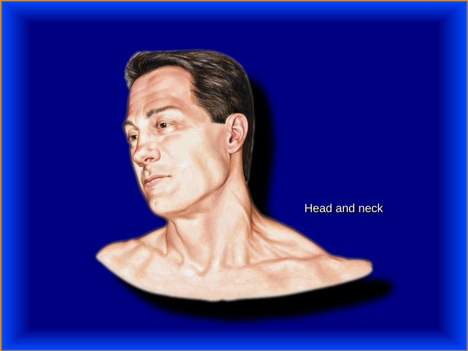
Head and neck
Human ear consists of:
● external ear
● middle ear
● internal ear

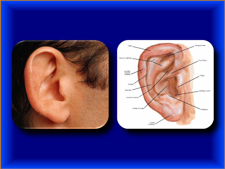
External ear
external auditory canal (meatus)
auricle


Auditory and vestibular apparatus in situ


Cartilage and muscles of the auricle
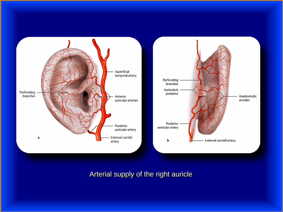
Arterial supply of the right auricle
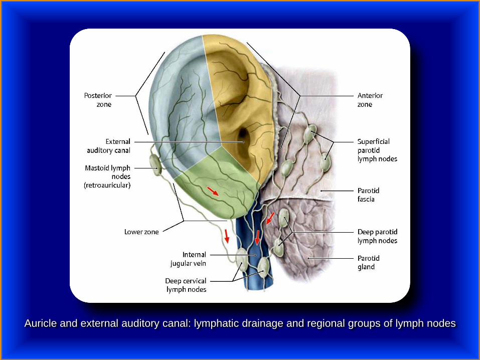
Auricle and external auditory canal: lymphatic drainage and regional groups of lymph nodes
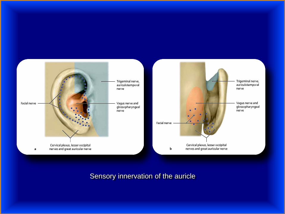
Sensory innervation of the auricle


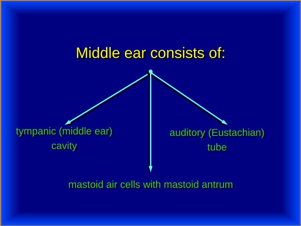
Middle ear consists of:
tympanic (middle ear)
cavity
auditory (Eustachian)
tube
mastoid air cells with mastoid antrum

External auditory canal, tympanic membrane, and tympanic cavity

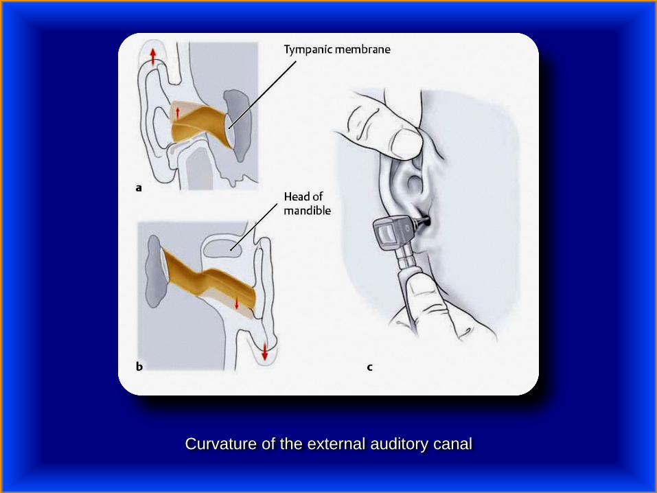
Curvature of the external auditory canal

Tympanic membrane

Auroscopic view of left tympanic membrane. Note that a bright cone of light is seen in the
anteroinferior quadrant of the membrane when it is illuminated.

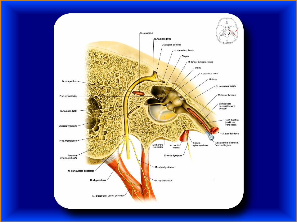
The left auditory apparatus as if viewed through a semi-transparent temporal bone.
Note the genu in the facial nerve at the site of the geniculate ganglion.
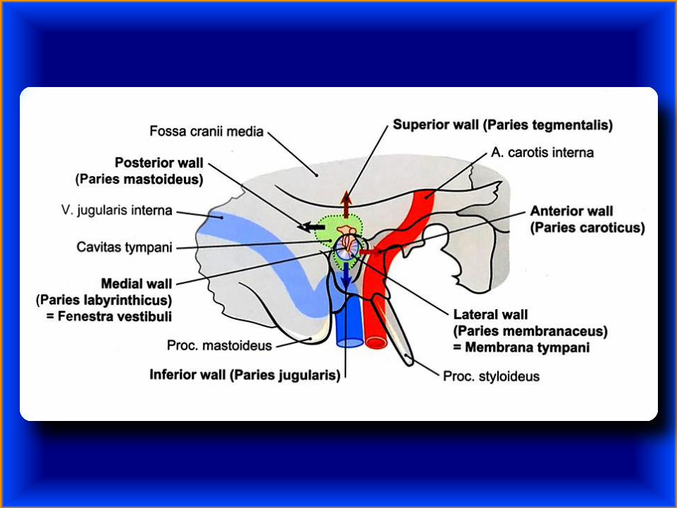
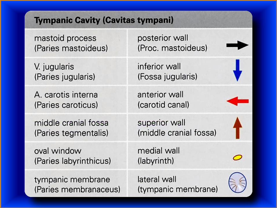


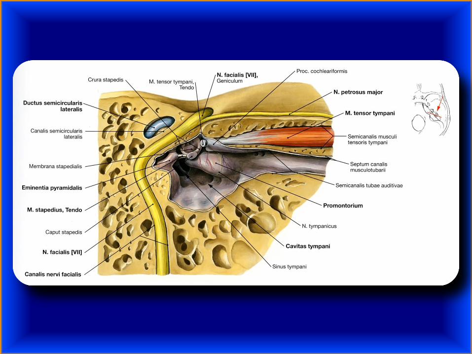
Walls of the tympanic cavity
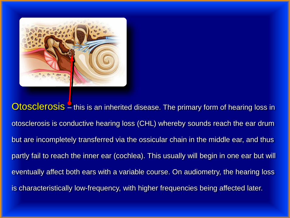
Otosclerosis – this is an inherited disease. The primary form of hearing loss in
otosclerosis is conductive hearing loss (CHL) whereby sounds reach the ear drum
but are incompletely transferred via the ossicular chain in the middle ear, and thus
partly fail to reach the inner ear (cochlea). This usually will begin in one ear but will
eventually affect both ears with a variable course. On audiometry, the hearing loss
is characteristically low-frequency, with higher frequencies being affected later.

Conductive hearing loss occurs when there is a problem conducting sound
waves anywhere along the route through the outer ear, tympanic membrane
(eardrum), or middle ear (ossicles). This type of hearing loss may occur in
conjunction with sensorineural hearing loss (mixed hearing loss) or alone.
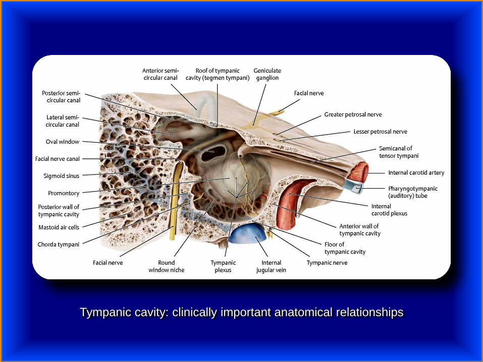
Tympanic cavity: clinically important anatomical relationships
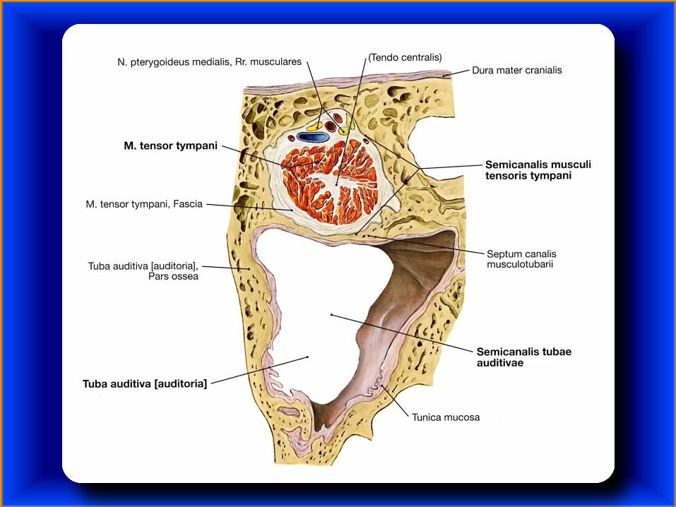

Pharyngotympanic (auditory) tube


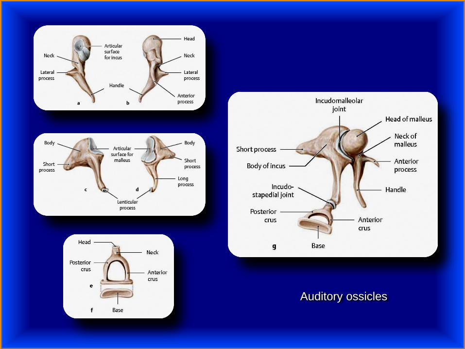
Auditory ossicles

Function of the ossicular chain
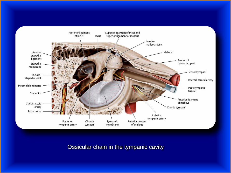
Ossicular chain in the tympanic cavity
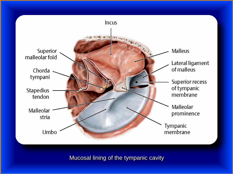
Mucosal lining of the tympanic cavity

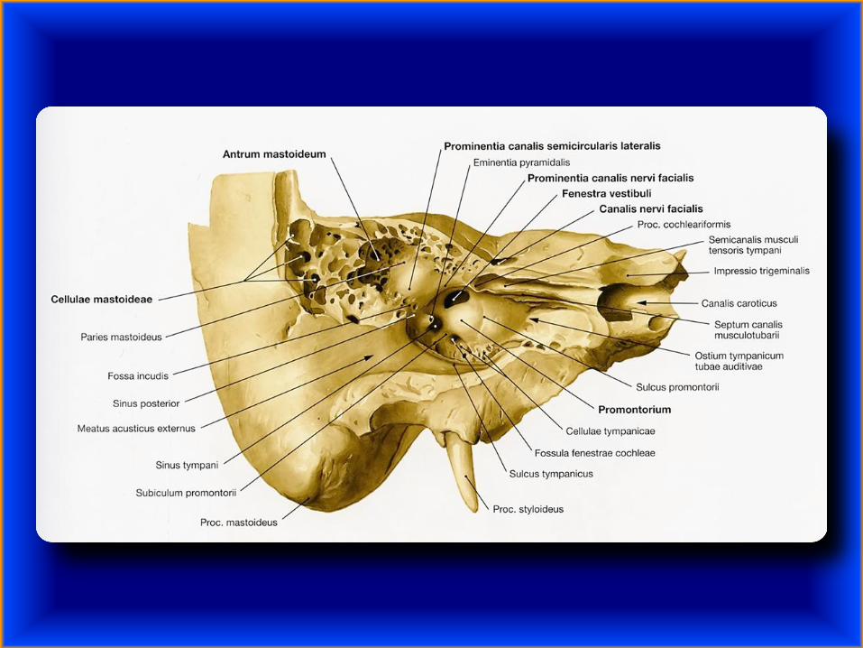
Clinically important levels of the tympanic cavity

Otitis media – is a group of inflammatory diseases of the middle ear –
Streptococcus pneumoniae, Haemophilus influenzae, Moraxella
catarrhalis, and Staphylococcus aureus.

Acute otitis media is very common in childhood. It is the most common
condition for which medical care is provided in children under five years
of age in the US.
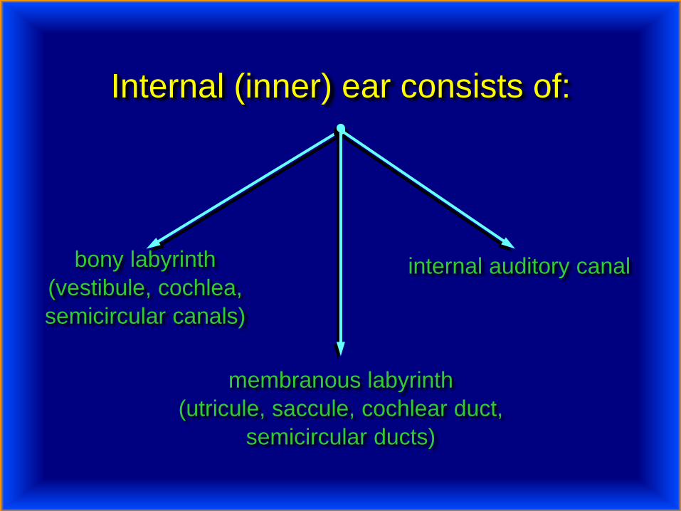
Internal (inner) ear consists of:
bony labyrinth
(vestibule, cochlea,
semicircular canals)
internal auditory canal
membranous labyrinth
(utricule, saccule, cochlear duct,
semicircular ducts)
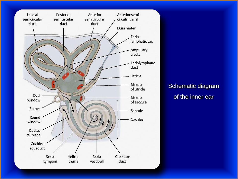
Schematic diagram
of the inner ear
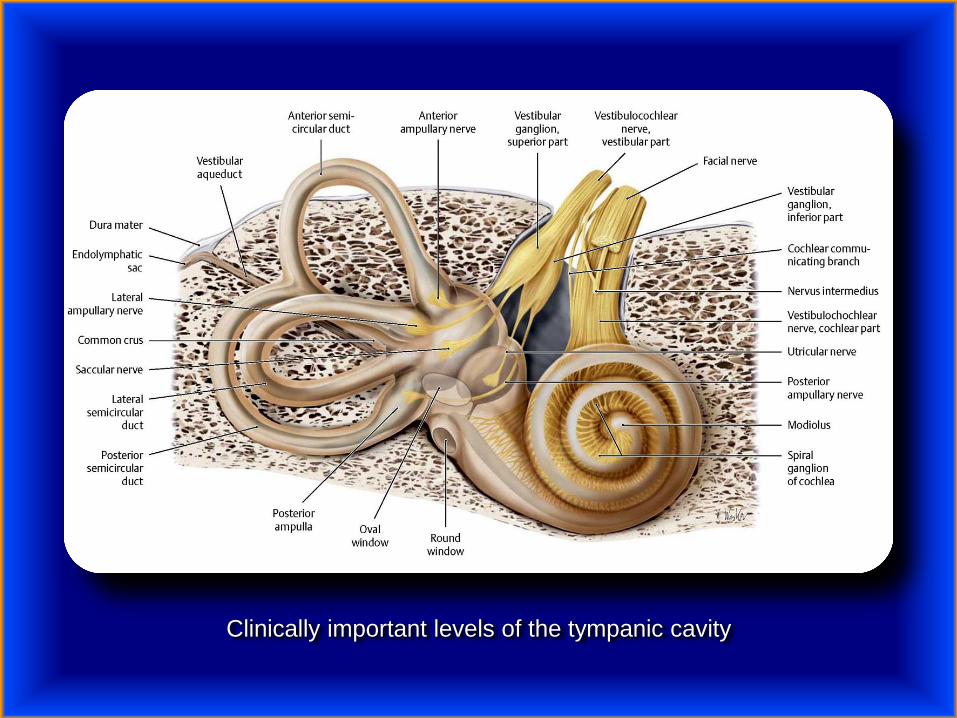
Clinically important levels of the tympanic cavity

Hyperacusis – is a debilitating hearing disorder characterized by an increased
sensitivity to certain frequency and volume ranges of sound (a collapsed tolerance
to usual environmental sound). A person with severe hyperacusis has difficulty
tolerating everyday sounds, some of which may seem unpleasantly or painfully
loud to that person but not to others.

Cochlear Hyperacusis – damage to the sound sensing organ
(cochlea) that results in the brain having sound sensitivities
around certain pitches;
Vestibular Hyperacusis – a form of Hyperacusis that also affects
a person’s Vestibular (balance) system resulting in nausea,
dizziness and the sensation of falling, in addition to sound
hypersensitivity and/or Tinnitus and hearing loss.

Causes include, but are not limited to:
Adverse drug reaction
Ankiety
Autism spectrum
Bell’s palsy
Chronic ear infections
Chronic fatigue syndrome
Ciprofloxacin antibiotic (quinolone family)
Depression
Developmental coordination disorder
Ear irrigation
Electroconvulsive Therapy
Facial nerve dysfunction (to stapedius)
Fibromyalgia
Head injury
Lyme disease
MAO inhibitor discontinuation syndrome
Migraine
Ménière's disease
Multiple Sclerosis
Noise-induced hearing loss
Posttraumatic stress disorder
Sensory Processing Disorder
Severe head trauma
Superior canal dehiscence syndrome (SCDS)
Surgery
Systemic lupus erythematosus (SLE)
Tay–Sachs disease
Temporomandibular joint disorder
Tension myositis syndrome
Tinnitus
Williams syndrome
Symptoms are ear pain, annoyance, and general intolerance to many
sounds that most people are unaffected by.
Hyperacusis can result in anxiety, stress and phonophobia.
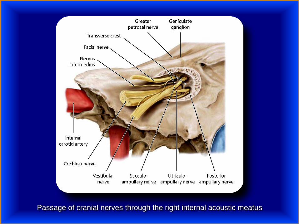
Passage of cranial nerves through the right internal acoustic meatus
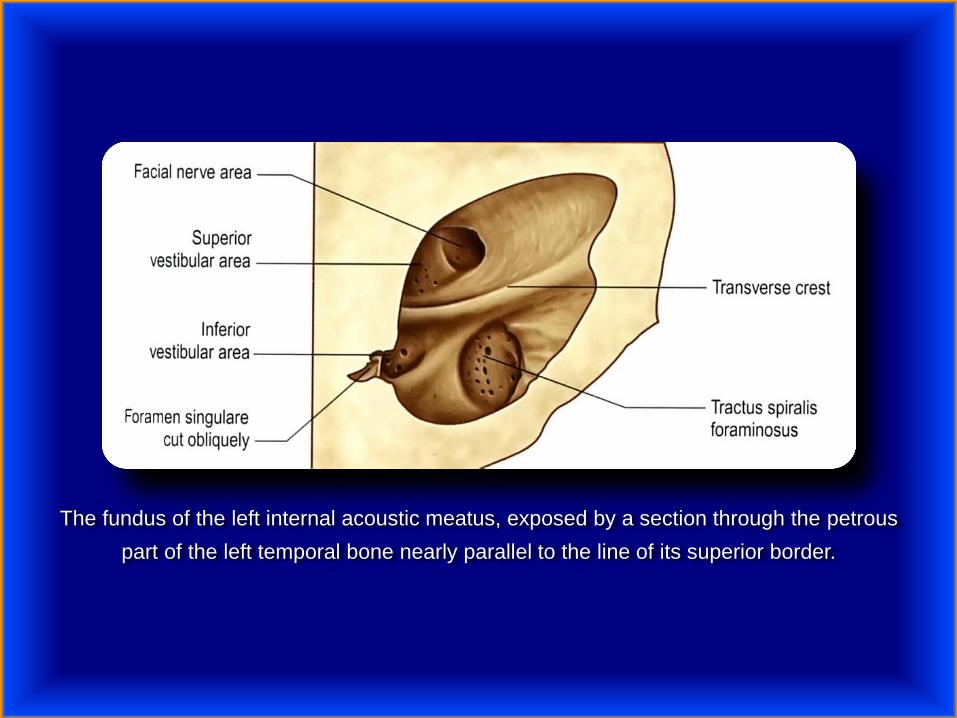
The fundus of the left internal acoustic meatus, exposed by a section through the petrous
part of the left temporal bone nearly parallel to the line of its superior border.
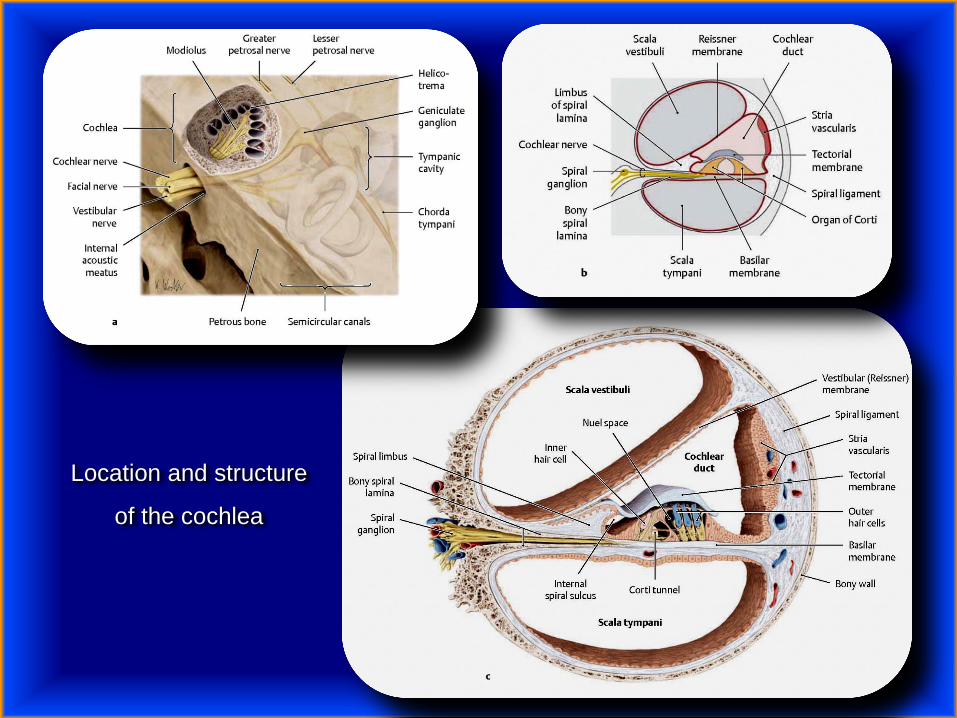
Location and structure
of the cochlea

Sound conduction during hearing

Sound conduction during hearing

Weber and Rinne Tests
Sensorineural hearing loss suggests a disorder of the internal ear or
the cochlear division of CN VIII.
Conductive hearing loss suggests a disorder of the external or middle ear
(eardrum, ear ossicles, or both).
The Weber and Rinne tests offer an easy way to differentiate between
sensorineural and conductive hearing loss.
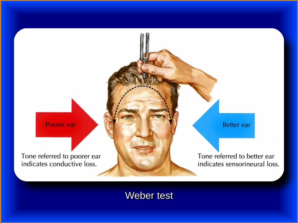
Weber test
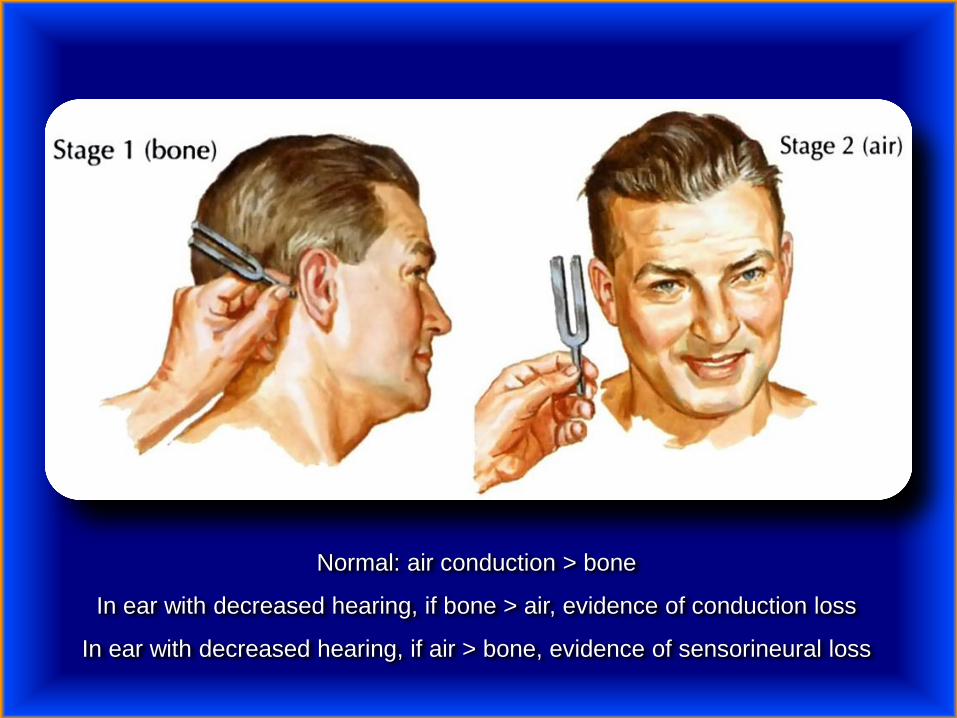
Normal: air conduction > bone
In ear with decreased hearing, if bone > air, evidence of conduction loss
In ear with decreased hearing, if air > bone, evidence of sensorineural loss
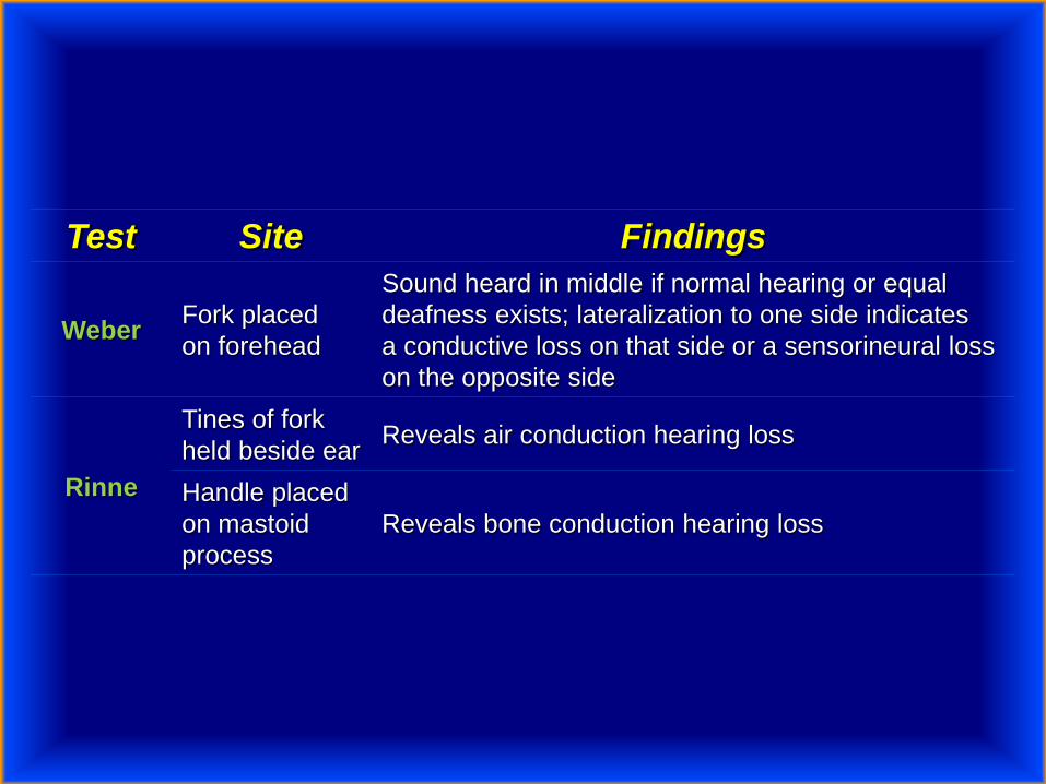
Test Site Findings
WeberFork placed
on forehead
Sound heard in middle if normal hearing or equal
deafness exists; lateralization to one side indicates
a conductive loss on that side or a sensorineural loss
on the opposite side
Rinne
Tines of fork
held beside earReveals air conduction hearing loss
Handle placed
on mastoid
process
Reveals bone conduction hearing loss


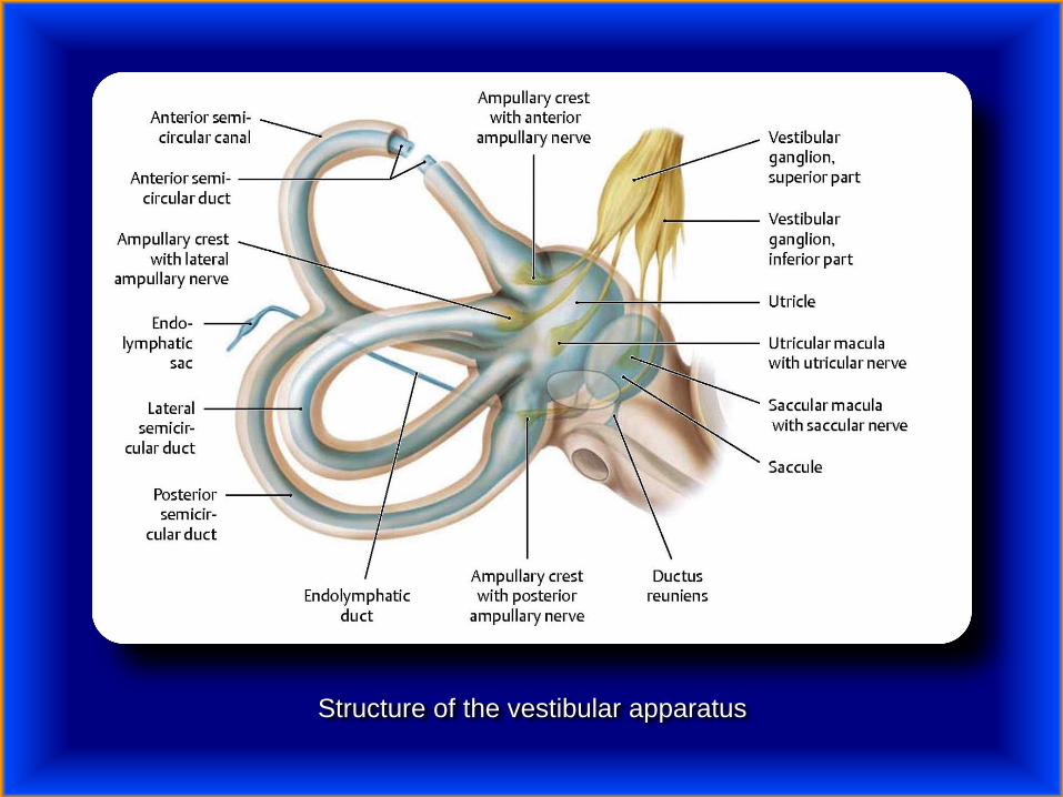
Structure of the vestibular apparatus
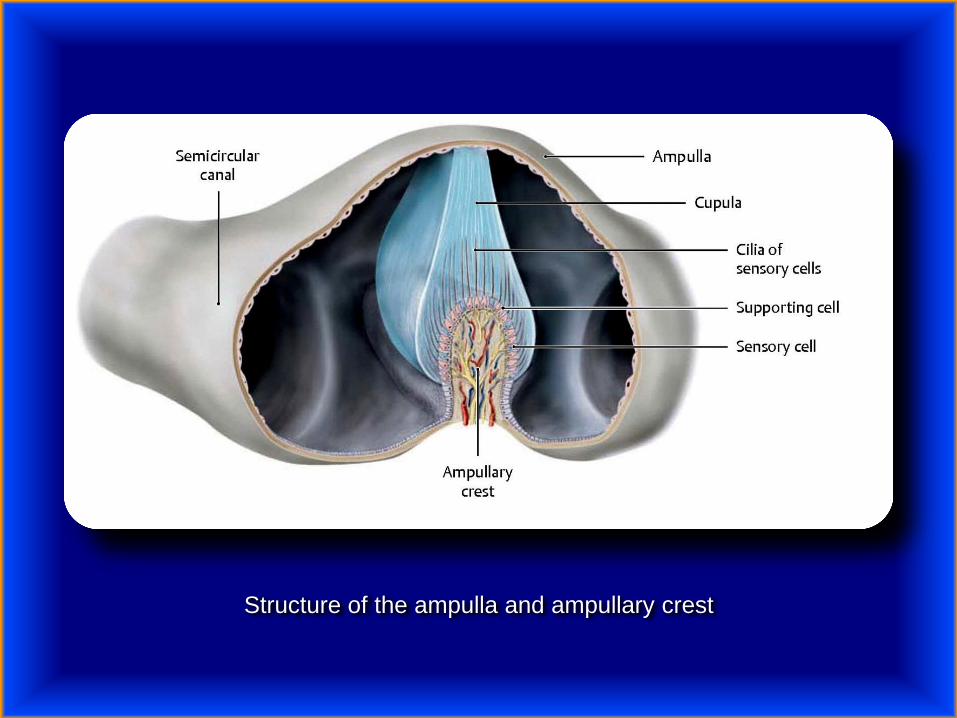
Structure of the ampulla and ampullary crest

Structure of the utricular and saccular maculae
Meniere’s disease – is a disorder of the inner ear that is characterized by episodes
of feeling like the world is spinning (vertigo), ringing in the ears (tinnitus), hearing
loss, and a fullness in the ear. Typically only one ear is affected, at least initially;
however, over time both ears may become involved. Episodes generally last from
20 minutes to a few hours. The time between episodes varies. The hearing loss
and ringing in the ears may become constant over time.
The cause of Ménière's disease is unclear but likely involves both genetic and
environmental factors.

In 1972, the academy defined criteria for diagnosing Ménière's disease as:
Fluctuating, progressive, sensorineural deafness.
Episodic, characteristic definitive spells of vertigo lasting 20 minutes to
24 hours with no unconsciousness, vestibular nystagmus always
present.
Tinnitus (ringing in the ears, from mild to severe) Often the tinnitus is
accompanied by ear pain and a feeling of fullness in the affected ear.
Usually the tinnitus is more severe before a spell of vertigo and
lessens after the vertigo attack.
Attacks are characterized by periods of remission and exacerbation.
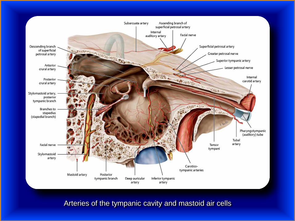
Arteries of the tympanic cavity and mastoid air cells
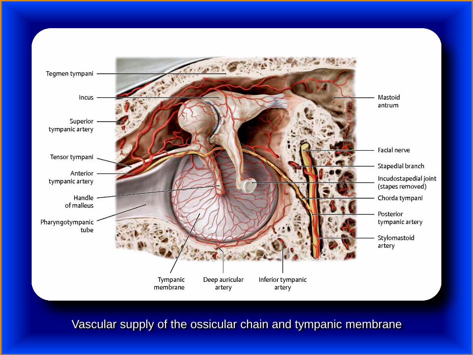
Vascular supply of the ossicular chain and tympanic membrane

Blood supply of the labyrinth

LaryngologistFot. J. Urbaniak
Thank you very much