Gipc1 has a dual role in Vangl2 trafficking and hair bundle integrity in the inner ear
11
RESEARCH ARTICLE 3775 Development 139, 3775-3785 (2012) doi:10.1242/dev.074229 © 2012. Published by The Company of Biologists Ltd INTRODUCTION In epithelia, two different, yet complementary, types of polarity regulate the orientation of individual cells. Apical-basal polarity refers to heterogeneity within a cell along the axis that extends between the basement membrane and the luminal surface with marked differences often observed between the luminal (apical) and basal and lateral (basal) surfaces. By contrast, planar cell polarity (PCP) refers to the uniform orientation of cells or cellular components within a horizontal plane of cells. The axis of orientation is therefore orthogonal to the axis of apical-basal polarity. In Drosophila, six ‘core genes’ were identified as a group of genes that function as a conserved signaling cassette for the specification of PCP: Van Gogh (Vang; also known as strabismus), flamingo (fmi; starry night), frizzled (fz), dishevelled (dsh), prickle (pk) and diego (Seifert and Mlodzik, 2007; Wang and Nathans, 2007; Zallen, 2007). In mammals, targeted or spontaneous mutations in homologs of these genes, such as cadherin EGF LAG seven-pass G-type receptor 1 (Celsr1), Van Gogh-like 2 (Vangl2), dishevelled1/2 (Dvl1/2) and Frizzled 3/6 (Fz3/6; Fzd3/6 – Mouse Genome Informatics), lead to defects in developmental processes that are thought to be mediated through PCP signaling, such as disruptions in closure of the neural tube and in the uniform orientation of stereociliary bundles in the inner ear (Montcouquiol et al., 2003; Curtin et al., 2003; Wang et al., 2005; Wang et al., 2006). The sensory epithelium of the mammalian cochlea, the organ of Corti (OC), comprises multiple cell types including mechanosensory hair cells (HCs) and non-sensory supporting cells (SCs). A bundle of modified microvilli, referred to as a stereociliary bundle, projects from the luminal surface of each HC, and all stereociliary bundles are orientated in the same direction, defining PCP within the epithelium. Defects in stereociliary bundle orientations have been used not only to demonstrate conservation of the PCP signaling pathway between vertebrates and invertebrates, but also to identify novel, mammalian-specific PCP components (Montcouquiol et al., 2003; Lu et al., 2004). In both invertebrates and vertebrates, the establishment of PCP often correlates with the asymmetric localization of core planar polarity proteins to the proximal (Vang and Pk) and distal (Fz, Dvl and Diego) apicolateral membranes (Seifert and Mlodzik, 2007). However, the molecular mechanisms that dictate these asymmetric distributions are not clear (Tree et al., 2002; Bastock et al., 2003; Takeuchi et al., 2003; Torban et al., 2004; Das et al., 2004; Jenny et al., 2005). The mechanisms controlling PCP protein trafficking are therefore of interest, and the elegant and sensitive read-out of disruptions in PCP within the mammalian cochlea make this an ideal system in which to study these mechanisms. MATERIALS AND METHODS Plasmids constructs Vangl2 full length was amplified from mouse cochlea and cloned into pEGFPC3 or pCLIG (Montcouquiol et al., 2006), or DsRed-monomer (Clontech). Site-directed mutagenesis was used to generate Vangl2 mutants lacking the last four amino acids (aa) (Vangl2 4 ), and the last 12 aa (Vangl2 12 ) (QuickChange, Stratagene). Rat myc-Gipc1 (pCM Vtag3C) and mouse synectin/GIPC (pEYFP-N1) cDNAs were obtained from R. Lefkowitz (Howard Hughes Medical Institute, Durham, NC, USA) and A. Horowitz (Dartmouth Medical School, Lebanon, NH, USA), respectively. Site-directed mutagenesis was used to induce mutations in the hydrophobic 1 Planar Polarity and Plasticity Group, Inserm U862, Neurocentre Magendie, Bordeaux, France. 2 University of Bordeaux, F-33000, Bordeaux, France. 3 Oregon Hearing Research Center, Oregon Health & Science University, Portland, OR 97239, USA. 4 CRCM, Inserm, U1068, Marseille, F-13009, France. 5 Institut Paoli-Calmettes, Marseille, France. 6 Aix-Marseille Université, F-13284, Marseille, France. 7 CNRS, UMR7258. 8 Laboratory of Cochlear Development, NIDCD, NIH, Bethesda, MD 22000, USA. *These authors contributed equally to this work ‡ Present address: New York University, Center for Genomics and Systems Biology, 12 Waverly Place, Room 705, New York, NY 10003, USA § Author for correspondence ([email protected]) Accepted 26 July 2012 SUMMARY Vangl2 is one of the central proteins controlling the establishment of planar cell polarity in multiple tissues of different species. Previous studies suggest that the localization of the Vangl2 protein to specific intracellular microdomains is crucial for its function. However, the molecular mechanisms that control Vangl2 trafficking within a cell are largely unknown. Here, we identify Gipc1 (GAIP C-terminus interacting protein 1) as a new interactor for Vangl2, and we show that a myosin VI-Gipc1 protein complex can regulate Vangl2 traffic in heterologous cells. Furthermore, we show that in the cochlea of MyoVI mutant mice, Vangl2 presence at the membrane is increased, and that a disruption of Gipc1 function in hair cells leads to maturation defects, including defects in hair bundle orientation and integrity. Finally, stimulated emission depletion microscopy and overexpression of GFP-Vangl2 show an enrichment of Vangl2 on the supporting cell side, adjacent to the proximal membrane of hair cells. Altogether, these results indicate a broad role for Gipc1 in the development of both stereociliary bundles and cell polarization, and suggest that the strong asymmetry of Vangl2 observed in early postnatal cochlear epithelium is mostly a ‘tissue’ polarity readout. KEY WORDS: Polarity, Traffic, Actin, Cochlea, Van Gogh-like 2, Gipc1, Myosin VI, Planar polarity, Mouse, Rat Gipc1 has a dual role in Vangl2 trafficking and hair bundle integrity in the inner ear Arnaud P. Giese 1,2, *, Jérome Ezan 1,2, *, Lingyan Wang 3 , Léa Lasvaux 1 , Frédérique Lembo 4,5,6,7 , Claire Mazzocco 1 , Elodie Richard 1 , Jérome Reboul 4,5,6,7,‡ , Jean-Paul Borg 4,5,6,7 , Matthew W. Kelley 8 , Nathalie Sans 1,2, *, John Brigande 3, * and Mireille Montcouquiol 1,2,§ DEVELOPMENT
Transcript of Gipc1 has a dual role in Vangl2 trafficking and hair bundle integrity in the inner ear
RESEARCH ARTICLE 3775
Development 139, 3775-3785 (2012) doi:10.1242/dev.074229© 2012. Published by The Company of Biologists Ltd
INTRODUCTIONIn epithelia, two different, yet complementary, types of polarityregulate the orientation of individual cells. Apical-basal polarityrefers to heterogeneity within a cell along the axis that extendsbetween the basement membrane and the luminal surface withmarked differences often observed between the luminal (apical)and basal and lateral (basal) surfaces. By contrast, planar cellpolarity (PCP) refers to the uniform orientation of cells or cellularcomponents within a horizontal plane of cells. The axis oforientation is therefore orthogonal to the axis of apical-basalpolarity. In Drosophila, six ‘core genes’ were identified as a groupof genes that function as a conserved signaling cassette for thespecification of PCP: Van Gogh (Vang; also known as strabismus),flamingo (fmi; starry night), frizzled (fz), dishevelled (dsh), prickle(pk) and diego (Seifert and Mlodzik, 2007; Wang and Nathans,2007; Zallen, 2007). In mammals, targeted or spontaneousmutations in homologs of these genes, such as cadherin EGF LAGseven-pass G-type receptor 1 (Celsr1), Van Gogh-like 2 (Vangl2),dishevelled1/2 (Dvl1/2) and Frizzled 3/6 (Fz3/6; Fzd3/6 – MouseGenome Informatics), lead to defects in developmental processesthat are thought to be mediated through PCP signaling, such asdisruptions in closure of the neural tube and in the uniform
orientation of stereociliary bundles in the inner ear (Montcouquiolet al., 2003; Curtin et al., 2003; Wang et al., 2005; Wang et al.,2006). The sensory epithelium of the mammalian cochlea, theorgan of Corti (OC), comprises multiple cell types includingmechanosensory hair cells (HCs) and non-sensory supporting cells(SCs). A bundle of modified microvilli, referred to as astereociliary bundle, projects from the luminal surface of each HC,and all stereociliary bundles are orientated in the same direction,defining PCP within the epithelium. Defects in stereociliary bundleorientations have been used not only to demonstrate conservationof the PCP signaling pathway between vertebrates andinvertebrates, but also to identify novel, mammalian-specific PCPcomponents (Montcouquiol et al., 2003; Lu et al., 2004).
In both invertebrates and vertebrates, the establishment of PCPoften correlates with the asymmetric localization of core planarpolarity proteins to the proximal (Vang and Pk) and distal (Fz, Dvland Diego) apicolateral membranes (Seifert and Mlodzik, 2007).However, the molecular mechanisms that dictate these asymmetricdistributions are not clear (Tree et al., 2002; Bastock et al., 2003;Takeuchi et al., 2003; Torban et al., 2004; Das et al., 2004; Jennyet al., 2005). The mechanisms controlling PCP protein traffickingare therefore of interest, and the elegant and sensitive read-out ofdisruptions in PCP within the mammalian cochlea make this anideal system in which to study these mechanisms.
MATERIALS AND METHODSPlasmids constructsVangl2 full length was amplified from mouse cochlea and cloned intopEGFPC3 or pCLIG (Montcouquiol et al., 2006), or DsRed-monomer(Clontech). Site-directed mutagenesis was used to generate Vangl2 mutantslacking the last four amino acids (aa) (Vangl2�4), and the last 12 aa(Vangl2�12) (QuickChange, Stratagene). Rat myc-Gipc1 (pCM Vtag3C)and mouse synectin/GIPC (pEYFP-N1) cDNAs were obtained from R.Lefkowitz (Howard Hughes Medical Institute, Durham, NC, USA) and A.Horowitz (Dartmouth Medical School, Lebanon, NH, USA), respectively.Site-directed mutagenesis was used to induce mutations in the hydrophobic
1Planar Polarity and Plasticity Group, Inserm U862, Neurocentre Magendie,Bordeaux, France. 2University of Bordeaux, F-33000, Bordeaux, France. 3OregonHearing Research Center, Oregon Health & Science University, Portland, OR 97239,USA. 4CRCM, Inserm, U1068, Marseille, F-13009, France. 5Institut Paoli-Calmettes,Marseille, France. 6Aix-Marseille Université, F-13284, Marseille, France. 7CNRS,UMR7258. 8Laboratory of Cochlear Development, NIDCD, NIH, Bethesda, MD 22000, USA.
*These authors contributed equally to this work‡Present address: New York University, Center for Genomics and Systems Biology, 12 Waverly Place, Room 705, New York, NY 10003, USA§Author for correspondence ([email protected])
Accepted 26 July 2012
SUMMARYVangl2 is one of the central proteins controlling the establishment of planar cell polarity in multiple tissues of different species.Previous studies suggest that the localization of the Vangl2 protein to specific intracellular microdomains is crucial for its function.However, the molecular mechanisms that control Vangl2 trafficking within a cell are largely unknown. Here, we identify Gipc1 (GAIPC-terminus interacting protein 1) as a new interactor for Vangl2, and we show that a myosin VI-Gipc1 protein complex can regulateVangl2 traffic in heterologous cells. Furthermore, we show that in the cochlea of MyoVI mutant mice, Vangl2 presence at themembrane is increased, and that a disruption of Gipc1 function in hair cells leads to maturation defects, including defects in hairbundle orientation and integrity. Finally, stimulated emission depletion microscopy and overexpression of GFP-Vangl2 show anenrichment of Vangl2 on the supporting cell side, adjacent to the proximal membrane of hair cells. Altogether, these results indicatea broad role for Gipc1 in the development of both stereociliary bundles and cell polarization, and suggest that the strong asymmetryof Vangl2 observed in early postnatal cochlear epithelium is mostly a ‘tissue’ polarity readout.
KEY WORDS: Polarity, Traffic, Actin, Cochlea, Van Gogh-like 2, Gipc1, Myosin VI, Planar polarity, Mouse, Rat
Gipc1 has a dual role in Vangl2 trafficking and hair bundleintegrity in the inner earArnaud P. Giese1,2,*, Jérome Ezan1,2,*, Lingyan Wang3, Léa Lasvaux1, Frédérique Lembo4,5,6,7, Claire Mazzocco1, Elodie Richard1, Jérome Reboul4,5,6,7,‡, Jean-Paul Borg4,5,6,7, Matthew W. Kelley8, Nathalie Sans1,2,*, John Brigande3,* and Mireille Montcouquiol1,2,§
DEVELO
PMENT
3776
pocket of the PDZ domain (Gipc1PDZ1dead, LGL/AAA, aa 143-145).pSUPER.gfp/neo vector (sh-GFP) and pSUPER.gfp/neo Gipc1 (shGIPC1a-GFP, shGIPC1b-GFP) were obtained from R. Wenthold (NIDCD, NIH,Bethesda, USA). GFP-Myosin VI pEGFP-C1 was obtained from T. W.Hasson (UCSD, San Diego, USA).
AntibodiesThe following primary antibodies were used: anti-Vangl2 (1:500;Montcouquiol et al., 2006), anti-Eea1 (1:500, BD Biosciences), anti-myc(1:1000, Covance, Ramona, CA, USA), anti-Gipc (M. Farquhar, UCSD,San Diego, USA, 1:800 and Proteus Biosciences, 1:400), anti-myosin VI(1:200, Proteus Biosciences), anti--catenin (1:1000, ChemiconInternational, Temecula, USA), anti-green fluorescent protein (GFP)(1:1000, Chemicon). Secondary antibodies were: Alexa Fluor-546/647 goatanti-rabbit and Alexa Fluor-546/647 goat anti-mouse (1:1000, Invitrogen),ATTO 647N anti-rabbit (1:400, Sigma), Star 635 anti-rabbit (Abberior) andMega 520 anti-mouse (Sigma).
Yeast two-hybrid screeningThe C-terminal portion of Vangl2 (aa 438-521) was used as a bait for thescreening and was subcloned into pGBTK7 vector (Clontech) in-framewith the Gal4 DNA-binding domain (Vangl2 DNA-DBD). Yeast two-hybrid screening and assays were performed as described in the Clontechprotocol (Matchmaker two-hybrid System). AH109 cells expressingGAL4-Vangl2 were combined with Y187 cells expressing an embryonicmouse cochlea cDNA library (Montcouquiol et al., 2006).
Construction and screening of PDZ-domain candidatesPDZ domains of interest [PDZ domain boundaries were obtained by cross-searching Interpro (V18), PFAM(V23) and SMART version 5.0(http://smart.embl-heidelberg.de)] were cloned in a Gateway pDONRvector and then transferred into the AD vector pACT2GW by Gatewayrecombinational cloning and transfected into the haploid Y187 yeast strain.Interactions between each PDZ domain and the product of pGBTK7-Vangl2, pGBTK7-Vangl2�4 transfected into the haploid yeast strain AH109were tested through mating of the two yeast strains.
Cell culture and transfectionsHEK293T and COS-7 cells were obtained from American Type CultureCollection (ATCC) and were cultured in DMEM (Gibco BRL)supplemented with fetal bovine serum and antibiotics. Transfections werecarried out using calcium phosphate co-precipitation (Sans et al., 2003).The cells were then cultured for an additional 24 hours beforeimmunostaining. For endocytosis, 24 hours after transfection, the cells wereincubated with 50 g/ml Alexa Fluor-568-conjugated transferrin(Invitrogen) in DMEM with L-glutamine and bovine serum albumin, for 3minutes at 37°C.
Cell lysis and subcellular fractionationTwenty-four hours after transfection with 10 g of indicated plasmids,HEK293T cells were harvested and suspended in cold 10 mM HEPESbuffer. A sucrose gradient fractionation solution was added (320 mMsucrose, 20 mM HEPES, 5 mM EDTA), with protease inhibitors (RocheDiagnostics, Indianapolis, IN, USA) (buffer A), and homogenized (Douncehomogenizer) to produce the homogenate (H). The homogenate wascentrifuged (8 minutes at 1000 g), and the supernatant collected; the pelletwas rinsed in buffer A, centrifuged again (7 minutes at 1000 g), and thefinal pellet was homogenized in buffer A (P1nuclear pellet). Thesupernatant from the first centrifugation was centrifuged again twice (7minutes at 1000 g then 20 minutes at 10,000 g). The pellet (P2plasmamembrane proteins) was homogenized in buffer A. The remainingsupernatant was centrifuged (2 hours at 140,000 g) and the supernatant(S3cytosol) and the pellet (P3microsomes: trans-golgi, endoplasmicreticulum, vesicles, all remaining small membranes) were collected inbuffer A. All the final pellets and lysates were resuspended in 2� SDSsample buffer. This experiment was carried out in triplicate.
Co-immunoprecipitation and immunoblottingDetergent extracts from olfactory bulbs of newborn mice or clearedtransfected cells were preincubated with specific antibodies to Vangl2,
Gipc1 or GFP for 1 hour, then antibodies were immobilized on Protein A/Gagarose beads (UltraLink resin, Thermo Scientific) overnight at 4°C.Washed beads were eluted with sample buffer and immunoprecipitatedproteins were analyzed by SDS-PAGE and immunoblotting. Anti-Vangl2,anti-Gipc1, anti-MyoVI (1:500 each), anti-myc, anti-GFP (1:5000) andanti-Gad65/67 (1:500, Chemicon) antibodies were used for theseexperiments. Immunoreactive proteins were visualized using achemiluminescence-based immunodetection of horseradish peroxidase(HRP) (Amersham).
MutantsFixed tissue of Snell’s waltzers mutant mice were a generous gift of KarenAvraham (Tel Aviv University, Israel). For comparison analysis, cochleaefrom control and mutant animals were immunolabeled in the same tube.
Dissection and in vitro electroporationsPregnant Sprague Dawley rats and CD1 mice were euthanized accordingto the European Communities Council Directives (86/609/EEC) and theFrench National Committee (87/848) recommendations. Embryonic day(E) 16.5 rat and E14.5 mice cochleae were dissected in cold HBSS(Invitrogen) as described previously (Montcouquiol et al., 2006).
In utero electroporationFor in utero electroporation, the PGK promoter of pSUPER.gfp/neo vectorwas replaced with the human elongation factor 1- promoter (EF1-;EEF1A1 – Human Gene Nomenclature Database). pSUPER-modifiedconstructs were injected and electroporated into E11.5 mouse otocysts asdescribed previously (Brigande et al., 2009). The organs of Corti wereharvested seven days later for analysis by immunocytochemistry.
MicroscopyImmunocytochemistry on cochleae were carried out as describedpreviously (Montcouquiol et al., 2008), except for images in Fig. 7A,B forwhich we used 10% trichloroacetic acid (TCA) for 1 hour at 4°C. Weobserved an overall stronger staining intensity with TCA fixation, coupledwith a small disruption of the integrity of the tissue (Hayashi et al., 1999).For optical sectioning of whole mounts, image acquisition were obtainedon an epifluorescence microscope system (AxioVision Z1; Carl Zeiss)fitted with an Apotome slide module with a 63� NA 1.4 Plan-Apochromatobjective and a digital camera (AxioCam MRm; Carl Zeiss), or on aconfocal microscope (TCS SP5; Leica). Imaging was carried out using az-step from 0.21 to 0.4 m. Stimulated emission depletion microscopy(STED) microscopy was performed with a TCS STED microscope (Leica),with a 63� 1.4 NA oil immersion and a 100� 1.4 NA oil immersionSTED objective. Images were processed using Volocity software(PerkinElmer). Deconvolution was carried out with Huygens 4.2 usingthree iterations. For quantification of hair cell/bundle phenotype, wedefined a PCP phenotype as a misorientated hair bundle within the planeof the epithelium but with a morphologically intact bundle.
Colocalization analysisThree 80�80 pixel regions were selected in the cytoplasm from each COS-7 cell. In each area, the value of colocalization was defined as thepercentage ratio between the number of positive vesicles for GFP-Vangl2,GFP-Vangl2�4 and the number of myc-Gipc1-, Eea1- or transferrin-positivevesicles using Axiovision software (Zeiss). Measurements from three areaswere averaged per cell. Means from different cells were averaged to obtaina final mean. Student’s t-test was used in order to determine statisticalsignificance.
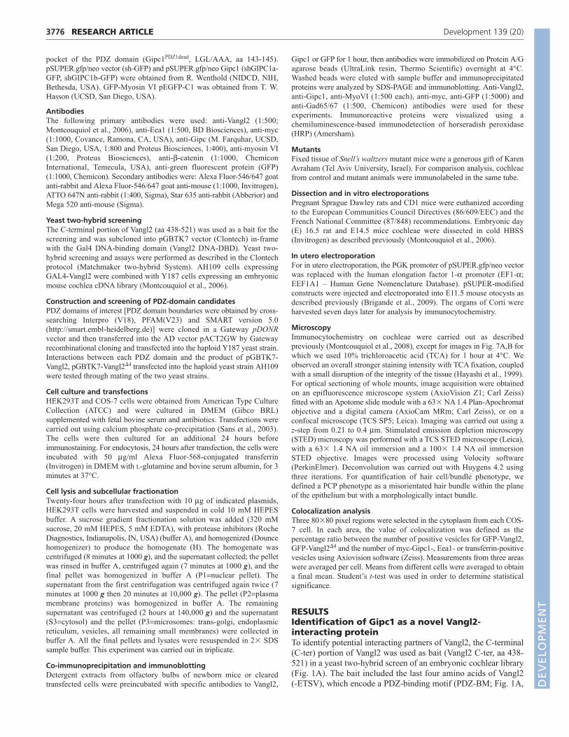
RESULTSIdentification of Gipc1 as a novel Vangl2-interacting proteinTo identify potential interacting partners of Vangl2, the C-terminal(C-ter) portion of Vangl2 was used as bait (Vangl2 C-ter, aa 438-521) in a yeast two-hybrid screen of an embryonic cochlear library(Fig. 1A). The bait included the last four amino acids of Vangl2 (-ETSV), which encode a PDZ-binding motif (PDZ-BM; Fig. 1A,
RESEARCH ARTICLE Development 139 (20)
DEVELO
PMENT
orange). Thirteen of the 63 clones identified as interacting withVangl2 encoded a portion of the protein Gipc1. Gipc1 is acytoplasmic scaffold protein with an N-terminal (N-ter) proline-rich domain (PRD), a central type II class PDZ domain, and a C-ter acyl carrier protein (ACP) domain (Fig. 1B). We confirmed theinteraction using a yeast two-hybrid assay with two of the thirteenclones, GIPC1C1 and GIPC1C15 (Fig. 1C). The deletion of the PDZ-BM (Vangl24), strongly reduced the interaction with Gipc1 in theyeast two-hybrid assay, but a further deletion in the C-ter of Vangl2(Vangl212) was needed to eliminate the interaction (Fig. 1C).These results suggest that the Vangl2 PDZ-BM can bind Gipc1 butthat, at most, eight aa upstream the PDZ-BM also participates inthis interaction. The previously characterized interaction betweenScribble1 (Scrib1) and Vangl2 was used as a positive control(Montcouquiol et al., 2006). Deletion assays demonstrated that thecentral PDZ domain of Gipc1 interacts with Vangl2, whereas thePRD and ACP domains do not (Fig. 1D). We also tested theinteraction of our bait on a group of candidate molecules with PDZdomains. Results confirmed that Gipc1, along with Scrib1, Dlg orMagi are amongst the strongest interactors for the last 83 aa ofVangl2 (supplementary material Table S1). All of the positiveinteractions were lost after deletion of the last four or 12 aa ofVangl2. We confirmed an interaction between Vangl2 and Gipc1using co-immunoprecipitation (Fig. 1E,F) and glutathione S-transferase (GST)-pull down assays (data not shown). Thisinteraction depends on the integrity of PDZ-BM of Vangl2 and thePDZ domain of Gipc1.
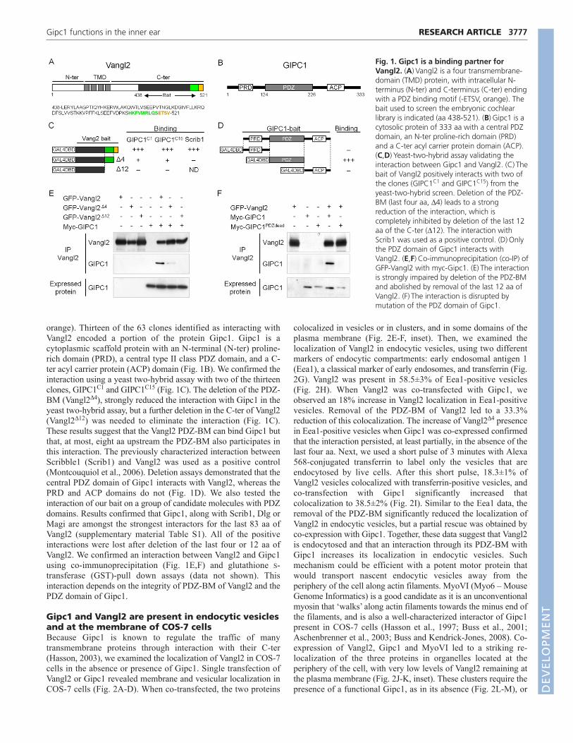
Gipc1 and Vangl2 are present in endocytic vesiclesand at the membrane of COS-7 cellsBecause Gipc1 is known to regulate the traffic of manytransmembrane proteins through interaction with their C-ter(Hasson, 2003), we examined the localization of Vangl2 in COS-7cells in the absence or presence of Gipc1. Single transfection ofVangl2 or Gipc1 revealed membrane and vesicular localization inCOS-7 cells (Fig. 2A-D). When co-transfected, the two proteins
colocalized in vesicles or in clusters, and in some domains of theplasma membrane (Fig. 2E-F, inset). Then, we examined thelocalization of Vangl2 in endocytic vesicles, using two differentmarkers of endocytic compartments: early endosomal antigen 1(Eea1), a classical marker of early endosomes, and transferrin (Fig.2G). Vangl2 was present in 58.5±3% of Eea1-positive vesicles(Fig. 2H). When Vangl2 was co-transfected with Gipc1, weobserved an 18% increase in Vangl2 localization in Eea1-positivevesicles. Removal of the PDZ-BM of Vangl2 led to a 33.3%reduction of this colocalization. The increase of Vangl24 presencein Eea1-positive vesicles when Gipc1 was co-expressed confirmedthat the interaction persisted, at least partially, in the absence of thelast four aa. Next, we used a short pulse of 3 minutes with Alexa568-conjugated transferrin to label only the vesicles that areendocytosed by live cells. After this short pulse, 18.3±1% ofVangl2 vesicles colocalized with transferrin-positive vesicles, andco-transfection with Gipc1 significantly increased thatcolocalization to 38.5±2% (Fig. 2I). Similar to the Eea1 data, theremoval of the PDZ-BM significantly reduced the localization ofVangl2 in endocytic vesicles, but a partial rescue was obtained byco-expression with Gipc1. Together, these data suggest that Vangl2is endocytosed and that an interaction through its PDZ-BM withGipc1 increases its localization in endocytic vesicles. Suchmechanism could be efficient with a potent motor protein thatwould transport nascent endocytic vesicles away from theperiphery of the cell along actin filaments. MyoVI (Myo6 – MouseGenome Informatics) is a good candidate as it is an unconventionalmyosin that ‘walks’ along actin filaments towards the minus end ofthe filaments, and is also a well-characterized interactor of Gipc1present in COS-7 cells (Hasson et al., 1997; Buss et al., 2001;Aschenbrenner et al., 2003; Buss and Kendrick-Jones, 2008). Co-expression of Vangl2, Gipc1 and MyoVI led to a striking re-localization of the three proteins in organelles located at theperiphery of the cell, with very low levels of Vangl2 remaining atthe plasma membrane (Fig. 2J-K, inset). These clusters require thepresence of a functional Gipc1, as in its absence (Fig. 2L-M), or
3777RESEARCH ARTICLEGipc1 functions in the inner ear
Fig. 1. Gipc1 is a binding partner forVangl2. (A)Vangl2 is a four transmembrane-domain (TMD) protein, with intracellular N-terminus (N-ter) and C-terminus (C-ter) endingwith a PDZ binding motif (-ETSV, orange). Thebait used to screen the embryonic cochlearlibrary is indicated (aa 438-521). (B)Gipc1 is acytosolic protein of 333 aa with a central PDZdomain, an N-ter proline-rich domain (PRD)and a C-ter acyl carrier protein domain (ACP).(C,D)Yeast-two-hybrid assay validating theinteraction between Gipc1 and Vangl2. (C)Thebait of Vangl2 positively interacts with two ofthe clones (GIPC1C1 and GIPC1C15) from theyeast-two-hybrid screen. Deletion of the PDZ-BM (last four aa, 4) leads to a strongreduction of the interaction, which iscompletely inhibited by deletion of the last 12aa of the C-ter (12). The interaction withScrib1 was used as a positive control. (D)Onlythe PDZ domain of Gipc1 interacts withVangl2. (E,F)Co-immunoprecipitation (co-IP) ofGFP-Vangl2 with myc-Gipc1. (E)The interactionis strongly impaired by deletion of the PDZ-BMand abolished by removal of the last 12 aa ofVangl2. (F)The interaction is disrupted bymutation of the PDZ domain of Gipc1.
DEVELO
PMENT
3778 RESEARCH ARTICLE Development 139 (20)
Fig. 2. Vangl2 and Gipc1 colocalize in endosomal vesicles that are redistributed peripherally by MyoVI. (A-F)Immunofluorescencemicroscopy of COS-7 cells transiently transfected with DsRed-Vangl2, myc-Gipc1 or a combination of the constructs, and labeled with phalloidin(blue), with a line scan corresponding to the dashed line for each condition. The arrows on the line scans represent cell boundaries labeled byphalloidin (A-D). Single expression of DsRed-Vangl2 (red) or myc-Gipc1 (green) led to vesicular, membrane and perinuclear staining. (E-F)DsRed-Vangl2 and myc-Gipc1 colocalize in cytoplasmic vesicles, clusters and at the plasma membrane. (G)Schematic of the localization of transferrin andEea1 in early endosomes containing Vangl2. (H)Sixty percent of GFP-Vangl2 vesicles colocalized with Eea1-labeled vesicles. This colocalization isincreased in the presence of myc-Gipc1, and severely impaired by removal of the PDZ-BM of Vangl2 (Vangl24). (I)Similar profiles are observedwhen we measured colocalization of Vangl2-positive vesicles with fluorescent transferrin after a 3-minute treatment. (J-K)DsRed-Vangl2, GFP-MyoVI and myc-Gipc1 co-expression leads to an almost complete colocalization of the three proteins, with a re-localization of these clusters closeto, but not at, the plasma membrane (magenta). Note on the line scan (K) that the peaks with high Vangl2 intensity (asterisks) do not correspond tothe plasma membrane peaks (arrows). (L-M)DsRed-Vangl2 and GFP-MyoVI are expressed in the cytoplasm, in vesicle-like cytoplasmic punctae,weakly at the membrane and in the perinuclear region of the cell. (N-O)A mutation in the PDZ domain of Gipc1 (myc-Gipc1PDZdead) prevents theformation of the clusters and their re-localization. Note the presence of Vangl2 at the membrane (inset). (P-Q)A short treatment (3 minutes) withAlexa Fluor568-conjugated transferrin (red) reveals colocalization with the clusters. Insets show magnifications of boxed area within the same panel.*P≤0.05, **P≤0.01, ***P≤0.001. Error bars represent the standard error of the mean of triplicates. Scale bar: 15m. D
EVELO
PMENT
when its central PDZ domain was mutated (Fig. 2N-O, inset), weobserved a strong reduction in the formation of clusters. COS-7cells co-transfected with Vangl2, Gipc1 and MyoVI and treated for3 minutes with fluorescent transferrin revealed a strongcolocalization of the clusters with transferrin, suggesting that theclusters were, at least in part, of endocytic nature (Fig. 2P-Q), assuggested previously (Aschenbrenner et al., 2003). These resultssuggest that Gipc1 can associate with both MyoVI and Vangl2simultaneously in clusters near the plasma membrane.
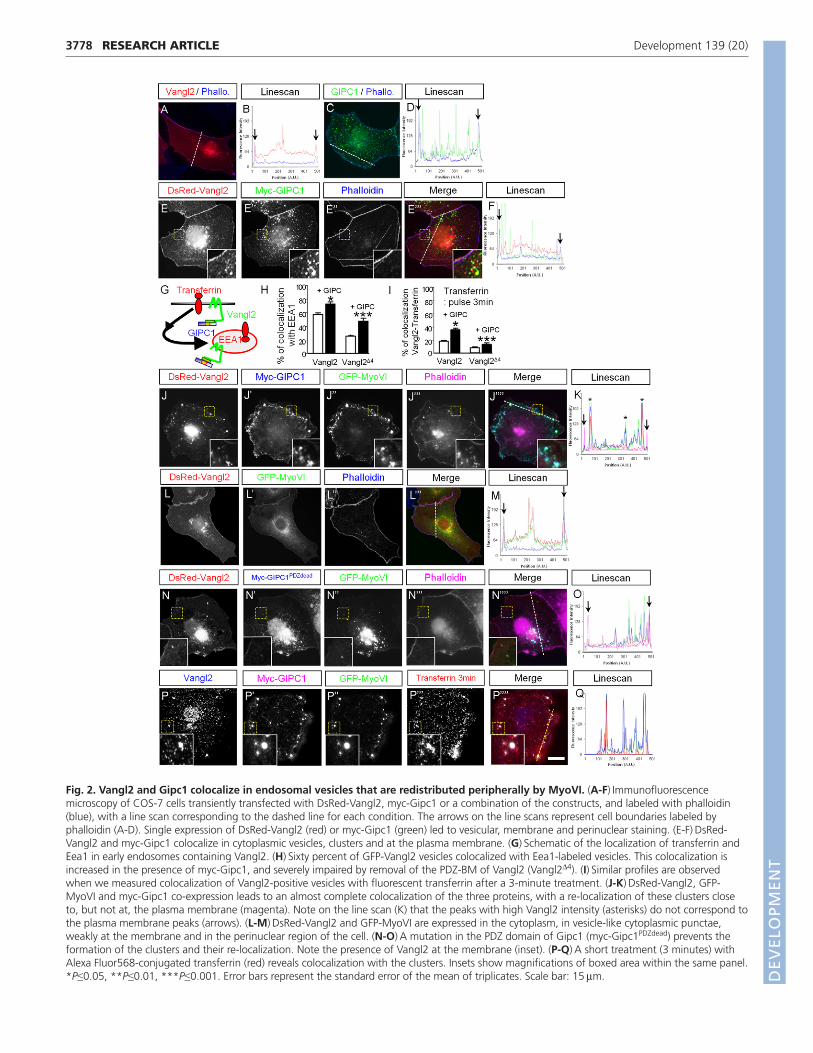
Gipc1 and MyoVI form a protein complex thatparticipates in Vangl2 removal from the plasmamembraneWe confirmed that Vangl2, MyoVI and Gipc1 can be associatedinto a molecular complex, by co-immunoprecipitation (co-IP) (Fig.3A,B). As anticipated, this complex was lost when the PDZ
domain of Gipc1 was mutated (Fig. 3A,B). We also show theexistence of a complex containing the three proteins in vivo, inmouse olfactory bulbs, by co-IP of Gipc1 with both endogenousVangl2 and MyoVI (Fig. 3C). Because of the limitation in proteincontent, we could not perform a similar co-IP in cochlear tissue.Alternatively, we show that overexpression of a GFP-Gipc1construct in HCs leads to the recruitment of endogenous Vangl2 inthe GFP-Gipc1 clusters (Fig. 3D).
To test the hypothesis that a protein complex consisting ofVangl2, Gipc1and MyoVI could move along the actin filament andaway from the membrane (Fig. 3E), we transfected HEK293T cellsand carried out subcellular fractionation (Fig. 3F). As a control forthe fractionation procedure, some cells were transfected with aconstruct that expressed either wild-type (WT) Vangl2 or theLooptail form of Vangl2 (Vangl2Lp). As expected, Vangl2Lp levelsin the membrane fraction were decreased by 58% compared with
3779RESEARCH ARTICLEGipc1 functions in the inner ear
Fig. 3. Gipc1 and MyoVI form a proteincomplex that participates in Vangl2removal from the plasma membrane.(A,B)Co-IP of Gipc1 and MyoVI with Vangl2.HEK293T lysates were subjected to IP with ananti-Vangl2 antibody and probed with anti-myc (for Gipc1) and anti-GFP (for MyoVI), orwith an anti-GFP antibody and probed withanti-Vangl2 and anti-myc (for Gipc1). Thethree proteins are present in a complex that isdisrupted when the PDZ domain of Gipc1 ismutated (Gipc1PDZdead). (C)Endogenous Gipc1,Vangl2 and MyoVI co-immunoprecipitate inlysates of mouse olfactory bulbs, but not withGad65/67. Asterisks denote IgG heavy chains.(D)Overexpression of Gipc1-YFP (green) intwo Ohc1 cells leads to the formation ofclusters containing endogenous Vangl2 (red).The image was taken just below the apicalsurface of the inner hair cell (Ihc) and outerhair cell (Ohc1); phalloidin labeling of the F-actin is in blue. (E)Vangl2 interacts with itsPDZ-BM with the central PDZ domain ofGipc1, which can interact with MyoVI. Thevesicular complex can be translocated along F-actin filaments by the motor function ofMyoVI. (F)Schematic of the fractionationprotocol. Fractions are as follows: H, totalhomogenate; S1, cell soma; P1, dense nuclei-associated membrane; S2, supernatant; P2,membrane fraction; S3, cytosolic; P3,microsomes. (G)Cellular fractionation assay.Antibodies against pan-cadherin and Gapdhshow the segregation of the cytosolic (Cyto)and membrane (Mb) fractions fromhomogenate (H), and the antibody againstmyc reveals the presence of Gipc1 in thecytosolic fraction (with a weaker expression ofthe mutated form). Vangl2 is present in themembrane fraction, with higher levels whenthe PDZ of Gipc1 is mutated (Gipc1PDZdead).
DEVELO
PMENT
3780
Vangl2 (data not shown) (Merte et al., 2010; Wansleeben et al.,2010). Next, similar fractionations were performed on cellstransfected with either Vangl2/Gipc1/MyoVI or Vangl2/Gipc1-PDZdead/MyoVI. Our results show an increase of 186±39% inVangl2 levels in the membrane fraction (normalized to pan-cadherin levels) when the PDZ domain of Gipc1 was mutatedcompared with WT Gipc1 (Fig. 3G). In addition, MyoVI levelswere increased in the cytosolic fraction when Gipc1 was mutated,suggesting retention of MyoVI in the cytoplasm. These results areconsistent with a process in which Gipc1 binds to Vangl2 at theplasma membrane, followed by a MyoVI-mediated removal ofvesicles containing Vangl2 away from the plasma membrane, intothe cytoplasm.
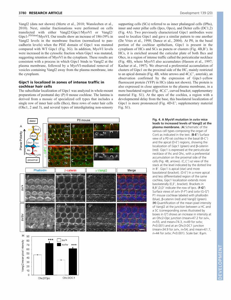
Gipc1 is localized in zones of intense traffic incochlear hair cellsThe subcellular localization of Gipc1 was analyzed in whole-mountpreparations of postnatal day (P) 0 mouse cochleae. The lamina isderived from a mosaic of specialized cell types that includes asingle row of inner hair cells (Ihcs), three rows of outer hair cells(Ohc1, 2 and 3), and several types of interdigitating non-sensory
supporting cells (SCs) referred to as inner phalangeal cells (IPhs),inner and outer pillar cells (Ipcs, Opcs), and Deiter cells (DC1,2)(Fig. 4A). Two previously characterized Gipc1 antibodies wereused to localize Gipc1 and gave a similar pattern to one another(De Vries et al., 1998; Dance et al., 2004). At P0, in the basalportion of the cochlear epithelium, Gipc1 is present in thecytoplasm of HCs and SCs as puncta or clusters (Fig. 4B,B�). InHCs, it is enriched around the cuticular plate of both Ihcs andOhcs, in a region of intense traffic called the pericuticular necklace(Fig. 4B), where MyoVI also accumulates (Hasson et al., 1997;Kachar et al., 1997). We observed a preferential accumulation ofclusters of Gipc1 on the proximal side of the HC, mainly restrictedto an apical domain (Fig. 4B, white arrows and 4C,C�, asterisk), anobservation confirmed by the expression of Gipc1-yellowfluorescent protein (YFP) in HCs (data not shown). The protein isalso expressed in close apposition to the plasma membrane, in amore basolateral region (Fig. 4C,C�, curved bracket; supplementarymaterial Fig. S1). At the apex of the cochlea, a region with adevelopmental delay from the base, this basolateral localization ofGipc1 is more pronounced (Fig. 4D-E�; supplementary materialFig. S1).
RESEARCH ARTICLE Development 139 (20)
Fig. 4. A MyoVI mutation in sv/sv miceleads to increased levels of Vangl2 at theplasma membrane. (A)Schematic of thevarious cell types composing the organ ofCorti as indicated in the text. (B-E�) Surfaceview of a P0 rat cochlea in the basal (B-C�)and the apical (D-E�) regions, showing thelocalization of Gipc1 (green) and -catenin(red). Gipc1 is expressed at the pericuticularnecklace of Ihc and Ohc, with a preferentialaccumulation on the proximal side of thecells (Fig. 4B, arrows). (C,C�) xz view of thestack at the level indicated by the dotted linein B�. Gipc1 is apical (star) and morebasolateral (bracket). (D-E�) In a more apicaland less differentiated region of the samecochlea, Gipc1 localization extends morebasolaterally (E,E�, bracket). Brackets inB,B�,D,D� indicate the row of Ipcs. (F-G�)Surface views of sv/+ (F-F�) and sv/sv (G-G�)P1 mouse cochleae labeled with phalloidin(blue), -catenin (red) and Vangl2 (green).(H)Quantification of the mean pixel intensityof Vangl2 at the junction between a HC anda SC (corresponding zones illustrated byboxes in G�) shows an increase in intensity atan Ohc2-Opc junction (mean47.2 for sv/+,n55; and mean74.3, n49 for sv/sv;P<0.001) and at an Ohc3-DC1 junction(mean34.9 for sv/+, n54; and mean61.7,n44 for sv/sv; P<0.001). Scale bar: 8m.
DEVELO
PMENT
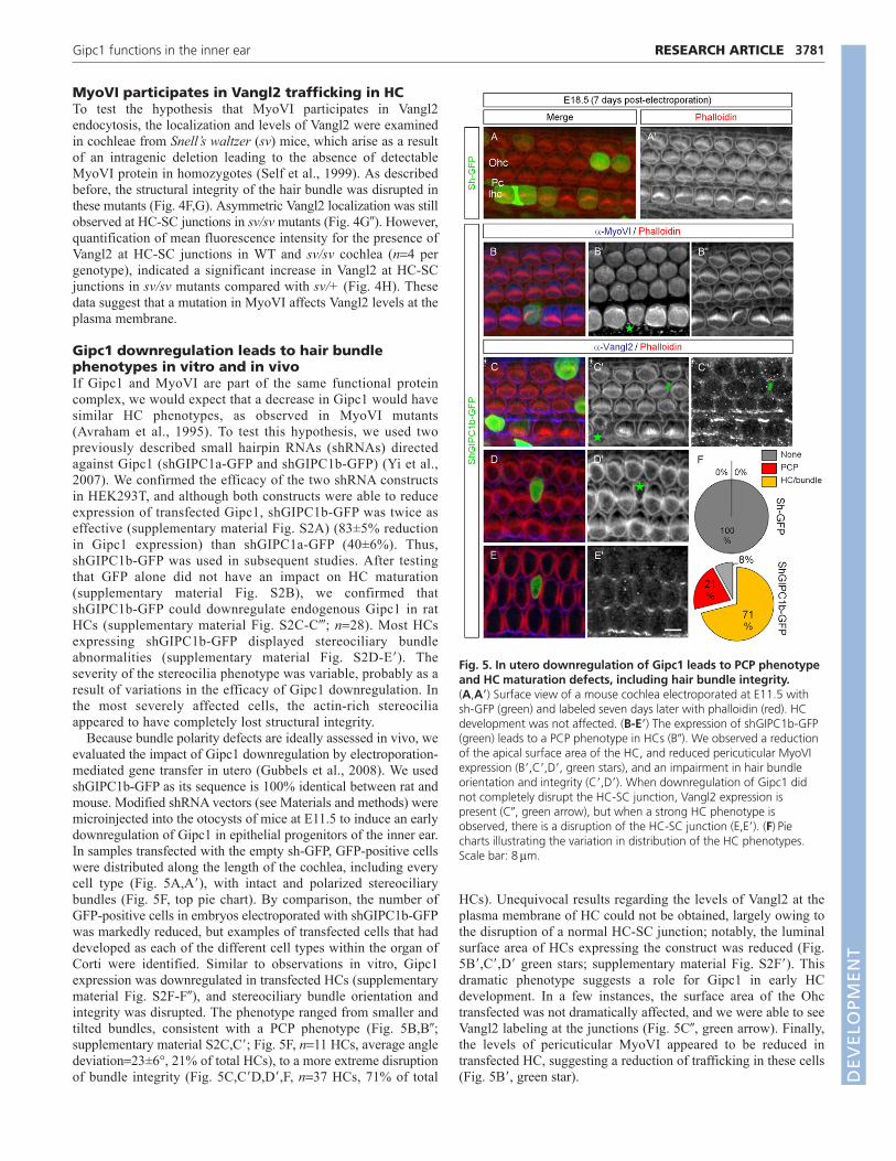
MyoVI participates in Vangl2 trafficking in HCTo test the hypothesis that MyoVI participates in Vangl2endocytosis, the localization and levels of Vangl2 were examinedin cochleae from Snell’s waltzer (sv) mice, which arise as a resultof an intragenic deletion leading to the absence of detectableMyoVI protein in homozygotes (Self et al., 1999). As describedbefore, the structural integrity of the hair bundle was disrupted inthese mutants (Fig. 4F,G). Asymmetric Vangl2 localization was stillobserved at HC-SC junctions in sv/sv mutants (Fig. 4G�). However,quantification of mean fluorescence intensity for the presence ofVangl2 at HC-SC junctions in WT and sv/sv cochlea (n4 pergenotype), indicated a significant increase in Vangl2 at HC-SCjunctions in sv/sv mutants compared with sv/+ (Fig. 4H). Thesedata suggest that a mutation in MyoVI affects Vangl2 levels at theplasma membrane.
Gipc1 downregulation leads to hair bundlephenotypes in vitro and in vivoIf Gipc1 and MyoVI are part of the same functional proteincomplex, we would expect that a decrease in Gipc1 would havesimilar HC phenotypes, as observed in MyoVI mutants(Avraham et al., 1995). To test this hypothesis, we used twopreviously described small hairpin RNAs (shRNAs) directedagainst Gipc1 (shGIPC1a-GFP and shGIPC1b-GFP) (Yi et al.,2007). We confirmed the efficacy of the two shRNA constructsin HEK293T, and although both constructs were able to reduceexpression of transfected Gipc1, shGIPC1b-GFP was twice aseffective (supplementary material Fig. S2A) (83±5% reductionin Gipc1 expression) than shGIPC1a-GFP (40±6%). Thus,shGIPC1b-GFP was used in subsequent studies. After testingthat GFP alone did not have an impact on HC maturation(supplementary material Fig. S2B), we confirmed thatshGIPC1b-GFP could downregulate endogenous Gipc1 in ratHCs (supplementary material Fig. S2C-C�; n28). Most HCsexpressing shGIPC1b-GFP displayed stereociliary bundleabnormalities (supplementary material Fig. S2D-E�). Theseverity of the stereocilia phenotype was variable, probably as aresult of variations in the efficacy of Gipc1 downregulation. Inthe most severely affected cells, the actin-rich stereociliaappeared to have completely lost structural integrity.
Because bundle polarity defects are ideally assessed in vivo, weevaluated the impact of Gipc1 downregulation by electroporation-mediated gene transfer in utero (Gubbels et al., 2008). We usedshGIPC1b-GFP as its sequence is 100% identical between rat andmouse. Modified shRNA vectors (see Materials and methods) weremicroinjected into the otocysts of mice at E11.5 to induce an earlydownregulation of Gipc1 in epithelial progenitors of the inner ear.In samples transfected with the empty sh-GFP, GFP-positive cellswere distributed along the length of the cochlea, including everycell type (Fig. 5A,A�), with intact and polarized stereociliarybundles (Fig. 5F, top pie chart). By comparison, the number ofGFP-positive cells in embryos electroporated with shGIPC1b-GFPwas markedly reduced, but examples of transfected cells that haddeveloped as each of the different cell types within the organ ofCorti were identified. Similar to observations in vitro, Gipc1expression was downregulated in transfected HCs (supplementarymaterial Fig. S2F-F�), and stereociliary bundle orientation andintegrity was disrupted. The phenotype ranged from smaller andtilted bundles, consistent with a PCP phenotype (Fig. 5B,B�;supplementary material S2C,C�; Fig. 5F, n11 HCs, average angledeviation23±6°, 21% of total HCs), to a more extreme disruptionof bundle integrity (Fig. 5C,C�D,D�,F, n37 HCs, 71% of total
HCs). Unequivocal results regarding the levels of Vangl2 at theplasma membrane of HC could not be obtained, largely owing tothe disruption of a normal HC-SC junction; notably, the luminalsurface area of HCs expressing the construct was reduced (Fig.5B�,C�,D� green stars; supplementary material Fig. S2F�). Thisdramatic phenotype suggests a role for Gipc1 in early HCdevelopment. In a few instances, the surface area of the Ohctransfected was not dramatically affected, and we were able to seeVangl2 labeling at the junctions (Fig. 5C�, green arrow). Finally,the levels of pericuticular MyoVI appeared to be reduced intransfected HC, suggesting a reduction of trafficking in these cells(Fig. 5B�, green star).
3781RESEARCH ARTICLEGipc1 functions in the inner ear
Fig. 5. In utero downregulation of Gipc1 leads to PCP phenotypeand HC maturation defects, including hair bundle integrity. (A,A�) Surface view of a mouse cochlea electroporated at E11.5 withsh-GFP (green) and labeled seven days later with phalloidin (red). HCdevelopment was not affected. (B-E�) The expression of shGIPC1b-GFP(green) leads to a PCP phenotype in HCs (B�). We observed a reductionof the apical surface area of the HC, and reduced pericuticular MyoVIexpression (B�,C�,D�, green stars), and an impairment in hair bundleorientation and integrity (C�,D�). When downregulation of Gipc1 didnot completely disrupt the HC-SC junction, Vangl2 expression ispresent (C�, green arrow), but when a strong HC phenotype isobserved, there is a disruption of the HC-SC junction (E,E�). (F)Piecharts illustrating the variation in distribution of the HC phenotypes.Scale bar: 8m.
DEVELO
PMENT
3782
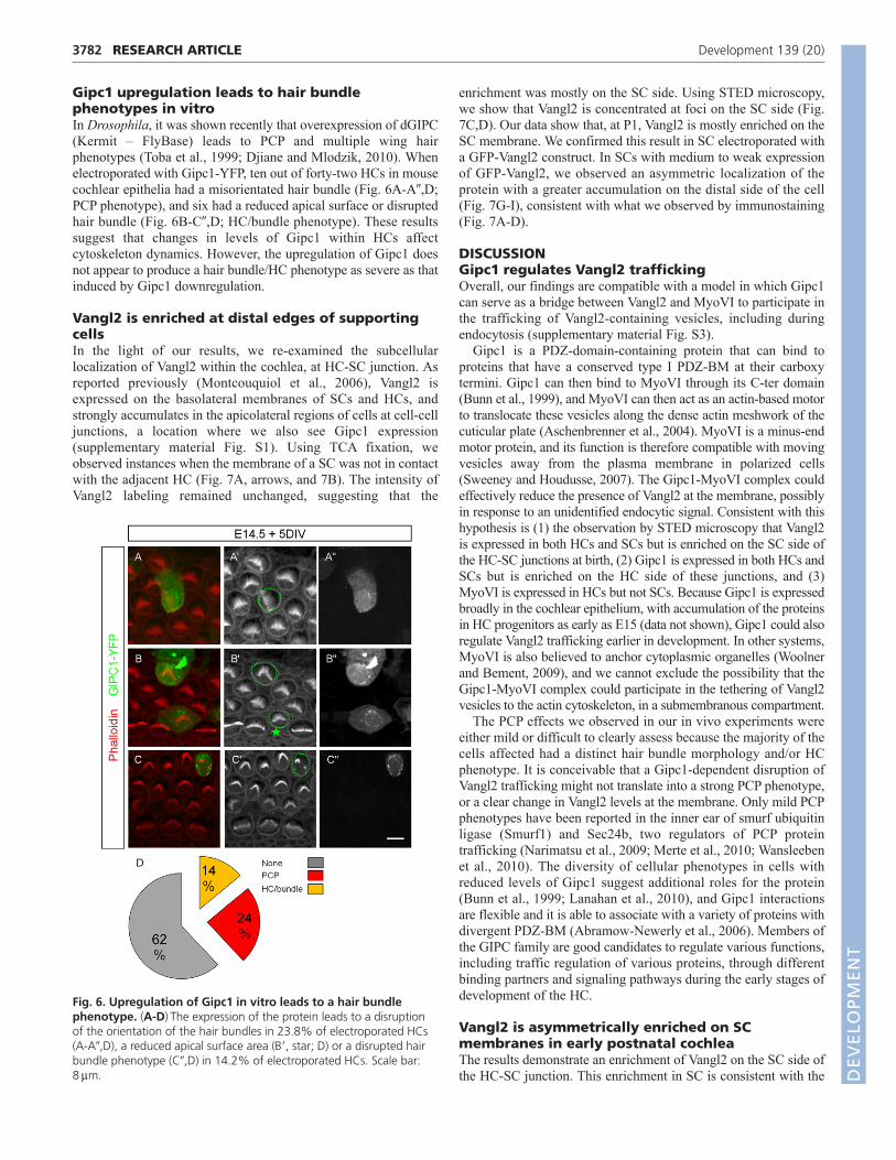
Gipc1 upregulation leads to hair bundlephenotypes in vitroIn Drosophila, it was shown recently that overexpression of dGIPC(Kermit – FlyBase) leads to PCP and multiple wing hairphenotypes (Toba et al., 1999; Djiane and Mlodzik, 2010). Whenelectroporated with Gipc1-YFP, ten out of forty-two HCs in mousecochlear epithelia had a misorientated hair bundle (Fig. 6A-A�,D;PCP phenotype), and six had a reduced apical surface or disruptedhair bundle (Fig. 6B-C�,D; HC/bundle phenotype). These resultssuggest that changes in levels of Gipc1 within HCs affectcytoskeleton dynamics. However, the upregulation of Gipc1 doesnot appear to produce a hair bundle/HC phenotype as severe as thatinduced by Gipc1 downregulation.
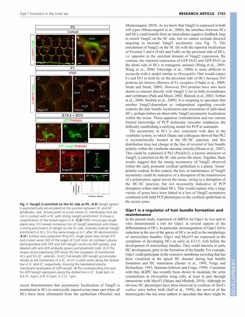
Vangl2 is enriched at distal edges of supportingcellsIn the light of our results, we re-examined the subcellularlocalization of Vangl2 within the cochlea, at HC-SC junction. Asreported previously (Montcouquiol et al., 2006), Vangl2 isexpressed on the basolateral membranes of SCs and HCs, andstrongly accumulates in the apicolateral regions of cells at cell-celljunctions, a location where we also see Gipc1 expression(supplementary material Fig. S1). Using TCA fixation, weobserved instances when the membrane of a SC was not in contactwith the adjacent HC (Fig. 7A, arrows, and 7B). The intensity ofVangl2 labeling remained unchanged, suggesting that the
enrichment was mostly on the SC side. Using STED microscopy,we show that Vangl2 is concentrated at foci on the SC side (Fig.7C,D). Our data show that, at P1, Vangl2 is mostly enriched on theSC membrane. We confirmed this result in SC electroporated witha GFP-Vangl2 construct. In SCs with medium to weak expressionof GFP-Vangl2, we observed an asymmetric localization of theprotein with a greater accumulation on the distal side of the cell(Fig. 7G-I), consistent with what we observed by immunostaining(Fig. 7A-D).
DISCUSSIONGipc1 regulates Vangl2 traffickingOverall, our findings are compatible with a model in which Gipc1can serve as a bridge between Vangl2 and MyoVI to participate inthe trafficking of Vangl2-containing vesicles, including duringendocytosis (supplementary material Fig. S3).
Gipc1 is a PDZ-domain-containing protein that can bind toproteins that have a conserved type I PDZ-BM at their carboxytermini. Gipc1 can then bind to MyoVI through its C-ter domain(Bunn et al., 1999), and MyoVI can then act as an actin-based motorto translocate these vesicles along the dense actin meshwork of thecuticular plate (Aschenbrenner et al., 2004). MyoVI is a minus-endmotor protein, and its function is therefore compatible with movingvesicles away from the plasma membrane in polarized cells(Sweeney and Houdusse, 2007). The Gipc1-MyoVI complex couldeffectively reduce the presence of Vangl2 at the membrane, possiblyin response to an unidentified endocytic signal. Consistent with thishypothesis is (1) the observation by STED microscopy that Vangl2is expressed in both HCs and SCs but is enriched on the SC side ofthe HC-SC junctions at birth, (2) Gipc1 is expressed in both HCs andSCs but is enriched on the HC side of these junctions, and (3)MyoVI is expressed in HCs but not SCs. Because Gipc1 is expressedbroadly in the cochlear epithelium, with accumulation of the proteinsin HC progenitors as early as E15 (data not shown), Gipc1 could alsoregulate Vangl2 trafficking earlier in development. In other systems,MyoVI is also believed to anchor cytoplasmic organelles (Woolnerand Bement, 2009), and we cannot exclude the possibility that theGipc1-MyoVI complex could participate in the tethering of Vangl2vesicles to the actin cytoskeleton, in a submembranous compartment.
The PCP effects we observed in our in vivo experiments wereeither mild or difficult to clearly assess because the majority of thecells affected had a distinct hair bundle morphology and/or HCphenotype. It is conceivable that a Gipc1-dependent disruption ofVangl2 trafficking might not translate into a strong PCP phenotype,or a clear change in Vangl2 levels at the membrane. Only mild PCPphenotypes have been reported in the inner ear of smurf ubiquitinligase (Smurf1) and Sec24b, two regulators of PCP proteintrafficking (Narimatsu et al., 2009; Merte et al., 2010; Wansleebenet al., 2010). The diversity of cellular phenotypes in cells withreduced levels of Gipc1 suggest additional roles for the protein(Bunn et al., 1999; Lanahan et al., 2010), and Gipc1 interactionsare flexible and it is able to associate with a variety of proteins withdivergent PDZ-BM (Abramow-Newerly et al., 2006). Members ofthe GIPC family are good candidates to regulate various functions,including traffic regulation of various proteins, through differentbinding partners and signaling pathways during the early stages ofdevelopment of the HC.
Vangl2 is asymmetrically enriched on SCmembranes in early postnatal cochleaThe results demonstrate an enrichment of Vangl2 on the SC side ofthe HC-SC junction. This enrichment in SC is consistent with the
RESEARCH ARTICLE Development 139 (20)
Fig. 6. Upregulation of Gipc1 in vitro leads to a hair bundlephenotype. (A-D)The expression of the protein leads to a disruptionof the orientation of the hair bundles in 23.8% of electroporated HCs(A-A�,D), a reduced apical surface area (B�, star; D) or a disrupted hairbundle phenotype (C�,D) in 14.2% of electroporated HCs. Scale bar:8m. D
EVELO
PMENT
recent demonstration that asymmetric localization of Vangl2 ismaintained in SCs in ototoxically injured avian inner ears when allHCs have been eliminated from the epithelium (Warchol and
Montcouquiol, 2010). As we know that Vangl2 is expressed in bothcell types (Montcouquiol et al., 2006), the interface between SCsand HCs could benefit from an intercellular negative-feedback loopto enrich Vangl2 on the SC side, but we cannot exclude directedtargeting to increase Vangl2 asymmetry (see Fig. 7). Theenrichment of Vangl2 on the SC fits with the reported localizationof Frizzled 3 and 6 (Fzd3 and Fzd6) on the proximal side of HCs,i.e. opposite to the enriched domain of Vangl2 expression. Bycontrast, the reported expression of GFP-Dvl2 and GFP-Dvl3 onthe distal side of HCs in transgenic animals (Wang et al., 2005;Wang et al., 2006; Etheridge et al., 2008) is more difficult toreconcile with a model similar to Drosophila. One would expectFz and Dvl to both be on the proximal side of HCs because Dvlproteins are known effectors of Fz receptors (Vladar et al., 2009;Strutt and Strutt, 2009). However, Dvl proteins have also beenshown to interact directly with Vangl2 C-ter in both invertebratesand vertebrates (Park and Moon, 2002; Bastock et al., 2003; Torbanet al., 2004; Suriben et al., 2009). It is tempting to speculate thatanother Vangl2-dependent or -independent signaling cascadecontrols the hair bundle localization and orientation of individualHCs, perhaps before an observable Vangl2 asymmetric localizationwithin the tissue. These apparent contradictions and our currentlimited knowledge of PCP molecular cascades emphasize thedifficulty establishing a unifying model for PCP in mammals.
The asymmetry in SCs is also consistent with data in thevestibular system, in which Deans and colleagues showed that Pk2is asymmetrically located at the HC/SC junction, and thisdistribution does not change at the line of reversal of hair bundlepolarity within the vestibular maculae (striola) (Deans et al., 2007).This could be explained if Pk2 (Prickle2), a known interactor ofVangl2, is enriched on the SC side across the tissue. Together, theseresults suggest that the strong asymmetry of Vangl2 observedwithin the early postnatal cochlear epithelium is a planar ‘tissue’polarity readout. In this context, the loss, or maintenance, of Vangl2asymmetry could be indicative of a disruption of the transmissionof a polarization signal across the tissue, owing to a disruption ofthe HC-SC junction, but not necessarily indicative of PCPdisruption within individual HCs. This would explain why a largevariety of genes have been linked to a loss of Vangl2 asymmetrycombined with mild PCP phenotypes in the cochlear epithelium inthe recent years.
Gipc1 is a regulator of hair bundle formation andmaintenanceIn the present study, expression of shRNA for Gipc1 in vivo and invitro demonstrated a role for Gipc1 in several aspects of thedifferentiation of HCs. In particular, downregulation of Gipc1 led toreduction in the size of the apices of HCs, as well as the morphologyof stereociliary bundles. Gipc1 and MyoVI are expressed in thecytoplasm of developing HCs as early as E13.5, well before thedevelopment of stereociliary bundles. They could function in earlyHC development, prior to the formation of the bundle. For example,Gipc1 could participate in the extensive membrane recycling that hasbeen visualized at the apical HC domain during hair bundleformation and HC maturation (Souter et al., 1995; Forge andRichardson, 1993; Denman-Johnson and Forge, 1999). Consistentwith this, dGIPC has recently been shown to modulate the actincytoskeleton in Drosophila wing cells, at least in part throughinteractions with MyoVI (Djiane and Mlodzik, 2010). Although noobvious HC phenotypes have been observed in cochleae of Snell’swaltzer mice before birth (Self et al., 1999), the survival of thehomozygotes has led some authors to speculate that there might be
3783RESEARCH ARTICLEGipc1 functions in the inner ear
Fig. 7. Vangl2 is enriched on the SC side at P0. (A,B)Vangl2 (green)is asymmetrically accumulated at the junction between SC and HC(phalloidin, red). Arrows point to zones where SC membranes that arenot in contact with a HC with strong Vangl2 enrichment. B shows amagnification of the boxed region in A. (C,D)STED microscopy (singleplane view, XY) reveals numerous foci of Vangl2 expression and showsa strong enrichment of Vangl2 on the SC side. Asterisks indicate Vangl2enrichment in SCs. D is the same image as in C after 3D deconvolution.(E-H�) Surface view projection (Proj XY), single plane view (single XY)and z-stack series (XZ) of the organ of Corti from rat cochlear cultureselectroporated with GFP and GFP-Vangl2 constructs (left panels), andlabeled with anti-GFP antibody (green) and phalloidin (red). (E-F)Theempty vector expressing GFP alone fills the cytoplasm of transfectedHCs and SCs (E�, asterisk). (G-H�) Full-length GFP-Vangl2 accumulatesdistally at the membrane of a SC. (H,H�) z-stack series along the dottedline in G� and G�, respectively, showing the basolateral plasmamembrane localization of GFP-Vangl2. (I)The corresponding line scanfor GFP-Vangl2 expression along the dotted line in H�. Scale bars: inA,E-H�, 4m; in B, 0.5m; in C,D, 1m.
DEVELO
PMENT
3784
a significant degree of functional redundancy among myosin proteinsduring mouse development and/or among other motor proteins thatmust be able to compensate for the loss of MyoVI. In humans,mutations in MYO6 cause hereditary hearing loss (DFNA22 andDFNB37 syndromes), which can be associated with hypertrophiccardiomyopathy (Ahmed et al., 2003; Melchionda et al., 2001).
In the course of this study, GIPC3, another member of the GIPCfamily was associated with progressive hearing loss in humans andmice (Charizopoulou et al., 2011). Interestingly, in the mutant mice,the identified mutation affected the PDZ domain of Gipc3, leadingto early postnatal disruption of hair bundle structural integrity. Inhumans, one of the identified mutations leads to a potentialtruncated construct, preventing interaction with MyoVI. Also, arecent study correlated hearing impairment in four patients with a359-kb deleted region in 19p13.12 harboring six genes, includingGIPC1, leading the authors to suggest that haploinsufficiency ofGIPC1 might contribute to hearing impairment through itsinteraction with MyoVI (Bonaglia et al., 2010). Altogether, theseresults strongly suggest that members of the GIPC family haveessential roles in HC maturation and auditory function.
AcknowledgementsWe thank the team of Bordeaux Imaging Center and Ulf Schwarz from LeicaMicrosystems, Germany, for their help in STED microscopy; the genotypingfacility; and Helene Doat from the animal facility for technical assistance.
FundingThis research was supported by a French National Institute of Health andMedical Research (INSERM) AVENIR grant [R04208GS to M.M. and R04210GSto N.S.]; Conseil Regional d’Aquitaine [M.M. and N.S.]; La Fondation pour laRecherche Medicale [M.M., N.S., J.E.]; Agence Nationale de la Recherche[ANR-08-; MNPS-040-01 to M.M. and ANR-07-NEUR-031-01 to N.S.];Aquitaine-INSERM Fellowship [A.P.G.]; International Reintegration Grants (IRG)Marie-Curie action [M.M.] and the European Commission Coordination Actionand the Network of European Neuroscience Institutes ENINET [contractnumber LSHM-CT-2005-19063; N.S. and M.M.]; and the National Institute onDeafness and other Communication Disorders [DC R01 008595, DC R01008595-03S2 (to J.B.) and P30 DC005983]. J.-P.B. is supported by LigueNationale Contre le Cancer [Label 2010 JPB], European Consortium forAnticancer Antibody Development (EUCAAD) (FP7 program), INCa and IBISa(Marseille Proteomic Platform).
Competing interests statementThe authors declare no competing financial interests.
Supplementary materialSupplementary material available online athttp://dev.biologists.org/lookup/suppl/doi:10.1242/dev.074229/-/DC1
ReferencesAbramow-Newerly, M., Roy, A. A., Nunn, C. and Chidiac, P. (2006). RGS
proteins have a signalling complex: interactions between RGS proteins andGPCRs, effectors, and auxiliary proteins. Cell Signal. 18, 579-591.
Ahmed, Z. M., Morell, R. J., Riazuddin, S., Gropman, A., Shaukat, S., Ahmad,M. M., Mohiddin, S. A., Fananapazir, L., Caruso, R. C., Husnain, T. et al.(2003). Mutations of MYO6 are associated with recessive deafness, DFNB37.Am. J. Hum. Genet. 72, 1315-1322.
Aschenbrenner, L., Lee, T. and Hasson, T. (2003). Myo6 facilitates thetranslocation of endocytic vesicles from cell peripheries. Mol. Biol. Cell 14, 2728-2743.
Aschenbrenner, L., Naccache, S. N. and Hasson, T. (2004). Uncoated endocyticvesicles require the unconventional myosin, Myo6, for rapid transport throughactin barriers. Mol. Biol. Cell 15, 2253-2263.
Avraham, K. B., Hasson, T., Steel, K. P., Kingsley, D. M., Russell, L. B.,Mooseker, M. S., Copeland, N. G. and Jenkins, N. A. (1995). The mouseSnell’s waltzer deafness gene encodes an unconventional myosin required forstructural integrity of inner ear hair cells. Nat. Genet. 11, 369-375.
Bastock, R., Strutt, H. and Strutt, D. (2003). Strabismus is asymmetricallylocalised and binds to Prickle and Dishevelled during Drosophila planar polaritypatterning. Development 130, 3007-3014.
Bonaglia, M. C., Marelli, S., Novara, F., Commodaro, S., Borgatti, R.,Minardo, G., Memo, L., Mangold, E., Beri, S., Zucca, C. et al. (2010).
Genotype-phenotype relationship in three cases with overlapping 19p13.12microdeletions. Eur. J. Hum. Genet. 18, 1302-1309.
Brigande, J. V., Gubbels, S. P., Woessner, D. W., Jungwirth, J. J. and Bresee,C. S. (2009). Electroporation-mediated gene transfer to the developing mouseinner ear. Methods Mol. Biol. 493, 125.
Bunn, R. C., Jensen, M. A. and Reed, B. C. (1999). Protein interactions with theglucose transporter binding protein GLUT1CBP that provide a link betweenGLUT1 and the cytoskeleton. Mol. Biol. Cell 10, 819-832.
Buss F. and Kendrick-Jones, J. (2008). How are the cellular functions of myosinVI regulated within the cell? Biochem. Biophys. Res. Commun. 369, 165-175.
Buss, F., Arden, S. D., Lindsay, M., Luzio, J. P. and Kendrick-Jones, J. (2001).Myosin VI isoform localized to clathrin-coated vesicles with a role in clathrin-mediated endocytosis. EMBO J. 20, 3676-3684.
Buss, F., Spudich, G. and Kendrick-Jones, J. (2004). MYOSIN VI: cellularfunctions and motor properties. Annu. Rev. Cell. Dev. Biol. 20, 649-676.
Charizopoulou, N., Lelli, A., Schraders, M., Ray, K., Hildebrand, M. S.,Ramesh, A., Srisailapathy, C. R., Oostrik, J., Admiraal, R. J., Neely, H. R. etal. (2011). Gipc3 mutations associated with audiogenic seizures andsensorineural hearing loss in mouse and human. Nat. Commun. 2, 201.
Curtin, J. A., Quint, E., Tsipouri, V., Arkell, R. M., Cattanach, B., Copp, A. J.,Henderson, D. J., Spurr, N., Stanier, P., Fisher, E. M., Nolan, P. M., Steel, K.P., Brown, S. D., Gray, I. C. and Murdoch, J. N. (2003). Mutation of Celsr1disrupts planar polarity of inner ear hair cells and causes severe neural tubedefects in the mouse. Curr. Biol. 13, 1129-1133.
Dance, A. L., Miller, M., Seragaki, S., Aryal, P., White, B. et al. (2004).Regulation of myosin-VI targeting to endocytic compartments. Traffic 5, 798-813.
Das, G., Jenny, A., Klein, T. J., Eaton, S. and Mlodzik, M. (2004). Diegointeracts with Prickle and Strabismus/Van Gogh to localize planar cell polaritycomplexes. Development 131, 4467-4476.
De Vries, L., Lou, X., Zhao, G., Zheng, B. and Farquhar, M. (1998). G. GIPC, aPDZ domain containing protein, interacts specifically with the C terminus ofRGS-GAIP. Proc. Natl. Acad. Sci. USA 95, 12340-12345.
Deans, M. R., Antic, D., Suyama, K., Scott, M. P., Axelrod, J. D. andGoodrich, L. V. (2007). Asymmetric distribution of prickle-like 2 reveals an earlyunderlying polarization of vestibular sensory epithelia in the inner ear. J.Neurosci. 27, 3139-3147.
Denman-Johnson, K. and Forge, A. (1999). Establishment of hair bundlepolarity and orientation in the developing vestibular system of the mouse. J.Neurocytol. 28, 821-835.
Djiane, A. and Mlodzik, M. (2010). The Drosophila GIPC homologue canmodulate myosin based processes and planar cell polarity but is not essential fordevelopment. PLoS ONE 5, e11228.
Etheridge, S. L., Ray, S., Li, S., Hamblet, N. S., Lijam, N. et al. (2008). Murinedishevelled 3 functions in redundant pathways with dishevelled 1 and 2 innormal cardiac outflow tract, cochlea, and neural tube development. PLoSGenet. 4, e1000259.
Forge, A. and Richardson, G. (1993). Freeze fracture analysis of apicalmembranes in cochlear cultures: differences between basal and apical-coil outerhair cells and effects of neomycin. J. Neurocytol. 22, 854-867.
Gubbels, S. P., Woessner, D. W., Mitchell, J. C., Ricci, A. J. and Brigande, J. V.(2008). Functional auditory hair cells produced in the mammalian cochlea by inutero gene transfer. Nature 455, 537-541.
Hasson, T. (2003). Myosin VI: two distinct roles in endocytosis. J. Cell Sci. 116,3453-3461.
Hasson, T., Gillespie, P. G., Garcia, J. A., MacDonald, R. B., Zhao, Y., Yee, A.G., Mooseker, M. S. and Corey, D. P. (1997). Unconventional myosins in inner-ear sensory epithelia. J. Cell Biol. 137, 1287-1307.
Hayashi, K., Yonemura, S., Matsui, T. and Tsukita, S. (1999).Immunofluorescence detection of ezrin/radixin/moesin (ERM) proteins with theircarboxylterminal threonine phosphorylated in cultured cells and tissues:application of a novel fixation protocol using trichloroacetic acid (TCA) as afixative. J. Cell Sci. 112, 1149-1158.
Jenny, A., Reynolds-Kenneally, J., Das, G., Burnett, M. and Mlodzik, M.(2005). Diego and Prickle regulate Frizzled planar cell polarity signalling bycompeting for Dishevelled binding. Nat. Cell Biol. 7, 691-697.
Kachar, B., Battaglia, A. and Fex, J. (1997). Compartmentalized vesicular trafficaround the hair cell cuticular plate. Hear. Res. 107, 102-112.
Lanahan, A. A., Hermans, K., Claes, F., Kerley-Hamilton, J. S., Zhuang, Z. W.,Giordano, F. J., Carmeliet, P. and Simons, M. (2010). VEGF receptor 2endocytic trafficking regulates arterial morphogenesis. Dev. Cell 18, 713-724.
Lu, X., Borchers, A. G., Jolicoeur, C., Rayburn, H., Baker, J. C. and Tessier-Lavigne, M. (2004). PTK7/CCK-4 is a novel regulator of planar cell polarity invertebrates. Nature 430, 93-98.
Melchionda, S., Ahituv, N., Bisceglia, L., Sobe, T., Glaser, F. et al. (2001).MYO6, the human homologue of the gene responsible for deafness in Snell’swaltzer mice, is mutated in autosomal dominant nonsyndromic hearing loss.Am. J. Hum. Genet. 69, 635-640.
Merte, J., Jensen, D., Wright, K., Sarsfield, S., Wang, Y., Schekman, R. andGinty, D. D. (2010). Sec24b selectively sorts Vangl2 to regulate planar cellpolarity during neural tube closure. Nat. Cell Biol. 12, 41-46.
RESEARCH ARTICLE Development 139 (20)
DEVELO
PMENT
Montcouquiol, M., Rachel, R. A., Lanford, P. J., Copeland, N. G., Jenkins, N.A. and Kelley, M. W. (2003). Identification of Vangl2 and Scrb1 as planarpolarity genes in mammals. Nature 423, 173-177.
Montcouquiol, M., Sans, N., Huss, D., Kach, J., Dickman, J. D., Forge, A.,Rachel, R. A., Copeland, N. G., Jenkins, N. A., Bogani, D., Murdoch, J.,Warchol, M. E., Wenthold, R. J. and Kelley, M. W. (2006). Asymmetriclocalization of Vangl2 and Fz3 indicate novel mechanisms for planar cell polarityin mammals. J. Neurosci. 26, 5265-5275.
Montcouquiol, M., Jones, J. M. and Sans, N. (2008). Detection of planarpolarity proteins in mammalian cochlea. Methods Mol. Biol. 468, 207-219.
Narimatsu, M., Bose, R., Pye, M., Zhang, L., Miller, B., Ching, P., Sakuma, R.,Luga, V., Roncari, L., Attisano, L., Wrana, J. L. (2009). Regulation of planarcell polarity by Smurf ubiquitin ligases. Cell 137, 295-307.
Park, M. and Moon, R. T. (2002). The planar cell-polarity gene stbm regulates cellbehaviour and cell fate in vertebrate embryos. Nat. Cell Biol. 4, 20-25.
Sans, N., Prybylowski, K., Petralia, R. S., Chang, K., Wang, Y. X., Racca, C.,Vicini, S. and Wenthold, R. J. (2003). NMDA receptor trafficking through aninteraction between PDZ proteins and the exocyst complex. Nat. Cell Biol. 5,520-530.
Seifert, J. R. and Mlodzik, M. (2007). Frizzled/PCP signalling: a conservedmechanism regulating cell polarity and directed motility. Nat. Rev. Genet. 8,126-138.
Self, T., Sobe, T., Copeland, N. G., Jenkins, N. A., Avraham, K. B. and Steel,K. P. (1999). Role of myosin VI in the differentiation of cochlear hair cells. Dev.Biol. 214, 331-341.
Souter, M., Nevill, G. and Forge, A. (1995). Postnatal development ofmembrane specialisations of gerbil outer hair cells. Hear. Res. 91, 43-62.
Strutt, H. and Strutt, D. (2009). Asymmetric localisation of planar polarityproteins: mechanisms and consequences. Semin. Cell Dev. Biol. 20, 957-963.
Suriben, R., Kivimäe, S., Fisher, D. A., Moon, R. T. and Cheyette, B. N. (2009).Posterior malformations in Dact1 mutant mice arise through misregulatedVangl2 at the primitive streak. Nat. Genet. 41, 977-985.
Sweeney, H. L. and Houdusse, A. (2007). What can myosin VI do in cells? Curr.Opin. Cell Biol. 19, 57-66.
Takeuchi, M., Nakabayashi, J., Sakaguchi, T., Yamamoto, T. S., Takahashi, H.,Takeda, H. and Ueno, N. (2003). The prickle related gene in vertebrates isessential for gastrulation cell movements. Curr. Biol. 13, 674-679.
Toba, G., Ohsako, T., Miyata, N., Ohtsuka, T., Seong, K. H. and Aigaki, T.(1999). The gene search system. A method for efficient detection and rapidmolecular identification of genes in Drosophila melanogaster. Genetics. 151,725-737.
Torban, E., Wang, H. J., Groulx, N. and Gros, P. (2004). Independent mutationsin mouse Vangl2 that cause neural tube defects in looptail mice impairinteraction with members of the Dishevelled family. J. Biol. Chem. 279, 52703-52713.
Tree, D. R., Shulman, J. M., Rousset, R., Scott, M. P., Gubb, D. and Axelrod, J.D. (2002). Prickle mediates feedback amplification to generate asymmetricplanar cell polarity signaling. Cell 109, 371-381.
Vladar, E. K., Antic, D. and Axelrod, J. D. (2009). Planar cell polarity signaling: the developing cell’s compass. Cold Spring Harb. Perspect. Biol. 1,a002964.
Wang, J., Mark, S., Zhang, X., Qian, D., Yoo, S. J., Radde-Gallwitz, K., Zhang,Y., Lin, X., Collazo, A., Wynshaw-Boris, A. and Chen, P. (2005). Regulationof polarized extension and planar cell polarity in the cochlea by the vertebratePCP pathway. Nat. Genet. 37, 980-985.
Wang, Y. and Nathans, J. (2007). Tissue/planar cell polarity in vertebrates: newinsights and new questions. Development 134, 647-658.
Wang, Y., Guo, N. and Nathans, J. (2006). The role of Frizzled3 and Frizzled6 inneural tube closure and in the planar polarity of inner-ear sensory hair cells. J.Neurosci. 26, 2147-2156.
Wansleeben, C., Feitsma, H., Montcouquiol, M., Kroon, C., Cuppen, E. andMeijlink, F. (2010). Planar cell polarity defects and defective Vangl2 traffickingin mutants for the COPII gene Sec24b. Development 137, 1067-1073.
Warchol, M. E. and Montcouquiol, M. (2010). Maintained expression of theplanar cell polarity molecule Vangl2 and reformation of hair cell orientation inthe regenerating inner ear. J. Assoc. Res. Otolaryngol. 11, 395-406.
Woolner, S. and Bement, W. M. (2009). Unconventional myosins actingunconventionally. Trends Cell Biol. 19, 245-252.
Yi, Z., Petralia, R. S., Fu, Z., Swanwick, C. C., Wang, Y. X., Prybylowski, K.,Sans, N., Vicini, S. and Wenthold, R. J. (2007). The role of the PDZ proteinGIPC in regulating NMDA receptor trafficking J. Neurosci. 27, 11663-11675.
Zallen, J. A. (2007). Planar polarity and tissue morphogenesis. Cell. 129, 1051-1063.
3785RESEARCH ARTICLEGipc1 functions in the inner ear
DEVELO
PMENT