Efficacy of Combined Therapy with Liposome-Encapsulated Meglumine Antimoniate and Allopurinol in...
10
Efficacy of Combined Therapy with Liposome-Encapsulated Meglumine Antimoniate and Allopurinol in Treatment of Canine Visceral Leishmaniasis Sydnei M. da Silva, a Izabela F. G. Amorim, b Raul R. Ribeiro, c * Erly G. Azevedo, c Cynthia Demicheli, d Maria N. Melo, a Wagner L. Tafuri, b Nelder F. Gontijo, a Marilene S. M. Michalick, a and Frédéric Frézard c Departamento de Parasitologia, a Departamento de Patologia, b Departamento de Fisiologia e Biofísica, Instituto de Ciências Biológicas, c and Departamento de Química, Instituto de Ciências Exatas, d Universidade Federal de Minas Gerais, Belo Horizonte, Minas Gerais, Brazil An innovative liposomal formulation of meglumine antimoniate (LMA) was recently reported to promote both long-term para- site suppression and reduction of infectivity to sand flies in dogs with visceral leishmaniasis. However, 5 months after treatment, parasites were still found in the bone marrow of all treated dogs. In order to improve treatment with LMA, the present study aimed to evaluate its efficacy in combination with allopurinol. Mongrel dogs naturally infected with Leishmania infantum were treated with six doses of LMA (6.5 mg Sb/kg of body weight/dose) given at 4-day intervals, plus allopurinol (20 mg/kg/24 h per os) for 140 days. Comparison was made with groups treated with LMA, allopurinol, empty liposomes plus allopurinol, empty liposomes, and saline. Dogs remained without treatment from day 140 to 200 after the start of treatment. The drug combination promoted both clinical improvement of dogs and significant reduction in the parasitic load in bone marrow and spleen on days 140 and 200 compared to these parameters in the pretreatment period. This is in contrast with the other protocols, which did not result in significant reduction of the bone marrow parasite load on day 200. Strikingly, the combined treatment, in contrast to the other regimens, induced negative quantitative PCR (qPCR) results in the liver of 100% of the dogs. Both xenodiagnosis and skin parasite determination by qPCR indicated that the drug combination was effective in blocking the transmission of skin par- asites to sand flies. Based on all of the parasitological tests performed on day 200, 50% of the animals that received the combined treatment were considered cured. V isceral leishmaniasis (VL) is a systemic parasitic disease which leads to high rates of morbidity and mortality in humans worldwide. Even with scientific advances related to diagnosis, treatment, and prevention over the past 10 years, VL still is a neglected disease leading to 60,000 human deaths/year. The clinical manifestations of VL are attributed to obligatory intracel- lular protozoa of the Leishmania donovani complex and, depend- ing on the etiological agent, the disease presents two distinct forms: anthroponotic VL that is endemic in India and Central Africa, caused by L. donovani, and zoonotic VL that occurs in countries of the Mediterranean basin, Central Asia, and Americas, caused by Leishmania infantum. Domestic dogs are the most im- portant urban reservoirs of L. infantum, which is transmitted to humans and dogs through bites of infected female sand flies of the genera Lutzomyia and Phlebotomus (Diptera: Psychodidae; Phle- botominae) in the New World and Old World, respectively (7, 33). The disease in dogs is characterized by a marked pleomor- phism, and the clinical signs vary according to the immune re- sponse of the animals toward the infection. In general, the main clinical signs of canine visceral leishmaniasis (CVL) are various degrees of dermopathy, lymphadenopathy, onychogryposis, weight loss, abnormalities of the musculoskeletal system, eye le- sions, hematopoietic disorders, renal disease, and lesions originat- ing from immune complex deposition in tissues (e.g., vasculitis and arthritis) (30). The treatment of dogs affected with VL has been practiced in Europe since the middle of the 20th century (1). Since then, the pentavalent antimonials, including meglumine antimoniate (MA) and sodium stibogluconate, have been the main class of drugs used to treat VL in both humans and dogs (1, 17). However, treatment with antimonial drugs does not promote parasitologi- cal cure in infected dogs, leading to frequent relapses (23) and necessitating continuous administration of the drugs, which are both poorly tolerated and expensive. The purine analog allopurinol is an alternative orally active drug (6) which presents low toxicity, is effective in reverting the clinical signs of CVL, and prevents recurrence of the disease (1, 8). However, allopurinol also is unable to promote parasitological cure in dogs with VL (23). Currently, the combination of MA and allopurinol constitutes the first-line pharmaceutical protocol for CVL. The basis for the use of this combination is the synergism between these drugs against Leishmania parasites, as previously demonstrated in vitro (22) and in vivo (1, 15, 24, 27). However, although most dogs recover clinically after therapy, complete elimination of the para- site is usually not achieved, and infected dogs may eventually re- lapse. Because of its limited parasiticidal efficacy and the potential risk of long-term treatment for selection of Leishmania strains Received 26 January 2012 Returned for modification 21 February 2012 Accepted 1 March 2012 Published ahead of print 12 March 2012 Address correspondence to Frédéric Frézard, [email protected]. * Present address: Centro de Ciências Agrárias, Ambientais e Biológicas da Universidade Federal do Recôncavo da Bahia, Cruz das Almas, Buenos Aires, Brazil. S.M.d.S. and I.F.G.A. contributed equally to this work. Copyright © 2012, American Society for Microbiology. All Rights Reserved. doi:10.1128/AAC.00208-12 2858 aac.asm.org Antimicrobial Agents and Chemotherapy p. 2858 –2867 June 2012 Volume 56 Number 6
Transcript of Efficacy of Combined Therapy with Liposome-Encapsulated Meglumine Antimoniate and Allopurinol in...

Efficacy of Combined Therapy with Liposome-EncapsulatedMeglumine Antimoniate and Allopurinol in Treatment of CanineVisceral Leishmaniasis
Sydnei M. da Silva,a Izabela F. G. Amorim,b Raul R. Ribeiro,c* Erly G. Azevedo,c Cynthia Demicheli,d Maria N. Melo,a Wagner L. Tafuri,b
Nelder F. Gontijo,a Marilene S. M. Michalick,a and Frédéric Frézardc
Departamento de Parasitologia,a Departamento de Patologia,b Departamento de Fisiologia e Biofísica, Instituto de Ciências Biológicas,c and Departamento de Química,Instituto de Ciências Exatas,d Universidade Federal de Minas Gerais, Belo Horizonte, Minas Gerais, Brazil
An innovative liposomal formulation of meglumine antimoniate (LMA) was recently reported to promote both long-term para-site suppression and reduction of infectivity to sand flies in dogs with visceral leishmaniasis. However, 5 months after treatment,parasites were still found in the bone marrow of all treated dogs. In order to improve treatment with LMA, the present studyaimed to evaluate its efficacy in combination with allopurinol. Mongrel dogs naturally infected with Leishmania infantum weretreated with six doses of LMA (6.5 mg Sb/kg of body weight/dose) given at 4-day intervals, plus allopurinol (20 mg/kg/24 hper os) for 140 days. Comparison was made with groups treated with LMA, allopurinol, empty liposomes plus allopurinol, emptyliposomes, and saline. Dogs remained without treatment from day 140 to 200 after the start of treatment. The drug combinationpromoted both clinical improvement of dogs and significant reduction in the parasitic load in bone marrow and spleen on days140 and 200 compared to these parameters in the pretreatment period. This is in contrast with the other protocols, which did notresult in significant reduction of the bone marrow parasite load on day 200. Strikingly, the combined treatment, in contrast tothe other regimens, induced negative quantitative PCR (qPCR) results in the liver of 100% of the dogs. Both xenodiagnosis andskin parasite determination by qPCR indicated that the drug combination was effective in blocking the transmission of skin par-asites to sand flies. Based on all of the parasitological tests performed on day 200, 50% of the animals that received the combinedtreatment were considered cured.
Visceral leishmaniasis (VL) is a systemic parasitic disease whichleads to high rates of morbidity and mortality in humans
worldwide. Even with scientific advances related to diagnosis,treatment, and prevention over the past 10 years, VL still is aneglected disease leading to �60,000 human deaths/year. Theclinical manifestations of VL are attributed to obligatory intracel-lular protozoa of the Leishmania donovani complex and, depend-ing on the etiological agent, the disease presents two distinctforms: anthroponotic VL that is endemic in India and CentralAfrica, caused by L. donovani, and zoonotic VL that occurs incountries of the Mediterranean basin, Central Asia, and Americas,caused by Leishmania infantum. Domestic dogs are the most im-portant urban reservoirs of L. infantum, which is transmitted tohumans and dogs through bites of infected female sand flies of thegenera Lutzomyia and Phlebotomus (Diptera: Psychodidae; Phle-botominae) in the New World and Old World, respectively (7,33). The disease in dogs is characterized by a marked pleomor-phism, and the clinical signs vary according to the immune re-sponse of the animals toward the infection. In general, the mainclinical signs of canine visceral leishmaniasis (CVL) are variousdegrees of dermopathy, lymphadenopathy, onychogryposis,weight loss, abnormalities of the musculoskeletal system, eye le-sions, hematopoietic disorders, renal disease, and lesions originat-ing from immune complex deposition in tissues (e.g., vasculitisand arthritis) (30).
The treatment of dogs affected with VL has been practiced inEurope since the middle of the 20th century (1). Since then,the pentavalent antimonials, including meglumine antimoniate(MA) and sodium stibogluconate, have been the main class ofdrugs used to treat VL in both humans and dogs (1, 17). However,
treatment with antimonial drugs does not promote parasitologi-cal cure in infected dogs, leading to frequent relapses (23) andnecessitating continuous administration of the drugs, which areboth poorly tolerated and expensive.
The purine analog allopurinol is an alternative orally activedrug (6) which presents low toxicity, is effective in reverting theclinical signs of CVL, and prevents recurrence of the disease (1, 8).However, allopurinol also is unable to promote parasitologicalcure in dogs with VL (23).
Currently, the combination of MA and allopurinol constitutesthe first-line pharmaceutical protocol for CVL. The basis for theuse of this combination is the synergism between these drugsagainst Leishmania parasites, as previously demonstrated in vitro(22) and in vivo (1, 15, 24, 27). However, although most dogsrecover clinically after therapy, complete elimination of the para-site is usually not achieved, and infected dogs may eventually re-lapse. Because of its limited parasiticidal efficacy and the potentialrisk of long-term treatment for selection of Leishmania strains
Received 26 January 2012 Returned for modification 21 February 2012Accepted 1 March 2012
Published ahead of print 12 March 2012
Address correspondence to Frédéric Frézard, [email protected].
* Present address: Centro de Ciências Agrárias, Ambientais e Biológicas daUniversidade Federal do Recôncavo da Bahia, Cruz das Almas, Buenos Aires, Brazil.
S.M.d.S. and I.F.G.A. contributed equally to this work.
Copyright © 2012, American Society for Microbiology. All Rights Reserved.
doi:10.1128/AAC.00208-12
2858 aac.asm.org Antimicrobial Agents and Chemotherapy p. 2858–2867 June 2012 Volume 56 Number 6

resistant to antimonial drug, WHO strongly supports the searchfor more effective therapeutic protocols (21, 23, 33).
In the 1970s, a major advance occurred when it was found thatliposome-encapsulated antimonial drugs were hundreds of timesmore effective than the free drugs against experimental VL basedon parasite suppression in the liver (4). Since then, much efforthas been devoted to the search for efficacious liposomal formula-tions in CVL (16). In this context, an innovative liposomal formu-lation of MA (LMA) was recently developed that has significantpharmaceutical and pharmacological advantages over conven-tional formulations (18, 29). Treatment of dogs naturally infectedby L. infantum with four intravenous (i.v.) doses of LMA at 6.5 mgSb/kg of body weight at 4-day intervals (28) promoted both long-term parasite suppression and reduction of infectivity to sandflies. However, parasites were still found in the bone marrow of alltreated dogs.
With the aim of further improving the treatment efficacy ofLMA in dogs, a new protocol was designed which combines LMAwith allopurinol and takes advantage of the synergistic antileish-manial actions of allopurinol and antimonial drugs. In the presentwork, the efficacy of the combination of LMA with allopurinol asa mean of achieving parasitological cure in dogs naturally infectedwith L. infantum has been established for the first time, throughclinical and parasitological parameters as well as assessment of theinfectivity of dogs to sand flies.
MATERIALS AND METHODSMaterials and drugs. Cholesterol and dicetylphosphate were purchasedfrom Sigma Co. (St. Louis, MO in the supplemental material). Distearoyl-phosphatidylcholine was obtained from Lipoid (Ludwigshafen, Ger-many). N-Methyl-D-glucamine and antimony pentachloride (SbCl5,99%) were obtained from Aldrich Chemical Co. (Milwaukee, WI). Allo-purinol was purchased from a commercial laboratory for pharmaceuticalproducts (Vetfarma, Brazil) as a formulation of oral capsules at individualdosages of 20 mg/kg of body weight. Meglumine antimoniate (MA) wassynthesized, as previously described (14), from equimolar amounts ofN-methyl-D-glucamine and pentavalent antimony oxyhydrate freshlyprepared from the hydrolysis of antimony pentachloride in water. Theresulting product contained 28% Sb by weight. As previously established(18), synthetic MA may be replaced by commercial MA (Glucantime;Sanofi-Aventis Farmacêutica Ltda., São Paulo, Brazil) in the preparationof the liposomal formulation.
Animals. The present study was approved by the Ethical Committeefor Animal Experimentation of the Universidade Federal de Minas Gerais(protocol number 211/2007), and all procedures were carried out accord-ing to the international guidelines (Principles of Laboratory Animal Care[26]).
Fifty-two mongrel dogs (32 males and 20 females) weighing 12.3 � 5.2kg (mean � standard deviation), of unknown age, and naturally infectedwith Leishmania infantum were obtained by donation from the Center forZoonosis Control of Ribeirão das Neves City (Minas Gerais State, Brazil),an area where canine visceral leishmaniasis is endemic. These dogs werepreviously identified by serological tests and captured as part of the activ-ities of the municipality’s Visceral Leishmaniasis Control Program.
The serological diagnosis was confirmed at the Serology Laboratory ofthe Institute of Biological Sciences (ICB), Federal University of MinasGerais (UFMG) using indirect immunofluorescence assay (IFAT) andenzyme-linked immunosorbent assay (ELISA) (5). All animals werefound to be positive by IFAT (�1:40 dilution) and ELISA (optical density,�0.100; 1:400 dilution). In addition, specific PCR of bone marrow aspi-rate was used to confirm the infection of the animals by L. infantum (13).The animals were kept in the experimental kennel of the ICB-UFMG withdrinking water and balanced commercial food (Nero Refeição; Total Ali-
mentos, Brazil) ad libitum during the entire experimental period. Prior totreatment, dogs were treated against intestinal helminths (Helfine cães;Agener União, Brazil) and ectoparasite infestations (Frontiline Top Spot;Merial, Brazil) and immunized against rabies (Defensor; Pfizer SaúdeAnimal, Brazil) and other infectious diseases (Vanguard HTLP 5/CV-L;Pfizer Saúde Animal, Brazil).
Preparation and characterization of liposomes. Liposome-encapsu-lated meglumine antimoniate (LMA) was prepared as previously de-scribed (29). Briefly, small unilamellar vesicles (SUVs) were obtained bymixing distearoylphosphatidylcholine, cholesterol, and dicetylphosphate(molar ratio of 5:4:1) following ultrasonication in deionized water at afinal lipid concentration of 55 g/liter. Then, the SUV suspension was fil-tered with a sterile 0.22-�m membrane and mixed with sucrose (sugar/lipid mass ratio of 3:1; final sugar concentration of 0.3 M), and the result-ing mixture was immediately frozen in liquid nitrogen and subsequentlydried (freeze dryer, 4.5 liters; Labconco, United Kingdom). At this point,the freeze-dried liposomal formulation may be stored at �20°C for at least6 months without any change of final vesicle size and drug encapsulationefficiency characteristics. The day before administration, the lyophilizedpowder was rehydrated with a solution of MA in water (antimony con-centration of 0.65 M, corresponding to 40% of the original SUV volume)and the resulting suspension was vortexed and incubated for 30 min at55°C. Then, the same volume of phosphate-buffered saline (PBS; 0.15 MNaCl, 0.01 M phosphate, pH 7.2) was added to the mixture, followed byvortexing and incubation for 30 min at 55°C. The resulting liposomesuspension was diluted in PBS and centrifuged (25,000 � g for 30 min at4°C) in order to separate LMA from nonencapsulated drug. After centrif-ugation, the liposome pellet was washed twice with isotonic saline andresuspended in sterile saline solution, and the final antimony concentra-tion was adjusted to 10 g/liter. The liposome suspension was stored at 4°Cuntil administration. The drug encapsulation efficiency was 37% � 5%, asdetermined by atomic absorption spectroscopy (AA600; PerkinElmer,Inc., United States). The mean hydrodynamic diameter of the vesicles was350 � 58 nm, with a polydispersity index of 0.30 � 0.07, as determined byphoton correlation spectroscopy (Malvern Zetasizer Nano ZS90; MalvernInstruments, United Kingdom). In parallel, lyophilized SUVs were rehy-drated with a solution of N-methyl-D-glucamine (0.65 M, pH 7.2) insteadof MA, using the same method as described above. The final suspension,called empty liposomes (LEMP), presented similar values of mean hydro-dynamic vesicle diameter and polydispersity index.
Treatment protocols. Dogs were randomly distributed into six groupsas follows. In the LMA�Allop group, eight animals were treated with sixdoses of LMA (6.5 mg Sb/kg of body weight/dose) given at 4-day intervalsand allopurinol (20 mg/kg of body weight/24 h per os) for 140 days startingfrom the first dose of LMA. In the LMA group, eight animals were treatedwith six doses of LMA (6.5 mg Sb/kg of body weight/dose) given at 4-dayintervals. In the Allop group, eight animals were treated with allopurinol(20 mg/kg of body weight/24 h per os) for 140 days and six doses ofisotonic saline given at the same volume and time intervals as LMA in theLMA�Allop group. In the LEMP�Allop group, eight animals weretreated with six doses of empty liposomes given at the same volume andtime intervals as LMA in the LMA�Allop group and allopurinol (20mg/kg of body weight/24 h per os) for 140 days. In the LEMP group, eightanimals were treated with six doses of empty liposomes given at the samevolume and time intervals as LMA in the LMA group. In the saline group,12 animals received six doses of isotonic saline given at the same volumeand time intervals as LMA in the LMA�Allop group.
Following day 140 after the beginning of treatment, all animals of thesix experimental groups were kept in the kennel for 60 days without anyintervention, representing a total experimental period of 200 days. Ani-mals were clinically monitored during the experimental period and weresubmitted to clinical and parasitological evaluations just before and ondays 140 and 200 after the beginning of treatment. At day 200, the animalswere euthanized using an overdose of sodium thiopental (i.v.) under gen-eral anesthesia.
Liposomal Meglumine Antimoniate plus Allopurinol
June 2012 Volume 56 Number 6 aac.asm.org 2859

During the experimental period, seven animals died: two dogs in theLMA�Allop group, two in the LMA group, and one in the LEMP group,due to wounds caused by fights, and two dogs (one in the saline group andone in the LEMP group), most probably due to the natural evolution ofthe disease.
Clinical evaluation. Just before treatment and on days 140 and 200after the start of treatment, the dogs were inspected for the presence ofclinical signs of CVL, and serum and plasma (using EDTA as an antico-agulant) were collected for quantitative evaluation of anti-Leishmania an-tibodies (IFAT), hemogram, and levels of serum urea, creatinine, alanineaminotransferase, aspartate aminotransferase, alkaline phosphatase, totalproteins, globulins, albumins, and albumin/globulin (A/G) ratio.
Based on the results of the physical examination, levels of anti-Leish-mania antibodies determined by IFAT (5), and laboratory findings inhemogram and serum biochemistry, the animals were classified accordingto a previously proposed staging system (30), with a few modifications, asshown in Table 1. Each animal received a score ranging from 0 to 4, wherescore 0 corresponds to absence of clinical signs of CVL, negative serology(IFAT � 1:40), and no abnormalities in hemogram and serum biochem-istry, and score values from 1 to 4 correspond to the mild, moderate,severe, and extremely severe disease clinical stage, respectively.
Parasitological evaluation. Quantitative real-time PCR (qPCR) wasused to determine the parasite loads in bone marrow and spleen justbefore treatment and on days 140 and 200 after the start of treatment.Dogs were submitted to general anesthesia using the combination of 2mg/kg of body weight of xylazine chloridrate (Calmium; União QuímicaFarmacêutica S/A, Brazil) and 11 mg/kg of body weight of ketamine chlo-ridrate (Ketamina Agener; União Química Farmacêutica S/A, Brazil) byintramuscular injection. Then, 1.0 ml of sternal bone marrow aspirate wascollected, followed by 1.0 ml of spleen aspirate after previous localizationof the organ with portable ultrasound equipment (SonoSite SonoHeartElite Superior 180 Plus; SonoSite Inc., United States). At euthanasia (day200 after starting treatment), samples of liver and skin of the internal faceof the right ear were collected to perform qPCR, in addition to the bonemarrow and spleen aspirates. Skin samples were also obtained just beforetreatment to be used as controls. All samples were stored at �80°C untilrequired for further processing.
Total DNA extraction of the samples was carried out with the DNeasy
blood and tissue kit (Qiagen, Inc., United States), used according to themanufacturer’s instructions. In order to quantify parasite burdens, prim-ers (forward, 5= TGT CGC TTG CAG ACC AGA TG 3=, and reverse, 5=GCA TCG CAG GTG TGA GCA C 3=) that amplified a 90-bp fragment ofa single-copy-number gene of DNA polymerase of L. infantum (GenBankaccession number AF009147) were used (9). PCR was carried out with afinal reaction mixture volume of 25 �l containing 200 nM forward andreverse primers, 1� SYBR Green PCR master mix (Applied Biosystems,United States), and 5 �l of template DNA. The PCR conditions were asfollows: an initial denaturation step at 95°C for 10 min, followed by 40cycles of denaturation at 95°C for 15 s and annealing/extension at 60°C for1 min. Standard curves were prepared for each run using 10-fold serialdilutions in DNase-/RNase-free water ranging from 1 to 106 molecules/�lof pGEM-T Easy vector system plasmids (Promega, United States) con-taining the 90-bp fragment of the DNA polymerase gene of L. infantum (3,9). The same procedure was carried out for the housekeeping �-actin geneusing primers that amplified a 307-bp fragment (forward, 5= CTT CTACAA CGA GCT GCG CG 3=, and reverse, 5= TCA TGA GGT AGT CGGTCA GG 3=) in order to verify the integrity of the samples and to normal-ize the initial concentrations of DNA. The number of copies of L. infan-tum in the samples was adjusted using the �-actin correction factor ob-tained for each sample. In all assays, the efficiency of the amplification wasclose to 100% and the standard curves presented correlation coefficientsbetween plasmid concentrations and threshold cycle (CT) values rangingfrom 0.97 to 0.99. In our assays, standard curves allowed the detection ofa limit of 0.8 parasites/ml of bone marrow and 0.25, 0.84, and 1.1 para-sites/mg of liver, spleen, and skin, respectively. The reaction mixtureswere processed and analyzed in the ABI Prism 7500 sequence detectionsystem (Applied Biosystems, United States).
Bone marrow aspirates (1.0 ml) were collected on day 200 and imme-diately seeded into a biphasic culture medium, NNN (Novy, McNeal, andNicole) medium, enriched with minimum essential medium (MEM �;Gibco, United States). Cultures were maintained at 23 � 1°C and exam-ined each 10 days for 30 days in order to identify the presence of promas-tigote forms.
Xenodiagnosis. Xenodiagnosis was performed as previously de-scribed (12), with some modifications, in order to verify the ability oftreated dogs to infect the sand flies. Briefly, just before treatment and on
TABLE 1 Staging system used for classification of dogs with visceral leishmaniasis
Clinical stage IFAT titer Clinical signs Clinicopathological abnormalities Score
Stage I: mild disease Negative or�1:160
Peripheral lymphadenopathy and/or oneslight dermatological alteration, suchas papular dermatitis, exfoliativedermatitis, seborrheic dermatitis, oronychogryposis
No clinicopathological abnormalities, creatinine1.4 mg/dl, urea 25 mg/dl, and A/G ratio�0.6
1
Stage II: moderate disease �1:80 Peripheral lymphadenopathy associatedwith two or more signs, as follows:cutaneous alterations (papulardermatitis, exfoliative dermatitis,seborrheic dermatitis,onychogryposis, or ulcerativedermatitis), anorexia, mucopurulentconjunctivitis, keratoconjunctivitis,fever, and epistaxis
Mild nonregenerative anemia and/orhypergammaglobulinemia,hypoalbuminemia, creatinine 1.4 mg/dl,urea 25 mg/dl, and A/G ratio �0.6
2
Stage III: severe disease �1:160 Clinical signs of stage II and lesionsassociated with immune complex,such as vasculitis, arthritis, and uveitis
Mild nonregenerative anemia andhypergammaglobulinemia,hypoalbuminemia, creatinine 1.4-2 mg/dl,urea 25 mg/dl, and A/G ratio �0.6
3
Stage IV: very severedisease
�1:640 Clinical signs of stage III and signs ofchronic kidney disease, such asnephrotic syndrome and uremic crisis
Nonregenerative anemia andhypergammaglobulinemia,hypoalbuminemia, creatinine 2 mg/dl, urea25 mg/dl, and A/G ratio �0.6
4
da Silva et al.
2860 aac.asm.org Antimicrobial Agents and Chemotherapy
days 140 and 200 after the start of treatment, animals were submitted togeneral anesthesia as described above and the internal surface of the rightear was shaved. Then, 40 to 50 4-day-old females of Lutzomyia longipalpisfrom the colony of the Laboratory of Physiology of HaematophagousInsects (Department of Parasitology, ICB/UFMG) were placed in a roundplastic box called a FleboContainer (11), and the sand flies were allowed tofeed directly on the right ear for 30 min in a dark room. After the bloodmeal, the sand flies were fed daily with a 50% fructose solution in distilledwater and kept at 28°C in the insectary for 5 days. On the fifth day, thefemales of L. longipalpis were dissected in a drop of PBS solution andmidguts were examined under an optical microscope at 400� magnifica-tion to verify the presence of promastigote forms and to determine theinfection ratio. The estimated number of promastigotes in each midgutwas determined as follows: �, absence of promastigotes; �, presence of 1to 50 promastigotes; ��, 51 to 200 promastigotes; and ���, 201promastigotes (32).
Cure criteria. The criteria established to consider an animal as curedof CVL on day 200 after the start of treatment, independent of the treat-ment protocol received, were (i) absence of parasites in bone marrowaspirate culture, (ii) negative results for parasitological evaluations byqPCR of bone marrow, spleen, liver, and ear skin, and (iii) negative resultsin xenodiagnosis.
Statistical analysis. Statistical analyses were performed with the aid ofGraphPad Prism version 5.00 for Windows (GraphPad Software, UnitedStates). According to the Kolmogorov-Smirnov test, experimental datawere not normally distributed. All data were not normally distributed.Then, the Kruskal-Wallis test followed by Dunn’s multiple comparisontest or the Mann-Whitney test was used to compare clinical stage andtissue parasitic loads. The Friedman test was used to compare the clinicalstages and the tissue parasite loads in each group just before and on days140 and 200 after the start of treatment. Comparison of qPCR results forskin samples before and after treatment was performed using Wilcoxonmatched pairs. Fisher’s exact test was used to compare the sand fly infec-tion efficiencies and the proportions of cured dogs between differentgroups. A significance level of 95% was applied in all statistical tests.
RESULTS
Treatment of mongrel dogs naturally infected with Leishmaniainfantum was performed with six doses of LMA (6.5 mg Sb/kg/dose) given at 4-day intervals plus allopurinol (20 mg/kg/24 h peros) (LMA�Allop) for 140 days starting from the first dose of LMA.The efficacy of the combined treatment was evaluated on the basisof clinical and parasitological parameters determined just beforetreatment and on days 140 and 200 after the start of treatment.Comparison was performed with experimental groups treatedwith LMA, allopurinol (Allop), or empty liposomes plus allopuri-nol (LEMP�Allop). Control groups received either saline orempty liposomes (LEMP).
Clinical parameters. Before treatment, all animals presentedclinical signs of CVL, as established by the inclusion criteria. Der-matological alterations, such as exfoliative dermatitis, seborrheicdermatitis, localized alopecia, ulcerative dermatitis over jointprominences, and onychogryposis, were the main clinical signsobserved (90.4% of the animals). The other most frequent clinicalsigns were lymphadenopathy and mucopurulent conjunctivitis/keratoconjunctivitis, observed in 75.0% and 32.7% of the animals,respectively. Normocytic normochromic anemia (31.0%), throm-bocytopenia (23.0%), leukocytosis (24.5%), serum urea (84.6%),and globulins (32.6%) above and A/G ratio (69.2%) below thereference values were the main abnormalities found in hemo-grams and serum biochemistry of the animals. All animals werealso positive according to IFAT, and 95% of these dogs presentedhigh levels of anti-Leishmania antibody titers (�1:640).
Taking into account clinical, laboratory, and serological dataand a recently proposed clinical classification of CVL (30), eachanimal received a specific score, the highest score correspondingto the most severe pattern of the disease. The distribution of ani-mals was as follows: 7.7% of dogs were in stage I, 51.9% in stage II,and 40.4% in stage III.
Treatment of infected dogs with LMA in combination withallopurinol (LMA�Allop) promoted a marked reduction in anti-Leishmania antibody titers. Quantitative serology of this group, asdetermined by IFAT endpoint, showed a 20.3-fold reduction inantibody titer, ranging from 1:8,133 before treatment to 1:400 onday 200. For comparison, during the same period, the average titerreductions in the other groups were as follows: LMA, 1.9-fold(from 1:3,867 to 1:2,000); Allop, 1.3-fold (from 1:3,940 to1:3,040); LEMP�Allop, 2.5-fold (from 1:2,800 to 1:1,140); LEMP,1.4-fold (from 1:5,013 to 1:3,627); and saline, 2.5-fold (from1:6,749 to 1:2,755). Interestingly, only in the LMA�Allop groupwas the median IFAT titer significantly lower on days 140 and 200than prior to treatment (P 0.05; Friedman).
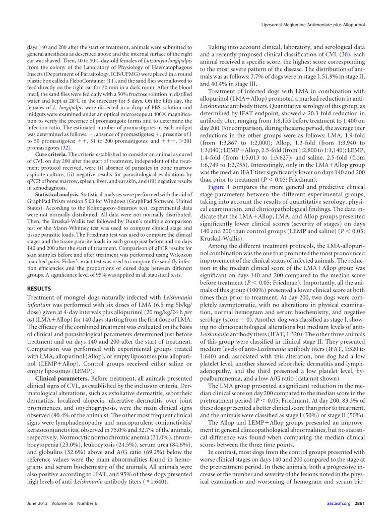
Figure 1 compares the more general and predictive clinicalstage parameters between the different experimental groups,taking into account the results of quantitative serology, physi-cal examination, and clinicopathological findings. The data in-dicate that the LMA�Allop, LMA, and Allop groups presentedsignificantly lower clinical scores (severity of stages) on days140 and 200 than control groups (LEMP and saline) (P 0.05;Kruskal-Wallis).
Among the different treatment protocols, the LMA-allopuri-nol combination was the one that promoted the most pronouncedimprovement of the clinical status of infected animals. The reduc-tion in the median clinical score of the LMA�Allop group wassignificant on days 140 and 200 compared to the median scorebefore treatment (P 0.05; Friedman). Importantly, all the ani-mals of this group (100%) presented a lower clinical score at bothtimes than prior to treatment. At day 200, two dogs were com-pletely asymptomatic, with no alterations in physical examina-tion, normal hemogram and serum biochemistry, and negativeserology (score � 0). Another dog was classified as stage I, show-ing no clinicopathological alterations but medium levels of anti-Leishmania antibody titers (IFAT, 1:320). The other three animalsof this group were classified in clinical stage II. They presentedmedium levels of anti-Leishmania antibody titers (IFAT, 1:320 to1:640) and, associated with this alteration, one dog had a lowplatelet level, another showed seborrheic dermatitis and lymph-adenopathy, and the third presented a low platelet level, hy-poalbuminemia, and a low A/G ratio (data not shown).
The LMA group presented a significant reduction in the me-dian clinical score on day 200 compared to the median score in thepretreatment period (P 0.05; Friedman). At day 200, 83.3% ofthese dogs presented a better clinical score than prior to treatment,and the animals were classified as stage I (50%) or stage II (50%).
The Allop and LEMP�Allop groups presented an improve-ment in general clinicopathological abnormalities, but no statisti-cal difference was found when comparing the median clinicalscores between the three time points.
In contrast, most dogs from the control groups presented withworse clinical stages on days 140 and 200 compared to the stage atthe pretreatment period. In these animals, both a progressive in-crease of the number and severity of the lesions noted in the phys-ical examination and worsening of hemogram and serum bio-
Liposomal Meglumine Antimoniate plus Allopurinol
June 2012 Volume 56 Number 6 aac.asm.org 2861

chemistry parameters were observed. Saline-treated animalspresented a significantly greater median clinical score on day 200than before treatment (P 0.05; Friedman).
Parasitological evaluations in bone marrow and spleen. Par-asitological evaluation of dogs by qPCR indicated that the LMA-allopurinol combined treatment was the most effective protocolfor reducing the parasite burden in bone marrow and spleen. Asillustrated in Fig. 2, this combination promoted a significant re-duction in bone marrow parasite loads on days 140 and 200 com-pared to the results of the LEMP�Allop, LEMP, and saline proto-cols (P 0.05; Kruskal-Wallis). Similarly, the LMA�Allop grouppresented with significantly lower parasite loads in the spleen ondays 140 and 200 than the LEMP and saline groups (P 0.05;Kruskal-Wallis) (Fig. 2).
The effectiveness of the LMA-allopurinol combination for re-ducing the parasite burden in the bone marrow and spleen wasalso evident when the parasite loads were compared before and ondays 140 and 200 after the start of treatment. The parasite burdensin both tissues were significantly reduced on days 140 and 200compared to the burdens in the pretreatment period (P 0.05;Friedman). On day 140, five animals (83.3%) were negative byqPCR in both the bone marrow and spleen. At the end of theexperimental period (day 200), three of these animals remainednegative in both the spleen and bone marrow. Another dog wasstill negative only in the spleen. The median numbers of parasitesin bone marrow and spleen aspirates, as determined by qPCR onday 140, were about 770 and 245 times lower, respectively, thanbefore treatment. This substantial reduction in the parasite loadwas still observed at the end of the experimental period (parasiteburdens were about 660 and 104 times lower, respectively).
The LMA and Allop groups showed results similar to those forthe LMA�Allop group with respect to the reduction of parasiteloads in spleen and bone marrow on day 140 (Fig. 2). However, onday 200, treatment with LMA or allopurinol (with or withoutempty liposomes) did not show a significant reduction of the bonemarrow parasite burden compared to that in the pretreatmentperiod, confirming the inability of these drugs alone to maintainlow parasite levels in this tissue after interruption of treatment.
Parasitological evaluations in the liver. The parasite loads inthe liver of treated dogs on day 200 after the start of treatment weredetermined by qPCR. Significant parasite suppression in the liverof dogs in comparison to the parasite suppression in controlgroups was achieved in the groups that received LMA (with orwithout allopurinol) but not in those receiving allopurinol with-out LMA. All animals (100%) treated with the LMA-allopurinolcombination presented negative qPCR results. As shown in Fig. 3,the parasite burdens in the livers of the LMA�Allop group weresignificantly lower than the parasite burdens in the LEMP andsaline groups (P 0.05; Kruskal-Wallis). In the LMA group, fiveanimals (87.3%) were found to be negative and the median num-ber of parasites in this organ was significantly lower than that inthe saline group (P 0.05; Kruskal-Wallis).
Parasitological evaluations in the skin. Parasitological evalua-FIG 1 Clinical staging of dogs naturally infected with Leishmania (L.) infan-tum before and after treatment with liposomal meglumine antimoniate(LMA), allopurinol (Allop), empty liposomes (LEMP), or the LMA�Allop orLEMP�Allop combination. (A) Staging prior to treatment. (B and C) Stagingon days 140 and 200, respectively, after the start of treatment. LMA (6.5 mgSb/kg/dose), empty liposomes (same dose of lipid), and saline (same volume)were given intravenously as six doses at 4-day intervals. Allopurinol was givenat 20 mg/kg/24 h per os for 140 days. Clinical scores are defined in detail inTable 1. Score 0, absence of clinical signs and clinicopathological alterationssuggestive of CVL and negative serology; score 1, mild clinical stage; score 2,
moderate stage; score 3, severe stage; score 4, extremely severe disease stage.Data are shown as dot plots, and lines correspond to the median of each group(n � 6 to 11). *(e,f) and *(f), P 0.05 according to Kruskal-Wallis test fol-lowed by Dunn’s multiple comparison test, for comparison with LEMP (e)and/or saline (f).
da Silva et al.
2862 aac.asm.org Antimicrobial Agents and Chemotherapy

tion was carried out by qPCR in the ear skin of dogs before treatmentand on day 200 after the start of treatment. No statistical differencewas observed between the six groups in the median number of para-sites in the ear skin on day 200 (P 0.05; Kruskal-Wallis). Neverthe-less, as illustrated in Fig. 4, the LMA�Allop group was the only groupto show a significantly lower parasite load than the saline group (P 0.05; Mann-Whitney). Furthermore, the parasite loads in the skin of
animals from the LMA�Allop, LMA, and LEMP�Allop groups weresignificantly lower on day 200 than before treatment (P 0.05; Wil-coxon matched pairs). Prior to treatment, one animal (16.7%) fromthe LMA�Allop group was negative according to skin qPCR. At day200, the proportion of skin-negative dogs from this group was 83.3%,corresponding to the highest proportion of negative dogs among allexperimental groups. In the dog whose qPCR remained positive, the
FIG 2 Parasite burdens in the bone marrow (A, B, and C) and spleen (D, E, and F) of dogs naturally infected with Leishmania (L.) infantum before and aftertreatment with liposomal meglumine antimoniate (LMA), allopurinol (Allop), empty liposomes (LEMP), or the LMA�Allop or LEMP�Allop combination.Parasite burdens prior to treatment (A and D), at day 140 (B and E), and at day 200 (C and F) are shown. LMA (6.5 mg Sb/kg/dose), empty liposomes (same doseof lipid), and saline (same volume) were given intravenously as six doses at 4-day intervals. Allopurinol was given at 20 mg/kg/24 h per os for 140 days, startingfrom the first dose of LMA. Parasite burdens were determined by qPCR as described in Materials and Methods. Data are shown as dot plots, and lines correspondto the median of each group (n � 6 to 11). *(d,e,f), *(e,f), *(e), and *(f), P 0.05 according to Kruskal-Wallis test followed by Dunn’s multiple comparison test,for comparison with LEMP�Allop (d), LEMP (e), and/or saline (f).
Liposomal Meglumine Antimoniate plus Allopurinol
June 2012 Volume 56 Number 6 aac.asm.org 2863

median number of parasites decreased about 314-fold (from 69,776amastigotes prior to treatment to 222 parasites on day 200).
Xenodiagnosis. The effect of the treatment of dogs on theirinfectivity to sand flies was investigated through xenodiagnosis inthe different experimental groups just before and on days 140 and200 after the start of treatment, using females of Lutzomyia longi-palpis. As shown in Table 2, the LMA�Allop and Allop groupsexhibited the best results in the xenodiagnosis evaluations.
In the LMA�Allop group, three animals (50%) infected sandflies just before treatment, with 38% infection efficiency (given bythe ratio of infected to fed sand flies in positive dogs), whereasxenodiagnosis was negative in all animals (100%) on days 140 and200. As shown in Table 2, significant reductions in the proportionsof infected sand flies were observed on both day 140 and 200
compared to the proportions in the pretreatment period (P 0.05; Fisher’s exact test).
Among the other groups, the Allop but not the LMA andLEMP�Allop groups showed 100% noninfective dogs on day 200.In the LMA, Allop, and LEMP�Allop groups, significant reduc-tions in the proportions of infected sand flies on days 140 and 200were also observed compared to the proportions in the pretreat-ment period (P 0.05; Fisher’s exact test) (Table 2). This is incontrast with the LEMP and saline groups, in which the propor-tions of infected sand flies were significantly increased on days 140and 200 compared to the proportions in the pretreatment period.
Parasitological cure. Each animal was evaluated on day 200using different parasitological tests, including culture from bonemarrow aspirates, qPCR of bone marrow, spleen, liver, and skinsamples, and xenodiagnosis. Animals that showed negative resultsin all the parasitological tests were considered cured of CVL. Ac-cording to these cure criteria, the LMA-allopurinol combinationpromoted parasitological cure in 50% of treated animals. An ad-ditional immunohistochemistry test was performed, as describedpreviously (28), in those tissues tested by qPCR. Strikingly, nega-tive results were obtained in all samples collected from the sixanimals of this group (data not shown). Among the other fiveexperimental groups, parasitological cure was achieved in onlyone animal that was treated with allopurinol.
As illustrated in Fig. 5, the proportion of cured dogs achievedafter treatment with the LMA-allopurinol combination was sig-nificantly greater than that in the saline group (P 0.05; Fisher’sexact test).
DISCUSSION
With the aim of further improving the treatment efficacy of CVLwith an innovative liposomal formulation of MA, a new therapeu-tic regimen was designed which combines this formulation withallopurinol, a well-tolerated and effective leishmaniostatic drugcapable of reaching all infected sites. In comparison to our previ-ous protocol (28), LMA was given at the same dosage (6.5 mgSb/kg/dose) and time intervals, but animals received six dosesinstead of four. Thus, a more effective protocol was also expectedfrom the more prolonged treatment with LMA. The protocol used
FIG 3 Parasite burdens in the liver of dogs naturally infected with Leishmania(L.) infantum after treatment with liposomal meglumine antimoniate (LMA),allopurinol (Allop), empty liposomes (LEMP), or the LMA�Allop orLEMP�Allop combination. LMA (6.5 mg Sb/kg/dose), empty liposomes(same dose of lipid), and saline (same volume) were given intravenously as sixdoses at 4-day intervals. Allopurinol was given at 20 mg/kg/24 h per os for 140days, starting from the first dose of LMA. Parasite burdens were determined byqPCR as described in Materials and Methods. Data are shown as dot plots, andlines correspond to the median of each group (n � 6 to 11). *(e,f) and *(f), P 0.05 according to Kruskal-Wallis test followed by Dunn’s multiple comparisontest, for comparison with LEMP (e) and/or saline (f).
FIG 4 Parasite burdens in the ear skin of dogs naturally infected with Leishmania (L.) infantum before and after treatment with liposomal meglumineantimoniate (LMA), allopurinol (Allop), empty liposomes (LEMP), or the LMA�Allop or LEMP�Allop combination. Parasite burdens prior to treatment (A)and at day 200 (B) are shown. LMA (6.5 mg Sb/kg/dose), empty liposomes (same dose of lipid), and saline (same volume) were given intravenously as six dosesat 4-day intervals. Allopurinol was given at 20 mg/kg/24 h per os for 140 days, starting from the first dose of LMA. Data are shown as dot plots, and linescorrespond to the median of each group (n � 6 to 11). *(f), P 0.05 for comparison with saline group according to the Mann-Whitney test, for comparison withsaline (f).
da Silva et al.
2864 aac.asm.org Antimicrobial Agents and Chemotherapy

for the administration of allopurinol (20 mg/kg/24 h) differedslightly from the conventional one in that a frequency of dosing of12 h was used (15, 20). Animals also remained without any ther-apeutic intervention from day 140 to 200, to uncover possiblerelapse of CVL.
Analysis of the clinical, parasitological, and xenodiagnosisfindings indicates at least an additive effect for LMA and allopuri-nol, since animals treated with this protocol presented much bet-
ter clinical and parasitological profiles than those treated witheach drug alone.
Among the six experimental groups, the LMA�Allop groupwas the one that exhibited the most pronounced improvement ofclinical signs and clinicopathological parameters. However, theimprovements were gradual and improvements were also ob-served in the groups treated with LMA or allopurinol alone. Dur-ing the experimental period, most of the animals (72% � 19%)treated with these protocols showed weight gain, complete regres-sion or marked reduction in the number and degree of skin le-sions, absence of lymphadenopathy and eye lesions, and altera-tions in the color of the mucous membranes (data not shown).Improvement in the clinical condition was observed until day 140and was also accompanied by the normalization of most hemo-gram and serum biochemistry parameters, such as protein elec-trophoresis, A/G ratio, number of erythrocytes, hemoglobin con-centration, hematocrit, and platelets (data not shown).
From day 140 to the end of the experimental period, a tendencytoward improvement was observed only in the LMA�Allopgroup, suggesting that, besides reversion of the physical and clin-icopathological abnormalities, the drug combination promoted along-term action resulting in the prevention of disease relapse. Incontrast, the LMA, Allop, and LEMP�Allop groups presented aslight worsening of their clinical status from day 140 to 200. Thisfact can be attributed to the inability of allopurinol to preventrelapse after interruption of its use (10, 27) and to the transitorypositive effect of LMA on the same parameters, as previously de-scribed by our group (28).
The clinical improvement observed in the LMA�Allop groupwas accompanied by a significant reduction in the parasitic load,as determined by qPCR. The choice of qPCR to assess the parasiteburdens in the tissues of the dogs was based on the high sensitivityand specificity of the technique for the absolute quantification ofparasites (25). The LMA-allopurinol combination had the great-est impact on parasite burden, reducing by hundreds of times thenumbers of parasites in bone marrow, spleen, and skin followingtreatment.
TABLE 2 Frequency of positive dogs in xenodiagnosis and proportion and intensity of infection of Lutzomyia longipalpis fed on dogs naturallyinfected with Leishmania (L.) infantum before and after treatment
Treatment groupa
Frequency of positive dogs inxenodiagnosis (%)b
Proportion of infected sand flies(%)c Intensity of infection (%)d
Prior totreatment Day 140 Day 200
Prior totreatment Day 140 Day 200
Prior to treatment Day 140 Day 200
� �� ��� � �� ��� � �� ���
LMA�Allop 50 0 0 19.1 0e 0e 42.5 40 17.5 0 0 0 0 0 0LMA 50 16.7 33.3 11.9 1.4e 1.9e 28 12 60 33.3 66.7 0 75 25 0Allop 50 0 0 35.0 0e 0e 11.2 11.2 77.6 0 0 0 0 0 0LEMP�Allop 62.5 0 12.5 34.3 0e 2.5e 22.9 24 53.1 0 0 0 28.6 57.1 14.3LEMP 33.3 33.3 50 16.7 26.2f 30.0f 28.6 22.9 48.5 23.7 14.5 61.8 22.2 20.6 57.2Saline 36.4 45.5 63.6 3.9 11.4f 16.4f 46.7 33.3 20 45.8 25 29.2 41.6 27.7 30.7a Liposomal meglumine antimoniate (LMA; 6.5 mg Sb/kg/dose), empty liposomes (LEMP; same dose of lipid), and saline (same volume) were given intravenously as six doses at4-day intervals; allopurinol (Allop) was given at 20 mg/kg/24 h per os for 140 days, starting from the first dose of LMA.b Proportions of dogs in each group whose promastigotes were identified in the midgut of Lutzomyia longipalpis females 5 days after their blood meal on the internal surface of theright ear of these animals.c Proportions of infected Lutzomyia longipalpis females in relation to total numbers of insects dissected 5 days after their blood meal on each experimental group of dogs.d Distribution of midgut infections according to the estimated numbers of promastigotes, categorized as follows: �, 1 to 50 promastigotes; ��, 51 to 200 promastigotes; and���, 201 promastigotes.e P 0.05 according to Fisher’s exact test, showing a significantly lower proportion of infected sand flies than in the pretreatment period.f P 0.05 according to Fisher’s exact test, showing a significantly greater proportion of infected sand flies than in the pretreatment period.
FIG 5 Proportions of dogs cured of Leishmania (L.) infantum infection aftertreatment with liposomal meglumine antimoniate (LMA), allopurinol (Al-lop), empty liposomes (LEMP), or the LMA�Allop or LEMP�Allop combi-nation. LMA (6.5 mg Sb/kg/dose), empty liposomes (same dose of lipid), andsaline (same volume) were given intravenously as six doses at 4-day intervals.Allopurinol was given at 20 mg/kg/24 h per os for 140 days, starting from thefirst dose of LMA. Dogs were considered cured when they showed negativeresults in all the parasitological tests performed on day 200, including culturefrom bone marrow aspirates, qPCR of bone marrow, spleen, liver, and skin,and xenodiagnosis. *, P 0.05 according to Fisher’s exact test.
Liposomal Meglumine Antimoniate plus Allopurinol
June 2012 Volume 56 Number 6 aac.asm.org 2865

Importantly, treatment with the drug combination resulted insignificant decreases in the parasite loads in the bone marrow andspleen on days 140 and 200 compared to the parasite loads in thepretreatment period. This is in contrast with the other groups,which did not show significant reductions in the bone marrowparasite loads on day 200. Furthermore, the drug combinationreduced the parasite loads in bone marrow to a significantlygreater degree than the LEMP-allopurinol protocol. These results,taken together, indicate at least additive effects of LMA and allo-purinol.
The importance of the use of splenic aspirates in monitoringtreatment was previously demonstrated in dogs infected with Eh-rlichia canis treated with doxycycline (19). To the best of ourknowledge, the present study is the first to evaluate the efficacy oftreatment of CVL according to the parasite burden in the spleenby using qPCR prior to and during treatment. The use of an ultra-sound device to guide the aspiration of the spleen allowed the safecollection of samples from this organ without complications suchas hemorrhages or ruptures. Given the importance of the spleen inthe context of CVL (31), evaluation of the parasite burden in thisorgan during treatment could help in monitoring relapses andresponse to therapeutics.
A major benefit of LMA-allopurinol combination is its abilityto produce negative qPCR results in the liver of all treated dogs.This remarkable effect is most probably due to the extremely highaccumulation of antimony in the liver promoted by the liposomeformulation (29). In accordance with this interpretation, the LMAgroup showed only one animal (18.7%) with positive qPCR ofliver tissue, whereas the Allop and LEMP�Allop groups presented37.5 and 62.5% positive animals.
Because of their hematophagic behavior, sand flies need directcontact with the skin of vertebrate hosts. The skin of dogs is the sitewhere transmission of parasites occurs, both from infected dogs tononinfected sand flies and from infected sand flies to noninfecteddogs, which spreads the disease to other dogs and humans (33).
Therefore, one of the most important objectives of the treat-ment of CVL is the blockade of transmission to sand flies by elim-inating parasites in the skin or, at least, reducing the number ofparasites to such a level that transmission does not occur (24).Thus, some authors (24) have proposed reduction in the infectiv-ity of dogs to sand flies through treatment as a key measure incontrol programs designed to eradicate active foci of CVL.
In our study, the capacity of L. longipalpis to be infected with L.infantum after feeding in infected dogs was investigated throughxenodiagnosis. Although it is the most accurate experimental toolfor assessing the epidemiologic impact of treatment of CVL, xe-nodiagnosis is not a simple methodology and basically is restrictedto research institutions (11, 24). Since our data show a positivecorrelation between the xenodiagnosis result and the number ofparasites in the ear skin (Spearman’s correlation, 0.6518; P 0.0001), in accordance with a recent study (5), skin qPCR may beused as an alternative protocol to assess the infectivity of dogs tothe sand flies.
Both the xenodiagnosis and skin parasite load results showedthat the LMA-allopurinol combination was the most effectiveprotocol for inhibiting the transmission of skin parasites to L.longipalpis. Indeed, it promoted the blockade of transmission ofparasites to sand flies on both days 140 and 200 and resulted in thehighest percentage (83.3%) of dogs negative according to skinqPCR. This is in contrast with the results obtained with the LMA
and LEMP�Allop groups, which both presented infective animalson day 200 and lower percentages of negative dogs (50 and 25%,respectively).
An important question to be answered is whether LMA-allo-purinol combined treatment is capable of promoting parasitolog-ical cure of infected dogs. This is a crucial question for the controlof visceral leishmaniasis, since no fully effective treatment hasbeen reported so far (2, 30) and an effective treatment would blockthe transmission of the parasite to sand flies and humans andreduce the risk of emergence of resistance to antimony. Based onthe absence of parasites in bone marrow, spleen, liver, and ear skinand negative xenodiagnosis on day 200, the LMA-allopurinolcombination was found to promote parasitological cure in 50% ofthe dogs treated. In the other groups, cured animals were foundonly in the Allop group, but at a much lower rate (8%). Thesefindings confirm our previous report that LMA alone cannot pro-mote parasitological cure (28).
The criteria used here for parasitological cure are based on theabsence of parasites in critical sites of infection and on the block-ade of parasite transmission to the sand flies at a specific time.Importantly, this evaluation was performed in treated dogs after a60-day period without treatment, to allow the occurrence of pos-sible relapses. Thus, in the LMA�Allop group, two of five animalsinitially negative in bone marrow and spleen (on day 140) becamepositive on day 200. In this context, our claim for achievement ofparasitological cure should be taken cautiously, since not all tis-sues were evaluated and one cannot completely exclude the pos-sibility of other relapses after a longer period of time withouttreatment.
As a major advance, the present study displays for the first timea new, highly effective therapeutic alternative for CVL, based onthe combination of nanotechnology and conventional therapy,with the prospect of achieving parasitological cure. In future stud-ies, new protocols based on this combination should be designedto confirm the achievement of cure in treated dogs and to furtherenhance the cure rate of CVL. A higher cure rate may be expectedfrom an increase in the number of doses of LMA, from the use ofallopurinol for a more prolonged period of time, and from theimprovement of the liposomal formulation for more effectivelyreaching less accessible sites of infection.
ACKNOWLEDGMENTS
We acknowledge the Brazilian agencies CNPq (grants 303046/2009-0,473534/2010-0, and 473601/2009-5), FAPEMIG (grants REDE–221/08,REDE– 40/11, APQ– 01935-09, APQ– 01355-09, PRONEX 2009, andPPM– 00382-11), and CAPES for financial support. R.R.R. was the recip-ient of a postdoctoral fellowship from FAPEMIG.
We are also grateful to Oscar Bruna-Romero from ICB-UFMG for hishelpful assistance with qPCR assays.
REFERENCES1. Alvar J, Canavate C, Molina R, Moreno J, Nieto J. 2004. Canine
leishmaniasis. Adv. Parasitol. 57:1– 88.2. Alvar J, et al. 1994. Canine leishmaniasis: clinical, parasitological and
entomological follow-up after chemotherapy. Ann. Trop. Med. Parasitol.88:371–378.
3. Alves CF, et al. 2009. Expression of IFN-gamma, TNF-alpha, IL-10 andTGF-beta in lymph nodes associates with parasite load and clinical form ofdisease in dogs naturally infected with Leishmania (Leishmania) chagasi.Vet. Immunol. Immunopathol. 128:349 –358.
4. Alving C. 1986. Liposomes as drug carriers in leishmaniasis and malaria.Parasitol. Today 2:101–107.
da Silva et al.
2866 aac.asm.org Antimicrobial Agents and Chemotherapy

5. Amorim IF, et al. 2011. Toll receptors type-2 and CR3 expression ofcanine monocytes and its correlation with immunohistochemistry andxenodiagnosis in visceral leishmaniasis. PLoS One 6:1–10.
6. Balana-Fouce R, Reguera RM, Cubria JC, Ordonez D. 1998. The phar-macology of leishmaniasis. Gen. Pharmacol. 30:435– 443.
7. Baneth G, Koutinas AF, Solano-Gallego L, Bourdeau P, Ferrer L. 2008.Canine leishmaniasis—new concepts and insights on an expanding zoo-nosis: part one. Trends Parasitol. 24:324 –330.
8. Baneth G, Shaw SE. 2002. Chemotherapy of canine leishmaniosis. Vet.Parasitol. 106:315–324.
9. Bretagne S, et al. 2001. Real-time PCR as a new tool for quantifyingLeishmania infantum in liver in infected mice. Clin. Diagn. Lab. Immunol.8:828 – 831.
10. Cavaliero T, et al. 1999. Clinical, serologic, and parasitologic follow-upafter long-term allopurinol therapy of dogs naturally infected with Leish-mania infantum. J. Vet. Intern. Med. 13:330 –334.
11. da Costa-Val AP, et al. 2007. Canine visceral leishmaniasis: relationshipsbetween clinical status, humoral immune response, haematology and Lut-zomyia (Lutzomyia) longipalpis infectivity. Vet. J. 174:636 – 643.
12. da Silva SM, et al. 2010. First report of infection of Lutzomyia longipalpisby Leishmania (Leishmania) infantum from a naturally infected cat ofBrazil. Vet. Parasitol. 174:150 –154.
13. da Silva SM, et al. 2009. First report of vertical transmission of Leishma-nia (Leishmania) infantum in a naturally infected bitch from Brazil. Vet.Parasitol. 166:159 –162.
14. Demicheli C, et al. 2003. Pentavalent organoantimonial derivatives: twosimple and efficient synthetic methods for meglumine antimonate. Appl.Organomet. Chem. 17:226 –231.
15. Denerolle P, Bourdoiseau G. 1999. Combination allopurinol and anti-mony treatment versus antimony alone and allopurinol alone in the treat-ment of canine leishmaniasis (96 cases). J. Vet. Intern. Med. 13:413– 415.
16. Frézard F, Demicheli C. 2010. New delivery strategies for the old penta-valent antimonial drugs. Expert Opin. Drug Deliv. 7:1343–1358.
17. Frézard F, Demicheli C, Ribeiro RR. 2009. Pentavalent antimonials: newperspectives for old drugs. Molecules 14:2317–2336.
18. Frézard F, Michalick MS, Soares CF, Demicheli C. 2000. Novel methodsfor the encapsulation of meglumine antimoniate into liposomes. Braz. J.Med. Biol. Res. 33:841– 846.
19. Harrus S, et al. 2004. Comparison of simultaneous splenic sample PCRwith blood sample PCR for diagnosis and treatment of experimental Eh-rlichia canis infection. Antimicrob. Agents Chemother. 48:4488 – 4490.
20. Koutinas AF, et al. 2001. A randomised, blinded, placebo-controlledclinical trial with allopurinol in canine leishmaniosis. Vet. Parasitol. 98:247–261.
21. Maia C, Campino L. 2008. Methods for diagnosis of canine leishmaniasisand immune response to infection. Vet. Parasitol. 158:274 –287.
22. Martinez S, Looker DL, Berens RL, Marr JJ. 1988. The synergistic actionof pyrazolopyrimidines and pentavalent antimony against Leishmaniadonovani and L. braziliensis. Am. J. Trop. Med. Hyg. 39:250 –255.
23. Miró G, Cardoso L, Pennisi MG, Oliva G, Baneth G. 2008. Canineleishmaniosis—new concepts and insights on an expanding zoonosis: parttwo. Trends Parasitol. 24:371–377.
24. Miró G, Galvez R, Fraile C, Descalzo MA, Molina R. 2011. Infectivity toPhlebotomus perniciosus of dogs naturally parasitized with Leishmania in-fantum after different treatments. Parasit. Vectors 4:52.
25. Mortarino M, et al. 2004. Quantitative PCR in the diagnosis of Leishma-nia. Parassitologia 46:163–167.
26. National Institutes of Health. 1985. Principles of laboratory animal care.NIH publication 85-23. National Institutes of Health, Bethesda, MD.
27. Noli C, Auxilia ST. 2005. Treatment of canine Old World visceral leish-maniasis: a systematic review. Vet. Dermatol. 16:213–232.
28. Ribeiro RR, et al. 2008. Reduced tissue parasitic load and infectivity tosand flies in dogs naturally infected by Leishmania (Leishmania) chagasifollowing treatment with a liposome formulation of meglumine antimo-niate. Antimicrob. Agents Chemother. 52:2564 –2572.
29. Schettini DA, et al. 2006. Improved targeting of antimony to the bone mar-row of dogs using liposomes of reduced size. Int. J. Pharm. 315:140–147.
30. Solano-Gallego L, et al. 2009. Directions for the diagnosis, clinical stag-ing, treatment and prevention of canine leishmaniosis. Vet. Parasitol. 165:1–18.
31. Strauss-Ayali D, Jaffe CL, Burshtain O, Gonen L, Baneth G. 2004.Polymerase chain reaction using noninvasively obtained samples, for thedetection of Leishmania infantum DNA in dogs. J. Infect. Dis. 189:1729 –1733.
32. Travi BL, Ferro C, Cadena H, Montoya-Lerma J, Adler GH. 2002.Canine visceral leishmaniasis: dog infectivity to sand flies from non-endemic areas. Res. Vet. Sci. 72:83– 86.
33. World Health Organization. 2010. Control of the leishmaniases, p 201.Report of a meeting of the WHO Expert Committee on the Control ofLeishmaniases, Geneva, 22–26 March 2010. Technical report series 949.World Health Organization, Geneva, Switzerland.
Liposomal Meglumine Antimoniate plus Allopurinol
June 2012 Volume 56 Number 6 aac.asm.org 2867