Double-walled microspheres loaded with meglumine antimoniate: preparation, characterization and in...
10
2013 http://informahealthcare.com/ddi ISSN: 0363-9045 (print), 1520-5762 (electronic) Drug Dev Ind Pharm, Early Online: 1–10 ! 2013 Informa Healthcare USA, Inc. DOI: 10.3109/03639045.2013.777734 RESEARCH ARTICLE Double-walled microspheres loaded with meglumine antimoniate: preparation, characterization and in vitro release study Ali Navaei 1 , Morteza Rasoolian 1 , Arash Momeni 1,2 , Shahriar Emami 1 , and Mohammad Rafienia 3 1 Biomedical Engineering Department, Amirkabir University of Technology, Tehran, Iran, 2 School of Biomedical Engineering, Dalhousie University, Halifax, Nova Scotia, Canada, and 3 Biosensor Research Center, Isfahan University of Medical Sciences, Isfahan, Iran Abstract Objective: The objective of this study was to fabricate double-walled poly(lactide-co-glycolide) (PLGA) microspheres to increase encapsulation efficiency and avoid rapid release of hydrophilic drugs such as meglumine antimoniate. Methods: In this study, double-walled and one-layered microspheres of PLGA were prepared using the emulsion solvent evaporation technique to better control the release of a hydrophilic drug, meglumine antimoniate (Glucantime Õ ), which is the first choice treatment of cutaneous leishmaniasis. The effect of hydrophobic coating on microspheres’ size, morphology, encapsula- tion efficiency and drug release characteristics was evaluated. Furthermore, the presence of antimony in meglumine antimoniate made it possible to observe the drug distribution within the microspheres’ cross section by means of energy dispersive X-ray spectroscopy. Results: Drug distribution images confirmed accumulation of the drug within the inner core of double-walled microspheres. In addition, these microspheres encapsulated the drug more efficiently up to 87% and demonstrated reduced initial burst and prolonged release compared to one-layered microspheres. These superiorities make double-walled microspheres an optimum candidate for sustained delivery of hydrophilic drugs. Conclusion: Double-walled microspheres provide some advantages over traditional micro- spheres overcoming most of their limitations. Double-walled microspheres were found to be more efficient than their corresponding one-layered microspheres in terms of encapsulation efficiencies and release characteristics. Keywords Degradation rate, double emulsion, drug distribution, EDX, poly(lactide-co-glycolide) History Received 23 July 2012 Revised 24 January 2013 Accepted 13 February 2013 Published online 17 April 2013 Introduction Developing efficient drug delivery systems with appropriate release characteristics has always been one of the principal concerns of researchers 1 . Sustained drug release enhances thera- peutic effects and eventually leads to more clinical success. Biodegradable polymeric microspheres are one of the best choices for this objective because of their capabilities for being injected and adjusting release rate 2 . Poly (lactide-co-glycolide) (PLGA) has been widely used to fabricate biodegradable microspheres due to its biocompatibility, biodegradability and ability to encapsulate vari- ous therapeutic agents such as hydrophilic and hydrophobic drugs, DNA, proteins, etc. 3 However, there are some common problems associated with conventional one-layered PLGA microspheres loaded with hydro- philic substances, including high initial burst, short release period, low encapsulation efficiency (%EE) 4,5 , inability to provide zero- order release and lack of delayed or pulsatile release 6,7 . Double- walled microspheres have been investigated as an alternative to overcome these problems 3,6,8 . Employing a polymeric shell with less drug content not only prevents the high initial burst, but also makes it possible to obtain more advanced release schedules such as delayed or pulsatile release by altering the shell material or thickness 4 . In the case of PLGA, drug release may take place as a result of two processes: drug diffusion and/or polymer degrad- ation by homogeneous bulk erosion 1,9 . The rate of diffusion is dependent on parameters like diffusion and partition coefficient, which are influenced by drug physico-chemical properties such as molecular size, hydrophilicity, charge, as well as polymer characteristics 1 . Lower affinity to drug and lower degradation rate of the shell compared to the PLGA core increases the %EE and release period by disturbing the mentioned mechanisms. The outer layer acts as a barrier inhibiting the drug from leaking out during the preparation procedure 3 . In addition, proteins, DNA and other delicate substances could be encapsulated within the inner core while degradation or denaturation is avoided by generating an appropriate medium in the core 4 . Over the last decades, different methods have been developed to prepare double-walled microspheres 10,11 . Coating processes involving the use of fluidized beds have been employed to fabricate double-walled microspheres 12 . Mathiowitz et al. mod- ified this method to overcome its disadvantages including large particle size 13,14 . They also developed a one-step preparation method for fabricating double-walled microspheres based on the Address for correspondence: Morteza Rasoolian, Biomedical Engineering Department, Amirkabir University of Technology, Tehran, Iran. E-mail: [email protected] Drug Development and Industrial Pharmacy Downloaded from informahealthcare.com by Dalhousie University on 04/18/13 For personal use only.
Transcript of Double-walled microspheres loaded with meglumine antimoniate: preparation, characterization and in...

2013
http://informahealthcare.com/ddiISSN: 0363-9045 (print), 1520-5762 (electronic)
Drug Dev Ind Pharm, Early Online: 1–10! 2013 Informa Healthcare USA, Inc. DOI: 10.3109/03639045.2013.777734
RESEARCH ARTICLE
Double-walled microspheres loaded with meglumine antimoniate:preparation, characterization and in vitro release study
Ali Navaei1, Morteza Rasoolian1, Arash Momeni1,2, Shahriar Emami1, and Mohammad Rafienia3
1Biomedical Engineering Department, Amirkabir University of Technology, Tehran, Iran, 2School of Biomedical Engineering, Dalhousie University,
Halifax, Nova Scotia, Canada, and 3Biosensor Research Center, Isfahan University of Medical Sciences, Isfahan, Iran
Abstract
Objective: The objective of this study was to fabricate double-walled poly(lactide-co-glycolide)(PLGA) microspheres to increase encapsulation efficiency and avoid rapid release of hydrophilicdrugs such as meglumine antimoniate.Methods: In this study, double-walled and one-layered microspheres of PLGA were prepared usingthe emulsion solvent evaporation technique to better control the release of a hydrophilic drug,meglumine antimoniate (Glucantime�), which is the first choice treatment of cutaneousleishmaniasis. The effect of hydrophobic coating on microspheres’ size, morphology, encapsula-tion efficiency and drug release characteristics was evaluated. Furthermore, the presence ofantimony in meglumine antimoniate made it possible to observe the drug distribution within themicrospheres’ cross section by means of energy dispersive X-ray spectroscopy.Results: Drug distribution images confirmed accumulation of the drug within the inner core ofdouble-walled microspheres. In addition, these microspheres encapsulated the drug moreefficiently up to 87% and demonstrated reduced initial burst and prolonged release comparedto one-layered microspheres. These superiorities make double-walled microspheres anoptimum candidate for sustained delivery of hydrophilic drugs.Conclusion: Double-walled microspheres provide some advantages over traditional micro-spheres overcoming most of their limitations. Double-walled microspheres were found to bemore efficient than their corresponding one-layered microspheres in terms of encapsulationefficiencies and release characteristics.
Keywords
Degradation rate, double emulsion, drugdistribution, EDX, poly(lactide-co-glycolide)
History
Received 23 July 2012Revised 24 January 2013Accepted 13 February 2013Published online 17 April 2013
Introduction
Developing efficient drug delivery systems with appropriaterelease characteristics has always been one of the principalconcerns of researchers1. Sustained drug release enhances thera-peutic effects and eventually leads to more clinical success.Biodegradable polymeric microspheres are one of the best choicesfor this objective because of their capabilities for being injected andadjusting release rate2. Poly (lactide-co-glycolide) (PLGA) hasbeen widely used to fabricate biodegradable microspheres due to itsbiocompatibility, biodegradability and ability to encapsulate vari-ous therapeutic agents such as hydrophilic and hydrophobic drugs,DNA, proteins, etc.3
However, there are some common problems associated withconventional one-layered PLGA microspheres loaded with hydro-philic substances, including high initial burst, short release period,low encapsulation efficiency (%EE)4,5, inability to provide zero-order release and lack of delayed or pulsatile release6,7. Double-walled microspheres have been investigated as an alternative toovercome these problems3,6,8. Employing a polymeric shell with
less drug content not only prevents the high initial burst, but alsomakes it possible to obtain more advanced release schedules suchas delayed or pulsatile release by altering the shell material orthickness4. In the case of PLGA, drug release may take place as aresult of two processes: drug diffusion and/or polymer degrad-ation by homogeneous bulk erosion1,9. The rate of diffusion isdependent on parameters like diffusion and partition coefficient,which are influenced by drug physico-chemical properties such asmolecular size, hydrophilicity, charge, as well as polymercharacteristics1. Lower affinity to drug and lower degradationrate of the shell compared to the PLGA core increases the %EEand release period by disturbing the mentioned mechanisms. Theouter layer acts as a barrier inhibiting the drug from leaking outduring the preparation procedure3. In addition, proteins, DNA andother delicate substances could be encapsulated within the innercore while degradation or denaturation is avoided by generatingan appropriate medium in the core4.
Over the last decades, different methods have been developedto prepare double-walled microspheres10,11. Coating processesinvolving the use of fluidized beds have been employed tofabricate double-walled microspheres12. Mathiowitz et al. mod-ified this method to overcome its disadvantages including largeparticle size13,14. They also developed a one-step preparationmethod for fabricating double-walled microspheres based on the
Address for correspondence: Morteza Rasoolian, Biomedical EngineeringDepartment, Amirkabir University of Technology, Tehran, Iran. E-mail:[email protected]
Dru
g D
evel
opm
ent a
nd I
ndus
tria
l Pha
rmac
y D
ownl
oade
d fr
om in
form
ahea
lthca
re.c
om b
y D
alho
usie
Uni
vers
ity o
n 04
/18/
13Fo
r pe
rson
al u
se o
nly.
spreading equilibria between two fluids suspended as emulsifieddroplets in a solvent7. Alternatively, the water-in-oil-in-oil-in-water (W/O/O/W) method has also been used to encapsulatevarious drugs3,15. Shi et al. entrapped both hydrophilic BSA andhydrophobic CyA in PLGA/POE microspheres and achieved highencapsulation efficiencies15. Zheng fabricated double-walledmicrospheres for sustained delivery of hydrophilic drugs andtherapeutic proteins with low initial burst and prolonged release3.
Meglumine antimoniate (Glucantime�) is a highly hydrophilicdrug and the first choice treatment of cutaneous leishmaniasis(CL) caused by different strains of leishmania parasites, whichsurvive and divide within skin macrophages in the mammalianhost16,17. Treatment of CL by Glucantime� needs regular local orsystemic injections because of rapid clearance of the drug fromthe site of action18. Therefore, sustained release through subcuta-neously injected double-walled microspheres at the lesion sitecould be an effective alternative in the treatment.
In this study, double-walled (W/O/O/W and S/O/O/W emul-sions) and one-layered (W/O/W and S/O/W emulsions) micro-spheres of PLGA-containing Glucantime� were prepared tocompare their encapsulation efficiencies and release profiles.Particle size of microspheres is an important factor for properinjection using a standard needle and large size of double-walledmicrospheres has always been a problem8; therefore, attemptswere made to decline the size of double-walled microspheres inthis study. Also, the presence of antimony in Glucantime�
structure provides an opportunity to evaluate the drug distributionwithin the microspheres.
Materials and methods
Materials
Resomer� RG504H (poly(D,L-lactic–co-glycolic acid with LA:GA ratio 50:50) (PLGA, MW (weight average molecular weight)48 000–78 000)), Resomer� RG756 (poly (D,L-lactic–co-glycolicacid with LA:GA ratio 75:25) (PLGA, MW 48 000–78 000)) andpolyvinyl alcohol (PVA) (MW 31 000–50 000) were purchasedfrom Boehringer Ingelheim, Germany, and used without modifi-cation. Meglumine antimoniate (Glucantime�) was obtained fromAventis Pharma Laboratory, France. Phosphate buffer saline(PBS) used for in vitro release test was obtained from PierceRockford, IL, containing 0.1 M sodium phosphate, 0.15 M sodiumchloride with pH 7.2. All other materials or solvents used were ofanalytical grade.
Preparation of double-walled microspheres
Glucantime�-loaded double-walled microspheres were preparedusing W/O/O/W and oil-in-oil-in-water (S/O/O/W) emulsionsolvent evaporation techniques. In the W/O/O/W method, 0.1 gRG504H polymer was dissolved in 0.5 ml dichloromethane(DCM) and emulsified with 0.2 ml aqueous solution ofGlucantime� (50 mg/ml) using magnetic stirrer (1000 rpm for5 min). This emulsion was mixed with 0.5 ml DCM solution ofRG756 polymer (0.2 mg/ml) under magnetic stirring (1000 rpmfor 5 min). This mixture was then added dropwise to a 50 mlaqueous solution of 0.5% (w/v) PVA and homogenized at8000 rpm (600 Diax model, Heidolph, Germany) for 10 min.The mixture was then stirred for 5 h until complete evaporation oforganic solvent and formation of solid microspheres. Themicrospheres were filtered by vacuum pump and washed threetimes with de-ionized distilled water to remove residual surfac-tant. Subsequently, the microspheres were freeze-dried (Alpha1–2 LD model, Christ Co., Germany) at 0.03 m bar and �57 �C,then collected and kept at �25 �C till use (W-type microspheres).In the case of S/O/O/W (S-type microspheres), 10 mg of the drug
powder obtained by lyophilization of Glucantime� solution wasused instead of the drug aqueous solution.
Preparation of one-layered microspheres
Preparation of the one-layered microspheres were carried out bywater-in-oil-in-water (W/O/W) and oil-in-water (S/O/W) emul-sion solvent evaporation techniques. All the steps were as sameas those performed for the fabrication of double-walled micro-spheres except that DCM solution of RG756 polymer wasdisregarded.
Thermal analysis of microspheres (differential scanningcalorimetry)
To investigate phase separation of two polymers, thermal analysisof microspheres using a differential scanning calorimeter (DSC)(DSC Q100 model, TA Instruments Company, New Castle, DE)equipped with a refrigerated cooling system was performed.Approximately 50 mg of microspheres were sealed in aluminumpans and heated from �20 �C to þ100 �C in the first ramp.Samples were then cooled to �10 �C and reheated to þ100 �C inthe second ramp. All the stages were performed at a rate of10 �C/min3. Acquired data were then analyzed by TA UniversalAnalyzer Software (TA instruments, New Castle, DE) in order toidentify glass transition temperatures (Tg).
Morphology and drug distribution study
The morphology of double-walled microspheres was observed byscanning electron microscopy (SEM, VEGAIILUM\TESCAN,Czech Republic) at an accelerating voltage of 20 kV.Microspheres were cross-sectioned using a razor blade andcoated with a layer of gold using an auto fine coater (K450Xmodel, Emitech Inc., England) to observe the internal structure ofthe S-type microspheres and surface morphology of both S and Wtypes. In addition, since Glucantime� has antimony (51Sb) in itsstructure, it is possible to view drug distribution within micro-spheres by energy-dispersive X-ray spectroscopy (EDX) (INCA,Oxford Instruments, England).
Fourier transform infrared spectroscopy
An infrared spectrophotometer in the frequency range of 400–4000 cm�1 (EQUINOX 55 model, Bruker, England) wasemployed to identify the presence of drug within microspheresand confirm the purity of the drug powder obtained bylyophilization of Glucantime� solution.
Particle size measurement
The diameter of the double-walled microspheres was determinedusing an optical microscope (Ellipse E200 and Ellipse TS100,Nikon, Japan) and Image Pro Plus software (VERSION 1.3,Media Cybernetic Co., England). Certain numbers of double-walled microspheres (W/O/O/W and S/O/O/W) were randomlychosen by software and their mean diameter was measured.
Drug loading and encapsulation efficiency
In order to determine drug loading (%DL), 10 mg of microsphereswas dissolved in 1 ml DCM and then 2 ml distilled deionizedwater was added to the organic solution and vortexed for 10 min.The biphasic mixture was centrifuged at 7000 rpm for 10 min toextract the drug. The drug concentration was determined by highperformance liquid chromatography (HPLC) (Model 535 UV/Visible detector, Bio-Tek., Kontron Instruments, Italy) at 207 nmwith a reverse phase C18 column (150� 4.6 mm, 5 mm, Waters�,Waters Corp., Milford, MA). The mobile phase was PBS (pH 7.2)
2 A. Navaei et al. Drug Dev Ind Pharm, Early Online: 1–10
Dru
g D
evel
opm
ent a
nd I
ndus
tria
l Pha
rmac
y D
ownl
oade
d fr
om in
form
ahea
lthca
re.c
om b
y D
alho
usie
Uni
vers
ity o
n 04
/18/
13Fo
r pe
rson
al u
se o
nly.

with a flow rate of 1 ml/min. Sample of 20ml was directly injectedinto the HPLC and quantification was performed by integration ofthe peak area at retention time of 1.76 min. Calibration wascarried out by diluted Glucantime� solutions (25–200mg/ml).
%EE and %DL were calculated based on the followingformula:
%DL ¼ MD
MD þMP
� 100, %EE ¼ MD
M0
� 100 ð1Þ
where MD is the amount of encapsulated drug, MP is the totalamount of polymer used to fabricate microspheres and M0 is theoriginal amount of drug.
In vitro release study
To achieve drug release profiles, 20 mg of microspheres (one-layered and double-walled) was dispersed in 5 ml PBS (pH 7.2)and placed in water bath (Memmert WB10 model, MemmertGmbH, Germany) at 37 �C. The samples were shaken three timesa day to approach dynamic environment of the body. At certaintimes, 250ml of supernatant was gathered after centrifuging at7000 rpm and replaced with the same amount of fresh PBS (pH7.2). The collected samples were analyzed using HPLC at thesame conditions that were applied for determining %EE and %DL.Since the supernatant contains only the released drug and polymerdegradation products in addition to PBS, and the retention time ofthe drug in the column is different from that of degradationproducts (data not shown), PBS alone was used to correct thebaseline for every measurement. This procedure was performedfor 30 d and cumulative release profiles were obtained.
Results
Fabrication of double-walled microspheres
In this study, individual polymeric solutions of PLGA (LA:GA,75:25) and PLGA (LA:GA, 50:50) in DCM (20% w/v) with amass ratio of 1:1 were mixed and emulsified in PVA aqueoussolution to form S/O/O/W and W/O/O/W emulsions. Organicsolvent evaporation caused polymer hardening and microsphereswere formed.
Identification of phase separation by DSC
There are several methods to prove the formation of double-walled structure including Fourier transform infrared spectros-copy (FTIR)3 and solubility study4. Formation of double-walledmicrospheres is a result of the phase which could be identifiedusing DSC. The DSC thermogram shows two distinct glasstransition temperatures (Figure 1), indicating two separatepolymeric phases. The two separate transitions were detected at51.59 �C and 55.51 �C.
Identification of double-walled structure, morphology ofmicrospheres and drug distribution profile
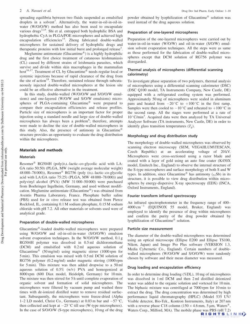
Morphology of double-walled microspheres (both S and W type)and cross-sectional view of S-type microspheres are shown inFigure 2. One should note that the samples used for SEM imagingwere fabricated by magnetic stirrer instead of homogenizer toachieve large particle size and be able to observe their crosssection by cutting with a razor blade. Double-walled structure isillustrated in Figure 2(A) for S-type microspheres. Porous surfacewas the common morphological characteristic of both S and Wtypes (Figure 2B and C) due to their similar preparationprocedure.
In double-walled microspheres, it is essential to determinewhere the major amount of the drug is entrapped as itsignificantly affects the drug release8. Presence of antimony inthe structure of Glucantime� promises the application of EDX tostudy distribution profile of the drug. As illustrated in Figure 3,the drug is encapsulated predominantly near the center for bothW-type (Figure 3A and B) and S-type (Figure 3C and D)microspheres.
Fourier transform infrared spectroscopy
As shown in Figure 4, FTIR spectrum of the lyophilizedGlucantime� is consistent with that of the reference pure drug19
(Figure 4A). Absorption band at 2300–3000 cm�1 is assigned toNHþ2 stretch in the structure of the drug. Additionally, the bands at1633 and 1049 cm�1 are due to C–O and Sb–O–C stretchvibrations, respectively20. FTIR spectra of S-type and W-typemicrospheres consist of both the drug and polymers absorptionbands (Figure 4B and C). FTIR results of the two types ofmicrospheres showed no significant difference. The peaksbetween 1395 and 1460 cm�1 were assigned to the maincharacteristic bands of RG756 and RG504H. The bands at1390–1450 and 1430 cm�1 were due to C–H stretch of methyl andmethylene groups, respectively20.
Particle size
The optical microscope images of microspheres are shown inFigure 5. The mean diameters of S-type and W-type microsphereswere 31� 13 and 52� 24mm, respectively. High-speed hom-ogenization in the last emulsification step effectively reduces theparticle size and makes it suitable for injection with a standardneedle. In this study, homogenizing at 8000 rpm resulted in alower mean particle size compared to the previous reports in theliterature3,4,8,21.
Encapsulation efficiency and drug loading
There are numerous studies on encapsulation efficiency, %DL andin vitro release of one-layered and double-walled microspheresentrapping hydrophilic drugs8,22–26, but there is no reportcomparing S- and W-type one-layered and double-walledmicrospheres.
As seen in Table 1, in both S- and W-type microspheres, %EEand %DL are higher in double-walled microspheres compared tocorresponding one-layered ones.
Figure 1. The DSC thermogram of double-walled W/O/O/Wmicrosphere.
DOI: 10.3109/03639045.2013.777734 Glucantime-loaded double-walled microspheres 3
Dru
g D
evel
opm
ent a
nd I
ndus
tria
l Pha
rmac
y D
ownl
oade
d fr
om in
form
ahea
lthca
re.c
om b
y D
alho
usie
Uni
vers
ity o
n 04
/18/
13Fo
r pe
rson
al u
se o
nly.

Release of Glucantime�
In this study, both one-layered and double-walled microsphereswere investigated and compared in terms of in vitro releaseprofiles. Figure 6 shows release profiles of Glucantime� from alltypes of microspheres over 30 d. The in vitro release of double-walled microspheres showed reduced initial burst and prolongedsustained release compared to the corresponding one-layeredmicrospheres (Table 1).
In the release profiles of PLGA microspheres, it is known thatafter the burst phase, a slow release phase known as the lag phasebegins8,27–29. At the lag phase, Glucantime� release rate declined
and almost remained constant for 20 and 15 d in the case of S and Wdouble-walled microspheres, respectively. After 18 d, more than80% of the total amount of encapsulated drug was depleted from theone-layered microspheres. In contrast, in double-walled micro-spheres, only about 40% of the drug was released after the sameperiod. No significant difference in initial burst and sustainedrelease between S- and W-type microspheres was observed.
Discussion
Double-walled microspheres were prepared using S/O/O/W andW/O/O/W emulsions. In this procedure, the internal aqueous
Figure 2. SEM images of the double-walled microspheres: double-walled structure (A); porous surface of the W (B) and S (C) double-walledmicrospheres.
4 A. Navaei et al. Drug Dev Ind Pharm, Early Online: 1–10
Dru
g D
evel
opm
ent a
nd I
ndus
tria
l Pha
rmac
y D
ownl
oade
d fr
om in
form
ahea
lthca
re.c
om b
y D
alho
usie
Uni
vers
ity o
n 04
/18/
13Fo
r pe
rson
al u
se o
nly.

solution containing the drug is either emulsified with the firstpolymer solution or the drug powder is dispersed into it. Then theother polymer solution in the same solvent is added, emulsified andthe resulting emulsion is then transferred into an aqueous solution ofa surfactant and emulsified again to form oil droplets within thecontinuous aqueous phase. Surfactant makes the droplets sphericaland stabilizes them in the aqueous medium until completeevaporation of organic solvent and polymer hardening (Figure 7).As illustrated schematically in Figure 8, the polymer–organicsolvent system is in the single-phase region at the beginning ofsolvent evaporation step (point A in Figure 8) because of highconcentration of DCM. Gradual evaporation of DCM increases thepolymer concentration and phase separation begins to occur upon
reaching the critical concentration at point B. Phase separationcontinues until complete evaporation of DCM (point C) andsubsequently microspheres harden and double-walled microspheresform. In this study, two different PLGA grades (RG504H (50:50)and RG756 (75:25)) were used in order to exploit phase separationthrough DCM evaporation and obtain double-walled structure.
Two separate transitions observed at 51.59 �C and 55.51 �C inDSC thermogram are in correspondence with RG504H andRG756 glass transition temperatures, respectively30. If twopolymers had formed a blend, a single Tg would have beenobserved in DSC thermogram, but our result shows the phaseseparation of polymers which is essential for the formation ofdouble-walled structure.
Figure 3. SEM/EDX images of the double-walled microspheres (S and W); SEM image zoom in of the W (A) (inset: large zoom) and S (C) (inset: largezoom) double-walled microspheres; distribution profile of Glucantime� within the W (B) (inset: elements which are existed in EDX analyzed zone) andS (D) (inset: elements which are existed in EDX analyzed zone) double-walled microspheres.
DOI: 10.3109/03639045.2013.777734 Glucantime-loaded double-walled microspheres 5
Dru
g D
evel
opm
ent a
nd I
ndus
tria
l Pha
rmac
y D
ownl
oade
d fr
om in
form
ahea
lthca
re.c
om b
y D
alho
usie
Uni
vers
ity o
n 04
/18/
13Fo
r pe
rson
al u
se o
nly.

Figure 4. FTIR spectrum of the lyophilizedGlucantime� (A), the S (B) and W (C)double-walled microspheres.
Figure 5. Optical microscope images of the S (A) and W (B) double-walled microspheres selected to measure particle size.
6 A. Navaei et al. Drug Dev Ind Pharm, Early Online: 1–10
Dru
g D
evel
opm
ent a
nd I
ndus
tria
l Pha
rmac
y D
ownl
oade
d fr
om in
form
ahea
lthca
re.c
om b
y D
alho
usie
Uni
vers
ity o
n 04
/18/
13Fo
r pe
rson
al u
se o
nly.

Figure 6. Cumulative Glucantime� release profiles of W/O/O/W and W/O/W (A) and S/O/O/W and S/O/W (B).
Table 1. Release characteristics, encapsulation efficiency and drug loading of one-layered and double-walled microspheres.
Microsphere type %Burst %Released drug until the day 18 %EE %DL
One-layered S 31.74� 1.86 80.08� 2.46 51� 2.81 2.48� 0.73One-layered W 35.75� 1.66 83.64� 2.61 43� 2.64 2.11� 0.88Double-walled S 17.43� 1.39 33.42� 2.11 87� 1.55 4.16� 0.48Double-walled W 21.57� 1.71 44.83� 2.35 81� 1.83 3.89� 0.69
Based on the first 24 h.
DOI: 10.3109/03639045.2013.777734 Glucantime-loaded double-walled microspheres 7
Dru
g D
evel
opm
ent a
nd I
ndus
tria
l Pha
rmac
y D
ownl
oade
d fr
om in
form
ahea
lthca
re.c
om b
y D
alho
usie
Uni
vers
ity o
n 04
/18/
13Fo
r pe
rson
al u
se o
nly.

Evaporation of organic solvent during the preparation ofmicrospheres using emulsion solvent evaporation technique leavespores at the surface of particles15 which is shown in SEM images.
Because of its hydrophilic nature, Glucantime� is expected tobe loaded in RG504H, which is more hydrophilic. This observa-tion can be explained by means of distribution theory31, which hasbeen successfully applied to predict distribution of some
therapeutic agents in biphasic systems32. The theory can berepresented as
logXRG504H
XRG756
¼ Vdrug
�drug � �RG756=DCM
� �2
� �RG504H=DCM � �drug
� �2
( )
ð2:3ÞRTð2Þ
where XRG504H and XRG756 are the drug concentrations in theRG504H and RG756 phases, respectively; �drug, �RG504H/DCM and�RG756/DCM are solubility parameters of the drug, RG504H phaseand RG756 phase, respectively; Vdrug is the molecular volume ofthe drug; R is the gas constant and T is absolute temperature. Sincethe difference between solubility parameters of the drug andRG504H is less than that of the drug and RG756, the drugconcentration would be higher in the RG504H layer. In the EDXimages of both W- and S-type microspheres, Glucantime� is highlyencapsulated near the center of the microspheres (Figure 3B andD). Therefore, it could be concluded that in these double-walledmicrospheres, RG504H which contains the major amount of thedrug, forms the inner layer. It should be noted that the presence ofother elements in EDX analysis in addition to those present in themeglumine antimoniate and the Resomers could be attributed tothe blade that is used for cross sectioning.
High-speed homogenization in the last emulsification stepeffectively reduced the microspheres size making them suitable forinjection with a standard needle. The smaller mean particle size ofS-type microspheres compared to W ones could be attributed to thepresence of drug aqueous solution in the first emulsification step ofW microspheres. Different factors affect the size of microspheresduring preparation procedure. Lee et al. investigated the effect ofpolymer mass ratios on PLLA/PLGA double-walled microspheressize and concluded that changes in mass ratios of PLLA to PLGA
Figure 7. Schematic representation of the double-walled microspheres preparation method.
Figure 8. Schematic representation of polymer-solvent system phasediagram and double-walled structure formation path ABC: after the lastemulsification step, the system is in the single-phase region (point A),solvent evaporation starts and polymer concentration increases to reach acritical point when phase separation begins (point B), gradual phaseseparation causes double-walled structure to form and complete evapor-ation of organic solvent results in obtaining solid double-walledmicrospheres (point C).
8 A. Navaei et al. Drug Dev Ind Pharm, Early Online: 1–10
Dru
g D
evel
opm
ent a
nd I
ndus
tria
l Pha
rmac
y D
ownl
oade
d fr
om in
form
ahea
lthca
re.c
om b
y D
alho
usie
Uni
vers
ity o
n 04
/18/
13Fo
r pe
rson
al u
se o
nly.

(w/w) results in the variations of shell thickness and core diameterwhile mean particle size remains constant4. They proposed thatparticle size is dependent only upon fabrication conditions such asstirring speed and surfactant concentration4,33.
During DCM evaporation in the preparation procedure ofdouble-walled microspheres, RG504H which is loaded withGlucantime� does not come into contact with the external aqueousphase. As illustrated in Figure 7, RG504H solidifies faster becauseof its more hydrophilic nature and lower tendency to DCM.It solidifies while the outer layer (RG756) has not hardened yetbecause of DCM. In this stage, DCM prevents the drug frompartitioning out to the external aqueous phase until its completeevaporation and solidification of RG756. This process results inhigh %EE for hydrophilic drugs in double-walled microspheres.
In double-walled microspheres, presence of more hydrophobicRG756 outer layer with negligible drug content resulted in asignificantly lower initial burst of about 20% which is almostequal to one half of that for one-layered ones. The porous surfaceobserved in the SEM image of double-walled microspheres(Figure 2) could be accounted for the persistence of the burstrelease. Zheng and Liang developed a one-step modification toreduce the initial burst release from PLGA microspheres. Theyadded a small amount of alginate into the internal aqueous phaseof W/O/W emulsion; meanwhile calcium chloride and chitosanwere put into the external one. After modification, the initialrelease was inhibited markedly, and the entrapment efficiency wasslightly increased. They observed that almost all the pores at thesurface of the microspheres had been closed with this modifica-tion method34. Lag phase was not observed in one-layered PLGAmicrospheres probably because of the hydrophilic nature ofRG504H and Glucantime�.
Conclusion
Double-walled microspheres provide some advantages overtraditional microspheres, overcoming most of their limitations.In this work, double-walled and one-layered microspheres weresuccessfully fabricated and loaded with Glucantime� employingdifferent emulsion solvent evaporation techniques. Double-walledmicrospheres were found to be more efficient than their corres-ponding one-layered microspheres in terms of %EE and releasecharacteristics. SEM/EDX was also successfully utilized toinvestigate drug distribution within the microspheres.Furthermore, microspheres with small particle size suitable forinjection with a standard needle were achieved using a high-speedhomogenizer. In future studies, these microspheres containingGlucantime�, the most well-known therapeutic for CL, could beevaluated in vivo in order to demonstrate their potential inimproving current treatments of this disease.
Declaration of interest
The authors report no declaration of interest.
References
1. Hillery AM, Lloyd AW, Swarbrick J. Drug delivery and targeting.London & New York: Taylor & Francis Inc; 2005.
2. Lee AP, Lee JL. BioMEMS and biomedical. New York: Springer;2006.
3. Zheng W. A water-in-oil-in-oil-in-water (W/O/O/W) method forproducing drug-releasing, double-walled microspheres. Int J Pharm2009;374:90–5.
4. Lee TH, Wang J, Wang CH. Double-walled microspheres for thesustained release of a highly water soluble drug: characterization andirradiation studies. J. Control Release 2002;83:437–52.
5. Lu TL, Sun WG, Zhao W, Chen T. Preparation of amifostinepolylactide-co-glycolide microspheres and its irradiation protective
to mouse through oral administration. Drug dev Ind Pharm 2011;37:1473–80.
6. Pekarek KJ, Jacob JS, Mathiowitz E. One-step preparation ofdouble-walled microspheres. Adv Mater 1994;6:684–7.
7. Pekarek KJ, Jacob JS, Mathiowitz E. Double-walled polymermicrospheres for controlled drug release. Nature 1994;367:258–60.
8. Tan E, Lin R, Wang CH. Fabrication of double-walled microspheresfor the sustained release of doxorubicin. J Colloid Interf Sci 2005;291:135–43.
9. Siegel SJ, Kahn JB, Metzger K, et al. Effect of drug type on thedegradation rate of PLGA matrices. Eur J Pharm Biopharm 2006;64:287–93.
10. Berkland C, Pollauf E, Packa DW, Kim K. Uniform double-walledpolymer microspheres of controllable shell thickness. J ControlRelease 2004;96:101–11.
11. Andreas K, Zehbe R, Kazubek M, et al. Biodegradable insulin-loaded PLGA microspheres fabricated by three different emulsifi-cation techniques: investigation for cartilage tissue engineering.Acta Biomater 2011;7:1485–95.
12. Parrot EL. Pharmaceutical technology. Minneapolis: Burgess; 1970.13. Mathiowitz E, Dor P, Amato C, Langer R. Polyanhydride micro-
spheres: 3. Morphology and characterization of systems made bysolvent removal. Polymer 1970;31:547–55.
14. Mathiowitz E, Kline D, Langer R. Morphology of polyanhydridemicrosphere delivery systems. Scan. Microsc. J. 1990;4:329–40.
15. Shi M, Yang YY, Chaw CS, et al. Double walled POE/PLGAmicrospheres: encapsulation of water-soluble and water-insolubleproteins and their release properties. J Control Release 2003;89:167–77.
16. Layegh P, Pezeshkpoor F, Soruri A, et al. Efficacy of cryotherapyversus intralesional meglumine antimoniate (glucantime) for treat-ment of cutaneous leishmaniasis in children. Am J Trop Med Hyg2009;80:172–5.
17. Vargas-Gonzalez A, Canto-Lara S, Damian-Centeno A, Andrade-Narvaez FJ. Response of cutaneous leishmaniasis (Chiclero’s ulcer)to treatment with meglumine antimoniate in southeast Mexico. Am JTrop Med Hyg 1999;61:960–3.
18. Herwaldt B, Berman J. Recommendations for treating leishmaniasiswith sodium stibogluconate (Pentostam) and review of pertinentclinical studies. Am J Trop Med Hyg 1992;46:296–306.
19. Demicheli C, de Figueiredo TL, Carvalho S, et al. Physico-chemicalcharacterization of meglumine antimoniate. Biometals 1999;12:63–6.
20. Pavia DL, Lampman GM, Kriz GS. Introduction to spectroscopy.Belmont, CA: Brooks/Cole; 2009.
21. Rahman N, Mathiowitz E. Localization of bovine serum albumin indouble-walled microspheres. J Control Release 2004;94:163–75.
22. Ito F, Honnami H, Kawakami H, et al. Preparation and properties ofPLGA microspheres containing hydrophilic drugs by the SPG(Shirasu porous glass) membrane emulsification technique. ColloidSurf B 2008;67:20–5.
23. Peltonen L, Aitta J, Hyvonen S, et al. Improved entrapmentefficiency of hydrophilic drug substance during nanoprecipitationof poly(L)lactide nanoparticles. AAPS PharmSciTec 2004;5:115–20.
24. Tan H, Ye J. Surface morphology and in vitro release performanceof double-walled PLLA/PLGA microspheres entrapping a highlywater-soluble drug. App Surf Sci 2008;255:353–6.
25. Lin R, Ng LS, Wang CH. In vitro study of anticancer drugdoxorubicin in PLGA-based microparticles. Biomaterials 2005;26:4476–85.
26. Lee W, Loei C, Widjaja E, Joachim Loo S. Altering the drug releaseprofiles of double-layered ternary-phase microparticles. J ControlRelease 2011;151:229–38.
27. Eun Oh J, Nam Y, Lee K, Gwan Park T. Conjugation of drug topoly(D,L-lactic-co-glycolic acid) for controlled release from bio-degradable microspheres. J Control Release 1999;57:269–80.
28. Stubbe BG, De Smedt SC, Demeester J. Programmed polymericdevices for pulsed drug delivery. Pharm Res 2004;21:1732–40.
29. Duvvuri S, Janoria KG, Mitra AK. Effect of polymer blending on therelease of ganciclovir from PLGA microspheres. Pharm Res 2006;23:215–23.
30. Rafienia M, Hojjati Emami S, Mirzadeh H, et al. Influence ofpoly(lactide-co-glycolide) type and gamma irradiation on thebetamethasone acetate release from the in situ forming systems.Curr Drug Deliv 2009;6:184–91.
DOI: 10.3109/03639045.2013.777734 Glucantime-loaded double-walled microspheres 9
Dru
g D
evel
opm
ent a
nd I
ndus
tria
l Pha
rmac
y D
ownl
oade
d fr
om in
form
ahea
lthca
re.c
om b
y D
alho
usie
Uni
vers
ity o
n 04
/18/
13Fo
r pe
rson
al u
se o
nly.

31. Snyder JJ. Kirkland R, eds. Modern practice of liquid chromatog-raphy. New York: Wiley–Interscience; 1971.
32. Matsumoto A, Matsukawa Y, Suzuki T, et al. The polymer-alloysmethod as a new preparation method of biodegradable microspheres:principle and application to cisplatin-loaded microspheres. J ControlRelease 1997;48:19–27.
33. Leach E, Noh K, Mathiowitz E. Effects of manufacturing conditionson the formation of double-walled polymer microspheres.J Microencapsul 1999;16:153–67.
34. Zheng C, Liang W. A one-step modified method to reduce theburst initial release from PLGA microspheres. Drug Deliv 2010;17:77–82.
10 A. Navaei et al. Drug Dev Ind Pharm, Early Online: 1–10
Dru
g D
evel
opm
ent a
nd I
ndus
tria
l Pha
rmac
y D
ownl
oade
d fr
om in
form
ahea
lthca
re.c
om b
y D
alho
usie
Uni
vers
ity o
n 04
/18/
13Fo
r pe
rson
al u
se o
nly.