Intraotic Administration of Gentamicin: A New Method to Study Ototoxicity in the Crista Ampullaris...
7
The Laryngoscope Lippincott-Raven Publishers, Philadelphia 0 1997 The American Laryngological, Rhinological and Otological Society, Inc. Intraotic Administration of Gentamicin: A New Method to Study Ototoxicity in the Crista Ampullaris of the Bullfrog Angelica Carranza, MD; Ivan Lopez, PhD; Philip Castellano, MD; Larry Hoffman, PhD; Vincente Honrubia, MD, DMSc A new method of local gentamicin administra- tion was tested in the bullfrog inner ear to achieve ototoxic-induced hair cell destruction. Gelfoam pledgets soaked with known amounts of gentam- icin were inserted into the perilymphatic cisterna of the bullfrog through a ventral surgical approach. A dose of 1.20 mg gentamicin, consistent with a per- ilymphatic concentration of 65 pg/ml, resulted in the desired ototoxic-induced hair cell damage, that is, complete hair cell destruction with minimal dis- ruption of other components of the sensory epithe- lium. This study demonstrates that this is a useful and simple method to investigate the process of vestibular ototoxicity and hair cell regeneration, including aspects of hair cell destruction and re- pair. Key Words: Bullfrog cristae ampullaris-Gen- tamicin ototoxicity-Hair cell regeneration. Laryngoscope, 102137-143,1997 INTRODUCTION Aminoglycoside antibiotics, including gentam- icin (GM), are known to induce degenerative changes in the vestibular labyrinth. These changes range from swelling and vacuolization of the sen- sory hair cells and fusion of the sensory stereocilia to complete hair cell loss and sensory epithelium obliteration (1). In the past, intensive work has been From the Division of Head & Neck Surgery,Victor Goodhill Ear Cen- ter, University of California at Los Angeles School of Medicine, Los Ange- les, CA, U.S.A. Preliminary results of this work were presented at the 1994 Meeting of the Association of Research in Otolaryngology, St. Petersburg, FL, Feb- ruary 1994 (abstract 138). This work was supported by grants DC01404 and DC00008, the Na- tional Enterprise Endowment Fund, and a National Medical Fellowship to Dr. Carranza. Editor’s Note: This Manuscript was accepted for publication July 15, 1996. Send Reprint Requests to Vincente Honrubia, MD, DMSc, Division of Head and Neck Surgery, University of California, Los Angeles, 62-129 CHS, Los Angeles, CA 90095-1624, USA. done in the cochlea and vestibule to investigate aminoglycoside ototoxicity (2-5). Recently, the dis- covery of possible hair cell regeneration after oto- toxicity has motivated investigators to study the process in different animal models to elucidate the mechanism of sensory epithelium repair and hair cell regeneration (6-11). Amphibians have often been the models used to study vestibular ototoxicity and hair cell regenera- tion (8,12,13).Omura et al. (13) compared three dif- ferent methods of GM administration in the bullfrog to study the ototoxic effects: intraperitoneal, intra- muscular, and intralabyrinthine injections. Of the three approaches, the intralabyrinthine injection re- sulted in the most severe damage in the shortest pe- riod. In this method a fine needle was inserted through the palatal bone of the bullfrog, and GM was blindly administered into the perilymphatic cis- terna. These authors recognize that with this tech- nique there is the possibility of rupturing the mem- branous labyrinth, resulting in mixing the perilymphatic and endolymphatic fluids which could itself lead to hair cell destruction. Baird et al. (8) also used intraotic injections of GM. Through a ventral approach in the roof of the mouth of the bull- frog, a hole was made using a deburred 25-gauge needle to place GM blindly into the inner ear space. Both aforementioned approaches represent a quick and effective method to induce ototoxicity in the in- ner ear of the bullfrog, albeit with uncertainty as to the precise fluid compartment where the drug was applied due to the possible trauma to the mem- branes separating the labyrinth spaces. This communication describes a method of in- troducing known amounts of GM for ototoxic evalua- tion through a controlled surgical approach into the bullfrog’s perilymphatic space. The results demon- strate that the method produces dose-dependent de- grees of ototoxic damage, as well as the occurrence of Laryngoscope 107: January 1997 Carranza et al.: Gentamicin Ototoxicity 137
-
Upload
independent -
Category
Documents
-
view
0 -
download
0
Transcript of Intraotic Administration of Gentamicin: A New Method to Study Ototoxicity in the Crista Ampullaris...
The Laryngoscope Lippincott-Raven Publishers, Philadelphia 0 1997 The American Laryngological, Rhinological and Otological Society, Inc.
Intraotic Administration of Gentamicin: A New Method to Study Ototoxicity in the Crista Ampullaris of the Bullfrog Angelica Carranza, MD; Ivan Lopez, PhD; Philip Castellano, MD; Larry Hoffman, PhD; Vincente Honrubia, MD, DMSc
A new method of local gentamicin administra- tion was tested in the bullfrog inner ear to achieve ototoxic-induced hair cell destruction. Gelfoam pledgets soaked with known amounts of gentam- icin were inserted into the perilymphatic cisterna of the bullfrog through a ventral surgical approach. A dose of 1.20 mg gentamicin, consistent with a per- ilymphatic concentration of 65 pg/ml, resulted in the desired ototoxic-induced hair cell damage, that is, complete hair cell destruction with minimal dis- ruption of other components of the sensory epithe- lium. This study demonstrates that this is a useful and simple method to investigate the process of vestibular ototoxicity and hair cell regeneration, including aspects of hair cell destruction and re- pair.
Key Words: Bullfrog cristae ampullaris-Gen- tamicin ototoxicity-Hair cell regeneration.
Laryngoscope, 102137-143,1997
INTRODUCTION Aminoglycoside antibiotics, including gentam-
icin (GM), are known to induce degenerative changes in the vestibular labyrinth. These changes range from swelling and vacuolization of the sen- sory hair cells and fusion of the sensory stereocilia to complete hair cell loss and sensory epithelium obliteration (1). In the past, intensive work has been
From the Division of Head & Neck Surgery, Victor Goodhill Ear Cen- ter, University of California a t Los Angeles School of Medicine, Los Ange- les, CA, U.S.A.
Preliminary results of this work were presented at the 1994 Meeting of the Association of Research in Otolaryngology, St. Petersburg, FL, Feb- ruary 1994 (abstract 138).
This work was supported by grants DC01404 and DC00008, the Na- tional Enterprise Endowment Fund, and a National Medical Fellowship to Dr. Carranza.
Editor’s Note: This Manuscript was accepted for publication July 15, 1996.
Send Reprint Requests to Vincente Honrubia, MD, DMSc, Division of Head and Neck Surgery, University of California, Los Angeles, 62-129 CHS, Los Angeles, CA 90095-1624, USA.
done in the cochlea and vestibule to investigate aminoglycoside ototoxicity (2-5). Recently, the dis- covery of possible hair cell regeneration after oto- toxicity has motivated investigators to study the process in different animal models to elucidate the mechanism of sensory epithelium repair and hair cell regeneration (6-11).
Amphibians have often been the models used to study vestibular ototoxicity and hair cell regenera- tion (8,12,13). Omura et al. (13) compared three dif- ferent methods of GM administration in the bullfrog to study the ototoxic effects: intraperitoneal, intra- muscular, and intralabyrinthine injections. Of the three approaches, the intralabyrinthine injection re- sulted in the most severe damage in the shortest pe- riod. In this method a fine needle was inserted through the palatal bone of the bullfrog, and GM was blindly administered into the perilymphatic cis- terna. These authors recognize that with this tech- nique there is the possibility of rupturing the mem- branous labyrinth, resulting in mixing the perilymphatic and endolymphatic fluids which could itself lead to hair cell destruction. Baird et al. (8) also used intraotic injections of GM. Through a ventral approach in the roof of the mouth of the bull- frog, a hole was made using a deburred 25-gauge needle to place GM blindly into the inner ear space. Both aforementioned approaches represent a quick and effective method to induce ototoxicity in the in- ner ear of the bullfrog, albeit with uncertainty as to the precise fluid compartment where the drug was applied due to the possible trauma to the mem- branes separating the labyrinth spaces.
This communication describes a method of in- troducing known amounts of GM for ototoxic evalua- tion through a controlled surgical approach into the bullfrog’s perilymphatic space. The results demon- strate that the method produces dose-dependent de- grees of ototoxic damage, as well as the occurrence of
Laryngoscope 107: January 1997 Carranza et al.: Gentamicin Ototoxicity 137
end organ and hair cell repair. Preliminary findings have been reported in abstract form (14).
MATERIALS AND METHODS A number of experiments were conducted on bull-
frogs to develop a simple and suitable surgical approach to the perilymphatic cisterna that would allow insertion of known amounts of GM without disrupting the mem- brane surrounding the endolymphatic space. Forty-five healthy adult bullfrogs (Rana catesbeiana) weighing 150 to 200 g were used. The study was approved by UCLA's Chancellor's Animal Research Committee, and all animal experiments were performed in compliance with The Guiding Principles in the Care and Use of Laboratory An- imals.
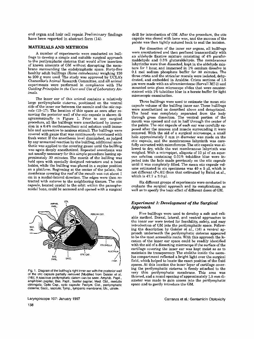
The inner ear of this animal contains a relatively large perilymphatic cisterna, positioned on the ventral side of the inner ear between the saccule and the otic cap- sule (15-17). The location of this space as seen after re- moving the posterior wall of the otic capsule is shown di- agrammatically in Figure 1. Prior to any surgical procedure, all the bullfrogs were anesthetized by immer- sion in a 0.4% methanesulfonic acid solution until immo- bile and unreactive to noxious stimuli. The bullfrogs were covered with gauze that was continuously moistened with fresh water. If the anesthesia level diminished, as judged by any unwanted reaction by the bullfrog, additional anes- thetic was applied to the covering gauze until the bullfrog was again deeply anesthetized. Repeated anesthesia was not usually necessary for this simple procedure lasting ap- proximately 30 minutes. The mouth of the bullfrog was held open with specially designed retractors and a head holder, while the bullfrog was placed in a supine position on a platform. Beginning at the center of the palate, the membrane covering the roof of the mouth was cut about 1 cm in a medial-lateral direction. The edges were then re- tracted with sutures to the neighboring tissues. The otic capsule, located caudal to the orbit within the parasphe- noidal bone, could be accessed and opened with a surgical
LITR. AMPHIR I . .. .
I I I OTIC'CAP.
BAS. I PAPIL.
Fig. 1. Diagram of the bullfrog's right inner ear with the posterior wall of the otic capsule partially removed [Modified from Geisler et al. (1 S)]. A spacious perilymphatic cistern can be seen. Amphib. Papil., amphibian papilar; Bas. Papil., basilar papilar; Med. Obl., medulla oblongata; Optic Cap., optic capsule; Perilym. Cist., perilymphatic cisterna; Sacc., saccule; Tymp., tympanic membrane; Utr., utricle.
drill for introduction of GM. After the procedure, the otic capsule was closed with bone wax, and the mucosa of the palate was then tightly sutured back to seal the incision.
For dissection of the inner ear organs, all bullfrogs were anesthetized and then perfused transcardially with an aldehyde fixative mixture consisting of 4% parafor- maldehyde and 0.5% glutaraldehyde. The membranous labyrinths were then dissected, kept in the aldehyde mix- ture for 1 hour, and immersed in 1% osmium dissolve in 0.1 mol sodium phosphate buffer for 30 minutes. The three crista and the utricular macula were isolated, dehy- drated, and embedded in Araldite. Crista sections of 1.5 pm were made with an ultramicrotome (Sorvall MT-2) and mounted onto glass microscope slides that were counter- stained with 1% toluidine blue in a borate buffer €or light microscopic examination.
Three bullfrogs were used to estimate the mean otic capsule volume of the bullfrog inner ear. These bullfrogs were anesthetized as described above and decapitated. The head was completely separated from the body through gross dissection. The ventral portion of the mouth was opened and cut in half through the center of the palate. The otic capsule of each ear was carefully ex- posed after the mucosa and muscle surrounding it were removed. With the aid of a surgical microscope, a small hole approximately 2 mm in diameter was made on the otic capsule, and the membranous labyrinth was care- fully extracted with microforceps. The otic capsule was al- lowed to dry, while the wet membranous labyrinth was weighed. With a micropipet, aliquots of 10 p1 of an aque- ous solution containing 0.01% toluidine blue were in- jected into the hole made previously on the otic capsule until it was completely filled. The mean otic capsule vol- ume estimated in six specimens was 40 e 5 p1, which is not different (P<.Ol) from that estimated by Baird et al., which is 47.7 3.0 pl.
Six different groups of experiments were conducted to evaluate the surgical approach and its complications, as well as to specify the toxic effect of different doses of GM.
Experiment 1: Development of the Surgical Approach
Five bullfrogs were used to develop a safe and reli- able method. Dorsal, lateral, and ventral approaches to the inner ear were tested for feasibility, safety, and easy introduction of GM into the perilymphatic space. Follow- ing the description by Geisler et al., (16) a ventral ap- proach underneath the perilymphatic cisterna appeared to be the most accessible route. With this approach the lo- cation of the inner ear space could be readily identified with the aid of a dissecting microscope if the surface of the cartilage covering the inner ear was kept moist so as to maintain its transparency. The otoliths inside the saccu- lus compartment reflected a bright light over the surgical field, which helped to locate the exact position of the fluid spaces. At this location the inner layer of cartilage cover- ing the perilymphatic cisterna is firmly attached to the very thin perilymphatic membrane. This area was thinned, and a round opening of approximately 1.5 mm di- ameter was made to gain access into the perilymphatic space and to gently introduce the GM.
Laryngoscope 107: January 1997
1 38
Carranza et al.: Gentamicin Ototoxicity
Experiment 2: Preparation of the GM Pledgets After some trials it was decided to use Gelfoam pled-
gets as the vehicle of transporting GM into the inner ear. Under sterile conditions, Gelfoam pieces were cut into ap- proximately equal sizes of 4 mm3 (2 x 2 x 1 mm) and weighed. They were saturated with a GM sulfate solution of 40 mg/ml (Elkins-Sinn, Inc., Cherry Hill, NJ) and weighed again. The weight of the GM-soaked pledget mi- nus the weight of the dry pledget was determined, divided by the density of water, and multiplied by the concentra- tion of GM used: wt of GM-soaked pledget - wt of dry pledget
density of water (1.0 mg/ml) 4o mg/ml
Approximately 0.33 2 0.02 mg of GM was absorbed in each pledget. These pieces were then stored in the freezer to so- lidify until a few minutes prior to their use.
Experiment 3: Site of Drug Distribution Experiments were conducted in four bullfrogs to ver-
ify that the insertion of pledgets did not disrupt the en- dolymphatic membrane. Using the ventral surgical ap- proach, a Gelfoam pledget soaked with methylene blue in a 0.1% aqueous solution was introduced into the inner ear. Twelve to 36 hours after surgery, the perilymphatic space was opened and the perilymphatic fluid remained bluish. The endolymphatic membrane was rinsed with sterile saline solution (0.1% NaC1) to remove methylene blue stain from its surface. With the aid of the surgical micro- scope, the integrity of the membranous labyrinth and the absence of methylene blue in the endolymphatic space were confirmed. It was verified that the surgical procedure did not disrupt or produce gross distortion to the endolym- phatic membrane and spaces such that the drug will have been retained in the perilymphatic space after injection.
Experiment 4: Dose-Dependent Effects of GM To evaluate dose-dependent effects of GM, three
doses of GM ranging from 0.3 to 3.0 mg were applied to three groups of four bullfrogs. One group received 0.3 mg of GM (1 pledget), a second group received 1.2 mg of GM (4 pledgets), and a third group received 3.0 mg of GM (10 pledgets). All bullfrogs were allowed to survive 1 week af- ter surgery. The aim of this locally applied treatment was to find the one exact dose (i.e., single treatment) that pro- duced the same destruction of sensory cells achieved by others through repeated intramuscular injections (e.g., Forge et al.) (9).
Experiment 5: Morphological Controls A group of five normal bullfrogs not subjected to any
surgical procedures was used to study the normal mor- phology of the cristae ampullaris (CA). To serve as con- trols, another group of four bullfrogs received the same surgical procedure as the experimental bullfrogs but with- out GM treatment. In one bullfrog, only the fenestra was made without the insertion of pledgets. The other bull- frogs received the same number of pledgets as the GM- treated amphibians (1,4, or 10 sterile pledgets), but were impregnated with saline solution.
One week after surgery all the bullfrogs were sacri- ficed so that the cytoarchitecture of the cristae could be
examined. Emphasis was placed on the study of the verti- cal semicircular canal cristae (SCC), especially the poste- rior SCC. As is known, the horizontal SCC is smaller and asymetrical since only half of the crista developes in these animal species (16,171. All the vestibular receptors were evaluated, including the three cristae and the macula utriculi. However, meticulous observations, which in- cluded the study of serial sectioning of the whole cristae length and evaluation of each slide, were conducted in the posterior SCC only.
Experiment 6: Time-Dependent Effects of GM A dose of 1.2 mg of GM that had been found to pro-
duce the immediate desired results (cf. experiment 4 re- sults) was selected to study long-term ototoxic effects. Three groups of four bullfrogs were treated with four pled- gets containing a total of 1.2 mg of GM. These bullfrogs were allowed to survive 1,2, or 4 weeks after surgery.
RESULTS Vestibular Sensory Epithelium of Normal and Control Bullfrogs
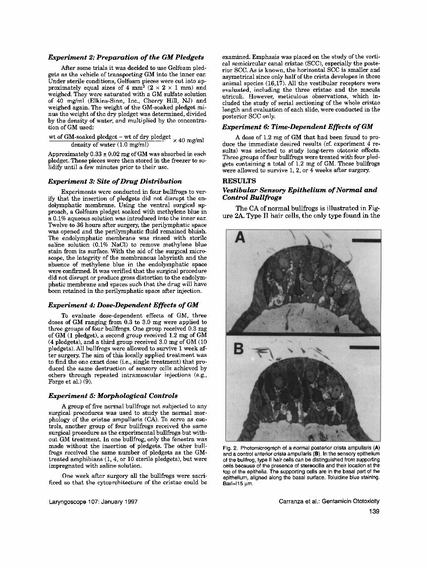
The CA of normal bullfrogs is illustrated in Fig- ure 2A. Type I1 hair cells, the only type found in the
Fig. 2. Photomicrograph of a normal posterior crista ampullaris (A) and a control anterior crista ampullaris (B). In the sensory epithelium of the bullfrog, type II hair cells can be distinguished from supporting cells because of the presence of stereocilia and their location at the top of the epithelia. The supporting cells are in the basal part of the epithelium, aligned along the basal surface. Toluidine blue staining. Barl=115 pm.
Laryngoscope 107: January 1997 Carranza et al.: Gentamicin Ototoxicity
139
bullfrog vestibular organ, are large and clearly dif- ferentiated. On their endolymphatic surface they show a number of stereocilia that protrude from the top of the cells. Supporting cells are aligned over the basal surface of the sensory epithelium. Their processes extend toward the surface of the crista, where they separate the receptor cells, as observed by other authors (17).
In the control bullfrogs subjected to surgery and insertion of 1, 4, or 10 saline-soaked Gelfoam pled- gets, the surface wound was in the process of heal- ing and the otic capsule was sealed without visual evidence of leakage. The cristae were normal in ap- pearance, and hair cells were present with their stereocilia on the apical surface, without evidence of damage. The supporting cells were basally located and consistently of normal morphology. A demon- strative photomicrograph from the crista of the an- terior SCC is shown in Figure 2B.
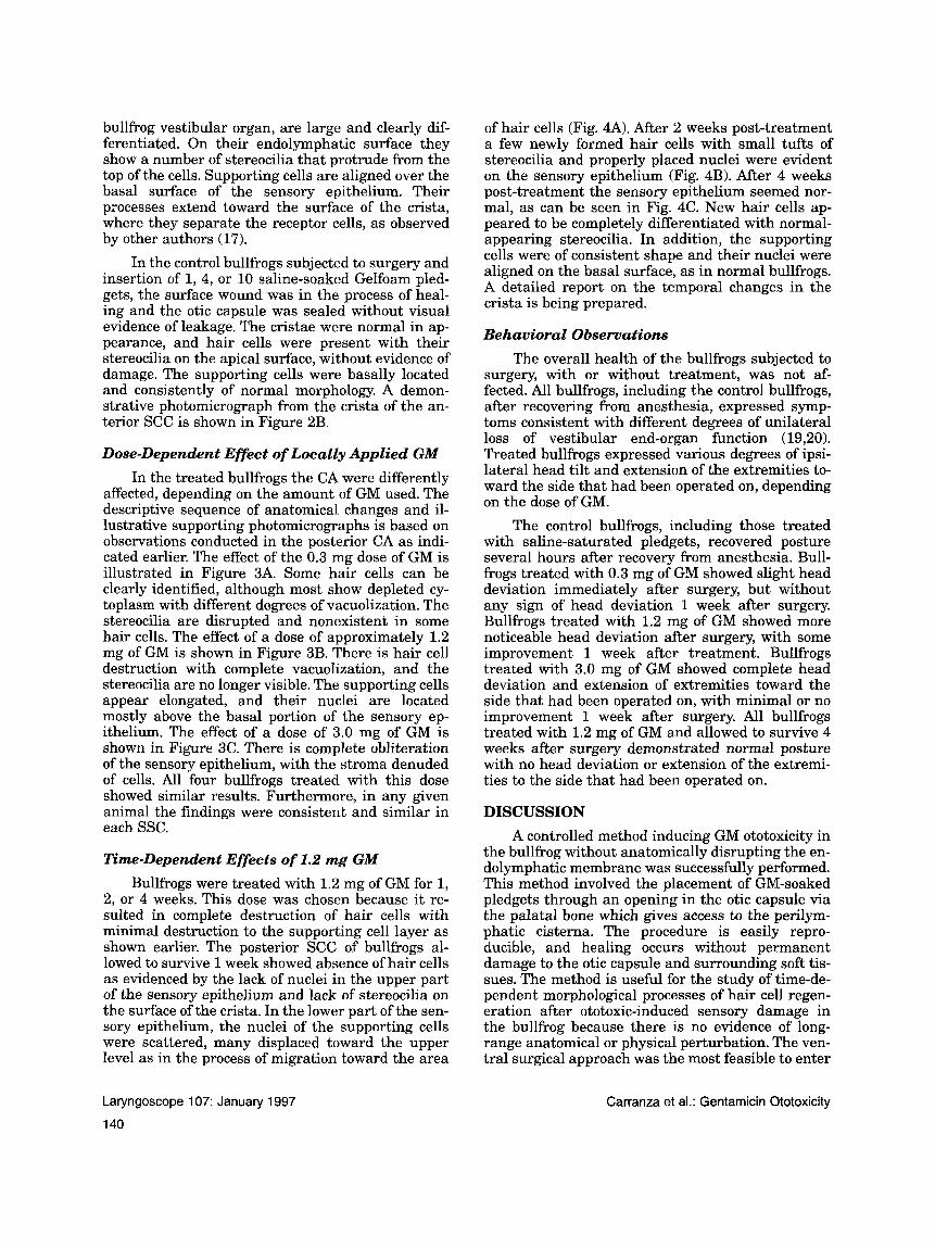
Dose-Dependent Effect of Locally Applied GM In the treated bullfrogs the CA were differently
dected, depending on the amount of GM used. The descriptive sequence of anatomical changes and il- lustrative supporting photomicrographs is based on observations conducted in the posterior CA as indi- cated earlier. The effect of the 0.3 mg dose of GM is illustrated in Figure 3A. Some hair cells can be clearly identified, although most show depleted cy- toplasm with different degrees of vacuolization. The stereocilia are disrupted and nonexistent in some hair cells. The effect of a dose of approximately 1.2 mg of GM is shown in Figure 3B. There is hair cell destruction with complete vacuolization, and the stereocilia are no longer visible. The supporting cells appear elongated, and their nuclei are located mostly above the basal portion of the sensory ep- ithelium. The effect of a dose of 3.0 mg of GM is shown in Figure 3C. There is complete obliteration of the sensory epithelium, with the stroma denuded of cells. All four bullfrogs treated with this dose showed similar results. Furthermore, in any given animal the findings were consistent and similar in each SSC.
Time-Dependent Effects of 1.2 mg GM Bullfrogs were treated with 1.2 mg of GM for 1,
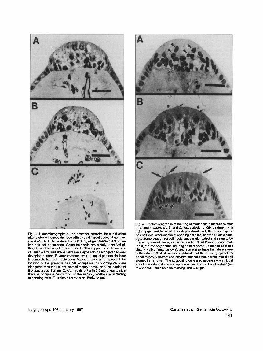
2, or 4 weeks. This dose was chosen because it re- sulted in complete destruction of hair cells with minimal destruction to the supporting cell layer as shown earlier. The posterior SCC of bullfrogs al- lowed to survive l week showed absence of hair cells as evidenced by the lack of nuclei in the upper part of the sensory epithelium and lack of stereocilia on the surface of the crista. In the lower part of the sen- sory epithelium, the nuclei of the supporting cells were scattered, many displaced toward the upper level as in the process of migration toward the area
of hair cells (Fig. 4A). After 2 weeks post-treatment a few newly formed hair cells with small tufts of stereocilia and properly placed nuclei were evident on the sensory epithelium (Fig. 4B). After 4 weeks post-treatment the sensory epithelium seemed nor- mal, as can be seen in Fig. 4C. New hair cells ap- peared to be completely differentiated with normal- appearing stereocilia. In addition, the supporting cells were of consistent shape and their nuclei were aligned on the basal surface, as in normal bullfrogs. A detailed report on the temporal changes in the crista is being prepared.
Behavioral Observations The overall health of the bullfrogs subjected to
surgery, with or without treatment, was not af- fected. All bullfrogs, including the control bullfrogs, after recovering from anesthesia, expressed symp- toms consistent with different degrees of unilateral loss of vestibular end-organ function (19,20). Treated bullfrogs expressed various degrees of ipsi- lateral head tilt and extension of the extremities to- ward the side that had been operated on, depending on the dose of GM.
The control bullfrogs, including those treated with saline-saturated pledgets, recovered posture several hours after recovery from anesthesia. Bull- frogs treated with 0.3 mg of GM showed slight head deviation immediately after surgery, but without any sign of head deviation 1 week after surgery. Bullfrogs treated with 1.2 mg of GM showed more noticeable head deviation after surgery, with some improvement 1 week after treatment. Bullfrogs treated with 3.0 mg of GM showed complete head deviation and extension of extremities toward the side that had been operated on, with minimal or no improvement 1 week after surgery. All bullfrogs treated with 1.2 mg of GM and allowed to survive 4 weeks after surgery demonstrated normal posture with no head deviation or extension of the extremi- ties to the side that had been operated on.
DISCUSSION A controlled method inducing GM ototoxicity in
the bullfrog without anatomically disrupting the en- dolymphatic membrane was successfully performed. This method involved the placement of GM-soaked pledgets through an opening in the otic capsule via the palatal bone which gives access to the perilym- phatic cisterna. The procedure is easily repro- ducible, and healing occurs without permanent damage to the otic capsule and surrounding soft tis- sues. The method is useful for the study of time-de- pendent morphological processes of hair cell regen- eration after ototoxic-induced sensory damage in the bullfrog because there is no evidence of long- range anatomical or physical perturbation. The ven- tral surgical approach was the most feasible to enter
Laryngoscope 107: January 1997
140
Carranza et al.: Gentamicin Ototoxicity
the perilymphatic space, making it possible to insert as many as 10 pledgets of 4 mm3 in size into this space. To investigate the possibility of damage to the endolymphatic membrane and mixing of inner ear fluids via this approach, pledgets soaked with meth- ylene blue dye were inserted into the perilymphatic cisterna by means of the same surgical approach as with the treated bullfrogs, and the absence of dye in the endolymphatic space was confirmed, proving that with our method there is no disruption of the membranous labyrinth to an extent that would al- low mixing of fluid compartments at variance of what might happen in other intraotic approaches (8,13).
The technique of intraotic administration of GM eliminated toxic side effects that could have resulted in systemic damage to other organs or neuromuscu- lar blockade from calcium depletion. These toxic ef- fects are observed when GM is given systemically, not only because of the high doses, but also because of the prolonged time period required to induce oto- toxicity (18). The intraotic application minimizes these secondary complications.
The effect of GM on the posterior CA of the bull- frog was found to be dose-dependent. A dose of 0.3 mg of GM caused minimal damage to the sensory epithelium, whereas a dose of 3.0 mg completely de- stroyed the sensory epithelium. Doses of 1.2 mg of GM, which can be provided by four pledgets of 4 mm3 soaked with commercial GM solution, induced ototoxicity with complete hair cell damage without disrupting the stroma of the sensory epithelium.
Prior studies have shown that a perilymphatic concentration of about 20 pg/ml, obtained with daily intramuscular injections of 100 mg/kg for 4 weeks, is needed to produce ototoxicity in animal species such as the guinea pig (18). Assuming that the volume of the inner ear of the bullfrog is approximately 40 p1, a dose of about 0.80 yg of GM should result in a 20 pg/ml concentration. Omura et al. (13) found that an intraotic dose of 0.24 pg of GM, blindly injected via a fine needle, produced similar effects to those in our study. This finding is also consistent with Baird et al., who demonstrated that intraotic injections of 10 pg/ml resulted in the uniform destruction of hair cells in the sacculus, but only selective destruction in the utricle of the bullfrog (8). Baird et al., also found regenerating hair cells in the saccular and utricular maculae as early as 24 to 48 hours postin- jection, and cellular morphology was normal at 7 to 9 days postinjection, earlier than in the present study. Likely, the accelerated recovery was due to more limited damage by the more diluted GM con- centration.
The possibility of hair cell regeneration was in- vestigated in a time-dependent manner. A dose of 1.2 mg of GM-equivalent to a perilymphatic concentra- tion of 65 pg/ml-resulted in complete destruction of
hair cells with minimal damage to the supporting cells. Two weeks post-treatment, new hair cells began to appear in the posterior CA. The recovery of the sensory epithelium has been postulated to be the re- sult of supporting cells experiencing mitotic prolifer- ation andor transdifferentiation or, in other words, the evolution of supporting cells, arrested in their de- velopment, toward their final expression as hair cells (6-11). By 4 weeks post-treatment the posterior CA appeared normal, with no signs of ototoxic damage. Another study in our laboratory, this time with chin- chillas, demonstrated similar potential hair cell re- generation in mammals. Using bromodeoxyuridine, it was shown that the process involves the mitotic pro- liferation of supporting cells and their subsequent differentiation into hair cells (21).
In summary, the bullfrog appears to be a promising animal model for the investigation of vestibular ototoxicity and hair cell regeneration as shown in this report. The documented experimental method adds a new dimension to this preparation which has provided basic information about hair cell excitability (22), molecular biology (231, organ func- tion (241, vestibular nerve physiology (25,26), and regeneration (27). Access to the perilymphatic space is best done through a ventral surgical approach, which allows the insertion of GM-soaked Gelfoam pledgets without any complications. A dose of 1.2 mg of GM, consistent with a perilymphatic concentra- tion of 65 mg/ml, results in sufficient ototoxicity to destroy the majority of hair cells in the bullfrog. This model can be used to study the process of hair cell regeneration from a morphological, physiologi- cal, or functional point of view.
ACKNOWLEDGMENT The authors thank Rita Watson for enthusiasti-
cally assisting with the histological preparations and Haya Leiner for the final proofreading of the manuscript.
BIBLIOGRAPHY 1. Wersall J. Structural damage to the organ of Corti and
vestibular epithelia caused by aminoglycoside ototoxicity in the guinea pig. In: Lerner SA, Matz GJ, Hawkins JE, eds. Aminoglycoside ototoxicity. Boston: Little, Brown and Co., 1981:197-214.
2. Wersall J. The ototoxic potential of netilmicin compared with amikacin. An animal study in guinea pigs. Scan J Infect Dis Suppl 1980;supp123:104-13.
3. Aran JM. Current perspectives on inner ear toxicity. Oto- laryngol Head Neck Surg 1995;112:133-44.
4. Baregi R, Grill V, Narducci ZR, et al. Gentamicin ototoxicity: histological and ultrastructural alterations after trans- tympanic administration. Pharmacol Res 1990;22:63544.
5. Takumida M, Bagger-Sjoback D, Harada Y, et al. Sensory hair fusion and glycocalyx changes following gentamicin expo- sure in the guinea pig vestibular organs. Acta Otolaryngol Suppl (Stockh) 1989;supp1107:39-47.
6. Weisleder P, Rube1 EW. Hair cell regeneration after strepto- mycin toxicity in the avian vestibular epithelium. J Comp Neurol 1993;331:97-110.
Laryngoscope 107: January 1997 Carranza et al.: Gentamicin Ototoxicity
142
7. Rubel EW, Dew LA, Roberson DW. Mammalian vestibular hair cell regeneration. Science 1995;267:701-3.
8. Baird RA, Torres MA, Shuff NR. Hair cell regeneration in the bullfrog vestibular otolith organs following aminoglycoside toxicity. Hear Res 1993;65:164-74.
9. Forge A, Li L, Corwin JT, e t al. Ultrastructural evidence for hair cell regeneration in the mammalian inner ear. Science
10. Girod DA, Rubel EW. Hair cell regeneration in the avian cochlea: if it works in birds, why not in man? Ear Nose Throat J 1987;70:343-54.
11. Lippe WR, Westbrook EW, Ryal BM. Hair cell regeneration in the chicken cochlea following aminoglycoside toxicity. Hear Res 1991;56:203-10.
12. Kimura RS, Iverson NA, Southard RE. Selective lesions of the vestibular labyrinth. Ann Otol Rhino1 Laryngol 1988;
13. Omura R, Harada Y, Suzuki M, et al. Structural and physio- logical change of the hullfrog semicircular canal due to gentamicin intoxication. Acta Otolaryngol Suppl (Stockh) 1989;supp1468:41-7.
14. Carranza A, Castellano P, Lopez I, e t al. Gentamicin ototoxi- city and hair cell regeneration in the cristae ampullaris of Rana catesbeiana. Assoc for Research in Otolaryngology. 1994;17:Abstract No. 138.
15. Ramprashad F, Landolt JP, Money KE, e t al. Comparative morphometric study of the vestibular system of the verte- brata: reptilia, aves, amphibian and Pisces. Acta Otolaryn- go1 Suppl (Stockh) 1986;supp1427:1-42.
16. Geisler C, Van Bereijk W, Frishkopf L. The inner ear of the bullfrog. J Morphol 1964;114:43-58.
17. Hillman DE. Morphology of peripheral and central vestibular system. In: Frog neurobiology. New York: Springer Verlag,
1993;259:1616-9.
97577-84.
1976:452-80.
Laryngoscope 107: January 1997
18. Tran Ba Huy P, Manuel C, Meulemans A. Kinetics of amino- glycoside antibiotics in perilymph and endolymph in ani- mals. In: Aminoglycoside ototoxicity. Boston: Little, Brown and Co.; 1981:81-97.
19. Frishkopf LS, Goldstein MH. Responses to acoustic stimuli from single units in the eighth nerve of the bullfrog. J Acoust SOC Am 1963;35:1219-28.
20. Precht W, Llinas R, Clarke M. Physiological responses of frog vestibular fibers to horizontal angular rotation. Exp Brain Res 1971;13:378-407.
21. Lopez I, Carranza A, Castellano P, et al. Hair cell regenera- tion after local gentamicin administration in the crista am- pullaris of the bullfrog. Otolaryngol Head Neck Surg 1994; 111:146. Abstract No. 60.
22. Hudsped AJ, Lewis RS. Kinetic analysis of voltage- and ion- dependent conductances in saccular hair cells of the bull- frog, Rana Catesbeiana. JPhysiol (Lond) 1988;400:237-74.
23. Gillespie PG, Hudspeth AJ. High-purity isolation of bullfrog hair bundles and subceIIuIar and topological IocaIization of constituent proteins. J Cell Biol 1991;112:62540.
24. Valli P, Botta L, Zucca G, et al. Functional organization of the peipheral efferent vestibular system in the frog. Brain Res
25. Honrubia V, Hoffman LF, Sitko S, et al. Anatomic and physi- ological correlates in the bullfrog vestibular nerve. J Neu- rophysiol 1989;62:688-701.
26. Honrubia V, Sitko S, Kimm J, et al. Physiological and anatomical characteristics of primary vestibular affer- ent neurons in the bullfrog. Znt J Neurosci 1981;15: 197-206.
27. Newman A, Honrubia V. Regeneration of the eighth cranial nerve in the bullfrog, Rana Catesbeiana. Exp Neurol 1986;
1986;362:92-7.
115~115-20.
Carranza et al.: Gentamicin Ototoxicity
143