Studies of a co-chaperone of the androgen receptor, FKBP52, as candidate for hypospadias
Upload
independentCategory
view
0download
0
AL
S*U
A
AbvetapitsnfsmtsfioctFcsmrArp
General and Comparative Endocrinology 113, 59–68 (1999)Article ID gcen.1998.7181, available online at http://www.idealibrary.com on
0CA
ndrogen Receptors and Sexual Dimorphisms in thearynx of the Bullfrog
unny K. Boyd,* Katherine D. Wissing,* Joyce E. Heinsz,* and Gail S. Prins†Department of Biological Sciences, University of Notre Dame, Notre Dame, Indiana 46556; and †Department ofrology, University of Illinois at Chicago, Chicago, Illinois 60612
ccepted September 5, 1998
avmocebiDtcdowpfw
eFgfaeidaa
s in most anuran amphibians, both male and femaleullfrogs (Rana catesbeiana) vocalize. Sex differences inocalizations in the bullfrog may be due to sex differ-nces in the larynx. We examined the laryngeal muscleo determine whether it possessed androgen receptorsnd whether there were morphological sexual dimor-hisms in the larynx. Using a polyclonal antibody andmmunocytochemistry, we found androgen receptors inhe laryngeal dilator muscle of both sexes. Males pos-essed approximately 13% more receptor-positive muscleuclei than females. We also stained the dilator muscleor the presence of succinate dehydrogenase. Density oftaining for the enzyme was significantly greater in maleuscle than in female muscle, indicating greater oxida-
ive capacity of muscle in males. This procedure alsohowed both a significantly greater cross-sectional areaor the dilator muscle in males and a greater area forndividual fibers. Male muscle consisted almost entirelyf fast-twitch oxidative/glycolytic fibers. Female muscleontained a mixture of fast-twitch glycolytic fibers andwo subclasses of fast-twitch oxidative/glycolytic fibers.inally, both the length and width of the entire laryngealomplex and the length and width of the dilator wereignificantly greater in males than in females. In sum-ary, laryngeal muscle of bullfrogs possessed androgen
eceptors and is thus likely to be androgen sensitive.ndrogens, during development or at adulthood, may be
esponsible for the anatomic and enzymatic sexual dimor-hisms in the larynx. r 1999 Academic Press
i
59016-6480/99 $30.00opyright r 1999 by Academic Pressll rights of reproduction in any form reserved.
Most male and female anuran amphibians vocalize,lthough there is usually a sex difference in the type ofocalization. In the majority of species, an ‘‘advertise-ent call’’ is given exclusively by males, but both sexes
ften give a ‘‘release call.’’ This sexual dimorphism inalling behavior may be a function of sexual differ-nces at several levels. First, these dimorphisms maye due to differences in the primary vocal organ which
s the larynx with its associated cartilages and muscles.ifferences may also occur in secondary contributors
o the vocalization, including the vocal sacs, buccalavity, and thoracic musculature. Finally, behavioralimorphisms may be due to sex differences in nervousr endocrine control of vocal effectors. The extent tohich sexual differences in vocalizations are due toeripheral anatomical differences between males and
emales, or to neural or hormonal differences, is notell understood.In bullfrogs, as in most other anurans, only males
ver give an advertisement call (Capranica, 1968).emale bullfrogs possess a functional larynx and canive release calls so the lack of advertisement calling inemales is clearly not due to a complete lack of theppropriate peripheral tissues. Morphological differ-nces in the larynx may nonetheless constrain callingn female bullfrogs. There are significant structuralifferences between male and female larynges in manynuran amphibian species. In Rana pipiens, for ex-mple, overall laryngeal size is about two times greater
n males than in females (McClelland and Wilczynski,
1dr1STa1md
adsflofimhdoimKath
oaocPmlmiatwa1opa1
tae
M
A
aBvbsafw(2
sbimm2
fsiofisw0irm(NCsma(0V
60 Boyd et al.
CA
989). The larynx of male frogs has been uniformlyescribed as larger, stronger, and more complex inepresentatives from most families (Ridewood, 1897,900; Trewavas, 1933; Schmidt, 1972b; Eichelberg andchneider, 1974; Kusunoki et al., 1986; Marsh andaigen, 1987; McClelland and Wilczynski, 1989; Ryannd Drewes, 1990; Kelley, 1996; McClelland et al.,997). Sex differences in the types of calls given byale and female bullfrogs may be due to sexual
ifferences in larynx morphology.Larger larynges in males have led to the common
ssumption that this dimorphism is androgen depen-ent. Yet evidence for androgen mediation comes fromtudies on only one species, the South African clawedrog Xenopus laevis (reviewed in Kelley, 1996). Thearynx of Xenopus is strikingly sexually dimorphic inverall size, muscle and cartilage mass, and muscleber number and type. Gonadectomy, steroid-replace-ent therapy, and antisteroid treatment experiments
ave convincingly shown that these dimorphisms areependent on the presence of androgens during devel-pment. Androgen receptors have been demonstrated
n the Xenopus larynx using both radioligand bindingethods and molecular methods (Segil et al., 1987;elley et al., 1989; Fischer and Kelley, 1991; Fischer et
l., 1993, 1995). The presence of androgen receptors inhe larynx or androgen dependence of larynx structureas not been shown in any other amphibian.Whether androgens might control larynx morphol-
gy in other anurans is unclear because the vocalpparatus of Xenopus diverges significantly from thatf other frogs and toads (Ridewood, 1897, 1900; Wits-hi et al., 1953; Rabb, 1960; Gans, 1973; Kelley, 1996).ipids lack vocal cords altogether but the dilatoruscle and both the cricoid and the thyrohyal carti-
ages are greatly enlarged. Arytenoid cartilages areodified into rods and sound is produced by ‘‘click-
ng’’ these cartilages apart. In the majority of othernurans, including bullfrogs, sound is produced whenhe dilator muscle opens the glottis and allows air flow
hich causes vibration of the vocal cords and associ-ted structures (McAlister, 1959; Martin, 1972; Schmidt,972a; Gans, 1973). We therefore examined the larynxf the bullfrog because the mechanism of soundroduction and function of the dilator muscle in thismphibian is more typical of anurans (Schmidt, 1965,
973; deJongh and Gans, 1969). Specifically, we sought wopyright r 1999 by Academic Pressll rights of reproduction in any form reserved.
o determine whether the bullfrog larynx possessedndrogen receptors and whether any sex differencesxisted in larynx morphology.
ATERIALS AND METHODS
ndrogen Receptor Distribution
The entire laryngeal complex was removed fromdult bullfrogs (Rana catesbeiana) and stored at 280°C.ullfrogs were obtained from C. D. Sullivan (Nash-ille, TN) and killed with an overdose of the anestheticenzocaine (0.2%) before dissection. There were noignificant differences in body weight between malesnd females (mean 6 SEM: males 334 6 25 g andemales 361 6 44 g). Laryngeal dilator muscle tissue
as dissected free, embedded in OCT compoundMiles, Inc), and sectioned (20 µm) in a cryostat at
17°C. Every other section was placed on a separatelide to generate matched pairs of sections. One mem-er of each pair was used for androgen receptor
mmunocytochemistry and the other member, for he-atoxylin and eosin staining. Sections were thawounted on subbed slides and immediately frozen at20°C.Representative slides (each containing two sections)
rom every animal were stained together, using theame reagent preparations, to reduce technical variabil-ty. All steps took place at room temperature unlesstherwise noted. Slides were dried for 10 min and thenxed for 10 min in 4% paraformaldehyde (with 10%aturated picric acid in 0.1 M phosphate buffer). Theyere next rinsed in phosphate-buffered saline (PBS;
.05 M with 0.9% NaCl) twice for 5 min each time andncubated in 0.5% H2O2 in PBS for 30 min. They wereinsed in PBS/gel (PBS with 0.1% gelatin) twice for 10in each time and blocked with 4% normal goat serum
NGS) plus 20% avidin D (avidin/biotin blocking kito. SP-2001 from Vector Laboratories, Burlingame,A) for 30 min. After blocking serum was poured off,
lides were incubated overnight at 4°C with the pri-ary antibody solution. Primary antibody was the
ffinity-purified rabbit polyclonal antibody PG-21Prins et al., 1991; 2 µg/ml antiserum in PBS/gel with.3% Triton-X 100, 2% NGS, and 20% biotin fromector avidin/biotin blocking kit). The next day, slides
ere rinsed twice (5 min each) in PBS/gel then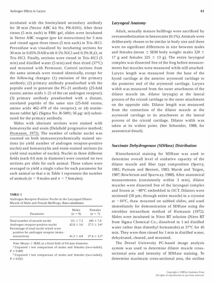
ifriaP3Tmattape(uamt
hHctnyfisaeo
L
odwa1cmLhtwdpofapta
S
dd11mmasainSfwmd
ss
T
AM
TAP
P
P
Androgen Effects in Larynx 61
ncubated with the biotinylated secondary antibodyor 30 min (Vector ABC kit No. PK-6101). After threeinses (5 min each) in PBS/gel, slides were incubatedn Vector ABC reagent (per kit instructions) for 5 minnd rinsed again three times (5 min each) in Tris–HCl.eroxidase was visualized by incubating sections for0 min in 0.05% DAB with 0.1% NiCl and 0.3% H2O2 inris–HCl. Finally, sections were rinsed in Tris–HCl (5in) and distilled water (5 min) and then dried (37°C)
nd mounted with Permount. Control sections fromhe same animals were treated identically, except forhe following changes: (1) omission of the primaryntibody; (2) primary antibody preadsorbed with theeptide used to generate the PG-21 antibody (25-foldxcess; amino acids 1–21 of the rat androgen receptor);3) primary antibody preadsorbed with a distant,nrelated peptide of the same size (25-fold excess;mino acids 462–478 of the receptor); or (4) nonim-une rabbit IgG (Sigma No. R-5001; 56 µg/ml) substi-
uted for the primary antibody.Slides with alternate sections were stained with
ematoxylin and eosin (Delafield progressive method;umason, 1972). The number of cellular nuclei was
ounted on both immunocytochemically stained sec-ions (to yield number of androgen receptor-positiveuclei) and hematoxylin and eosin-stained sections (toield total number of nuclei). Nuclei in three differentelds (each 0.6 mm in diameter) were counted on twoections per slide for each animal. These values wereveraged to yield a single value for each parameter forach animal so that n in Table 1 represents the numberf animals (n 5 9 males and n 5 7 females).
ABLE 1
ndrogen Receptor-Positive Nuclei in the Laryngeal Dilatoruscle of Male and Female Bullfrogs, Rana catesbeiana
ParameterMales(n 5 9)
Females(n 5 7)
otal number of muscle nuclei 111 6 7.3 100 6 7.4ndrogen receptor-positive nuclei 42.8 6 3.6 27.5 6 3.6a
ercentage of total nuclei which werepositive for androgen receptor immu-noreactivity 41.2 6 4.8 27.4 6 3.2b
Note. Means 6 SEM; in a fixed field of 0.6 mm diameter.a Unpaired t test comparison of males and females (two-tailed),# 0.009.b Unpaired t test comparison of males and females (two-tailed),
d# 0.032.
aryngeal Anatomy
Adult, sexually mature bullfrogs were sacrificed byveranesthetization in benzocaine (0.1%). Animals wereeliberately chosen to be similar in body size and thereere no significant differences in size between males
nd females (mean 6 SEM body weight: males 320 6
7 g and females 325 6 13 g). The entire laryngealomplex was dissected free of the frog before measure-ents but muscles were measured while still attached.
arynx length was measured from the base of theyoid cartilage at the anterior arytenoid cartilage to
he posterior end of the arytenoid cartilage. Larynxidth was measured from the outer attachment of theilator muscle (m. dilator laryngis) at the lateralrocess of the cricoid cartilage to the outer attachmentn the opposite side. Dilator length was measuredrom the connection of the dilator muscle at therytenoid cartilage to its attachment at the lateralrocess of the cricoid cartilage. Dilator width was
aken at its widest point. (See Schneider, 1988, fornatomical detail).
uccinate Dehydrogenase (SDHase) Distribution
Histochemical staining for SDHase was used toetermine overall level of oxidative capacity of theilator muscle and fiber type composition (Sperry,981; Putnam and Bennett, 1983; Marsh and Taigen,987; Rowlerson and Spurway, 1988). After anatomicaleasurements (consistently within 3 min), dilatoruscles were dissected free of the laryngeal complex
nd frozen at 280°C embedded in OCT. Dilators wereectioned (50 µm; through entire muscle) in a cryostatt 216°C, thaw mounted on subbed slides, and usedmmediately for demonstration of SDHase using theitroblue tetrazolium method of Humason (1972).lides were incubated in Nitro BT solution (Nitro BTrom Sigma Chemical Co.; dissolved in 1 ml distilled
ater rather than dimethyl formamide) at 37°C for 45in. They were then rinsed for 1 min in distilled water,
ehydrated, cleared, and mounted.The Drexel University PC-based image analysis
ystem was used to determine dilator muscle cross-ectional area and intensity of SDHase staining. To
etermine maximum cross-sectional area, the outlineCopyright r 1999 by Academic PressAll rights of reproduction in any form reserved.

odsmfswsiriv
wfwh(bmdco
R
ctwwplttrw(
ptbtbns
rdsmatcntfa
mewbaimfF
tdfig1a9vmn
F
ScMhc
62 Boyd et al.
CA
f the two largest sections from each animal wasrawn and the area was computed (in mm2) by theoftware, based on prior calibration with a stageicrometer. Both area measurements were averaged
or each animal. Relative overall intensity of SDHasetaining was measured by determining the gray levelithin a 1.4-mm2 box, centered over each section (two
ections per animal measured). A box this size wouldnclude between 20 and 100 fibers. Gray-level valuesanged from 0 (darkest) to 230 (lightest). Lighterntensity SDHase staining thus has a higher gray levelalue than darker intensity SDHase staining.The NTS5 tracing software system (Eutectics, Inc)as used to determine fiber diameter on tissue stained
or SDHase. The outline of at least 20 fibers per animalas drawn (403 power) and each fiber was coded as
aving dark, medium, or light staining for SDHaseSassoon et al., 1987). The software computed fiber areaased on prior calibration. Both male and femaleuscle possessed an eccentric area with smaller and
arker fibers. Data presented are only from the moreentral, homogeneous area which comprised the bulkf the muscle.
ESULTS
The laryngeal muscle of bullfrogs contained signifi-ant levels of androgen receptor-like immunoreac-ive material (Figs. 1C and 1D). This immunoreactivity
as located primarily over muscle fiber nuclei andas found in both sexes. Light immunoreactivity wasresent over fiber cytoplasm. Immunoreactivity was
ikely specific because staining was abolished whenhe antibody was preadsorbed with the peptide usedo produce the antibody (Fig. 1F), when nonimmuneabbit serum IgG replaced the primary antibody, andhen primary or secondary antibodies were omitted
data not shown). Staining was not affected when
IG. 1. Histochemical staining of the bullfrog laryngeal dilator mDHase staining in a male. (C) Androgen receptor-like immunocytocal staining in a female. (E) Staining remains when primary antibody
aterials and Methods). (F) Specific staining disappears when the aistology differs between A, B, and C–F because tissue stained for SD
ut longitudinally. Bar, 50 µm.opyright r 1999 by Academic Pressll rights of reproduction in any form reserved.
rimary antibody was preadsorbed with another pep-ide which was also part of the rat androgen receptorut not used for antibody production (Fig. 1E). Fur-her, immunoreactivity was also observed in anotherullfrog laryngeal muscle, the anterior constrictor, butot in the bullfrog esophageal smooth muscle (data nothown).
The distribution of androgen receptor-like immuno-eactivity was sexually dimorphic in bullfrog laryngealilator muscle (Table 1; Figs. 1C and 1D). Males hadignificantly more immunoreactive nuclei than fe-ales. The overall density of muscle nuclei in males
nd females (determined on a fixed-size field of hema-oxylin and eosin-stained sections) was not signifi-antly different. When the number of immunoreactiveuclei was expressed as a percentage of total nuclei in
hat field, the significant difference between males andemales remained. Males had approximately 13% morendrogen receptor-positive nuclei than females.There were significant sex differences also in larynxorphology (Table 2). The length and width of the
ntire larynx and also of the laryngeal dilator muscleas significantly greater (by about 30%) in male
ullfrogs, compared to females. The cross-sectionalrea of the dilator muscle was 42% larger in males thann females. Enzymatic activity in the laryngeal dilator
uscle, as determined by overall intensity of stainingor SDHase, was significantly greater in males (Table 2;igs. 1A and 1B).Finally, there was a sex difference in the size and
ype of muscle fibers which made up the bullfrogilator. Overall, the cross-sectional area of individualbers on SDHase-stained sections was significantlyreater in males than in females (Table 2; Figs. 1A andB). In addition, SDHase staining in male muscleppeared more homogeneous (Figs. 1B and 2A), with8% of cells having a medium-intensity staining andery light-staining fibers being virtually absent. Inales, fibers with different intensities of staining did
ot have significantly different areas (Fig. 2A). In
(A) Succinate dehydrogenase (SDHase) staining in a female. (B)l staining in a male. (D) Androgen receptor-like immunocytochemi-
dsorbed with a peptide from elsewhere in the androgen receptor (seeis preadsorbed with the peptide used to create the antibody. Grossas cut in cross-section while that stained for androgen receptor was
uscle.hemicais preantibodyHase w

63Copyright r 1999 by Academic Press
All rights of reproduction in any form reserved.

fissprmfa0fsftdF
D
mlflcfitouX
figav
T
MF
LLDDDD
M
s
F
s(fi
64 Boyd et al.
CA
emales, three classes of fibers with different stainingntensity could be recognized (Figs. 1A and 2B). Dark-tained fibers represented about 4.3% of the total fiberample, medium-stained fibers represented 39.4% (com-arable to most male fibers), and light-stained fibersepresented 56.3% of the fibers. In female bullfrog
uscle, there were significant differences in fiber areaor fibers with different staining intensities (Figs. 1And 2B; one-way ANOVA df 2,157; F 5 17.822; P ,
.001). Mean fiber area (6SEM) for dark fibers inemales was 697 6 127 µm2, mean area for medium-tained fibers was 1354 6 89 µm2, and mean fiber areaor light fibers was 1878 6 71 µm2. Areas of each of thehree fiber types in females (dark, medium, and light)iffered significantly from all other types (post hocisher’s LSD test).
ISCUSSION
Sex differences in laryngeal morphology of bullfrogsay be due to effects of plasma androgens on the
arynx. The larynx muscle tissue of both male andemale bullfrogs contained specific androgen receptor-ike immunoreactivity, but males exhibited a signifi-antly higher density of androgen receptors. This is therst time that androgen receptors have been located in
he anuran larynx with immunocytochemistry. Previ-usly, ligand-binding and molecular methods weresed to identify androgen receptors in the larynx ofenopus (Fischer and Kelley, 1991; Kelley, 1996). Our
ABLE 2
orphological Parameters of the Larynx in Male (n 5 5) andemale (n 5 8) Bullfrogs (Means 6 SEM)
Parametera Males Females
arynx length (cm) 2.26 6 0.05 1.49 6 0.02arynx width (cm) 2.52 6 0.10 1.87 6 0.05ilator muscle length (cm) 1.48 6 0.08 1.08 6 0.04ilator muscle width (cm) 0.66 6 0.05 0.43 6 0.02ilator cross-sectional area (mm2) 174.9 6 14 100.7 6 9ensity of SDHase staining (graylevel) 113.8 6 4 160.4 6 4uscle fiber cross-sectional area(all fiber types; µm2) 2080 6 121 1620 6 59
a Unpaired t test comparison between males and females found
tignificant differences (P # 0.001) in all parameters.opyright r 1999 by Academic Pressll rights of reproduction in any form reserved.
ndings in bullfrogs support the hypothesis that andro-en sensitivity of the larynx is a common feature ofnurans. Spectral and temporal properties of frogocalizations are dependent on larynx muscle fiber
IG. 2. Frequency histograms of individual muscle fiber cross-ectional area in the laryngeal dilator muscle of male (A) and femaleB) bullfrogs. Legend indicates the darkness of staining of individualbers for SDHase. Bin width was 100 µm2.
ype and mass of laryngeal structures (McAlister, 1961;

DMmi
tssrAga1a1ahbX1aboMtsaam
ultcaaaa(atssatl
ac
ssmgiomtfiascwbdba1nt
dfdSBaalmMFSfsR1fge
mc
Androgen Effects in Larynx 65
rewry et al., 1982; Ryan, 1986; Tobias and Kelley, 1988;cClelland et al., 1996). Sexual differences in larynxorphology may thus contribute to sexual differences
n calling.The antibody we used likely recognized an authen-
ic androgen receptor protein in bullfrogs because thetructure of the androgen receptor is relatively con-erved across vertebrates. The amphibian androgeneceptor has been cloned from Xenopus (GenBankccession No. U67129). Within the DNA-binding re-ion, rat and frog receptors are identical except for onemino acid substitution (He et al., 1990; Fischer et al.,993). Our antibody was raised against the first 21mino acids of the rat androgen receptor (Prins et al.,991). There is 71% homology between the first 21mino acids of the rat and frog receptors. On the otherand, there is minimal overlap in this same regionetween the Xenopus androgen receptor and otherenopus steroid hormone receptors (e.g., Weller et al.,987). It is thus unlikely that this antibody recognizednother steroid receptor in bullfrogs. This antibody haseen previously used to identify androgen receptors inther tissues in amphibians (Dorlochter et al., 1994;atsumoto et al., 1996). Similarity of androgen recep-
or structure between amphibians and rats is alsoupported by cross-reactivity of other antibodies (Davisnd Moore, 1996), by ligand-binding studies (Lupo etl., 1993), and by cDNA probe hybridization experi-ents (Varriale and Serino, 1994).Sex differences in androgen responsiveness may
nderlie morphological differences in the bullfrogarynx. Male bullfrog laryngeal dilator muscle con-ained 13% more androgen receptor-positive cell nu-lei than female muscle. This difference alone mayccount for sexual differences in larynx morphologynd the call which is ultimately produced. In Xenopus,ndrogens stimulate myogenesis, increases in fiberrea and sex-typical changes in fiber type compositionKelley, 1996). Androgens also increase fiber diameternd change fiber type in forelimb muscles involved inhe sexual amplectic clasp (Muller et al., 1969; Rubin-tein et al., 1983; Dorlochter et al., 1994). Bullfrogs wereacrificed during the breeding season when plasmandrogens are high (Licht et al., 1983; Boyd, 1992). Athis time of year, plasma androgens are usually equiva-
ent in both sexes so sex differences in circulating gndrogens are unlikely to account for all differences inalling behavior.
Muscle fiber type in the bullfrog larynx was alsoexually dimorphic. Based on the area and SDHasetaining of fibers in the bullfrog larynx, the male larynxuscle contained primarily fast-twitch oxidative/
lycolytic fibers (FOG fibers; corresponding to amphib-an type 2 or mammalian type IIa fibers; nomenclaturef Gans and De Gueldre, 1992). In size and staining,ale bullfrog laryngeal fibers were most similar to
hose in the frog hindlimb muscle although ratio ofber types differs (Lannergren and Smith, 1966; Smithnd Ovalle, 1973; Putnam and Bennett, 1983; Rowler-on and Spurway, 1988). Male bullfrog larynx may alsoontain fast-twitch glycolytic fibers because the fibersith the largest diameter were also lightest staining
ut statistical analysis did not prove the existence of aistinctly different set of fibers. Laryngeal muscles ofoth Xenopus and Hyla versicolor have been describeds consisting entirely of FOG fibers (Marsh and Taigen,987; Sassoon et al., 1987). Our finding of this predomi-ant fiber type in bullfrog larynx muscle is thus similar
o two other anuran species.Muscle fiber types in laryngeal dilator muscles
iffered significantly between male and female bull-rogs. Female bullfrog dilator muscle contained threeistinct classes of fibers (based on Lannergren andmith, 1966; Smith and Ovalle, 1973; Putnam andennett, 1983; Lannergren and Hoh, 1984; Rowlersonnd Spurway, 1988; Rowlerson, 1994). Large-diameternd light-staining fibers were likely fast-twitch glyco-ytic fibers (corresponding to amphibian type 1 and
ammalian type IIB; Gans and De Gueldre, 1992).edium-size and medium-staining fibers were likely
OG fibers and similar to those in male bullfrogs.mall, dark fibers were probably also FOG fibers butrom a subgroup with a slower twitch which corre-ponds to amphibian type 3 (and mammalian type I;owlerson and Spurway, 1988; Gans and De Gueldre,992; Rowlerson, 1994). In Xenopus and Hyla arborea,emales also possess a mixture of fiber types in laryn-eal muscle (Eichelberg and Schneider, 1974; Sassoont al., 1987).
Species differences in call rate, call amplitude, andechanisms of sound production are likely to be
orrelated with variation in the composition of laryn-
eal muscles. Bullfrog laryngeal dilator muscle fibersCopyright r 1999 by Academic PressAll rights of reproduction in any form reserved.

wi5tbg1imobmawBadrttsbd(b(w
cflGmamdboobfccs(mag
tna
A
HpGG
R
B
B
C
C
C
D
d
D
D
E
E
F
F
66 Boyd et al.
CA
ere significantly larger than fibers in the same musclen Xenopus (2080 µm2 area in bullfrogs compared with–25 µm2 in Xenopus; Sassoon et al., 1987). It is unlikelyhat this difference is due to differences in body sizeetween the two species because muscle fiber size isenerally unrelated to body size (Putnam and Bennett,983; Marsh and Taigen, 1987). The intensity of stain-ng for SDHase also differed significantly between
ale Xenopus (‘‘very dark’’ corresponding to highxidative capacity; Sassoon et al., 1987) and maleullfrogs (‘‘medium’’). Fiber type composition andetabolic enzyme levels of muscle are related to
ctivity levels in frogs. This is true across species,ithin species, and within individuals (Engel, 1965;ennett, 1974; Cummings, 1979; Sperry, 1981; Givennd McKay, 1990). Very small fiber diameter and veryark staining for SDHase in Xenopus larynx mayepresent specializations for high call rates. The adver-isement call of male Xenopus requires contraction ofhe dilator muscle at a rate of about 71 times perecond (Kelley and Gorlick, 1990). In contrast, theullfrog advertisement call is much slower, with theilator muscle contracting only about twice a second
Capranica, 1965). The energetic costs of calling inullfrogs are modest, compared to some other anuranse.g., Taigen et al., 1985), and this may be associated
ith lower levels of SDHase in laryngeal muscle.The laryngeal dilator muscle plays a critical role in
all production by opening the glottis and allowing airow from the lungs to the buccal cavity (deJongh andans, 1969). Androgen receptor localization in thisuscle supports the hypothesis that this tissue is
ndrogen sensitive. Plasma androgens in bullfrogsay thus be responsible for morphological sexual
ifferences in the larynx and sex differences in callingehavior. In male bullfrogs, the dilator muscle is largerverall, individual fibers have larger diameters andxidative capacity is greater, compared to the femaleullfrog dilator muscle. Male laryngeal muscle there-ore has the potential to generate more force whichould, in turn, result in greater call duration and longeralling bouts. The advertisement call of males doeshow these differences from the female release callCapranica, 1965). It is unlikely however that these
uscle differences represent absolute constraints ondvertisement calling by female bullfrogs. Females
ive no advertisement call whatsoever—not even oneopyright r 1999 by Academic Pressll rights of reproduction in any form reserved.
hat is shorter and simpler. Dimorphisms in the centralervous system thus likely account for the lack of andvertisement call in female bullfrogs.
CKNOWLEDGMENTS
We thank Kevin Bartz, Lynn Birch, Sharon Emerson, Lisa Guerra-osler, Cindy Mitchell-Herpolsheimer, and Michelle Tito for helperforming and interpreting these experiments. Supported by NIHrant HD24653 and NSF Grant IBN95–14305 to S.K.B. and NIHrant DK40890 to G.S.P.
EFERENCES
ennett, A. F. (1974). Enzymatic correlates of activity metabolism inanuran amphibians. Am. J. Phys. 226, 1149–1151.
oyd, S. K. (1992). Sexual differences in hormonal control of releasecalls in bullfrogs. Horm. Behav. 26, 522–535.
apranica, R. R. (1965). ’’The Evoked Vocal Response of the Bull-frog.’’ MIT Press, Cambridge, MA.
apranica, R. R. (1968). The vocal repertoire of the bullfrog (Ranacatesbeiana). Behavior 31, 302–325.
ummings, J. W. (1979). Physiological and biochemical adaptationsto training in Rana pipiens. J. Comp. Physiol. 134, 345–350.avis, G. A., and Moore, F. L. (1996). Neuroanatomical distributionof androgen and estrogen receptor-immunoreactive cells in thebrain of the male roughskin newt. J. Comp. Neurol. 372, 294–308.
eJongh, H. J., and Gans, C. (1969). On the mechanism of respirationin the bullfrog, Rana catesbeiana: A reassessment. J. Morphol. 127,259–290.orlochter, M., Astrow, S. H., and Herrera, A. A. (1994). Effects oftestosterone on a sexually dimorphic frog muscle: Repeated invivo observations and androgen receptor distribution. J. Neurobiol.25, 897–916.rewry, G. E., Heyer, W. R., and Rand, A. S. (1982). A functionalanalysis of the complex call of the frog Physalaemus pustulosus.Copeia 1982, 636–645.
ichelberg, H., and Schneider, H. (1974). The fine structure of thelarynx muscles in female tree frogs, Hyla a. arborea L. (Anura,Amphibia). Cell. Tissue Res. 152, 185–191.
ngel, W. K. (1965). Diseases of the neuromuscular junction andmuscle. In ‘‘Neurohistochemistry’’ (C. W. M. Adams, ,Ed.), pp.622–672. Elsevier, Amsterdam.
ischer, L. M., and Kelley, D. B. (1991). Androgen receptor expressionand sexual differentiation of effectors for courtship song inXenopus laevis. Semin. Neurosci. 3, 469–480.
ischer, L., Catz, D., and Kelley, D. (1993). An androgen receptormRNA isoform associated with hormone-induced cell prolifera-
tion. Proc. Natl. Acad. Sci. USA. 90, 8254–8258.
F
G
G
G
H
H
K
K
K
K
L
L
L
L
M
M
M
M
M
M
M
M
M
P
P
R
R
R
R
R
R
R
R
S
S
S
Androgen Effects in Larynx 67
ischer, L. M., Catz, D., and Kelley, D. B. (1995). Androgen-directeddevelopment of the Xenopus laevis larynx: Control of androgenreceptor expression and tissue differentiation. Dev. Biol. 170,115–126.ans, C. (1973). Sound production in the Salientia: Mechanisms andevolution of the emitter. Am. Zool. 13, 1179–1194.ans, C., and De Gueldre, G. (1992). Striated muscle: physiology andfunctional morphology. In ‘‘Environmental Physiology of theAmphibians’’ (M. E. Feder, and W. W. Burggren, Eds.), pp.277–313. Univ. of Chicago Press, Chicago, IL.iven, M. F., and McKay, D. M. (1990). Variation in the citratesynthase activity in calling muscles of carpenter frogs, Ranavirgatipes. Copeia 1990, 863–867.e, W. W., Fischer, L. M., Sun, S., Bilhartz, D. L., Zhu, X., Young,C. Y.-F., Kelley, D. B., and Tindall, D. J. (1990). Molecular cloning ofandrogen receptors from divergent species with a polymerasechain reaction technique: Complete cDNA sequence of the mouseandrogen receptor and isolation of androgen receptor cDNAprobes from dog, guinea pig and clawed frog. Biochem. Biophys.Res. Commun. 171, 697–704.umason, G. L. (1972) ’’Animal Tissue Techniques,’’ 3rd ed. Free-man, San Francisco.
elley, D. B. (1996). Sexual differentiation in Xenopus laevis. In ‘‘TheBiology of Xenopus’’ (R. C. Tinsley, and H. R. Kobel, Eds.), pp.143–176. Oxford Univ. Press, Oxford.
elley, D. B., and Gorlick, D. L. (1990). Sexual selection and thenervous system. BioScience 40, 275–283.
elley, D., Sassoon, D., Segil, N., and Scudder, M. (1989). Develop-ment and hormone regulation of androgen receptor levels in thesexually dimorphic larynx of Xenopus laevis. Dev. Biol. 131, 111–118.
usunoki, M., Satou, M., and Ueda, K. (1986). An electromyographicanalysis of calling behavior in the Japanese toad, Bufo japonicus.Zool. Sci. 3, 395–399.
annergren, J., and Hoh, J. F. Y. (1984). Myosin isoenzymes in singlemuscle fibres of Xenopus laevis: Analysis of five different functionaltypes. Proc. R. Soc. London B 222, 401–408.
annergren, J., and Smith, R. S. (1966). Types of muscle fibres in toadskeletal muscle. Acta Phys. Scand. 68, 263–274.
icht, P., McCreery, B. R., Barnes, R., and Pang, R. (1983). Seasonaland stress related changes in plasma gonadotropins, sex steroids,and corticosterone in the bullfrog, Rana catesbeiana. Gen. Comp.Endocrinol. 50, 124–145.
upo, C., Lodi, L., Giacoma, C., and Halliday, T. R. (1993). Testoster-one binding sites in the brain, plasma sex hormones and reproduc-tive behaviour in males of the toad Bufo bufo. Behav. Proc. 30,93–102.arsh, R. L., and Taigen, T. L. (1987). Properties enhancing aerobiccapacity of calling muscles in gray tree frogs Hyla versicolor. Am. J.Phys. 252, R786–R793.artin, W. F. (1972). Evolution of vocalization in the genus Bufo. In‘‘Evolution in the Genus Bufo’’ (W. F. Blair, ,Ed.), pp. 279–309. Univ.of Texas Press, Austin, TX.atsumoto, A., Arai, Y., Toyoda, F., Kikuyama, S., and Prins, G. S.(1996). Immunohistochemical analysis of androgen receptor in theabdominal glands of the cloaca of male red-bellied newts, Cynops
pyrrhogaster. Zool. Sci. 13, 429–433.cAlister, W. H. (1959). The vocal structures and method of callproduction in the genus Scaphiopus Holbrook. Texas J. Sci. 11,60–77.cAlister, W. H. (1961). The mechanics of sound production inNorth American Bufo. Copeia 1961, 86–95.cClelland, B. E., and Wilczynski, W. (1989). Sexually dimorphiclaryngeal morphology in Rana pipiens. J. Morphol. 201, 293–299.cClelland, B. E., Wilczynski, W., and Ryan, M. J. (1996). Correla-tions between call characteristics and morphology in male cricketfrogs (Acris crepitans). J. Exp. Biol. 199, 1907–1919.cClelland, B. E., Wilczynski, W., and Rand, A. S. (1997). Sexualdimorphism and species differences in the neurophysiology andmorphology of the acoustic communication system of two neotropi-cal hylids. J. Comp. Physiol. A 180, 451–462.uller, E. R. A., Galavazi, G., and Szirmai, J. A. (1969). Effect ofcastration and testosterone treatment on fiber width of the flexorcarpi radialis muscle in the male frog (Rana temporaria L.). Gen.Comp. Endocrinol. 13, 275–284.
rins, G. S., Birch, L., and Greene, G. L. (1991). Androgen receptorlocalization in different cell types of the adult rat prostate.Endocrinology 129, 3187–3199.
utnam, R. W., and Bennett, A. F. (1983). Histochemical, enzymatic,and contractile properties of skeletal muscles of three anuranamphibians. Am. J. Phys. 244, R558–R567.
abb, G. B. (1960). On the unique sound production of the surinamtoad, Pipa pipa. Copeia 1960, 368–369.
idewood, W. G. (1897). On the structure and development of thehyobranchial skeleton and larynx in Xenopus and Pipa, withremarks on the affinities of the Aglossa. J. Linn. Soc. London Zool.26, 53–128.
idewood, W. G. (1900). On the hyobranchial skeleton and larynx ofthe new aglossal toad, Hymenochirus boettgeri. J. Linn. Soc. LondonZool. 27, 454–460.
owlerson, A. (1994). An outline of fibre types in vertebrate skeletalmuscle: histochemical identification and myosin isoforms. Bas.Appl. Myol. 4, 333–352.
owlerson, A. M., and Spurway, N. C. (1988). Histochemical andimmunohistochemical properties of skeletal muscle fibres fromRana and Xenopus. Histochem. J. 20, 657–673.
ubinstein, N. A., Erulkar, S. D., and Schneider, G. T. (1983). Sexualdimorphism in the fibers of a ‘‘clasp’’ muscle of Xenopus laevis.Exp. Neurol. 82, 424–431.
yan, M. J. (1986). Factors influencing the evolution of acousticcommunication: Biological constraints. Brain Behav. Evol. 28, 70–82.
yan, M. J., and Drewes, R. C. (1990). Vocal morphology of thePhysalaemus pustulosus species group (Leptodactylidae): Morpho-logical response to sexual selection for complex calls. Biol. J. Linn.Soc. 40, 37–52.
assoon, D. A., Gray, G. E., and Kelley, D. B. (1987). Androgenregulation of muscle fiber type in the sexually dimorphic larynx ofXenopus laevis. J. Neurosci. 7, 3198–3206.
chmidt, R. S. (1965). Larynx control and call production in frogs.Copeia 1965, 143–147.
chmidt, R. S. (1972a). Action of intrinsic laryngeal muscles during
release calling in leopard frog. J. Exp. Zool. 181, 233–244.Copyright r 1999 by Academic PressAll rights of reproduction in any form reserved.

S
S
S
S
S
S
T
T
T
V
W
W
68 Boyd et al.
CA
chmidt, R. S. (1972b). Release calling and inflating movements inanurans. Copeia 1972, 240–245.
chmidt, R. S. (1973). Vocal cord mechanisms of release calling innorthern leopard frog. Copeia 1973, 624–627.
chneider, H. (1988). Peripheral and central mechanisms of vocaliza-tion. In ‘‘The Evolution of the Amphibian Auditory System’’ (B.Fritzsch, M. J. Ryan, W. Wilczynski, T. E. Hetherington, and W.Walkowiak, Eds.), pp. 537–558. Wiley, New York.
egil, N., Silverman, L., and Kelley, D. B. (1987). Androgen-bindinglevels in a sexually dimorphic muscle of Xenopus laevis. Gen. Comp.Endocrinol. 66, 95–101.
mith, R. S., and Ovalle, W. K. (1973). Varieties of fast and slowextrafusal muscle fibres in amphibian hind limb muscles. J. Anat.116, 1–24.
perry, D. G. (1981). Fiber type composition and postmetamorphicgrowth of anuran hindlimb muscles. J. Morphol. 170, 321–345.
aigen, T. L., Wells, K. D., and Marsh, R. L. (1985). The enzymatic
opyright r 1999 by Academic Pressll rights of reproduction in any form reserved.
basis of high metabolic rates in calling frogs. Phys. Zool. 58,719–726.
obias, M. L., and Kelley, D. B. (1988). Electrophysiology anddye-coupling are sexually dimorphic characteristics of individuallaryngeal muscle fibers in Xenopus laevis. J. Neurosci. 8, 2422–2429.
rewavas, E. (1933). The hyoid and larynx of the Anura. Phil. Trans.R. Soc. London B Biol. Sci. 222, 401–525.
arriale, B., and Serino, I. (1994). The androgen receptor mRNA isup-regulated by testosterone in both the harderian gland andthumb pad of the frog, Rana esculenta. J. Steroid Biochem. Mol. Biol.51, 259–265.eller, I. J., Lew, D., and Shapiro, D. J. (1987). The Xenopus laevisestrogen receptor: Sequence homology with human and avianreceptors and identification of multiple estrogen receptor messen-ger ribonucleic acids. Mol. Endocrinol. 1, 355–362.itschi, E., Bruner, J. A., and Van Bergeijk, W. A. (1953). Sound
production by Xenopus. Anat. Rec. 117, 636.Copyright © 2022 FDOKUMEN