Novel Regulators of Feeding and Cardiovascular Physiology ...
Upload
independentCategory
view
3download
0
The Laryngoscope Lippincott-Raven Publishers, Philadelphia 0 1998 The American Larvnpoloeical. Rhinological and Otologicil Soci&y, Inc.
Regulators of Proliferation and Apoptosis in Carcinoma of the Larynx Laura C. Whisler, MD; Nancy B. Wood, PhD; David D. Caldarelli, MD; James C. Hutchinson, MD; William R. Panje, MD; Michael Friedman, MD; Harvey D. Preisler, MD; Sue Leurgans, PhD; Jan Nowak, MD, PhD; John S. Coon, MD, PhD
Expression of interrelated gene products regulat- ing cell proliferation and apoptosis may be disordered in squamous cell carcinoma (SCC) of the larynx com- pared with normal squamous mucosa. Certain of these abnormalities, alone or in combination, may be of prognostic significance in low-stage carcinomas of the larynx. A retrospective study of archival material was made. Expression of the Bcl-2 family of apoptosis- related genes (bcl-2, bcl-X, mcl-1, and bax) and the pro- liferation- and apoptosis-related genes p53 and cyelin D-1 were determined in 40 low-T-stage laryngeal car- cinomas and in uvular epithelium from patients with- out SCC. Among the antiapoptotic members of the Bcl-2 family, Bcl-X and Mcl-1 showed more intense and widespread staining than Bcl-2 itself in both normal squamous mucosa and SCC. The well-ordered expres- sion patterns of Bcl-2-related proteins founci in nor- mal epithelium were lost in SCC, and patterns of ex- pression varied widely among individual tumors. Also, mean expression levels for Bax and cyclin D-1 were significantly lower than in normal epithelium (P = .036 and P = .009, respectively), whereas expres- sion of p53 was higher in tumors (P = .034). Expression of Bcl-X and Mcl-1 was greater in poorly differenti- ated than in well-differentiated tumors (P = .014 and P = .031, respectively). No associations were seen be- tween marker expression patterns and clinical out- come in this group of patients. Bcl-x and Mcl-1 appear to be the most abundantly expressed antiapoptotic proteins of the Bcl-2 family in both normal squamous mucosa and SCC of the larynx. Multiple genes regu- lating proliferation and apoptosis are expressed ab-
Presented at the Meeting of the Middle Section of the American Laryngological, Rhinological and Otological Society, Inc., Kansas City, Mis- souri, January 25,1997.
From the Departments of Otolaryngology and Bronchoesophagology (L.c.w., D.D.c., J.c.H., w.R.P., J.s.c., M.F.), Pathology (N.B.w., J.s.c.), Preventive Medicine (s.L.), and Oncology (H.D.P.), Rush Medical College, Rush-Presby- t e n a n a t . Luke’s Medical Center, Chicago, Illinois; and the Advanced Cen- ter for Specialty Care (M.F.) and the Department of Pathology (J.N.), Illinois Masonic Medical Center, Chicago, Illinois.
Supported in part by the Stanton A. Friedberg Chair of Otolaryn-
Send Reprint Requests to Laura Whisler, MD, Department of Oto- laryngology and Bronchoesophagology, Rush-Presbyterian-St. Luke’s Med- ical Center, 1653 West Congress Parkway, Chicago, IL, 60612, U.S.A.
gology.
normally in laryngeal SCC compared with normal ep- ithelium. In particular, loss or measurable decrease in expression of the proapoptotic protein Bax in tumors may contribute to the deranged growth control of SCC. Further study is needed to evaluate the prog- nostic significance of particular patterns of disor- dered expression of proteins regulating proliferation and apoptosis in SCC of different head and neck sites.
Laryngoscope, 108630-638,1998
INTRODUCTION Squamous cell carcinoma (SCC) of the glottic larynx
is biologically distinct from SCC of other sites in the head and neck.1 The resistance of the glottis to microscopic spread of tumor appears to be related in part to the paucity of lymphatics. Patients with TlNO and T2NO glot- tic carcinoma have a 5-year cure rate of approximately 90% when treated with local excision or radiation therapy. Supraglottic tumors are usually diagnosed a t a more ad- vanced stage, with a 5-year cure rate for TlNO and T2NO lesions of approximately 80% to 85%.1 Nevertheless, some of these patients do not respond well to treatment and de- velop local recurrence, metastases, or a second primary SCC. Understanding the biology of SCC at the molecular level may allow discrimination of low-stage laryngeal SCC likely to progress or respond poorly to conventional treat- ment, and may lead to new therapeutic approaches.
Recently there has been progress in understanding how cell proliferation and programmed cell death (apop- tosis) are regulated in normal cells and how this regula- tion is frequently disrupted in cancer. A network of inter- related gene products that interact to regulate these vital processes in virtually all multicellular organisms, includ- ing humans, has been identified.2-4 The details of these in- teractions appear to be cell type-specific, however, and de- lineating the precise role of the various factors in most human tissues is at an early stage. Bcl-2, p53, and cyclin D-1 genes are abnormal or are abnormally expressed in many human cancers, but reports concerning the signifi- cance for patient outcome vary from tumor to tumor and are often contradictory, even for a particular tumor or group of tumors, specifically SCC of the head and neck.
Laryngoscope 108: May 1998
630
Whisler et al.: Larynx Carcinoma
The basic objective of this investigation was to find out which members of the Bcl-2 family of apoptosis- related proteins are expressed in normal squamous mu- cosa of the head and neck and in SCC, using laryngeal car- cinoma as a model. Although Bcl-2 itself has been previously studied by ourselves and others, expression of other potentially important family members, particularly Bcl-X, Mcl-1, and Bax,4,5 have not been described in this context. We sought to determine whether the expression patterns observed in normal squamous epithelium were disrupted in SCC and might therefore potentially account for the proliferative abnormalities and treatment resis- tance of this malignancy. Since members of the Bcl-2 fam- ily interact importantly with p53 and cyclin D-1, it was of interest to determine whether abnormalities of the Bcl-2 family were associated with expression patterns of these other factors.6-9 In addition, we sought associations be- tween expression patterns of this family of factors and clinical outcome in low-stage laryngeal cancers, because early identificatiosi of patients unlikely to respond to stan- dard treatment might be of particular clinical utility, as well as scientific interest, in this group of patients.
MATERIALS AND METHODS Patients
Information was obtained from the records of patients treated for T1 and T2 laryngeal SCC between 1990 and 1995 at Rush or Illinois Masonic medical centers, who had at least 1 year of follow-up (mean, 5 years), unless they had died of their tumor within a year of presentation. The archival paraffin tissue blocks of 40 patients fulfilling these criteria were available for study; 36 of these patients were treated surgically and four by radiation. Seven women and 33 men (age range, 37 to 87 years; mean age, 60 years) were included. Five patients with nodal metastases at presentation (all of whom had supraglottic tumors) were ex- cluded from outcome analysis, leaving 25 glottic and 10 supra- glottic tumors without detectable nodal or distant metastases for clinical outcome analysis. Favorable treatment outcome was de- fined as undetectable disease at the most recent follow-up. Unfa- vorable treatment outcome was defined as follows: 1. tumor re- currence, either locally or distant; 2. development of a second SCC of the aerodigestive tract; or 3. death from tumor.5 To deter- mine whether the expression of regulators of apoptosis and pro- liferation in SCC differed from normal squamous mucosa, archival tissue blocks from a reference population of 14 non- smoking adult patients without SCC (matched for gender with the tumor population and with a mean age of 45 years) who had undergone uvulopalatopharyngoplasty for obstructive sleep ap- nea were studied.
Measurement of Marker Expression In preparation for antibody staining, paraffin tissue sec-
tions (5 pm, freshly cut) were deparaffinized and rehydrated us- ing standard technique and subjected to a microwave antigen re- trieval for 48 minutes in citrate buffer.10 The tissue was stained using the Ventana ES320 Histo-stainer (Ventana Medical Sys- tems, Tucson, AZ), using supplied diaminobenzidine and avidin- biotin conjugate immunoperoxidase chemistry.llJ2 The procedure was substantially the same for all markers, except for the pri- mary antibody (Ab). Tissue sections were stained at a 1:40 con- centration for Bcl-2, using the monoclonal Ab (clone 124) supplied by DAKO (Carpenteria, CA) as described.7 For p53 the DO-1 monoclonal Ab (Calbiochem, San Diego, CA) was used at a con- centration of 1:lOOO as described.9 Cyclin D-1 expression was de-
tected as described,’s using the NCL-CYCLIN D-1 monoclonal Ab (Novacastra, Newcastle-upon-Tyne, UK) at a concentration of 1:25. Rabbit polyclonal Ab (Santa Cruz, Santa Cruz, CA) was used for detection of Bax (Ab N-20, lot C216, a t 1:400), Bcl-X (Ab S-18, lot (2126, a t 1:100), and Mcl-1 (Ab S-19, lot K085, a t 1:200). Optimal rabbit antibody concentrations were determined by titration, using subsets of benign lymphoid cells visualized in archival sections of human tonsils extirpated for inflammatory disease. A single block was selected for analysis for each patient on the basis of having the greatest area of viable tumor. Im- munostaining of the entire tumor area on each slide was scored subjectively on a rating scale of 0 to 4+ (cell counting was not per- formed) as described,I3 by two observers (L.c.w. and J.s.c.) inde- pendently, who always agreed within one scoring point on each of the cases. Scoring differences were resolved by consensus. Less than 1% of positive cells rated 0, 1% to 10% rated 1+, 10% to 35% rated 2+, 36% to 70% rated 3+, and more than 70% rated 4+. Nor- mal small lymphocytes, present in every tumor section, served as an internal positive control for Bcl-2 staining. Previously selected positive and negative control slides were stained for all other markers (for p53, a breast carcinoma with a mutation in exon 7). Human tonsils were used as controls for Bax, Bcl-X, and Mcl-1, where the previously reported and characteristic cytoplasmic ex- pression patterns in large and small lymphocytes were ob- served.14-16 Staining specificity for these proteins was further verified by specific inhibition of staining by a 10-fold excess of the immunizing peptide (Santa Cruz).
Statistical Analysis Because tumor marker level was recorded on a scale of 0 to
4, all analyses of markers employ nonparametric statistics. The distributions of markers in tumor tissue were compared with the distributions in uvular tissue using Wilcoxon’s rank sum test. The distributions of marker levels for different differentiation grades were compared using the Kruskal-Wallis test. Spearman’s correlation coefficient was used to measure the associations among the markers. The Mann-Whitney U test was used to test for associations between marker expression and clinical outcome. All P values are two-tailed. Statistical significance was identified as P < .05.
RESULTS Comparison of Expression Patterns
Because expression of Bax, Bcl-X, and Mcl-1 had not previously been described in squamous mucosa or SCC of the head and neck, we investigated their ex- pression to determine whether these proteins might potentially play a role in regulating apoptosis in this particular tissue and its carcinomas. It was also deemed important to compare expression of each pro- tein in normal epithelium and carcinoma to determine whether altered expression could potentially be linked with behavioral abnormalities of tumor cells com- pared with normal squamous mucosa. In this ex- ploratory study, expression patterns were compared descriptively, and the proportion of immunostained cells of SCC versus benign squamous cells were com- pared semiquantitatively via a widely used scoring system.
In addition to the previously documented expres- sion of Bcl-2 in SCC and normal squamous epithe- lium,7 multiple other members of the Bcl-2 family were also detectable in our patients. Bcl-2 itse\fi,in- variably showed a finely granular cytoplasmic distrib-
Laryngoscope 108: May 1998 Whisler et al.: Larynx Carcinoma
63 1
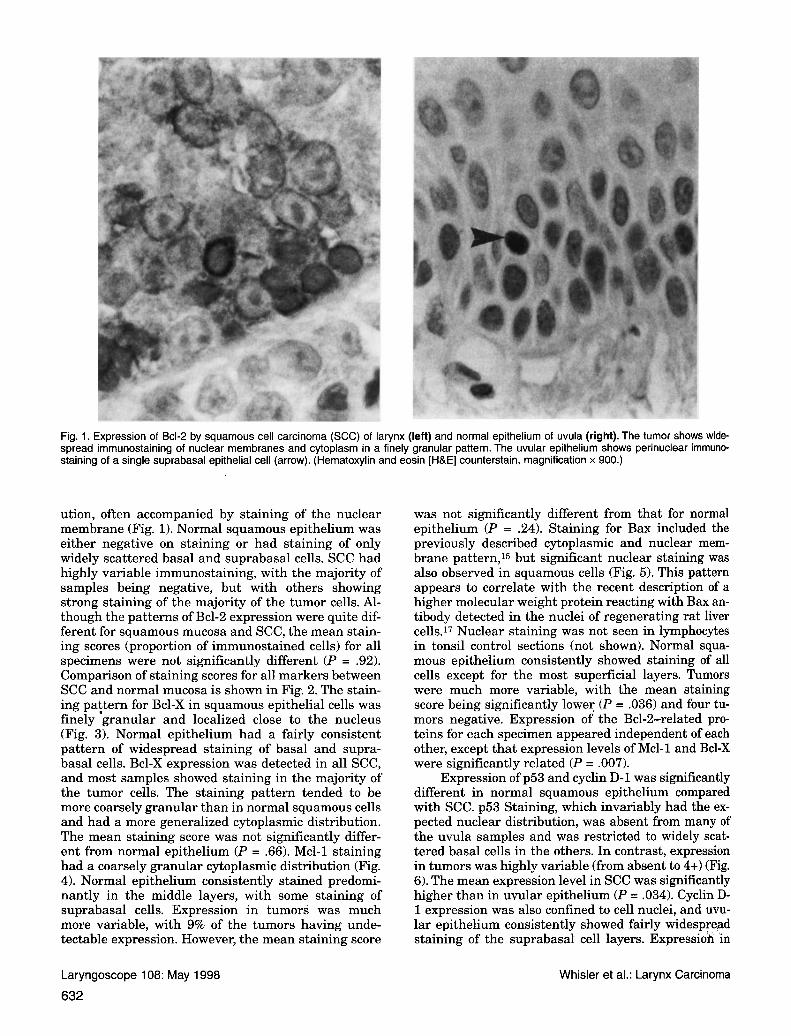
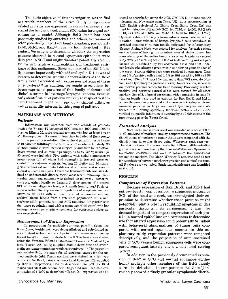
Fig. 1. Expression of Bcl-2 by squamous cell carcinoma (SCC) of larynx (left) and normal epithelium of uvula (right). The tumor shows wide- spread immunostaining of nuclear membranes and cytoplasm in a finely granular pattern. The uvular epithelium shows perinuclear immuno- staining of a single suprabasal epithelial cell (arrow). (Hematoxylin and eosin [H&E] counterstain, magnification x 900.)
ution, often accompanied by staining of the nuclear membrane (Fig. 1). Normal squamous epithelium was either negative on staining or had staining of only widely scattered basal and suprabasal cells. SCC had highly variable immunostaining, with the majority of samples being negative, but with others showing strong staining of the majority of the tumor cells. Al- though the patterns of Bcl-2 expression were quite dif- ferent for squamous mucosa and SCC, the mean stain- ing scores (proportion of immunostained cells) for all specimens were not significantly different (P = .92). Comparison of staining scores for all markers between SCC and normal mucosa is shown in Fig. 2. The stain- ing paitern for Bcl-X in squamous epithelial cells was finely granular and localized close to the nucleus (Fig. 3). Normal epithelium had a fairly consistent pattern of widespread staining of basal and supra- basal cells. Bcl-X expression was detected in all SCC, and most samples showed staining in the majority of the tumor cells. The staining pattern tended to be more coarsely granular than in normal squamous cells and had a more generalized cytoplasmic distribution. The mean staining score was not significantly differ- ent from normal epithelium (P = .66). Mcl-1 staining had a coarsely granular cytoplasmic distribution (Fig. 4). Normal epithelium consistently stained predomi- nantly in the middle layers, with some staining of suprabasal cells. Expression in tumors was much more variable, with 9% of the tumors having unde- tectable expression. However, the mean staining score
was not significantly different from that for normal epithelium (P = .24). Staining for Bax included the previously described cytoplasmic and nuclear mem- brane pattern,l5 but significant nuclear staining was also observed in squamous cells (Fig. 5). This pattern appears to correlate with the recent description of a higher molecular weight protein reacting with Bax an- tibody detected in the nuclei of regenerating rat liver cells.17 Nuclear staining was not seen in lymphocytes in tonsil control sections (not shown). Normal squa- mous epithelium consistently showed staining of all cells except for the most superficial layers. Tumors were much more variable, with the mean staining score being significantly lower (P = .036) and four tu- mors negative. Expression of the Bcl-2-related pro- teins for each specimen appeared independent of each other, except that expression levels of Mcl-1 and Bcl-X were significantly related (P = .007).
Expression of p53 and cyclin D-1 was significantly different in normal squamous epithelium compared with SCC. p53 Staining, which invariably had the ex- pected nuclear distribution, was absent from many of the uvula samples and was restricted to widely scat- tered basal cells in the others. In contrast, expression in tumors was highly variable (from absent to 4+) (Fig. 6) . The mean expression level in SCC was significantly higher than in uvular epithelium (P = .034). Cyclin D- 1 expression was also confined to cell nuclei, and uvu- lar epithelium consistently showed fairly widespread staining of the suprabasal cell layers. Expressioh 'in
Laryngoscope 108: May 1998
632
Whisler et al.: Larynx Carcinoma
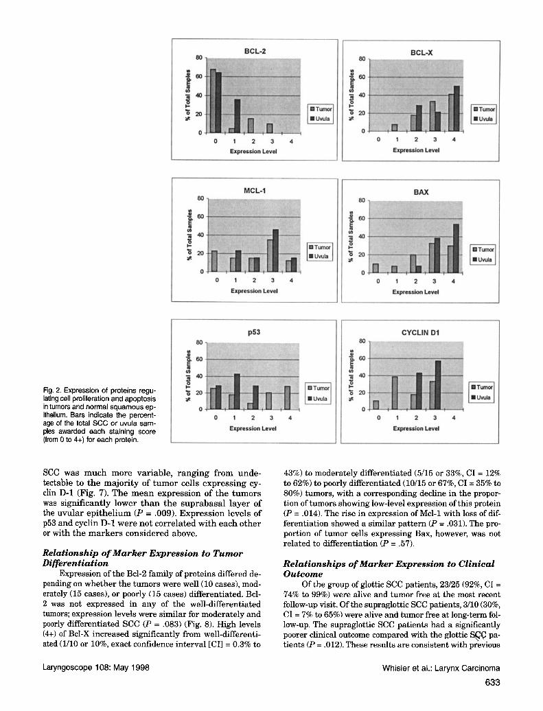
Fig. 2. Expression of proteins regu- lating cell proliferation and apoptosis in tumors and normal squamous ep- ithelium. Bars indicate the percent- age of the total SCC or uvula sam- ples awarded each staining score (from 0 to 4+) for each protein.
BCL-2 80
E " s m %
0 0 1 2 3 4
Expression Level
I I MCL-I 80
:" Uvula
8 I-
o m %
L.
0 0 1 2 3 4
Expression Level
P53 80
I- Tumor
% s m
0 0 1 2 3 4
Expression Level
SCC was much more variable, ranging from unde- tectable to the majority of tumor cells expressing cy- clin D-1 (Fig. 7). The mean expression of the tumors was significantly lower than the suprabasal layer of the uvular epithelium (P = .009>. Expression levels of p53 and cyclin D-1 were not correlated with each other or with the markers considered above.
Relationship of Marker Expression to Tumor Differentiation
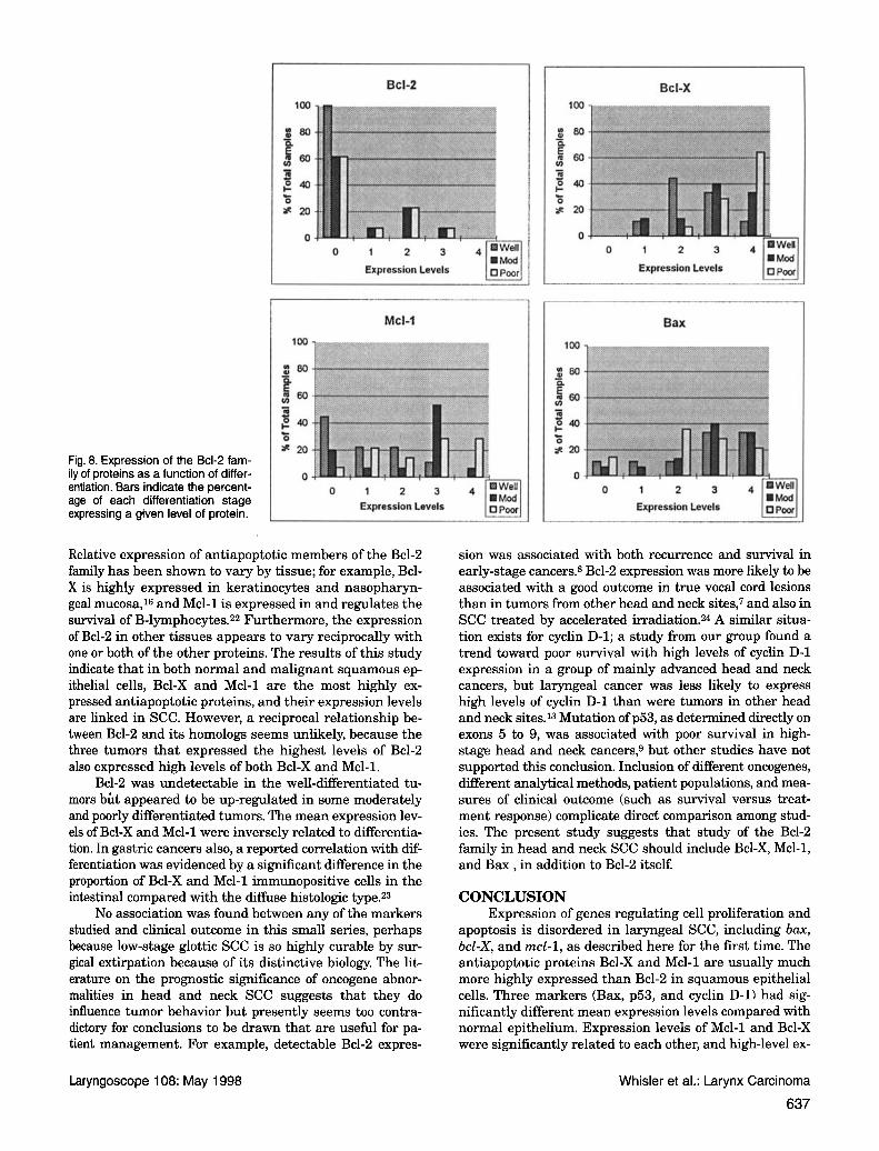
Expression of the Bcl-2 family of proteins differed de- pending on whether the tumors were well (10 cases), mod- erately (15 cases), or poorly (15 cases) differentiated. Bcl- 2 was not expressed in any of the well-differentiated tumors; expression levels were similar for moderately and poorly differentiated SCC (P = .083) (Fig. 8). High levels (4+) of Bcl-X increased significantly from well-differenti- ated (1/10 or lo%, exact confidence interval [CI] = 0.3% to
I BCL-X
0 1 2 3 4
Expndon Level
BAX
0 1 2 3 4
Expndon Level
CYCLIN D1
0 1 2 3 4
Expression Level
43%) to moderately differentiated (5/15 or 33%, CI = 12% to 62%) to poorly differentiated (10/15 or 67%, CI = 35% to 80%) tumors, with a corresponding decline in the propor- tion of tumors showing low-level expression of this protein (P = .014). The rise in expression of Mcl-1 with loss of dif- ferentiation showed a similar pattern (P = .031). The pro- portion of tumor cells expressing Bax, however, was not related to differentiation (P = 57).
Relationships of Marker Expression to Clinical Outcome
Of the group of glottic SCC patients, 23/25 (92%, CI = 74% to 99%) were alive and tumor free at the most recent follow-up visit. Of the supraglottic SCC patients, 3/10 (30%, CI = 7% to 65%) were alive and tumor free at long-term fol- low-up. The supraglottic SCC patients had a significantly poorer clinical outcome compared with the glottic S& pa- tients (P = .012). These results are consistent with previous
Laryngoscope 108: May 1998 Whisler et al.: Larynx Carcinoma
633
Fig. 3. Expression of Bcl-X protein by SCC of larynx (left) and normal epithelium of uvula (right). The tumor shows widespread, coarsely gran- ular cytoplasmic immunostaining. Uvular epithelium shows more finely granular cytoplasmic staining, predominantly of basal and suprabasal cells, frequently close to the nucleus (arrow). (H&E counterstain, magnification x 900.)
Fig. 4. Expression of Mcl-1 protein by SCC of larynx (left) and normal squamous epithelium of uvula (right). Most of the tumor cells show gran- ular cytoplasmic immunostaining (arrow). Uvular epithelial cells show immunostaining predominantly in the middle cell layers (arrow). (H&E counterstain, magnification x 900.)
Laryngoscope 108: May 1998
634
Whisler et al.: Larynx Carcinoma
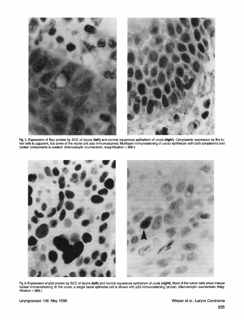
Fig. 5. Expression of Bax protein by SCC of larynx (left) and normal squamous epithelium of uvula (right). Cytoplasmic expression by the tu- mor cells is apparent, but some of the nuclei are also immunostained. Multilayer immunostaining of uvular epithelium with both cytoplasmic and nuclear components is evident. (Hematoxylin counterstain, magnification x 900.)
Fig. 6. Expression of p53 protein by SCC of larynx (left) and normal squamous epithelium of uvula (right). Most of the tumor cells show intense nuclear immunostaining. In the uvula, a single basal epithelial cell is shown with p53 immunostaining (arrow). (Hematoxylin counterstain,'m'ag- nification x 900.)
Laryngoscope 108: May 1998 Whisler et al.: Larynx Carcinoma
635
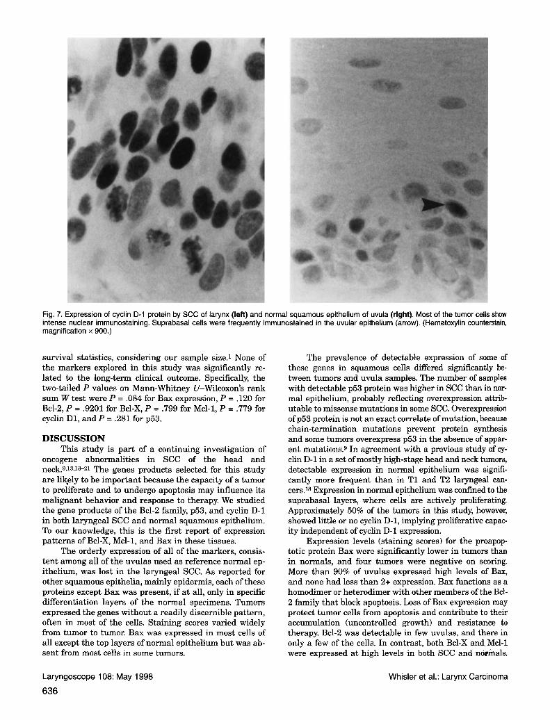
Fig. 7. Expression of cyclin D-1 protein by SCC of larynx (left) and normal squamous epithelium of uvula (right). Most of the tumor cells show intense nuclear immunostaining. Suprabasal cells were frequently immunostained in the uvular epithelium (arrow). (Hematoxylin counterstain, magnification x 900.)
survival statistics, considering our sample size.1 None of the markers explored in this study was significantly re- lated to the long-term clinical outcome. Specifically, the two-tailed P values on Mann-Whitney U-Wilcoxon's rank sum W test were P = .084 for Bax expression, P = .120 for Bcl-2, P = .9201 for Bcl-X, P = .799 for Mcl-1, P = .779 for cyclin D1, and P = .281 for p53.
DISCUSSION This study is part of a continuing investigation of
oncogene abnormalities in SCC of the head and neck.gJ3JS21 The genes products selected for this study are likely to be important because the capacity of a tumor to proliferate and to undergo apoptosis may influence its malignant behavior and response to therapy. We studied the gene products of the Bcl-2 family, p53, and cyclin D-1 in both laryngeal SCC and normal squamous epithelium. To our knowledge, this is the first report of expression patterns of Bcl-X, Mcl-1, and Bax in these tissues.
The orderly expression of all of the markers, consis- tent among all of the uvulas used as reference normal ep- ithelium, was lost in the laryngeal SCC. As reported for other squamous epithelia, mainly epidermis, each of these proteins except Bax was present, if a t all, only in specific differentiation layers of the normal specimens. Tumors expressed the genes without a readily discernible pattern, often in most of the cells. Staining scores vaned widely from tumor to tumor. Bax was expressed in most cells of all except the top layers of normal epithelium but was ab- sent from most cells in some tumors.
The prevalence of detectable expression of some of these genes in squamous cells differed significantly be- tween tumors and uvula samples. The number of samples with detectable p53 protein was higher in SCC than in nor- mal epithelium, probably reflecting overexpression attrib- utable to missense mutations in some SCC. Overexpression of p53 protein is not an exact correlate of mutation, because chain-termination mutations prevent protein synthesis and some tumors overexpress p53 in the absence of appar- ent mutations.9 In agreement with a previous study of cy- clin D-1 in a set of mostly high-stage head and neck tumors, detectable expression in normal epithelium was signifi- cantly more frequent than in T1 and T2 laryngeal can- cers.18 Expression in normal epithelium was confined to the suprabasal layers, where cells 'are actively proliferating. Approximately 50% of the tumors in this study, however, showed little or no cyclin D-1, implying proliferative capac- ity independent of cyclin D-1 expression.
Expression levels (staining scores) for the proapop- totic protein Bax were significantly lower in tumors than in normals, and four tumors were negative on scoring. More than 90% of uvulas expressed high levels of Bax, and none had less than 2+ expression. Bax functions as a homodimer or heterodimer with other members of the Bcl- 2 family that block apoptosis. Loss of Bax expression may protect tumor cells from apoptosis and contribute to their accumulation (uncontrolled growth) and resistance to therapy. Bcl-2 was detectable in few uvulas, and there in only a few of the cells. In contrast, both Bcl-X and Mcl-1 were expressed at high levels in both SCC and notmals.
Laryngoscope 108: May 1998 Whisler et al.: Larynx Carcinoma
636
Bcl-2
entiation. Bars indicate the percent- age of each differentiation stage expressing a given level of protein.
0 1 2 3 4
Expression Levels
0 1 2 3 4 Expression Levels
MCl-1 I00
240 f
* 20 Fig. 8. Expression of the Bcl-2 fam- ily of proteins as a function of differ- I o
Relative expression of antiapoptotic members of the Bcl-2 family has been shown to vary by tissue; for example, Bcl- X is highly expressed in keratinocytes and nasopharyn- geal mucosa,16 and Mcl-1 is expressed in and regulates the survival of B-lymphocytes.22 Furthermore, the expression of Bcl-2 in other tissues appears to vary reciprocally with one or both of the other proteins. The results of this study indicate that in both normal and malignant squamous ep- ithelial cells, Bcl-X and Mcl-1 are the most highly ex- pressed antiapoptotic proteins, and their expression levels are linked in SCC. However, a reciprocal relationship be- tween Bcl-2 and its homologs seems unlikely, because the three tumors that expressed the highest levels of Bcl-2 also expressed high levels of both Bcl-X and Mcl-1.
Bcl-2 was undetectable in the well-differentiated tu- mors b6t appeared to be up-regulated in some moderately and poorly differentiated tumors. The mean expression lev- els of Bcl-X and Mcl-1 were inversely related to differentia- tion. In gastric cancers also, a reported correlation with dif- ferentiation was evidenced by a significant difference in the proportion of Bcl-X and Mcl-1 immunopositive cells in the intestinal compared with the diffuse histologic type.23
No association was found between any of the markers studied and clinical outcome in this small series, perhaps because low-stage glottic SCC is so highly curable by sur- gical extirpation because of its distinctive biology. The lit- erature on the prognostic significance of oncogene abnor- malities in head and neck SCC suggests that they do influence tumor behavior but presently seems too contra- dictory for conclusions to be drawn that are useful for pa- tient management. For example, detectable Bcl-2 expres-
I I BcI-X
0 1 2 3 4
Expression Levels
Bax
100
O1 80 Y, ,a e j40 c
3120
0 I
Expression Levels
sion was associated with both recurrence and survival in early-stage cancers.8 Bcl-2 expression was more likely to be associated with a good outcome in true vocal cord lesions than in tumors from other head and neck sites,7 and also in SCC treated by accelerated irradiation.24 A similar situa- tion exists for cyclin D-1; a study from our group found a trend toward poor survival with high levels of cyclin D-1 expression in a group of mainly advanced head and neck cancers, but laryngeal cancer was less likely to express high levels of cyclin D-1 than were tumors in other head and neck sites.13 Mutation of p53, as determined directly on exons 5 to 9, was associated with poor survival in high- stage head and neck cancers: but other studies have not supported this conclusion. Inclusion of different oncogenes, different analytical methods, patient populations, and mea- sures of clinical outcome (such as survival versus treat- ment response) complicate direct comparison among stud- ies. The present study suggests that study of the Bcl-2 family in head and neck SCC should include Bcl-X, Mcl-1, and Bax , in addition to Bcl-2 itself.
CONCLUSION Expression of genes regulating cell proliferation and
apoptosis is disordered in laryngeal SCC, including bax, bcl-X, and mcl-1, as described here for the first time. The antiapoptotic proteins Bcl-X and Mcl-1 are usually much more highly expressed than Bcl-2 in squamous epithelial cells. Three markers (Bax, p53, and cyclin D-1) had sig- nificantly different mean expression levels compared with normal epithelium. Expression levels of Mcl-1 and Bcl-X were significantly related to each other, and high-level ex-
Laryngoscope 108: May 1998 Whisler et al.: Larynx Carcinoma
637
pression was related to loss of tumor differentiation. Fur- ther study is needed to evaluate the prognostic signifi- cance of specific abnormalities of these oncogenes.
ACKNOWLEDGMENT
cellent technical support. The authors wish to thank Kathe Clodfelter for ex-
BIBLIOGRAPHY 1. Bailey BJ. Early glottic cancer. In: Bailey BJ, Johnson JT, eds.
Head and Neck Surgery-Otolaryngology. Philadelphia: J B Lippincott; 1993:1313-33.
2. Harris CC. Structure and function of the p53 tumor suppres- sor gene: clues for rational cancer therapeutic strategies. J Nut Cancer Inst 1996;88:1442-55.
3. HosokawaY, Arnold A. Cyclin Dl/pradl as a central target in oncogenesis. J Lab Clin Med 1996;127:246-52.
4. Reed J, Miyashita T, Takayama S, et al. Bcl-2 family proteins: regulators of cell death involved in the pathogenesis of cancer and resistance to therapy. J Cellular Biochem
5. Miyashita T, Krajewski S, Krajewska M, et al. Tumor sup- pressor p53 is a regulator of bcl-2 and bax gene expression in vitro and in vivo. Oncogene 1994;9:1799-805.
6. Bellacosa A, Almadori G, Cavallo S, et al. Cyclin D1 gene am- plification in human laryngeal squamous cell carcinomas: prognostic significance and clinical implications. Clin Can- cer Res 1996;2:175-80.
7. Friedman M, Grey P, Venkatesan T, Bloch I, Caldarelli D, Coon J. Prognostic significance of Bcl-2 expression in lo- calized squamous cell carcinoma of the head and neck. Ann Otol Rhino1 Laryngol 1997;106:445-50.
8. Gallo 0, Bodddi V, Calzolari A, Simonetti L, Trovati M, Bianchi S. Bcl-2 protein expression correlates with recur- rence and survival in early stage head and neck cancer treated by radiotherapy. Clin Cancer Res 1996;2:261-7.
9. Wood N, Kotelnikov V, Caldarelli D, et al. Mutation of p53 in squamous cell cancer of the head and neck: relationship to tumor cell proliferation. Laryngoscope 1997;107:
10. Shi S, Key ME, Kalra KI. Antigen retrieval in formalin-fixed, paraffin-embedded tissue: an enhancement method for im- munohistochemical staining based on microwave oven
1996;60:23-32.
827-33.
heating of tissue sections. J Histochem Cytochern 1991;39: 741-8.
11. Ventana 320 Operators Manual. 1992. 12. Elias J. Immunohistochernistry: A Practical Approach to Di-
agnosis. Chicago: ASCP Press; 1990:20-6. 13. Kelanic SM, Kotelnikov V, Caldarelli DD, et al. Significance
of cyclin D-1 expression in squamous cell carcinoma of the head and neck. Head Neck 1997; (in press).
14. Krajewski S, Bodrug S, Krajewska M, et al. Immunohisto- chemical analysis of Mcl-1 protein in human tissue. Am J Pathol 1995;146:1309-19.
15. Krajewski S, Krajewska M, Shabaik A, Miyashita T, Wang H, Reed J. Immunohistochemical determination of in vivo dis- tribution of Bax, a dominant inhibitor of Bcl-2. Am J Pathol 1994;145:1323-36.
16. Krajewski S, Krajewska M, Shabaik A, et al. Immunohisto- chemical analysis of in vivo patterns of Bcl-X expression. Cancer Res 1994;54:5501-7.
17. Kren BT, Trembley JH, Krajewski S, Behrens TW, Reed JC, Steer CJ. Modulation of apoptosis-associated genes Bcl-2, Bcl-x, and Bax during rat liver regeneration. Cell Growth Differ 1996;7:1633-42.
18. Kotelnikov V, Coon J, Mundle S, et al. Cyclin D1 expression in squamous cell carcinomas of the head and neck and oral mucosa in relation to proliferation and apoptosis. Clin Cancer Res 1997;3:95-102.
19. Kotelnikov V, Coon J, Taylor S, et al. Proliferation of epithelia of noninvolved mucosa in patients with head and neck can- cer. Head Neck 1996;18:522-8.
20. Kotelnikov V, Coon J, Haleem A, et al. Cell kinetics of head and neck cancers. Clin Cancer Res 1995;1:527-37.
21. Mundle S, KotelnikovV, Wood N, et al. Assessment of apoptosis in relation to proliferation and mutational status of p53 gene in head and neck cancers. Int J Oncol 1996;8:1257-64.
22. Lomo J, Smeland EB, Krajewski S, Reed JC, Blomhoff HK. Expression of the bcl-2 homologue Mcl-1 correlated with survival of peripheral blood B-lymphocytes. Cancer Res
23. Krajewska M, Fenoglio-Preiser CM, Krajewski S, et al. Im- munohistochemical analysis of Bcl-2 family proteins in adenocarcinomas of the stomach. Am J Pathol 1996;149:
24. Wilson GD, Grover R, Richman, Daley FM, Saunders MI, Dis- che S. Bcl-2 expression correlates with favourable outcome in head and neck cancer treated by accelerated radiother- apy. Anticancer Res 1996;16:2403-8.
1996;56:40-3.
1449-57.
Laryngoscope 108: May 1998
638 Whisler et al.: Larynx Carcinoma
Copyright © 2022 FDOKUMEN