Organohalogen compounds in human breast milk from Republic of Buryatia, Russia
Upload
independentCategory
view
0download
0
This article appeared in a journal published by Elsevier. The attachedcopy is furnished to the author for internal non-commercial researchand education use, including for instruction at the authors institution
and sharing with colleagues.
Other uses, including reproduction and distribution, or selling orlicensing copies, or posting to personal, institutional or third party
websites are prohibited.
In most cases authors are permitted to post their version of thearticle (e.g. in Word or Tex form) to their personal website orinstitutional repository. Authors requiring further information
regarding Elsevier’s archiving and manuscript policies areencouraged to visit:
http://www.elsevier.com/copyright
Author's personal copy
Exposure and effects assessment of persistent organohalogen contaminants in arcticwildlife and fish☆
Robert J. Letcher a,⁎, Jan Ove Bustnes b, Rune Dietz c, Bjørn M. Jenssen d, Even H. Jørgensen e,h,Christian Sonne c, Jonathan Verreault a,1, Mathilakath M. Vijayan f, Geir W. Gabrielsen g
a Wildlife and Landscape Science Directorate, Science and Technology, Branch, Environment Canada, Carleton University, Ottawa, ON, Canada K1A 0H3b Norwegian Institute for Nature Research, Unit for Arctic Ecology, The Polar Environmental Centre, N-9296 Tromsø, Norwayc University of Aarhus, National Environmental Research Institute, Department of Arctic Environment, Roskilde, DK-4000, Denmarkd Department of Biology, Norwegian University of Science and Technology, Trondheim, NO-7491, Norwaye Norwegian College of Fishery Science, University of Tromsø, N-9037 Tromsø, Norwayf Department of Biology, University of Waterloo, Waterloo, Ontario, Canadag Norwegian Polar Institute, Tromsø, NO-9296, Norwayh Norwegian Institute for Nature Research, Polar Environmental Centre, N-9096 Tromsø, Norway
a b s t r a c ta r t i c l e i n f o
Article history:Received 4 May 2009Received in revised form 8 October 2009Accepted 14 October 2009Available online 12 November 2009
Keywords:Circumpolar arcticWildlifeFishOrganohalogen compounds (OHCs)MetabolitesExposureBiological effects
Persistent organic pollutants (POPs) encompass an array of anthropogenic organic and elemental substancesand their degradation and metabolic byproducts that have been found in the tissues of exposed animals,especially POPs categorized as organohalogen contaminants (OHCs). OHCs have been of concern in thecircumpolar arctic for decades. For example, as a consequence of bioaccumulation and in some casesbiomagnification of legacy (e.g., chlorinated PCBs, DDTs and CHLs) and emerging (e.g., brominated flameretardants (BFRs) and in particular polybrominated diphenyl ethers (PBDEs) and perfluorinated compounds(PFCs) including perfluorooctane sulfonate (PFOS) and perfluorooctanic acid (PFOA) found in Arctic biotaand humans. Of high concern are the potential biological effects of these contaminants in exposed Arcticwildlife and fish. As concluded in the last review in 2004 for the Arctic Monitoring and Assessment Program(AMAP) on the effects of POPs in Arctic wildlife, prior to 1997, biological effects data were minimal andinsufficient at any level of biological organization. The present review summarizes recent studies onbiological effects in relation to OHC exposure, and attempts to assess known tissue/body compartmentconcentration data in the context of possible threshold levels of effects to evaluate the risks. This reviewconcentrates mainly on post-2002, new OHC effects data in Arctic wildlife and fish, and is largely based onrecently available effects data for populations of several top trophic level species, including seabirds (e.g.,glaucous gull (Larus hyperboreus)), polar bears (Ursus maritimus), polar (Arctic) fox (Vulpes lagopus), andArctic charr (Salvelinus alpinus), as well as semi-captive studies on sled dogs (Canis familiaris). Regardless,there remains a dearth of data on true contaminant exposure, cause–effect relationships with respect tothese contaminant exposures in Arctic wildlife and fish. Indications of exposure effects are largely based oncorrelations between biomarker endpoints (e.g., biochemical processes related to the immune and endocrine
Science of the Total Environment 408 (2010) 2995–3043
Abbreviations: ALB, thyroid hormone binding albumin; AMAP, Arctic Monitoring and Assessment Program; BDE-209, 2,2′,3,3′,4,4′,5,5′-decabromodiphenyl ether; BFR,brominated flame retardant; BGS, brain growth spurt; BMD, bone mineral density; BMR, basal metabolic rate; CHL, chlordane; Con A, concanavalin; CP, chloroparaffin; CYP,cytochrome P450; CBz, chlorobenzene; DNA, deoxyribonucleic acid; E2, 17β-estradiol; EDC, endocrine disrupting compound; EFI, epithelial follicular index; EHV, herpes virus; EIV,influenza virus; FA, fluctuating asymmetry; FABP, fatty acid binding protein; FSH, follicle stimulating hormone; GH, growth hormone; GST, glutathione-S-transferase; HBCD,hexabromocyclododecane; HCH, hexachlorocyclohexane; Hg, mercury; HP, haptoglobin; HPT, hypothalamus–pituitary–thyroid; IGF-I, insulin-like growth factor I; IgG,immunoglobulin G; IgM, immunoglobin M; LH, luteinizing hormone; LOEL, lowest observed effect level; MeO-, methoxyl-; MeSO2-, methylsulfonyl-; mRNA, messenger ribonucleicacid; OC, organochlorine; OHC, organohalogen contaminant; 25 OHD, 25-hydroxy-vitamin D3; OH-, hydroxyl-; 4-OH-HpCS, 4-hydroxy-heptachlorostyrene; P4, progesterone; PAH,polycyclic aromatic hydrocarbon; PBDE, polybrominated diphenyl ether; PCB, polychlorinated biphenyl; PCDD, polychlorinated dibenzo-p-dioxin; PCDF, polychlorinateddibenzofuran; PCP, pentachlorophenol; PFC, perfluorinated compound; PFCA, perfluorinated carboxylic acid; PFOA, perfluorooctanoic acid; PFOS, perfluorooctane sulfonate; PFSA,perfluorinated sulfonate; PHA, phytohemagglufinin; POP, persistent organic pollutant; p,p′-DDD, bis(p-chlorophenyl)-1,1-dichloroethane; p,p′-DDE, bis(p-chlorophenyl)-1,1-dichloroethene; p,p′-DDT, bis(p-chlorophenyl)-1,1,1-trichloroethane; PRC, prolactin; REO, reovirus; SLE, St. Lawrence river estuary; T, testosterone; T4, thyroxine; T3, 3,3′,5-triiodo-L-thyronine; TBBPA, tetrabromobisphenol A; TBG, thyroid binding globulin; TCDD, 2,3,7,8-tetrachloro-dibenzo-p-dioxin; TEF, toxic equivalency factor; TEQ, toxic equivalent; TET,tetanus toxoid; TH, thyroid hormone; TTR, transthyretin.☆ This paper is a contribution to the AMAP POPs assessment.⁎ Corresponding author. Tel.: +1 613 998 6696; fax: +1 613 998 0458.
E-mail addresses: [email protected] (R.J. Letcher), [email protected] (J.O. Bustnes), [email protected] (R. Dietz), [email protected] (B.M. Jenssen),[email protected] (E.H. Jørgensen), [email protected] (C. Sonne), [email protected] (J. Verreault), [email protected] (M.M. Vijayan),[email protected] (G.W. Gabrielsen).
1 Current address: Département des sciences biologiques, Université du Québec à Montréal, C.P. 8888, Succursale Centre-ville Montreal, QC, Canada H3C 3P8.
0048-9697/$ – see front matter © 2009 Elsevier B.V. All rights reserved.doi:10.1016/j.scitotenv.2009.10.038
Contents lists available at ScienceDirect
Science of the Total Environment
j ourna l homepage: www.e lsev ie r.com/ locate /sc i totenv
Author's personal copy
system, pathological changes in tissues and reproduction and development) and tissue residue levels ofOHCs (e.g., PCBs, DDTs, CHLs, PBDEs and in a few cases perfluorinated carboxylic acids (PFCAs) andperfluorinated sulfonates (PFSAs)). Some exceptions include semi-field studies on comparative contaminanteffects of control and exposed cohorts of captive Greenland sled dogs, and performance studies mimickingenvironmentally relevant PCB concentrations in Arctic charr. Recent tissue concentrations in several arcticmarine mammal species and populations exceed a general threshold level of concern of 1part-per-million(ppm), but a clear evidence of a POP/OHC-related stress in these populations remains to be confirmed. Thereremains minimal evidence that OHCs are having widespread effects on the health of Arctic organisms, withthe possible exception of East Greenland and Svalbard polar bears and Svalbard glaucous gulls. However, thetrue (if any real) effects of POPs in Arctic wildlife have to be put into the context of other environmental,ecological and physiological stressors (both anthropogenic and natural) that render an overall complexpicture. For instance, seasonal changes in food intake and corresponding cycles of fattening and emaciationseen in Arctic animals can modify contaminant tissue distribution and toxicokinetics (contaminantdeposition, metabolism and depuration). Also, other factors, including impact of climate change (seasonal iceand temperature changes, and connection to food web changes, nutrition, etc. in exposed biota), disease,species invasion and the connection to disease resistance will impact toxicant exposure. Overall, furtherresearch and better understanding of POP/OHC impact on animal performance in Arctic biota arerecommended. Regardless, it could be argued that Arctic wildlife and fish at the highest potential risk ofPOP/OHC exposure and mediated effects are East Greenland, Svalbard and (West and South) Hudson Baypolar bears, Alaskan and Northern Norway killer whales, several species of gulls and other seabirds from theSvalbard area, Northern Norway, East Greenland, the Kara Sea and/or the Canadian central high Arctic, EastGreenland ringed seal and a few populations of Arctic charr and Greenland shark.
© 2009 Elsevier B.V. All rights reserved.
1. OHC exposure in Arctic wildlife and fish
The circumpolar Arctic includes land masses and waters within thepolitical boundaries of Canada, Greenland (Denmark), Norway, Sweden,Finland, the Russian Federation and Alaska (United States of America)(Fig. 1). There have been minimal direct use within the circumpolarArctic of chemical substances classified as persistent organic pollutants(POPs), and those used have been comprised largely of organohalogen(chlorinated, brominated and fluorinated) compounds (OHCs). How-ever, long-range atmospheric transport, and to a lesser general extentvia ocean currents and rivers, to the Arctic occurs for POPs (and/orprecursors and degradation products) sourced in more southerlylatitudes (Braune et al., 2005; de Wit et al., 2004, 2006). As aconsequence, lipophilic POPs accumulate in organismswithin especiallymarine food webs and thus there is a concern for the health of exposedwildlife and fish as well as for humans who consume country foods.
The last AMAP assessment of POPs and associations and relationshipsbetween OHC exposure and biomarkers of effects in Arctic biota(including wildlife and fish) included any new published informationup to approximately 2004 (Braune et al., 2005; Fisk et al., 2005; deWit etal., 2004, 2006).Within the last 4 to 5 years there has been a considerableamount of new effects information published on OHCs in Arctic wildlifeand fish, and is the subject of the present review. As will be summarized,dataonOHC levels andeffects infish in theArctic is, for example, scarce incomparisonwith animals living inmarine (mainly coastal) environmentssuch as polar bear (Ursus maritimus) and glaucous gull (Larushyperboreus), which are apex species in the Arctic marine food web.Regardless, POP and effects studies on animals living in polar environ-ments are hampered due to challenging, difficult and/or expensivelogistics. Furthermore, there are numerous natural (e.g., ecological andphysiological) and anthropogenic factors (e.g., Arcticwarming in relationto introduction of new species and pathogens to the Arctic, and changesin the food web and prey–predator interactions) that can influence and/or confound the exposure to and effects of OHCs in many Arctic animalsand inparticular those that exhibit strong seasonal adaptations at variouslevels of biological organization (e.g., cellular, organ,whole organismandpopulation). This includes temporal changes in bioenergetics betweenperiods of fat accumulation and fat mobilization, which in turn caninfluence the toxicokinetics of POPs and the corresponding, tissue-specific effect sensitivity toward POPs. Changes in POP toxicokineticsinclude factors such as altered deposition andmobilization. Long periodsof emaciation, and associated fat mobilization and redistribution of
accumulated POPs, seems tomake these animals particularly sensitive toPOPs. Also, enzyme-catalyzed metabolism can occur in which the POPcanbedetoxifiedor toxified tometabolites that canalsobepersistent andsubject to unique toxicokinetics in the exposed organism includingunique tissue-specific toxicities.
In (marine)wildlife species feeding at higher levels of theArctic foodweb, and as will be discussed in this review, POP/OHC exposure can behigh enough to exceed putative threshold levels that have beenpreviously estimated for non-target and non-Arctic species (Fisk et al.,2005). In these non-target species studies, e.g., on captive or non-Arcticspecies, exposure to specific POPs/OHCs have been shown to result indeleterious and observable effects via mode(s) of action and mechan-isms that are a function of the contaminant type and treatment level.However, difficulties in extrapolation relating to differences insensitivity for animal groups (e.g., comparative toxicology) not livingin the Arctic arewell appreciated. This raises the question as towhetherdocumented effects observed in non-Arctic species investigated inlaboratory studies can be directly transferred and applied to Arcticspecies. Equally importantly, such laboratory studies often exposecaptive animals to a single POP or OHC (or technical products) at highdoses for short periods of time and use non-food routes of administra-tion (not orally ingested). This makes it difficult to extrapolate theseeffects seen at high acute doses to possible adverse effects at lower butmore chronic (multi-generational) exposures as is the case for Arcticwildlife and fish. Also, free-ranging wildlife and fish as exposed to acomplex cocktail of knownPOPs/OHCs asopposed to simplemixtures orcompounds generally used in experimental, lab-based designs.
Among the possible mechanisms that legacy and novel OHCs mayelicit effects, e.g., endocrine and immune disruptive potentials, havebeen reported for OHCs that biomagnify to relatively high concentra-tions in Arctic wildlife and fish (de Wit et al., 2004; Fisk et al., 2005).Thus, there is great cause for concern that the health, reproductionpotential and survival of exposed species may be affected (Fisk et al.,2005). For example, the vulnerability of offspring (fetus and neonate)in highly exposed cohorts of Arctic wildlife where contaminanttransfer from the mother is occurring at a time of critical (andsensitive) developmental sensitivity to stressors. In addition, depend-ing on the time period Arctic wildlife and fish are likely coping withother additional anthropogenic stressors such as Arctic warming andthe subsequently complex impacts on ecosystems.
An intensely focused environmental stress element in the Arctic isclimate change caused by global warming and/or temperature
2996 R.J. Letcher et al. / Science of the Total Environment 408 (2010) 2995–3043

Author's personal copy
changes (Graversen et al., 2008). There is an established link betweenrecent climate change and phenological, geographical and composi-tional changes to ecosystems across many regions of the world(Parmesan and Yohe, 2003). Although the magnitude of warming isregionally variable, it has been reported that for the Arctic themagnitude is nearly twice that of the global average (Johannessenet al., 2004; Graversen et al., 2008). A changing environment canaffect wildlife populations under abnormal and possibly increasedstressor conditions that are outside of cyclic/seasonal conditions,ranging from habitat loss and alteration to new and more virulentdiseases. Climate-related change that will influence OHCs and otherPOPs may result in increased levels, and at the minimum perturba-tions, of contaminant exposure at various levels of the food web. Thismodulation in both contaminant exposure and health status could bedeleterious in that certain Arctic wildlife and fish species arechallenged by stressors beyond their capacity to tolerate chemicalexposures that elicit a biological response or worse a toxicologicaleffect (Jenssen, 2006).
Among the other biological and physiological factors that areimportant in interpreting contaminant and effect data and the healthof Arctic wildlife and fish are life history parameters and lipid content(Ylitalo et al., 2001). Lipid content is critical for assessing health andplays a key role in the tissue dynamics of many lipophilic OHCs, and iscommonly oversimplified from a biological and health perspective.
For example, Krahn et al. (2001) evaluated OCs and lipid profiles inblubber of gray whales (Eschrichtius robustus) from the eastern NorthPacific stock (hunter-killed in the Arctic, biopsied free ranging, andstranded whales). Significantly higher lipid levels were found in theblubber of subsistence animals (Arctic) that were sampled followingsummer feeding in the Bering and Chukchi Seas, compared to lipidlevels in the biopsied and stranded animals. Lipid class profiles fromblubber of presumably healthy gray whales (i.e. from subsistence andbiopsy sampling) contained primarily triglycerides and were verydifferent from those of stranded animals that showed lipid decom-position (increased proportions of free fatty acids, cholesterol andphospholipids). Higher concentrations of OC contaminants werefound in stranded juvenile gray whales, compared to juvenilesubsistence whales, and were thought to result from retention ofOCs in blubber of the stranded animals as lipid stores aremobilized forenergy and total lipid levels decrease, rather than from a difference indiet or feeding areas. OC concentrations in various tissues (blubber,liver, kidney, muscle, and brain) were similar on a lipid weight basis,except for brain, which had lower lipid-adjusted OC concentrationsbecause the blood–brain barrier can limit contaminant transfer(Krahn et al., 2001). Alternately, delayed OC transfer to the brainhypothetically does not explain lower brain OC levels with respect topseudo-equilibrium concentrations established across the blood–brain barrier. Lower brain concentrations are likely related to the
Fig. 1.Map of the circumpolar showing Arctic wildlife and fish species and populations where there is OHC exposure information available in the last 7 years. For a given species, theshaded areas indicate exposure “hotspots”. See Tables 1–4 for listings and references of OHC concentration levels.
2997R.J. Letcher et al. / Science of the Total Environment 408 (2010) 2995–3043
Author's personal copy
higher polarity of brain lipids (e.g., phospholids) compared to e.g.,adipose tissue. Regardless, assessing toxicokinetic distribution isimportant especially for target organs (e.g., brain).
The overall goal of the present AMAP exercise is to review thestate-of-the-science with respect to what new information has beenpublished in approximately the last 7 years on effects and responsesin key, target Arctic wildlife and fish species and populations (Fig. 1),and in relation to exposure to OHCs and precursors, as well as theirdegradation and/or metabolism byproducts. This also includes, in thecase of seabirds, those species and populations that seasonallymigrate into and out of the Arctic region for at least part of the year.For both marine and terrestrial (and marine-feeding terrestrial)mammals there presently remains a bias for effects oriented studies inkey species from “hotspot” Arctic areas where OHC exposure data hashistorically been shown to be more plentiful and shown to be higherrelative to species from other circumpolar regions (Letcher et al.,2000b; Braune et al., 2005; de Wit et al., 2004, 2006). In the followingsub-section we review reports from over approximately the lastdecade as to the type and highest levels of classes of known OHCs anddegradation and metabolite products in Arctic wildlife and fish.Extensive reviews of specific OHCs (and other POPs) in Arctic biota arebeyond the scope of the present review and are discussed in OHCclass-specific reviews being published in the present special AMAPissue.
1.1. Ursids and canids
Prior to about 7 years ago, East Greenland, Svalbard and the KaraSea regions of the Arctic had been documented as having the highestlevels of OHCs and degradation products in the tissues of ursids andcanids (Norstrom et al., 1998; de Wit et al., 2004; Braune et al., 2005).Listed in Table 1, from data available over the last decade, are themaximum exposure concentrations reported for the sum (Σ) ofchlorinated, brominated and/or fluorinated OHCs in the tissues andthe main body compartment, blood, of free-ranging mammalianwildlife within the Arctic. With respect to ursids and canidsessentially, all of the OHC data presently reported for polar bears(Tables 1 and 2 and summarized from references therein) are frompopulations spanning the circumpolar Arctic with the exception of thevast territory of the Russian Arctic region (Fig. 1). Some miminal dataexists for perfluorinated compounds (PFCs), perfluorinated sulfonates(PFSAs) and perfluorinated carboxylates (PFCAs) in mink (Mustelavison) from the Yukon Territory and Arctic fox (Vulpes lagopus) fromWestern Hudson Bay. The ΣPFSA concentration in the liver of theArctic fox is as high as 250 ng/g (ww). For polar bears regardless ofpopulation, the general order of tissue concentrations are Σpoly-chlorinated biphenyl (PCB)≈Σchlordanes (CHL)≈ΣPFSA (essentiallyall PFOS)>ΣDDT (i.e., p,p′-DDT (bis(p-chlorophenyl)-1,1,1-trichlor-oethane), p,p′-DDE (bis(p-chlorophenyl)-1,1-dichloroethene) and p,p′-DDD (bis(p-chlorophenyl)-1,1-dichloroethane), and in some cases o,p′-DDT, o,p′-DDE and/or o,p′-DDD)>Σchlorobenzene (CBz)≈Σhexchloro-cyclohexane (HCH)≈ΣToxaphene≈ΣPFCA>Σpolybrominated diphe-nyl ether (PBDE)>hexabromocyclododecane (HBCD) flame retardant.In the case of ΣPCB, ΣCHL and ΣPFSA, these concentrations exceeded1 ppm (ww) in bears from reported populations. Mainly for EastGreenland bears, and to a lesser extent Svalbard bears, levels ofΣhydroxylated (OH)-PCB, Σmethylsulfonyl (CH3SO2=MeSO2)-PCB,3-MeSO2-p,p′-DDE metabolites have been reported (Table 2), and atlevels comparable to ΣDDTs and ΣCBzs (Table 1). In contrast levels ofΣOH-PBDEs, Σmethoxylated(MeO)-PBDEs, pentachlorophenol (PCP)and 4-OH-heptachlorstyrene (4-OH-HpCS) are at very low ppb levelsor below detection (Table 2).
For polar bears from the East Greenland and Svalbard regions thereare substantial reports on OHC concentration associations withchanges in various (e.g., endocrine- and immune-related) biomarkerresponses, although these do not directly establish cause–effect
relationships (e.g., Oskam et al., 2003, 2004a; Haave et al., 2003;Braathen et al., 2004; Lie et al., 2004, 2005; Sonne et al., 2004, 2005a,b,c,2006a,b, 2007a,b, 2008a; Fisk et al., 2005; Kirkegaard et al., 2005;Verreault et al., 2008a; Muir et al., 2006). In some Svalbard and/orRussian investigations, effect studies have been restricted to studyinghealth variables that can be analyzed in blood (plasma or serum) inrelation to OHC content in plasma and/or adipose biopsies. This isbecause the polar bear is especially protected in these regions. Allcircumpolar bear populations are protected as per the internationalpolar bear treaty of 1973, although only some populations are subjectto aboriginal hunts. Samples were obtained during the handling ofpolar bears in connection with research activities, i.e., chemicalimmobilization and deployment of satellite collars or conventionaltags. A substantial amount of information has been achieved fromthese studies. This includes correlative relationships and suggestedassociations between OHC levels and hormone levels, vitamins andimmune status as well as associations with contaminant levels andpolar bear movements.
For the East Greenland region, it was possible to obtain samplesfrom a large number of organ tissues from polar bears obtainedfrom the traditional hunt. Histopathological investigations on polarbears were started in East Greenland in 1999. These studies haveprovided a unique opportunity to investigate the potential organ-specific effects by assessments of OHC exposure in relation tochanges in biomarker measurements. However, these studies arealso based on correlational and descriptive analyses. To improve theunderstanding and disentangling the potential effects of the cocktailof exposure to contaminants and food stress, experimentalexposure studies have been performed using sledge (or sled) dogs(Canis familiaris) and domesticated Arctic fox, which are possiblesurrogate model species for other Canidae species including polarbears. In captive sled dog and to a lesser extent Arctic fox studies,which included a cohort fed a naturally POP contaminated diet ofminke whale (Balaenoptera acutorostrata) blubber, it has beenpossible over recent years to define and compare OHC exposed andunexposed (reference) groups in direct relation to an array ofeffects, e.g. reproductive organs and other internal organs, theskeletal system, immune and endocrine systems, and POP dietaryaccumulation, biotransformation and toxicokinetics (and associatedenzyme systems) (e.g., Sonne et al., 2006c, 2007c,d, 2008b,c,d,e,f,2009a,b,c; Kirkegaard et al., 2010a,b,c; Verreault et al., 2008a,b,2009a,b,c). To our knowledge, there are no studies that haveexamined the possible effects of POPs/OHCs on free-living canids,including Arctic foxes. However, a contaminant exposure study hasbeen reported using domesticated (farmed) Arctic foxes (Hallanger,2006; Rogstad, 2007).
1.2. Marine mammals
Arctic cetaceans and pinnipeds in this review focuses mostly onmarine mammals were new OHC-related effects information exists;bowhead whale (Balaena mysticetus), beluga whale (Delphinapterusleucas), harbour porpoise (Phocoena phocoena) and ringed seal. Thesespecies are selected based on their 1) circumpolar distribution, 2)potential as models for other species (e.g., ringed seal for ice seals,bowhead for mysticetes, beluga whales for odontocetes), 3) availablePOPs/OHC data (Tables 1 and 2), 4) physiologic and pathologic data(enzymology, endocrinology, lesions noted, etc.) evaluated in contextof OHCs and ecology (e.g., biomagnification and trophodynamics),and 5) use as subsistence species in indigenous communities. Amongmarine mammals, understanding dose–response relationships foraquatic mammals have been attempted especially for the adversehealth effects of PCB exposure (Kannan et al., 2000) and thetoxicokinetics and trends of PCBs in beluga whales (Hickie et al.,1999, 2000). Enzyme-catalyzed metabolism of POPs has been shownto have an apparently small influence on the concentrations and
2998 R.J. Letcher et al. / Science of the Total Environment 408 (2010) 2995–3043

Author's personal copy
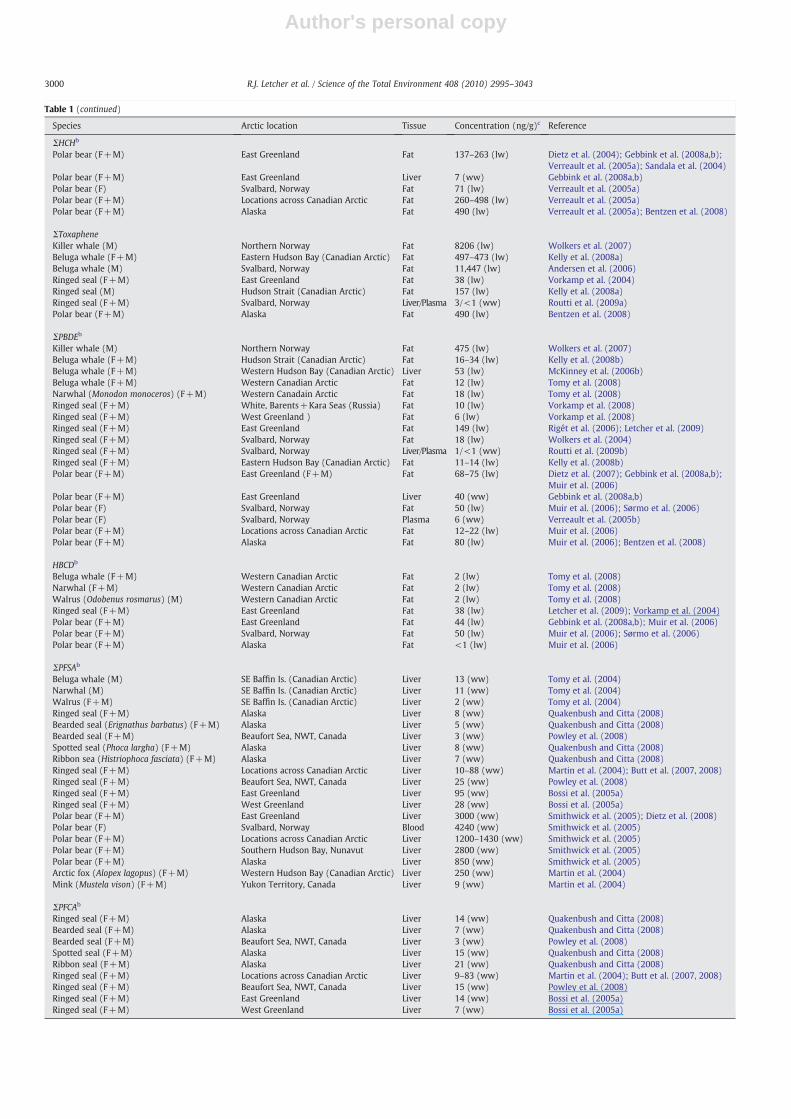
Table 1A comprehensive selection of recently reported, highest exposure levels of major classes of persistent halogenated organic contaminants in free-ranging mammalian wildlife specieswithin the Arctic: geometric or arithmetic means of concentrations (or ranges of means) in major storage tissues (fat, liver or muscle).a
Species Arctic location Tissue Concentration (ng/g)c Reference
ΣPCBb
Bowhead whale (Balaena mysticetus) (F+M) Alaska Fat 451 Hoekstra et al. (2003a)Killer whale (Orcinus orca) (F+M; transients) Alaska Fat 230,000 (lw) Ylitalo et al. (2001)Killer whale (M) Northern Norway Fat 26,940 (lw) Wolkers et al. (2007)Beluga whale (Delphinapterus leucas) (F+M) Hudson Strait (Canadian Arctic) Fat 661–3690 (lw) Kelly et al. (2008a)Beluga whale (F+M) Western Hudson Bay (Canadian Arctic) Liver 1737 (lw) McKinney et al. (2006b)Beluga whale (M) Svalbard, Norway Fat 3198–10075 (lw) Andersen et al. (2001)Ringed seal (Phoca hispida) (F+M) Svalbard, Norway Liver/Plasma 45/22 (lw) Routti et al., (2008a,b)Ringed seal (M) Hudson Strait (Canadian Arctic) Fat 602 (lw) Kelly et al. (2008a)Ringed seal (F+M) West Greenland Fat 200 (lw) Vorkamp et al. (2008)Ringed seal (F+M) East Greenland Fat 1370 (lw) Rigét et al. (2006); Letcher et al. (2009);
Vorkamp et al. (2004)Stellar sea lions (Eumetopias jubatus)(F+M; all pups)
Alaska–Bering Sea Blood 3692–7797 (lw) Myers et al. (2008)
Stellar sea lions (F+M; all pups) Russia–Bering Sea Blood 4600–18000 (lw) Myers et al. (2008)Polar bear (Ursus maritimus) (F+M) East Greenland Fat 7818 (lw) Dietz et al. (2004); Gebbink et al. (2008a,b);
Verreault et al. (2005a); Sandala et al. (2004)Polar bear (F+M) East Greenland Liver 2354 (ww) Gebbink et al. (2008a,b)Polar bear (F) Svalbard, Norway Fat 5972 (lw) Verreault et al. (2005a)Polar bear (F+M) Greenland, Denmark Fat 5414–9100 (lw) Dietz et al. (2004); Verreault et al. (2005a)Polar bear (F+M) Locations across Canadian Arctic Fat 1138–2802 (lw) Verreault et al. (2005a)Polar bear (F+M) Alaska Fat 2174 (lw) Verreault et al. (2005a); Bentzen et al. (2008)
ΣCHLb
Killer whale (M) Northern Norway Fat 6565 (lw) Wolkers et al. (2007)Beluga whale (F+M) Western Hudson Bay (Canadian Arctic) Liver 808 (lw) McKinney et al. (2006b)Beluga whale (M) Svalbard, Norway Fat 2099–6143 (lw) Andersen et al. (2001)Ringed seal (F+M) Svalbard, Norway Liver/Plasma 13/3 (ww) Routti et al. (2009a)Ringed seal (F) Northern Baffin Bay, Canadian Arctic Fat 194 (ww) Borgå et al. (2005)Ringed seal (F+M) East Greenland Fat 400 (lw) Vorkamp et al. (2004)Polar bear (F+M) East Greenland Fat 1776 (lw) Dietz et al. (2004); Gebbink et al. (2008a,b);
Verreault et al. (2005a); Sandala et al. (2004)Polar bear (F+M) East Greenland Liver 4114 (ww) Gebbink et al. (2008a,b)Polar bear (F) Svalbard, Norway Fat 1517 (lw) Verreault et al. (2005a)Polar bear (F+M) Locations across Canadian Arctic Fat 1819–2457 (lw) Verreault et al. (2005a)Polar bear (F+M) Alaska Fat 2007 (lw) Verreault et al. (2005a); Bentzen et al. (2008)
ΣDDTb
Killer whale (F+M; transients) Alaska Fat 320000 (lw) Ylitalo et al. (2001)Beluga whale (F+M) Hudson Strait (Canadian Arctic) Fat 520–2521(lw) Kelly et al. (2008a)Beluga whale (F+M) Western Hudson Bay (Canadian Arctic) Liver 284 (lw) McKinney et al. (2006b)Beluga whale (M) Svalbard, Norway Fat 3272–6770 (lw) Andersen et al. (2001)Ringed seal (F+M) East Greenland Fat 1200 (lw) Vorkamp et al. (2004)Ringed seal (F+M) West Greenland Fat 220 (lw) Vorkamp et al. (2008)Ringed seal (M) Hudson Strait (Canadian Arctic) Fat 413 (lw) Kelly et al. (2008a)Stellar sea lions (F+M; all pups) Alaska–Bering Sea Blood 2127–5464 (lw) Myers et al. (2008)Stellar sea lions (F+M; all pups) Russia–Bering Sea Blood 3600–15000 (lw) Myers et al. (2008)Polar bear (F+M) East Greenland Fat 309 (lw) Dietz et al. (2004); Gebbink et al. (2008a,b);
Verreault et al. (2005a); Sandala et al. (2004)Polar bear (F) Svalbard, Norway Fat 209 (lw) Verreault et al. (2005a)Polar bear (F+M) Greenland, Denmark Fat 309–559 (lw) Dietz et al. (2004); Verreault et al. (2005a)Polar bear (F+M) Locations across Canadian Arctic Fat 65–210 (lw) Verreault et al. (2005a)Polar bear (F+M) Alaska Fat 165 (lw) Verreault et al. (2005a); Bentzen et al. (2008)
ΣCBzb
Killer whale (F+M; transients) Alaska Fat 127,000 (lw) Ylitalo et al. (2001)Beluga whale (F+M) Hudson Strait (Canadian Arctic) Fat 112–377 (lw) Kelly et al. (2008a)Ringed seal (F+M) East Greenland Fat 16 (lw) Vorkamp et al. (2004)Ringed seal (F+M) West Greenland Fat 10 (lw) Vorkamp et al. (2008)Ringed seal (M) Hudson Strait (Canadian Arctic) Fat 78 (lw) Kelly et al. (2008a)Polar bear (F+M) East Greenland Fat 79–187 (lw) Dietz et al. (2004); Gebbink et al. (2008a,b);
Verreault et al. (2005a); Sandala et al. (2004)Polar bear (F+M) East Greenland Liver 12 (ww) Gebbink et al. (2008a,b)Polar bear (F) Svalbard, Norway Fat 105 (lw) Verreault et al. (2005a)Polar bear (F+M) Locations across Canadian Arctic Fat 98–191 (lw) Verreault et al. (2005a)Polar bear (F+M) Alaska Fat 118 (lw) Verreault et al. (2005a); Bentzen et al. (2008)
ΣHCHb
Beluga whale (F+M) Hudson Str. (Canadian Arctic) Fat 95–119 (lw) Kelly et al. (2008a)Beluga whale (F+M) Western Hudson Bay (Canadian Arctic) Liver 45 (lw) McKinney et al. (2006b)Beluga whale (M) Svalbard, Norway Fat 68–510 (lw) Andersen et al. (2001)Ringed seal (F+M) East Greenland Fat 67 (lw) Vorkamp et al. (2004)Ringed seal (F+M) West Greenland Fat 40 (lw) Vorkamp et al. (2008)Ringed seal (M) Hudson Strait (Canadian Arctic) Fat 145 (lw) Kelly et al. (2008a)
(continued on next page)
2999R.J. Letcher et al. / Science of the Total Environment 408 (2010) 2995–3043
Author's personal copy
Table 1 (continued)
Species Arctic location Tissue Concentration (ng/g)c Reference
ΣHCHb
Polar bear (F+M) East Greenland Fat 137–263 (lw) Dietz et al. (2004); Gebbink et al. (2008a,b);Verreault et al. (2005a); Sandala et al. (2004)
Polar bear (F+M) East Greenland Liver 7 (ww) Gebbink et al. (2008a,b)Polar bear (F) Svalbard, Norway Fat 71 (lw) Verreault et al. (2005a)Polar bear (F+M) Locations across Canadian Arctic Fat 260–498 (lw) Verreault et al. (2005a)Polar bear (F+M) Alaska Fat 490 (lw) Verreault et al. (2005a); Bentzen et al. (2008)
ΣToxapheneKiller whale (M) Northern Norway Fat 8206 (lw) Wolkers et al. (2007)Beluga whale (F+M) Eastern Hudson Bay (Canadian Arctic) Fat 497–473 (lw) Kelly et al. (2008a)Beluga whale (M) Svalbard, Norway Fat 11,447 (lw) Andersen et al. (2006)Ringed seal (F+M) East Greenland Fat 38 (lw) Vorkamp et al. (2004)Ringed seal (M) Hudson Strait (Canadian Arctic) Fat 157 (lw) Kelly et al. (2008a)Ringed seal (F+M) Svalbard, Norway Liver/Plasma 3/<1 (ww) Routti et al. (2009a)Polar bear (F+M) Alaska Fat 490 (lw) Bentzen et al. (2008)
ΣPBDEb
Killer whale (M) Northern Norway Fat 475 (lw) Wolkers et al. (2007)Beluga whale (F+M) Hudson Strait (Canadian Arctic) Fat 16–34 (lw) Kelly et al. (2008b)Beluga whale (F+M) Western Hudson Bay (Canadian Arctic) Liver 53 (lw) McKinney et al. (2006b)Beluga whale (F+M) Western Canadian Arctic Fat 12 (lw) Tomy et al. (2008)Narwhal (Monodon monoceros) (F+M) Western Canadain Arctic Fat 18 (lw) Tomy et al. (2008)Ringed seal (F+M) White, Barents+Kara Seas (Russia) Fat 10 (lw) Vorkamp et al. (2008)Ringed seal (F+M) West Greenland ) Fat 6 (lw) Vorkamp et al. (2008)Ringed seal (F+M) East Greenland Fat 149 (lw) Rigét et al. (2006); Letcher et al. (2009)Ringed seal (F+M) Svalbard, Norway Fat 18 (lw) Wolkers et al. (2004)Ringed seal (F+M) Svalbard, Norway Liver/Plasma 1/<1 (ww) Routti et al. (2009b)Ringed seal (F+M) Eastern Hudson Bay (Canadian Arctic) Fat 11–14 (lw) Kelly et al. (2008b)Polar bear (F+M) East Greenland (F+M) Fat 68–75 (lw) Dietz et al. (2007); Gebbink et al. (2008a,b);
Muir et al. (2006)Polar bear (F+M) East Greenland Liver 40 (ww) Gebbink et al. (2008a,b)Polar bear (F) Svalbard, Norway Fat 50 (lw) Muir et al. (2006); Sørmo et al. (2006)Polar bear (F) Svalbard, Norway Plasma 6 (ww) Verreault et al. (2005b)Polar bear (F+M) Locations across Canadian Arctic Fat 12–22 (lw) Muir et al. (2006)Polar bear (F+M) Alaska Fat 80 (lw) Muir et al. (2006); Bentzen et al. (2008)
HBCDb
Beluga whale (F+M) Western Canadian Arctic Fat 2 (lw) Tomy et al. (2008)Narwhal (F+M) Western Canadian Arctic Fat 2 (lw) Tomy et al. (2008)Walrus (Odobenus rosmarus) (M) Western Canadian Arctic Fat 2 (lw) Tomy et al. (2008)Ringed seal (F+M) East Greenland Fat 38 (lw) Letcher et al. (2009); Vorkamp et al. (2004)Polar bear (F+M) East Greenland Fat 44 (lw) Gebbink et al. (2008a,b); Muir et al. (2006)Polar bear (F+M) Svalbard, Norway Fat 50 (lw) Muir et al. (2006); Sørmo et al. (2006)Polar bear (F+M) Alaska Fat <1 (lw) Muir et al. (2006)
ΣPFSAb
Beluga whale (M) SE Baffin Is. (Canadian Arctic) Liver 13 (ww) Tomy et al. (2004)Narwhal (M) SE Baffin Is. (Canadian Arctic) Liver 11 (ww) Tomy et al. (2004)Walrus (F+M) SE Baffin Is. (Canadian Arctic) Liver 2 (ww) Tomy et al. (2004)Ringed seal (F+M) Alaska Liver 8 (ww) Quakenbush and Citta (2008)Bearded seal (Erignathus barbatus) (F+M) Alaska Liver 5 (ww) Quakenbush and Citta (2008)Bearded seal (F+M) Beaufort Sea, NWT, Canada Liver 3 (ww) Powley et al. (2008)Spotted seal (Phoca largha) (F+M) Alaska Liver 8 (ww) Quakenbush and Citta (2008)Ribbon sea (Histriophoca fasciata) (F+M) Alaska Liver 7 (ww) Quakenbush and Citta (2008)Ringed seal (F+M) Locations across Canadian Arctic Liver 10–88 (ww) Martin et al. (2004); Butt et al. (2007, 2008)Ringed seal (F+M) Beaufort Sea, NWT, Canada Liver 25 (ww) Powley et al. (2008)Ringed seal (F+M) East Greenland Liver 95 (ww) Bossi et al. (2005a)Ringed seal (F+M) West Greenland Liver 28 (ww) Bossi et al. (2005a)Polar bear (F+M) East Greenland Liver 3000 (ww) Smithwick et al. (2005); Dietz et al. (2008)Polar bear (F) Svalbard, Norway Blood 4240 (ww) Smithwick et al. (2005)Polar bear (F+M) Locations across Canadian Arctic Liver 1200–1430 (ww) Smithwick et al. (2005)Polar bear (F+M) Southern Hudson Bay, Nunavut Liver 2800 (ww) Smithwick et al. (2005)Polar bear (F+M) Alaska Liver 850 (ww) Smithwick et al. (2005)Arctic fox (Alopex lagopus) (F+M) Western Hudson Bay (Canadian Arctic) Liver 250 (ww) Martin et al. (2004)Mink (Mustela vison) (F+M) Yukon Territory, Canada Liver 9 (ww) Martin et al. (2004)
ΣPFCAb
Ringed seal (F+M) Alaska Liver 14 (ww) Quakenbush and Citta (2008)Bearded seal (F+M) Alaska Liver 7 (ww) Quakenbush and Citta (2008)Bearded seal (F+M) Beaufort Sea, NWT, Canada Liver 3 (ww) Powley et al. (2008)Spotted seal (F+M) Alaska Liver 15 (ww) Quakenbush and Citta (2008)Ribbon seal (F+M) Alaska Liver 21 (ww) Quakenbush and Citta (2008)Ringed seal (F+M) Locations across Canadian Arctic Liver 9–83 (ww) Martin et al. (2004); Butt et al. (2007, 2008)Ringed seal (F+M) Beaufort Sea, NWT, Canada Liver 15 (ww) Powley et al. (2008)Ringed seal (F+M) East Greenland Liver 14 (ww) Bossi et al. (2005a)Ringed seal (F+M) West Greenland Liver 7 (ww) Bossi et al. (2005a)
3000 R.J. Letcher et al. / Science of the Total Environment 408 (2010) 2995–3043
Author's personal copy
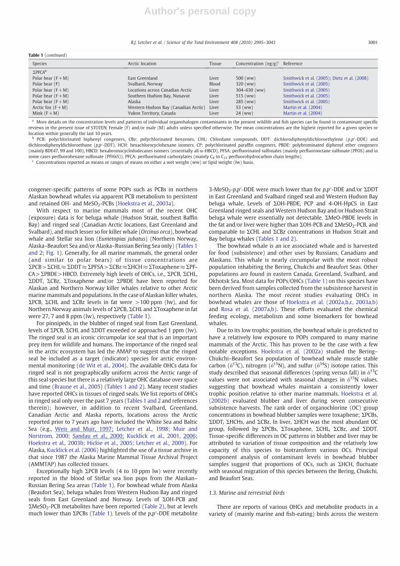
congener-specific patterns of some POPs such as PCBs in northernAlaskan bowhead whales via apparent PCB metabolism to persistentand retained OH- and MeSO2-PCBs (Hoekstra et al., 2003a).
With respect to marine mammals most of the recent OHC(exposure) data is for beluga whale (Hudson Strait, southest BaffinBay) and ringed seal (Canadian Arctic locations, East Greenland andSvalbard), andmuch lesser so for killer whale (Orcinus orca), bowheadwhale and Stellar sea lion (Eumetopias jubatus) (Northern Norway,Alaska–Beaufort Sea and/or Alaska–Russian Bering Sea only) (Tables 1and 2; Fig. 1). Generally, for all marine mammals, the general order(and similar to polar bears) of tissue concentrations areΣPCB>ΣCHL≈ΣDDT≈ΣPFSA>ΣCBz≈ΣHCH≈ΣToxaphene≈ΣPF-CA>ΣPBDE>HBCD. Extremely high levels of OHCs, i.e., ΣPCB, ΣCHL,ΣDDT, ΣCBz, ΣToxaphene and/or ΣPBDE have been reported forAlaskan and Northern Norway killer whales relative to other Arcticmarinemammals and populations. In the case of Alaskan killerwhales,ΣPCB, ΣCHL and ΣCBz levels in fat were >100 ppm (lw), and forNorthern Norway animals levels of ΣPCB, ΣCHL and ΣToxaphene in fatwere 27, 7 and 8 ppm (lw), respectively (Table 1).
For pinnipeds, in the blubber of ringed seal from East Greenland,levels of ΣPCB, ΣCHL and ΣDDT exceeded or approached 1 ppm (lw).The ringed seal is an iconic circumpolar ice seal that is an importantprey item for wildlife and humans. The importance of the ringed sealin the arctic ecosystem has led the AMAP to suggest that the ringedseal be included as a target (indicator) species for arctic environ-mental monitoring (de Wit et al., 2004). The available OHCs data forringed seal is not geographically uniform across the Arctic range ofthis seal species but there is a relatively large OHC database over spaceand time (Braune et al., 2005) (Tables 1 and 2). Many recent studieshave reported OHCs in tissues of ringed seals. We list reports of OHCsin ringed seal only over the past 7 years (Tables 1 and 2 and referencestherein); however, in addition to recent Svalbard, Greenland,Canadian Arctic and Alaska reports, locations across the Arcticreported prior to 7 years ago have included the White Sea and BalticSea (e.g., Weis and Muir, 1997; Letcher et al., 1998; Muir andNorstrom, 2000; Sandau et al., 2000; Kucklick et al., 2001, 2006;Hoekstra et al., 2003b; Hickie et al., 2005; Letcher et al., 2009). ForAlaska, Kucklick et al. (2006) highlighted the use of a tissue archive inthat since 1987 the Alaska Marine Mammal Tissue Archival Project(AMMTAP) has collected tissues.
Exceptionally high ΣPCB levels (4 to 10 ppm lw) were recentlyreported in the blood of Stellar sea lion pups from the Alaskan–Russian Bering Sea areas (Table 1). For bowhead whale from Alaska(Beaufort Sea), beluga whales from Western Hudson Bay and ringedseals from East Greenland and Norway. Levels of ΣOH-PCB andΣMeSO2-PCB metabolites have been reported (Table 2), but at levelsmuch lower than ΣPCBs (Table 1). Levels of the p,p′-DDE metabolite
3-MeSO2-p,p′-DDE were much lower than for p,p′-DDE and/or ΣDDTin East Greenland and Svalbard ringed seal and Western Hudson Baybeluga whale. Levels of ΣOH-PBDE, PCP and 4-OH-HpCS in EastGreenland ringed seals andWestern Hudson Bay and/or Hudson Straitbeluga whale were essentially not detectable. ΣMeO-PBDE levels inthe fat and/or liver were higher than ΣOH-PCB and ΣMeSO2-PCB, andcomparable to ΣCHL and ΣCBz concentrations in Hudson Strait andBay beluga whales (Tables 1 and 2).
The bowhead whale is an ice associated whale and is harvestedfor food (subsistence) and other uses by Russians, Canadians andAlaskans. This whale is nearly circumpolar with the most robustpopulation inhabiting the Bering, Chukchi and Beaufort Seas. Otherpopulations are found in eastern Canada, Greenland, Svalbard, andOkhotsk Sea. Most data for POPs/OHCs (Table 1) on this species havebeen derived from samples collected from the subsistence harvest innorthern Alaska. The most recent studies evaluating OHCs inbowhead whales are those of Hoekstra et al. (2002a,b,c, 2003a,b)and Rosa et al. (2007a,b). These efforts evaluated the chemicalfeeding ecology, metabolism and some biomarkers for bowheadwhales.
Due to its low trophic position, the bowhead whale is predicted tohave a relatively low exposure to POPs compared to many marinemammals of the Arctic. This has proven to be the case with a fewnotable exceptions. Hoekstra et al. (2002a) studied the Bering–Chukchi–Beaufort Sea population of bowhead whale muscle stablecarbon (δ13C), nitrogen (δ15N), and sulfur (δ34S) isotope ratios. Thisstudy described that seasonal differences (spring versus fall) in δ13Cvalues were not associated with seasonal changes in δ15N values,suggesting that bowhead whales maintain a consistently lowertrophic position relative to other marine mammals. Hoekstra et al.(2002b) evaluated blubber and liver during seven consecutivesubsistence harvests. The rank order of organochlorine (OC) groupconcentrations in bowhead blubber samples were toxaphene; ΣPCBs,ΣDDT, ΣHCHs, and ΣCBz. In liver, ΣHCH was the most abundant OCgroup, followed by ΣPCBs, ΣToxaphene, ΣCHL, ΣCBz, and ΣDDT.Tissue-specific differences in OC patterns in blubber and liver may beattributed to variation of tissue composition and the relatively lowcapacity of this species to biotransform various OCs. Principalcomponent analysis of contaminant levels in bowhead blubbersamples suggest that proportions of OCs, such as ΣHCH, fluctuatewith seasonal migration of this species between the Bering, Chukchi,and Beaufort Seas.
1.3. Marine and terrestrial birds
There are reports of various OHCs and metabolite products in avariety of (mainly marine and fish-eating) birds across the western
Table 1 (continued)
Species Arctic location Tissue Concentration (ng/g)c Reference
ΣPFCAb
Polar bear (F+M) East Greenland Liver 500 (ww) Smithwick et al. (2005); Dietz et al. (2008)Polar bear (F) Svalbard, Norway Blood 320 (ww) Smithwick et al. (2005)Polar bear (F+M) Locations across Canadian Arctic Liver 304–630 (ww) Smithwick et al. (2005)Polar bear (F+M) Southern Hudson Bay, Nunavut Liver 515 (ww) Smithwick et al. (2005)Polar bear (F+M) Alaska Liver 285 (ww) Smithwick et al. (2005)Arctic fox (F+M) Western Hudson Bay (Canadian Arctic) Liver 53 (ww) Martin et al. (2004)Mink (F+M) Yukon Territory, Canada Liver 24 (ww) Martin et al. (2004)
a More details on the concentration levels and patterns of individual organohalogen contaminants in the present wildlife and fish species can be found in contaminant specificreviews in the present issue of STOTEN. Female (F) and/or male (M) adults unless specified otherwise. The mean concentrations are the highest reported for a given species orlocation within generally the last 10 years.
b PCB: polychlorinated biphenyl congeners, CBz: polychlorinated benzenes, CHL: Chlordane compounds, DDT: dichlorodiphenyldichloroethylene (p,p′-DDE) anddichlorodiphenyldichloroethane (p,p′-DDT), HCH: hexachlorocyclohexane isomers, CP: polychlorinated paraffin congeners, PBDE: polybrominated diphenyl ether congeners(mainly BDE47, 99 and 100), HBCD: hexabromocyclododecanes isomers (essentially all α-HBCD), PFSA: perfluorinated sulfonates (mainly perfluorooctane sulfonate (PFOS) and insome cases perfluorohexane sulfonate (PFHxS)), PFCA: perfluorinated carboxylates (mainly C8 to C13 perfluorohydrocarbon chain lengths).
c Concentrations reported as means or ranges of means on either a wet weight (ww) or lipid weight (lw) basis.
3001R.J. Letcher et al. / Science of the Total Environment 408 (2010) 2995–3043

Author's personal copy
Table 2A comprehensive selection of recently reported, highest exposure levels of classes of metabolites, degradation products or related compounds of persistent organohalogencontaminants reported in free-ranging wildlife and fish species within the Arctic: geometric or arithmetic mean concentrations in major storage tissues (fat, liver or blood).a
Species Arctic location Tissue Concentration(ng/g)c Reference
ΣOH-PCBb
Glaucous gull (Larus hyperboreus) (F+M) Bear Is. (Svalbard), Norway Liver/Blood
28/52 (ww) Verreault et al. (2005c, 2007a)
Glaucous gull Bear Is. (Svalbard), Norway Egg <1 (ww) Verreault et al. (2005c)Bowhead whale (Balaena mysticetus) (F+M) Alaska Plasma 2 (ww) Hoekstra et al. (2003a)Beluga whale (Delphinapterus leucas) (F+M) Western Hudson Bay (Canadian Arctic) Liver 3 (lw) McKinney et al. (2006b)Ringed seal (Phoca hispida) (F+M) Svalbard, Norway Plasma <1 (ww) Routti et al. (2008a,b)Polar bear (Ursus maritimus) (F+M) East Greenland Fat 59 (ww) Gebbink et al. (2008a,b)Polar bear (F+M) East Greenland Liver 322 (ww) Gebbink et al. (2008a,b)Polar bear (F+M) East Greenland Blood 827 (ww) Gebbink et al. (2008a,b); Sandala et al. (2004)Polar bear (F) Svalbard, Norway Plasma 173 (ww) Verreault et al. (2005b)
ΣMeSO2-PCBb
Glaucous gull (F+M) Bear Is. (Svalbard), Norway Liver/Blood 25/2 (ww) Verreault et al. (2005c, 2007a)Glaucous gull Bear Is. (Svalbard), Norway Egg 92 (lw) Verreault et al. (2005c)Bowhead whale (F+M) Alaska Fat 7 (ww) Hoekstra et al. (2003a)Beluga whale (F+M) Western Hudson Bay (Canadian Arctic) Liver 77 (lw) McKinney et al. (2006b)Ringed seal (F+M) East Greenland Fat 36 (lw) Letcher et al. (2009)Ringed seal (F+M) Svalbard, Norway Liver 2 (ww) Routti et al. (2008a,b)Polar bear (F+M) East Greenland Fat 214 (ww) Gebbink et al. (2008a,b); Sandala et al. (2004)Polar bear (F+M) East Greenland Liver 322 (ww) Gebbink et al., (2008a,b)Polar bear (F+M) East Greenland Blood 107 (ww) Gebbink et al., (2008a,b); Sandala et al., (2004)
3-MeSO2-p,p′-DDEb
Glaucous gull (F+M) Bear Is. (Svalbard), Norway Liver/Blood 1/<1 (ww) Verreault et al. (2007a)Ringed seal (F+M) East Greenland Fat 2 (lw) Letcher et al. (2009)Beluga whale (F+M) Western Hudson Bay (Canadian Arctic) Liver 22 (lw) McKinney et al. (2006b)Polar bear (F+M) East Greenland Fat 6 (ww) Gebbink et al. (2008a,b); Sandala et al. (2004)Polar bear (F+M) East Greenland Liver 29 (ww) Gebbink et al. (2008a,b)Polar bear (F+M) East Greenland Blood 1 (ww) Gebbink et al. (2008a,b); Sandala et al. (2004)
BCPSb
Glaucous gull (F+M) Bear Is. (Svalbard), Norway Plasma 20–26 (lw) Verreault et al. (2005c)
PCPb
Glaucous gull (F+M) Bear Is. (Svalbard), Norway Plasma <1 (ww) Verreault et al. (2005c)Ringed seal (F+M) East Greenland Fat 1 (ww) Letcher et al. (2009)Ringed seal (F+M) Svalbard, Norway Blood <1 (ww) Routti et al., (2008a,b)Polar bear (F+M) East Greenland Fat 1 (ww) Gebbink et al. (2008a,b)Polar bear (F+M) East Greenland Liver 4 (ww) Gebbink et al. (2008a,b)Polar bear (F+M) East Greenland Blood <1 (ww) Gebbink et al. (2008a,b); Sandala et al. (2004)
4-OH-HpCSb
Glaucous gull (F+M) Bear Is. (Svalbard), Norway Plasma <1 (ww) Verreault et al. (2005c)Polar bear (F+M)r East Greenland Fat 1 (ww) Gebbink et al. (2008a,b)Polar bear (F+M) East Greenland Liver 8 (ww) Gebbink et al. (2008a,b)Polar bear (F+M) East Greenland Blood 10 (ww) Gebbink et al. (2008a,b); Sandala et al. (2004)
ΣOH-PBDEb
Glaucous gull (F+M) Bear Is. (Svalbard), Norway Liver/Blood 4/4 (ww) Verreault et al. (2005b, 2007a)Beluga whale (F+M) Hudson Strait (Canadian Arctic) Fat, Blood <1 (lw) Kelly et al. (2008a)Beluga whale (calves) Hudson Strait (Canadian Arctic) Fat <1 (lw) Kelly et al. (2008a)Beluga whale (M) Hudson Strait (Canadian Arctic) Liver <1 (lw) Kelly et al. (2008a)Beluga whale (F+M) Western Hudson Bay (Canadian Arctic) Liver <1 (lw) McKinney et al. (2006b)Ringed seal (F+M) Svalbard, Norway Plasma <1 (ww) Routti et al. (2009b)Ringed seal (F+M) East Greenland Fat 1 (lw) Letcher et al. (2009)Polar bear (F+M) East Greenland Fat 1 (ww) Gebbink et al., (2008a,b)Polar bear (F+M) East Greenland Blood 3 (ww) Gebbink et al., (2008a,b)Polar bear (F) Svalbard, Norway Plasma <1 (ww) Verreault et al. (2005b)
ΣMeO-PBDEb
Polar cod (Boreogadus saida) (F+M) Northeastern Hudson Bay (Canadian Arctic) Muscle 10 (lw) Kelly et al. (2008a)White-winged scoter (Melanitta deglandi) (F+M) Northeastern Hudson Bay (Canadian Arctic) Muscle 2 (lw) Kelly et al. (2008a)Common eider (Somateria mollissima) (F+M) Northeastern Hudson Bay (Canadian Arctic) Muscle 1 (lw) Kelly et al. (2008a)Glaucous gull (F+M) Bear Is. (Svalbard), Norway Liver/Blood 32/3 (ww) Verreault et al. (2005b, 2007a)Beluga whale (F+M) Hudson Strait (Canadian Arctic) Fat 62–100 (lw) Kelly et al. (2008a)Beluga whale (F+M) Hudson Strait (Canadian Arctic) Blood 10–31 (lw) Kelly et al. (2008a)Beluga whale (M) Hudson Strait (Canadian Arctic) Liver 310 (lw) Kelly et al. (2008a)Beluga whale (calves) Hudson Strait (Canadian Arctic) Fat 310 (lw) Kelly et al. (2008a)Beluga whale (F+M) Western Hudson Bay (Canadian Arctic) Liver 100 (lw) McKinney et al. (2006b)Ringed seal (F+M) East Greenland Fat 5 (lw) Letcher et al. (2009)Ringed seal (M) Hudson Strait (Canadian Arctic) Fat 7 (lw) Kelly et al. (2008a)Polar bear (F+M) East Greenland Fat/Blood 4/<1 (ww) Gebbink et al., (2008a,b)Polar bear (F+M) East Greenland Fat 1 (ww) Gebbink et al., (2008a,b)Polar bear (F) Svalbard, Norway Plasma <1 (ww) Verreault et al. (2005b)
3002 R.J. Letcher et al. / Science of the Total Environment 408 (2010) 2995–3043
Author's personal copy
hemispheric Arctic but largely in the muscle, liver, plasma and/or eggof black-legged kittiwake (Rissa tridactyla) from Northern Norway,Canadian high Arctic areas and Barents Sea, northern fulmar(Fulmarus glacialis) from the Canadian high Arctic, Alaska (AleutianArchipelago) and Svalbard (Bear Is.), and glaucous gull from NorthernBaffin Bay but mainly from Svalbard (Bear Is.) (Tables 2 and 3). Otherrecent reports of OHCs in birds include those from NortheasternHudson Bay (white-winged scoter (Melanitta deglandi) and commoneider (Somateria mollissima)), South (peregrine falcon (Falco pere-grinus)) and East (black guillemot (Cepphus grylle)) Greenland andSvalbard (Arctic tern (Sterna paradisaea). Generally, for all Arcticbirds, the general order of tissue (or egg) concentrations areΣPCB>ΣCHL≈ΣDDT≈ΣToxaphene≈ΣCP (chlorinated paraffins)>ΣCBz≈ΣHCH≈ΣPFSA>ΣPBDE≈HBCD>ΣPFCA. Higher levels ofΣPCB of>1 ppm (lw or ww) have been reported for black-leggedkittiwake, great (Larus marinus) and lesser (Larus fuscus ) black-backed and herring (Larus argentatus) gulls (Northern Norway),glaucous (Bear Is.) and ivory (Canadian central-high Arctic) gulls, andnorthern fulmars (Bear Is. and Canadian central-high Arctic). For thelast three bird species/populations, levels of ΣCHL and ΣDDT have alsobeen reported to be>1 ppm (lw or ww). Higher OHC concentrationshave also been recently reported in the eggs of ivory gulls fromSvalbard, and Frans Josef Land and Severnaya Zemlya in the Russianarctic (Miljeteig et al., 2007). ΣDDT levels>1 ppm have also beenreported for black-legged kittiwakes (Northern Norway). High levelsof ΣPBDE (>1 ppm) were recently reported in the liver of northernfulmars (Bear Is.).
ΣOH-PCB, ΣMeSO2-PCB, 3-MeSO2-p,p′-DDE, ΣOH-PBDE, PCP, 4-OH-HpCS and ΣMeO-PBDE concentrations have only been reported inthe liver, plasma and/or egg of glaucous gull (Svalbards, Bear Is.)(Table 2). The only exception is the recent report of ΣMeO-PBDElevels in the muscle of white-winged scoter and common eider(Northeastern Hudson Bay) (Table 2). Of these contaminant classes,ΣOH-PCB, ΣMeSO2-PCB and ΣMeO-PBDE levels were in to 2 to 52 ng/g(ww) range, ΣOH-PBDE around 4 ng/g (ww), and the remainder atvery low to sub-ppb levels in the liver or plasma of glaucous gull(Bear Is.). ΣOH-PCB and ΣMeSO2-PCB levels were at least 3 orders ofmagnitude lower than ΣOH-PCBs in the glaucous gull liver (Table 1).
OHC exposure-effects related studies have mainly been on themarine seabird the glaucous gull. There have been limited studies onOHC exposure-related effects in terrestrial birds having a seasonal orannual distribution within the Arctic. Among the effect studies carriedout with terrestrial birds in the last 7 years, the species of focus hasbeen the American kestrel (Falco sparverius) and peregrine falcon(Falco peregrinus). These two species were selected for the purpose ofthe present review as they also have a northern distribution in Europeand North America.
Marine birds migrating (mainly for breeding) or based perma-nently in the Arctic and Subarctic regions have received particularresearch attention with respect to the effects of environmentalchemical pollution. Since 2002, an impressive number of studieshave been published on a wide range of species spanning Europe andNorth America and including predatory, fish-eating and bottom-feeding species. However, most of the investigations conducted thusfar have been on species from Europe, creating an importantcontaminant effect knowledge gap for species in North America (deWit et al., 2004; Fisk et al., 2005).
In a recent study on terrestrial grey sparrows (Passer dometicus)from North-Norway (Helgeland) very high levels of the PBDEcongener 2,2′,3,3′,4,4′,5,5′-decabromoDE (BDE-209) were reportedin liver samples of the birds (Ciesielski et al., 2008). These sparrowsnested at farms, and this may indicate that the high levels wereassociated with living in an environment heavily impacted by humanactivity. In contrast, very low levels were reported in livers of willow(Lagopus lagopus) and rock ptarmigans (Lagopus mutus) (Ciesielskiet al., 2008).
1.4. Marine and freshwater fish
Levels of POPs, including OHCs, have historically been reported tobe generally low in Arctic fish (deWit et al., 2006; Braune et al., 2005).In a few species/populations with high contaminant levels, PCBshave generally been the dominant contaminant class, whereas levelsof p,p′-DDT are known to normally be somewhat lower (de Wit et al.,2004; Evenset et al., 2005). Recent reports (roughly in the last 7 years)have reported various OHC classes in the muscle, liver and to a muchlesser extent whole body, of mainly Arctic charr (Salvelinus alpinus)(Bear Is. and East and Northern Greenland) and polar cod (Boreogadussaida) (Barents Sea, Northeastern Hudson Bay and NortheasternBaffin Bay) (Table 4). OHCs have also been reported in the liver ofAtlantic cod (Gadus morhua) (Barents Sea), a few species of marineflatfish (Barents Sea, West Greenland and Southeastern Baffin Bay),and three species of freshwater fish from Northern Québec (Canada)(Table 4). Finally, OHCs have been reported in the liver and/or muscleof Greenland shark (Somniosus microcephalus) (Iceland and South-eastern Baffin Bay). Generally, for all fish reported, the order of tis-sue concentrations are ΣPCB>ΣCHL≈ΣCP>ΣDDT≈ΣToxaphene≈ΣCBz≈ΣHCH≈ΣPBDE≈ΣPFSA≈ΣPFCA>HBCD (Table 4). Highlevels of ΣPCB of>1 ppm (lw or ww) have been reported in themuscle and/or liver for two species and populations of long-lived fish,Arctic charr (Bear Is.) and Greenland shark (Iceland and SoutheasternBaffin Bay). For the same charr population levels of ΣCP have beenreported to be>1 ppm. For Southeastern Baffin Bay, Greenland sharklevels of ΣCHL and ΣDDT have also been reported to be >1 ppm. Thereare no known reports of ΣOH-PCB, ΣMeSO2-PCB, 3-MeSO2-p,p′-DDE,ΣOH-PBDE, PCP, 4-OH-HpCS and ΣMeO-PBDE concentrations in Arcticfish. The lone exception is for MeSO2-PCBs and -p,p′-DDEs, whichwere not detected (<0.01 ng/g lw) in whole body homogenates ofArctic cod samples collected in 1993 from the Resolute Bay area(Letcher et al., 1998). The only known exception is one recent studythat reported ΣMeO-PBDE levels of about 10 ng/g (lw) in the muscleof polar cod from Northeastern Hudson Bay (Table 2), and wasexceedingly low relative to other major OHC classes in polar cod butcomparable to ΣPBDE, ΣPFSA, ΣPFCA and HBCD levels (Table 4).
2. Biological effects in relation to OHC levels in Arcticwildlife and fish
As illustrated in Fig. 1 and discussed in Section 1, based onOHCdatasummarized in Tables 1–4, recent reports on OHCs in Arctic wildlifeand fish indicate that there are several “hotspot” species andpopulations with respect to exposure, which should be considered tobe at heightened risk of possible OHC-mediated effects. In general,
Notes to Table 2:a More details on the concentration levels and patterns of metabolites, degradation products or related compounds of organohalogen contaminants in the present wildlife and fish
species may be found in contaminant specific reviews in the present issue of STOTEN. Female (F) and/or male (M) adults unless specified otherwise. Themean concentrations are thehighest reported for a given species or location within about the last 7 years.
b OH-PCB: hydroxylated polychlorinated biphenyl congeners, PCP: pentachlorophenol, 4-OH-HpCS: 4-hydroxy-heptachlorostyrene, MeSO2-PCB: methylsulfone-PCB congeners,3-MeSO2-p,p′-DDE: 3-methylsulfone-dichlorodiphenyldichloroethylene, BCPS: bis(4-chlorophenyl)sulfone, OH-PBDE: hydroxylated polybrominated diphenyl ether congeners,MeO-PBDE: methoxylated-PBDE congeners.
c Concentrations reported as means or ranges of means on either a wet weight (ww) or lipid weight (lw) basis.
3003R.J. Letcher et al. / Science of the Total Environment 408 (2010) 2995–3043
Author's personal copy
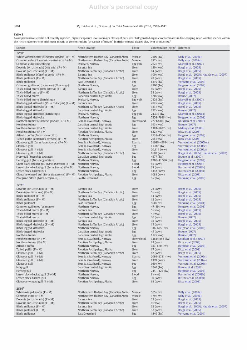
Table 3A comprehensive selection of recently reported, highest exposure levels of major classes of persistent halogenated organic contaminants in free-ranging avian wildlife species withinthe Arctic: geometric or arithmetic means of concentrations (or ranges of means) in major storage tissues (fat, liver or muscle).a
Species Arctic location Tissue Concentration (ng/g)c Reference
ΣPCBb
White-winged scoter (Melanitta deglandi) (F+M) Northeastern Hudson Bay (Canadian Arctic) Muscle 2500 (lw) Kelly et al. (2008a)Common eider (Somateria mollissima) (F+M) Northeastern Hudson Bay (Canadian Arctic) Muscle 287 (lw) Kelly et al. (2008a)Common eider (hatchlings) Svalbard, Norway Egg yolk 262 (lw) Murvoll et al. (2007)Dovekie (or Little auk) (Alle alle) (F+M) Barents Sea Liver 130 (ww) Borgå et al. (2005)Dovekie (or Little auk) (F+M) Northern Baffin Bay (Canadian Arctic) Liver 14 (ww) Borgå et al. (2005)Black guillemot (Cepphus grylle) (F+M) Barents Sea Liver 100 (ww) Borgå et al. (2005); Haukås et al. (2007)Black guillemot (F+M) Northern Baffin Bay (Canadian Arctic) Liver 47 (ww) Borgå et al. (2005)Black guillemot East Greenland Egg 6410 (lw) Vorkamp et al. (2004)Common guillemot (or murre) (Uria aalge) Northern Norway Egg 2300 (lw) Helgason et al. (2008)Thick-billed murre (Uria lomvia) (F+M) Barents Sea Liver 49 (ww) Borgå et al. (2005)Thick-billed murre (F+M) Northern Baffin Bay (Canadian Arctic) Liver 33 (ww) Borgå et al. (2005)Thick-billed murre Canadian central high Arctic Egg 120 (ww) Braune (2007)Thick-billed murre (hatchlings) Svalbard, Norway Egg yolk 2429 (lw) Murvoll et al. (2007)Black-legged kittiwake (Rissa tridactyla) (F+M) Barents Sea Liver 492 (ww) Borgå et al. (2005)Black-legged kittiwake (F+M) Northern Baffin Bay (Canadian Arctic) Liver 123 (ww) Borgå et al. (2005)Black-legged kittiwake Canadian central high Arctic Egg 177 (ww) Braune (2007)Black-legged kittiwake (hatchlings) Svalbard, Norway Egg yolk 24,536 (lw) Murvoll et al. (2006a)Black-legged kittiwake Northern Norway Egg 7254–7938 (lw) Helgason et al. (2008)Northern fulmar (Fulmarus glacialis) (F+M) Bear Is. (Svalbard), Norway Liver/Blood 7273/8106 (lw) Knudsen et al. (2007)Northern fulmar Canadian central high Arctic Egg 163 (ww) Braune (2007)Northern fulmar (F+M) Canadian central high Arctic Liver 1383–3637 (lw) Mallory et al. (2006)Northern fulmar (F+M) Aleutian Archipelago, Alaska Liver 622 (ww) Ricca et al. (2008)Atlantic puffin (Fratercula arctica) Northern Norway Egg 2535–4594 (lw) Helgason et al. (2008)Tufted puffin (Fratercula cirrhata) (F+M) Aleutian Archipelago, Alaska Liver 265 (ww) Ricca et al. (2008)Glaucous gull (Larus hyperboreus) (F+M) Bear Is. (Svalbard), Norway Plasma 31646–49894 (lw) Verreault et al. (2005b,c)Glaucous gull Bear Is. (Svalbard), Norway Egg 11,786 (lw) Verreault et al. (2005c)Glaucous gull (F+M) Bear Is. (Svalbard), Norway Liver 20,114 (ww) Verreault et al. (2007a)Glaucous gull (F+M) Northern Baffin Bay (Canadian Arctic) Liver 1680 (ww) Borgå et al. (2005); Haukås et al. (2007)Ivory gull (Pagophila eburnea) Canadian central high Arctic Egg 4877 (lw) Braune et al. (2007)Herring gull (Larus argentatus) Northern Norway Egg 8786–11,596 (lw) Helgason et al. (2008)Great black-backed gull (Larus marinus) (F+M) Northern Norway Blood 95 (ww) Helberg et al. (2005)Lesser black-backed gull (Larus fuscus) (F+M) Northern Norway Blood 28 (ww) Bustnes et al. (2008b)Lesser black-backed gull Northern Norway Egg 1342 (ww) Bustnes et al. (2006b)Glaucous-winged gull (Larus glaucescens) (F+M) Aleutian Archipelago, Alaska Liver 1083 (ww) Ricca et al. (2008)Peregrine falcon (Falco peregrinus) South Greenland Egg 55 (lw) Vorkamp et al. (2009)
ΣCHLb
Dovekie (or Little auk) (F+M) Barents Sea Liver 24 (ww) Borgå et al. (2005)Dovekie(or Little auk) (F+M) Northern Baffin Bay (Canadian Arctic) Liver 5 (ww) Borgå et al. (2005)Black guillemot (F+M) Barents Sea Liver 14 (ww) Borgå et al. (2005)Black guillemot (F+M) Northern Baffin Bay (Canadian Arctic) Liver 12 (ww) Borgå et al. (2005)Black guillemot East Greenland Egg 960 (lw) Vorkamp et al. (2004)Common guillemot (or murre) Northern Norway Egg 47–89 (lw) Helgason et al. (2008)Thick-billed murre (F+M) Barents Sea Liver 5 (ww) Borgå et al. (2005)Thick-billed murre (F+M) Northern Baffin Bay (Canadian Arctic) Liver 4 (ww) Borgå et al. (2005)Thick-billed murre Canadian central high Arctic Egg 30 (ww) Braune (2007)Black-legged kittiwake (F+M) Barents Sea Liver 38 (ww) Borgå et al. (2005)Black-legged kittiwake (F+M) Northern Baffin Bay (Canadian Arctic) Liver 15 (ww) Borgå et al. (2005)Black-legged kittiwake Northern Norway Egg 336–605 (lw) Helgason et al. (2008)Black-legged kittiwake Canadian central high Arctic Egg 45 (ww) Braune (2007)Northern fulmar Canadian central high Arctic Egg 112 (ww) Braune (2007)Northern fulmar (F+M) Bear Is. (Svalbard), Norway Liver/Blood 3363/1556 (lw) Knudsen et al. (2007)Northern fulmar (F+M) Aleutian Archipelago, Alaska Liver 93 (ww) Ricca et al. (2008)Atlantic puffin Northern Norway Egg 383–870 (lw) Helgason et al. (2008)Tufted puffin (F+M) Aleutian Archipelago, Alaska Liver 17 (ww) Ricca et al. (2008)Glaucous gull (F+M) Northern Baffin Bay (Canadian Arctic) Liver 82 (ww) Borgå et al. (2005)Glaucous gull (F+M) Bear Is. (Svalbard), Norway Plasma 2086–2721 (lw) Verreault et al. (2005c)Glaucous gull (F+M) Bear Is. (Svalbard), Norway Liver 1399 (ww) Verreault et al. (2007a)Glaucous gull Bear Is. (Svalbard), Norway Egg 969 (lw) Verreault et al. (2005c)Ivory gull Canadian central high Arctic Egg 3240 (lw) Braune et al. (2007)Herring gull Northern Norway Egg 744–1125 (lw) Helgason et al. (2008)Lesser black-backed gull (F+M) Northern Norway Blood 8 (ww) Bustnes et al. (2008b)Lesser black-backed gull Northern Norway Egg 30 (ww) Bustnes et al. (2006b)Glaucous-winged gull (F+M) Aleutian Archipelago, Alaska Liver 68 (ww) Ricca et al. (2008)
ΣDDTb
White-winged scoter (F+M) Northeastern Hudson Bay (Canadian Arctic) Muscle 565 (lw) Kelly et al. (2008a)Common eider (F+M) Northeastern Hudson Bay (Canadian Arctic) Muscle 179 (lw) Kelly et al. (2008a)Dovekie (or Little auk) (F+M) Barents Sea Liver 32 (ww) Borgå et al. (2005)Dovekie (or Little auk) (F+M) Northern Baffin Bay (Canadian Arctic) Liver 9 (ww) Borgå et al. (2005)Black guillemot (F+M) Barents Sea Liver 32 (ww) Borgå et al. (2005); Haukås et al. (2007)Black guillemot (F+M) Northern Baffin Bay (Canadian Arctic) Liver 52 (ww) Borgå et al. (2005)Black guillemot East Greenland Egg 1340 (lw) Vorkamp et al. (2004)
3004 R.J. Letcher et al. / Science of the Total Environment 408 (2010) 2995–3043

Author's personal copy
Table 3 (continued)
Species Arctic location Tissue Concentration (ng/g)c Reference
ΣDDTb
Common guillemot (or murre) Northern Norway Egg 962–1117 (lw) Helgason et al. (2008)Thick-billed murre (F+M) Barents Sea Liver 25 (ww) Borgå et al. (2005)Thick-billed murre (F+M) Northern Baffin Bay (Canadian Arctic) Liver 32 (ww) Borgå et al. (2005)Thick-billed murre Canadian central high Arctic Egg 103 (ww) Braune (2007)Black-legged kittiwake (F+M) Barents Sea Liver 98 (ww) Borgå et al. (2005)Black-legged kittiwake (F+M) Northern Baffin Bay (Canadian Arctic) Liver 56 (ww) Borgå et al. (2005)Black-legged kittiwake Northern Norway Egg 806–1562 (lw) Helgason et al. (2008)Black-legged kittiwake Canadian central high Arctic Egg 43 (ww) Braune (2007)Northern fulmar Canadian central high Arctic Egg 124 (ww) Braune (2007)Northern fulmar (F+M)r Bear Is. (Svalbard), Norway) Liver 1289 (lw) Knudsen et al. (2007)Northern fulmar (F+M) Aleutian Archipelago, Alaska Liver 15 (ww) Ricca et al. (2008)Atlantic puffin Northern Norway Egg 1104–1569 (lw) Helgason et al. (2008)Tufted puffin (F+M) Aleutian Archipelago, Alaska Liver 4 (ww) Ricca et al. (2008)Glaucous gull (F+M) Northern Baffin Bay (Canadian Arctic) Liver 2380 (ww) Borgå et al. (2005); Haukås et al. (2007)Glaucous gull (F+M) Bear Is. (Svalbard), Norway Plasma 10,245–15076 (lw) Verreault et al. (2005c)Glaucous gull Bear Is. (Svalbard), Norway Egg 3559 (lw) Verreault et al. (2005c)Ivory gull Canadian central high Arctic Egg 10,744 (lw) Braune et al. (2007)Herring gull Northern Norway Egg 2241–4184 (lw) Helgason et al. (2008)Great black-backed gull (F+M) Northern Norway Blood 30 (ww) Helberg et al. (2005)Lesser black-backed gull (F+M) Northern Norway Blood 16 (ww) Bustnes et al. (2008b)Lesser black-backed gull Northern Norway Egg 652 (ww) Bustnes et al. (2006b)Glaucous-winged gull (F+M) Aleutian Archipelago, Alaska Liver 15 (ww) Ricca et al. (2008)Peregrine falcon South Greenland Egg 40 (lw) Vorkamp et al. (2009)
ΣCBzb
White-winged scoter (F+M) Northeastern Hudson Bay (Canadian Arctic) Muscle 71 (lw) Kelly et al. (2008a)Common eider (F+M) Northeastern Hudson Bay (Canadian Arctic) Muscle 48 (lw) Kelly et al. (2008a)Black guillemot East Greenland Egg 400 (lw) Vorkamp et al. (2004)Ivory gull Canadian central high Arctic Egg 586 (lw) Braune et al. (2007)Black-legged kittiwake Canadian central high Arctic Egg 18 (ww) Braune (2007)Thick-billed murre Canadian central high Arctic Egg 32 (ww) Braune (2007)Northern fulmar Canadian central high Arctic Egg 14 (ww) Braune (2007)Northern fulmar (F+M) Bear Is. (Svalbard), Norway Liver/Blood 602/583 (lw) Knudsen et al. (2007)Northern fulmar (F+M) Aleutian Archipelago, Alaska Liver 82 (ww) Ricca et al. (2008)Tufted puffin (F+M) Aleutian Archipelago, Alaska Liver 48 (ww) Ricca et al. (2008)Glaucous-winged gull (F+M) Aleutian Archipelago, Alaska Liver 116 (ww) Ricca et al. (2008)Peregrine falcon South Greenland Egg <1 (lw) Vorkamp et al. (2009)
ΣHCHb
White-winged scoter (F+M) Northeastern Hudson Bay (Canadian Arctic) Muscle 9 (lw) Kelly et al. (2008a)Common eider (F+M) Northeastern Hudson Bay (Canadian Arctic) Muscle 15 (lw) Kelly et al. (2008a)Herring gull Northern Norway Egg 15–32 (lw) Helgason et al. (2008)Black-legged kittiwake Northern Norway Egg 20–34 (lw) Helgason et al. (2008)Black-legged kittiwake Canadian central high Arctic Egg 8 (ww) Braune (2007)Thick-billed murre Canadian central high Arctic Egg 11 (ww) Braune (2007)Black guillemot East Greenland Egg 170 (lw) Vorkamp et al. (2004)Common guillemot (or murre) Northern Norway Egg 14 (lw) Helgason et al. (2008)Northern fulmar Canadian central high Arctic Egg 6 (ww) Braune (2007)Northern fulmar (F+M) Bear Is. (Svalbard), Norway Liver 16 (lw) Knudsen et al. (2007)Northern fulmar (F+M) Aleutian Archipelago, Alaska Liver 48 (ww) Ricca et al. (2008)Atlantic puffin Northern Norway Eggs 15–29 (lw) Helgason et al. (2008)Tufted puffin (F+M) Aleutian Archipelago, Alaska Liver 24 (ww) Ricca et al. (2008)Glaucous gull (F+M) Bear Is. (Svalbard), Norway Plasma 74–109 (lw) Verreault et al. (2005c)Glaucous gull Bear Is. (Svalbard), Norway Egg 48 (lw) Verreault et al. (2005c)Ivory gull Canadian central high Arctic Egg 175 (lw) Braune et al. (2007)Glaucous-winged gull (F+M) Aleutian Archipelago, Alaska Liver 80 (ww) Ricca et al. (2008)Peregrine falcon South Greenland Egg <1 (lw) Vorkamp et al. (2009)
ΣToxapheneWhite-winged scoter (F+M) Northeastern Hudson Bay (Canadian Arctic) Muscle 119 (lw) Kelly et al. (2008a)Common eider (F+M) Northeastern Hudson Bay (Canadian Arctic) Muscle 192 (lw) Kelly et al. (2008a)Black guillemot East Greenland Egg 1350 (lw) Vorkamp et al. (2004)Northern fulmar (F+M) Bear Is. (Svalbard), Norway Liver 406 (lw) Knudsen et al. (2007)Glaucous gull (F+M) Bear Is. (Svalbard), Norway) Plasma 1800–2400 (lw) Verreault et al. (2005c)Glaucous gull Bear Is. (Svalbard), Norway Egg 1829 (lw) Verreault et al. (2005c)
ΣCPb
Dovekie (or Little auk) (M) Bear Is. (Svalbard), Norway Liver/Muscle 2600/1100 (lw) Reth et al. (2006)Black-legged kittiwake (F+M) Bear Is. (Svalbard), Norway Liver/Muscle 970/500 (lw) Reth et al. (2006)
ΣPBDEb
Arctic tern (Sterna paradisaea) Svalbard, Norway Egg 41 (lw) Jenssen et al. (2007)White-winged scoter (F+M) Northeastern Hudson Bay (Canadian Arctic) Muscle 71 (lw) Kelly et al. (2008a)Common eider (F+M) Northeastern Hudson Bay (Canadian Arctic) Muscle 20 (lw) Kelly et al. (2008a)Common eider (hatchlings) Svalbard, Norway Egg yolk 2 (lw) Murvoll et al. (2007)
(continued on next page)
3005R.J. Letcher et al. / Science of the Total Environment 408 (2010) 2995–3043

Author's personal copy
concentrations of OHCs and/or PCB and p,p′-DDE metabolites havebeen shown to be especially high for polar bears from Western andSouthern Hudson Bay, East Greenland and/or Svalbard. For bothcetaceans and pinnipeds and amongmeasured OHCs, ΣPCB, ΣCHL andΣDDT exposure levels continue to be the most substantial, and OHCexposure levels are generally highest for Alaskan and NorthernNorway killer whales, Svalbard beluga whales, East Greenland ringedseals and Bering Sea Stellar sea lions. For (marine) birds, amongmeasured OHCs exposure levels, ΣPCBs, ΣCHLs and ΣDDTs continue togenerally be the most substantial, and especially for black-leggedkittiwake great and lesser black-backed gulls and herring gulls(Northern Norway), glaucous (Bear Is.) and ivory (Canadian central-high Arctic) gulls. High OHC concentrations have also been recentlyreported in the eggs of ivory gulls from Frans Josef Land and SevernayaZemlya) gulls, and northern fulmars (Bear Is. and Canadian central-high Arctic). For fish, Arctic charr (Bear Is.) and Greenland shark(Iceland and Southeastern Baffin Bay) are of highest OHC exposureconcern.
Since the last Arctic wildlife effects review that focused onCanadian (North American) species (Fisk et al., 2005), there hasbeen a substantial amount of new information on OHC exposure in
relation to e.g., endocrine and immune function in free-ranging Arcticwildlife species and populations, specifically Svalbard glaucous gullsand Svalbard and East Greenland polar bears. With respect toendocrine disrupting compounds (EDCs), over the last decade legacyandmore recently emerging (brominated and fluorinated) OHCs havedemonstrated endocrine disruptive potential in in vitro and in vivostudies on non-Arctic wildlife and fish. Although the major concernabout EDCs are related to exposure and effects in humans, the effectsof EDCs on wildlife and ecosystem functions are potentially very large.A good example of an EDC, and one of the classic OHCs, the syntheticpesticide DDT, and in particular o,p′-DDT, and the DDT metabolites ofo,p′-DDE and p,p′-DDE have estrogenic effects (Wojtowicz et al.,2007), either by acting as estrogenic receptor agonists (Di Lorenzoet al., 2002) or as androgen receptor agonists (Kelce et al., 1995).Furthermore, there are numerous reports that other insecticides suchas β-HCH, cis- and trans-chlordane, dieldrin, endosulfan, mirex,oxychlordane, toxaphenes and trans-nonachlor have reproductiveand endocrine effects (Colborn et al., 1993). Several classic industrialchemicals detected in Arctic fish and wildlife, such as polychlorinateddibenzo-p-dioxins (PCDDs) and PCBs, have also been reported to haveendocrine disruptive properties (Colborn et al., 1993). More recently
Table 3 (continued)
Species Arctic location Tissue Concentration (ng/g)c Reference
ΣPBDEb
Black guillemot (F+M) Barents Sea Liver 3 (ww) Haukås et al. (2007)Black guillemot East Greenland Egg 83 (lw) Vorkamp et al. (2004)Thick-billed murre (hatchlings) Svalbard, Norway Egg yolk 90 (lw) Murvoll et al. (2007)Northern fulmar (F+M) Bear Is. (Svalbard), Norway Liver 5255 (lw) Knudsen et al. (2007)Glaucous gull (F+M) Northern Baffin Bay (Canadian Arctic) Liver 59 (ww) Haukås et al. (2007)Glaucous gull (F+M) Bear Is. (Svalbard), Norway Liver/Blood 522/52 (ww) Verreault et al. (2005b, 2007a)Black-legged kittiwake (hatchlings) Svalbard, Norway Egg yolk 461 (lw) Murvoll et al. (2006a)Ivory gull Canadian central high Arctic Egg 44 (lw) Braune et al. (2007)Lesser black-backed gulls (F+M) Northern Norway Blood 2 (ww) Bustnes et al. (2008b)
HBCDb
Arctic tern Svalbard, Norway Egg 5 (lw) Jenssen et al. (2007)Common eider (hatchlings) Svalbard, Norway Egg yolk 6 (lw) Murvoll et al. (2007)Thick-billed murre (hatchlings) Svalbard, Norway Egg yolk 35 (ww) Murvoll et al. (2007)Northern fulmar (F+M) Bear Is. (Svalbard), Norway Liver 15 (lw) Knudsen et al. (2007)Glaucous gull (F+M) Bear Is. (Svalbard), Norway Liver/Blood 76/3 (ww) Verreault et al. (2005b, 2007a)Black-legged kittiwake (hatchklings) Svalbard, Norway Egg yolk 118 (lw) Murvoll et al. (2006a)Ivory gull Canadian central high Arctic Egg 2 (lw) Braune et al. (2007)
ΣPFSAb
Black guillemot (F+M) Barents Sea Liver 14 (ww) Haukås et al. (2007)Black-legged kittiwake (F+M) SE Baffin Is. (Canadian Arctic) Liver 10 (ww) Tomy et al. (2004)Northern fulmar (F+M) Bear Is. (Svalbard), Norway Liver 5 (ww) Knudsen et al. (2007)Northern fulmar (F+M) Canadian central high Arctic Liver 1 (ww) Martin et al. (2004)Common loon (F+M) Northern Québec (Canadian Arctic) Liver 20 (ww) Martin et al. (2004)Glaucous gull (F+M) Barents Sea Liver 66 (ww) Haukås et al. (2007)Glaucous gull (F+M) SE Baffin Is. (Canadian Arctic) Liver 20 (ww) Tomy et al. (2004)Glaucous gull (F+M) Bear Is. (Svalbard), Norway Plasma 134 (ww) Verreault et al. (2005d)Glaucous gull (F+M) Bear Is. (Svalbard), Norway Liver 105 (ww) Verreault et al. (2005d)Glaucous gull Bear Is. (Svalbard), Norway Egg 104 (ww) Verreault et al. (2005d)Herring gull Northern Norway Egg 42 (ww) Verreault et al. (2007b)Lesser black-backed gull (F+M) Northern Norway Blood 35 (ww) Bustnes et al. (2008b)
ΣPFCAb
Black guillemot (F+M) Barents Sea Liver 1 (ww) Haukås et al. (2007)Common loon (F+M) Northern Québec (Canadian Arctic) Liver 2 (ww) Martin et al. (2004)Glaucous gull (F+M) Barents Sea Liver 2 (ww) Haukås et al. (2007)Glaucous gull (F+M) Bear Is. (Svalbard), Norway Plasma 102 (ww) Verreault et al. (2005d)Glaucous gull Bear Is. (Svalbard), Norway Egg 42 (ww) Verreault et al. (2005d)Herring gulls Northern Norway Egg 7 (ww) Verreault et al. (2007b)Lesser black-backed gull (F+M) Northern Norway Blood 8 (ww) Bustnes et al. (2008b)
a More details on the concentration levels and patterns of individual organohalogen contaminants in the present wildlife and fish species can be found in contaminant specificreviews in the present issue of STOTEN. Female (F) and/or male (M) adults unless specified otherwise. The mean concentrations are the highest reported for a given species orlocation within generally the last 10 years.
b PCB: polychlorinated biphenyl congeners, CBz: polychlorinated benzenes, CHL: Chlordane compounds, DDT: dichlorodiphenyldichloroethylene (p,p′-DDE) anddichlorodiphenyldichloroethane (p,p′-DDT), HCH: hexachlorocyclohexane isomers, CP: polychlorinated paraffin congeners, PBDE: polybrominated diphenyl ether congeners(mainly BDE47, 99 and 100), HBCD: hexabromocyclododecanes isomers (essentially all α-HBCD), PFSA: perfluorinated sulfonates (mainly perfluorooctane sulfonate (PFOS) and insome cases perfluorohexane sulfonate (PFHxS)), PFCA: perfluorinated carboxylates (mainly C8 to C13 perfluorohydrocarbon chain lengths).
c Concentrations reported as means or ranges of means on either a wet weight (ww) or lipid weight (lw) basis.
3006 R.J. Letcher et al. / Science of the Total Environment 408 (2010) 2995–3043
Author's personal copy
it has also been demonstrated that several novel industrial chemicalsincluding several BFRs such as PBDEs and tetrabromobisphenol A(TBBPA) (Hamers et al., 2006, 2008; van der Ven et al., 2006, 2008;Harju et al., 2007; Kuiper et al., 2007; Morgado et al., 2007) and PFCs(Oakes et al., 2005; Liu et al., 2007; Chang et al., 2008; Jensen andLeffers, 2008) have effects on multiple endocrine systems.
For Arctic mammals, several studies have documented associationsbetween OHCs and hormone levels, e.g., plasma hormone levels inSvalbard polar bears and in captive studies on domesticated Arctic fox.In Svalbard bears associations have been shown for thyroid hormones(Skaare et al., 2001; Braathen et al., 2004), cortisol (Oskam et al.,2004a), testosterone (Oskam et al., 2003), and progesterone (Haave etal., 2003). For seabirds, and specifically the top predator, glaucous gull,from Svalbard, there has been several reports showing associationsbetween exposure to OHCs and/or POPs to altered levels of e.g., sexsteroid hormones (Verboven et al., 2008a; Verreault et al., 2006,2008b), thyroid hormones (Verreault et al., 2004, 2007c) and prolactin(Verreault et al., 2008b). In Svalbard Arctic charr and polar bearsalterations in cortisol levels have also been associated with POPexposure (Jørgensen et al., 2002a,b; Oskam et al., 2004a). The followingsub-sections highlight relatively new reports of OHC exposure andlevels and associations with changes in various biomarkers in Arcticwildlife and fish. Regardless, in all but a few cases these are correlativerelationships between tissue or blood OHC concentrations andbiomarker responses, and thus only suggestive of cause–effect linkages.
2.1. Ursids and canids
OHC-related effects are of most concern in polar bears from“hotspots” of high OHC exposure including Hudson Bay, East Green-land and Svalbard relative to other circumpolar regions such as(Tables 1 and 2, Fig. 1). For example, it has been speculated that polarbears from Svalbard may suffer from OHC/POP exposure-relatedpopulation effects that could have resulted from reproductiveimpairment of females, lower survival rates of cubs, or increasedmortality of reproductive females (Derocher et al., 2003). The possiblereproductive impairment of polar bears at Svalbard is based on thescarcity of females that are 16 years or older and have cubs. However,as will be described, much of the studies reported for East Greenlandand Svalbard bears assess correlations between OHC tissue or bodycompartment residue levels and biomarker-specific measurementamong individuals. However, this “weight of evidence” from multipleassociative effect-OHC level indicators is compelling and providesstrong suggestions of effects.
OHCs have been reported to be relatively high in some populationsof Arctic canids (Fuglei et al., 2007; Hoekstra et al., 2003c), and inparticular in Arctic foxes at Svalbard which have a marine feedingregime and a diet consisting of mainly seabirds, seabird eggs andcarcasses of marinemammals (Fuglei et al., 2007). In free-living Arcticfoxes from Svalbard, it was shown that body condition showed asignificant inverse relationship with PBDEs, CHLs and HCB levels. Theauthors suggested that the increased tissue contaminant concentra-tions were due to depletion of adipose tissue as a consequence of theirnatural emaciation cycle during the spring and summer (Fuglei et al.,2007). Thus, the relationship between reduced body condition andhigh adipose tissue levels of POPs is not believed to be caused by thePOPs. It should also be noted that foxes that were feeding at hightrophic levels had higher tissue contaminant levels (Sonne et al.,2008f). Regardless, more direct evidence is needed that establishescause–effect relationships with recognized mechanisms, which hasbeen provided to some degree by effects studies using semi-captivesled dogs and Arctic foxes.
2.1.1. VitaminsPotential effects mechanisms for OHCs and POPs may be on
vitamin homeostasis. Vitamin A and E are important anti-oxidants,
and are also important for the normal development and health ofanimals. Vitamin D is an important factor in bone formation andmetabolism and eggshell formation. OHCs may also affect thehomeostasis of several vitamins, including vitamin A, E and D(Spear and Moon, 1986; Brouwer et al., 1989; Kato et al., 1989;Käkelä et al., 1999; Lilienthal et al., 2000).
A study on Greenland sledge dog cohorts fed a porcine (Suis scrofa)fat control diet or exposed to a natural cocktail of OHCs via a minkewhale (Balaenoptera acutorostrata) blubber diet (up to 21 months)showed that 25-hydroxyvitamin D3 (25OHD), a liver metabolite ofvitamin D3, was significantly lower in exposed individuals of thematernal generation, although they had received quantitativelyhigher amounts of vitamin D3 in the diet than controls (Kirkegaardet al., 2010a). Vitamin D3 and 25OHD in plasma of all whale blubberexposed groups, and also 25OHD in liver of exposed offspring dogswas significantly higher than pork fat fed controls. The studyconcluded that a natural dietary cocktail of OHCs (and possiblyother POPs) from marine mammals in amounts of 50–200 g/day mayreduce liver levels of vitamin 25OHD in the sled dogs. For the samesled dog cohort study, Kirkegaard et al. (2010b) also reported thatretinol (vitamin A) and α-tocopherol (vitamin E) concentrationswere measured to be 66–266 fold and 5 fold higher, respectively,in the whale blubber fed to the exposed dogs compared to the lard fedto the control dogs. Samples taken at the end of the study withexposure varying between 12 to 19 months, showed negativecorrelations between hepatic retinol and ΣDDT concentrations, andbetween hepatic vitamin A and ΣPBDE when including all exposedanimals. A negative correlation between kidney vitamin E and ΣPCB(measured in adipose tissue) was observed, while positive significantcorrelations between kidney retinol and ΣCHL and dieldrin wereshown as well. Hepatic vitamin E concentrations were significantlylower in the exposed group compared to controls, most likely due to acombination of OHC exposure and high dietary intake of unsaturatedfatty acids.
In an exposure study on farmed arctic foxes, where pups wereexposed to a diet containing naturally POP contaminatedminkewhaleblubber from weaning (2 months of age), it was reported that at anage of 22 months the exposed animals had lower plasma and vitaminE levels than control animals that were exposed to a diet containingclean pig fat (Rogstad, 2007).
2.1.2. Endocrine system
2.1.2.1. Thyroid hormones. As summarized in Table 5, with respect toeffects of OHCs on thyroid hormones (THs), a study on polar bearsfrom the Svalbard area suggested that females appear to be moresusceptible than males (Braathen et al., 2004). However, in theBraathen et al. (2004) study it is worth noting that condition, whichmay strongly influence the TH levels, was not included as a variable inthe statistical analysis. In females with cubs-of-the year, negativerelationships were found between ΣPCB and free thyroxine (FT4), free3,3′,5-triiodo-L-thyronine (FT3) and the ratios of total thyroxine (TT4)and total triodothyronine (TT3) (TT4:TT3). In females with older cubsand/or without cubs, ΣPCB was negatively related to TT4 andpositively related to TT3:FT3. In males, ΣPCB was negatively relatedto FT3 and positively related to FT4:FT3. It was also noted that PCB-118showed opposite relationships between some of the TH variables.Thus, PCB levels have been shown to be correlated with five THvariables in the female Svalbard polar bears (TT4, FT4, FT3, TT3:FT3, andTT4:TT3), but only two TH variables in males (FT3 and FT4:FT3). T3 is anactive TH formwith respect to tissue-/cell-specific, TH-mediated geneexpression and activity e.g., metabolism and thermo-regulation. SinceFT3 is an active TH form, the effect on this hormone, and its balancewith TT3 and FT4 is of particular concern, and also strongly suggeststhat PCBs may cause physiological related effects in this bearpopulation. This suggests that the effects of OHCs on TH homeostasis
3007R.J. Letcher et al. / Science of the Total Environment 408 (2010) 2995–3043

Author's personal copy
Table 4A comprehensive selection of recently reported, highest exposure levels of major classes of persistent halogenated organic contaminants in free-ranging fish species within theArctic: geometric or arithmetic means of concentrations (or ranges of means) in major storage tissues (fat, liver or muscle).a
Species Arctic location Tissue Concentration(ng/g)c Reference
ΣPCBb
Arctic charr (Salvelinus alpinus) (F+M) Bear Is. (Svalbard), Norway Muscle 2700 (ww) Skotvold et al. (1998)Arctic charr (F+M) East Greenland Muscle 530–870 (lw) Vorkamp et al. (2004)Arctic charr (F+M) North Greenland Muscle 9 (ww) Cleemann et al. (2002)Atlantic cod (Gadus morhua) (F+M) Barents Sea (several locations) Liver 165–392 (ww) Stange and Klungsøyr (1997)Polar cod (Boreogadus saida) (F+M) Barents Sea (several locations) Liver/Whole body 47–91/3 (ww) Stange and Klungsøyr (1997);
Borgå et al. (2005); Haukås et al. (2007)Polar cod (F+M) Northeastern Hudson Bay
(Canadian Arctic)Muscle 60 (lw) Kelly et al. (2008a)
Polar cod (F+M) Northern Baffin Bay, Canadian Arctic Whole body 2 (ww) Borgå et al. (2005)Long rough dab (Hippoglossoides platessoides) (F+M) Barents Sea (several locations) Liver 16–57 (ww) Stange and Klungsøyr (1997)Greenland halibut (or Greenland turbot)(Reinhardtius hippoglossoides) (F+M)
West Greenland Liver 112 (ww) Berg et al. (1997)
Greenland halibut (or Greenland turbot) (F+M) Southeast Baffin Is. (Canadian Arctic) Muscle 58 (lw) Fisk et al. (2002)Greenland shark (Somniosus microcephalus) (F+M) Southeast Baffin Is. (Canadian Arctic) Liver 3442 (lw) Fisk et al. (2002)Greenland shark (F) Iceland Liver/Muscle 4400/4100 (lw) Strid et al. (2007)
ΣCHLb
Arctic charr (F+M) East Greenland Muscle 330–430 (lw) Vorkamp et al. (2004)Polar cod (F+M) Barents Sea Whole body 4 (ww) Borgå et al. (2005)Polar cod (F+M) Northern Baffin Bay (Canadian Arctic) Whole body 3 (ww) Borgå et al. (2005)Polar cod (F+M) Barents Sea (several locations) Liver 25–46 (ww) Stange and Klungsøyr (1997)Atlantic cod (F+M) Barents Sea (several locations) Liver 75–140 (ww) Stange and Klungsøyr (1997)Long rough dab (F+M) Barents Sea (several locations) Liver 7–18 (ww) Stange and Klungsøyr (1997)Greenland halibut (or Greenland turbot) (F+M) Southeast Baffin Is. (Canadian Arctic) Muscle 162 (lw) Fisk et al. (2002)Greenland shark (F+M) Southeast Baffin Is. (Canadian Arctic) Liver 1815 (lw) Fisk et al. (2002)
ΣDDTb
Arctic charr (F+M) East Greenland Muscle 310–500 (lw) Vorkamp et al. (2004)Polar cod (F+M) Barents Sea Liver/Whole body 21/3 (ww) Borgå et al. (2005); Haukås et al. (2007)Polar cod (F+M) Northeastern Hudson Bay
(Canadian Arctic)Muscle 50 (lw) Kelly et al. (2008a)
Polar cod (F+M) Northern Baffin Bay (Canadian Arctic) Whole body 3 (ww) Borgå et al. (2005)Polar cod (F+M) Barents Sea (several locations) Liver 11–45 (ww) Stange and Klungsøyr (1997)Atlantic cod (F+M) Barents Sea (several locations) Liver 98–175 (ww) Stange and Klungsøyr (1997)Long rough dab (F+M) Barents Sea (several locations) Liver 7–30 (ww) Stange and Klungsøyr (1997)Greenland halibut (or Greenland turbot) (F+M) Southeast Baffin Is. (Canadian Arctic) Muscle 78 (lw) Fisk et al. (2002)Greenland shark (F+M) Southeast Baffin Is. (Canadian Arctic) Liver 7195 (lw) Fisk et al. (2002)
ΣCBzb
Arctic charr (F+M) East Greenland Muscle 45–53 (lw) Vorkamp et al. (2004)Polar cod (F+M) Northeastern Hudson Bay
(Canadian Arctic)Muscle 27 (lw) Kelly et al. (2008a)
Greenland halibut (or Greenland turbot) (F+M) Southeast Baffin Is. (Canadian Arctic) Muscle 55 (lw) Fisk et al. (2002)Greenland shark (F+M) Southeast Baffin Is. (Canadian Arctic) Liver 52 (lw) Fisk et al. (2002)
ΣHCHb
Arctic charr (F+M) East Greenland Muscle 21–26 (lw) Vorkamp et al. (2004)Polar cod (F+M) Northeastern Hudson Bay
(Canadian Arctic)Muscle 10 (lw) Kelly et al. (2008a)
Greenland halibut (or Greenland turbot) (F+M) Southeast Baffin Is. (Canadian Arctic) Muscle 81 (lw) Fisk et al. (2002)Greenland shark (F+M) Southeast Baffin Is. (Canadian Arctic) Liver 53 (lw) Fisk et al. (2002)
ΣToxapheneArctic charr (F+M) East Greenland Muscle 220–240 (lw) Vorkamp et al. (2004)Polar cod (F+M) Northeastern Hudson Bay
(Canadian Arctic)Muscle 33 (lw) Kelly et al. (2008a)
ΣCPb
Arctic charr (F) Bear Is. (Svalbard), Norway Liver/Muscle 400/1440 (lw) Reth et al. (2006)Atlantic cod (F) Iceland/Northern Norway Liver 150 (lw) Reth et al. (2006)
ΣPBDEb
Arctic charr (F+M) East Greenland Muscle 26–46 (lw) Vorkamp et al. (2004)Polar cod (F+M) SE Baffin Is.+E. Hudson Bay, Canada Whole body 23 (lw) Tomy et al. (2008)Polar cod (F+M) Svalbard, Norway+Barents Sea Liver/Whole body 2 (ww)/3 (lw) Sørmo et al. (2006); Wolkers et al. (2004);
Haukås et al. (2007)Polar cod (F+M) Northeastern Hudson Bay
(Canadian Arctic)Muscle 10 (lw) Kelly et al. (2008a)
HBCDb
Polar cod (F+M) Svalbard, Norway Whole body 1.9 (lw) Wolkers et al. (2004); Sørmo et al. (2006)Polar cod (F+M) SE Baffin Is.+E. Hudson Bay, Canada Whole body 2 (lw) Tomy et al. (2008)
3008 R.J. Letcher et al. / Science of the Total Environment 408 (2010) 2995–3043

Author's personal copy
could be related to a number of factors. Thyroid system EDCs, e.g., thosestructurally and chemically resembling T4 and T3, may target multipleTH control pathways related to the hypothalamic–pituitary–thyroidaxis and in a chemical-dependent manner (Boas et al., 2006).Mechanisms of interference could include TH production and metab-olism (e.g., TH degradation and interconversion), thyroid receptorbinding, cellular uptake and interaction with thyroid hormonebinding proteins such as TH binding albumin and transthyretin (TTR)(Ucán-Marín et al., 2010).
In a study on polar bears from the highly OHC exposed EastGreenland population, no histological abnormalities were detected inthyroid glands (Kirkegaard et al., 2005). This suggested that thepossible effect of OHCs on the TH balance of polar bears was notrelated to damage of the thyroid gland. Sonne et al. (2009a) veryrecently reported on the impact from dietary OHC exposure onthyroid gland pathology in farmed male Arctic foxes (V. lagopus). Theexposed group was fed a diet based on wild minke whale blubber asmain fat source in order to mimic the exposure to OHC cocktails in theArtic environment. Among the findings, the study shows that the OHCmixture in minke whale blubber may effect the development ofthyroid gland cysts, C-cell hyperplasia and increases in the prevalenceof cystic remnants of embryonic ducts. The authors speculated thatthe mechanism causing these effects could include endocrinedisruption of the HPT (hypothalamus–pituitary–thyroid)-axis, adisturbance of the calcium homeostasis/metabolism or energymetabolism or immune suppression.
A study on Greenland sledge dog cohorts fed a porcine fat controldiet or exposed to a natural cocktail of OHCs via aminkewhale blubberdiet (up to 21 months) showed that the exposed dogs had circulatingblood concentrations levels of OHCs thatwere associatedwith changesin TH levels (Kirkegaard et al., 2010c). Significantly lower FT3, TT3, FT4and TT4 in exposed as compared to control bitches across 10–18 months of age, and FT4 across all observations (6–18 months)was observed. A significantly higher TT4:FT4 ratio in exposed ascompared to controls, lower thyroid gland weight with increasingΣDDT and an increase in TT3 with increasing dieldrin were observed.The data suggested that OHC exposure time of at least 10–18 monthsmay be crucial in low-dose OHC exposure studies, and there may bechronic effects on TH dynamics.
The results of a contaminant exposure study using domesticated(farmed) Arctic foxes was recently completed (Hallanger, 2006;Rogstad, 2007). Reports so far have shown that there were nodifferences in plasma TH levels in juvenile (6 months old) foxes thathad been exposed to the POP contaminated diet containing naturally
POP contaminated minke whale blubber, and the control animalsgiven a diet containing clean pig fat (Hallanger, 2006). However, theresults showed that in exposed 22 months old foxes the ratio betweenFT4 and FT3 (FT4:FT3) was lower in the exposed foxes (Rogstad, 2007).
2.1.2.2. Cortisol. Concerning cortisol, a study on 121 male and 130female polar bears from the Svalbard area showed that concentrationsof OC pesticides combined with PCBs and their interactions couldaccount for over 25% of the variation in the cortisol concentration(Oskam et al., 2004a) (Table 5). Even though the relationships showedthat the OC levels related negatively and the PCBs contributedpositively to the variation in plasma cortisol, the overall relationshipwas negative between levels of chlorinated OHCs and plasma cortisol.The authors concluded that despite the complexity of stress responsesand the interactions with environmental factors, the study demon-strated that high concentrations of these OHCs in polar bears mightalter plasma cortisol concentrations. Since glucocorticoids arenecessary for the normal fetal development, it was suggested thatdisturbances of the regulation of this endocrine system during criticalstages in fetal life may induce abnormal neurobehavioural function inadult life (Oskam et al., 2004a; Ropstad et al., 2006).
2.1.2.3. Reproductive hormones.With respect to reproductive hormoneeffects, correlations between OHCs and sex hormones have beenreported in both female and male polar bears. A study on 121 malepolar bears from the Svalbard area showed that the combination ofOCs and PCBs accounted for 57% of the variation in the plasmatestosterone concentration (Oskam et al., 2003). The testosteroneconcentrations were negatively correlated with both OC and PCBconcentrations in themale polar bears. More recently, a study on polarbears from East Greenland confirmed the relationship betweenchlorinated OHCs and testosterone (Smette, 2007). In this studytestosterone was extracted from blood samples taken from immatureand adult male polar bears by Greenland hunters. The resultssuggested that in immature males, contaminants such as trans-chlordane, CB118, dieldrin, and CB153 were important determinantsfor the variation in the blood plasma concentration. In adult males,compounds such as trans-chlordane, p,p′-DDE, BDE47, and CB128were suggested to be important determinants in the variation oftestosterone. In an experimental study on farmed arctic foxes, it wasreported that juvenile (6 months old) foxes that were exposed to POPcontaminated feed (naturally POP contaminated minke whaleblubber) had significantly lower plasma testosterone levels thancontrol animals (Hallanger, 2006). However, no between-group
Table 4 (continued)
Species Arctic location Tissue Concentration(ng/g)c Reference
ΣPFSAb
Polar cod (F+M) Beaufort Sea, NWT, Canada Whole body 1 (ww) Powley et al. (2008)Polar cod (F+M) Barents Sea (F+M) Liver 2 (ww) Haukås et al. (2007)Polar cod (F+M) SE Baffin Is. (Canadian Arctic) Liver 2 (ww) Tomy et al. (2004)White sucker (Catostomus commersonii) (F+M) Northern Québec (Canadian Arctic) Liver 8 (ww) Martin et al. (2004)Brook trout (Salvelinus fontinalis) (F+M) Northern Québec (Canadian Arctic) Liver 39 (ww) Martin et al. (2004)Lake whitefish (Coregonus clupeaformis) (F+M) Northern Québec (Canadian Arctic) Liver 12 (ww) Martin et al. (2004)
ΣPFCAb
Polar cod (F+M) Beaufort Sea, NWT, Canada Whole body 1 (ww) Powley et al. (2008)Polar cod (F+M) Barents Sea Liver 7 (ww) Haukås et al. (2007)White sucker (F+M) Northern Québec (Canadian Arctic) Liver 15 (ww) Martin et al. (2004)Brook trout (F+M) Northern Québec (Canadian Arctic) Liver 18 (ww) Martin et al. (2004)Lake whitefish (F+M) Northern. Québec (Canadian Arctic) Liver 17 (ww) Martin et al. (2004)
a More details on the concentration levels and patterns of individual organohalogen contaminants in the present wildlife and fish species can be found in contaminant specificreviews in the present issue of STOTEN. Female (F) and/or male (M) adults unless specified otherwise. The mean concentrations are the highest reported for a given species orlocation within generally the last 10 years.
b PCB: polychlorinated biphenyl congeners, CBz: polychlorinated benzenes, CHL: Chlordane compounds, DDT: dichlorodiphenyldichloroethylene (p,p′-DDE) anddichlorodiphenyldichloroethane (p,p′-DDT), HCH: hexachlorocyclohexane isomers, CP: polychlorinated paraffin congeners, PBDE: polybrominated diphenyl ether congeners(mainly BDE47, 99 and 100), HBCD: hexabromocyclododecanes isomers (essentially all α-HBCD), PFSA: perfluorinated sulfonates (mainly perfluorooctane sulfonate (PFOS) and insome cases perfluorohexane sulfonate (PFHxS)), PFCA: perfluorinated carboxylates (mainly C8 to C13 perfluorohydrocarbon chain lengths).
c Concentrations reported as means or ranges of means on either a wet weight (ww) or lipid weight (lw) basis.
3009R.J. Letcher et al. / Science of the Total Environment 408 (2010) 2995–3043

Author's personal copy
differences were found in 22 months old foxes (Rogstad, 2007). Thismay indicate age-related effects of POPs on testosterone in males.
In female polar bears from the Svalbard area, a positive correlationbetween PCBs and progesterone (P4) has been reported (Haave et al.,2003). One of the effects of P4 is to exert a negative feedback onsecretion of gonadotropin-releasing hormone from the hypothala-mus, thus inhibiting secretion of follicle-stimulating hormone (FSH)and luteinizing hormone (LH) from the anterior pituitary (Richards,1996). A PCB-induced increase of P4 may therefore affect FSH/LH-stimulated maturation of follicles in females and prevent normalovulation from taking place. It has been reported that slightly elevatedP4 concentrations in heifers at the time of artificial insemination led tosignificantly lower fertility (Waldman et al., 2001). Thus, functionallyit is possible that PCB-induced rise in P4 concentrations in polar bearsmay reduce their reproduction (Haave et al., 2003). The estradiolconcentrations in the same study were not related to OHCconcentrations in female polar bears (Haave et al., 2003).
In a study on Greenland sledge dog cohorts fed a porcine fat controldiet or exposed to a natural cocktail of OHCs via a minke whale blubberdiet, sledge dog males were followed for up to six months and testos-terone concentrations, testes/baculum morphology and bone mineraldensity (BMD) were determined (Kirkegaard et al., 2010d). Plasmatestosterone concentrations were lower in the exposed group relative tothe control animals although not significantly.
A goat model, and also a murine model, have been used to evaluateendocrine and reproductive effects of selected chlorinated compounds(Lundberg et al., 2006; Lyche et al., 2004a,b,c, 2006; Oskam et al., 2004b,2005; Ropstad et al., 2006). In the goat studies, females were exposed tothe non-dioxin like PCB-153 and the dioxin-like PCB-126 duringgestation and lactation, and effects were examined in their offspring9 months post-partum. At that time themean concentrations of PCB-153and -126 in adipose tissue of the goat kids were 5800 and 0.49 ng/g lipidweight, respectively (Lyche et al., 2004b). These levels are relativelysimilar to those reported in polar bears (Ropstad et al., 2006). In female
Table 5Summary of correlations between selected biological parameters (retinoid and hormone levels, and immune response) and organohalogen concentrations (blood plasma) reportedin free-ranging Svalbard Glaucous gulls and Svalbard and East Greenland polar bears.a
Biomarker Svalbard Glaucous gulls Svalbard and East Greenland polar bears
Both sexesb Malesb Femalesb References Both sexesb Malesb Femalesb References
Retinoid levelsRetinol (vitamin A) (liver) ↑↓ (ΣPCBs)
↓ (HCB)Henriksen et al., (2000)
Retinol (vitamin A) (plasma) ↓ (ΣPCB, HCB,HCH)
↔ ↔ Skaare et al., (2001);Braathen et al., (2004)
Retinyl palmitate (liver) ↔ Henriksen et al., (2000)
Hormone levelsFree thyroxine (T4) (plasma) ↓ (HCB, Oxy) ↔ Verreault et al. (2004) ↔ ↓ (ΣPCB) Braathen et al. (2004)Total T4 (plasma) ↓ (HCB, Oxy) ↔ Verreault et al. (2004) ↔ ↓ (ΣPCB) Braathen et al. (2004)Free triiodothyronine (T3) (plasma) ↔ ↔ Verreault et al. (2004) ↓ (ΣPCB) ↓ (ΣPCB) Braathen et al. (2004)Total T3 (plasma) ↔ ↔ Verreault et al. (2004) ↔ ↔ Braathen et al. (2004)Free T4/T3 ratios (plasma) ↓ (HCB, Oxy,
DDE, ΣPCB)↔ Verreault et al., (2004),
(2007c)↑ (ΣPCB) ↔ Braathen et al. (2004)
Total T4/T3 ratios (plasma) ↓ (HCB, Oxy,DDE, ΣPCB)
↔ Verreault et al., (2004),(2007c)
↔ ↓ (ΣPCB) Braathen et al. (2004)
Total T3/Free T3 ratios (plasma) ↔ ↔ Verreault et al. (2004) ↔ ↑ (ΣPCB) Braathen et al. (2004)Total T4/Free T4 ratios (plasma) ↓ (Oxy) ↔ Verreault et al. (2004) ↓ (ΣPCB, HCB)
↑ (DDE)↔ ↔ Skaare et al., (2002);
Braathen et al., (2004)Testosterone (T) (plasma) ↔ ↔ Verreault et al. (2006) ↓ (ΣPCB)
↓ (ΣOCP)Oskam et al. (2003)
17β-estradiol (E2) (plasma) ↔ ↔ Verreault et al. (2006) ↔ Haave et al. (2003)Progesterone (P4) (plasma) ↑ (ΣPCB, ΣDDT,
ΣCHL ΣPBDE)↔ Verreault et al. (2006) ↑(ΣPCB) Haave et al. (2003)
Corticosterone (g. gulls)or cortisol(p. bears) (plasma)
↑ (ΣOHC) Verboven et al. (2009a) ↑ (ΣPCB)↓ (ΣOCP)
Oskam et al., (2004)
Immune responseWhite cell indices (blood) ↑ (Oxy, PCBs) ↑ (HCB, Oxy,
DDE, PCBs)Bustnes et al. (2004)
Antibody response to diphtheria toxoid ↔ ↓ (HCB, Oxy) Bustnes et al. (2004)Antibody titre against influenza virus(EIV)
↓ (ΣPCB) Sagerup et al. (2009) ↓ (ΣPCB) Lie et al. (2004)
Antibody titre against reovirus (REO) ↔ Sagerup et al. (2009) ↓ (ΣPCB) Lie et al. (2004)Antibody titre against tetanus toxoid (TET) ↔ Sagerup et al. (2009) ↑ (ΣPCB) Lie et al. (2004)Immunoglobulin-G and -M (blood) ↓ (ΣPCB) Sagerup et al. (2009) ↓ (ΣPCB, HCB) Lie et al. (2004)Lymphocyte response toPhytohemagglutinin (PHA)
↑ (ΣPCB) Sagerup et al. (2009) ↓ (ΣPCB, ΣOCP) Lie et al. (2005)
Lymphocyte response to pokeweedmitogen (PWM)
↔ Sagerup et al. (2009) ↑ (ΣPCB)↓ (ΣOCP)
Lie et al. (2005)
Lymphocyte response to concanavalin(Con A)
↑ (ΣPCB) Sagerup et al. (2009) ↓ (ΣPCB) Lie et al. (2005)
Lymphocyte response tolipopolysaccharid (LPS)
↑ (ΣPCB)↑ (ΣOCP)
Lie et al. (2005)
Lymphocyte response to mycobacteriumavium paratubercolosis (PPD)
↑ (ΣPCB)↓ (ΣOCP)
Lie et al. (2005)
a Statistical analyses were conducted separately on males and females and/or on both sexes combined. ↓ : negative correlation; ↑ : positive correlation; ↔ : no change. Whenspecified, the organohalogens (sums or individual compounds) that were negatively or positively correlated with these biological parameters are identified in parentheses.
b PCB: polychlorinated biphenyl congeners, HCB: hexachlorobenzene, Oxy.: oxychlordane, DDE: dichlorodiphenyldichloroethylene, DDT: dichlorodiphenyldichloroethanecompounds, CHL: chlordane compounds, PBDE: polybrominated diphenyl ether congeners, HCH: hexachlorocyclohexane isomers, OHC: sum of various chlorinated and brominated,persistent organohalogen contaminants (OHCs) as well as their metabolic products, OCP: sum of various organochlorine pesticides.
3010 R.J. Letcher et al. / Science of the Total Environment 408 (2010) 2995–3043

Author's personal copy
offspring, exposure to PCB-153 during gestation and lactation resulted insignificantly lowered offspring prepubertal LH concentrations, delayedpuberty, and that concentrationsof Pwere higher during the luteal phaseof the estrous cycle (Lycheet al., 2004b;Ropstadet al., 2006). PCB-126didnot cause any marked effects. In male offspring, PCB-153 exposurecaused significantly smaller testis diameter. Male offspring exposed toPCB-153 differed significantly from the control group with respect toplama LHand testosterone levels, had a lower ratio of interstitiumarea toseminiferous tubules area and proportion of diploid cells, and alsoshowed a significantly higher percentage of sperm with damaged DNA(Oskam et al., 2005). Furthermore, maternal exposure to PCB-126 alsoaffected testosterone levels in male offspring (Oskam et al., 2005).
Even though it is difficult to predict any adverse reproductiveeffects from OHCs based on correlative studies for free-ranging bears,and even more difficult to demonstrate causality, the relationshipsbetween the OHCs and reproductive hormones in recent studiesstrongly suggest that some OHC alone or as complex mixture canmodulate the reproductive system in male and female polar bears.This strong suggestion is also supported by results from experimentalstudies on various model species such as domesticated arctic foxes,mice and goats as surrogates for polar bears (Lundberg et al., 2006;Lyche et al., 2004a,b,c, 2006; Oskam et al., 2004b, 2005; Ropstad et al.,2006; Sonne et al., 2009a). Thus, high OHC levels in male cubs may bea reason for concern since disturbed testis development can lead tosubsequent impairment of reproductive health in adulthood. Further-more, the continuous presence of high concentrations of OHCs andperhaps POPs in general throughout their life could possiblypotentiate any reproductive toxicity that may have occurred duringfetal and early postnatal development.
2.1.3. Reproductive pathologyThe reproductive organs are susceptible to changes in homeostasic
function that are induced by OCs (Colborn et al,. 1993; Damstra et al.,2002). The functioning of reproductive organs involves a complexinteraction and timing between endocrine (hormonal) and immuneparameters. Impaired fertility has been associated with a negativeimpact from OHCs in both male and female mammals. Among theeffects noted are testicular dysfunctions, such as low sperm count andaltered spermatozoa morphology in males, and pathological changessuch as endometriosis, leiomyomas, occlusions and stenosis in thefemale reproductive tract (Bergman and Olsson, 1985; Bergman, 1999;Campagna et al., 2001; Damstra et al., 2002). Such effects may lead to areduction in the number of successful births which, if it occurs at highfrequencies, could potentially have an effect at the population level.Pathological problems may also lead to life-long sterility. PCBs areknown to have estrogenic properties (e.g. Matthews and Zacharewski,2000) while some of the OH-PCB metabolites are anti-estrogenic (e.g.Kramer et al., 1997; Moore et al., 1997) and others estrogenic (e.g.Andersson et al., 1999). In laboratory mice and rat studies, prenatal andneonatal oral exposure to various PCB congeners has shown that themost likely severe impact is on the brain growth spurt (BGS) wherepermanent spontaneous changes in behaviour, learning and memoryoccurred (e.g. Eriksson and Fredriksson, 1998; Holene et al., 1999).However, prenatal exposure is maternally derived. The toxicity isprobably mediated via decreases in brain dopamine concentrationsprobably due to accumulations of specific PCB congeners in the braintissue as shown in studies using laboratory rat strains (e.g. Shain et al.,1991; Seegal and Schantz 1994; Kodavanti et al., 1995, 1996;Mariussenet al., 1999). Similar findings have been reported in humans from theFaroe Island Cohort Study (e.g. Grandjean and Landrigan, 2006);however, this was confounded by exposure to Hg.
2.1.3.1. Reproductive organs. Reproductive organs from East Greenlandpolar bears from 1999–2001 have been examined to investigatepotential negative impacts from OHCs (Sonne et al., 2006c). Multipleregression analyses showed a significant inverse relationship between
various OHC groups and testes length and baculum length/weight,respectively, in both subadult and adult males. Decreasing baculumbone mineral density (BMD) was significantly correlated withincreasing concentrations of CHLs, dieldrin, PCBs, PBDEs and HCB inboth subadults and adults. In females, a significant inverse relation-ship was found between OHCs and ovary and uterine horn length andweight (Sonne et al., 2006c). Sonne et al. (2007b) also investigated thespatial and temporal trends in sexual organ size of polar bears. Theseresults supported the idea that there may be an impact fromxenoendocrine contaminants on the size of East Greenland polarbear genitalia. To what extent these findings have an effect on the EastGreenland polar bear sperm and ovocyte quality/quantity and uterusand penis size/robustness and hence the reproduction is uncertain.
A clinical survey on East Greenland male sledge dogs revealed arare congenital malformation of the urethra and penis correspondingto severe third degree perineal and penile hypospadias (Sonne et al.,2008a). This effect was related to high dioxin toxic-equivalency (TEQ)values, among others OHCs, and hence teratogenic changes from EDCsin the neonatal environment could not be excluded. Genetic-baseddefects may also play a role although hypospadias is very rare in dogsand such malformation means that the animal will not be able toreproduce. It was suggested that this congenital malformation wasdue to endocrine organ pathology/tumours of the dam, enzyme/receptor defects (mutation) in the pup and/or in uteri exposure toxenoestrogenic OHCs (Sonne et al., 2008a).
In a study on Greenland sledge dog cohorts fed a porcinefat control diet or exposed to a natural cocktail of OHCs via a minkewhale blubber diet, sledge dog males were followed for up tosix months and testes/baculum morphology were determined(Kirkegaard et al., 2010d). Baculum weight was lower althoughnot significantly in the exposed group. Daily peri- and postnatalexposure to ca. 100 g of polluted minke whale blubber resultedin reduction of testes weight in Greenland sledge dogs. The studyconcluded that the results showed that especially the size ofsexual organs may be affected by endocrine disrupting environmentalpollutants in sledge dogs.
2.1.3.2. Enlarged clitoris/pseudohermaphorditism. Wiig et al. (1998)reported on four suspected pseudohermaphroditic female polar bearsexamined during live-capture around Svalbard for the purpose ofsatellite tagging. It was suggested that the enlarged clitorises werecongenital and could have been caused by an enzyme defect (21-hydroxylase deficiency), androgen producing tumour or a highexposure to OHCs during the foetal stage or early development ofthe reproductive organs. However, the authors did not have access tonecropsy/biopsy samples from the individuals and it could thereforenot be definitively concluded that this phenomenon in factcorresponded to the patho-anatomical diagnosis of a pseudoher-maphrodite. In another study, polar bear sexual organs werecollected from 44 East Greenland polar bears from 1999 to 2002,and only one aberrant adult female was identified. The bear had asignificantly enlarged clitoris resembling in size, form and colourthose of previously reported pseudohermaphroditic polar bears fromSvalbard (Wiig et al., 1998; Sonne et al., 2005a). Except for theenlarged clitoris, all dimensions of the external and internalreproductive organs of the bear were similar to a reference groupof 23 normal adult female polar bears from East Greenland collectedin 1999–2002. The aberrant bear was a female genotype, andmacroscopic examination of her internal reproductive organsindicated that she was reproductively functional. The histologicalexamination of the clitoral enlargement revealed intense chroniculcerative and perivascular clitoriditis similar to acral lick dermatitisfrequently seen in domestic dogs. The authors concluded that therewere no signs of pseudohermaphroditic hyperplasia of clitoral tissuedue to androgenic or antiestrogenic endocrine disruption. The levelsof OHCs and dioxin TEQ values were also lower than concentrations
3011R.J. Letcher et al. / Science of the Total Environment 408 (2010) 2995–3043

Author's personal copy
that were considered to be of toxicological risk. It was thereforeconcluded that some of the previously reported adult female polarbear pseudohermaphrodites from Svalbard (Wiig et al., 1998) mayhave been misdiagnosed. Wiig et al. (1998) also reported on twoyearlings from one of the adult Svalbard females exhibiting enlargedclitorises (i.e. all three bears from this family had the sameabnormality raising the question of abnormality versus geneticderivation). It is likely that the yearling phenomenon was a tera-togenic lesion and explanations for that are similar to those sug-gested for the above hypospadias dog (pathology/tumours of thedam, enzyme/receptor defects (mutation) in the pup and/or in uteriexposure to environmental xenoestrogens). These results empha-sized that examinations for pseudohermaphrodism in arctic wildlifeneed to consider that occurrences can be due to natural events vianumerous etiologies (Sonne et al., 2005a).
2.1.4. Immune systemResults from studies by Bernhoft et al. (2000) and Lie et al. (2004,
2005) have suggested that serum immunoglobulin G (IgG) levels, andhumoral (anti-body response following immunization) and cellularimmunity (antigen and mitogen induced lymphocyte proliferation)may be impaired by OHCs in the Svalbard subpopulation of polarbears.
In assessing whether a cause–effect relationship exists withrespect to immune toxicity from a typical Greenlandic peoples naturalintake of OHCs from marine mammal blubber, results from acontrolled study on domestic West Greenland sledge dogs wererecently reported (Sonne et al., 2006b). The exposed sledge dog groupwas fed a diet based on minke whale blubber rich in OHCs and n-3fatty acids, and with exposure levels similar to those of Inuits andpolar bears. The control group was fed a diet based on uncontami-nated pork fat. The study documented that a daily intake of 50–200 gof minke whale blubber for 21–52 weeks caused an impairment ofboth the non-specific and specific cellular immune system in thesledge dogs. Acute phase complement protein and cytokine RNAexpression was also studied in the same cohort of sledge dogs afterexposure for up to ca. 100 weeks, and suppression was observed forliver haptoglobin (HP) and fatty acid binding protein (FABP) in theexposed group (Sonne et al., 2007b). This study concluded that thecombination of OHCs and marine n3/n6-fatty acids as well asmicroelements had an impact on the immune status/reactions insledge dogs when compared to the pork fat. These sledge dog resultssuggested that possibly polar bears suffer from similar decreasedresistance to diseases by a comparable intake of marine mammalblubber due to the immunomodulatory content of both OHCs and thefatty acid n3/n6 profiles, vitamins and microelements (Lie et al., 2004,2005).
It has been documented that high concentrations of PCBs and/orOC pesticides correlate with reduced specific lymphocyte functionand thus may produce impaired resistance against infections inSvalbard polar bears (Lie et al., 2005). Samples of lymph nodes(axillary and inguinal), spleen, thymus and thyroid tissue from a totalof 82 polar bears from East Greenland 1999–2002 were examinedhistologically (Kirkegaard et al., 2005). High secondary follicle countwas found in spleen (21%) and lymph nodes (20%), and this wassignificantly higher in subadults compared to adults of both sexes.Most of the correlations between concentrations of OHCs and theamount of secondary follicles in lymph nodes were insignificant, butΣPBDE showed a significant and modest positive correlation. Inspleen, a significant relationship between low concentrations of OHCsin adipose tissue and few/absent secondary follicles was found withrespect to concentrations of ΣCHLs, ΣHCHs, HCB and dieldrin. Nohistopathological abnormalities (e.g. neoplasia) were found in spleen,lymph nodes, thymus or thyroid. Kirkegaard et al. (2005) suggestedthat, based on the available sample exposure of polar bears to OHCs, itwas unlikely to have resulted in adverse effects on the tissues in
question, although moderate relationships were found with ΣCHL,ΣHCH, HCB and dieldrin concentrations.
In a study where polar bears from Svalbard and Canada wererecaptured 32–40 days after immunization with inactivated influenzavirus, reovirus, and herpes virus and tetanus toxoid, negativeassociations were found between ΣPCBs and serum immunoglobu-lin-G levels and between ΣPCB and increased titers against influenzavirus and reovirus (Lie et al., 2004). A positive correlation wasreported between ΣPCB and increased antibodies against tetanustoxoid. Furthermore, there were correlations between PCB and OCconcentrations and lymphocyte proliferation responses (Lie et al.,2005). Thus, as concluded in the previous AMAP assessment (de Witet al., 2004), high OC levels may impair the polar bears ability toproduce antibodies and may thus produce impaired resistance toinfections.
Studies in arctic ursids and canids are consistent with cause–effects of PCB exposure on the immune system of captive goats. In astudy by Ropstad et al. (2006), goats were assessed during the first6 weeks of the offspring life of female goats that had been exposed toCB-153 and CB-126 during gestation and lactation. Offspring exposedto PCBs had significantly higher numbers of white blood cells at an ageof 2 weeks, and the lymphocyte response to phytohemagglufinin(PHA) and to concanavalin (Con A) was significantly lower at ages of2, 4, and 8 weeks (Lyche et al., 2004a). CB-153 also suppressed thematernal and neonatal immunity, as demonstrated by reducedtransfer of maternal IgG and specific antibodies (Lyche et al., 2006).CB-126 exposure of the mothers resulted in a significantly lowerconcentration of monocytes in 2, 4 and 8 weeks old offspring.Furthermore, exposure to CB-126 resulted in a significant reductionof IgG levels in the pregnant mothers (Lyche et al., 2004a, 2006).
Since several PFCAs/PFSAs have been shown to be immunotoxic inlaboratory animals (Keil et al., 2008; Lau et al., 2007; Peden-Adamset al., 2008), PFCs may be a potential immunodisruptor in polar bearssuch as those from East Greenland that have been shown to have PFCAand particularly high PFSA (mainly PFOS) concentrations (Table 1).Concentrations of several PFCAs and PFSAs (e.g., PFOS) have been onthe rise in recent years (Dietz et al., 2008). PFOS concentratons inSvalbard bears are comparable to East Greenland bears, but lower PFC(and in particular PFOS) concentrations have been reported inCanadian and Alaskan bears (Smithwick et al., 2005; Letcher, 2009)(Table 1).
2.1.5. Internal organs
2.1.5.1. Liver. In rats and mink, several studies have associated acuteexposure to PCBs with liver toxicity (Jonsson et al., 1981; Bergmanet al., 1992a; Kelly, 1993; Chu et al., 1994;MacLachlan andCullen, 1995;Parkinson, 1996). In marine wildlife, chronic exposure to OHCs, suchas PCBs, DDTs, and PBDEs has been related to toxic effects on severalorgan systems (Bergman and Olsson, 1985; Schumacher et al., 1993;Bergman, 1999; Bergman et al., 2001). An investigation into liverhistology of East Greenland polar bears was initiated during 1999–2002 (Sonne et al., 2005b). Lightmicroscopy changes revealed nucleardisplacement from the normal central cytoplasmic location inparenchymal cells, mononuclear cell infiltrations (12–16%), mildbile duct proliferation accompanied by fibrosis (8%), and fataccumulation in hepatocytes and pluripotent Ito cells (75–100%).For adult females, increases in hepatocytic intracellular fat weresignificantly correlated with concentrations of ΣHCHs, as was the casefor lipid granulomas and HCB in adult males. Based on theserelationships and the nature of the chronic inflammation, it wassuggested that these effects were caused by factors including long-term exposure to various groups of OHCs, i.e. OCs, BFR, PFCs andmercury, and recurring infections with environmental micropatho-gens (virus bacteria) as well as parasites (Sonne et al., 2005b, 2007e,2008a). It should also be strongly emphasized that the lesions are a
3012 R.J. Letcher et al. / Science of the Total Environment 408 (2010) 2995–3043

Author's personal copy
result of environmental as well as endogenous factors. Of environ-mental factors known to induce similar lesions are bacteria, virus,fungi and parasites as well as contaminants (Sonne et al. 2005b,2007e, 2008a). Overall, the most important endogenous individualfactors in the development of liver lesions in polar bears is age, whichis also the case for other free-ranging mammals (Sonne et al. 2005b,2007e, 2008a).
The possible links between liver changes and OHC exposure inpolar bears was supported by Sonne et al. (2008b) who found similarhistopathological liver changes in the OHC exposed group of sledgedogs previously mentioned. Sonne et al. (2008a) also investigatedliver histopathology in relation with PFCA and PFSA (essentially allPFOS) concentrations in East Greenland polar bears. Although nostatistical relationships were found the authors suggested a potentialrelationship due to the nature of the chronic lesions.
The results of a contaminant exposure study using domesticated(farmed) Arctic foxes was recently completed (Hallanger, 2006). Inaddition to a control cohort, an exposure cohort was fed, similarly torecent captive sledge dog studies, i.e., a diet that contained blubberfrom naturally POP contaminated minke whale blubber. When foxeshad been fed for 15 and 22 months, there were higher prevalences ofglomerular, tubular and interstitial hepatic lesions than the controlgroup (Sonne et al., 2008c).
2.1.5.2. Kidney. Kidney samples from East Greenland polar bearscollected from 75 individuals during 1999 to 2002 showed OHC-related lesions of diffuse glomerular capillary wall thickening (foundin 22% of the animals examined), glomerular mesangial deposits(74%), tubular epithelial cell hyperplasia (21%), hyalinization of thetubular basement membrane, tubular dilatation, atrophy and necrosis(36%), tubular medullary hyaline casts (15%), interstitial fibrosis(30%) and mononuclear cell infiltration (51%) (Sonne et al., 2006b).With the exception ofmononuclear cell infiltrations, these parameterswere all correlated with age, whereas none was associated with thesex of the animals. In an age-controlled statistical analysis ofcovariance, increases in glomerularmesangial deposits and interstitialfibrosis were significantly correlated with ΣPBDE concentrations insubadults. In adult males, statistically significant positive correlationswere found for tubular epithelial cell hyperplasia and dieldrinconcentration, diffuse glomerular capillary wall thickening andΣCHL concentrations, and tubular medullary hyaline casts andΣCHL, ΣPBDE, ΣPCB and ΣHCH. The lesions were consistent withthose reported previously in highly OHC-contaminated Baltic sealpopulations and exposed laboratory animals (Bergman et al., 2001). Inconclusion, age was an important parameter in the development ofpolar bear renal lesions. However, based on the above findings as wellas the nature of the findings, it was suggested that long-termexposure to OHCs may be a cofactor in renal lesion occurrence,although other cofactors, such as exposure to Hg and recurrentinfections from viruses, bacteria and parasites, could not be ruled out(Sonne et al., 2006c). In a recent experiment on OHC exposed (via anaturally contaminanted minke whale diet) sledge dogs, likewisesignificantly higher frequencies of glomerular, tubular and interstitiallesions were found in the exposed group (Sonne et al., 2007c).Furthermore, higher urine protein:creatinine ratio and plasma urealevels were found in the exposed group, which indicated a negativeimpact on kidney function via tubular and glomerular dysfunctions(Sonne et al., 2008d). Similar histopathological lesions were found inOHC exposed Arctic foxes also fed polluted minke whale blubber(Sonne et al., 2008e).
Sonne et al. (2009a) also investigated thyroid histology in the OCexposed (farmed) Arctic foxes. Four OHC-related histopathologicalchanges were observed: flat-epithelial-cell true thyroid cysts, rem-nants of simple squamous epithelial cell embryonic ducts anddisseminated thyroid C-cell hyperplasia. The results indicated thatthe consumption of a minke whale blubber diet was a causative factor
in the disruption of the HPT-axis, a disturbance of the calciumhomeostasis/metabolism or energy metabolism or immune suppres-sion. Because concentrations of OHCs are higher in wild Arctic foxes itis likely that these animals suffer from similar OHC-induced thyroidgland pathological and functional changes.
2.1.6. Skeletal system
2.1.6.1. Bone mineral density. Bonemineral composition inmammals isbased on a complex set of interrelated mechanisms and is influencedby various nutritional and environmental factors (e.g. Ganong, 2005;Sarazin et al., 2000; Johansson and Melhus, 2001; Johansson et al.,2002; Promislow et al., 2002; Michaelsson et al., 2003). In marinemammals such as grey seal, ringed seal, harbour seal (Phoca vitulina),or in a reptile, the alligator (Alligator mississippiensis), osteopenia andmacroscopic pathology have been examined in bone during distinctperiods of exposure to anthropogenic pollutants (Zakharov andYablokov, 1990; Bergman et al., 1992b; Mortensen et al., 1992;Schandorff, 1997; Sonne-Hansen, et al., 2002; Lind et al., 2003, 2004).The studies showed relationships between OHCs and exosmosises,periodontitis, loss of alveolar bone structures, osteoporosis, wideningof the canine opening, and enlargement of the foramen. In an analysisof BMD in skulls of polar bears from East Greenland sampled during1892–2002, BMD in skulls sampled in the period of OHC introductioninto the Arctic (1966–2002) was significantly lower than in skullssampled in the pre-OHC period (1892–1932) for subadult females,subadult males, and adult males but not adult females (Sonne et al.,2004). In addition, a negative correlation was found between OHCsand skull BMD for ΣPCBs and ΣCHL in subadults and for dieldrin andΣDDT in adult males. For ΣPBDE in subadults, an indication of arelationship was detected. It was therefore concluded that bonemineral composition in East Greenland polar bears may have been afunction of OHC exposure (Sonne et al., 2004). Furthermore, Sonneet al. (2008f, 2009b) investigated the impact from PCBs on BMD andbiomechanical properties in semi-controlled studies of sledge dogsand arctic foxes. In these studies no statistically significant relation-ships were found between PCBs and BMD (measured as controls vs.exposed). Emaciation (seasonal fasting) seemed to decrease thebiomechanical properties of femoral bone tissue in arctic fox(lowered stiffness). For a study on Greenland sledge dog cohortsfed a porcine fat control diet or exposed to a natural cocktail of OHCsvia a minke whale blubber diet, sledge dog males were followed forup to six months and bone mineral density was determined(Kirkegaard et al., 2010d). BMD was lower in the exposed groupalthough not significantly.
Using a goat model, a study on bone tissue composition of thefemale offspring of the goats revealed that the trebecular bonemineral density was higher in kids that had been exposed to CB-153during gestation and lactation as compared to the control group(Lundberg et al., 2006). No such effects were observed in the groupexposed to CB-126, nor did any of the two compounds affect bonestrength (Lundberg et al., 2006).
2.1.6.2. Gross skull pathology and asymmetry. Laboratory studies haveshown that OHCs can induce periodontitis in mink (Render et al.,2000a,b, 2001), and in humans PCBs seem to interfere with normaltooth development (Rogan, 1979; Miller, 1985; Gladen et al., 1990). Invarious studies of wildlife including marine mammals, relationshipsbetween exposure to OHCs and exostosis, periodontitis, osteoporosisand widening of canine alveoli have been documented (Zakharov andYablokov, 1990; Bergman et al., 1992b; Mortensen et al., 1992; DeGuise et al., 1995; Schandorff, 1997). A time–trend study of skullpathology on East Greenland and Svalbard polar bears sampled during1892–2002 was reported to investigate the possible negative healthimpacts in polar bears (Sonne et al., 2007d). Of seven differentpathological changes, differences could only be observed for tooth
3013R.J. Letcher et al. / Science of the Total Environment 408 (2010) 2995–3043
Author's personal copy
wear and periodontitis. In East Greenland, the prevalence of toothwear was significantly higher in polar bears collected in the pre-contamination period than in bears sampled during periods afterintroduction of and contamination of the environment by OHCs.Considering periodontitis, prevalence was not significantly differentbetween pre-contamination and contamination periods. Polar bearsfrom Svalbard had significantly higher prevalence of tooth wear andperiodontitis than polar bears from East Greenland. Hence, a cleargeographical difference was found but no evidence for an associationbetween skull pathology and exposure to OHCs in East Greenland andSvalbard polar bears (Sonne et al., 2007d).
Interactions of OHCs with hormone receptors have occasionallybeen associated with endocrine disruption and stress in vertebrates.With respect to skull pathology, this has been shown to lead toelevated blood corticosteroid levels that may induce fluctuatingasymmetry (FA) of the skull (Colborn et al., 1993; Feldman, 1995;Borisov et al., 1997; de March et al., 1998; Bergman, 1999; Damstraet al., 2002). Sonne et al. (2005c) investigated FA in skulls of 283 polarbears sampled in East Greenland from 1892 to 2002. Thirteen metricbilateral traits in skull and lower jaw were measured and comparedbetween polar bears born before 1960 (pre-OHC exposure period)and after 1961 (OHC exposure period). The degree of fluctuatingasymmetry did not differ statistically between the two periods in 10 ofthe 13 traits. In fact, when significant differences were found in four ofthe traits, the fluctuating asymmetry was lower in skulls sampledafter 1960. A time trend analysis did find fluctuations over time forfive traits, but the relationship was weak as the trend appeared tooccur by chance due to the high number of regressions analysed. Acorrelation analysis of FA versus the sum concentrations of variousclasses of OHCs in adipose tissue from a subsample of 94 recentlycollected polar bears (1999–2002) did not show a trend either. Hence,this study could not document any relationship between skullasymmetry in polar bears and periods with different exposure toOHCs (Sonne et al., 2005c). It was therefore concluded that thedifferences were likely to be influenced by nutritional status, geneticfactors, a sub-effect exposure to OHCs or other confoundingenvironmental factors such as temperature differences within thetwo investigated periods. In a recent paper, Bechshøft et al. (2008)investigated eight bilateral metric traits from East Greenland andSvalbard with respect to temporal trends from 1950 to 2004. Three of24 trait vs. group combinations (subadults, adult female and adultmales) showed a significant negative slope with OHC concentrations.The general decrease in FA during 1950–2000 may be explained bythe declining OHC concentrations found within the same period(Derocher et al., 2003; Dietz et al., 2004). Thus, no indications werefound for a linkage between FA and OHCs that were measured.
2.1.7. Neurological and behaviour effectsRelationships between spatial behaviour (e.g., habitat use) and
levels of PCBs have been reported in female polar bears in the Svalbardand Barents Sea region (Olsen et al., 2003). However, it is not likelythat these relationships reflect that the contaminants affect the spatialbehaviour of the polar bears. Rather, the relationships between spatialbehaviour and their PCB levels are due to that animals that have homerange areas in the eastern parts of the Svalbard–Barents Sea regionsconsume seals that have higher concentrations of OHCs as comparedto bears which have habitats further west. In addition, the finding thatlevels of PCBs were positively associated with the annual home rangesize of the animals is most likely due to the expenditure of moreenergy and therefore also greater food consumption. Thus, animalswith large home range sizes will be exposed to larger amounts ofOHCs than animals with low home range sizes.
Disruption of the TH homeostasis caused by OHCs has been linkedto neurodevelopmental and behavioural disorders in humans andlaboratory animals (Lilienthal et al., 2006; Meerts et al., 2004). Sincethere are several reports that polar bears with the highest levels of
PCBs also have low plasma concentrations of THs (Braathen et al.,2004; Skaare et al., 2001) (Table 5), there are reasons to be concernedthat thyroid disruption may result in neurodevelopmental andbehavioural disorders in polar bears. However, no studies have beendesigned to focus on this issue, probably because it is difficult to assesssuch effects in free-living apex predators. There are no reports ofeffects related to neurotoxic effects of OHCs in neither polar bears norother ursids or canids. However, in a recent study by Basu et al.(2009), several neurochemical biomarkers were measured in thebrain stem (medulla oblongata) from 82 polar bears hunted in EastGreenland, and the relationships to various OHC levels were assessedin the same tissue. There were no significant correlations betweendopamine-2, GABA-A, muscarinic cholinergic, nicotinic cholinergicreceptors, cholinesterase, monoamine oxidase enzyme, neurochem-ical biomarkers and the concentrations of numerous classes of OHCsand metabolites or degradation products (e.g., ΣPCBs, ΣCHLs, ΣOH-and ΣMeSO2-PCB metabolites, ΣDDT and ΣPBDEs).
2.1.8. Toxicokinetics and biotransformation and enzyme systemsMetabolism (or biotransformation) is an important factor in the
fate of the OHCs via processes that render the OHC more or less toxic,is target tissue specific (occurring mainly in the liver) and is expected(but not necessarily) to result in the excretion and depuration of theformed metabolite(s). Lipid cycling, bioenergetics, seasonal physio-logical changes can all have an influence on OHC/POP fate andtoxicokinetics (e.g., metabolism). Although not a measure of anoutright adverse effect, i.e., it is an expected or normal response, OHCoxidative metabolism can be mediated via cytochrome P450 (CYP)monooxygenases (constituent and induced), and result in theformation of oxygenated or hydroxylated metabolites that arethemselves persistent and/or bioaccumulative OHCs such as hepa-chlor epoxide and oxychlordane (from heptachlor and cis- and trans-chlordane, respectively) and OH- and MeSO2-PCBs derived from PCBmetabolism (Letcher et al., 2000a) (Table 2). Other enzymes may alsobe involved in OHC degradation and metabolism such as thosemediating the debromination of BFRs including PBDEs and HBCDs ashas been indicated in vitro as being a possible metabolic processoccurring in the liver of polar bears (Letcher et al., 2009). One couldalso speculate that it is possible that enzymes that are regulated andmetabolized by e.g., hormones such as THs could be involved in themediation of the degradation and interconversion of POPs. Forexample, TH deiodinases, sulfatases and glucocorticoidases thatmediate the half-life or interconversion of THs, could also bemediating the metabolism of POP and/or there (e.g., OH-containing)metabolites. There is evidence, although not confirmed, that THdeiodinases in liver and intestinal microsomes of common carp(Cyprinus carpio) mediate the debromination in vitro of brominateddiphenyl ether BDE-99 to BDE-47 (Benedict et al., 2007).
The potential chemically-induced effects reported in captivesledge dogs, and wild top predator mammals such as polar bears,may be elicited, in part, through enzyme (e.g., CYP)-mediatedbiotransformation of major POP classes (e.g., PCBs and PBDEs), andsubsequent formation and retention of OH-substituted metabolites(i.e., OH-PCBs and OH-PBDEs). Recently, major OH-PCB, MeSO2-PCBand OH-PBDE congeners have been reported as residues in liver, brain,fat and/or blood of polar bears mainly from the Canadian high Arcticand/or East Greenland populations (Gebbink et al., 2008a,b; Sandalaet al., 2004; Verreault et al., 2005a) (Table 2). For East Greenlandbears, bioaccumulation and/or biomagnification from a ringed sealblubber diet has also been reported for various OHCs (Letcher et al.,2009). In the case of apparent MeSO2-PCB and OH-PBDE metabolites,PCP and 4-OH-HpCS, it was concluded that MeSO2-PCBs weremetabolites formed in both the bears and ringed seals, and OH-PBDEs, PCP and 4-OH-HpCS were not necessarily metabolites but alsoaccumulated from, e.g. natural sources and accumulated in the ringedseal and polar bear food web. In the case of OH-PCBs, these were
3014 R.J. Letcher et al. / Science of the Total Environment 408 (2010) 2995–3043

Author's personal copy
metabolites of PCBs formed entirely in the bears. In the captive sledgedog studies with exposure and control cohorts, the comparativeconcentrations and compositional patterns of OH-PCBs, OH-PBDEs,MeSO2-PCBs, PCP and 4-OH-HpCS demonstrated a similar finding asin the free-ranging East Greenland polar bear studies (Verreault et al.,2008a). In addition, the catalytic activity of major xenobiotic-metabolizing phase I and II hepatic microsomal enzymes in captivesledge dog cohorts, were shown to be induced by increased exposureto a POP-contaminant, minke whale blubber diet (Verreault et al.,2009a). It may be concluded that for some POPs (such as PCBs) and forsome (especially top predator) Arctic wildlife species and popula-tions, biotransformation to more biologically active metabolites (e.g.,OH-PCBs) may be more important with respect to biological effects.OH-PCBs and OH-PBDE formation from PCBs and PBDEs, respectively,represent examples of biotransformation mechanisms “activating”organohalogen, depending on the OHC class and congener, topotential EDCs. However, one must consider the reproductive statusand other components of the diet that may affect CYP enzymes andthe level of induction and role in biotransformation that can enhanceexcretion and/or bioactivation of toxicants in a chemical specificmanner. Regardless, the CYP1A induction and enzyme activity arewell established as sensitive biomarkers for exposure to dioxin-likepollutants in vertebrates (Goksøyr, 1995; Bucheli and Fent, 1995).Further, the occurrence of organ dioxin-like toxicity in some cases hasbeen shown to be correlated with CYP1A expression in vertebrates.Andreasen et al. (2002) for example concluded that the cardiovascularsystem is a primary target of 2,3,7,8-tetrachloro-dibenzo-p-dioxin(TCDD) mediated developmental toxicity in zebrafish.
2.2. Marine mammals
In the last decade, there has been numerous reviews of OHCs andpotential effects in cetaceans and pinnipeds (e.g., O'Hara and Rice,1996; MacDonald, 2000; O'Hara and O'Shea, 2001, 2005; Pfeiffer,2002; O'Hara and Becker, 2003; O'Shea and Tanabe, 2003; Vos et al.,2003; de Wit et al., 2004; Houde et al., 2005). The present reviewfocuses and/or highlights several effects-related studies on key Arcticmarine mammals.
2.2.1. Cetaceans
2.2.1.1. Bowhead whale. Hoekstra et al. (2002a) studied the Bering–Chukchi–Beaufort Sea population of bowhead whale, and seasonalfluctuation in δ13C values was consistent for all age classes, suggestingthat the Bering and Beaufort seas are both important regions forfeeding. This implied that POPs exposure varied seasonally and anyassessment must consider the time of year, which certainly applies toall other Arctic wildlife and fish. Despite the chemical evidence for noadverse effects, the bowhead whale continues to be studied since it isan endangered species, important for subsistence users, relativelyeasily accessible to hunters and scientists for study as a fresh, wellcharacterized animal and numerous other reasons. As part of anongoing health assessment, Rosa et al. (2007a) evaluated vitamin Aand E concentrations in liver, blubber and serum. Serum, liver andblubber retinol and OC concentrations in serum and blubber wereexamined with no significant correlations noted. In a related study,Rosa et al. (2007b) evaluated serum TH and histomorphology ofthyroid tissue (e.g., epithelial–follicular index or EFI). Some biologicalvariation was noted as TH concentrations in pregnant/lactatingwhales were significantly lower than other cohorts and differencesin the EFI between subadult and adults in spring and fall. Noassociation was noted for serum TH concentrations and select OHCconcentrations in serum, blubber or liver. Cell culture techniques havesucceeded in cell culture lines being established for toxicity testing ofbowhead whales and North Atlantic right whales (Eubalaena glacialis)(Godard et al., 2006).
Chiral OHCs such as atropisomeric PCBs are subject to process (e.g.,biotransformation) that can give rise to changes in enantiomer ratiosin Arctic biota (Warner et al., 2005). Hoekstra et al. (2002c) evaluatedblubber and liver from bowhead whale and zooplankton for PCBconcentrations and determined the enantiomeric fractions of eightchiral PCB congeners (CB-91, -95, -135, -136, -149, -174, -176, and-183) to quantify enantiomer-specific accumulation. Accumulation ofseveral chiral PCBs (CB-91, -135, -149, -174, -176, and -183) inblubber was enantiomer-specific relative to bowhead liver andzooplankton, suggesting that biotransformation processes withinthe bowhead whale are enantioselective. The enantiomeric fractionsfor CB-95 and CB-149 were significantly correlated with body lengthin male and female whales, while enantiomeric fractions for CB-91correlated with length in males only. Results suggest that enantiose-lective accumulation of CB-91, -95, and -149 is influenced by PCBconcentrations, age, and/or the modification of an uncharacterizedstereoselective process (or processes) during sexual maturity.Hoekstra et al. (2003b) applied these enantiomer specific processesto biomagnification in the Arctic food web as well. These findings thenled to assessment of actual metabolites and indicate that despite lowconcentrations in tissues the bowhead whale is responding (unlikelyan adverse effect per se) to the presence of the POPs. Hoekstra et al.(2003a) further assessed blubber and plasma to quantify theconcentrations of MeSO2- and OH-PCB metabolites (Table 2). TheMeSO2-PCBs in blubber was dominated by 4-MeSO2-substitutedcongeners, the most abundant being 4-MeSO2-CB70, 3- MeSO2-CB132, and 4- MeSO2-CB64. MeSO2-PCB concentrations in blubberwere low compared to concentrations previously reported in othermarine mammals (Table 2). Similar ratios of MeSO2-PCB metabolitesto parent PCB congeners among marine mammals suggest thatCYP2B-like biotransformation influences the formation and clearanceof MeSO2-PCBs. ΣOH-PCB concentrations in bowhead plasma werelow compared to humans and many marine mammals (only twodetected; 4-OH-CB130 and 4-OH-CB187) (Table 2). These detailedstudies suggested that the bowhead whale is very likely not sufferingfrom adverse health effects of OHC exposure, but did indicate selectivebiotransformation and accumulation processes.
2.2.1.2. Killer whales. To our knowledge, there have been no effectslinked or related to POP or OHC exposure for killer whales in Arcticwaters, or in animals that migrate in or out of Arctic waters. However,Wolkers et al. (2007) reported that in the blubber of one subadult andeight male adults sampled in northern Norway mean ΣPCB andpesticide levels were similar, approximately 25μg/g lipid, and ΣPBDEswere approximately 0.5μg/g lipid, and stated that killer whale fromthese waters are among the most OHC contaminanted arctic animals,with levels exceeding those in (Svalbard) polar bears (Table 1). Ylitaloet al. (2001) reported on organochlorines (including dioxin-like PCBcongeners and DDTs) and PCBs in biopsy blubber samples of free-ranging resident and transient killer whales from the Kenai Fjords/Prince William Sound (Alaska), and acquired during the 1994–1999field seasons. Concentrations of organochlorines in transient killerwhales (marine mammal-eating) were extremely high (ΣPCB=230μg/g lw) and much higher than those found in resident animals(fish-eating) apparently due to differences in diets of these two killerwhale eco-types. Certain life-history parameters such as sex, age andreproductive status also influenced the concentrations of OCs in theAlaskan killer whales. Reproductive female whales contained muchlower levels of organochlorines than sexually immature whales ormature male animals in the same age class. Finally, very recently,various OC pesticides, PCBs, PBDEs, OH-PCBs, OH-PBDEs, MeO-PBDEsand other OHCs were reported in the blood of a single captive(female) killer whale fed a natural diet of Pacific herring (Clupeapallasii) (Bennett et al., 2009). ΣPCBs, ΣDDTs, the antimicrobialcompound triclosan and ΣOH-PCB were at the highest concentrationsof 32, 45, 9 and 6.5 ng/g ww, respectively. It was concluded that this
3015R.J. Letcher et al. / Science of the Total Environment 408 (2010) 2995–3043
Author's personal copy
captive whale provided a rare glimpse at the accumulation, retentionand metabolism of several classes of persistent contaminants in akiller whale, and because of the Pacific Ocean origins of this whale'sdiet of herring, these results also provided an indication of whatmetabolites might be expected in the coastal marine mammals ofBritish Columbia and Alaska (including the Bering Sea). Given theOHC-effects associated reported for East Greenland and Svalbardpolar bears, further effects studies are warranted in killer whalesgiven that several OHC class (i.e., ΣPCB, ΣCHL and ΣCBz) exceed100 ppm (lw) and are around 10 ppm (lw) in blubber from Alaskanand Northern Norway populations, respectively (Table 1).
2.2.1.3. Beluga whale. The belugawhale is a toothedwhale (odontocete)known to feed in the upper trophic levels and inhabits Arctic watersas well as marine areas of well known industrial contamination(St. Lawrence River Estuary (SLE) in Québec, Canada) and may beimpacted at the individual animal and population levels as describedin Martineau et al. (2003), Gauthier et al. (2003), and Brousseau et al.(2003). In the Arctic some populations of beluga whales are low andof concern including those in Cook Inlet (near Anchorage, Alaska USA)and Hudson Bay (Canada) as well as in the SLE. With respect to OHCs,levels are relatively high (Tab. 1), and appear to be similar in belugawhales at Svalbard and eastern Canada (Andersen et al., 2001, 2006).Hoevever, levels of toxaphenes appear to be somewhat lower inAlaska as compared to in eastern Canada and Svalbard (Andersen et al.,2006).
Numerous investigations assessing the health of this species haveutilized in vitro (e.g., DeGuise et al., 1995a, 1998; Gauthier et al.,1999a,b, 2003; Brousseau et al., 2003) and in vivo (e.g., Martineau etal., 1988, 1994, 2003; Lair et al., 1997; Mikaelian et al., 2000) tools,including bioassays (Lapierre et al., 1999; Fournier et al., 2000). It isnot possible to review all of these efforts in the present review, butrather use examples to better present some of the current knowledgeconcerning POPs/OHCs in beluga whales with an emphasis on theimmune system and prevalence of neoplasia as shown for SLEanimals. Readers are encouraged to seekmore details in the publishedstudies (Andersen et al., 2001; O'Hara and O'Shea, 2001; Vos et al.,2003).
Belugas from the SLE have been studied for approximately twentyyears and are contaminated by high concentrations of POPs includingOHCs, where for instance levels of PCBs have been reported in therange of 80 ug/g (lw) (e.g., Martineau et al., 1987; Letcher et al.,2000b). Thus, in this population of belugas, levels of OHCs in generalare about 10-fold higher than in Arctic populations of belugas ineastern Canada and Svalbard (Andersen et al., 2001).
Many studies have attempted to determine if POP exposures areresulting in harmful biological effects in arctic belugas by comparingthe contaminants and biological data with the heavily polluted andmore intensively examined SLE beluga population (more easilyaccessed and examined stranded animals). Some reports haveassociated high levels of POPs with apparently severe lesions (likelylethal), widespread infections and a high rate of neoplasia (anabnormal proliferation of cells) for the SLE beluga population (e.g.,Martineau et al., 1987, 1994, De Guise et al., 1995a, 1998). This rate ofneoplasia is alarming as compared to other wildlife populations(Martineau et al., 2002). Comparisons to more northerly populationsare complicated by logistical constraints and variations in bias ofsampling. For example, hunter-killed animals will be selecteddifferently as compared to live or found dead stranded animals as isthe case for the SLE or Cook Inlet (Alaska) whales. Thus, a basiccomparison of prevalence rates of neoplasia or other pathologies areconfounded by the selection bias as well as age and sex of animalsevaluated; along with other variable environmental factors (e.g.,proximity to specific industries and municipalities). A case of a lethalneoplasia outside the SLE has been documented as a poorlydifferentiated carcinoma within the brainstem of a captive beluga
whale (Ridgway et al., 2002). Stranded SLE belugas are affected byrelatively high rates of cancer of the digestive tract and could beassociated with exposure to the POP class of polycyclic aromatichydrocarbons (PAHs) (some PAHs are well known mutagens andcarcinogens such as benzo[a]pyrene) from local aluminum smelters(Martineau et al., 2002). This type of exposure in the high Arctic iscurrently unlikely for the many stocks of whales. However, withexpansion of certain industries into the arctic region (e.g., hydroelec-tric and/or hydrothermal power; petroleum processing and mining)this situation could change in the future. Still, one should use greatcaution in linking the neoplasia that is well documented for the SLEand chemical contamination with the chemical contamination ofarctic beluga whales and potential adverse health effects.
It is known that PCBs exhibit tumor-promoting effects in experi-mental animals. However, not all congeners are involved as shown byVondracek et al. (2005) who demonstrated in vitro using rat liverepithelial WB-F344 cells that the dioxin-like PCB congeners (CB-126,CB-105), and 4-OH-CB79, a metabolite of the planar CB-77 congener,induced cell proliferation in a concentration-dependent manner; whilethe ‘non-dioxin-like’ compounds that are not aryl hydrocarbon receptoragonists (CB-47, 153, and 4-OH-CB187) had no effect on cellproliferation at concentrations up to 10 mM. This indicates the needto consider dioxin-like congeners and metabolites such as OH-PCBs inthese marine mammals when discussing neoplastic responses and/orrisks, as well as when considering the immune system. Thus, reportingΣPCB concentrations and comparing among stocks and other cohortscan overlook a large toxicological consideration, e.g. congener-specificprofiles and concentrations.
Some investigators claim that comparing the SLE population(more contaminated) to Arctic populations (less contaminated)provides a contrasting “dose–response” opportunity. This simplisticview should be considered with caution regarding contaminantexposure and response comparisons for two very distinct populations(geographically isolated). Severity and prevalence of lesions compar-isons by populations and their correlation with POP concentrationswithin specific cohorts (e.g., age categories) may help characterize theeffects of these compounds on beluga, but this approach should beused with caution. One limitation is the lack of health (clinical oradverse health effects measures) or biomedical data even thoughtissue contaminant concentrations in arctic belugas have beenmonitored for approximately two decades (e.g., Muir et al., 1990,1996a,b; Béland et al., 1991, 1993; Wade et al., 1997; Krahn et al.,1999; MacDonald, 2000).
Research reports strongly suggest that OHCs adversely affect theimmune system of animals at many levels (e.g., Davis and Safe, 1989;Kerkvliet et al., 1990; Andersson et al., 1991; Tryphonas et al., 2000).Enhanced vulnerability to infectious agents is a concern with immunesuppression (e.g., Imanishi et al., 1980) and thus could be anunderlying factor to marine mammal mortality events (even thoughthe diagnosis is “viral”). Considering immunotoxic effects of OHCs arewell characterized in laboratory animals, it should be noted that somesimilar correlations have been demonstrated in wild and captivemarine mammals (Lahvis et al., 1995; De Swart et al., 1996; De Guiseet al., 1995b, 1998; Ross et al., 2000; van Loveren et al., 2000).However, the correlative nature of these findings needs to beemphasized and more direct evidence is needed that establishes acause-effect relationship with a recognized mechanism. As empha-sized for evidence of effects for East Greenland polar bears, thisprovides stronger, direct evidence as opposed to a “weight ofevidence” from multiple associative effects indicators.
A daunting enterprise is to consider the very realistic exposurescenarios that involve chemical mixtures. Consideration of singlechemical toxicity is easier for study (actual toxicity) and assessing risk(potential toxicity). Levin et al. (2005) and Mori et al. (2006)indicated there is increasing evidence to support that OHCs canproduce immunotoxic effects in marine mammals as mixtures of
3016 R.J. Letcher et al. / Science of the Total Environment 408 (2010) 2995–3043

Author's personal copy
OHCs on phagocytosis and lymphocyte proliferation, respectively. Allspecies (marinemammals andmice) were not equally sensitive to theadverse effects of OHCs on either neutrophils or monocytesphagocytosis. Except for harbour seals, all mixtures that significantlymodulated neutrophil or monocyte phagocytosis contained at leastone non-coplanar PCB. The authors indicated non-coplanar congenersexplain variability in phagocytosis responses. Mori et al. (2006)determined OHCs significantly altered lymphocyte proliferation in thespecies tested (mostly non-coplanar PCBs). Synergistic (non-additive)and antagonistic interactions between OHCs were detected in most ofthe species tested and this highlights the complexity of consideringchemical mixtures. The dioxin TEQ approach and the traditionalmouse model both failed to predict experimentally induced effects onboth phagocytosis and lymphocyte proliferation in the marinemammals tested in this in vitro system, thus reducing confidence inTEQs and mouse models in risk assessment of OHC mixtures formarine mammal immune systems (Levin et al., 2005; Mori et al.,2006). Testing the relative sensitivity to immunomodulatory effects ofcontaminants (including contaminant mixtures) for varying marinemammals likely have important implications for risk assessment,conservation and management. However, dependence on in vitromodels still allows for a great deal of uncertainty. Although there arelarger ethical issues, consideration is warranted of limited in vivostudies with captive odontocetes (especially beluga whales) toimprove basic understanding of the immune system in concert within vitro methods. We need to know what stressor(s) can affectimmunity, and what may result with respect to adverse healthoutcomes (e.g., opportunistic cases presented). Levin et al. (2005) andMori et al. (2006) indicated phylogeny does not predict immunesystem toxicity of OHCs. To highlight the complexity of this issue theauthors indicated; “Overall, our data suggest the presence of species-specific sensitivities to different mixtures, in which OCs interactionsmay be complex and that may exert their effects through dioxin-likeor dioxin-independent pathways.”. These types of effects may beinvolved with recent epizootics, especially harbour seals (Ross et al.,1995, 2000; van Loveren et al., 2000). These negative effects are alsoconsistent with the infections caused by opportunistic agents asobserved in SLE beluga (Martineau et al., 1994).
Cook Inlet (Alaska) belugas have not been as intensively assessedas for the SLE. POP/OHC data are available (Krahn et al., 1999; Becker,2000) but there is very limited live capture or necropsy basedinformation on health and disease status. The Cook Inlet belugas areknow to have much lower concentrations of OHCs than those whichhave been reported for belugas throughout Alaska, as well as forwestern Hudson Bay beluga (Letcher et al., 2000b) (Table 1), andmuch lower than SLE whales. In the case of Cook Inlet belugas, it wassuggested that a different source (geographic or food web) of thesecompounds may exist. Again, this indicates the need to take greatcaution when comparing possible effects among regions or stocks.
As was described for polar bears and sled dogs, CYP enzymeinduction and catalytic activity has commonly been assessed inmarine mammals as a biomarker for OHC exposure (e.g., White et al.,1994; Letcher et al., 1996; McKinney et al., 2004, 2006a; Wilson et al.,2005). For example, Wilson et al. (2005) and McKinney et al. (2004)examined CYP1A1 protein expression immunohistochemically innumerous organs of beluga whales from the Arctic (western HudsonBay) and SLE. They concluded that the pattern and extent of CYP1A1staining in whales from all three locations were similar to those seenin animal models in which CYP1A has been highly induced, indicatinga relatively high-level expression in these whales. CYP1A1 inductionhas been related to toxic effects of planar, aromatic and dioxin-likecompounds including PAHs in some species by activation viabiotransformation of parent compounds to carcinogens (or otheractions). In SLE beluga whales, the apparently induced CYP1A1expression in combination with relatively high concentrations of OHCand PAH contaminants (and metabolites) indicated that CYP1A1
could be involved in the development of neoplastic lesions in SLEwhales. The induction of enzyme in itself is not a toxic response but isimportant in understanding bioactivation and the response of thewhales (e.g., McKinney et al., 2006a). McKinney et al. (2006a,b)reported on PBDEs and PCBs and OH- and/or MeSO2-containgingmetabolites in the liver of beluga whales from SLE in comparsion toWestern Hudson Bay (Tables 1 and 2), as well as looking at the PCBand PBDE congener-specific in vitro metabolism using liver micro-somes from Western Hudson Bay individuals.
2.2.2. PinnipedsSome pinniped species have demonstrated associations with
adverse health effects, as well as apparent induction of xenobiotic-metabolizing enzyme systems (e.g., CYPs) and OHC metaboliteformation, and concentrations of some OHCs. The present pinnipedsection focuses on ringed seal and grey seal studies. Other speciesstudied are worthy of brief mention as well, especially harbour sealsand northern fur seals (Callorhinus ursinus). Beckmen et al. (1999,2003) indicated that pups of primiparous northern fur seal damswereexposed to higher concentrations via milk than other pups, and thesepups were immunologically suppressed in comparison to pups ofmultiparous (older) dams. Harbour seals represent a relatively wellstudied species and reports have associated POPs concentrations withenhanced susceptibility to infectious agents (e.g., Reijnders et al.,1997; van Loveren et al., 2000), immune suppression (Ross et al.,1995, 2000; DeSwart et al., 1996; Levin et al., 2005; Neale et al., 2005;Mori et al., 2006), and reproductive or endocrine disruption(Reijnders, 1980, 1986, 1994; Reijnders et al., 1999). Immunotox-icological effects have also been reported in grey seals (Sørmo et al.,2009). We encourage interested readers to consider the harbour seal,ringed seal and/or grey seal as a model(s) for effects of contaminantson pinnipeds, especially pups (Debier et al., 2003a,b; Jenssen et al.,2003; Sørmo et al., 2005).
By far, the majority of OHC effects-related reports have been onArctic and non-Arctic populations of ringed seals, although it is alsoimportant to point out the apparent adverse health effects of OHCsreported for ringed seals primarily from the Baltic Sea region (e.g.,Helle et al., 1976a,b, 1983; Olsson et al., 1992, 1994; O'Hara andBecker, 2003; Nyman et al., 2003; Routti et al., 2008a) and the DutchWadden Sea (Reijnders, 1980, 1986). As described in O'Hara andBecker (2003), linking contaminants to health effects and/orpopulation declines in pinnipeds occurred in earlier studies of ringedseals as well as for grey and harbour seals in the Baltic Sea during the1980 s (e.g., Olsson et al., 1992, 1994). Other studies have shown theutility of the harbour seal to assess OHCs with respect to the immunesystem, thyroid, and vitamin A status (e.g., Mos et al., 2002, 2006,2007a,b; Tabuchi et al., 2006). An apparent decline in somepopulations of seals occurred in the Baltic during the 1950 s and ahypothesis that OHCs may be involved was formulated (Olsson et al.,1992). Critical to this evaluation was the observation of reproductiveimpairment and symptoms suggesting immune dysfunction, pathol-ogy of bone tissue, proliferation of gastrointestinal parasites andlesions. Studies of free-ranging grey seals and ringed seals from theBaltic Sea (Bergman et al., 2001) have shown an association betweenOHCs and renal lesions. In dose–response and case-control experi-ments with OHCs, toxic effects on renal tissue have been found in rats(Bruckner et al., 1974; McCormack et al., 1978; Wade et al., 2002).Impacts on recruitment were documented as decreases in fecundityconcurrent with lesions (abnormalities) of the female reproductiveorgans (i.e., uterine stenosis or occlusions) (Olsson, 1978’Olsson et al.,1994; Helle et al., 1976a, 1983). A 60% decrease in the number offemales becoming pregnant occurred in ringed seals from PCB-contaminated Bothnian Bay (Helle et al., 1976a). O'Hara and Becker(2003) elaborated on the above with a focus on grey seals, and thestrength of this study was the intensive assessment of animals formultidisciplinary evaluation with a pre-planned approach to
3017R.J. Letcher et al. / Science of the Total Environment 408 (2010) 2995–3043
Author's personal copy
processing (post mortem examination and sampling with follow upanalyses) of stranded animals. This well designed approach could be amodel for assessing the health and potential impacts of contaminantson arctic pinnipeds (ringed seal) by region either via hunter kill,bycatch or stranded opportunities. This above approach usedqualitative, semi-quantitative and quantitative measures to get anoverall “picture” of the seal epidemiology. Numerous organ systemsappear to be involved with the clinical presentation but adrenalchanges (adrenocortical hyperplasia and adenomas) may be drivingthe other observed changes as the complex of lesions and tissuechanges are consistent with hyperadrenocorticism (e.g., Cushing'ssyndrome). Temporal relationships with decreasing OHCs in seals andthe reduction of uterine obstructions with a concurrent increase inpregnancies offer additional evidence of a correlative link. However,O'Hara and Becker (2003) noted that Finnish scientists still reportrelatively high prevalence of uterine occlusions in ringed seals of theBaltic Sea. Debier et al. (2003a,b) emphasized the importance oflactation and pup (grey seal) exposure to PCBs and that exposures forthis cohort may remain high. For seals it should be emphasized thatnot only the adrenal gland should be considered as a target, but somevitamins (e.g., vitamin A) and THs could be linked with the adrenalgland and the noted lesions and clinical effects via EDCs and possiblyother OHCs (Hall et al., 2003; Jessen et al., 2003; Sørmo et al., 2005;Routti et al., 2008a). It is critical to consider the numerous direct andindirect positive and negative endocrine feedback mechanismsinvolved and that identifying the “primary” target (e.g., receptor)and lesion (e.g., histological change or altered enzyme function) isdaunting without controlled studies.
In vivo studies of harbour seals fed contaminated Baltic fishshowed a cause-effect relationship between OHC exposure and effectson humoral (antibody response) and cell-mediated (lymphocyteproliferation) immunity (De Swart et al., 1994, 1995; Ross et al., 1995,1996a,b,c). In a study on free-living Baltic and Atlantic grey seal pups,it was shown that T-cell and T/B-cell mitogen responses weresignificantly lower in the more PCB-contaminated pups from theBaltic Sea as compared to the reference pups from the much lesscontaminated coast of Norway (Sørmo et al., 2009). It was shown thatdioxin-like mono-ortho PCBs explained the variation of immunosup-pressive responses.
Considering these relatively low concentrations in the westernArctic this review of apparent adverse health effects focuses on theeastern Arctic. However, reference material is critical for any studyand matched samples and analyses (chemical and health) frompopulations with varying exposures to OHCs would be more powerfulthen isolated assessments of single populations. The OHCs may bedecreasing but other OHCs may not be and some may be increasing,thus adverse effects may be maintained or arise due to chemicalsother than OHCs that have been identified and are routinelymeasuredin wildlife.
Similar to the Arctic species discussed thus far, there is no evidenceor study that exposure of pinnipeds to PFC such as the highlybioaccumulative PFOS is associated with effects. Bossi et al. (2005a,b)report that perfluorinated acids were detected in livers of fish, birdsand marine mammals from Greenland and the Faroe Islands (Table 1)indicating another OHC class of consideration with respect topotential adverse health effects. As has been shown for East Greenlandpolar bears, over the last few decades PFOS continues to increase andbiomagnification of PFOS has been shown for its marine food chain(Bossi et al., 2005b; Dietz et al., 2008).
2.2.3. Other marine mammalsIn free-ranging cetaceans, OHC immunotoxic effects, through
mitogen-induced lymphocyte response and IgG concentration, havebeen suggested in bottlenose dolphins (Tursiops truncatus) (Lahviset al., 1995), striped dolphins (Stenella coeruleoalba), and harbour seal(Troisi et al., 2001), as well in SLE beluga whale as was described in
the previous sub-section (Martineau et al., 1994, De Guise et al., 1995,1998). In harbour porpoise the concentrations of some OHCs haveshown strong correlative relationships with thymus atrophy likely byincreasing lymphocyte depletion from lymphatic organs (Siebertet al., 2002). Exposure of mice to BDE-47, the dominant PBDEcongener found in Arctic wildlife tissues, suppressed the proliferationof lymphocytes and the production of antibodies (Darnerud andThuvander, 1998). Likewise thymotoxic effects occurred in miceexposed to BDE-71 (Fowles et al., 1994). Siebert et al. (2002) alsofound a correlation between elevated concentrations of p,p′-DDE andspleen depletion in harbour porpoise. In wildlife, histopathologicalchanges in lymphoid organs have been correlated to concentrations ofOHCs in harbour seal, grey seal, ringed seal and harbour porpoise(Bergman and Olsson, 1985; Schumacher et al., 1993; Bergman et al.,2001; Siebert et al., 2002).
Some non-arctic marine mammal species or stocks studies offerinsights to potential adverse effects of OHCs (and/or associations withother stress factors). PCBs are a factor found to be strongly associatedwith harbor porpoise mortality as potential immunosuppressants(e.g., Jepson et al., 1999, 2005). Jepson et al. (2005) described asignificant, positive relationship between PCB concentrations andnematode burdens in harbour porpoise from the UK. The observedassociation was confounded by sex, age and cause of death and thePCBswere clearly not the sole determinants of nematode burdens; buta role was suspected. These parasite infestations may also beassociated with infectious agents that contribute to mortality (e.g.,Jepson et al., 2000; Jepson, 2003), most of which were pneumoniasassociated with various combinations of parasitic, bacterial andmycotic agents. The risk of infection from PCB exposure in theharbour porpoise in a controlled situation was recently reported byHall et al. (2006).
2.2.4. Toxicokinetics and biotransformation and enzyme systemsAs already described for ursids, canids and cetaceans, a lot can be
learned about how animals respond to OHC exposure via assessmentsof e.g., OHC enantiomer selective processes and the generation andretention and/or accumulation of specific metabolites. For example,Warner et al. (2005) evaluated chiral PCBs in biota from theNorthwater Polynya (NOW) in the Canadian Arctic to examinepotential biotransformation of chiral PCB atropisomers, includingringed seals. Highly non-racemic enantiomeric fractions wereobserved in ringed seals and racemic enantiomeric fractions werefound in prey (zooplankton and fish). These findings demonstrated astereoselective accumulation and/or biotransformation of indi-vidual PCB stereoisomers. As for the legacy OHCs, congener-specificaccumulation and food chain transfer of PBDEs has been documentedfor ringed seals and its food web. Wolkers et al. (2004) recentlyevaluated the congener-specific accumulation of PBDEs in polar cod toringed seal from Svalbard (Table 1). The concentrations wererelatively low with congeners 47, 99, and 100 as most dominant. Inringed seal the pattern was simple as compared to polar cod, withBDE-47 representing 90% of total PBDEs. Wolkers et al. (2004)considered differences in concentrations and patterns to be a resultof differences in PBDE metabolism and accumulation, although con-sideration of variations in prey was not adequately considered.Regardless, these profiles are important to consider with respect tothe response of the host. In studies on CYP enzyme activity, OHC levelsand patterns and OHCmetabolism in ringed seals, there are three veryrecent reports contrasting individuals from the exposure contrastedBaltic Sea and Svalbard populations (Tables 1 and 2). Routti et al.(2008b) reported that animals from the two populations were con-trasted by the activity of CYP enzymes (as measured by EROD,benzyloxyresorufin-O-dealkylation, methoxyresorufin-O-demethylationand pentoxyresorufin-O-dealkylation activities) and Phase II activities(as measured by uridine diphosphophate glucuronosyl transferaseand glutathione-S-transferase (GST) activities). The level of ΣPCBs had
3018 R.J. Letcher et al. / Science of the Total Environment 408 (2010) 2995–3043

Author's personal copy
a positive impact on CYP and GST enzyme activities leading tobiotransformation of several PCB congeners, which were precursorsfor the major OH-PCBs (plasma) and MeSO2-PCBs (liver) detected inseals from both populations. For the same wild seal cohorts, Routti etal. (2009a) reported that among organochlorine pesticides, p,p′-DDEand ΣCHLs were the highest in concentration, and with increasinghepatic contaminant concentrations and activities of CYP enzymes,the concentration ratios of 3-methylsulfonyl-p,p′-DDE/p′p-DDE andpentachlorophenol/hexachlorobenzene increased. Relative concen-trations of the CHLmetabolites, oxychlordane and heptachlor epoxide,to ΣCHLs were higher in the seals from Svalbard compared to theseals from the Baltic, while the trend was opposite for cis- and trans-nonachlor. It was concluded that the observed differences in theOHC patterns in the seals from the two populations are probablyrelated to the catalytic activity of xenobiotic-metabolizing enzymes,and also to differences in dietary exposure. Finally, and again for thesame ringed seal cohorts, Routti et al. (2009b) reported that ΣPBDEconcentrations in liver were six times higher in the ringed seals fromthe Baltic Sea compared to the seals from Svalbard. BDE-47 was thedominant congener, and the BDE-47/ΣPBDE ratio was higher in theseals from Svalbard compared to that for Baltic seals, while the trendwas opposite for BDE-153 and -154. In plasma samples, OH-PBDEswere detectable in 74 % and 93 % of samples from Svalbard and BalticSea, respectively and the congeners dominated by bioaccumulationof naturally occurring congeners. However, low levels of 3-OH-BDE47and 4′-OH-BDE49 were detected in the higher contaminant exposedBaltic ringed seals, which suggested minor oxidative biotransforma-tion of BDE-47. In the Baltic seals, relative concentrations of BDE-153and -154 to ΣPBDEs increased and BDE-28/ΣPBDE decreased withincreasing ΣOHC concentration, which suggested BDE-153 and -154aremore persistent than BDE-28. In the case of OH-PCBs and OH-PBDEformation, and as suggested for e.g. polar bears, this represents abiotransformation mechanism by which organohalogens are “activat-ed” to potential EDCs in ringed seals.
2.3. Birds
In the present assessment, the review of OHC exposure andassociation to effects in birds includes species that spend at least someportion of the year (e.g., breeding season) in the Arctic. With respectto these species (e.g., American kestrel) there are recent captivedosing studies with PBDEs have shown effects in exposed birds versuscontrols, and thus these studies are highlighted in the present review.
2.3.1. Terrestrial — American kestrel and peregrine falcon
2.3.1.1. Endocrine system and vitamins. Experiments with PCB-exposedAmerican kestrels showed that both baseline and stress-inducedcorticosterone levels were lower compared to control birds of thesame age (Love et al., 2003). This suggests that PCB exposure canimpair the corticosterone stress response and increase the birds'susceptibility to environmental stressors. It has also been reportedthat kestrels exposed to PCBs had significantly depressed TH levelscompared to unexposed individuals (Smits et al., 2002). Moreover, inanother study, eggs of American kestrels were injected withenvironmentally relevant concentrations of penta-BDE congeners(DE-71 technical mixture), and the nestlings were fed doses of thismixture for 29 days post-hatching. The exposed kestrel chicks hadlower plasma T4, plasma retinol, hepatic retinol and retinyl palmitatelevels compared to control birds (Fernie et al., 2005a,b). In this sameinvestigation, PBDE exposure also induced hepatic oxidative stress(i.e. increased GSSG:glutathione ratio, increase in lipid oxidation andincreased oxidized glutathione), with more pronounced effectsobserved in females.
2.3.1.2. Carotenoids and iris color. Carotenoids are disproportionatelycomponents of animal colour signals such as those used in sexualcommunication and signalling between offspring and their parents,and in warning colours (Møller et al., 2000). They are a large group ofmore than 600 different biochemicals, with similar properties,synthesised by bacteria, fungi, algae and plants. The signallingfunctions of carotenoids are reflecting other physiological propertiesof these biochemicals. They play important roles in immuno-regulation in vertebrates by stimulating the production, capacitiesand functions of different immunological components (Bendich,1989; Chew, 1993; Olson and Owens, 1998). Bortolotti et al.(2003a) showed experimentally that PCB dosing in American kestrelsdisrupted both color expressions and plasma carotenoid levels; that is,the breeding PCB-exposed birds became duller in plumage relative tothe controls. In winter, adult males exposed to PCBs were duller, whilejuveniles were brighter than control birds. It was suggested that PCBconfounded carotenoids through endocrine disruption (Bortolottiet al., 2003a). Also, it has been reported that PCB exposure altered theiris color of American kestrels, which may indicate that PCB has thepotential to alter important social signals in birds (Bortolotti et al.,2003b).
2.3.1.3. Reproduction. A recent study by Fernie et al. (2009) wasreported on captive American kestrel females dietarily exposed toenvironmentally relevant concentrations of DE-71 mixture andunintentionally to HBCD. Relative to control birds there was delayedegg laying and smaller eggs being laid, thinner eggshells anddifferential weight loss during embryonic development, and reducedfertility and reproductive success (Fernie et al., 2009). The degree ofegg shell thinning in the ΣPBDE and HBCD exposed group wascomparable to previous studies with captive kestrels exposed to15000 ng/g of DDT (Porter et al., 1969). In the exposed kestrels theΣPBDE and HBCD concentrations in eggs were 1130 ng/g ww and16 ng/g ww, respectively. In the control bird eggs the ΣPBDE andHBCD concentrations were 288 ng/g ww and 3 ng/g ww, respectively.A study of peregrine falcon diets in British Columbia showed that a10% inclusion of prey species such as starlings, robins, gulls andmagpies result in p,p'-DDE concentrations in falcon eggs greater than15000 ng/g, and negative reproductive effects would be expectedbased on these levels. In fact, only pigeons were considered a safe dietfor peregrine falcons. These assessments give a plausible explanationas to why peregrine falcons still cannot overcome the difficultiesrelated to their establishment in certain areas (Elliott et al., 2005).Moreover, a recent study on peregrines from Sweden showed that theaverage brood size for individual females decreased with increasingconcentrations of ΣPBDE in eggs, suggesting that PBDEs mightinfluence reproduction in this species (Johansson et al., 2009). Inreference to Table 3, thus far and recently in whole eggs for any Arcticbird, no ΣPBDE concentration has been reported higher than ~9 ng/gww as shown for black guillemot eggs from East Greenland (Vorkampet al., 2004), and for HBCD<1 ng/g ww in eggs of Arctic tern fromSvalbard (Jenssen et al., 2007) and ivory gulls from the centralCanadian high Arctic (Braune et al., 2007). It would appear that thereis currently a low risk from ΣPBDE and HBCD exposure in the eggs ofArctic birds with respect to the reproductive and developmentaleffects reported for captive kestrels in Fernie et al. (2009).
2.3.1.4. Development. Experiments with American kestrels havedemonstrated developmental effects associated with exposure toboth PCBs and PBDEs (Fernie et al., 2003a,b, 2006). In these studies, itwas found that the development of second generation birdswas alteredby in ovo exposure of the parents to PCBs; i.e., the growth patternslevelswere altered in thefirst generation and the TH levelswere alteredin second generation offspring as a result of PCB exposure. It wassuggested that these changes may be caused via different mechanismsincluding maternal deposition of PCBs, paternal behavior or
3019R.J. Letcher et al. / Science of the Total Environment 408 (2010) 2995–3043
Author's personal copy
neurobehavioral and endocrine-thyroid functions in the nestlings(Fernie et al., 2003a). Reported developmental effects related to PCBexposure also included embryonic underdevelopment and edema, andincreased incidences of multiple deformities within the kestrel clutches(Fernie et al., 2003b). In American kestrel eggs that were injected withenvironmentally relevant concentrations of penta-BDE congeners, andchicks that were also fed doses of this mixture for 29 days, PBDEs didnot affect hatching and fledging success (Fernie et al., 2006). However,the PBDE-exposed nestlings ate more food, grew faster and reached alarger body size. It was suggested by the authors that larger nestlingsizemay be detrimental to their bone structure andmay carry excessiveenergy costs. It has also been shown that PCB-exposure in Americankestrels alters brood patch size in both breeding and non-breedingbirds (Fisher et al., 2006a).
2.3.1.5. Immune system. Immunological effects have been documentedin American kestrel males exposed to PCBs relative to controls; forexample, an increase in total white blood cell counts associated with adecrease in heterophile-to-lymphocyte ratio (Smits et al., 2002). Inaddition, PCB-exposed birds had a significantly greater skin response toPHA (T-cell mediated immunity) than did the controls. In another studyin which American kestrel eggs were injected with an environmentallyrelevant penta-BDEmixture, and the chicks fed doses of thismixture for29 days after hatching, the PBDE-exposed nestlings had increased PHAresponses and a reduced antibody-mediated response (Fernie et al.,2005a). In addition, in kestrels exposed to PBDEswere found alterationsin the spleen (fewer germinal centers), bursa (reduce apoptosis) andthymus (increased macrophages).
2.3.1.6. Behaviour. Captive American kestrels were exposed to a PCBmixture prior to pairing and throughout the entire incubation period(Fisher et al., 2006b). The exposure resulted in longer incubationperiods and altered incubation behavior; i.e., seven of the 14 behaviorvariables were related to the PCB treatment, but the effects werelargely biased toward disruptedmale behavior. Moreover, Fernie et al.(2003a) found that dietary exposure to PCBs resulted in particularlyaggressive courtship interactions, clutch abandonment and theoccurrence of cracked eggs. Fernie et al. (2008) also reported thatAmerican kestrels exposed to a relevant PBDE-containing DE-71mixture over a short period showed alteration in the quality of the pairbonds, and both males and females showed an altered reproductivebehavior pattern.
2.3.2. Marine — Glaucous gulls
2.3.2.1. Endocrine system. A number of glaucous gull field studiescarried out on Bjørnøya (Bear Is., Svalbard) during the incubationperiod have reported significant relationships between circulatinglevels of endogenous hormones and blood (or plasma) concentrationsof major OHC classes, including OCs, BFRs and OH-containinganalogues. Overall, these studies strongly suggest that the highexposure to OHCs may contribute at disrupting endocrine functionsand homeostasis in nesting glaucous gulls from Bjørnøya.
Using a low and a high POP/OHC-exposed nesting colony selectedbased on previously documented differences in feeding ecology (Bustneset al., 2000), Verreault et al. (2004) reported that incubating maleglaucousgulls fromthemost contaminated colonyexhibited significantlylower plasma levels of T4 compared to the less exposed colony. Assummarized in Table 5, with respect to effects of OHCs on THs, forSvalbard glaucous gulls significant negative relationships were reportedbetween blood ΣPCB, ΣDDT, HCB and oxychlordane concentrations, andplasma T4 levels. The lower levels of T4 in males were associated with aslight, although non-significant level increase of the metabolically activeT3. No significant result was found for females. However, in a subsequentstudy, Verreault et al. (2007a) reported for Bjørnøya glaucous gulls, thatfor both males and females, levels of free and total T4 and T3 were not
associated significantly with the variation of concentrations of any of theOHCs determined. In another study using the same glaucous gull nestingsites on Bjørnøya, circulating levels of progesterone (P4) in incubatingmales were found to be significantly and positively correlated withplasmaconcentrations of PCBs,DDTs, chlordanes andPBDEs (Verreault etal., 2006). No such correlation was found for females as well as betweenconcentrations of any of the selected OHCs and plasma levels oftestosterone (T) in either sex. The 17β-estradiol (E2) was below theradioimmunoassay detection limit in both male and female plasma. In afollow up study, Verboven et al. (2008) reported positive correlationsbetween concentrations of selected OCs and BFRs, and T and E2 levels inthe yolk of unincubated, third-laid eggs of glaucous gulls. In this study,eggs from nests in which two sibling eggs hatched or failed to hatchdiffered in POP profiles (i.e., contribution of different POP classes) and inthe relative levels of T and E2.
A study was undertaken to examine the variation of plasmaprolactin (PRL) levels in relation to POP exposure in incubatingglaucous gulls from Bjørnøya (Verreault et al., 2008b). PRL is ananterior pituitary hormone that is closely associated with thereproduction, and particularly in parental behaviors. Levels of PRLwere determined in plasma collected from males and females 3 minfollowing capture (i.e., baseline PRL) and after a 30 min standardizedcapture and restraint protocol (i.e., handling PRL). The baseline PRLlevels and the rate of decrease in PRL levels during this 30-mincapture/restraint period tended to vary negatively with plasma POPconcentrations (OC, BFR and OH-metabolite concentrations wereextracted using principal component analysis) in males, but not infemales. No significant relationship was found between plasmahandling PRL levels and POP concentrations in either sex.
A major concern is that TH-dependent processes, such as thyroidhormone transport, are susceptible to chemical stress and can bedisrupted by thyroidogenic, xenobiotic compounds accumulated in anorganism. TTR, thyroid hormone binding albumin (ALB), and thyroidbinding globulin (TBG) are the major hormone transport proteins ofTHs in all vertebrates including birds (McKinnon et al., 2005). Asreported recently by Ucán-Marin et al. (2009a,b), for the first time fora bird species TTR and ALB cDNAwere isolated, cloned and sequencedfrom the brain and/or liver of Svalbard glaucous gull. Identical TTRnucleotide and amino acid sequences were obtained for liver andbrain (Ucán-Marin et al. (2010). Recombinant TTR (rTTR) wasexpressed and purified, and used to generate concentration depen-dent, competitive TTR binding curves with each of T3 or T4 bytreatment with CB-187, BDE47, 4-OH-CB187, 6-OH-BDE47, 4′-OH-BDE49, 4-MeO-CB187, and 6-MeO-BDE47. Relative to the nonsub-stituted BDE-47 and CB-187 and their MeO-substituted analogs, theOH-substituted analogs all had greater affinity and more potentcompetitive binding than both T3 and T4. Similar results were foundusing the same ligands and concentrations in competitive bindingstudies with gull rALB protein (Ucán-Marin et al., 2009; 2010).Furthermore, known circulating levels of 4-OH-CB187, and to a lesserextent for 6-OH-BDE47, and 4′-OH-BDE49, in the plasma of free-ranging Svalbard glaucous gulls (Verreault et al., 2005b, 2006)(Table 2) were comparable to the concentration of in vitro competitivepotency of T3 and T4 with gull rTTR and rALB. It was suggested thatenvironmentally relevant and selected OH-containing PCBs, and to alesser extent OH-PBDE congeners have the potential to be physiolog-ically effective in these gull species via perturbation of T3 and T4transport. In the context of overall thyroid hormone binding in birds,OHC binding interactions with TTR may be of lesser importancethan ALB since in birds the proportion of circulating TH-bindingtransport proteins is low for TTR. According toMcNabb et al. (2003) inchicken the circulating T4 is bound 75% to ALB, 17% to TTR, and 7.5% toan α-TBG.
2.3.2.2. Reproduction. In glaucous gull females on Bjørnøya, there wasincreasing frequencies of non-viable eggs with increasing OHC levels
3020 R.J. Letcher et al. / Science of the Total Environment 408 (2010) 2995–3043
Author's personal copy
in the blood. Moreover, chicks from females with high OC residueswere in relatively poor body condition (Bustnes et al., 2003), and thegrowth rate was relatively poor in these chicks. The latter effect wasonly found in females having long feeding trips, suggesting acombined effect of OHCs and potential natural stressors (Bustneset al., 2005). In a study by Verboven et al. (2009a), Bjørnøya glaucousgulls exhibiting relatively high plasma concentrations of ΣCHLand HBCD were found to lay smaller third-laid eggs in a clutch ofthree. Eggs into which females had deposited relatively lowconcentrations of ΣPCB and relatively high concentrations of ΣDDTalso were smaller. Moreover, positive correlations were reportedbetween plasma POP patterns (i.e., relative contribution of com-pounds to ΣOHC) in glaucous gull females and changes in total waterand lipid content in yolk of their eggs. These results suggest that eggquality may not only be affected by the direct transfer of OHCs fromthe mother to the egg, but also through associated changes in egg sizeand composition.
2.3.2.3. Development and survival. In glaucous gulls from Bjørnøya,there was a significant drop in adult survival rate with increasingblood residues of OHCs. For example, a 10-fold increase in the levels ofoxychlordane in the blood (from 5 to 50 ng/g wet weight) wasassociatedwith a 29% reduction of the survival rate of females and 16%in males (Bustnes et al., 2003). Bustnes et al. (2002) found thatglaucous gulls with high blood concentrations of OHCs, especiallyHCB, were more likely to have asymmetric primary wing feathers; i.e.,differences in the length of the feathers between the right and the leftwing.
2.3.2.4. Immune system. In glaucous gulls from Bjørnøya, the levels ofwhite blood cells were positively related to blood concentrations ofseveral major OHCs, and the immune response to novel antigens wassignificantly lower in females with high OHC concentrations, especiallyHCB (Bustnes et al., 2004). Furthermore, Sagerup et al. (2000) found thatindividuals with high OHC levels had higher parasite (nematodes)burdens than birds with low levels, and a parasite removal experimentfound some evidence of a trade-off between immune function anddefense against OHCs (Bustnes et al., 2006a). This study showed that inuntreatedmale glaucous gulls therewas a negative relationship betweennesting success and OHC concentrations, while in males receiving ananti-parasite drug there were no such effects. In a laboratory design bySagerup et al. (2009), chicks of glaucous gulls were given a naturallycontaminatedmarine diet (seabird eggs) or a control diet (hen eggs). Allchicks were immunized with herpes virus (EHV), reovirus (REO),influenza virus (EIV) and tetanus toxoid (TET). After eight weeks offeeding, the experimental group fed seabird eggs had3- to 13-fold higherconcentrations of HCB, oxychlordane, p,p′-DDE and PCBs relative to thecontrol group fed hen eggs. The experimental group had significantlylower antibody titre against the EIV and lower concentration of IgG andimmunoglobulin-M (IgM) in the blood. The experimental group also hada significantly higher peripheral blood lymphocyte response to PHA andto spleen lymphocytes stimulatedwithConA andPCB. This indicates thatthe occurring OHC exposure in the Arctic affects the immune system andreduces the infection resistance of glaucous gull chicks.
2.3.2.5. Bioenergetics. Based on previous findings suggesting thatcirculating thyroid hormone homeostasis may be perturbed in highlyPOP/OHC-exposed glaucous gulls (males) breeding on Bjørnøya(Verreault et al., 2004), a follow up study was conducted toinvestigate the associations between plasma T4 and T3 levels, plasmaPOP concentrations and basal metabolic rate (BMR) (i.e., minimaloxygen consumption) (Verreault et al., 2007a). In both males andfemales combined, negative associations were found between BMRand plasma concentrations of ΣPCBs, ΣDDTs and particularly ΣCHLs,thus suggesting potentially altered functions of the basal metabolism.However, levels of free and total T4 and T3 were not associated
significantly with the variation of BMR or concentrations of any of theOHCs determined. A companion study by Verboven et al. (2009b)using the same colonies on Bjørnøya showed the nest temperature ofglaucous gull males and females was negatively correlated with theconcentrations of certain OHCs, BFRs (PBDEs and HBCD) and OH-PCBmetabolites in plasma of the incubating parent. To test the parentalcontrol of incubation conditions in relation to OHC exposure, theenergetic cost of incubation was augmented by artificially increasingclutch size from two to four eggs using dummy eggs. Clutchenlargement in glaucous gulls was followed by a decrease in nesttemperature, although this decrease was not associated with plasmaOHC concentrations.
2.3.2.6. Genotoxicity. The formation of DNA adducts was investigated inliver of captive glaucous gulls in Ny-Ålesund (Svalbard) fed hen eggs(control group) or naturally POP/OHC-contaminated seabird eggs (ex-posed group) (Østby et al., 2005). DNA adducts were detected in liversamples of all birds with the exception of one individual, and the exposedgroup had significantly higher levels of DNA adducts relative to the controlgroup. However, therewas no correlation between DNA adduct levels andblood concentrations of the selected OCs. Using the same dietary-exposedglaucousgull cohorts inNy-Ålesund,Krøkjeet al. (2006)demonstrated thatfor males and females, the fraction of damaged metaphases (i.e.,chromosomal aberrations) and DNA strand breaks in liver were quanti-tatively higher in the exposed group relative to the control.
2.3.2.7. Toxicokinetics and biotransformation enzyme systems. Hepatichomologues tomammalian CYP1A proteins were quantified in captiveglaucous gulls in Ny-Ålesund (Svalbard) fed hen eggs (control group)or naturally POP/OHC-contaminated seabird eggs (exposed group)(Østby et al., 2005). The levels of CYP1A protein in liver weresignificantly higher in males of the exposed group relative to those ofthe control group, and were positively correlated to the bloodconcentrations of most OHCs. The metabolism of OHCs such as PCBsand PBDE are not well understood in wildlife and particularly in birds(Hakk and Letcher, 2003; Letcher et al., 2000a). However, putative OH-PCB and OH-PBDE metabolites have been reported in the tissues ofcertain avian species (Table 2). Several OH-PCBandOH-PBDE congenerswere recently quantified in the plasma of adult glaucous gulls from theNorwegian Arctic. i.e., 6-OH-BDE47, 4′-OH-BDE49, 4-OH-CB187, tendedto dominate (Verreault et al., 2005a,b). As was described for ursids,canids andmarinemammals, OH-PCBs in the plasma of birds is to CYP-mediated PCB biotransformation. However, some OH-PBDE congenerscan also bioaccumulate in aquatic food webs as natural productsproduced by marine organisms such as sponges and algae. As wasreported by Ucán-Marin et al. (2010), several OH-PCB and OH-PBDEcongeners can competitively and potently displace T4 and T3 from gullrTTR and rALB. As a consequence, this could result in the release of FT4and FT3, which can enhance T4 and T3 metabolism and excretion.
2.3.2.8. Behaviour. In glaucous gulls on Bjørnøya, the time away fromthe nest site during incubation pauses was positively related to bloodconcentrations of OHCs, especially PCBs, suggesting endocrine orneurotoxic effects on the feeding behavior (Bustnes et al., 2001, 2005).A study by Verboven et al. (2008b) confirmed previous findings byBustnes et al. (2001, 2005) suggesting that nest-site attendance (i.e.,presence of the non-incubating parent) in Bjørnøya glaucous gullswas negatively correlated with plasma concentrations of major OHCs(e.g., organochlorines and BFRs). However, in this study the amount oftime the eggs were being incubatedwas not associatedwith any of theorganochlorine and BFR concentrations.
2.3.3. Marine — Great black-backed gulls
2.3.3.1. Reproduction. In a large scale study of great black-backed gullsbreeding along the coast of northern Norway, several different
3021R.J. Letcher et al. / Science of the Total Environment 408 (2010) 2995–3043

Author's personal copy
ecological variables suggested adverse relationships with OHCs(Helberg et al., 2005; Bustnes et al., 2008a). Reproductive variablessuch as laying date, egg size, nesting success, hatching condition ofchicks and early chick survival were adversely correlated to bloodconcentrations of OHCs (PCBs, p,p′-DDE, HCB and oxychlordane) inboth males and females. In general, these studies suggested that theconcentrations and reproductive effects of OHCs were greater whenthe environmental conditions were poorer. More specifically, whenthere was little food available for the gulls, the levels of OHCsincreased in their blood and the effects of OHCs became morepronounced.
2.3.3.2. Development and survival. Among great black-backed gulls atthree colonies along the northern Norwegian Coast there wasincreasing wing feather asymmetry (i.e., differences in the length ofthe feathers between the right and the left wing) with increasingblood residues of OHCs at the site where birds exhibited the highestblood concentrations of OHCs (Bustnes et al., 2007). In great black-backed gulls breeding along the coast of northern Norway, there was asignificant drop in adult return rate between the breeding seasons forbirds that had more elevated blood residues of p,p′-DDE at one of thethree study locations investigated (Bustnes et al., 2008a). In thatparticular colony the levels of OHCs were in general higher comparedto the other two colonies.
2.3.3.3. Immune system. In breeding great black-backed gulls fromcoastal northern Norway, it was examined whether white blood cellsand carotenoid color, both measurements related to immune statusand function, were related to blood concentrations of OHCs (Bustneset al., 2007). However, no correlation was found. This suggests thatconcentrations of OHCs were likely too low to result in measurableeffects on these immune parameters.
2.3.4. Marine — Lesser black-backed gullsIn lesser black-backed gulls from the coast of northern Norway, the
blood levels of most OCs quantified were lower compared to largergull species such as the glaucous gull and the great black-backed gull(Bustnes et al., 2006b). However, the levels of p,p′-DDE wererelatively high, which also was reported in a study of Finnish lesserblack-backed gulls (Hario et al., 2000, 2004). This distinction waspostulated to be the result of the bird'smigration route as themajorityof these lesser black-backed gulls winter in eastern Africa where p,p′-DDE is still used today in many areas (Hario et al., 2004; Bustnes et al.,2006b). No effects of p,p′-DDEwere found on eggshell thickness in theNorwegian study (Bustnes et al., 2006b), but in Finnish gulls frequentchick deaths have been attributed to high loadings of p,p′-DDE as wellas other OC compounds (Hario et al., 2000, 2004). In one Norwegianlesser black-backed gull colony, the probability of chick loss washigher in males with high blood levels of OCs, and the return rate ofthese individuals between the breeding seasons was negativelyrelated to increasing OC levels (Bustnes et al., 2008b). Moreover,Bustnes et al. (2008c) reported that the blood levels of perfluorinatedcompounds (PFCs) were as high as those of OCs in lesser black-backedgulls, although PFSAs and PFCAs were not found to negatively affectany ecological parameters (Bustnes et al., 2008b).
2.3.5. Marine — Ivory gullsOne species that may be particularily vulnerable to lipid-soluble
OCs is the ivory gull since it has a largely lipid-based diet fromscavenging (e.g., seal and whale carcasses). The populations has alsodeclined substantially over the last few years, e.g. in the CanadianArctic (Braune et al., 2007). Eggs from ivory gulls were collected fromthe Canadian Arctic in 1976, 1987 and 2004 and analyzed for a widerange of OHCs (OC pesticides, PCBs, PCDDs and PCDFs, PBDEs andHBCD) (Braune et al., 2007). One objective in this study was todetermine if the contaminant burdens in ivory gulls may have played
a role in the population decline of the species. Concentrations of mostOCs decreased orwere stable over the time period, while those of BFRsgenerally increased, particularly for BDE47. The egg OC levels found inthis study were in general lower compared to published toxicologicalthreshold values for eggs of wild birds, but synergistic/additivesublethal effects could not be ruled out.
In a study of Svalbard and Russia Arctic ivory gull eggs, Miljeteiget al. (2007) found negative associations between eggshell thicknessand concentrations of a wide range of contaminants including OCs,BFRs and selected PFCs (e.g., PFOS and several PFCAs). Comparisons ofeggshell thickness betweenmuseum archived samples from 1930 andthe eggs collected in Russia in 2006 showed a 13% reduction ineggshell thickness. The authors concluded that the ivory gullpresently suffers from reproductive effects, potentially mediated byelevated OC levels. It was also concluded in this study that thesefindings may warrant further investigation as the levels of p,p′-DDEmeasured in ivory gull eggs were nearly as high as those reported inperegrine falcon and sparrowhawk eggs from Britain in the 1960 s.
Thirty-five eggs of ivory gulls were collected in the Norwegian(Svalbard) and Russian Arctic in 2006 (Miljeteig et al., 2007). Higherlevels of organochlorine pesticides and PCBswere determined in theseeggs compared to other Arctic seabirds (e.g., glaucous gulls and black-legged kittiwakes), with particularly high concentrations for p,p′-DDE.Positive correlations were reported between retinol and contaminantlevels in the eggs, while concentrations of the antioxidant vitamin Ewere decreasing with increasing contaminant levels. The authors ofthis study suggest that the ivory gull may be affected by contaminant-induced oxidative stress.
2.3.6. Marine — Black-legged kittiwakesLevels of vitamin A, retinyl palmitate and vitamin Eweremeasured
in plasma and liver of black-legged kittiwake hatchlings fromKongsfjorden (Svalbard) and Runde (an island along the coast ofNorthern Norway) (Murvoll et al., 2006a). No significant associationwas found between concentrations of OHCs (OCs and BFRs) in eggyolk sacs and vitamin levels or morphological variables (e.g., hatching,yolk sac and liver mass, hematocrit, and head and tarsus length).However, multivariate regressions showed a non-significant tendencyfor association between liver vitamin E levels and yolk sac OHCconcentrations.
2.3.7. Marine — Northern fulmarsIn a study of breeding northern fulmars from Bjørnøya, levels of
total and unbound T4 and T3 were determined in plasma as well asretinol and retinyl palmitate levels in liver (Knudsen et al., 2007).Among the OHCs determined (OCs and polychlorinated dibenzo-p-dioxins (PCDDs) and polychlorinated dibenzofurans (PCDFs)) inblood and liver samples, only the di-ortho-CB-146 concentrationssignificantly and positively explained some of the level variation intotal and unbound T4 and T3, retinol and retinyl palmitate. In anotherstudy by Mallory et al. (2007), liver levels of retinol and retinylpalmitate and plasma levels of retinol and total T4 and T3 also weredetermined in northern fulmars from two colonies in the CanadianArctic: Cape Vera on northern Devon Island and Migratory BirdSanctuary on Prince Leopold Island. None of these biomarkerssignificantly differed between male and female northern fulmars, orbetween the two breeding colonies investigated. No correlation wasfound between liver OC concentrations and total T4 levels and retinolin plasma. However, this study reported significant negative relation-ships between circulating total T3 levels and liver p,p′-DDD concen-trations in both males and females; negative correlations betweenliver retinyl palmitate and pentachlorobenzene in females; andpositive correlations between liver retinol and retinyl palmitate andheptachlor epoxide in males.
Relationships between liver OC concentrations, parasite (gastro-intestinal helminths) prevalence and physiological indices of chronic
3022 R.J. Letcher et al. / Science of the Total Environment 408 (2010) 2995–3043

Author's personal copy
stress (spleen size and heterophil:lymphocyte ratios) were examinedin northern fulmars collected during the breeding season in Cape Vera,Nunavut (Canadian Arctic) (Mallory et al., 2007). Spleen size was notsignificantly related to either OC concentrations or presence of parasites(mainly cestodes). Liver OC concentrations and heterophil:lymphocyteratios in northern fulmars having burdens of cestodes did not differsignificantly from those in individuals with no or little cestodes at thisbreeding colony.
Hepatic EROD activity determined in breeding northern fulmarsfrom Bjørnøya correlated positively with liver concentrations of TEQsbased on concentrations of PCDDs/Fs and non-ortho and mono-orthoPCBs (G.W. Gabrielsen, personal communication). Moreover, innorthern fulmars from Devon Island and Prince Leopold Island inthe Canadian Arctic, significant positive relationships were foundbetween hepatic EROD activity and liver concentrations of most of theOC classes monitored for (B.M. Braune, personal communication).
2.3.8. Marine — Eiders and other ducksIn a study of newly hatched commoneider chicks fromKongsfjorden
(Svalbard), Murvoll et al. (2007) investigated the retinol, retinylpalmitate andα-tocopherol status in plasma and liver aswell as variousmorphological variables (e.g., hatching, yolk sac and liver mass,hematocrit, and head and tarsus length). Significant positive relation-shipswere reported in this studybetweeneggyolk sac concentrationsofPCBs, HCH and oxychlordane, and liver vitamin E levels. However, noneof the morphological traits were related to OHC concentrations in thesecommon eider chicks.
Common eiders nesting in the Baltic Sea (eight colonies), whichare exposed to high concentrations of potentially genotoxic PAHs andOHCs, and a less polluted area, the Beaufort Sea (two colonies), wereinvestigated for genetic damage via analysis of DNA content variationin blood samples (Matson et al., 2004). No significant difference ingenetic damage was observed in this study among the common eidercolonies within either the Baltic Sea or the Beaufort Sea. However,common eider colonies from the Baltic Sea had significantly elevatedestimates of genetic damage compared to those from the Beaufort Sea.
EROD activity was measured in liver of Steller's eiders (Polystictastelleri) and Harlequin ducks (Histronicus histronicus) from Unalaska(high contamination) and Popof/Unga Islands (low contamination) inthe eastern Aleutian Islands, Alaska (Miles et al., 2007). Hepatic ERODactivity was significantly higher in Harlequin ducks compared toSteller's eiders, and significantly higher in individuals from industrialsites compared to those from nonindustrial sites. No relationship wasfound between blood PCB concentrations and EROD activity in any ofthese species. Dosing of captive Steller's eiders with known CYP1Ainducers also yielded higher EROD activity, although the resultsindicated that wild Steller's eiders are exposed to CYP1A inducingOHCs at concentrations greater than those used in this laboratorydesign.
2.3.9. GuillemotsThe plasma and liver status of vitamin A, retinyl palmitate and
vitamin E as well as selected morphological variables (e.g., hatching,yolk sac and liver mass, hematocrit, and head and tarsus length) wereinvestigated in newly hatched Brünnich's guillemot (thick-billedmurre) chicks from Kongsfjorden (Svalbard) (Murvoll et al., 2007).Negative and significant relationships were found between yolk sacHCB, oxychlordane and p,p′-DDE concentrations, and liver α-tocopherol levels, although these relationships became weakerwhen liver mass was included as covariable. Moreover, morphologicaltraits tended to be negatively, but non-significantly, related to eggyolk sac OC and BFR concentrations in Brünnich's guillemot hatch-lings. In another investigation, levels of retinol and retinyl palmitatewere quantified in liver of black guillemot nestlings from Saglek Bay(Labrador, Canada), which accumulate high PCB concentrations fromcontaminated marine sediments in the proximity of a former military
site (Kuzyk et al., 2003; Environmental Sciences Group, 2002). Blackguillemot nestlings from the highly and moderately PCB-exposedgroups (Beach and Islands, respectively) generally exhibited lowerliver levels of retinol and retinyl palmitate relative to the low PCB-exposed (reference) group, although some differences existedbetween males and females. Levels of total T4 and T3 were measuredin thyroid glands and plasma samples of these nestlings, although nosignificant difference was found among the three PCB exposuregroups. In contrast, tendencies in the dataset suggested that bothplasma T and E2 levels were elevated in black guillemot nestlings fromboth the Beach and Island groups, relative to the reference group.However, the important number of samples with non-detectablelevels of these hormones (mainly T) hampered a more in depthstatistical comparison. Furthermore, levels of plasma corticosteronedid not vary as a function of the PCB exposure groups.
Cell-mediated immunitywas examined in locally PCB-contaminatedblack guillemot nestlings from Saglek Bay (Labrador, Canada) via fieldmeasurement of the skin-swelling response to an intradermal injectionof PHA (Environmental Sciences Group, 2002). Significant immunesuppression, measured as a lower age-adjusted stimulation index, wasfound in the highly PCB-exposed nestlings (Beach group).
EROD and malic enzyme activities as well as porphyrin concentra-tions were determined in liver of black guillemot nestlings fromSaglek Bay (Labrador, Canada) (Kuzyk et al., 2003; EnvironmentalSciences Group, 2002). Livers of nestlings collected from the high PCB-exposed group (Beach) were enlarged relative to the low PCBexposure group (reference) (significant for females only), and hadhigher EROD activity. Hepatic EROD activity also was elevated innestlings from the moderately PCB-exposed group (Islands) com-pared to the reference group birds. Liver malic enzyme activity andporphyrin concentrations did not vary as a function of these PCBexposure groups.
Hatching success was estimated in black guillemot nestlings fromSaglek Bay (Labrador, Canada) based on the ratio of the number ofeggs that hatched to the number of eggs that were laid during theperiod July–September (Environmental Sciences Group, 2002).Hatching success rates were not different among the low (reference),moderate (Islands) and high (Beach) PCB-exposed groups, and wereconsistent with those reported elsewhere for black guillemotpopulations (Cairns, 1980).
Histological examination of reproductive systems was conductedin black guillemot nestlings from Saglek Bay (Labrador, Canada)(Environmental Sciences Group, 2002). Reproductive systems wereexamined qualitatively for any gross morphological differences inovaries, oviducts, testes or Wolffian ducts, and compared among thethree PCB exposure groups. In males, sections were analyzed for totalarea, medullary area, capsule area, number of Sertoli cells and numberof primordial germ cells. Testes weights were also determined.Females were analyzed for number of oocytes, number of oocyteswith deposits, oocyte stage, oocyte area, number of primordial germcells and total ovary weight. Results of histology of black guillemotnestling's reproductive systems found evidence of altered reproduc-tive development for males and, to a limited extent, females. In thecase of males, structural abnormalities appeared to be present athigher frequencies among the high PCB-exposed group (Beach) andmoderately PCB-exposed group (Islands) nestlings than among thelow PCB exposure group (reference) nestlings. However, given thesmall sample size, no statistical justification was found for relatingthis apparent trend to PCB exposure. For females, statistical analysis ofrelative ovary weights and PCB exposure was suggestive of a possiblerelationship, but overall inconclusive.
2.3.10. Herons and shagsIn great blue herons (Ardea heridias) and black-crowned night
herons (Nycticorax nycticorax) on the St-Lawrence River in NorthAmerica, there were differences in plasma and liver retinoid and
3023R.J. Letcher et al. / Science of the Total Environment 408 (2010) 2995–3043
Author's personal copy
thyroid hormone levels between the colonies investigated, which wasconsistent with differences in levels of PCBs between these colonies(Champoux et al., 2002). In a companion investigation, plasmaretinols were negatively correlated with several OCs in these birds(Champoux et al., 2006). It was also reported in this study that in greatblue heron eggs, the molar ratio retinol:retinyl palmitate was stronglycorrelated with PCB andmirex concentrations. Moreover, in a study ofEuropean shag (Phalacrocorax aristotelis) nestlings, plasma retinollevels were negatively correlated with concentrations of PCBs andsome OC pesticides (Murvoll et al., 2006b).
In black-crowned night heron, measures of genotoxic effects inbird embryos from more OC-contaminated colonies in Illinois,Minnesota and Virginia were the same as from the less contaminatedcolonies (Levengood et al., 2007).
In great blue herons and black-crowned night herons, differencesin ERODactivity andporphyrinswere observedbetween colonies fromthe St-Lawrence River in North America, being consistent withdifferences in levels of PCBs between those colonies (Champouxet al., 2002, 2006). In another study, increased enzyme induction withincreasing PCB levels also was found in black-crowned night heronembryos (Levengood et al., 2007). In great blue herons from thewesternUSA, birds frombreeding colonieswith high concentrations ofPCBs and p,p′-DDE had significantly lower nest attendance relative toreference colonies with lower levels of these contaminants (Thomasand Anthony, 2003).
Harris et al. (2003) reported that the predominant factorsinfluencing reproductive success in great blue herons in 23 coloniesalong the southern coast of British Columbia was disturbance byhumans and bald eagles combined with the loss and degradation ofnesting habitats, but not the sublethal toxicity mediated by organo-chlorine concentrations measured in these birds.
In grey heron colonies from the UK, there has been high mortalityof nestlings, many of these suffering from skeletal deformities. Deadchicks and eggs from these same colonies had levels of PCBs, PCDDsand PCDFs within the range sufficiently high to cause deformities(Thompson et al., 2006).
A recent field study on European shag in Northern Norwayreported significant positive associations between FA of wing bonelength of 21 days old chicks and hepatic concentrations CB-105, -118,-138, -153, and -180. There were no effects of other OHCs, such asPBDEs, DDTs, HCHs, chlordans and toxaphens on the FA. It wassuggest that the effects of the PCBs on FA may be due to effectsof these compounds on wing bone growth and structure (Jenssenet al., 2010).
Japanese studies have suggested that common cormorants(Phalacrocorax carbo) may suffer from oxidative stress due tochemically induced (OCs and other contaminants) formation ofreactive oxygen and subsequent formation of antioxidant speciesand subsequent reduction of antioxidant resistance (Nakayama et al.,2006).
2.3.11. Eagles and ospreysA long term study (1964–1999) of white-tailed sea eagles
(Haliaeetus albicilla) from three different regions in Sweden (BalticSea Coast, inland central Sweden and Lapland) found highlysignificant correlations between nest productivity and p,p′-DDE levelsin eggs (249 eggs from 205 clutches), and somewhat weaker effects ofPCBs (Helander et al., 2002). In this study, the estimated lowestobserved effect level (LOEL) in eggs for depressed productivity was120 ug/g for p,p′-DDE and 500 ug/g for PCBs. Also, p,p′-DDE wasrelated to eggshell thickness, but it was concluded that eggshellthinning may be a parallel syndrome of p,p′-DDE poisoning ratherthan the mechanism of reproductive failure. Moreover, at the BalticSea Coast colony there was a great improvement in the productivity ofwhite-tailed sea eagles with reductions of p,p′-DDE and PCBs in theenvironment, and presently the production is almost the same as in
lowest polluted areas such as the Lapland, and the Baltic population isincreasing. This study is a strong demonstration of the time neededfrom the banning of a detrimental agent such as p,p′-DDT to recoveryof the population (~25 years), and the mean brood size had notreached normal background level yet. A another study from southernFinland where OCs were measured in 11 dead white-tailed sea eagles,the organ levels of OC pesticides and PCBs were moderate, and notexpected to cause adverse effects (Krone et al., 2006). In Greenland,levels of OC pesticides and PCBs in white-tailed sea eagles alsowere moderate and indicative of no health effect (Krone et al., 2004).In a large scale study of bald eagles (Haliaeetus leucocephalus)from different colonies in the Great Lakes area (North America),levels of p,p′-DDE and PCBs were measured in 309 nestlings from 10subpopulations (Bowerman et al., 2003). Geometric mean concentra-tions of p,p′-DDE and PCBs were inversely related to productivity andsuccess rates of eagles in nine of the 10 subpopulations. In Lake Superiorin Wisconsin (USA), however, a temporal study (1989-2001) of baldeagles measuring p,p′-DDE and PCBs in nestling plasma failed to findany correlation between reproductive rate and contaminant concentra-tions (Dykstra et al., 2005). In this study, the p,p′-DDE and PCB levelswere also below the estimated thresholds for impairment of reproduc-tion, and it was concluded that p,p′-DDE and PCBs no longer limit thereproductive rate of this particular bald eagle population. In theAleutianIslands, OCs and mercury were measured in 136 unhatched bald eagleeggs between 2000 and 2002 (Anthony et al., 2007). Eggshell thicknessand productivity were normal and indicative of healthy populations asthe concentrations of most contaminants were below threshold levelsfor effects on reproduction. A study of osprey (Pandion haliaetus) eggsfrom regions of the Chesapeake Bay (USA) of particular contaminantconcern reported higher levels of PCBs, p,p′-DDE and several other OCpesticides, in addition to PBDEs and PFCs (e.g., PFOS), relative toadjacent, less contaminated regions (Rattner et al., 2004). Although theproductivity was marginal for sustaining the local populations in theseregions of concern, therewas no evidence linkingmarginal productivityto p,p′-DDE, PCB or aryl hydrocarbon receptor-active PCB congenerexposure in osprey eggs. However, in another osprey study by Toschiket al. (2005) from the Delaware River and Bay, the concentrations of p,p′-DDE, heptachlor epoxide, chlordane and PCBswere predictive of hatchingsuccess, suggesting that in this area, contaminants continue to be astressor on osprey productivity. Moreover, in an area along the lowerportion of the Columbia River, the number of osprey nests increasedfrom 94 nests in 1997 to 255 nests in 2004 (Henny et al., 2008).These authors concluded that this increase was associated with higherreproductive rate in recent years and lower levels of most OCpesticides, PCBs and PCDFs. In this study, a comparison betweenobserved egg residues in 2004 and effect level information for ospreyssuggests that very few nests were affected by OCs.
The dioxin congener TCDD was added to hepatocyte cultures frombald eagle embryos to determine their sensitivity to induction of CYP1Aand porphyrin accumulation (Kennedy et al., 2003). There was noporphyrin accumulation, but both CYP1A catalytic activity EROD assayand immunodetectable CYP1A were induced by high concentrations ofTCDD. This suggests that bald eagles are relatively insensitive to some ofthe effects of TCDD and related dioxin-like compounds, which wasconsistent with recent field data on bald eagles.
2.4. Marine and freshwater fish
Due to the fact that most fish are relatively short lived, and occupylow trophic positions in aquatic, high-latitude food chains, the levelsof POPs/OHCs in fish are generally low, although as we have shown(Table 4), there are exceptions. Local hot-spots in fresh water systemswith high contaminant levels within a spatially limited area may befound. Awell-known example of this in the high Arctic is the high OHClevels in the Arctic charr population residing in Lake Ellasjøen,Bjørnøya (Bear Island) south of Svalbard. This local “hotspot” is
3024 R.J. Letcher et al. / Science of the Total Environment 408 (2010) 2995–3043
Author's personal copy
thought to be caused by transport of contaminants by seabirds fromthe marine environment and their deposition in the guano (Evensetet al., 2005). Indeed some of the highest PCB levels (>5 µg/g muscleww) in the Arctic ecosystem have been recorded in tissues of Arcticcharr from this lake (Skotvold et al., 1998, 1999; Evenset et al., 2004)(Table 4). To our knowledge, it is likely others sites may exist, e.g.,high OHC levels have been reported in fish from Lake Nordlaguna onJan Mayen in Svalbard (Gabrielsen et al., 1997, 2007).
Compared to avian and mammalian top-predators, very limiteddata exists on possible effects of POPs/OHCs in fish in the Arctic.Evaluation of the possible risk posed by contaminants on fish speciesand/or populations in the Arctic must, therefore, be based on the fewenvironmentally relevant experimental effect studies with PCB thathave been carried out with Arctic fish (Jørgensen et al., 2006).Furthermore, possible risk is also supported by results from effectstudies on other fish species, and data from one comparative study onArctic charr from Lake Ellasjøen (high POP levels) and Lake Øyangen(low POP levels), Bjørnøya (M. Vijayan, unpublished data). It must beemphasized that the mechanisms of actions of the various POPs,constituting the suite of contaminants found in Arctic animals, havebeen reviewed in fish before (Hahn et al., 2004) and it is beyond thescope of this review.
2.4.1. Toxicokinetics and biotransformation enzyme systemsAs previously mentioned, threshold levels for CYP1A induction
may be considered an indicator of thresholds for biological effects. Inenvironmentally realistic experimental studies with Arctic charr giventechnical PCB mixtures (Aroclor 1260/1254) the lowest observableeffect level (LOEL) for hepatic CYP1A induction was reported to be1 μg PCB/g ww (Jørgensen et al., 1999, 2002a). This LOEL wasconsistent for both catalytic activity (EROD) and CYP1A proteincontent (Jørgensen et al., 1999). However, recently a significantCYP1A protein induction in Arctic charr was seen with a liver PCBconcentration as low as 200 ng/g ww, when the fish were emaciatedafter the long winter fasting period (Vijayan et al., 2006). This showsthat lipid mobilization, and associated changes in tissue distributionand bioavailability of lipophilic pollutants, may strongly affect thebiological activity of these pollutants during periods of negativeenergy balance. For instance, a high content of storage lipids(triglycerids) in the liver must be considered to lead to a decreasedcytoplasma:lipid equilibrium of the lipophilic compound, therebyreducing the concentration of PCB at the site of action (cytoplasm)and the corresponding CYP1A expression.
LOEL for hepatic CYP1A induction may, therefore, differ betweenspecies with different strategies for lipid deposition. For instance,hepatic CYP1A induction was more than 3-fold lower in Atlantic codthan in rainbow trout (Oncorhynchus mykiss) despite a substantiallyhigher TCDD concentration in the fatty liver of the cod relative to thelean liver in trout (Hektoen et al., 1994). Taking into consideration thesubstantial temporal variations in body fatness within fish species inthe Arctic, and differences between these species in their body fatdistribution, it appears difficult to set a common threshold level forhepatic CYP1A expression for all fish species. Further, seasonaladaptations are associated with endocrine changes and compensatorymechanisms associated with long-term cold acclimation in ectother-mic fish, which may also affect the activity of hepatic biotransforma-tion enzymes (Jørgensen and Wolkers, 1999). However, existing dataindicate that LOEL for hepatic CYP1A induction is far below 1 μg PCB/gww, at least in very lean fish. In support of this, a significantly higherhepatic CYP1A activity was found in out-migrant juvenile chinooksalmon (O. tshawytscha) from an urban estuary with liver PCBconcentration of 450 ng/g ww than in conspecifics with lower liverPCB concentrations from other estuaries (Stein et al., 1995).
2.4.1.1. Experimental data on PCB toxicokinetics and effects in Arctic fish.Relatively little is knownabout PCB effects in arcticfish. Few studies have
taken into account the seasonal fluctuations in the fat deposition andmobilization that is inherent to the high Arctic animals, includingfish (see below). To our knowledge, there are just a few experimentalstudies thathaveaddressed the impactof environmentally realistic levelsof PCBs, associated with the seasonal feeding and fasting cycles, in Arcticfish (Jørgensenet al., 2006). These studies used the technical PCBmixtureAroclor 1254 as the contaminant because PCBs are the dominatingorganic pollutant in the Arctic wildlife and fish (Tables 1–4). Also,the congener composition found in Arctic fish is similar to that of Aroclor1254 (Gundersen et al., 2000; Muir et al., 1988).
It is well established that inter-species differences in adipositymayhave consequences for the tissue distribution of lipophilic POPs(Hektoen et al., 1992) and the sensitivity of a given fish towardlipophilic POPs (Lassiter and Hallam, 1990; Geyer et al., 1993). Manyanimals in the Arctic exhibit marked seasonal cycles of “fattening” andemaciation, and an important question was, therefore, if and howsuch temporal changes in adiposity affect the toxicokinetics oflipophilic pollutants as well as the sensitivity of the animal towardsuch pollutants. Contamination in animals with marked seasonalchanges in food intake and body fatness follows a pattern withsuccessive cycles of POP deposition (season with high food intake)and disposition (season with low food intake or anorexia). In theexperimental studies, the fish were, therefore, first contaminated byoral administration and thereafter held both with and without foodfor several months in order to i) attain a realistic scenario ofdeposition and disposition, and ii) test the effect of fasting andemaciation on dose-response relationships (Jørgensen et al., 2006).
2.4.1.2. POP toxicokinetics associated with winter fasting and emacia-tion. In one experimental set-up with anadromous Arctic charr, PCBdisposition and hepatic CYP1A activity was studied during their over-winter fasting (Jørgensen et al., 2002a). The fish were orallycontaminated after their summer feeding in September, after whichthey were left without food until May the following year to mimictheir natural feeding habit. Irrespective of the dose administered, only15% of the body burden of PCBwas lost between October andMay. Therate of loss corresponded to a PCB half-life in excess of 800 days,which is much greater than the<120 days recorded previously forfish (Niimi and Oliver, 1983). The long half-life of PCB recorded in thisstudymay be an effect of the lowwater temperatures prevailing in theArctic throughout winter, but may also reflect previous findings thatthe Arctic charr have a low capacity to eliminate PCB compared toother fish (Bright et al., 1995; Andersson et al., 2001).
The low rate of elimination of PCB recorded in arctic charr willresult in an increased bioaccumulation of OHCs in this species, and anincreased risk of redistribution toward vital organs during periods offat mobilization. Indeed, in the group given an environmentallyrealistic PCB dose (1 μg PCB/g fish) in September, a 9-fold increase inmean brain PCB concentration during winter (from 0.857 μg/g wwmeasured in the fish sampled in October to 5.780 μg/g ww in thosesampled inMay)was observed (Jørgensen et al., 2002a). This increase,most of which took place during the last part of the winter, wasaccompanied with a net loss of PCB from the muscular and skeletallipid depots, and a net input to metabolically active and vital tissuessuch as the liver and brain. The dramatic increase in brain PCBconcentration probably reflects the energy demand, and associatedsubstrate repartitioning, to this tissue during fasting, and emphasizesthe risk seasonal emaciation may pose to animals contaminated withpersistent, lipophilic pollutants. The reported 2-fold increase in liverPCB concentration recorded during winter was much lower than inthe brain. However, liver is a major organ of xenobiotic biotransfor-mation and, consequently, the input of PCB to this organ may havebeen counteracted by a concurrent elimination of PCB. This assump-tion is supported by the observation that the activity of liverbiotransformation enzymes (i.e. CYP1A) increased 12-fold fromOctober to May in the group treated with 1 μg PCB/fish in September.
3025R.J. Letcher et al. / Science of the Total Environment 408 (2010) 2995–3043
Author's personal copy
There was also a strong (80%) decrease in the liver triacylglycerolconcentration during winter in these fish (Jørgensen et al., 2002a).Lipids are stored as fat droplets in specialized cells in the liver ofsalmonid fish (Robertson and Bradley, 1992) and lipid mobilizationmay lead to an increased cytoplasma:lipid equilibrium of PCB(Bertelsen et al., 1998). This may lead to a higher bioavailability ofPCB for the activation of cytosolic aryl hydrocarbon receptor and theassociated induction of liver CYP1A expression. These results pointedto a dramatic increase in the toxicity of persistent, lipophilicpollutants with fasting and lipid mobilization. Also, these studiesemphasize that nutritional status must be taken into account whenliver CYP1A enzyme induction is used as a biomarker in riskassessments.
2.4.2. Endocrine, immunological and metabolic effectsLittle is known about the effect of contaminants on the
physiological responses to environmental stressors in fish in theArctic. A series of laboratory studies to address this aspect was carriedout with either fed or four month fasted Arctic charr that wereexposed to Aroclor 1254 (Jorgensen et al., 2006). Subsequently, thesefish were exposed to stressors, including an acute handling distur-bance (Jørgensen et al., 2002a) and a disease challenge with Aero-monas salmonicida (inducing furunculosis in fish) (Maule et al., 2005).The results showed that extended fasting resulted in a dose-relatedeffect of PCB on stress response in charr. Specifically, plasma cortisollevels in unstressed fish were lower in all PCB treated groupscompared to the untreated controls in the fasted fish (Jørgensenet al., 2002a). Also, the acute stressor-induced plasma cortisolelevation was muted by PCB exposure in a dose-related manner inthese fish. As elevation in stressor-induced plasma cortisol responseis an evolutionarily conserved adaptive response (Vijayan et al., 2005;Hontela and Vijayan, 2008), the muted response with PCB exposuresuggests reduced capacity for the animal to cope with secondarystressors. The attenuated cortisol response may be related to a de-crease in the interrenal tissue sensitivity toward adrenocorticotropichormone stimulation, interrenal exhaustion or altered turnover of thehormone, all of which have been reported in fish exposed to PCBs(Vijayan et al., 1997; Hontela and Vijayan, 2008). Unlike the fastedfish, in fed fish the high PCB dose elevated pre-stress plasma cortisolconcentrations, whereas no effects of the high PCB dose were seen onpost-stress cortisol levels. This clearly suggests that PCBs effect isstrongly influenced by the nutritional state of the animal. In addition,other changes that were associated with extended fasting and PCB inArctic charr included disruption of the brain glucocorticoid receptorupregulation (Aluru et al., 2004) and lower plasma glucose concen-tration and activities of several liver enzymes involved in intermedi-ary metabolism (i.e. lactate dehydrogenase, alanine aminotransferase,glucose-6-phosphate dehydrogenase) (Vijayan et al., 2006). Thesedata revealed that even the low dose of PCB, which resulted in a tissueburden of ~200 ng/g liver w.w., impact metabolism in fasted but notfed arctic charr (Vijayan et al., 2006). This demonstrates that PCBcontent well within the levels found in arctic fish attenuate theanimals ability to cope with stress and disrupt metabolic pathways(e.g. amino acid catabolism and gluconeogenesis) vital for animalperformance, including growth and metabolism.
In the disease challenge experiment it was found that skin mucuslysozyme activity was reduced in all fasted PCB groups before theywere challenged with A. salmonicida and that mortality during thedisease experiment increased in a dose-dependent manner with PCBin fasted Arctic charr (Maule et al., 2005). These finding correspondsto other studies which have demonstrated that PCB can decreaseresistance of fish to bacterial and viral pathogens, and modulateadaptive immune responses both in laboratory and field studies(Thuvander and Carlstein, 1991; Thuvander and Darnrud, 1999;Arkoosh and Collier, 2002). Based on these results we hypothesize
that PCBs impact the immune integrity of the most contaminated fishin the Arctic.
It is very important to emphasise that these experimentsdemonstrated PCB impact at liver concentrations as low as 200 ng/gww in fasted fish (Vijayan et al., 2006), and that the effects compriseda wide range of bodily functions, including immune, endocrine andmetabolic mechanisms (Vijayan et al., 2005). These PCB levels arebelow the threshold for CYP1A induction reported earlier (Jørgensenet al., 1999) and supports the notion that this threshold, and thoseassociated with biological effects, may be far below 1 μg PCB/g ww infasted animals. It is also important to emphasize that nutritionalstatus affect dose–response relationships substantially. For examplein the disease challenge experiment, a high PCB dose which resultedin a liver PCB concentration of approximately 9 μg PCB/g ww did noteffect disease resistance in fed fish, whereas a 30-fold lower tissue PCBconcentration affected fish that had been fasted (Maule et al., 2005).Based on these studies, it appears that the existing dose–responserelationships for PCBs derived from laboratory animals, maintained ina positive nutritional state, may not be applicable for Arctic animals.
2.4.3. Effects on smoltification and seawater preferenceMany Arctic animals undertake directed migrations in order to
cope with the strong spatial and temporal variations in foodavailability and feeding opportunity that is typical for the Arcticenvironment. For example, some freshwater fish (i.e. members of thefamily salmonidae) leave their oligotrophic freshwater environmentsin spring and migrate to the sea where they utilize the abundant foodresources of the Arctic oceans. The Arctic charr, which is thenorthernmost distributed salmonid fish, and a very important foodsource for many high-Arctic communities, undertake annual migra-tions to the sea to feed every summer, whereby they accumulateresources for winter survival and sexual maturation (Dempson andKristofferson, 1987). Prior to seaward migration, these fish undergo apreparatory process termed parr-smolt transformation (or smoltifica-tion), by which they develop seawater tolerance (ability to hypoos-moregulate in full-strength seawater) and metabolic adaptations thatenhance survival, food utilisation and energy accumulation during theshort residence in the sea (Hoar, 1988). Since seawater entry maycause severe osmotic stress to these fish, a successful smoltification ispivotal for survival, growth and energy allocation during the feedingresidence in seawater (Usher et al., 1991), as well as subsequentreproduction after the return to freshwater (Dutil, 1986). In the Arcticcharr, the endocrine-orchestrated smoltification process is completedduring a period when these fish are emaciated and therefore areconsidered to be particularly vulnerable to POPs/OHCs.
Possible effects of PCB on smoltification in anadromous Arcticcharr were studied in semi-wild charr that were hatched in captivityand released in a natural system where they established ananadromous (sea migrating) life strategy (Jørgensen et al., 2004).Upon return to freshwater after the feeding residence in the sea, theywere captured and maintained in captivity under natural light andtemperature conditions and exposed to different concentrations ofAroclor 1254 (Jørgensen et al., 2004). Thereafter, several indices of thesmoltification process were investigated throughout winter andspring. In order to mimic their natural life-style, the fish were notfed during this period. Subsequently, the fish were transferred toseawater during summer, where they were fed, and back tofreshwater again in late summer where they were held untilmaturation. The results showed that the charr that were exposed tothe high PCB dose had either a transient or permanent reduction inplasma levels of hormones involved in the regulation of thesmoltification process, including growth hormone (GH), insulin-likegrowth-factor I (IGF-I), T4 and T3, during winter and spring. Thisreduced endocrine response in the high PCB group corresponded withan impaired hypoosmoregulatory ability in seawater in May and June.Also, these fish showed reduced growth and survival in seawater
3026 R.J. Letcher et al. / Science of the Total Environment 408 (2010) 2995–3043
Author's personal copy
(Jørgensen et al., 2004). The effect of PCB on seawater tolerance andperformance in this study was thought to be due to disruption inhormone dynamics. However, such dysfunctions may also occur atmuch lower tissue PCB burdens than seen in this study. For instance,plasmaT4 concentrationswere reduced in coho salmon (O. kisutch) fedAroclor 1254 that amounted to a liver PCB concentration of<1 μg/gww (Folmar et al., 1982).
In a recent experiment, aqueous exposure of Atlantic salmon to10 μg Aroclor 1254/l during the yolk-sac stage resulted in a dramaticreduced preference for seawater (motivation for seaward migration)at the smolt stage a year later, whereas exposure during thesmoltification period reduced both seawater preference, gill Na+, K+-ATPase (indicator of hypoosmoregulatory ability) and plasmalevels of hormones involved in the regulation of the smoltificationprocess (triiodothyronine and cortisol) (Lerner et al., 2007). The totalbody burden of PCB in the salmon exposed during the smoltificationperiod (516 ng/g ww) is well within the range detected in Arctic fish.It is worth noting that the effect of exposure to PCB may varydepending on the developmental stages, and that short-termexposure during early life stages may have long-term impacts onbehavioral mechanisms that are of utmost importance for growth andmaturation in fish living in the high north. One key aspect ofsmoltification is thought to be a role for thyroid hormone inimprinting the odour of their home stream (Hoar, 1988). Indeed,Leatherland and Sonstegard (1980) reported reduced plasma T4 andT3 titers in rainbow trout (O. mykiss) fed amixture of Aroclor 1245 and1254. Consequently, PCB-related effect on the thyroid axis may alsoaffect the ability of these fish to return to their natal river.
2.4.4. ReproductionTo the best of our knowledge, no environmentally relevant
experimental studies on the effect of POPs/OHCs on reproduction inhigh-latitude fish have been reported. There are, however, results inother studies with fish exposed to PCB suggesting that adverse effectsmay occur in fish in the Arctic. In two separate experiments, Niletilapia (Oreochromis niloticus) larvae and adults were dietary exposedto different doses of Aroclor 1254 (Coimbra and Reis-Henriques, 2005,2007). Tilapia larvae was fed 50 ng Aroclor/g feed for 40 daysfollowing the absorption of the yolk sac, whereas adult fish(<1 year old) was fed three doses of Aroclor (0, 0.5 and 4.5 μg/gfeed) for 10 and 20 days. In the former experiment, tissue concentra-tions of PCB after the exposure period were in the order 10–30 ng/gtissue ww. Although no data exists on POP levels in larvae of Arcticfish, egg POP concentration tends to be around 50% of those found inadult, mature females (Miller, 1994), and hence, the concentration intilapia larvae are relevant for the contaminant load that can be foundin young life-stages of Arctic fish. Briefly, the experiment with tilapialarvae revealed both ovary and testicular alterations and a decline inT4 plasma concentrations 18 months after the intermittent PCBexposure (Coimbra and Reis-Henriques, 2007).
The tissue concentration of PCB in the adult tilapia exposed toAroclor were not reported (Coimbra and Reis-Henriques, 2005), butusing a realistic assimilation efficiency (80%) a tissue concentration ofabout 1 μg/g ww seems likely. Hence the body burden of PCB is withinthe burdens found in Arctic fish (Table 1). This short-time exposure toa realistic PCB body burden caused depressed plasma progesteroneand testosterone levels in males. In the adult fish exposed to the high-dose PCB, morphological alterations were seen in the testicular andgerminal tissues. In Atlantic croaker (Micropogoinas undulates)exposed to Aroclor 1254 during gonadal recrudescence, there was aloss of in vitro pituitary gonadotropic response to luteinizinghormone-releasing hormone stimulation (Khan and Thomas, 1997).Although brain PCB concentration appeared high in these fish(~13 μg/g ww), it is important to remember that a realistic PCBburden (1 μg/g tissue ww) caused a brain PCB concentration that wasnot much lower (~6 μg/g ww) in Arctic charr after the winter fasting.
Further evidence which points to PCB-derived effects on repro-duction in the most polluted Arctic fish stems from a field study onArctic charr in Lake Geneva (Monod, 1985). In that study, eggs from18feral, mature female charr were collected and fertilized individuallywith males captured from the same lake. From each spawn the eggswere divided in two parts; one which was incubated and laterrecorded for embryo development and mortality and one which wasanalyzed for PCB. Egg PCB concentrations from the sampled femalesranged from 100 to 500 ng/g ww or 13 to 78 μg/g lw; concentrationswhich probably would be found in the gonads of some individuals ofArctic charr from Lake Ellasjøen at Bjørnøya. There was a significantpositive correlation between mortality in the early embryonic phaseand PCB concentration in this study, when PCB concentrations wereexpressed on a lipid weight basis, but not when they were expressedon a wet weight basis (Monod, 1985). The explanation for thisparadox is possibly that a high PCB concentration on a lipid weighbasis reflects low lipid contents in these eggs, and a higherbioavailability of PCBs during the early embryonic development.This, again, illustrates that lipid status and feeding cycles should betaken into account when studying the effects of lipophilic pollutantsin the Arctic.
In conclusion it seems that low PCB levels in eggs (<500 ng/g ww)affect their survival after fertilization and that larvae exposed to verylow PCB levels (10–30 ng/g ww) may suffer from reproductivedysfunctions later in life. In an epidemiological investigation inthe Great Lakes, there was a negative correlation between lake trout(S. namaycush) egg PCB concentration (124–314 ng/g w.w.) and eggand fry survival (Mac and Edsall, 1991). Although the eggs in the latterstudywere exposed tomany other chemicals, it is clear that PCB levelsfar below 1 μg/g ww in early life stages (eggs, larvae) impactreproduction later in life. These studies also support the proposalthat early life stages of fish are more vulnerable to pollutants thanadult stages, which complicates the determination of thresholdconcentration for contaminants further.
2.4.5. A special case of DDT and toxaphene in Arctic fishThe levels of p,p′-DDT are generally below those of PCB in the
arctic biota, but occasionally levels similar to, or above, those of PCBcan be found (Tables 1, 3 and 4), as has been the case for Greenlandshark (Fisk et al., 2002). In Arctic charr from Bjørnøya, the p,p′-DDTconcentration in muscle was approximately 6-fold lower than PCBconcentrations, but still relatively high in the individual with thehighest concentration (423 ng/g ww; Evenset et al., 2004). Littlerelevant experimental evidence is available for evaluating if these p,p′-DDT levels may pose a threat to fish in the Arctic. In a study withmaturing brook trout fed technical DDT for a period of 156 days(Macek, 1968), the whole fish contained 0.78 (controls) to 7.60 μg/gww p,p′-DDT (high dose fish) at the end of the contamination period.There were no observable effects of DDT exposure on the adult fishduring the contamination period. However, cumulative mortality ofeggs and fry were higher if one or both gametes came from DDT-treated fish than when both gametes were from untreated controls(Macek, 1968). Of the various p,p′-DDT metabolites, p,p′-DDE wasfound to be themost prevalent in arctic charr from Ellasjøen, Bjørnøya(Evenset et al., 2004). This metabolite, which is considered to be anti-androgenic, did not affect plasma sex steroid levels or gonadaldevelopment in summer flounder (Paralichthys dentatus) injectedwith very high doses (15, 30 and 60 μg/g fish weight) of p,p′-DDE(Mills et al., 2001; Zaroogian et al., 2001). The tissue p,p′-DDT levelsobtained in the experiment with brook trout (Macek, 1968)correspond to those that have been found in Greenland shark (Fisket al., 2002). However, p,p′-DDT concentrations in the shark weremeasured in the fatty liver, and the concentrations at target sites werenot estimated. Data on toxicokinetics of p,p′-DDT associated withfasting and emaciation, to the best of our knowledge, has not beenreported. It is possible that fish in the Arctic (e.g. Greenland shark) is
3027R.J. Letcher et al. / Science of the Total Environment 408 (2010) 2995–3043

Author's personal copy
affected by p,p′-DDT, but too little data is available to suggestthreshold concentrations for effects.
Not many environmental realistic, experimental studies ontoxaphene toxicity have been reported. In an experiment with Arcticcharr, groups were orally administered a low and a high dose oftoxaphene, after which several physiological parameters werecompared between toxaphene and control groups 60 days later(Blanar et al., 2005). Toxaphene levels in the muscle of these fishranged at the time of sampling between 0.03 (control fish) and920 ng/gww (high dose fish). The toxaphene level in the high and lowdose groups seems to be very high compared to the levels observed infish in the Arctic in general (<100 ng/g tissueww; deWit et al., 2004),but comparable to levels observed in Yukon Flats in Alaska, whereburbot (Lota lota) liver contained up to 420 ng toxaphene/g liver ww(Mueller and Matz, 2000). The short-term exposure to toxaphene didnot affect post-stress plasma cortisol levels, or vitamin A and E levels,in Arctic charr. On the other hand a similar exposure (104 days) with adose of toxaphene, causing a muscle toxaphene concentrationbetween 300 and 600 ng/g ww, markedly decreased fish growth(Blanar et al., 2005). Taken into consideration the short-lastingcontaminant exposure these fish were exposed to, the threshold fortoxaphene effects must be below the tissue levels obtained in thesefish and probably below the highest levels of toxaphene found inArctic fish. However, pertinent studies, including fed and fastedanimals, are necessary before a threshold concentration of thischemical eliciting a toxic effect in Arctic fish can be established.
2.4.6. Comparative studies on Arctic charr in Lake Ellasjøen and Øyangenat Bear Island
POP/OHC contamination in wild animals constitute a mixture ofindustrial pollutants such as PCBs and chlorinated pesticides such asp,p′-DDT (in addition to other pollutants such as heavy metals etc.),and this mixture of pollutants represents a toxic burden that isdifficult or impossible to imitate in laboratory experiments. Compar-ative studies on wild populations suffering from different pollutantloads and the multitude of stressors characterizing high-latitudeenvironments are therefore needed for a reliable assessment of possi-ble impacts.
In contrast to the situation with high pollutant loads associatedwith accidents and discharge from local industrial activities, the highpollutant load in Arctic charr from Lake Ellasjøen, represents a localup-concentration of long-range transported POPs/OHCs to the maindeposition area in the Norwegian Arctic (Pacyna and Oehme, 1988;Evenset et al., 2004). Further, the Arctic charr population in LakeØyangen, located only 6 km north of Lake Ellasjøen, has substantially(10–40 times) lower contaminant loading than Arctic charr from LakeEllasjøen (Evenset et al., 2004). Consequently, these lakes on Bjørnøyaprovide a natural system for assessing biomarker responses and effectparameters related to long-term contaminants exposure in fish livingunder similar environmental conditions. Further, the biomarkerresponses and effects documented in the experimental studies withPCB in Arctic charr (Jørgensen et al., 2006), represents a usefulbackground for the validation and interpretation of results obtained incomparative studies with these two charr populations, since PCBrepresents the dominating POP in these fish.
Sampling of charr from these two lakes for biomarker studies havebeen conducted twice. The first sampling was carried out in the late1990 s, whereas a new sampling was undertaken in 2002. Comparedto a LOEL as low as 200 ng/g ww determined for PCB related hepaticCYP1A responses in experimental studies with Arctic charr (Vijayanet al., 2006), the level of POPs in feral charr in Lake Ellasjøen should beexpected to show hepatic CYP1A responses. Indeed, a strong hepaticCYP1A induction was seen in the Arctic charr sampled in LakeEllasjøen in the late 1990 s. Actually the hepatic CYP1A activity incharr from Lake Ellasjøen was higher than the maximum levelsrecorded in laboratory studies with Arctic charr (Jørgensen et al.,
1999, 2002a), and almost 16 fold higher than in charr from LakeØyangen. The hepatic CYP1A induction in Lake Ellasjøen charr wasstronger than expected and motivated further studies on theseremote charr populations in order to reveal whether the performanceof the charr in Lake Ellasjøen are affected by their contaminant load.
Logistical difficulties, including the need to capture a sufficientnumber of comparable fish, are a formidable challenge in ecotox-icological studies on feral fish in the Arctic. So far, it has not beenpossible to finance a comprehensive study on the two populations ofcharr and the biomarker study performed on the fish sampled in 2002was aimed at revealing more information about whether or not thetwo charr populations differ in POP-related biomarker responses. Thetissues sampled in 2002 comprised only 3 large males from each lake.Restricting the samples to large individuals was necessary because thelakes have two sympatric morphs of charr, small dwarfs and largercannibalistic individuals, of which the contaminant load is highest inthe latter (Evenset et al., 2005). The three fish sampled from each lakewere all immature and of similar size.
The liver contaminant loadwas predominantly PCBs and therewas a25-fold higher mean PCBs load in the Lake Ellasjøen compared toindividuals from LakeØyangen (M. Vijayan, unpublished). Thiswas alsoclearly reflected in a higher CYP1A mRNA level and protein expressionin the Ellasjøen charr compared to the Øyangen charr. However, liverheat shock protein 70 (hsp70) and 90 (hsp90), two key indicators ofcellular stress response (Iwama et al., 2006), gene and proteinexpression was not affected by the contaminants, whereas the brainlevels of both these proteins were significantly higher in the Ellasjøencharr compared to the Øyangen charr (M. Vijayan, unpublished). Therewas also a reduction in brain glucocorticoid receptor (a key signalingmolecule for stress hormone cortisol action and plasma regulation ofthis steroid) expression in arctic charr from Lake Ellasjøen compared toLake Øyangen. Together, these results suggest that brain function maybe more sensitive to contaminants, probably as a result of a higheraccumulation of POPs in the brain than in other tissues during periods offat mobilization.
This study is the first to suggest a tissue-specific biological effectassociatedwith contaminant exposure in fish residing in these remoteArctic lakes. While the mechanism is unknown, further studies arewarranted to understand the toxicant dynamics, specifically associ-ated with the feeding and fasting cycles, to establish threshold levelsfor PCBs effects. Based on experimental studies with laboratory rearedfish, our results clearly suggest that long-term or multi-generationalexposure to contaminants in charr residing in these Arctic lakes maylead to impaired animal performance.
3. Climate change and other stressors in Arctic wildlife and fish
Recently, there has been focus on the effects of other non-POPanthropogenic stressors on biodiversity and ecosystem functioning inthe Arctic. Global climate change is one such factor that has receivedfocused and growing attention during the past years (Jenssen, 2006).It has been argued that the global warming will have a huge negativeimpact on arctic ecosystems. A central prediction is that changingclimate will lead to structural changes in species composition withinecosystems (Callaghan et al., 2004; Ims and Fuglei, 2005). This willresult in changes in prey availability for predators, which mayinfluence both the accumulation and effects of POPs. For instance, overthe past two decades, it has been shown that increasingly earlierbreakup of the ice has been linked to lower body condition, birth andsurvival rates inWestern Hudson Bay polar bears (Regehr et al., 2007;Stirling et al., 1999). The impacts of climate change on polar bearsmayalso be a dynamic factor in the type, complexity and level of OHCexposure, and subsequently on OHC-related effects. For example, itwas very recently reported that temporal diet variation (shift in preyseal species) (1991 to 2007) in polar bears from the Western HudsonBay subpopulation was related to changes in the timing of the annual
3028 R.J. Letcher et al. / Science of the Total Environment 408 (2010) 2995–3043

Author's personal copy
sea ice breakup, and was linked to increases in the tissue concentra-tions of several OHCs (McKinney et al., 2009).
There are other significant anthropogenic stressors, such as over-fishing, hunting, habitat destruction, increased traffic due to increasedtourism, introduction of new species and pathogens to the Arctic, fooddeprivation, and changes in prey-predator interactions. The Arctic ischaracterized by extreme low environmental temperatures duringwinter and large seasonal fluctuations in light conditions. Arcticmammals are also challenged by large, and often unpredictabletemporal variations in climate conditions and food availability, andsomemust take advantage of a short summer season for reproduction.These factors are also a threat for the future exploitation of harvestablebio-resources in the Arctic (fisheries, indigenous hunting).
Recent evidence suggests that a combination of POPs and otherstressors may have strong adverse effects, and that even low levels ofPOPs may be hazardous. Hence, the effects of POPs on populationsseems to be state-dependent and can increase with exposure to otherstressors such as predation, pathogens and food deprivation (Relyaand Mills, 2001; Sih et al., 2004; Gill and Elliott, 2003; Bustnes et al.,2006a, 2008a). Together with POPs, these non-POP anthropogenicfactors have the potential of directly affecting Arctic organisms,biodiversity and ecosystem health. POPs and all of these otheranthropogenic stressors do not act as single independent stressors,but that they act in concert and are dynamic. Unfortunately, there is adearth of knowledge on the combined effects of these multiplestressor on Arctic animals.
Future environmental changes are expected to increase stress onnorthern wildlife and fish populations through influence on theirecosystems (Boonstra, 2004). Therefore, to understand the threats tonorthern ecosystems from changing physical environments, it isnecessary to have knowledge about the current impact of variousPOPs and other pollutants. This will require more studies that dealwith natural populations and and an ability to measure interactiveeffects of different stressors including pollutants.
4. Synthesis and summary
As has been outlined in this review, since the last AMAPassessment and related reviews (de Wit et al., 2004, 2006; Brauneet al., 2005; Fisk et al., 2005; Gabrielsen et al., 2007), substantialamounts of new knowledge regarding exposure to OHCs and theirdegradation byproducts (Fig. 1, Tables 1–4), and effects (e.g., Table 5)have been suggested by correlations with various biomarkermeasurements from selected, free-ranging mammal, bird and fishpopulations that generally feed at the top of the Arctic marine foodweb (e.g., Alaska, Hudson Bay (Canada), East Greenland, NorthernNorway and/or Svalbard). Based on the “weight of evidence” fromreports on OHC effects in Arctic wildlife and fish, there are several key“hotspot” species and populations (Fig. 2). We also note other veryrecent reviews on POPs and effects in specific Arctic species andpopulations such as on Svalbard glaucous gulls (Verreault et al., 2010).Also, a recent paper by Sonne et al. (2009c) reported effects on
Fig. 2. Arctic species and populations (“hotspots”) of concern based on “weight of evidence” of stress and effects related to OHC exposure and other stressors. Also see Fig. 1 andTables 1–4 with respect to summary of known OHC exposure in key Arctic wildlife and fish.
3029R.J. Letcher et al. / Science of the Total Environment 408 (2010) 2995–3043

Author's personal copy
reproductive performance in East Greenland polar bears andrelationship to OHCs using a physiologically-based pharmacokineticmodeling approach.
4.1. A specific case of concern for endocrine-related effects
Because endocrine systems are important for Arctic mammals torespond adequately to environmental stressors, there is concern thatPOPs with endocrine disrupting properties may interfere withacclimatization and adaptations to the extreme Arctic environmentalconditions (Jenssen, 2006). THs, sex steroid hormones and glucocor-ticosteroids seem to be the most vulnerable endocrine variables toeffects of OHCs in Arctic wildlife and fish. Important functions of THsare the regulation of metabolic processes including thermoregulation,growth and differentiation of tissues, and neurodevelopment. THs arealso important in adaptations to natural periods of fasting, tomoulting, and for learning and for cognitive abilities (Jenssen,2006). Thus, TH homeostasis disruption effected/caused by EDCsmay lead to suboptimal responses to extreme temperatures, fasting,and disturbance of moulting in arctic mammals. In polar bearslearning and cognitive abilities are probably important factors forsuccessful hunting. Thus, perturbation of TH homeostasis by EDCsmay impair learning and cognitive abilities and thus survival,especially of young developing polar bears (Jenssen, 2006).
Testosterone and progesterone are essential for reproduction, butthey are also important in sexual behaviour. Disturbances of thesehormones may affect reproduction both directly and indirectly.Glucocorticosteriods are involved in a range of physiological process-es including reproduction, immune function, behaviour andmetabolicadaptation to stress. Disruption of this endocrine system may lead toreduced reproduction and survival. Even thoughmost of the reports ofendocrine related effects in arctic mammals are based on studiesshowing significant correlations between pollutant levels andresponses, the reported endocrine responses are in general accor-dancewith those observed in controlled studies on laboratory animals(Jenssen, 2006).
Rapid climate change is likely to pose additional stress to arcticmammals, and because endocrine systems are important for enablinganimals to respond adequately to environmental stress, there isconcern that EDCs may interfere with acclimatization and adaptationsto rapid climate changes caused by human release of greenhousegasses (Jenssen, 2006). Thus, the weight-of-evidence clearly rein-forces the risk that arctic free-ranging mammals, EDCs interfere withendocrine systems that are important for a life in the Arctic.
4.2. “Hotspot” species and populations of OHC exposure and effects
Based on the present evidence summarized in this AMAP review, itis believed that effects of biological significance related (possibly) toPOP/OHC exposure are occurring in some arctic species in somespecific areas including 1) polar bears may be at higher risk forinfections and reproductive effects that could lead to decreasedsurvival and a chronic stress on population health (although not yetstudied) due to immune, endocrine and reproductive effects, 2)energetic stress, and other direct and indirect, climate change relatedfactors, may modulate these impacts, 3) glaucous gulls with high POPlevels may be at risk due to behavioral, immune, endocrine andreproductive effects and subsequent impacts on survival, 4) ivorygulls show potential reproductive effects, and 5) captive arctic charrexperiments have demonstrated impact of environmentally realisticPCB exposure loads on metabolic, immune and endocrine systems.With respect to ivory gulls and reproductive effects, this may berelated to eggshell thinning, although no broken eggs have ever beenfound in these Canadian and Svalbard colonies and eggs all seemed tohatch normally (Braune et al., 2007;Miljeteig et al., 2007). It should bere-emphasized that this review does not consider exposure to non-
OHC substances. Hg exposure in arctic biota is singularly a major issueof concern with respect to effects and is the subject of a current butseparate AMAP review exercise.
4.2.1. MammalsIn recent years, toxico-pathological studies conducted with East
Greenland polar bears have reported on several correlative associa-tions between blood and tissue (mainly adipose) OHC and Hgconcentrations, and selected biomarkers of biological effects, e.g.,histology, structure and size of various organs and bone mineralcomposition. Also, OHCs may be related to a reduced size ofreproductive organs in both male and female East Greenland polarbears. Reproductive effects on steroid hormone parameters andassociations with concentrations of specific OHCs have been reportedin both female and male polar bears from Svalbard. Previousobservations of supposed pseudohermaphroditism in female polarbears from Svalbard could not be verified from a thoroughpathological examination of a single animal from the Greenlandhunt with a similarly enlarged clitoris. Tissue alterations in liver andkidney have likewise been found in the East Greenland polar bearsthat could be linked to specific OHCs. However, no OHC-relatedalterations were found in lymph nodes, spleen, thymus or thyroidgland. Relationships between OHCs and THs have been reported inboth male and female polar bears. Studies on effects on the skeletalsystem in East Greenland polar bears documented a reduction in BMDin relation to OHC concentrations. However, no relationship wasfound between skull pathology and organohalogens in East Green-land. Fluctuating skull asymmetry in polar bears showed variableresults dependant on the analytical method used. Some of the lackingskeletal effects may have been due to sub-effect exposure to OHCs,influence of nutritional status, genetic factors or other confoundingenvironmental factors such as climatic oscillations. Furthermore,Greenland exposure studies on captive sledge dogs have documentedthat daily intake of marine mammal blubber from Greenland maycause an impairment of the immune system in top predators.However, considering the potential uncertainties related to the lifehistory and physiological condition of randomly-captured free-ranging polar bears, a major concern with these health riskassessments is the lack of confirmation of cause–effect linkagesbetween chronic environmental POP/OHC exposure and apparentadverse biological effects. Based on ethical and practical grounds,direct dosing studies (exposure–response) to examine chemically-related stress in polar bears and other top Arctic predators are notfeasible.
In Norwegian (Svalbard) and/or Russian investigations, healtheffect studies have been restricted to the determination of POPs/OHCsusing blood samples and adipose biopsies as the polar bear isprotected in these regions, although bears are still occasionallyhunted in Russia. This was the case where samples were obtainedduring the handling of polar bears in connection with chemicalimmobilization and deployment of satellite collars or conventionaltags. However, a substantial amount of information has been achievedfrom those parameters having adverse effects on hormone levels,vitamins and immune status as well as correlative associations withcontaminant levels and polar bear movements within their homerange. There are also new reports on tissue distribution and bodyburdens and associated toxicokinetics, as well as bioaccumulation andin some cases biomagnification from the ringed seal blubber diet, ofmajor chlorinated and brominated OHCs and in connection withestablishedmetabolites in East Greenland bears. For example, OH-PCBmetabolites are mainly a blood localization issue, and thus dependenton continual metabolic formation from precursor PCBs. OHC andmetabolite exposure variation has as yet unknown effects andtoxicological implications.
During the last five years, arctic sled dogs and foxes have beenused as applicable surrogate species for the polar bear (and other
3030 R.J. Letcher et al. / Science of the Total Environment 408 (2010) 2995–3043

Author's personal copy
canids and ursids) as these species can be studied in captivity and/orunder more controlled conditions, and with the inclusion of controlgroups. Recently reports have been published where sled dogs andarctic foxes have been used as surrogates for polar bears and otherArctic top predators (e.g., wolf). Specifically, over a two-year periodjuvenile sled dogs in West Greenland were fed either blubber fromminke whale (including accumulated contaminants) or commercialpork fat for human consumption. The West Greenland minke whalehas a comparable load of contaminants as West Greenland ringedseals and was considered representative of the diet of polar bears.Compared to the control animals (given a pork fat diet), the sled dogsthat were fed blubber showed altered liver and kidney functions andmorphology, insufficient immune response and impaired cellularimmunity. For many of the parameters investigated, these effects aresimilar to those that have recently been reported in East Greenlandpolar bears. Looking at the postulated mechanism behind the effects,they appear to be linked to changes in enzymes that can transformOHCs such as PCBs and PBDEs into OH-PCB and to a lesser extentputative OH-PBDE metabolites, respectively. Based on studies in wildpopulations in the Canadian high Arctic and Eastern Greenland andthe sled dog studies, biotransformation into these metabolites may bemore important for the toxic effect than the levels of the originallyaccumulated (untransformed) compound.
There have been recent reports on lower trophic feeding arcticmarine mammals (e.g., Alaskan/Beaufort Sea bowhead whales andgray whales) showing very low OHC concentrations in tissues (i.e.,blubber) (Table 1 and 2) and with no notable indication of adverseeffects on histology, physiology, enantiomer-specific OHC accumula-tion and biotransformation. Studies as a model mysticete species havedemonstrated their importance as a subsistence resource to justifyfurther study. However, no responses to POPs have been reported,which is contrast to studies on non-arctic populations of belugawhales and ringed seals (Tables 1 and 2).
The bowhead whale has not been as intensively studied as othermarine mammals (e.g., harbour seals, beluga whales, bottlenosedolphins). There may be good reason considering the low concentra-tions of OHCs in tissues of this largemysticete (Tables 1 and 2) and thelack of reported evidence of adverse health effects. The levels of POPs/OHCs in whales depend on the eating habits of different species.Bowhead and gray whales, which are low in the food web, have lowlevels of contaminants. Recent studies have confirmed the very lowOHC levels in blubber of these species, with no signs of effects on thebiology on the animals. For this assessment, we conclude the bowheadwhale shows no evidence of adverse health effects due to OHCs/POPsand based on lower trophic level feeding is likely of lower risk ascompared to other cetaceans. However, studies are likely to continuein the context of the health assessment and as a subsistence resource.
Beluga feed at a higher level in the foodweb and populations in thehighly polluted SLE are known to show adverse effects, includingimpacts on the immune system and tumors. Arctic beluga whales andother odontocetes (harbour porpoise, killer whales, etc.) are morecontaminated than some of the mysticetes and are worthy ofassessment. Recent studies have linked specific feeding habits(consumption of marine mammals) with higher POPs exposure inArctic (Alaskan and Northern Norway) killer whales (Tables 1 and 2).Some have speculated these higher exposures may be compromisingbut logistical constraints prevent an adequate assessment. At present,it is not known whether odontocetes in the Arctic may show moresubtle effects, but a previous AMAP assessment (Fisk et al., 2005) thatmade comparison to known effect levels in other animals concludedthat levels of PCBs in some toothed whales are high enough to raiseconcerns. Further studies of biological impacts on toothed whaleswould therefore bewarranted, although effect studies on Faroese pilotwhales are known to be underway.
Beluga whales in the Arctic are less POP/OHC contaminated thanthose from the SLE. The latter populations are known to be suffering
adverse effects such as on the immune system and increasedneoplasia. Considering the high trophic level, circumpolar distribu-tion, and importance to indigenous peoples, further study of arcticbeluga populations is warranted to determine if more subtle effectsare noted as compared to those from the SLE. Ringed seals present asimilar scenario, as the beluga do, with arctic populations lesscontaminated than the more stressed Baltic Sea population. Reportsfrom Baltic seals have shown gross histological lesions (e.g., uterinelesions, claw and bone lesions). In Baltic gray seals bone lesions maybe associated with contaminant-mediated vitamin D and thyroiddisruption. There are no new studies of biological effects on seals inthe Arctic. However, based on comparisons to known effect levels inother species, the previous AMAP assessment (Fisk et al., 2005)concluded that contaminant levels at some sites (East Greenland;Fig. 1) were high enough to raise concerns about effects on theimmune system, on their levels of THs and vitamin A, and onreproduction.
4.2.2. BirdsIn recent years, effect studies on wild terrestrial birds having an
arctic distribution have been limited to peregrine falcons. Most ofthese studies have concluded that the use of the pesticide DDT, whichhas driven many local bird populations to extinction or to the brink ofextinction, has now ceased and most populations are in a recoveryphase. However, these studies also provide a perspective as to thetime scale needed from the ban of a persistent pollutant such as DDTto a partial population recovery, and also why some local populationsmay have problems re-establishing themselves. Hence, peregrinefalcons may be vulnerable to a reversal of the effect of p,p’-DDE giventhat changes occur in the environment, and this may also include achange in availability of different prey items (e.g., Elliott et al., 2005).The laboratory based studies conducted on American kestrels sincethe 1990 s at McGill University have gained great insight into a widerange of effects and mechanisms of toxicity of environmentallyrelevant concentrations of major POPs/OHCs, most recently empha-sized by studies on BFR (PBDE) dietary exposure of American kestrels(Fernie et al., 2005a,b, 2006, 2008). These include, among others,endocrine, immunological, developmental, reproductive and behav-ioral effects. In addition, several captive American kestrel studiespublished before 2002 (outside the scope of the present review) havereported other adverse effects on the immune system (Smits andBortolotti, 2001), reproduction (Fernie et al., 2001a,b), courtshipbehavior (Fisher et al., 2001) and changes in egg composition (Fernieet al., 2000).
Similar to marine mammals, some arctic populations of seabirdsfeeding at the top of themarine food web are exposed to high levels ofPOPs. Previous assessments have concluded that levels were highenough to cause concern for a number of physiological and ecologicaleffects. Although most of these bird species from the Arctic have notexperienced population threatening declines as opposed to someterrestrial birds (e.g., falcons), subtle adverse biological effectsmediated by chronic contaminant exposure have been suggested aspotential stressor at the population level in glaucous gulls and ivorygulls in Svalbard and/or in Eastern Russian Arctic (Miljeteig et al.,2007), and perhaps also subspecies of the lesser black-backed gulls inNorthern Norway (Bustnes et al., 2006b).
At one particular breeding location, Bear Island, halfway betweenthe main Svalbard islands and Norway, observations were madealready in the early 1970 s that breeding glaucous gulls exhibitingabnormal behaviour had alarmingly high tissue PCB and p,p′-DDElevels. Reports of dead or dying glaucous gulls on Bear Island have alsobeen recorded sporadically since then. This island is a known “hotspot”for PCBs as well as numerous other OHCs (Tables 2 and 3), and studiesreported in the previous assessment confirmed that POPs wereassociated with effects on their enzymatic system, development andbehaviour (deWit et al., 2004). Effects in glaucous gulls have nowbeen
3031R.J. Letcher et al. / Science of the Total Environment 408 (2010) 2995–3043
Author's personal copy
documented for many organizational levels of the biological systems,including endocrine, immunological, bioenergetics, genetic, enzymat-ic, behavioral, reproductive, metabolism, survival and developmentaleffects (Fig. 2). For example, some results show that specific POPs areassociated with energy expenditure and potentially the ability tothermoregulate, heat transfer to the eggs during incubation and nestattendance behaviour.
There is at present increasing evidence that other marine birdsmay also experience deleterious effects from exposure to long-rangetransported POPs in the arctic environment. Observations of glaucousgulls at Bear Island and Great black-backed gulls from the northernNorwegian coast have shown that the effects of specific OHCs may beenhanced when the birds are exposed to additional stressors such asparasite infection, predation, pathogens, climate change and/or foodscarcity. This suggests that a changing environment with anincreasing natural and anthropogenic stress on wildlife avianpopulations may modulate (e.g., increase) the impact of pollutants.It is suggested that POPs can also contribute to affect the health ofother arctic marine bird populations such as the highly contaminatedivory gull and great skua (Stercorarius skua ), although this is yet to beestablished. At present, the species that appear to be the mostvulnerable or sensitive are the predatory and fish-eating larids inwhichmany occupy top trophic positions in their respective Arctic (orSubarctic) marine food webs (e.g., glaucous gulls, ivory gulls, lesserblack-backed gulls and great black-backed gulls).
4.2.3. FishArctic fish with high POP levels are either long-lived, fat marine
fish, or populations of freshwater fish that live in local “hotspots”.Specific examples are marine Greenland shark (Davis Strait/Baffin(Canada) and Iceland) and freshwater Arctic charr (Svalbard/Bear Is.(Bjørnøya), Norway) (Fig. 1; Tables 2 and 4). For arctic charr, there isnew evidence of biological effects at Lake Ellasjøen on Bjørnøya. Thislake receives a high load of contaminants from guano from a nearbyseabird colony and the POP levels are much higher than in anotherlake on the island that receives less guano. This is reflected in thehigher liver CYP1A and heat shock proteins (indicator of cellularstress) expression in Lake Ellasjøen char. In addition to theseobservations, there are experiments showing that environmentallyrelevant levels of POPs/OHCs can be redistributed from adiposetissues to critical tissues, including brain and liver in response toseasonal long-term fasting in arctic charr. Also, laboratory studieswith fasted arctic charr exposed to environmentally relevant levels ofPOPs/OHCs suggests that the animal performances, including stressresponse, immune response and seawater tolerance, are affected bythe contaminants.
4.3. POP exposure in relation to possible effect thresholds
There is currently no new information (post-2002; de Wit et al.,2004; Fisk et al., 2005) on specific threshold data for OHC exposure forterrestrial or marine birds or mammals residing in the Arctic.However, wildlife from the northern ecosystems are experiencingincreasing stress, particularly from two sources: climate change andexposure to OHCs, which include the legacy OHCs and an increasingcomplexity of new organic contaminants of current production andusage (Boonstra, 2004; Muir and Howard, 2006) (Tables 2 and 3).
A great deal of caution must be exercised in establishing POPexposure level thresholds in relation to physiological and biochemicalresponses that manifest in an animal as a (deleterious) effect. As hasbeen summarized, and applicable to all “hotspot” species andpopulations, essentially all “effects” studies to date in three key Arcticspecies, glaucous gull, polar bear and arctic charr (Fig. 2), have beenlargely based on conclusions from examination of correlativerelationships between biomarker responses and POP (mainly OHC)concentrations in tissues and blood. One recent exception has been
the collection of reports from a semi-captive studies with WestGreenland sledge dogs and with Arctic charr. Regardless, there areother numerous factors to consider that can influence and modulateOHC/POP exposure in Arctic wildlife and fish, not the least of which isthe impact of climate change as already described.
OHC/POP modulating factors include OHC metabolism, andformation of persistent OHCmetabolites, which can be distinct agentsof OHC-mediated effect rather than via precursor OHCs directly. Otherfactors include seasonal fluctuations in exposures and subsequenteffects, and in relation to changes in e.g., physiological, bioenergeticand dietary condition and status of species. Thus, there are “critically”effective periods and biological/physiological considerations in termsof exposure and “sensitivity” of an effect response. There are trans-generational and age factors, survival, morphometrics and bodycondition. There is also the possibility of adaptive responses to POPexposure which could reduce the effect sensitivity for an exposedspecies or population over time. Therefore, one has to be cautious inbuilding a “weight of evidence” and in establishing effects thresholdsbased on POP exposure and incorporating data from studies on non-Arctic species such as captive laboratory animals (e.g., monkeys andrats), or using in vitro models in attempts to establish cause–effectrelationships for arctic species.
Multiple stressor effects in relation to POP exposure thresholdsmust also consider the changing and dynamics of POP trends(temporal, geographic) and the complexity of the types of POPs/OHCs. That is, the complexity and synergism of effects in relation tothe (known and unknown) POP “cocktail” of exposure (Tables 1–4). Ina situation of multiple chemical exposures, the single chemicals mayact independently as in a single exposure, or a number of thechemicals may interact to modulate the effects of the total multipleexposure. An additive effect occurs when the combined effect of twochemicals corresponds to the sum of the effects of each chemical givenalone. The interactions between different chemical components in amixturemay also result in either a weaker (antagonistic) or a stronger(synergistic) combined effect than the additive effect that would beexpected from knowledge about the toxicity and mode of action ofeach individual compound. Potentiation occurs when the toxicity of achemical on a certain tissue or organ system is enhanced when giventogether with another chemical that alone does not have toxic effectson the same tissue or organ system.
POP/OHC interactions may take place in the toxicokinetic phase(i.e. processes of uptake, distribution, metabolism and excretion) or inthe toxicodynamic phase (i.e. effects of chemicals on the receptor,cellular target or organ). For compounds that are active as the parentcompound, enzyme inhibition may reduce detoxication and thusenhance toxicity, whereas enzyme induction can enhance detoxica-tion and thereby reduce toxicity. However, a majority of the chemicalswhich enter the body are metabolised (biotransformed). Metabolismcan either increase (bioactivation) or decrease the toxicity of acompound, and there are a number of possible interactions that caninfluence the outcomes. Inducers of the microsomal CYP enzymes arewell known to result in either increased production of active toxicmetabolites or reduced toxicity caused by increased detoxication,depending on which enzymes and pathways are affected and thebiological activity of the parent compound and its metabolites(Colborn et al., 1993; Letcher et al., 2000a; Damstra et al., 2002;Hakk and Letcher, 2003; Crofton et al., 2005; Fisk et al., 2005; Boaset al., 2006; Alexander et al., 2008).
With respect to exposure to mixtures of contaminants, most focushas been put on compounds that have a similar mode of action(simple similar action, also termed Loewe additivity and doseaddition). The best examples are PCDDs, PCDFs and dioxin-likePCBs, which are aryl hydrocarbon receptor agonsists (van den Berget al., 1998), and estrogenic mimics that are the estradiol hormonereceptor agonists (Matthiessen and Johnson, 2007). The toxicequivalent factor (TEF) concept is based on the assumption that
3032 R.J. Letcher et al. / Science of the Total Environment 408 (2010) 2995–3043

Author's personal copy
structurally related chemicals exert their toxic effects by a similarmechanism of action (simple similar action), but they differ inpotency. The TEF approach has been used for the risk assessment ofmixtures of PCDD/Fs and dioxin-like PCBs in arctic animals in the twoprevious AMAP reports on POPs. A re-evaluation of human andmammalian TEFs for dioxins and dioxin-like compounds has recentlybeen performed byWHO (van den Berg et al., 2006). Finally, there is ageneral lack of data for many new OHCs and other POPs with respectto a toxic equivalency approach for arctic wildlife and fish.
Due to release of estrogenic compounds to aquatic environmentsthere has recently been particular focus on exposure to mixtureswhich contain different estrogenic compounds (Thorpe et al., 2006).Currently available information suggests that mixtures of estrogeniccompounds act no more than additively (Brian et al., 2007;Matthiessen and Johnson, 2007). There is however, also concernabout antiestrogenic properties of some chemicals, especially of drugs(Kawahara and Yamashita, 2000). Thus, in the environment animalscan be exposed to mixtures of both oestrogenic and antiestrogeniccompounds (Houtman et al., 2004). There does not seem to be anystudies on the additive effects of multiple estrogenic compounds inarctic animals. In addition, other environmental factors than con-taminants, such as temperature, pH, salinity and nutritional status,may influence the biological response to a chemical challenge, andthus act as confounding factors in the analysis of mixture effects.
There is also serious concern about combined effects of mixtureson the thyroid system of arctic animals. Studies on free-ranging fish,birds, seals and polar bears have shown correlative associationsbetween pollutant levels, in particular PCBs and levels of circulatingTHs (Brown et al., 2004; Braathen et al., 2004; Jørgensen et al., 2004;Sørmo et al., 2005; Verreault et al., 2004). The modes/mechanisms ofaction of thyroid disrupters are multiple, and little is known about thecombined effects of several chemicals that interfere with the thyroidhomeostasis. However, it has been documented that exposure tomixtures of thyroid-disrupting chemicals may cause both dose-dependent additivity and synergism (Crofton et al., 2005), butdissimilar modes of action may also play an important role. ManyPOP/OHC concentrations also covary so that it is not possible to stateequivocally that a certain level of POP/OHC exposure is the cause ofthe observed effect.
Threshold levels of individual OHCs have mainly been establishedin laboratory animals for effects on reproduction, neurobehavioraldevelopment, and immune suppression. There are major speciesdifferences in e.g., susceptibility, sensitivity and differing mechanismsto the effects of POPs and the environmental complexity of themixture of POPs, and thus taking such a threshold approach may notbe applicable and is difficult to establish for arctic species. There areeither none or insufficient effects data for many newly identified POPsin arctic wildlife such as brominated flame retardants, perfluorinatedcompounds and current-use pesticides and other industrial chemicals,as well as for biologically active metabolites of some POPs such as OH-containing organohalogens (e.g., OH-PCBs). Thus, it is not yet possibleto assess the effects of current levels of these contaminants in arcticbiota. Results from field experiments with surrogate species havegiven added weight to the possible link between some POPs andspecific effects. The implications of these findings are that populationsof key species (e.g., east Greenland and Svalbard polar bears andSvalbard glaucous gull), and possibly other highly contaminatedspecies, that are being affected by current levels of some POPs.
There is no information on possible effects of contaminants in fishin the Arctic at the population level. However, in general, and based onarctic charr dosing studies, it may be concluded that the levels ofPOPs/OHCs in fish in the Arctic are below the threshold levels foradverse effects and that only a few species and/or individuals showlevels which are above thresholds for adverse biological effects. Datafrom experimental laboratory studies mimicking the Arctic life-style,and the few comparative data obtained from high- and low
contaminated arctic charr from Bjørnøya, leads to the propositionthat several arctic species of fish (e.g. Greenland shark, arctic charr,Greenland halibut and blue hake) may suffer from POP-relatedperformance dysfunction. This conclusion is strengthened by the factthat feral animals, in contrast to laboratory contaminated animals, areexposed to multigenerational, life-long contamination and that theyare contaminated with a suite of contaminants. It is also possible toconclude that for Arctic charr the threshold ΣPCB levels for adverseeffects may be as low as 100 ng/g tissue ww seasonally, and evenlower for early life stages (eggs, fry). However, there is a lack ofstudies on tissue toxicant levels, their seasonal changes in tissuedistribution, and possible effects in feral fish in the Arctic, and suchstudies are highly needed. For example, seasonal fat deposition and fatmobilization may lead to high tissue accumulation of POPs/OHCsduring the fattening period and a subsequent abnormally high inter-and intra-tissue redistribution of lipophilic pollutants during theemaciation period, which could lead to a potentiation of their toxicity.Extending to wildlife, as for fish there are also sensitive periods ofdevelopment, e.g., critical development periods for young animalssuch as during the denning period for mammals and chick rearingperiods for birds (periods where these animals use an exceptionalamount of body lipids).
In the present effects assessment, and based on the aforemen-tioned rationale, a different approach was taken with respect to theconcept of POP/OHC exposure level thresholds for effects, as wastaken in the last AMAP effects assessment (de Wit et al., 2004; Fisket al., 2005). A pragmatic perspective on what may be considered anexposure threshold of heightened effects risk for any given OHC, classof OHC and POP in an Arctic wildlife or fish species, would be toconsider the 1 ppm level in any target tissue, body compartment oregg as a general bioindicator of higher risk of a deleterious impact onhealth and likely via a complex and combined mode(s) of actions,regardless of whether correlative (Fig. 2) or cause–effect informationis known. As has been summarized and discussed, there are severalarctic species in which at least one class of OHC exceeds to 1 ppm (lwor ww) level (Tables 1–4). For example, East Greenland, Svalbard and(West and South) Hudson Bay polar bears, Alaskan and NorthernNorway killer whales, several species of gulls and other seabirds fromthe Svalbard area, Northern Norway, East Greenland, the EasternRussian Arctic and/or the Canadian central high Arctic, East Greenlandringed seal and a few populations of arctic charr and Greenland shark(Fig. 1). One could argue that this perspective follows the precau-tionary principle approach, i.e., not having scientific certainty is not ajustification for not regulating (Hanekamp and Bast, 2007). A 1 ppmOHC (class) exposure threshold is a cautious definition of deleterious,OHC-associated risks to arctic wildlife and fish and promotes apreventive prescription rather than a more precautionary dimensionsuch as regulation to control human activities to reduce or eliminateOHC exposure risks (Sandin et al., 2004).
5. Knowledge gaps and recommendations
The body of evidence is clearly growing with respect to OHC-mediated effects on arctic wildlife and fish. The trophic level thatspecies occupy in the arctic marine food web, and their effectsensitivity, may predispose individuals to POP-mediate effects thatmay be adverse. POP-mediated effects data are lacking for otherpotentially more vulnerable species (e.g., ivory gull, glaucous gull,great skua and polar bear) and for most species during periods ofphysiological sensitivity (e.g., growth period, reproductive and fastingperiods). There continues to be a lack of data on POP effects in Arcticwildlife and especially Russian, Canadian and Alaskan wildlife, whereRussian wildlife are highly contaminated with POPs (e.g., in theWhiteand Kara Seas) and thus are of high concern.
The very nature of POP exposure in Arctic wildlife is defined asbeing a complex mixture of known (Tables 1, 3 and 4) and unknown
3033R.J. Letcher et al. / Science of the Total Environment 408 (2010) 2995–3043
Author's personal copy
chemicals, which includes a growing number of persistent OHC, andtheir degradation andmetabolic byproducts (Table 2). There is clearlya need for continued characterization of unknown POPs, includingOHCs, at levels that are deleterious to arctic wildlife, and with respectto the combined effects of the POPs cocktail to which they areexposed.
For marine mammals, focused attention in the immediate termshould be given to the ringed seal (e.g., East Greenland) since presentand temporal trends of OHC concentrations (Tables 1 and 2) in Arcticpopulations are showing a gradient of exposure, e.g. ranging from lowin Alaska and western Canada to much higher in East Greenland,Svalbard, Western Russia and the Baltic region. Many groups,including AMAP, have identified the ringed seal as a keystone species,which supports the need to assess the overall health of sealpopulations, including the role contaminants may play. Focusedattention for possible effects should also be given to killer whales,especially those frequenting the waters of Northern Norway, Alaskaand Svalbard.
In selected species, such as top predator Svalbard and EastGreenland polar bears and Svalbard glaucous gulls, there is a “weightof evidence” that suggest linkages between exposures to concentra-tions of known POPs and modulation/effect on biomarkers of e.g.immuno-suppression and endocrine disruption. At present these areexceptional cases as similar “weights of evidence” simply do not existas yet for suggestive effects in other arctic species, i.e. strong linkagesbetween contaminant exposure and effects. However, adverse effectshave been found in arctic wildlife near some contaminated sites (e.g.,military bases).
There is a general lack of basic ecological and physiologicalinformation for arctic wildlife that makes it difficult to assess potentialchanges caused by contaminants, or any anthropogenic stresses, andin the context of other environmental stressors such as climatechange. This is important for determining baseline levels of hormones,vitamins, blood variables, immune factors, etc., and other factors thataffect these (e.g., time of day, time of year, reproductive state, healthstatus, fasting, etc.). It is not yet possible to conclude that changes inthese imply increased risk.
Most effect thresholds established for POPs have been generatedusing non-arctic animals. Comparison of threshold effects levels tocurrent levels in arctic organisms may be confounded by otherstressors, e.g., seasonal physiological changes. Ideally, effects studiesand risk assessment need to focus on exposure thresholds for arcticspecies, although this is an ethical and logistic challenge, and partialsuccess has been achieved using surrogate and captive models, e.g.,sled dogs and arctic fox for mammals (including humans). Physiolog-ical changes have been observed in seabirds (largely from Svalbard)with higher levels of OHCs, including PCBs, PBDEs and theirmetabolites. The impact of these changes and other sensitive effectson population dynamics should be studied. The health of wildlife nearother sources of local contaminants should also be assessed.
Biological effects of new chemicals, as well as persistent metabo-lites such as OH-PCBs and OH-PBDEs and MeSO2-PCBs and -p,p′-DDEhave been shown. Correlative/associative evidence for, e.g., immune,endocrine and pathological effects in East Greenland polar bear hasbeen reported. However, whether there is a health impact at thepopulation or ecosystem level, or what the relative importance is ofthese contaminant stressors compared to the complexity of multipleenvironmental stresses, is currently unknown and unstudied. The POPstress is complex considering newly established contaminants such asPBDEs and PFCs. Interpretation of correlative hormone studies ishampered by lack of information on what other variables may affectthese in wild populations. This makes drawing conclusions from somebiomarker studies tenuous.
The present review has summarized a number of investigationsthat have revealed potential chemically-related stress in certainhighly or moderately contaminated arctic terrestrial and marine bird
species. However, important knowledge gaps on the mechanisms oftoxicity and impact of anthropogenic or natural stressors in Arcticavian contaminants research prevent drawing a holistic picture of thecurrent health status of these potentially more sensitive bird species.We recommend that future effect studies of POPs on terrestrial andmarine birds, given the valuable information on effect mechanismsprovided by the America kestrel studies, should focus on thecombined effects of POPs and other anthropogenic or naturalstressors. Using laboratory and/or semi-field designs, these studiesshould also aim to identify the effects of single classes of contami-nants, including the POPs of most environmental concern (e.g., legacyOCs) but also more recently identified or suggested POP candidates(e.g., BFRs, PFCs and current-use pesticides), and their combinedeffects. For ethical and logistical reasons, given the demand orresources and time for e.g. semi-field study designs, and the realitythat naturally complex POP exposures occur in wildlife and fish in thefield, it would be most logical to consider study designs based onexposures to complex mixtures of (known and unknown) POPs via adiet that reflects the food basket of the Arctic species in question.Furthermore, populations of various Arctic avian species are showingdeclining trends and enhanced contaminant exposure coupled with apredicted rapid change of the Arctic climate may be importantcontributing factor causing these declines. We encourage especiallythose studies dealing with wild populations in which interactiveeffects of anthropogenic and natural stress parameters, and pollutantexposure can be measured.
A major challenge for understanding the impacts of POPs in wildlifeis to link the effects/responses observed in animals to a specific cause,such as exposure to bioaccumulative contaminants and their precursorand degradation products. Many studies rely on correlations betweentissue concentrations of contaminants and effects using a biomarkerapproach. Biomarkers are a measure of changes in physiological oranatomical state and indicative of potential POP-mediated effects. Insome cases an effect can be defined as adverse, within the naturalresponse variation, and can cause reversible or irreversible changes inan organism. However, in addition to logistical constraints such asselection of study site, season, and time window during the life cycle,other weaknesses include limited knowledge of the life history andgeneral condition of randomly-captured animals. POP-related effectsmust be considered in the context of other or additional stressors,including anthropogenic activities, parasite infection, predation, micro-pathogens (disease), climate change and food scarcity. The potentialadverse effects due to POPs/OHCs may be expressed as observedchanges in animal performances, including impaired reproduction,altered response and stress responses (to infectious agents), but thisremains to be established in Arctic wildlife and fish.
Chimate change is the dominant stressor to arctic ecosystems inbiota. Climate change as an effects stressor to Arctic wildlife and fishclearly can be linked through changes in the level and type of POP/OHC exposure. This has been recently demonstrated for WesternHudson Bay polar bears, where temporal changes in sea ice conditions(1991–2007) was found to be linked to polar bear diet/food web(McKinney et al., 2009). In turn, several chlorinated and brominatedOHC levels increased faster (e.g., ΣPBDE) and went from decreasing toincreasing (i.e., ΣCHLs and ΣPCBs), relative to levels had diet notchanged. These diet-influenced trends may continue with increas-ingly warmer temperatures and diminished sea ice under predictedclimate change scenarios (Johannessen et al., 2004). These resultsindicate that there is a need to further investigate the interrelation-ships between climate change and effects on factors (such as diet)that can change the levels and trends of OHC exposure, and possiblyon OHC-mediated effects, which are likely to be specific to Arcticregions and/or species populations.
In our recommendations we may say that we encourage/recommend to do more co-operation between scientists in the Arctic(as has been done with circumpolar polar bears) with regard to
3034 R.J. Letcher et al. / Science of the Total Environment 408 (2010) 2995–3043
Author's personal copy
studies on spatial and temporal trends. More harmonization ofcollection of samples as well as analysis done by the same laboratorywill improve our impact to management people. One should considerthe sample archives as sources for materials to assess metabolites,nutrients and biomarkers as well. Scientists should also indicate thetissue types and storage conditions for use in assessing impacts ofcontaminants (e.g., histology, serum chemistries), not just ware-housing for chemical analyses only.
Acknowledgements
Our studies reported here were supported by funding from theNorwegian Research Council, Norwegian, Danish and Canadian IPYprograms, and in Canada by the Northern Contaminants Program (NCP;Department of Indian and Northern Affairs), Chemicals ManagementPlan (CMP; Environment Canada and Health Canada) and the NaturalSciences and Engineering Research Council of Canada. We gratefullyacknowledge the contribution of Drs. Alec Maule, NeelakanteswarAluru, Øyvind Aas-Hansen, Jan-Eirik Killie, Anita Evenset, GuttormEvensen and Steve Wiseman for the Arctic charr studies. We thank Dr.Todd O'Hara (Institute of Arctic Biology, University of Alaska Fairbanks,Fairbanks, Alaska, 99775-7000, U.S.A.) for his input into the earlydevelopment of this review. We also thank the Arctic co-operationProgramme under the Nordic Council ofMinisters for providing fundingfor workshops held in developing this review.
References
Alexander J, Hetland RB, Vikse R, Dybing E, Eriksen GS, Farstad W, et al. Combined toxiceffects of multiple chemical exposures. Oslo, Norway: Norwegian ScientificCommittee for Food Safety; 2008.
Aluru N, Jørgensen EH, Maule AG, Vijayan M. PCB disruption on the hypothalamus–pituitary-interrenal axis involves brain glucocorticoid receptor downregulation inanadromous Arctic charr. Am J Physiol 2004;287:R787–793.
Andersen G, Kovacs KM, Lydersen C, Skaare JU, Gjertz I, Jenssen BM. Concentrations andpatterns of organochlorine contaminants in white whales (Delphinapterus leucas)from Svalbard, Norway. Sci Total Environ 2001;264:267–81.
Andersen G, Føreid S, Skaare JU, Jenssen BM, Lydersen C, Kovacs KM. Levels oftoxaphene congeners in white whales (Delphinapterus leucas) from Svalbard,Norway. Sci Total Environ 2006;357:128–37.
Andersson L, Nikolaidis E, Brunstrom B, Bergman Å, Dencker L. Effects of polychlori-nated biphenyls with Ah receptor affinity on lymphoid development in the thymusand the bursa of Fabricius of chick embryos in ovo and in mouse thymus anlagen invitro. Toxicol Appl Pharmacol 1991;107:183–8.
Andersson PL, Blom A, Johannisson A, Pesonen M, Tysklind M, Berg AH, et al. Assessmentof PCBs and hydroxylated PCBs as potential xenoestrogens: in vitro studies based onMCF-7 cell proliferation and induction of vitellogenin in primary culture of rainbowtrout hepatocytes. Arch Environ Contam Toxicol 1999;37: 145–50.
Andersson PL, Berg AH, Bjerselius R, Norrgren L, Olsèn H, Olsson P-E, et al.Bioaccumulation of selected PCBs in zebra fish, three-spined stickleback andArctic charr after three different routes of exposure. Arch Environ Contam Toxicol2001;40:519–30.
Anthony RG, Miles AK, Ricca MA, Estes JA. Environmental contaminants in bald eagleeggs from the Aleutian archipelago. Environ Toxicol Chem 2007;26:1843–55.
Andreasen EA, Spitsbergen JM, Tanguay RL, Stegeman JJ, Heideman W, Peterson RE.Tissue specific expression of AHR2, ARNT2, and CYP1A in zebrafish embryos andlarvae: effects of developmental stage and 2,3,7,8-tetrachlorodibenzo-p-dioxinexposure. Toxicol Sci 2002;68:403–19.
Arkoosh MR, Collier TK. Ecological risk assessment paradigm for salmon: analyzingimmune function to evaluate risk. Hum Ecol Risk Assess 2002;8:1265–76.
Basu N, Scheuhammer AM, Sonne C, Dietz R, Letcher RJ. Is mercury in the environmentof neurotoxic concern to polar bears? Environ Toxicol Chem 2009;28:133–40.
Bechshøft TØ, Wiig Ø, Sonne C, Rigét FF, Dietz R, Letcher RJ, et al. Temporal and spatialvariation in metric asymmetry in skulls of polar bears (Ursus maritimus) from EastGreenland and Svalbard. Annal Zool Fenn 2008;45:15–31.
Beckmen KB, Ylitalo GM, Towell RG, Krahn MM, O'Hara TM, Blake JE. Factors affectingorganochlorine contaminant concentrations in milk and blood of northern fur seal(Callorhinus ursinus) dams and pups from St. George Island, Alaska. Sci TotalEnviron 1999;231:183–200.
Beckmen KB, Blake JE, Ylitalo GM, Stott JL, O'Hara TM. Organochlorine contaminantexposure and associations with hematological and humoral immune functionalassays with dam age as a factor in free-ranging northern fur seal pups (Callorhinusursinus). Mar Pollut Bull 2003;46:594–606.
Becker PR. Concentration of chlorinated hydrocarbons and heavy metals in AlaskaArctic marine mammals. Mar Pollut Bull 2000;40:819–29.
Benedict RT, Stapleton HM, Letcher RJ, Mitchelmore CL. Debromination of polybromi-nated diphenyl ether-99 (BDE-99) in carp (Cyprinus carpio) microflora andmicrosomes. Chemosphere 2007;69:987–93.
Bennett ER, Ross PS, Huff D, Alaee M, Letcher RJ. Polychlorinated and polybrominatedcompounds and metabolites in the plasma and diet of a captive killer whale(Orcinus orca). Mar. Pollut. Bull. 2009;58:1078–83.
Bentzen TW, Muir DCG, Amstrup SC, O'Hara TM. Organohalogen concentrations inblood and adipose tissue of Southern Beaufort Sea polar bears. Sci Total Environ2008;406:352–67.
Bendich A. Carotenoids and the immune response. J Nutrition 1989;119:112–5.Berg V, Ugland KI, Hareide NR, Aspholm PE, Polder A, Skaare JU. Organochlorine
contamination in deep-sea fish from the Davis Strait. Mar Environ Res1997;44:135–48.
Bergman A, Olsson M. Pathology of Baltic grey seal and ringed seal females with specialreference to adrenocortical hyperplasia: is environmental pollution the cause of awidely distributed disease syndrome? Finnish Game Res 1985;44:47–62.
Bergman A, Olsson M, Reiland S. Skull-bone lesions in the Baltic grey seal (Halichoerusgrypus). Ambio 1992a;21:517–9.
Bergman A, Bäcklin B-M, Järpild B, Grimelius L, Wilander E. Influence of commercialpolychlorinated biphenyls and fractions there of on liver histology in female mink(Mustela vison). Ambio 1992b;21:591–5.
Bergman A. Health condition of the Baltic grey seal (Halichoerus grypus) during twodecates. Apmis 1999;107:270–82.
Bergman A, Bergstrand A, Bignert A. Renal lesions in Baltic grey seals (Halichoerusgrypus) and ringed seals (Phoca hispida botnica). Ambio 2001;30:397–409.
Bernhoft A, Skaare JU, Wiig Ø, Derocher AE, Larsen HJS. Possible immunotoxic effects oforganochlorines in polar bears (Ursus maritimus) at Svalbard. J Toxicol EnvironHealth A 2000;57:561–74.
Bertelsen SL, Hoffman AD, Gallinat CA, Elonen CM, Nichols JW. Evaluation of log Kow
and tissue lipid content as predictors of chemical partitioning in fish. EnvironToxicol Chem 1998;17:1447–55.
Blanar CA, Curtis MA, Chan HM. Environmental dose of toxaphene does not affect thegrowth, stress response, and selected physiological parameters in juvenile Arcticcharr (Salvelinus alpinus). Bull Environ Contam Toxicol 2005;75:436–42.
Boas M, Feldt-Rasmussen U, Skakkebæk NE, Main KM. Environmental chemicals andthyroid function. Eur J Endocrinol 2006;154:599–611.
Boonstra R. Coping with changing northern environments: the role of the stress axis inbirds and mammals. Int Comp Biol 2004;44:95–108.
Borgå K, Gabrielsen GW, Skaare JU, Kleivane L, Norstrom RJ, Fisk AT. Why doorganochlorine differences between Arctic regions vary among trophic levels?Environ Sci Technol 2005;39:4343–52.
Borisov V, Baranov IAS, Valetsky AV, Zakharov VM. Developmental stability of the mink(Mustela vison) under the impact of PCB. Acta Theriol 1997:17–26 Suppl. 4.
Bortolotti GR, Fernie KJ, Smits JE. Carotenoid concentration and coloration of Americankestrel (Falco sparverius) disrupted by experimental exposure to PCBs. Funct Ecol2003a;17:651–7.
Bortolotti GR, Smits JE, Bird DM. Iris colour in American kestrel varies with age, sex, andexposure to PCBs. Physiol Biochem Zool 2003b;76:99–104.
Bossi R, Riget FF, Dietz R. Temporal and spatial trend of perfluorinated compounds inringed seal (Phoca hispida) fromGreenland. Environ Sci Technol 2005a;39:7416–22.
Bossi R, Riget FF, Dietz R, Sonne C, Fauser P, Dam M, et al. Preliminary screening ofperfluorooctane sulfonate (PFOS) and other fluorochemicals in fish, birds andmarinemammals from Greenland and the Faroe Islands. Environ Pollut 2005b;136:323–9.
Bowerman WW, Best DA, Giesy JP, Shieldcastle MC, Meyer MW, Postupalsky S, et al.Associations between regional differences in polychlorinated biphenyls anddichlorodiphenyldichloroethylene in blood of nestling bald eagles and reproduc-tive productivity. Environ Toxicol Chem 2003;22:371–6.
Braathen M, Derocher AE, Wiig Ø, Sørmo EG, Lie E, Skaare JU, et al. PCB induced effectson retinol and thyroid hormone status in polar bears (Ursus maritimus). EnvironHealth Perspect 2004;112:826–33.
Braune BM,Muir DCG, Stern G, Hoekstra PF, Dietz R, KwanM, et al. Spatial and temporaltrends of mercury and POPs in Canadian arctic biota. Sci Total Environ 2005;351–352:4–56.
Braune BM. Temporal trends of organochlorines and mercury in seabird eggs from theCanadian Arctic, 1975–2003. Environ Pollut 2007;148:599–613.
Braune BM, Mallory ML, Gilchrist HG, Letcher RJ, Drouillard KG. Levels and trends oforganochlorines and brominated flame retardants in ivory gull eggs from theCanadian Arctic, 1976 to 2004. Sci Total Environ 2007;378:403–17.
Brian JV, Harris CA, Scholze M, Kortenkamp A, Booy P, Lamoree M, et al. Evidence ofestrogenic mixture effects on the reproductive performance of fish. Environ SciTechnol 2007;41:337–44.
Brousseau P, De Guise S, Voccia I, Ruby S, Fournier M. Immune status of St. Lawrenceestuary beluga whales. In: Vos JG, Bossart GD, Fournier M, O'Shea TJ, editors.Toxicology of Marine Mammals. London: Taylor & Francis; 2003. p. 381–403.
Brouwer A, Reijnders PHJ, Koeman JH. Polychlorinated biphenyl (PCB)-contaminatedfish induces vitamin A and thyroid hormone deficiency in the common seal (Phocavitulina). Aquat Toxicol 1989;15:99–106.
Brown SB, Evans RE, Vandenbyllardt L, Finnson KW, Palace VP, Kane AS, et al. Alteredthyroid status in lake trout (Salvelinus namaycush) exposed to co-planar 3, 3 ′, 4, 4 ′,5-pentachlorobiphenyl. Aquat Toxicol 2004;67:75–85.
Bruckner JV, Khanna KL, Cornish HH. Polychlorinated biphenyl-induced alteration ofbiologic parameters in the rat. Toxicol Appl Pharmacol 1974;28:189–99.
Bucheli TD, Fent K. Induction of cytochrome P450 as a biomarker of environmentalcontamination in aquatic ecosystems. Crit Rev Environ Sci Technol 1995;25:201–68.
Bustnes JO, Erikstad KE, Bakken V, Mehlum F, Skaare JU. Feeding ecology and theconcentrationof organochlorines (OCs) in glaucous gulls. Ecotoxicology2000;9:175–82.
Bustnes JO, Folstad I, Erikstad KE, Fjeld M, Miland ØO, Skaare JU. Blood concentration oforganochlorines and wing feather asymmetry in glaucous gull. Funct Ecol2002;16:617–22.
3035R.J. Letcher et al. / Science of the Total Environment 408 (2010) 2995–3043
Author's personal copy
Bustnes JO, Erikstad KE, Skaare JU, Bakken V, Mehlum F. Ecological effects oforganochlorine pollutants in the Arctic: a study of the glaucous gull. Ecol Appl2003;13:504–15.
Bustnes JO, Hanssen SA, Folstad I, Erikstad KE, Hasselquist D, Skaare JU. Immunefunction and organochlorine pollutants in arctic breeding glaucous gull. ArchEnviron Contam Toxicol 2004;47:530–41.
Bustnes JO, Miland ØO, Fjeld M, Erikstad KE. Relationships between ecological variablesand four organochlorine pollutants in an arctic glaucous gull (Larus hyperboreus)population. Environ Pollut 2005;136:175–85.
Bustnes JO, Erikstad KE, Hanssen SA, Tveraa T, Folstad I, Skaare JU. Anti-parasitetreatment removes negative effects of environmental pollutants on reproduction inan Arctic seabird. Proc Biol Sci 2006a;273:3117–22.
Bustnes JO, Helberg M, Strann KB, Skaare JU. Environmental pollutants in endangeredvs. increasing subspecies of the lesser black-backed gull on the Norwegian Coast.Environ Pollut 2006b;144:893–901.
Bustnes JO, Kristiansen KO, HelbergM. Immune status, carotenoid colouration andwingfeather growth in relation to organochlorine contaminants in great black-backedgulls. Arch Environ Contam Toxicol 2007;53:96–102.
Bustnes JO, Tveraa T, Fauchald P, Helberg M, Skaare JU. The potential impact ofenvironmental variation on the concentrations and ecological effects of pollutantsin a marine avian top predator. Environ Int 2008a;34:193–201.
Bustnes JO, Erikstad KE, Lorentsen SH, Herzke D. Perfluorinated and chlorinatedpollutants as predictors of demographic parameters in an endangered seabird.Environ Pollut 2008b;156:417–24.
Bustnes JO, Borgå K, Erikstad KE, Lorentsen SH, Herzke D. Perfluorinated, brominatedand chlorinated compounds in a population of lesser black-backed gulls (Larusfuscus). Environ Toxicol Chem 2008c;27:1383–92.
Butt CM, Muir DCG, Stirling I, Kwan M, Mabury SA. Rapid response of arctic ringed sealsto changes in perfluoroalkyl production. Environ Sci Technol 2007;41:42–9.
Butt CM, Mabury SA, Kwan M, Wang A, Muir DCG. Spatial trends of perfluoroalkylcompounds in ringed seals (Phoca hispida) from the Canadian Arctic. EnvironToxicol Chem 2008;27:542–53.
Callaghan TV, Björn LO, Chernov Y, Chapin T, Christensen TR, Huntley B, et al. Effects onthe structure of Arctic Ecosystems in the short- and long-term perspectives. Ambio2004;33:436–47.
Cairns DK. Nesting density, habitat structure and human disturbance as factors in BlackGuillemot reproduction. Wilson Bull 1980;92:352–61.
Campagna C, Sirard M-A, Ayotte P, Bailey JL. Impaired maturation, fertilization, andembryonic development of porcine oocytes following exposure to an environ-mentally relevant organochlorine mixture. Biol Reprod 2001;65:554–60.
Champoux L, Rodrigue J, DesGranges JL, Trudeau S, Hontela A, Boily M, et al. Assessmentof contamination and biomarker responses in two species of herons on the St.Lawrence River. Environ Monit Assess 2002;79:193–215.
Champoux L, Rodrigue J, Trudeau S, Boily MH, Spear PA, Hontela A. Contamination andbiomarkers in the great blue heron, an indicator of the state of the St. LawrenceRiver. Ecotoxicol 2006;15:83–96.
Chang SC, Thibodeaux JR, Eastvold ML, Ehresman DJ, Bjork JA, Froehlich JW, et al.Thyroid hormone status and pituitary function in adult rats given oral doses ofperfluorooctanesulfonate (PFOS). Toxicol 2008;243:330–9.
Chew BP. Role of carotenoids in the immune response. J Dairy Sci 1993;76:2804–11.Chu I, Villeneuve DC, Yagminas A, Lecavalier P, Poon R, Feeley M, et al. Subchronic
toxicity of 3,3′,4,4′,5-pentachlorobiphenyl in the rat. 1. Clinical, biochemical,hamatological, and histopathological changes. Fund Appl Toxicol 1994;22:457–68.
Ciesielski T, Jenssen BM, Pedersen HC, Bæk K, Skaare JU, Ringsby TH. Decabrominateddiphenyl ether in ptarmigans and sparrows in Norway. Organohalogen compounds2008;70:1181–4.
Cleemann M, Riget F, Paulsen GB, de Boer J, Klungsøyr J, Aastrup P. Organochlorines inGreenland lake sediments and landlocked Arctic charr (Salvelinus alpinus). Sci TotEnviron 2002;45:173–85.
Coimbra AM, Reis-Henriques MA. Nile tilapia, Oreochromis niloticus L., reproductioninhibition by dietary exposure to Aroclor 1254. Bull Environ Contam Toxicol2005;75:407–12.
Coimbra AM, Reis-Henriques MA. Tilapia larvae Aroclor 1254 exposure: effects ongonads and circulating hormones during adulthood. Bull Environ Contam Toxicol2007;79:407–12.
Colborn T, Saal FSV, Soto AM. Developmental effects of endocrine-disrupting chemicalsin wildlife and humans. Environ Health Perspect 1993;101:378–84.
Crofton KM, Craft ES, Hedge JM, Gennings C, Simmons JE, Carchman RA, et al. Thyroid-hormone-disrupting chemicals: evidence for dose-dependent additivity or syner-gism. Environ Health Perspect 2005;113:1549–54.
Damstra T, Barlow S, Bergman A, Kavlock R, Kraak GVD. Global Assessment of theState-of-the-Science of Endocrine Disruptors. Geneva: World Health Organiza-tion; 2002.
Darnerud PO, Thuvander A. Studies on immunological effects of polybrominateddiphenyl ethers (PBDE) and polychlorinated biphenyls (PCB) exposure in rats andmice. Organohalogen Compounds 1998;35:415–8.
Davis D, Safe S. Dose–response immunotoxicities of commercial polychlorinatedbiphenyls (PCBs) and their interaction with 2, 3, 7, 8-tetrachlorodibenzo-p-dioxin.Toxicol Lett 1989;48:35–43.
Debier C, Pomeroi P, Dupont C, Joiris C, Comblin V, Le Boulenge C, et al. Quantitativedynamics of PCB transfer from mother to pup during lactation in UK grey sealsHalicheorus grypus. Mar Ecol Prog Ser 2003a;247:237–48.
Debier C, Pomeroy PP, Dupont C, Joiris C, Comblin V, Le Boulenge E, et al. Dynamics ofPCB transfer from mother to pup during lactation in UK grey seals Halichoerusgrypus: differences in PCB profile between compartments of transfer and changesduring the lactation period. Mar Ecol Prog Ser 2003b;247:249–56.
De Guise S, Martineau D, Béland P, Fournier M. Possible mechanisms of action ofenvironmental contaminants on St-Lawrence beluga whales (Delphinapterusleucas). Environ Health Perspect 1995a;103:73–7.
De Guise S, Flipo D, Boehm JR, Martineau D, Béland P, Fournier M. Immune functions inbeluga whales (Delphinapterus leucas): evaluation of phagocytosis and respiratoryburst with peripheral blood leukocytes using flow cytometry. Vet ImmunImmunopath 1995b;47:351–62.
De Guise S, Martineau D, Béland P, Fournier M. Effects of in vitro exposure of belugawhale leukocytes to selected organochlorines. J Toxicol Environ Health A1998;55:479–93.
de March BGE, de Wit CA, Muir DCG, Braune BM, Gregor DJ, Norstrom RJ. Persistentorganic pollutants. AMAP assessment report: Arctic pollution issues. Oslo: AMAP;1998. 183 pp.
Dempson JB, Kristofferson AH. Spatial and temporal aspects of the ocean migration ofanadromous Arctic charr. Am Fish Soc Symp 1987;1:340–57.
Derocher AE, Wolkers H, Colborn T, Schlabach M, Larsen TS, Wiig Ø. Contaminants inSvalbard polar bear samples archived since 1967 and possible population leveleffects. Sci Total Environ 2003;301:163–74.
De Swart RL, Ross PS, Vedder LJ, Timmerman HH, Heisterkamp S, van Loveren H, et al.Impairment of immune function in harbor seals (Phoca vitulina) feeding on fishfrom polluted waters. Ambio 1994;23:155–9.
De Swart RL, Ross PS, Timmerman HH, Vos HW, Reijnders PJH, Vos JG, et al. Impairedcellular immune response in harbour seals (Phoca vitulina) fed environmentallycontaminated herring. Clin Exp Immunol 1995;101:480–6.
De Swart RL, Ross PS, Vos JG, Osterhaus ADME. Impaired immunity in harbor seals [Phocavitulina] fed environmentally contaminated herring. Vet Q 1996;18(Suppl-8).
de Wit CA, Alaee M, Muir DCG. Levels and trends of brominated flame retardants in theArctic. Chemosphere 2006;64:209–33.
de Wit CA, Fisk A, Hobbs K, Muir D, Gabrielsen G, Kallenborn R, Krahn M, Norstrom R,Skaare J. Persistent Organic Pollutants. Assessment Report. Arctic Pollution Issues,Oslo, 2004, 310 pp.
Dietz R, Rigét FF, Sonne C, Letcher RJ, Born EW,Muir DCG. Seasonal and temporal trendsin polychlorinated biphenyls and organochlorine pesticides in East Greenland polarbears (Ursus maritimus), 1990–2001. Sci Total Environ 2004;331:107–24.
Dietz R, Rigét FF, Sonne C, Letcher RJ, Backus S, Born EW, Kirkegaard M, Muir DCG. Ageand seasonal variability of polybrominated diphenyl ethers in free-ranging EastGreenland polar bears (Ursus maritimus). Environ Pollut 2007;146:177–84.
Dietz R, Bossi R, Riget FF, Sonne C, Born EW. Increasing perfluoroalkyl contaminants ineast Greenland polar bears (Ursus maritimus): a new toxic threat to the Arctic bears.Environ Sci Technol 2008;42:2701–7.
Di Lorenzo D, Villa R, Biasiotto G, Belloli S, Ruggeri G, Albertini A, et al. Isomer-specificactivity of dichlorodyphenyltrichloroethane with estrogen receptor in adult andsuckling estrogen reporter mice. Endocrinol 2002;143:4544–51.
Dykstra CR, Meyer MW, Rasmussen PW, Warnke DK. Contaminant concentrations andreproductive rate of Lake Superior bald eagles, 1989–2001. J Great Lakes Res2005;31:227–35.
Elliott JE, Miller MJ,Wilson LK. Assessing breeding potential of peregrine falcons based onchlorinated hydrocarbon concentrations in prey. Environ Pollut 2005;134:353–61.
Environmental Sciences Group. Ecological risk assessment of PCB contaminatedsediments at Saglek, Labrador. Report No. RMC-CCE-ES-00-26. Kingston Canada:Royal Military College; 2002. 211 pp.
Eriksson P, Fredriksson A. Neurotoxic effects in adult mice neonatally exposed to 3, 3′, 4,4′, 5-pentachlorobiphenyl or 2, 3, 3′, 4, 4′-pentachlorobiphenyl. Changes in brainnicotinic receptors and behaviour. Environ Toxicol Pharmacol 1998;5:17–27.
Evenset A, Christensen GN, Skotvold T, Fjeld E, Schlabach M, Wartena E, et al. Acomparison of organic contaminants in two high Arctic lake ecosystems, Bjørnøya(Bear Island), Norway. Sci Total Environ 2004;318:125–41.
Evenset A, Christensen GN, Kallenborn R. Selected chlorobornanes, polychlorinatednaphthalenes and brominated flame retardants in Bjørnøya (Bear Island)freshwater biota. Environ Pollut 2005;136:419–30.
Feldman EC. Hyperadrenocorticism. In: Ettinger SJ, Feldman EC, editors. Textbook ofVeterinary Internal Medicine (Vol. II). Philadelphia, USA: W.B. Saunders Company;1995. p. 1538–78.
Fernie KJ, Bortolotti GR, Smits JE, Wilson J, Drouillard KG, Bird DM. Changes in eggcomposition of American kestrels exposed to dietary polychlorinated biphenyls.J Toxicol Environ Health Part A 2000;60:291–303.
Fernie KJ, Smits JE, Bortolotti GR, Bird DM. Reproduction success of American kestrelsexposed to dietary polychlorinated biphenyls. Environ Toxicol Chem2001a;20:776–81.
Fernie KJ, Smits JE, Bortolotti GR, Bird DM. In ovo exposure to polychlorinatedbiphenyls: reproductive effects on second-generation American kestrels. ArchEnviron Contam Toxicol 2001b;40:544–50.
FernieKJ, Smits J, Bortolotti G.Developmental toxicity of inovoexposure topolychlorinatedbiphenyls: I. Immediate and subsequent effects on first-generation nestling Americankestrels (Falco sparverius). Environ Toxicol Chem 2003a;22:554–60.
Fernie KJ, Bortolotti G, Smits J. Reproductive abnormalities, teratogenicity, anddevelopmental problems in American kestrels (Falco sparverius) exposed topolychlorinated biphenyls. J Toxicol Environ Health Part A 2003b;66:2089–103.
Fernie KJ, Mayne G, Shutt JL, Pekarik C, Grasman KA, Letcher RJ, et al. Evidence ofimmunomodulation in nestling American kestrels (Falco sparverius) exposed toenvironmentally relevant PBDEs. Environ Pollut 2005a;138:485–93.
Fernie KJ, Shutt JL, Mayne G, Hoffman D, Letcher RJ, Drouillard KG, et al. Exposure topolybrominated diphenyl ethers (PBDEs): changes in thyroid, vitamin A,glutathione homeostasis, and oxidative stress in American kestrels (Falcosparverius). Toxicol Sci 2005b;88:375–83.
Fernie KJ, Shutt JL, Ritchie IJ, Letcher RJ, Drouillard KG, Bird DM. Changes in the growth,but not the survival, of American kestrels (Falco sparverius) exposed to
3036 R.J. Letcher et al. / Science of the Total Environment 408 (2010) 2995–3043
Author's personal copy
environmentally relevant polybrominated diphenyl ethers. J Toxicol EnvironHealth Part A 2006;69:1541–54.
Fernie KJ, Shutt JL, Letcher RJ, Ritchie JI, Sullivan K, Bird DM. Changes in reproductivecourtship behaviors of adult American kestrels (Falco sparverius) exposed toenvironmentally relevant levels of the polybrominated diphenyl ether mixture, DE-71. Toxicol Sci 2008;102:171–8.
Fernie KJ, Shutt JL, Letcher RJ, Ritchie IJ, Bird DM. Environmentally relevantconcentrations of DE-71 and HBCD alter eggshell thickness and reproductivesuccess of American kestrels. Environ Sci Technol 2009;43:2124–30.
Fisher SA, Bortolotti GR, Fernie KJ, Smits JE, Marchant TA, Drouillard KG, et al. Courtshipbehavior of captive American kestrels (Falco sparverius) exposed to polychlori-nated biphenyls. Arch Environ Contam Toxicol 2001;41:215–20.
Fisher SA, Bortolotti GR, Fernie KJ, Bird DM, Smits JE. Brood patches of American kestrelsaltered by experimental exposure to PCBs. J Toxicol Environ Health Part A2006a;69:1603–12.
Fisher SA, Bortolotti GR, Fernie KJ, Bird DM, Smits JE. Behavioral variation and itsconsequences during incubation for American kestrels exposed to polychlorinatedbiphenyls. Ecotoxicol Environ Safety 2006b;63:226–35.
Fisk AT, Tittlemier SA, Pranschke JL, Norstrom RJ. Using anthropogenic contaminantsand stable isotopes to assess the feeding ecology of Greenland sharks. Ecology2002;83:2162–72.
Fisk AT, de Wit CA, Wayland M, Kuzyk ZZ, Burgess N, Letcher R, et al. An assessment ofthe toxicological significance of anthropogenic contaminants in Canadian arcticwildlife. Sci Total Environ 2005;351–352:57–93.
Folmar LC, Dickhoff WW, Zaugg WS, Hodgins HO. The effects of Aroclor 1254 and no. 2fuel oil on smoltification and sea water adaptation of coho salmon (Oncorhynchuskisutch). Aquatic Toxicol 1982;2:291–9.
Fournier M, Dégas V, Colborn T, Omara FO, Denizeau F, Potworowski RF, et al.Immunosuppression in mice fed on diets containing beluga whale blubber from theSt. Lawrence esturary and the Arctic populations. Toxicol Lett 2000;112:311–7.
Fowles JR, Fairbrother A, Baecher-Steppan L, Kerkvliet NI. Immunologic and endocrineeffects of the flame retardant entabromodiphenyl ether (DE-71) in C57BL/6 J mice.Toxicol 1994;86:49–61.
Fuglei E, Bustnes JO, Hop H, Mork T, Bjornfoth H, van Bavel B. Environmentalcontaminants in arctic foxes (Alopex lagopus) in Svalbard: relationships withfeeding ecology and body condition. Environ Pollut 2007;146:128–38.
Gabrielsen GW, Alsos IG, Brekke B. 1997. Undersøkelser av PCB i jord, fisk og sjøfugl iområdet rundt avfallsfyllingen på Jan Mayen. NP rapport nr. 104, 1997, 31 pp.
GanongWF. Review of Medical Physiology, 22. USA: Appleton and Lange; 2005. 928 pp.Gauthier JM, Dubeau H, Rassart É. Induction of micronuclei in vitro by organochlorine
compounds in beluga whale skin fibroblasts. Mut Res 1999a;43:87–95.Gauthier JM, Dubeau H, Rassart E, JarmanWM,Wells RS. Biomarkers of DNA damage in
marine mammals. Mut Res 1999b;44:427–39.Gauthier JM, Dubeau H, Rassart E. Evaluation of genotoxic effects of environmental
contaminants in cells of marine mammals, with particular emphasis on belugawhales. In: Vos JG, Bossart GD, Fournier M, O'Shea TJ, editors. Toxicology of MarineMammals. London: Taylor & Francis; 2003. p. 404–28.
Gebbink WA, Sonne C, Dietz R, Kirkegaard M, Riget FF, Born EW, et al. Tissue-specificcongener composition of organohalogen and metabolite contaminants in EastGreenland Polar Bears (Ursus maritimus). Environ Pollut 2008a;152:621–9.
Gebbink WA, Sonne C, Dietz R, Kirkegaard M, Born EW, Muir DCG, et al. Target tissueselectivity and burdens of diverse classes of brominated and chlorinatedcontaminants in polar bears (Ursus maritimus) from East Greenland. Environ SciTechnol 2008b;42:752–9.
Geyer HJ, Steinberg CE, Scheunert I, BrüggemannR, SchützW,Kerttrup A, et al. A reviewofthe relationship between acute toxicity (LC50) of γ-hexachlorocyclohexane (γ-HCH,lindane) and total lipid content of different fish species. Toxicol 1993;83:169–79.
Gill CE, Elliott JE. Influence of food supply and chlorinated hydrocarbon contaminantson breeding success of bald eagles. Ecotoxicol 2003;12:95–111.
Gladen BC, Taylor JS, Wu YC, Ragan NB, Rogan WJ, Hsu CC. Dermatological findings inchildren exposed transplacentally to heat-degraded polychlorinated biphenyls inTaiwan. J Dermatol 1990;122:799–808.
Godard CAJ, Wise SS, Kelly RS, Goodale B, Kraus S, Romano T, et al. Benzo[a]pyrenecytotoxicity in right whale (Eubalaena glacialis) skin, testis and lung cell lines.Environ Res 2006;62:S20–4.
Goksøyr A. Use of cytochrome P450 1A (CYP1A) in fish as a biomarker of aquaticpollution. Arch Toxicol Supp 1995;17:80–95.
Grandjean P, Landrigan PJ. Review: developmental neurotoxicity of industrialchemicals. Lancet 2006;16:2167–78.
Graversen RG, Mauritsen T, Tjernström M, Källén E, Svensson G. Vertical structure ofrecent Arctic warming. Nature 2008;451:53–6.
Haave M, Ropstad E, Derocher AE, Lie E, Dahl E, Wiig O, et al. Polychlorinated biphenylsand reproductive hormones in female polar bears at Svalbard. Environ HealthPerspect 2003;111:431–6.
Hahn ME, Merson RR, Karchner SI. Xenobiotic receptors in fishes: structural andfunctional diversity and evolutionary insights. In: Moon TW, Mommsen TP, editors.Biochemistry and Molecular Biology of Fishes, 6. Amsterdam: EnvironmentalToxicology. Elsevier; 2004. p. 191–228.
Hakk H, Letcher RJ. Metabolism in the toxicokinetics and fate of brominated flameretardants — a review. Environ Internat 2003;29:801–26.
Hall AJ, Kalantzi OI, Thomas GO. Polybrominated diphenyl ethers (PBDEs) in grey sealsduring their first year of life — are they thyroid hormone endocrine disrupters?Environ Pollut 2003;126:29–37.
Hall AJ, Hugunin K, Deaville R, Law RJ, Allchin CR, Jepson PD. The risk of infection frompolychlorinated biphenyl exposure in the harbor porpoise (Phocoena phocoena): acase–control approach, environ. Health Pespect 2006;114:704–11.
Hallanger IG. 2006. Vitamin and hormone status in PCB exposed juvenile male Arcticfox, Alopex lagopus. (MSc). Trondheim: University of Trondheim.
Hamers T, Kamstra JH, Sonneveld E, Murk AJ, Kester MHA, Andersson PL, et al. In vitroprofiling of the endocrine-disrupting potency of brominated flame retardants.Toxicol Sci 2006;92:157–73.
Hamers T, Kamstra JH, Sonneveld E, Murk AJ, Visser TJ, Van Velzen MJM, et al.Biotransformation of brominated flame retardants into potentially endocrine-disrupting metabolites, with special attention to 2, 2 ′, 4, 4 ′-tetrabromodiphenylether (BDE-47). Mol Nut Food Res 2008;52:284–98.
Hanekamp JC, Bast A. Hormesis in precautionary regulatory culture: modelspreferences and the advancement of science. Human Exp Toxicol 2007;26:855–73.
Hario M, Himberg K, Hollmén T, Rudbäck E. Polychlorinated biphenyls in diseasedlesser black-backed gull (Larus fuscus fuscus) chicks from the Gulf of Finland.Environ Pollut 2000;107:53–60.
Hario M, Hirvi JP, Hollmén T, Rudbäck E. Organochlorine concentrations in diseased vs.healthy gull chicks from the northern Baltic. Environ Pollut 2004;127:411–23.
Harju M, Hamers T, Kamstra JH, Sonneveld E, Boon JP, Tysklind M, et al. Quantitativestructure–activity relationship modeling on in vitro endocrine effects andmetabolic stability involving 26 selected brominated flame retardants. EnvironToxicol Chem 2007;26:816–26.
Harris ML, Elliott JE, Butler RW, Wilson LK. Reproductive success and chlorinatedhydrocarbon contamination of resident blue herons (Ardea Herodias) from coastalBritish Columbia, Canada, 1977 to 2000. Envion Pollut 2003;121:207–27.
Haukås M, Berger U, Hop H, Gulliksen B, Gabrielsen GW. Bioaccumulation of per- andpolyfluorinated alkyl substances (PFAS) in selected species from the Barents Seafood web. Environ Pollut 2007;148:360–71.
Hektoen H, Ingebrigtsen K, Brevik EM, Oehme M. Interspecies differences in tissuedistribution of 2, 3, 7, 8-terachlorodibenzo-p-dioxin between cod (Gadus morhua)and rainbow trout (Oncorhynchus mykiss). Chemosphere 1992;24:581–7.
Hektoen H, Bernhoft A, Ingebrigtsen K. Utne Skaare J, Goksøyr A. Response of hepaticxenobiotic metabolizing enzymes in rainbow trout (Oncorhynchus mykiss) and cod(Gadus morhua). Aquat Toxicol 1994;28:97–106.
Helander B, Olsson A, Bignert A, Asplund L, Litzén K. The role of DDE, PCB, coplanar PCBand eggshell parameters for reproduction in the white-tailed sea eagle (Haliaeetusalbicilla) in Sweden. Ambio 2002;31:386–403.
Helberg M, Bustnes JO, Erikstad KE, Kristiansen KO, Skaare JU. Relationships betweenreproductive performance and organochlorine pollutants in great-black backedgulls (Larus marinus). Environ Pollut 2005;134:475–83.
Helgason LB, Barrett R, Lie E, Polder A, Skaare JU, Gabrielsen GW. Levels and temporaltrends (1983–2003) of persistent organic pollutants (POPs) and mercury (Hg) inseabird eggs from Northern Norway. Environ Pollut 2008;155:190–8.
Helle E, OlssonM, Jensen S. PCB levels correlated with pathological changes in seal uteri.Ambio 1976a;5:261–3.
Helle E, OlssonM, Jensen S. DDT and PCB levels and reproduction in ringed seal from theBothnian Bay. Ambio 1976b;5:188–9.
Helle E, Hyvarinen PH,Wickstrom K. Levels of organochlorine compounds in inland sealpopulation in Eastern Finland. Mar Pollut Bull 1983;14:256–60.
Henriksen EO,GabrielsenGW, TrudeauS,Wolkers J, SagerupK, Skaare JU. Organochlorinesand possible biochemical effects in glaucous gulls (Larus hyperboreus) from Bjornoya,the Barents sea. Arch Environ Contam Toxicol 2000;38:234–43.
Hickie B, Mackay D, Koning JD. Lifetime pharmacokinetic model for hydrophobiccontaminants in marine mammals. Environ Toxicol Chem 1999;18:2622–33.
Hickie BE, Kingsley MCS, Hodson PV, Muir DCG, Beland P, Mackay D. A modelling-basedperspective on the past, present and future PCB contamination of the St.Lawrence beluga whale population (Delphinapterus leucas). Can J Fish Aquat Sci2000;57:101–12.
Hickie BE, Muir DCG, Addison RF, Hoekstra P. Modeling the temporal trends ofpersistent organic pollutants in Arctic ringed seal (Phoca hispida) populations. SciTotal Environ 2005;351–352:413–26.
Henny CJ, Grove RA, Kaiser JL. Osprey distribution, abundance, reproductive successand contaminant burdens along lower Columbia River, 1997/1998 versus 2004.Arch Environ Contam Toxicol 2008;5:525–34.
Hoar WS. The physiology of smolting salmonids. In: Hoar WS, Randall DJ, editors. FishPhysiology, Vol. XIB: Academic Press, New York; 1988. p. 275–344.
Hoekstra PF, Dehn LA, George JC, Muir DCG, O'Hara TM. Trophic ecology of bowheadwhales (Balaena mysticetus) compared to other arctic marine biota as interpretedfrom C, N, and S isotope signatures. Can J Zool 2002a;80:223–31.
Hoekstra PF, O'Hara TM, Pallant S, Solomon KR, Muir DCG. Bioaccumulation oforganochlorine contaminants in bowhead whales (Balaena mysticetus) fromBarrow, Alaska. Arch Environ Contam Toxicol 2002b;42:497–507.
Hoekstra PF, Wong CS, O'Hara TM, Solomon KR, Mabury S, Muir DCG. Enantiomer-specific accumulation of PCB atropisomers in the bowhead whale (Balaenamysticetus). Environ Sci Technol 2002c;36:1419–25.
Hoekstra PF, Letcher RJ, O'Hara TM, Backus SM, Solomon KR, Muir DCG. Hydroxylatedand methylsulfone-containing metabolites of PCBs in the plasma and blubber ofbowhead whales (Balaena mysticetus). Environ Toxicol Chem 2003a;22:2650–8.
Hoekstra PF, O'Hara TM, Karlsson H, Solomon KR, Muir DCG. Enantiomer-specificbiomagnification of alpha-hexachlorocyclohexane and selected chiral chlordane-related compounds in an arctic marine food web. Environ Toxicol Chem2003b;22:2482–91.
Hoekstra PF, Braune BM, O'Hara TM, Elkin B, Solomon KR, Muir DCG. Organochlorinecontaminant and stable isotope profiles in Arctic fox (Alopex lagopus) from theAlaskan and Canadian Arctic. Environ Pollut 2003c;122:423–33.
Holene E, Nafstad I, Skaare JU, Krogh H, Sagvolden T. Behavioural effects in female ratsof postnatal exposure to sub-toxic doses of polychlorinated biphenyl congener 153.Acta Paediatrica 1999;429:55–63.
3037R.J. Letcher et al. / Science of the Total Environment 408 (2010) 2995–3043
Author's personal copy
Hontela A, Vijayan MM. Adrenocortical toxicology in fishes. In: Harvey PW, Everett D,Springall C, editors. Adrenal Toxicology. Boca Raton (Florida): Taylor & Francis;2008. p. 233–54.
Houde M, Hoekstra PF, Solomon KR, Muir DCG. Organohalogen contaminants indelphinoid cetaceans. Rev Environ Contam Toxicol 2005;184:1–57.
Houtman CJ, Cenijn PH, Hamers T, Lamoree MH, Legler J, Murk AJ, et al. Toxicologicalprofiling of sediments using in vitro bioassays, with emphasis on endocrinedisruption. Environ Toxicol Chem 2004;23:32–40.
Imanishi J, Nomura H, Matsubara M, Kita M, Won SJ, Mizutani T, et al. Effect ofpolychlorinated biphenyl on viral infections in mice. Infect Immun 1980;29:275–7.
Ims RA, Fuglei E. Trophic interaction cycles in tundra ecosystems and the impact ofclimate change. BioScience 2005;55:311–22.
Iwama GK, Afonso LOB, Vijayan MM. Stress in fish. In: Evans DH, Claiborne JB, editors.The Physiology of Fishes, 3. Boca Raton (Florida): CRC Press; 2006. p. 319–42.
Jensen AA, Leffers H. Emerging endocrine disrupters: perfluoroalkylated substances.Internat J Androl 2008;31:161–9.
Jenssen BM, Haugen O, Sørmo EG, Skaare JU. Negative relationship between PCBs andplasma retinol in low-contaminated free-ranging gray seal pups (Halichoerusgrypus). Environ Res 2003;93:79–87.
Jenssen BM. Endocrine disrupting chemicals and climate change: a worst casecombination for Arctic marine mammals and seabirds? Environ Health Perspect2006;114:76–80.
Jenssen BM, Sørmo EG, Baek K, Bytingsvik J, Gaustad H, Ruus A, et al. Brominated flameretardants in North-East Atlantic ecosystems. Environ Health Perspect 2007;115(suppl 1):35–41.
Jenssen BM, Aarnes JB, Murvoll KM, Herzke D, Nygård T. Fluctuating wing asymmetryand hepatic concentrations of persistent organic pollutants are associated inEuropean shag (Phalacrocorax aristotelis) chicks. Sci Total Environ 2010;408:578–85.
Jepson PD, Bennett PM, Allchin CR, Law RJ, Kuiken T, Baker JR, et al. Investigatingpotential associations between chronic exposure to polychlorinated biphenyls andinfectious disease mortality in harbor porpoises from England and Wales. Sci TotalEnviron 1999;243:339–48.
Jepson PD, Baker JR, Kuiken T, Simpson VR, Kennedy S, Bennett PM. Pulmonarypathology of harbor porpoises stranded in England and Wales between 1990 and1996. Vet Rec 2000;146:721–8.
Jepson PD. 2003. Pathology and toxicology of stranded harbor porpoises (Phocoenaphocoena) inUKwaters. PhDthesis. Royal VeterinaryCollege, University of London,UK.
Jepson PD, Bennett PM, Deaville R, Allchin CR, Baker JR, Law RJ. Relationships betweenpolychlorinated biphenyls and health status in harbor porpoises (Phocoenaphocoena) stranded in theUnited Kingdom. Environ Toxicol Chem2005;24:238–48.
Johannessen OM, Bengtsson L, Miles MW, Kuzmina SI, Semenov VA, Alekseev GV, et al.Arctic climate change: observed and modelled temperature and sea–ice variability.Tellus A 2004;56:328–41.
Jonsson HT, Walker EM, Greene WB, Hughson MD, Hennigar GR. Effects of prolongedexposure to dietary DDT and PCB on rat liver morphology. Archives of EnvironContam Toxicol 1981;10:171–83.
Johansson S, Melhus H. Vitamin A antagonizes calcium response to vitamin D in man.J Bone Miner Res 2001;16:1899–905.
Johansson S, Lind PM, Hakansson H, Oxlund H, Örberg J, Melhus H. Subclinicalhypervitaminosis A causes fragile bones in rats. Bone 2002;31:685–9.
Johansson AK, Sellstöm U, Lindberg P, Bignert A, de Wit CA. Polybrominated diphenylether congener patterns, hexabromocyclododecane, and brominated biphenyl 153in eggs of peregrine falcons (Falco peregrinus) breeding in Sweden. Environ ToxicolChem 2009;28:9–17.
Jørgensen EH, Wolkers J. Effect of temperature on the P4501A response in winter- andsummer-acclimated Arctic charr (Salvelinus alpinus) after oral benzo(a)pyreneexposure. Can J Fish Aquat Sci 1999;56:1370–5.
Jørgensen EH, Bye BE, Jobling M. Influence of nutritional status on biomarker responsesto PCB in the Arctic charr (Salvelinus alpinus). Aquat Toxicol 1999;44:233–44.
Jørgensen EH, Foshaug H, Andersson P, Burkow IC, Jobling M. Polychlorinated biphenyltoxicokinetics and P4501A responses in anadromous Arctic charr during winteremaciation. Environ Toxicol Chem 2002a;21:1745–52.
Jørgensen EH, Vijayan M, Aluru N, Maule AG. Fasting modifies Aroclor 1254 impact onplasma cortisol, glucose and lactate responses to handling disturbance in Arcticcharr. Comp Biochem Physiol Part C 2002a;132:235–45.
Jørgensen EH, Vijayan MM, Aluru N, Maule AG. Fastingmodifies Aroclor 1254 impact onplasma cortisol, glucose and lactate responses to a handling disturbance in Arcticcharr. Comp Biochem Physiol Part C 2002b;132:235–45.
Jørgensen EH, Aas-Hansen Ø, Maule AG, Strand JET, Vijayan MM. PCB impairssmoltification and seawater performance in anadromous Arctic charr (Salvelinusalpinus). Comp Biochem Physiol Part C 2004;138:203–12.
Jørgensen EH, Vijayan MM, Killie J-EA, Aluru N, Aas-Hansen Ø, Maule A. Toxicokineticsand effects of PCBs in Arctic fish: a review of studies on Arctic charr. J ToxicolEnviron Health A 2006;69:37–52.
Kannan K, Blakenship AL, Jones PD, Giesy JP. Toxicity reference values for the toxiceffects of polychlorinated biphenyls to aquatic mammals. Human Ecol Risk Assess2000;6:181–201.
Käkelä R, Käkelä A, Hyvärinen H, Asikainen J, Dahl SK. Vitamins A(1), A(2), and E inminks exposed to polychlorinated biphenyls (Aroclor 1242((R))) and copper, viadiet based on freshwater or marine fish. Environ Toxicol Chem 1999;18:2595–9.
Kato N, Momota Y, Kusuhara T. Changes in distribution of alpha-tocopherol andcholesterol in serum-lipoproteins and tissues of rats by dietary PCB and dietarylevel of protein. J Nutr Sci Vitaminol 1989;35:655–60.
Kawahara T, Yamashita I. Estrogen-independent ovary formation in the medaka fish,Oryzias latipes. Zool Sci 2000;17:65–8.
Keil DE, Mehlmann T, Butterworth L, Peden-Adams MM. Gestational exposure toperfluorooctane sulfonate suppresses immune function in B6C3F1 mice. Toxicol Sci2008;103:77–85.
Kelce WR, Stone CR, Laws SC, Gray LE, Kemppainen JA, Wilson EM. Persistent DDTmetabolitep,p′-DDE is a potent androgen receptor antagonist. Nature1995;375:581–5.
Kelly BC, Ikonomou MG, Blair JD, Goba FAPC. Hydroxylated and methoxylatedpolybrominated diphenyl ethers in a Canadian Arctic marine food web. EnvironSci Technol 2008a;42:7069–77.
Kelly BC, Ikonomou MG, Blair JD, Goba FAPC. Bioaccumulation behaviour ofpolybrominated diphenyl ethers (PBDEs) in a Canadian Arctic marine food web.Sci Total Environ 2008b;401:60–72.
Kelly WR. The liver and biliary system. In: Jubb KVF, Kennedy PC, Palmer N, editors.Pathology of Domestic Animals. San Diego: Academic Press Inc.; 1993. p. 319–406.
Kennedy SW, Jones SP, Elliot JE. Sensitivity of Bald Eagle (Haliateetus leucocephalus)hepatocyte cultures to induction of cytochrome P450 1A by 2, 3, 7, 8-tetrachlorodibenzo-p-dioxin. Ecotoxicol 2003;12:163–70.
Kerkvliet NI, Baecher-Steppan L, Smith BB, Youngberg JA, HendersonMC, Buhler DR. Roleof the Ah locus in suppression of cytotoxic T lymphocyte activity by halogenatedaromatic hydrocarbons (PCBs and TCDD): structure–activity relationshipsandeffectsin C57Bl/6 mice congenic at the Ah locus. Fundam Appl Toxicol 1990;14:532–41.
Khan IA, Thomas P. Aroclor 1254-induced alterations in hypothalamic monoaminemetabolism in the Atlantic croaker (Micropogonias undulates): correlation withpituitary gonadotropin release. Neurotoxicol 1997;18:553–60.
Kirkegaard B, Sonne C, Leifsson PS, Dietz R, Born EW, Muir DCG, et al. Histology ofselected immunological organs in polar bear (Ursus maritimus) from East Green-land in relation to concentrations of organohalogen contaminants. Sci Total Environ2005;341:119–32.
KirkegaardM, Jakobsen J, Sonne C, Dietz R, Jensen AL, Letcher RJ. Altered vitamin D3 and25-hydroxyvitamin D3 in Greenland sled dogs (Canis familiaris) dietarily exposedto organohalogen polluted whale blubber. Ecotoxicol Environ. Safety 2010a;submitted (March 2010).
Kirkegaard M, Sonne C, Jakobsen J, Dietz R, Jenssen BM, Jensen AL, Letcher RJ.Organohalogens in a whale blubber supplemented diet affects hepatic retinol andrenal tocopherol concentrations in Greenland sled dogs (Canis familiaris). J. Toxicol.Environ. Health Part A 2010b;73(12):773–86.
KirkegaardM, Sonne C, Dietz R, Letcher RJ, Jensen AL, Hansen SS, Jenssen BM, GrandjeanP. Alterations in thyroid hormone status in Greenland sledge dogs exposed tonaturally OHC contaminated whale blubber. Sci. Total Environ. 2010c; submitted(March 2010).
Kirkegaard M, Sonne C, Dietz R, Jenssen BM, Leifsson PS, Bech Jensen J-E, Letcher RJ.Testosterone concentrations and male genital organ morphology in Greenlandsled dogs (Canis familiaris) dietary exposed to organohalogen contaminants.Toxicol. Environ. Chem. 2010d; accepted (Feb. 2010).
Knudsen LB, Borga K, Jørgensen EH, van Bavel B, Schlabach M, Verreault J, et al.Halogenated organic contaminants and mercury in northern fulmars (Fulmarusglacialis): levels, relationships to dietary descriptors and blood to liver comparison.Environ Pollut 2007;146:25–33.
Kodavanti PRS, Ward TR, McKinney JD, Tilson HA. Increased [3H]-phorbol ester bindingin rat cerebellar granule cells by polychlorinated biphenyl mixtures and congeners:structure–activity relationships. Toxicol Appl Pharmacol 1995;130:140–8.
Kodavanti PRS, Ward TR, McKinney JD, Tilson HA. Inhibition of microsomal andmitochondrial Ca2+-sequestration in rat cerebellum by polychlorinated biphenylmixtures and congeners: structure–activity relationships. ArchToxicol1996;70:150–7.
KrahnMM, Burrows DG, Stein JE, Becker PR, Schantz MM,Muir DCG, et al. White whalesfrom three Alaskan stocks — concentrations and patterns of persistent organo-chlorine contaminants in blubber. J Cetacean Res Manag 1999;1:239–50.
Krahn MM, Ylitalo GM, Burrows DG, Calambokidis J, Moore SE, Gosho M, et al.Organochlorine contaminant concentrations and lipid profiles in eastern NorthPacific gray whales (Eschrichtius robustus). J Cetacean Res Manag 2001;3:19–29.
Kramer VJ, Helferich WG, Bergman Å, Klasson-Wehler E, Giesy JP. Hydroxylatedpolychlorinated biphenyl metabolites are anti-estrogenic in a stably transfected humanbreast adenocarcinoma (MCF-7) cell line. Toxicol Appl Pharmacol 1997;144:363–76.
Krøkje Å, Bingham C, Tuven RH, Gabrielsen GW. Chromosome aberrations and DNAstrand breaks in glaucous gull (Larus hyperboreus) chicks fed environmentallycontaminated gull eggs. J Toxicol Environ Health A 2006;69:159–74.
Krone O,Willie F, Kenntner N, Boertman D, Tararuch F. Mortality factors, environmentalcontaminants, and parasites of white-tailed sea eagles from Greenland. Avian Dis2004;48:417–24.
Krone O, Stjernberg T, Kenntner N, Tataruch F, Koivusaari J, Nuuja I. Mortality factors,helminth burden, and contaminant residues in white-tailed sea eagles (Haliaeetusalbicilla) from Finland. Ambio 2006;35:98–104.
Kucklick JR, Krahn MM, Becker PR, Porter BJ, Schantz MM, York GS, et al. Persistentorganic pollutants in Alaskan ringed seal (Phoca hispida) and walrus (Odobenusrosmarus) blubber. J Environ Monit 2006;8:848–54.
Kucklick JR, Struntz WDJ, Becker PR, York GW, O'Hara TM, Bohonowych JE. Persistentorganochlorine pollutants in ringed seals and polar bears collected from northernAlaska. Sci Total Environ 2001;287:45–59.
Kuiper RV, Canton RF, Leonards PEG, Jenssen BM, Dubbeldam M, Wester PW, et al.Long-term exposure of European flounder (Platichthys flesus) to the flame-retardants tetrabromobisphenol A (TBBPA) and hexabromocyclododecane(HBCD). Ecotoxicol Environ Safety 2007;67:349–60.
Kuzyk ZZ, Burgess NM, Stow JP, Fox GA. Biological effects of marine PCB contaminationon black guillemot nestlings at Saglek, Labrador: liver biomarkers. Ecotoxicol2003;12:183–97.
Lahvis GP, Wells RS, Kuehl DW, Stewart JL, Rhinehart HL, Via CS. Decreasedlymphocyte-responses in free-ranging bottlenosed dolphins (Tursiops truncatus)
3038 R.J. Letcher et al. / Science of the Total Environment 408 (2010) 2995–3043
Author's personal copy
are associated with increased concentrations of PCBs and DDT in peripheral-blood.Environ Health Perspect 1995;103:67–72.
Lair S, Béland P, De Guise S, Martineau D. Adrenal hyperplastic and degenerativechanges in Beluga whales. J Wild Dis 1997;33:430–7.
Lapierre P, De Guise S, Muir DCG, Norstrom R, Béland P, Fournier M. Immune functionsin the Fisher rat fed Beluga whale (Delphinapterus leucas) blubber from thecontaminated St. Lawrence estuary. Environ Res 1999;A80:S104–12.
Lassiter RR, Hallam TG. Survival of the fattest: Implications for acute effects of lipophilicchemicals on aquatic populations. Environ Toxicol Chem 1990;9:585–95.
Lau C, Anitole K, Hodes C, Lai D, Pfahles-Hutchens A, Seed J. Perfluoroalkyl acids: areview of monitoring and toxicological findings. Toxicol Sci 2007;99:366–94.
Leatherland JF, Sonstegard RA. Effect of dietaryMirex and PCB in combination with fooddeprivation and testosterone administration on thyroid activity and bioaccumula-tion of organochlorines in rainbow trout Salmo gairdenri Richardson. J Fish Disease1980;3:115–24.
Lerner DT, Björnsson BT, McCormick SD. Effects of aqueous exposure to polychlorinatedbiphenyls (Aroclor 1254) on physiology and behaviour of smolt development ofAtlantic salmon. Aquat Toxicol 2007;81:329–36.
Letcher RJ, Norstrom RJ, Lin S, Ramsay MA, Bandiera SM. Immunoquantitation andmicrosomal monooxygenase activities of hepatic cytochromes P450 1A and P4502B and chlorinated hydrocarbon contaminant levels in polar bear (Ursusmaritimus). Toxicol Appl Pharmacol 1996;137:127–40.
Letcher RJ, Norstrom RJ, Muir DCG. Biotransformation versus bioaccumulation: Sourcesof methyl sulfone PCB and 4, 4′-DDE metabolites in the polar bear food chain.Environ Sci Technol 1998;32:1656–61.
Letcher RJ, Klasson-Wehler E, Bergman Å. Methylsulfone and hydroxylated metabolitesof polychlorinated biphenyls. In: Paasivirta J, editor. The Handbook of EnvironmentChemistry: New Types of Persistent Halogenated Compounds, 3. Heidelberg:Springer-Verlag; 2000a. p. 315–60.
Letcher RJ, Norstrom RJ, Muir DCG, Sandau CD, Koczanski K, Michaud R, et al.Methylsulfone polychlorinated biphenyl and 2, 2-bis(chlorophenyl)-1, 1-dichlor-oethylene metabolites in Beluga whale (Delphinapterus leucas) from the St.Lawrence River Estuary and Western Hudson Bay, Canada. Environ Toxicol Chem2000b;19:1378–88.
Letcher RJ, Gebbink WA, Sonne C, Dietz R, McKinney MA, Born EW. Bioaccumulationand biotransformation of brominated and chlorinated contaminants and theirmetabolites in ringed seals (Pusa hispida) and polar bears (Ursus maritimus) fromEast Greenland. Environ Internat 2009;35:1118–24.
Letcher RJ. Temporal and spatial trends of contaminants in Canadian polar bears: partIII. In: Smith S, Stow J, Edwards D, editors. (eds.), Synopsis of Research Conductedunder the 2008/2009. Ottawa: Northern Contaminants Program. Indian andNorthern Affairs Canada; 2009. p. 85–98.
Levin M, Morsey B, Mori C, Nambiar PR, De Guise S. PCBs and TCDD, alone and inmixtures, modulate marine mammal but not B6c3F1 mouse leukocyte phagocy-tosis. J Toxicol Environ Health Part A 2005;68:635–56.
Levengood JM,Wiedenmann L, Custer TW, Schaeffer DJ, Matson CW,MelanconMJ, et al.Contaminant exposure and biomarker response in embryos of black-crownednight-herons (Nycticorax nycticorax) nesting near Lake Calumet, Illinois. J GreatLakes Res 2007;33:791–805.
Lie E, Larsen HJS, Larsen S, Johansen GM, Derocher AE, Lunn NJ, et al. Does highorganochlorine (OC) exposure impair the resistance to infection in polar bears(Ursus maritimus)? Part I: effect of OCs on the humoral immunity. J Toxicol EnvironHealth A 2004;67:555–82.
Lie E, Larsen HJS, Larsen S, Johansen GM, Derocher AE, Lunn NJ, et al. Does highorganochlorine (OC) exposure impair the resistance to infection in polar bears(Ursus maritimus)? Part II: possible effects of OCs onmitogen- and antigen-inducedlymphocyte proliferation. J Toxicol Environ Health A 2005;68:457–84.
Lilienthal H, Fastabend A, Hany J, Kaya H, Roth-Harer A, Dunemann L, et al. Reducedlevels of 1, 25-dihydroxyvitamin D-3 in rat dams and offspring after exposure to areconstituted PCB mixture. Toxicol Sci 2000;57:292–301.
Lilienthal H, Hack A, Roth-Harer A, Grande SW, Talsness CE. Effects of developmentalexposure to 2, 2 ′, 4, 4 ′, 5-pentabromodiphenyl ether (PBDE-99) on sex steroids,sexual development, and sexually dimorphic behavior in rats. Environ HealthPerspect 2006;114:194–201.
Lind PM, Bergman A, Olsson M, Örberg J. Bone mineral density in male Baltic grey seal.Ambio 2003;32:385–8.
Lind PM, Milnes MR, Lundberg R, Bermudez D, Örberg J, Guillette LJ. Abnormal bonecomposition in female juvenile American alligators from a pesticide-polluted lake(Lake Apopka, Florida). Environ Health Perspect 2004;112:359–62.
Liu CS, Du YB, Zhou BS. Evaluation of estrogenic activities and mechanism of action ofperfluorinated chemicals determined by vitellogenin induction in primary culturedtilapia hepatocytes. Aquat Toxicol 2007;85:267–77.
Love OP, Shutt LJ, Silfies JS, Bortolotti GR, Smits JEG, Bird DM. Effects of dietary PCBexposure on adrenocortical function in captive American kestrels (Falco sparverius).Ecotoxicol 2003;12:199–208.
Lundberg R, Lyche JL, Ropstad E, Aleksandersen M, Ronn M, Skaare JU, et al. Perinatalexposure to PCB 153, but not PCB 126, alters bone tissue composition in female goatoffspring. Toxicol 2006;228:33–40.
Lyche JL, Larsen HJS, Skaare JU, Tverdal A, Dahl E, Johansen GM, et al. Effects of perinatalexposure to low doses of PCB 153 and PCB 126 on lymphocyte proliferation andhematology in goat kids. J Toxicol Environ Health Part A 2004a;67:889–904.
Lyche JL, Oskam IC, Skaare JU, Reksen O, Sweeney T, Dahl E, et al. Effects of gestational andlactational exposure to low doses of PCBs 126 and 153 on anterior pituitary andgonadal hormones and on puberty in female goats. Reprod Toxicol 2004b;19:87–95.
Lyche JL, Skaare JU, LarsenHJS,RopstadE. LevelsofPCB126andPCB153 inplasmaand tissuesin goats exposed during gestation and lactation. Chemosphere 2004c;55:621–9.
Lyche JL, Larsen HJS, Skaare JU, Tverdal A, Johansen GM, Ropstad E. Perinatal exposureto low doses of PCB 153 and PCB 126 affects maternal and neonatal immunity ingoat kids. J Toxicol Environ Health Part A 2006;69:139–58.
Mac MJ, Edsall CC. Environmental contaminants and the reproductive success of laketrout in the Great Lakes: an epidemiological approach. J Toxicol Environ Health1991;33:375–94.
Macek KJ. Reproduction in brook trout (Salvelinus alpinus) fed sublethal concentrationsof DDT. J Fish Res Bd Canada 1968;25:1787–96.
MacDonald C. The status of contaminants in fish and marine mammals in the InuvialuitSettlement Region. Northern Environmental Consulting & Analysis, for theFisheries Joint Management Committee, Inuvik, Northwest Territories; 2000.
MacLachlan NJ, Cullen JM. Liver, biliary system and exocrine pancreas. In: Carlton WW,McGavin MD, editors. Thomsons Special Veterinary Pathology. St. Louis: MosbyYear Book; 1995. p. 81–115.
Mallory ML, Braune BM, Forbes MRL. Contaminant concentrations in breeding and non-breeding northern fulmars (Fulmarus glacialis L.) from the Canadian high arctic.Chemosphere 2006;64:1541–4.
Mallory ML, McLaughlin JD, Forbes MR. Breeding status, contaminant burden andhelminth parasites of Northern Fulmars (Fulmarus glacialis) from the Canadian highArctic. Ibis 2007;149:338–44.
Mariussen E, Andersen JM, Fonnum F. The effect of polychlorinated biphenyls on theuptake of dopamine and other neurotransmitters into rat brain synaptic vesicles.Toxicol Appl Pharmacol 1999;161:274–82.
Martin JW, Smithwick MM, Braune BM, Hoekstra PF, Muir DCG, Mabury S. Identificationof long-chain perfluorinated acids in biota from the Canadian Arctic. Environ SciTechnol 2004;38:373–80.
Martineau D, Béland P, Desjardins C, Lagacé A. Levels of organochlorine chemicals intissues of Beluga whales (Delphinapterus leucas) from the St Lawrence Estuary,Quebec, Canada. Arch Environ Contam Toxicol 1987;16:137–47.
Martineau D, Lagacé A, Béland P, Higgins R, Armstrong D, Shugart L. Pathology ofstranded Beluga whales (Delphinapterus leucas) from the St. Lawrence Estuary,Quebec. J Comp Pathol 1988;98:287–311.
Martineau D, De Guise S, Fournier M. Pathology and toxicology of beluga whales fromthe St. Lawrence Estuary, Quebec, Canada. Past, present and future. Sci TotalEnviron 1994;154:201–15.
Martineau D, Lemberger K, Dallaire A, Labelle P, Lipscomb TP, Michel P, et al. Cancer inwildlife, a case study: Beluga from the St. Lawrence Estuary, Quebec, Canada.Environ Health Perspect 2002;110:1–8.
Martineau D, Mikaelian I, Lapointe J-M, Labelle P, Higgins R. Pathology of cetaceans. In:Vos JG, Bossart GD, Fournier M, O'Shea TJ, editors. Toxicology of Marine Mammals.London: Taylor & Francis; 2003. p. 333–80.
Matson CW, Franson JC, Hollmén T, Kilpi M, Hario M, Flint PL, et al. Evidence ofchromosomal damage in common eiders (Somateria mollissima) from the BalticSea. Mar Pollut Bull 2004;49:1066–71.
Matthews J, Zacharewski T. Differential binding affinities of PCBs, HO-PCBs, andAroclors with recombinant human, rainbow trout (Oncorhynchus mykiss), andgreen anole (Anolis carolinensis) estrogen receptors, using a semi-high throughputcompetitive binding assay. Toxicol Sci 2000;53:326–39.
Matthiessen P, Johnson I. Implications of research on endocrine disruption for theenvironmental risk assessment, regulation and monitoring of chemicals in theEuropean Union. Environ Pollut 2007;146:9–18.
Maule AG, Jørgensen EH, VijayanM. A. Aroclor 1254 reduces disease resistance and innateimmune responses in fasted Arctic charr. Environ Toxicol Chem 2005;24:117–24.
McCormack KM, Kluwe WM, Sanger VL, Hook JB. Effects of polybrominated biphenylson kidney function and activity of renal microsomal enzymes. Environ HealthPerspect 1978;23:153–7.
McKinney MA, Arukwe A, Martineau D, Dallaire AD, Béland P, De Guise S, et al.Characteracterization and profiling of hepatic cytochromes P450 and phase IIxenobiotic-metabolizing enzymes in Beluga whale (Delphinapterus leucas) fromthe St. Lawrence Estuary and the Canadian arctic. Aquat Toxicol 2004;69:35–49.
McKinney MA, De Guise S, Martineau D, Béland P, Arukwe A, Letcher RJ.Biotransformation of polybrominated diphenyl ethers and polychlorinated biphe-nyls in Beluga whale (Delphinapterus leucas) and rat mammalian model using an invitro hepatic microsmal assay. Aquat Toxicol 2006a;77:87–97.
McKinney MA, Martineau D, Dallaire AD, Béland P, De Guise S, LeBeuf M, et al.Organohalogen contaminants and metabolites in the liver of Beluga whales(Delphinapterus leucas) from two Canadian populations. Environ Toxicol Chem2006b;25(5):1246–57.
McKinney MA, Peacock E, Letcher RJ. Sea ice-associated diet change increases the levelsof contaminants in polar bears. Environ Sci Technol 2009;43:4334–9.
McKinnon B, Huika L, Kerry R, Robin M. Synthesis of thyroid hormone bindingproteins transthyretin and albumin by human trophoblast. J Clin EndocrinolMetab 2005;90:6714–20.
Meerts IATM, Hoving S, van den Berg JHJ, Weijers BM, Swarts HJ, van der Beek EM, et al.Effects of in utero exposure to 4-hydroxy-2, 3, 3 ′, 4 ′, 5-pentachlorobiphenyl(4-OH-CB107) on developmental landmarks, steroid hormone levels, and femaleestrous cyclicity in rats. Toxicol Sci 2004;82:259–67.
Michaelsson K, Lithell H, Vessby B, Melhus H. Serum retinol levels and the risk offracture. New Engl J Med 2003;348:287–94.
Mikaelian I, Boisclair J, Dubey JP, Kennedy S, Martineau D. Toxoplasmosis in Belugawhales (Delphinapterus leucas) from the St Lawrence Estuary: two case reports anda serological survey. J Comp Pathol 2000;122:73–6.
Miles AK, Flint PL, Trust KA, Ricca MA, Spring SE, Arrieta DE, et al. Polycyclic aromatichydrocarbon exposure in Steller's eiders (Polysticta stelleri) and Harlequin ducks(Histronicus histronicus) in the eastern Aleutian Islands, Alaska, USA. EnvironToxicol Chem 2007;26:2694–703.
3039R.J. Letcher et al. / Science of the Total Environment 408 (2010) 2995–3043
Author's personal copy
Miljeteig C, Strøm H, Gavrilo MV, Skaare JU, Jenssen BM, Gabrielsen GW. Organohalo-gens and mercury in ivory gull eggs. Report No. SFT 1006/2007. Oslo: NorwegianPollution Control Authority (SFT; 2007. 36 pp.
Miller RW. Congenital PCB poisoning: a re-evaluation. Environ Health Perspect1985;60:211–4.
Miller MA. Organochlorine concentration dynamics in Lake Michigan chinook salmon(Oncorhynchus tshawytscha). Arch Environ Contam 1994;27:367–74.
Mills LJ, Gutjahr-Gobell RE, Haebler RA, Borsay Horowitz DJ, Jayaraman S, Pruell RJ, et al.Effects of estrogenic (o, p′-DDT; octylphenol) and anti-androgenic (p,p′-DDE)chemicals on indicators of endocrine status in juvenile male summer flounder(Paralicthys dentatus). Aquat Toxicol 2001;52:157–76.
Møller AP, Biard C, Blount JD, Houston DC, Ninni P, Saino N, et al. Carotenoid-dependentsignals: indicators of foraging efficiency, immunocompetence or detoxificationability? Avian Poul Biol Rev 2000;11:137–59.
Monod G. Egg mortality of Lake Geneva charr (Salvelinus alpinus L.) contaminated byPCB and DDT derivates. Bull Environ Contam Toxicol 1985;35:531–6.
Moore M, Mustain M, Daniel K, Chen I, Safe S, Zacharewski T, et al. Antiestrogenicactivity of hydroxylated polychlorinated biphenyl congeners identified in humanserum. Toxicol Appl Pharmacol 1997;142:160–8.
Morgado I, Hamers T, van der Ven L, Power DM. Disruption of thyroid hormone bindingto sea bream recombinant transthyretin by ioxinyl and polybrominated diphenylethers. Chemosphere 2007;69:155–63.
Mori C, Morsey B, Levin M, Nambiar PR, De Guise S. Immunomodulatory effects of invitro exposure to organochlorines on T-cell proliferation in marine mammals andmice. J Toxicol Environ Health Part A 2006;69:283–302.
Mortensen PÅ, Bergman A, Bignert A, Hansen HJ, Härkönen T, Olsson M. Prevalence ofskull lesions in harbour seals (Phoca vitulina) in Swedish and Danish museumcollections: 1835–1988. Ambio 1992;21:520–4.
Mos L, Morsey B, Jeffries SJ, Yunker M, Raverty S, De Guise S, et al. Both chemical andbiological pollution contribute to immunological profiles of free-ranging harborseals. Environ Toxicol Chem 2006;25:3110–7.
Mos L, Tabuchi M, Dangerfield N, Jeffries SJ, Koop BF, Ross PS. Contaminant-associateddisruption of vitamin A and its receptor (retinoic acid receptor) in free-rangingharbour seals (Phoca vitulina). Aquat Toxicol 2007a;81:319–28.
Mos L, Tabuchi M, Dangerfield N, Jeffries SJ, Koop BF, Ross PS. Contaminant-associateddisruption of vitamin A and its receptor (PARR) in free-ranging harbour seals(Phoca vitulina). Aquat Toxicol 2007b;81:319–28.
Mueller KA, Matz AC. Organochlorine concentrations in burbot (Lota lota) livers fromFairbanks, Alaska, and Katuni, Tetlin and Yukon Flats National Wildlife Refuges,Alaska, 1998. U.S. Fish and Wildlife Service, Ecological Services, Fairbanks, AK,Technical Report NAES-TR-00-001; 2000. 49 pp.
Muir DCG, Norstrom RJ, Simon M. Organochlorine contaminants in Arctic marine foodchains: accumulation of specific polychlorinated biphenyls and chlordane-relatedcompounds. Environ Sci Technol 1988;22:1071–9.
Muir DCG, Ford CA, Stewart REA, Smith TG, Addison RF, Zinck ME, et al. Organochlorinecontaminants in Belugas from Canadian waters. Can Bull FishAquat Sci1990;224:165–90.
Muir DCG, Ford CA, Rosenberg B, Norstrom RJ, Simon M, Béland P. Persistentorganochlorines in Beluga whales (Delphinapterus leucas) from the St LawrenceRiver Estuary. 1. Concentrations and patterns of specific PCBs, chlorinatedpesticides and polychlorinated dibenzo-pdioxins and dibenzofurans. EnvironPollut 1996a;93:219–34.
Muir DCG, Koczanski K, Rosenberg B, Béland P. Persistent organochlorines in Belugawhales (Delphinaterus leucas) from the St. Lawrence River Estuary. II. Temporaltrends, 1982–1994. Environ Pollut 1996b;93:235–45.
Muir DCG, Norstrom RJ. Geographical differences and time trends of persistent organicpollutants in the Arctic. Toxicol Letters 2000;112:93–101.
Muir DCG, Backus S, Derocher AE, Dietz R, Evans TJ, Gabrielsen GW, et al. Brominatedflame retardants in polar bears (Ursus maritimus) from Alaska, the Canadian Arctic,Greenland and Svalbard. Environ Sci Technol 2006;40:449–55.
Muir DCG, Howard PH. Are there other persistent organic pollutants? A challenge forenvironmental chemists. Environ Sci Technol 2006;40:7157–66.
Murvoll KM, Skaare JU, Moe B, Anderssen E, Jenssen BM. Spatial trends and associatedbiological responses of organochlorines and brominated flame retardants inhatchlings of north Atlantic kittiwakes (Rissa tridactyla). Environ Toxicol Chem2006a;25:1648–56.
Murvoll KM, Skaare JU, Anderssen E, Jenssen BM. Exposure and effects of persistentorganic pollutants in European shag (Phalacrocorax aristotelis) hatchlings from thecoast of Norway. Environ Toxicol Chem 2006b;25:190–8.
Murvoll KM, Skaare JU, Jensen H, Jenssen BM. Associations between persistent organicpollutants and vitamin status in Brünnich's guillemot and common eiderhatchlings. Sci Total Environ 2007;381:134–45.
Myers MJ, Ylitalo GM, Krahn MM, Boyd D, Calkins D, Burkanov V, et al. Organochlorinecontaminants in endangered Steller sea lion pups (Eumetopias jubatus) fromwestern Alaska and the Russian Far East. Sci Total Environ 2008;396:60–9.
Nakayama K, Iwata H, Kim EY, Tashiro K, Tanabe S. Gene expression profiling incommon cormorant liver with an oligo array: assessing the potential toxic effects ofenvironmental contaminants. Environ Sci Technol 2006;40:1076–83.
Neale JC, Thomas KP, Ronald TS, Gershwin EM. PAH- and PCB-induced alterations ofprotein tyrosine kinase and cytokine gene transcription in harbour seal (Phocavitulina) PBMC. Clin Develop Immunol 2005;12:91–7.
Niimi AJ, Oliver BG. Biological half-lives of polychlorinated biphenyl (PCB) congeners inwhole fish and muscle of rainbow trout (Salmo gairdneri). Can J Fish Aquat Sci1983;40:1388–94.
Norstrom RJ, Belikov S, Born EW, Garner GW, Malone B, Olpinski S, et al. Chlorinatedhydrocarbon contaminants in polar bears from eastern Russia, North America,
Greenland, and Svalbard: biomonitoring of arctic pollution. Arch Environ ContamToxicol 1998;35:354–67.
Nyman M, Bergknut M, Fant ML, Raunio H, Jestoi M, Bengs C, et al. Contaminantexposure and effects in Baltic ringed and grey seals as assessed by biomarkers. MarEnviron Res 2003;55:73–99.
Oakes KD, Sibley PK, Martin JW, MacLean DD, Solomon KR, Mabury SA, et al. Short-termexposures of fish to perfluorooctane sulfonate: acute effects on fatty acyl-CoAoxidase activity, oxidative stress, and circulating sex steroids. Environ ToxicolChem 2005;24:1172–81.
O'Hara TM, Becker PR. Persistent organic contaminants in Arctic marine mammals. In:Vos JG, Bossart GD, Fournier M, O'Shea TJ, editors. Toxicology of Marine Mammals.London: Taylor & Francis; 2003. p. 168–205.
O'Hara TM, O'Shea TJ, Dierauf Gulland. Toxicology. Marine Mammal Medicine: Health,Disease and Rehabilitation, 22. Boca Raton, Florida: CRC Press; 2001. p. 471–520.
O'Hara TM, O'Shea TJ. Assessing the impacts of environmental contaminants. In:Reynolds JE, editor. Marine Mammal Research: Conservation Beyond Crisis.Baltimore (Maryland): The Johns Hopkins University Press; 2005. p. 63–84.
O'Hara TM, Rice CR. Polychlorinated biphenyls (PCBs). In: Fairbrother A, Locke L, HoffeGL, editors. Noninfectious Diseases of Wildlife, 2. Ames (Iowa): Iowa StateUniversity Press; 1996. p. 71–86.
Olsen GH, Mauritzen M, Derocher AE, Sørmo EG, Skaare JU, Wiig O, et al. Space-usestrategy is an important determinant of PCB concentrations in female polar bears inthe Barents Sea. Environ Sci Technol 2003;37:4919–24.
Olsson M. PCB and reproduction among Baltic seals. Finnish Game Res 1978;37:40–5.Olsson M, Andersson O, Bergman A, Blomkvist G, Frank A, Rappe C. Contaminants and
diseases in seals from Swedish waters. Ambio 1992;21:561–2.Olsson M, Karlsson B, Ahnland E. Diseases and environmental contaminants in seals
from the Baltic and Swedish west coast. Sci Total Environ 1994;154:217–27.Olson VA, Owens IPF. Costly sexual signals: are carotenoids rare, risky or required?
Trends Ecol Evol 1998;13:510–4.O'Shea TJ, Tanabe S. Persistent ocean contaminants and marine mammals: a
retrospective overview. In: Vos JG, Bossart GD, Fournier M, O'Shea TJ, editors.Toxicology of Marine Mammals. New York: Taylor & Francis; 2003. p. 99–134.
Oskam IC, Ropstad E, Dahl E, Lie E, Derocher AE, Wiig Ø, et al. Organochlorines affect themajor androgenic hormone, testosterone, in male polar bears (Ursus maritimus) atSvalbard. J Toxicol Environ Health 2003;66A:2119–39.
Oskam IC, Ropstad E, Lie E, Derocher AE, Wiig O, Dahl E, Larsen S, Skaare JU.Organochlorines affect the steroid hormone cortisol in free-ranging polar bears(Ursus maritimus) at Svalbard, Norway. J Toxicol Environ Health 2004a;67A:959–77.
Oskam IC, Lyche JL, Krogenaes A, Thomassen R, Skaare JU, Wiger R, et al. Effects oflong-term maternal exposure to low doses of PCB126 and PCB153 on thereproductive system and related hormones of young male goats. Reprod2004b;130:731–42.
Oskam IC, Lyche JL, Krogenaes A, Thomassen R, Skaare JU, Wiger R, et al. Effects of long-term maternal exposure to low doses of PCB126 and PCB153 on the reproductivesystem and related hormones of youngmale goats. Reproduction 2005;130:731–42.
Østby L, Gabrielsen GW, Krøkje A. Cytochrome P4501A induction and DNA adductformation in glaucous gulls (Larus hyperboreus), fed with environmentallycontaminated gull eggs. Ecotoxicol Environ Safety 2005;62:363–75.
Pacyna J, OehmeM. Long range transport of some organic compounds to the NorwegianArctic. Atmos Environ 1988;22:243–57.
ParkinsonA. Biotransformation of xenobiotics. In: Klaassen CD, editor. Casarett andDoull'sToxicology: The Basic Science of Poisons. New York: McGraw-Hill; 1996. p. 113–86.
Parmesan C, Yohe GA. Globally coherent fingerprint of climate change impacts acrossnatural systems. Nature 2003;421:37–42.
Peden-Adams MM, Keller JM, EuDaly JG, Berger J, Gilkeson GS, Keil DE. Suppression ofhumoral immunity in mice following exposure to perfluorooctane sulfonate.Toxicol Sci 2008;104:144–54.
Pfeiffer C. Molecular and Cell Biology of Marine Mammals. Melbourne, Florida: KriegerPublishing Co; 2002. 427 pp.
Powley CR, George SW, Russell MH, Hoke RA, Buck RC. Polyfluorinated chemicals in aspatially and temporally integrated food web in the Western Arctic. Chemosphere2008;70:664–72.
Promislow JHE, Goodman-Gruen D, Slymen DJ, Barrett-Connor E. Retinol uptake andbone mineral density in the elderly: the Rancho Bernardo study. J Bone Miner Res2002;17:1349–58.
Quakenbush LT, Citta JJ. Perfluorinated contaminants in ringed, bearded, spotted, and ribbonseals from the Alaskan Bering and Chukchi Seas. Mar Pollut Bull 2008;56:1802–14.
Rattner BA, McGowan PC, Golden NH, Hatfield JS, Toschik PC, Lukei RF, et al.Contaminant exposure and reproductive success of ospreys (Pandion haliaetus)nesting in Chesapeake Bay regions of concern. Arch Environ Contam Toxicol2004;47:126–40.
Regehr EV, Lunn NJ, Amstrup SC, Stirling I. Effects of earlier sea ice breakup on survivaland population size of polar bears in Western Hudson Bay. J Wildl Manage2007;71:2673–83.
Reijnders PJH. Organochlorine and heavy metal residues in harbor seals from theWadden Sea and their possible effects on reproduction. Netherlands J Sea Res1980;14:30–65.
Reijnders PJH. Reproductive failure in common seals feeding on fish from pollutedcoastal waters. Nature 1986;324:456–7.
Reijnders PJH. Toxicokinetics of chlorobiphenyls and associated physiologicalresponses in marine mammals, with particular reference to their potential forecotoxicological risk assessment. Sci Total Environ 1994;154:229–36.
Reijnders PJH, Ries EH, Tougaard S, Norgaard N, Heidemann G, Schwarz J, et al.Population development of harbor seals Phoca vitulina in the Wadden Sea after the1988 virus epizootic. J Sea Res 1997;38:161–8.
3040 R.J. Letcher et al. / Science of the Total Environment 408 (2010) 2995–3043
Author's personal copy
Reijnders PJH, Aguilar A, Donovan GP. Chemical Pollutants and Cetaceans. J CetaceanRes Manag 1999:273–84 Special Issue 1.
Render JA, Hochstein JR, Aulerich RJ, Bursian SJ. Proliferation of periodontal squamousepithelium in mink fed 2, 3, 7, 8-tetrachlorodibenzo-p-dioxin (TCDD). Vet HumToxicol 2000a;42:86-86.
Render JA, Aulerich RJ, Bursian SJ, Nachreiner RF. Proliferation of maxillary andmandibular periodontal squamous cells in mink fed 3, 3′4, 4′, 5-pentachlorobiphenyl(PCB 126). J Vet Diagn Invest 2000b;12:477–9.
Render JA, Bursian SJ, Rosenstein DS, Aulerich RJ. Squamous epithelial proliferation inthe jaws of mink fed diets containing 3, 3′4, 4′, 5-pentachlorobiphenyl (PCB 126) or2, 3, 7, 8-tetrachlorodibenzo-p-dioxin (TCDD). Vet Hum Toxicol 2001;43:22–6.
Reth M, Ciric A, Christensen GN, Heimstad ES, Oehme M. Short- and medium-chainchlorinated paraffins in biota from the European Arctic: differences in homologuegroup patterns. Sci Total Environ 2006;367:252–60.
Richards JAS. Molecular loci for potential drug toxicity in ovaries. Environ HealthPerspect 1996;70:159–61.
Ricca MA, Miles AK, Anthony RG. Sources of organochlorine contaminants and mercuryin seabirds from the Aleutian archipelago of Alaska: inferences from spatial andtrophic variation. Sci Total Environ 2008;406:308–23.
Ridgway SH, Marino L, Lipscomb TP. Description of a poorly differentiated carcinomawithin the brainstem of a white whale (Delphinapterus leucas) from magneticresonance images and histological analysis. Anatom Rec 2002;268:441–9.
Rigét F, Vorkamp K, Dietz R, Rastogi SC. Temporal trend studies on polybrominateddiphenyl ethers (PBDEs) and polychlorinated biphenyls (PCBs) in ringed seals fromEast Greenland. J Environ Monit 2006;8:1000–5.
Robertson JC, Bradley TM. Liver ultrastructure of juvenile Atlantic salmon (Salmo salar).J Morph 1992;211:41–54.
Rogan WJ. PCBs and cola-colored babies: Japan, 1968, and Taiwan, 1979. Teratol1979;26:259–61.
Rogstad TW. Vitamin and hormone status in PCB exposed male Alopex lagopus (MSc).Trondheim, Norway: Norwegian University of Science and Technology; 2007.
Ropstad E, Oskam IC, Lyche JL, Larsen HJ, Lie E, Haave M, et al. Endocrine disruptioninduced by organochlorines (OCs): field studies and experimental models. J ToxicolEnviron Health Part A 2006;69:53–76.
Rosa C, Blake JE, Mazzaro L, Hoekstra PF, Ylitalo GM, O'Hara TM. Vitamin A and E tissuedistribution with comparisons to organochlorine concentrations in the serum,blubber and liver of the bowhead whale (Balaena mysticetus). Comp BiochemPhysiol Part B 2007a;148:454–62.
Rosa C, O'Hara TM, Hoekstra PF, Refsal K, Blake JE. Serum thyroid hormoneconcentrations and thyroid histomorphology as biomarkers in bowhead whales(Balaena mysticetus). Can J Zool 2007b;85:609–18.
Ross PS, de Swart RL, Reijnders PJH, van LoverenH, Vos JG, OsterhausADME. Contaminant-related suppression of delayed-type hypersensitivity and antibody responses inharbor seals fed herring from the Baltic Sea. EnvironHealth Perspect 1995;103:162–7.
Ross PS, de Swart RL, Timmerman HH, Reijnders PJH, Vos JG, van Loveren H, et al.Suppression of natural killer cell activity in harbour seals (Phoca vitulina) fed BalticSea herring. Aquat Toxicol 1996a;34:71–84.
Ross PS, van Loveren H, de Swart RL, van der Vliet H, de Klerk A, Timmerman HH, et al.Host resistance to rat cytomegalovirus (RCMV) and immune function inadult PVG rats fed herring from the contaminated Baltic Sea. Arch Toxicol1996b;70:661–71.
Ross PS, de Swart RL, van der Vliet H, Willemsen L, de Klerk A, van Amerongen G, et al.Impaired cellular immune response in rats exposed perinatally to Baltic Sea herringoil or 2, 3, 7, 8-TCDD. Arch Toxicol 1996c;71:563–74.
Ross PS, Vos JG, Birnbaum LS, Osterhaus ADME. PCBs are a health risk for humans andwildlife. Science 2000;289:1878–9.
Routti H, Nyman M, Jenssen BM, Backman C, Koistinen J, Gabrielsen GW. Bone-relatedeffects of contaminants in seals may be associated with vitamin D and thyroidhormones. Environ Toxicol Chem 2008a;27:873–80.
Routti H, Letcher RJ, Arukwe A, van Bavel B, Yoccoz NG, Chu S-G, et al. Biotransformationand metabolites of PCBs in relation to Phase I and II xenobiotic-metabolizingenzyme activities in ringed seals (Phoca hispida) from Svalbard and the Baltic Sea.Environ Sci Technol 2008b;42:8952–8.
Routti H, van Bavel B, Letcher RJ, Arukwe A, Chu S-G, Gabrielsen GW. Concentrations,patterns and metabolites of organochlorine pesticides in relation to xenobioticPhase I and II enzyme activities in ringed seals (Phoca hispida) from Svalbard andthe Baltic Sea. Environ Pollut 2009a;157:2428–34.
Routti H, Letcher RJ, van Bavel B, Arukwe A, Chu S-G, Gabrielsen GW. Polybrominateddiphenyl ethers and their hydroxylated analogues in ringed seals (Phoca hispida)from Svalbard and the Baltic Sea. Environ Sci Technol 2009b;43:3494–9.
Sagerup K, Henriksen EO, Skorping A, Skaare JU, Gabrielsen GW. Intensity of parasiticnematodes increases with organochlorine levels in the glaucous gull. J Appl Ecol2000;37:532–9.
Sagerup K, Larsen HJ, Skaare JU, Johansen GM, Gabrielsen GW. The toxic effects ofmultiple persistent organic pollutant exposures on the post-hatch immunitymaturation of glaucous gulls. J Toxicol Environ Health A 2009;72:870–83.
Sandala GM, Sonne-Hansen C, Dietz R, Muir DCG, Valters K, Bennett ER, et al. Methylsulfone and hydroxylated PCBmetabolites in adipose andwhole blood of polar bear(Ursus maritimus) from East Greenland. Sci Total Environ 2004;331:125–41.
Sandau SD, McAlees AJ, Letcher RJ, Meerts IATM, Chittim B, Brouwer A, et al.Identification of 4-hydroxy-heptachlorostyrene in polar bear plasma and itsbinding affinity to transthyretin: a metabolite of octachlorostyrene? Environ SciTechnol 2000;34:3871–7.
Sandin P, Bengtsson B-E, Bergman Å, Brandt I, Dencker L, Eriksson P, et al. Precautionarydefaults: a new strategy for chemical risk management. Human Ecol Risk Assess2004;10:1–18.
SarazinM,AlexandreC, ThomasT. Influence onbonemetabolismofdietary traceelements,protein, fat, carbohydrates and vitamins. Joint Bone Spine 2000;67:408–18.
Schandorff S. Developmental stability and skull lesions in the harbour seal (Phocavitulina) in the 19th and 20th centuries. Ann Zool Fennici 1997;34:151–66.
Schumacher U, Zahler S, Horny HP, Heidemann G, Skirnisson K, Welsch U. Histologicalinvestigations on the thyroid glands of marine mammals (Phoca vitulina, Phocoenaphocoena) and the possible implications of marine pollution. J Wild Dis1993;29:103–8.
Seegal RF, Schantz SL. Neurochemical and behavioural sequelae of exposure to dioxinsand PCBs. In: Schecter A, editor. Dioxins and Health. New York: Plenum Publishers;1994. p. 409–47.
Siebert U, Vossen A, Baumgärtner W, Müller G, Beineke A, McLachlan M. Investigationsof the influence of pollutants on the endocrine and immune system of harbourporpoise in the German North and Baltic Seas. 2002; UBA F+E Vorhaben FKZ 2 9965 211/01: 1–308.
ShainW, Bush B, Seegal RF. Neurotoxicity of polychlorinated biphenyls: structure–activityrelationship of individual congeners. Toxicol Appl Pharmacol 1991;111:33–42.
Sih A, Bell AM, Kerby JL. Two stressors are deadlier than one. Trends Eco Evol2004;19:274–6.
Skaare JU, Bernhoft A, Wiig O, Norum KR, Haug E, Eide DM, et al. Relationships betweenplasma levels of organochlorines, retinol and thyroid hormones from polar bears(Ursus maritimus) at Svalbard. J Toxicol Environ Health Part A 2001;62:227–41.
Skaare JU, Larsen HJ, Lie E, Bernhoft A, Derocher AE, Norstrom RJ. Ecological riskassessment of persistent organic pollutants in the arctic. Toxicol 2002;181:193–7.
Skotvold T, Wartena Elleks MM, Schlaback M. Persistent organic contaminants in Arcticcharr (Salvelinus alpinus) on Bear Island. Organohalogen Compounds 1998;39:411–4.
Skotvold T, Wartena EMM, Christensen GN, Fjeld E, Schlaback M. Organochlorinecontaminants in biota and sediment from lakes on Bear Island. Akvaplan nivareport no. 510.1443.1 (ISBN 8244900490); 1999. 47 pp.
Smette EI, 2007. Associations between persistent organic pollutants and sex hormonesin polar bears (Ursus maritimus) from East Greenland. PhD thesis, NorwegianTechnical University Trondheim., Trondheim, Norway.
Smits JEG, Bortolotti GR. Antibody-mediated immunotoxicity in American kestrels(Falco sparverius) exposed to polychlorinated biphenyls. J Toxicol Environ HealthPart A 2001;62:217–26.
Smits JEG, Fernie KJ, Bortolotti GR, Marchant TA. Thyroid hormone suppression andcell-mediated immunomodulation in American kestrels (Falco sparverius) exposedto PCBs. Arch Environ Contam Toxicol 2002;43:338–44.
Smithwick M, Mabury SA, Solomon KR, Sonne C, Martin JW, Born EW, et al. Circumpolarstudy of perfluoroalkyl contaminants in polar bears (Ursus maritimus). Environ SciTechnol 2005;39:5517–23.
Sonne-Hansen C, Dietz R, Leifsson PS, Hyldstrup L, Rigét FF. Cadmium toxicity to ringedseals (Phoca hispida): an epidemiological study of possible cadmium inducednephropathy and osteodystrophy in ringed seals from Qaanaaq in NorthwestGreenland. Sci Total Environ 2002;295:167–81.
Sonne C, Dietz R, Born EW, Rigét FF, Kirkegaard M, Hyldstrup L, et al. Is bone mineralcomposition disrupted by organochlorines in East Greenland polar bears (Ursusmaritimus)? Environ Health Perspect 2004;112:1711–6.
Sonne C, Leifsson PS, Dietz R, Born EW, Letcher RJ, Kirkegaard M, et al. Enlarged clitorisin wild polar bears (Ursus maritimus) can be misdiagnosed as pseudohermaphro-ditism. Sci Total Environ 2005a;337:45–58.
Sonne C, Dietz R, Leifsson PS, Born EW, Kirkegaard M, Rigét FF, et al. Do organohalogencontaminants contribute to liver histopathology in East Greenland polar bears(Ursus maritimus)? Environ Health Perspect 2005b;113:1569–74.
Sonne C, Rigét FF, Dietz R, Kirkegaard M, Born EW, Letcher RJ, et al. Trends in fluctuatingasymmetry in East Greenland polar bears (Ursus maritimus) from 1892 to 2002 inrelation to organohalogen pollution. Sci Total Environ 2005c;341:81–96.
Sonne C, Leifsson PS, Dietz R, Born EW, Letcher RJ, Hyldstrup L, et al. Xenoendocrinepollutants may reduce size of sexual organs in East Greenland polar bears (Ursusmaritimus). Environ Sci Technol 2006a;40:5668–74.
Sonne C, Dietz R, Leifsson PS, Born EW, KirkegaardM, Letcher RJ, et al. Are organohalogencontaminants a co-factor in the development of renal lesions in East Greenland polarbears (Ursus maritimus)? Environ Toxicol Chem 2006b;25:1551–7.
Sonne C, Dietz R, Larsen HJS, Loft KE, Kirkegaard M, Letcher RJ, et al. Impairment ofcellular immunity in west Greenland sledge dogs (Canis familiaris) dietary exposedto polluted minke whale (Balaenoptera acutorostrata) blubber. Environ Sci Technol2006c;40:2056–62.
Sonne C, Dietz R, Born EW, Rigét FF, Leifsson PS, Bechshøft TØ, et al. Spatial andtemporal variation in size of polar bear (Ursus maritimus) sexual organs and its usein pollution and climate change studies. Sci Total Environ 2007a;387:237–46.
Sonne C, Rigét FF, Dietz R, Wiig Ø, Kirkegaard M, Born EW. Gross skull pathology in eastGreenland and Svalbard polar bears (Ursus maritimus) during 1892 to 2002 inrelation to organohalogen pollution. Sci Total Environ 2007b;372:554–61.
Sonne C, Fonfara S, Dietz R, Kirkegaard M, Letcher RJ, Shahmiri S, et al. MultipleCytokine and acute phase protein gene transcription in West Greenland sledgedogs (Canis familiaris) dietary exposed to organic environmental pollutants. ArchEnviron Contam Toxicol 2007c;53:110–8.
Sonne C, Leifsson PS, Dietz R, Kirkegaard M, Møller P, Jensen AL, et al. Renal lesions inGreenland sledge dogs (Canis familiaris) exposed to a natural dietary cocktail ofpersistent organic pollutants. Toxicol Environ Chem 2007d;89:563–76.
Sonne C, Dietz R, Leifsson PS, Asmund G, Born EW, Kirkegaard M. Are liver and renallesions in East Greenland Polar Bears (Ursus maritimus) associated with highmercury levels? Environ Health 2007e;6:11.
Sonne C, Bossi R, Dietz R, Leifsson PS, Rigét FF, Born EW. The potential correlationbetween perfluorinated acids and liver morphology in east Greenland polar bears(Ursus maritimus). Toxicol Environ Chem 2008a;90:275–83.
3041R.J. Letcher et al. / Science of the Total Environment 408 (2010) 2995–3043
Author's personal copy
Sonne C, Leifsson PS, Dietz R, Kirkegaard M, Møller P, Jensen AL, et al. Greenland sledgedogs (Canis familiaris) exhibit liver lesions when exposed to low-dose dietaryorganohalogen contaminated minke whale (Balaenoptera acutorostrata) blubber.Environ Res 2008b;106:72–80.
Sonne C, Dietz R, Born EW, Leifsson PS, Andersen S. Is there a link between Hypospadiasand organochlorine exposure in East Greenland sledge dogs (Canis familiaris)?Ecotoxicol Environ Safety 2008c;69:391–5.
Sonne C, Dietz R, Kirkegaard M, Letcher RJ, Shahmiri S, Andersen S, et al. Effects oforganohalogen pollutants on haematological and urine clinical–chemical para-meters in Greenland sledge dogs (Canis familiaris). Ecotoxicol Environ Safety2008d;69:381–90.
Sonne C, Rigét FF, Jensen JEB, Hyldstrup L, Teilmann J, Dietz R, et al. Does the nutritionprofile of vitamins, fatty acids and microelements counteract the negative impactfrom organohalogen pollutants on bone mineral density in Greenland sledge dogs(Canis familiaris)? Environ Internat 2008e;34:811–20.
Sonne C, Leifsson PS, Wolkers H, Jenssen BM, Fuglei E, Ahlstrøm Ø, et al.Organochlorine-induced histopathology in kidney and liver tissue from Arctic fox(Vulpes lagopus). Chemosphere 2008f;71:1214–24.
Sonne C, Wolkers H, Leifsson PS, Iburg T, Munro Jenssen B, et al. Chronic dietaryexposure to environmental organochlorine contaminants induces thyroid glandlesions in Arctic foxes (Vulpes lagopus). Environ Res 2009a;109:702–11.
Sonne C, Wolkers H, Rigét FF, Jensen JEB, Teilmann J, Jenssen BM, et al. Composition andbiomechanical properties of bone tissue from male arctic foxes (Vulpes lagopus)exposed to organochlorine contaminants and emaciation. Comp Biochem Physiol2009b;149:97–103.
Sonne C, Gustavson K, Rigét FF, Dietz R, Birkved M, Letcher RJ, et al. Reproductiveperformance in East Greenland polar bears (Ursus maritimus) may be affected byorganohalogen contaminants as shown by physiologically based pharmacokinetic(PBPK) modeling. Chemosphere 2009c;77:1558–68.
Sørmo EG, Jüssi I, Jüssi M, BraathenM, Skaare JU, Jenssen BM. Thyroid hormone status inBaltic and Atlantic gray seal (Halichoerus grypus) pups in relation to polychlori-nated biphenyls and organochlorine pesticides. Environ Toxicol Chem2005;24:610–6.
Sørmo EG, Salmer MP, Jenssen BM, Hop H, Bæk K, Kovacs KM. Biomagnification ofbrominated flame retardants in the polar bear food chain in Svalbard, Norway.Environ Toxicol Chem 2006;25:2502–11.
Sørmo EG, Larsen HJS, Johansen GM, Skaare JU, Jenssen BM. Immunotoxicity ofpolychlorinated biphenyls (PCB) in free-ranging gray seal pups with specialemphasis on dioxin-like congeners. J Tox Environ Health 2009;72:266–76.
Spear PA, Moon TW. Thyroid-vitamin A interactions in chicks exposed to 3, 4, 3′, 4′-tetrachlorobiphenyl: influence of low dietary vitamin A and iodine. Environ Res1986;40:188–98.
Stange K, Klungsøyr J. Organochlorine contaminants in fish and polycyclic aromatichydrocarbons in sediments from the Barents Sea. ICES J Mar Sci 1997;54:318–32.
Stein JE, HomT, Collier TK, BrownDW, VaranasiU. Contaminant exposure and biochemicaleffects in outmigrant juvenile chinook salmon from urban and nonurban estuaries ofPuget sound, Washington. Environ Toxicol Chem 1995;14:1019–29.
Stirling I, Lunn NJ, Iacozza J. Long-term trends in the population ecology of polar bearsin western Hudson Bay in relation to climate change. Arctic 1999;52:294–306.
Strid A, Jörundsdóttir H, Päpke O, Svararsson J, Bergman Å. Dioxins and PCBs inGreenland shark (Somniosus microcephalus) from the north-east Atlantic. Mar PollBull 2007;54:1514–22.
Tabuchi M, Veldhoen N, Dangerfield N, Jeffries SJ, Helbing CC, Ross PS. PCB-relatedalterationof thyroidhormonesand thyroidhormone receptor geneexpression in free-ranging harbor seals (Phoca vitulina). Environ Health Perspect 2006;114:1024–31.
Thomas CM, Anthony RG. Environmental contaminants and nesting behavior of greatblue herons from the Columbia and Willamette Rivers, Oregon and Washington.Northwest Sci 2003;77:320–30.
Thompson HM, Fernandes A, Rose M, White S, Blackburn A. Possible chemical causes ofskeletal deformities in grey heron nestlings (Ardea cinerea) in North Nottingham-shire, UK. Chemosphere 2006;65:400–9.
Thorpe KL, Gross-Sorokin M, Johnson I, Brighty G, Tyler CR. An assessment of the modelof concentration addition for predicting the estrogenic activity of chemicalmixtures in wastewater treatment works effluents. US Dept Health HumanSciences Public Health Science; 2006. p. 90–7.
Thuvander A, Carlstein M. Sublethal exposure of rainbow trout (Oncorhynchus mykiss)to polychlorinated biphenyls: effects on the humoral immune response to Vibrioanguilarum. Fish Shellfish Immunol 1991;1:77–86.
Thuvander A, Darnerud PO. Effects of polybrominated di-phenyl ether (PBDE) andpolychlorinated biphenyl (PCB) on some immunological parameters after oralexposure in rats and mice. Toxicol Environ Chem 1999;70:229–42.
Tomy GT, Budakowski W, Halldorson T, Helm PA, Stern GA, Friesen K, et al. Fluorinatedorganic compounds in an eastern Arctic marine food web. Environ Sci Technol2004;38:6475–81.
Tomy GT, Pleskach K, Oswald T, Halldorson T, Helm PA, Macinnis G, Marvin CH.Enantioselective bioaccumulation of hexabromocyclododecane and congener-specific accumulation of brominated diphenyl ethers in an eastern Canadian Arcticmarine food web. Environ Sci Technol 2008;42:3634–9.
Toschik PC, Rattner BA, McGowan PC, Christman MC, Carter DB, Hale RC, et al.Effects of contaminant exposure on reproductive success of ospreys (Pandionhaliaetus) nesting in Delaware River and bay, USA. Environ Toxicol Chem2005;24:617–28.
Troisi GM, Haraguchi K, Kaydoo DS, Nyman M, Aguilar A, Borrell A, et al.Bioaccumulation of polychlorinated biphenyls (PCBs) and dichlorodiphenylethane(DDE) methyl sulfones in tissues of seal and dolphin morbillivirus epizooticvictims. J Toxicol Environ Health Part A 2001;62:1–8.
Tryphonas H, Bryce F, Huang J, Lacroix F, Hodgen M. Ladouceur, DT, Hayward S. Effectsof toxaphene on the immune system of cynomolgus (Macaca fascicularis) monkeys.A pilot study. Food Chem Toxicol 2000;38:25–33.
Ucán-Marín F, Arukwe A, Mortensen A, Gabrielsen GW, Fox GA, Letcher RJ.Recombinant transthyretin purification and competitive binding with organohalo-gen compounds in two gull species (Larus argentatus and Larus hyperboreus).Toxicol Sci 2009;107:440–50.
Ucán-Marín F, Arukwe A, Mortensen A, Gabrielsen GW, Letcher RJ. Recombinantalbumin transport protein from gull species (Larus argentatus and hyperboreus) andhuman: chlorinated and brominated contaminant binding and thyroid hormones.Environ Sci Technol 2010;44:497–504.
Usher ML, Talbot C, Eddy FB. Effects of transfer to seawater on growth and feeding inAtlantic salmon (Salmo salar L.). Aquaculture 1991;94:309–26.
van den Berg M, Birnbaum L, Bosveld ATC, Brunstrom B, Cook P, Feeley M, et al. Toxicequivalency factors (TEFs) for PCBs, PCDDs, PCDFs for humans andwildlife. EnvironHealth Perspect 1998;106:775–92.
van der Ven LTM, Verhoef A, van de Kuil T, Slob W, Leonards PEG, Visser TJ, et al. A 28-day oral dose toxicity study enhanced to detect endocrine effects of hexabromo-cyclododecane in wistar rats. Toxicol Sci 2006;94:281–92.
van der Ven LTM, van de Kuil T, Verhoef A, Leonards PEG, SlobW, Canton RF, et al. A 28-day oral dose toxicity study enhanced to detect endocrine effects of a purifiedtechnical pentabromodiphenyl ether (pentaBDE) mixture in Wistar rats. Toxicol2008;245:109–22.
van Loveren H, Ross PS, Osterhaus ADME, Vos JG. Contaminant-induced immunosup-pression and mass mortalities among harbor seals. Toxicol Lett 2000;112:319–24.
Verboven N, Verreault J, Letcher RJ, Gabrielsen GW, Evans E. Maternally derivedtestosterone and 17-estradiol in the eggs of Arctic-breeding glaucous gulls inrelation to persistent organic pollutants. Comp Biochem Physiol-Part C: ToxicolPharmacol 2008;148:143–51.
Verboven N, Verreault J, Letcher RJ, Gabrielsen GW, Evans E. Nest temperature andparental behaviour of Arctic-breeding glaucous gulls exposed to persistent organicpollutants. Anim Behav 2009a;77:411–8.
Verboven N, Verreault J, Letcher RJ, Gabrielsen GW, Evans E. Differential investment ineggs by Arctic-breeding glaucous gulls exposed to persistent organic pollution. Auk2009b;126:123–33.
Verreault J, Skaare JU, Jenssen BM, Gabrielsen GW. Effects of organochlorinecontaminants on thyroid hormone levels in Arctic breeding glaucous gulls, Larushyperboreus. Environ Health Perspect 2004;112:532–7.
Verreault J, Muir DCG, Norstrom RJ, Stirling I, Fisk AT, Gabrielsen GW, et al. Chlorinatedhydrocarbon contaminants and metabolites in polar bears (Ursus maritimus) fromSvalbard, East Greenland, Alaska and the Canadian Arctic during 1999–2002. SciTotal Environ 2005a;351:369–90.
Verreault J, Gabrielsen GW, Chu SG, Muir DCG, Andersen M, Hamaed A, et al. Flameretardants andmethoxylated and hydroxylated polybrominated diphenyl ethers intwo Norwegian Arctic top predators: glaucous gulls and polar bears. Environ SciTechnol 2005b;39:6021–8.
Verreault J, Letcher RJ, Muir DCG, Chu SG, Gebbink WA, Gabrielsen GW. Neworganochlorine contaminants and metabolites in plasma and eggs of glaucousgulls (Larus hyperboreus) from the Norwegian Arctic. Environ Toxicol Chem2005c;24:2486–99.
Verreault J, HoudeM, Gabrielsen GW, Berger U, HaukåsM, Letcher RJ, et al. Perfluorinatedalkyl substances in plasma, liver, brain, and eggs of glaucous gulls (Larus hyperboreus)from the Norwegian Arctic. Environ Sci Technol 2005d;39:7439–45.
Verreault J, Letcher RJ, Ropstad E, Dahl E, Gabrielsen GW. Organohalogen contaminantsand reproductive hormones in incubating glaucous gulls (Larus hyperboreus) fromthe Norwegian Arctic. Environ Toxicol Chem 2006;25:2990–6.
Verreault J, Shahmiri S, Gabrielsen GW, Letcher RJ. Organohalogen and metabolically-derived contaminants and associations withwhole body constituents in NorwegianArctic glaucous gulls. Environ Internat 2007a;33:823–30.
Verreault J, Berger U, Gabrielsen GW. Trends of perfluorinated alkyl substances inherring gull eggs from two coastal colonies in northern Norway: 1983–2003.Environ Sci Technol 2007b;41:6671–7.
Verreault J, Bech C, Letcher RJ, Ropstad E, Dahl E, Gabrielsen GW. Organohalogencontamination in breeding glaucous gulls from the Norwegian Arctic: associationswith basal metabolism and circulating thyroid hormones. Environ Pollut2007c;145:138–45.
Verreault J, Dietz R, Sonne C, Gebbink WA, Shahmiri S, Letcher RJ. Comparative fate oforganohalogen contaminants in two top carnivores in Greenland: captive sledgedogs and wild polar bears. Comp Biochem Physiol-Part C: Toxicol Pharmacol2008a;147:306–15.
Verreault J, Verboven N, Gabrielsen GW, Letcher RJ, Chastel O. Changes in prolactin in ahighly organohalogen-contaminated Arctic top predator seabird, the glaucous gull.Gen Comp Endocrinol 2008b;156:569–76.
Verreault J, Maisonneuve F, Dietz R, Sonne C, Letcher RJ. Comparative hepatic activity ofxenobiotic-metabolizing enzymes and concentrations of organohalogens and theirhydroxylated analogues in captive Greenland sledge dogs (Canis familiaris).Environ Toxicol Chem 2009a;28:162–72.
Verreault J, Letcher RJ, Sonne C, Dietz R. Dietary, age and trans-generational effects onthe fate of organohalogen contaminants in captive sledge dogs in Greenland.Environ Internat 2009b;35:56–62.
Verreault J, Letcher RJ, Sonne C, Dietz R. In vitro metabolism of polychlorinatedbiphenyls and cytochrome P450 monooxygenase activities in dietary-exposedGreenland sledge dogs. Comp Biochem Physiol-Part C 2009c;150:91–100.
Verreault J, Gabrielsen GW, Bustnes JO. The Svalbard glaucous gull as bioindicatorspecies in the European Arctic: Insight from 35 years of contaminants research. RevEnviron Contam Toxicol 2010;205:77–116.
3042 R.J. Letcher et al. / Science of the Total Environment 408 (2010) 2995–3043

Author's personal copy
Vijayan MM, Feist G, Otto DME, Schreck CB, Moon TW. 3, 3′, 4, 4′-Tetrachlorobiphenylaffects cortisol dynamics and hepatic function in rainbow trout. Aquat Toxicol1997;37:87–98.
Vijayan MM, Prunet P, Boone AN. Xenobiotic impact on corticosteroid signaling. In:Moon TW, Mommsen TP, editors. Biochemistry and Molecular Biology of Fishes,Volume 6, Environmental Toxicology. Elsevier: Amsterdam; 2005. p. 365–94.
Vijayan MM, Aluru N, Maule AG, Jørgensen EH. Fasting augments PCB impact on livermetabolism in anadromous Arctic char. Toxicol Sci 2006;91:431–9.
Vondracek J, Machala M, Bryja V, Chramostova K, Krcmar P, Dietrich C, et al. Arylhydrocarbon receptor ativating polychlorinated biphenyls and their hydroxylatedmetabolites induce cell proliferation in contact-inhibited rat liver epithelial cells.Toxicol Sci 2005;83:53–63.
Vorkamp K, Christensen JH, Riget F. Polybrominated diphenyl ethers and organochlo-rine compounds in biota from the marine environment of East Greenland. Sci TotalEnviron 2004;331:143–55.
Vorkamp K, Riget FF, Glasius M, Muir DCG, Dietz R. Levels and trends of persistentorganic pollutants in ringed seals (Phoca hispida) from Central West Greenland,with particular focus on polybrominated diphenyl ethers (PBDEs). Environ SciTechnol 2008;34:499–508.
Vorkamp K, Thomsen M, Møller S, Falk K, Sørensen PB. Persistent organochlorinecompounds in peregrine falcon (Falco peregrinus) eggs from South Greenland:levels and temporal changes between 1986 and 2003. Environ Internat2009;35:336–41.
Vos JG, Bossart GD, Fournier M, O'Shea TJ. The effects of chemical contaminants onimmune function in harbour seals: results of a semi-field study. In: Vos JG, BossartGD, Fournier M, O'Shea TJ, editors. Toxicology of Marine Mammals. London: Taylor& Francis; 2003. p. 558–70.
Wade TL, Chambers L, Gardinall PR. Toxaphene, PCB, DDT, and chlordane analyses ofBeluga whale blubber. Chemosphere 1997;34:1351–7.
Wade MG, Foster WG, Younglai EW, McMahon A, Leingartner K, Yagminas A, et al.Effects of subchronic exposure to a complex mixture of persistent contaminants inmale rats: systemic, immune, and reproductive effects. Toxicol Sci 2002;67:131–43.
Waldman A, Reksen O, Landsverk K, Kommisrud E, Dahl E, Refsdal AO, et al.Progesterone concentrations in milk fat at first insemination — effects on non-return and repeat breeding. Anim Reprod Sci 2001;65:33–41.
Warner NA, Norstrom RJ, Wong CS, Fisk AT. Enantiomeric fractions of chiralpolychlorinated biphenyls provide insights on biotransformation capacity of arcticbiota. Environ Toxicol Chem 2005;24:2763–7.
Weis IM, Muir DCG. Geographical variation of persistent organochlorine concentrationsin blubber of ringed seal (Phoca hispida) from the Canadian arctic: univariate andmultivariate approaches. Environ Pollut 1997;96:321–33.
White RD, Hahn ME, Lockhart WL, Stegeman JJ. Catalytic and immunochemicalcharacterization of hepatic microsomal cytochromes P450 in Beluga whale(Delphinapterus leucas). Toxicol Appl Pharmacol 1994;126:45–57.
Wiig Ø, Derocher AE, Cronin MM, Skaare JU. Female pseudo-hermaphrodite polar bearsat Svalbard. J Wild Diseases 1998;34:792–6.
Wilson JY, Cooke SR, Moore MJ, Martineau D, Mikaelian I, Metner DA, et al. Systemiceffects of Arctic pollutants in beluga whales indicated by CYP1A1 expression.Environ Health Perspect 2005;113:1594–9.
Wolkers H, van Bavel B, Derocher AE, Wiig Ø, Kovacs KM, Lydersen C, et al. Congener-specific accumulation and food chain transfer of polybrominated diphenyl ethers intwo Arctic food chains. Environ Sci Technol 2004;38:1667–74.
Wolkers H, Corkeron PT, van Parijs SM, Simila T, van Bavel B. Accumulation and transferof contaminants in killer whales (Orcinus orca) from Norway: indications forcontaminant metabolism. Environ Toxicol Chem 2007;26:1582–90.
Wojtowicz AK, KajtaM, Gregoraszczuk EL. DDT- and DDE-induced disruption of ovariansteroidogenesis in prepubertal porcine ovarian follicles: a possible interaction withthe main steroidogenic enzymes and estrogen receptor beta. J Physiol Pharmacol2007;58:873–85.
Ylitalo GM, Matkin CO, Buzitis J, Krahn MM, Jones LL, Rowles T, et al. Influence of life-history parameters on organochlorine concentrations in free-ranging killer whales.Sci Total Environ 2001;281:183–203.
Zakharov MZ, Yablokov AV. Skull asymmetry in the Baltic grey seal: effects ofenvironmental pollution. Ambio 1900;19:266–9.
Zaroogian G, Gardner G, Borsay Horowitz D, Gutjahr-Gobell R, Haebler R, Mills L. Effectof 17β-estradiol, o, p′-DDT, octylphenol and p,p′-DDE on gonadal development andliver and kidney pathology in juvenile male summer flounder (Paralichthysdentatus). Aquat Toxicol 2001;54:101–12.
3043R.J. Letcher et al. / Science of the Total Environment 408 (2010) 2995–3043
Copyright © 2022 FDOKUMEN