Excessive sub-threshold motor preparation for non-target stimuli in normal aging
8
This article appeared in a journal published by Elsevier. The attached copy is furnished to the author for internal non-commercial research and education use, including for instruction at the authors institution and sharing with colleagues. Other uses, including reproduction and distribution, or selling or licensing copies, or posting to personal, institutional or third party websites are prohibited. In most cases authors are permitted to post their version of the article (e.g. in Word or Tex form) to their personal website or institutional repository. Authors requiring further information regarding Elsevier’s archiving and manuscript policies are encouraged to visit: http://www.elsevier.com/copyright
Transcript of Excessive sub-threshold motor preparation for non-target stimuli in normal aging

This article appeared in a journal published by Elsevier. The attachedcopy is furnished to the author for internal non-commercial researchand education use, including for instruction at the authors institution
and sharing with colleagues.
Other uses, including reproduction and distribution, or selling orlicensing copies, or posting to personal, institutional or third party
websites are prohibited.
In most cases authors are permitted to post their version of thearticle (e.g. in Word or Tex form) to their personal website orinstitutional repository. Authors requiring further information
regarding Elsevier’s archiving and manuscript policies areencouraged to visit:
http://www.elsevier.com/copyright

Author's personal copy
Excessive sub-threshold motor preparation for non-target stimuli in normal aging
Antonino Vallesi a,b,⁎, Donald T. Stuss b,c
a International School for Advanced Studies, SISSA, Trieste, Italyb Rotman Research Institute at Baycrest, Toronto, Canadac Departments of Psychology and Medicine, University of Toronto, Canada
a b s t r a c ta r t i c l e i n f o
Article history:Received 6 November 2009Revised 5 January 2010Accepted 6 January 2010Available online 14 January 2010
Keywords:Event-related potentialsNormal agingResponse suppressionLateralized readiness potentialGo/nogoStroop-like interference
Problems in suppressing neural activity related to distracting information increase with age. We investigatedwhether age-related changes in processing non-target material are present even when behavioralperformance is matched between age groups. Younger (19–36 years) and older (61–80 years) participantsperformed a go/nogo task with different degrees of cognitive interference for two types of nogo stimuli. Oneach block, either the left or the right hand was used for the go responses. EEG was recorded to compute theLateralized Readiness Potential (LRP), a measure of unilateral motor response preparation. Althoughperformance was similar in the two groups, older adults showed a pronounced LRP partial responsepreparation not only for high-conflict nogo stimuli, but even for low-conflict ones, when both age groupsperformed at ceiling. These results indicate that, even without age-related performance differences, olderindividuals show enhanced response preparation to non-target stimuli that can be detected with moresensitive measures such as the LRP. Negative correlations between nogo-LRPs and go-RTs in the older grouponly suggest the possibility that partial response preparation for nogo stimuli is the cost to pay to maintainoptimal speed to go stimuli in normal aging.
© 2010 Elsevier Inc. All rights reserved.
The hypothesis that older people have problems in suppressing theprocessing of distracting information (Hasher and Zacks, 1988) hasreceived support in different domains such as visual and auditoryselective attention (Madden and Langley, 2003; Wild-Wall andFalkenstein, 2010), reading (Connelly et al., 1991) and semanticpriming (Duchek et al., 1995). An age-related decline in thesuppression function especially occurs with non-target material thatproduces conflict because of its similarity to target stimuli (Juncos-Rabadan et al., 2008; Sweeney et al., 2001; Tun et al., 2002). However,no age-related behavioral impairment is usually reported whenirrelevant stimuli are easily distinguishable from targets on thebasis of salient perceptual (Scialfa et al., 1998), spatial (Carlson et al.,1995; Zeef et al., 1996), or semantic features (Connelly et al., 1991; Liet al., 1998).
One possible interpretation of these results is that normal agingdoes not affect the processing of irrelevant information that is easy todistinguish from relevant material. However, the absence of age-related behavioral changes does not necessarily suggest similarunderlying processing. This issue was investigated in a recent study(Vallesi et al., 2009c) using go/nogo tasks while recording event-related potentials (ERPs). The tasks included conflicting go and nogostimuli, obtained with complementary combinations of letters and
colours, and a low-conflict nogo condition, namely coloured numbersthat were easy to distinguish from the task-relevant letters. Subjectshad to respond with the (dominant) right hand to go stimuli only.Both older and young individuals performed at ceiling on low-conflictnogo stimuli, but the older group showed a bigger posterior P2 to thiskind of stimuli than to high-conflict nogo stimuli. Moreover, thecentral P3 associated to low-conflict nogo stimuli was morepronounced in the older adults than in younger adults. Thus, eventhough the overt performance data would suggest that aging does notaffect processing of easily distinguishable irrelevant information, theelectrophysiological results reveal the “hidden” story—there is adifference between young and old individuals at the neural level, evenwith this considerably simple condition.
Whether the nogo P3 component reflects an active inhibitoryprocess is still a matter of debate, with some studies confirming thisaccount (Roberts et al., 1994; Smith et al., 2008) and othersdisconfirming it (e.g., Falkenstein et al., 1999; Verleger et al., 2006).In line with the inhibition account, the amplitude of the nogo P3increases with stimuli invalidly cueing a go response, that is withincreased previous preparation (Smith et al., 2007). On this account,older adults might have needed to suppress partial responses to low-conflict nogo stimuli to a greater extent than young controls.
In this context, the findings in Vallesi et al's (2009c) study suggestthat the older individuals' attention was more attracted by low-conflict nogo stimuli at the perceptual level (posterior P2) and theyneeded to use more neural resources at the response suppression(central P3) stage. It is conceivable that the missing link between
NeuroImage 50 (2010) 1251–1257
⁎ Corresponding author. Cognitive Neuroscience Sector, International School forAdvanced Studies (SISSA-ISAS) Via Beirut 2-4, 34014 Trieste, Italy. Fax: +39 403787615.
E-mail address: [email protected] (A. Vallesi).
1053-8119/$ – see front matter © 2010 Elsevier Inc. All rights reserved.doi:10.1016/j.neuroimage.2010.01.022
Contents lists available at ScienceDirect
NeuroImage
j ourna l homepage: www.e lsev ie r.com/ locate /yn img

Author's personal copy
abnormal perceptual processing and the need for a greater suppres-sion is an inappropriate increase in partial response preparation forthese nogo stimuli with age. The central nogo N2–P3 complex wasslightly left-lateralized but, since only the right hand was used for goresponses, it was not possible to unequivocally attribute this leftlateralization to motor-related processes rather than to other left-lateralized processes (e.g., language).
To investigate more directly whether motor processes areinvolved, the current study used a modified version of the simpletask in Vallesi et al. (2009c), in which participants had to respond togo stimuli with the right and left hand in different blocks. By usingunimanual responses with both hands, it was possible to compute theLateralized Readiness Potential (LRP), a continuous electrophysiolog-ical index of covert response preparation (De Jong et al., 1988; Eimer,1998; Gratton et al., 1988; Vallesi et al., 2005). The LRP, which iscomputed from the event-related potentials recorded over motorcortical areas that control right and left hand movements, representsthe net increase of EEG negativity over the motor cortex contralateralto a prepared movement, and it is sensitive to partial unilateralresponse preparation (Eimer and Schlaghecken, 1998; Leuthold et al.,1996), even during nogo conditions (Shin et al., 2004).
While earlier studies have already shown that LRP is a validmeasure to detect age-related decline in suppressing inappropriateresponses elicited by conflicting information (e.g., Wild-Wall et al.,2008; Zeef et al., 1996), to the best of our knowledge, this is the firststudy of aging that records LRP in the context of a go/nogo task, inwhich the necessity to keep the response system in check ismaximally emphasized by the task demands. Given the documentedage-related selective attention problems in filtering out non-targetinformation (e.g., Hasher et al., 1999; Fabiani et al., 2006), and the ERPresults in our previous go/nogo study (Vallesi et al., 2009c), weexpected a disproportional early response preparation in the oldergroup as measured with LRP, with respect to the young controls, notonly with high-conflict nogo stimuli but also with low-conflict ones.
Method
Participants
Fourteen healthy older adults (6 females; mean age: 71 years,range: 61–80) and 14 younger controls (7 females; mean age:25 years, range: 19–36) gave their informed consent to volunteer forthe study. The participants had normal or corrected-to-normal sightand reported no history of neurological, psychiatric or neuropsycho-logical problems (e.g. memory). All were right-handed on the Oldfield(1971) questionnaire and had at least 13 years of education. Theyreceived 20 $ for their time. No older participant had dementia asassessed with the Mini Mental State Examination (range: 27–30). Thestudy was previously approved by the Baycrest Research Ethics Board.
Materials and task
Participants were tested individually in a sound-attenuated dimlylit room after a 64-channel EEG cap was mounted on their scalp.Visual stimuli were presented through a computer display at adistance of 60 cm.
The task was a modified version of that used in our previous works(Vallesi et al., 2009a, 2009c; see Fox et al., 2000, for a similar design).Go responses were given by pressing “B” in the computer keyboardwith the right or left hand in different blocks. Go/nogo stimuli wereletters and numbers coloured in red or blue. For half of the subjects, gostimuli were “blue O” and “red X”, and nogo stimuli were “red O” and“blue X” (high-conflict nogo) or the coloured numbers 2 and 3 (low-conflict nogo). The association between colour and go/nogo letterswas counterbalanced for the other half of the subjects (i.e., go stimuli:“red O” and “blue X”).
On each trial, a go/nogo stimulus was initially presented for300 ms at the centre of the computer screen. A blank screen followedthe stimulus offset for an interval that varied randomly between 2.4and 4.4 sec. Four blocks of trials were administered. On each block, 80go (50%), 40 high-conflict nogo (25%) and 40 low-conflict nogo (25%)stimuli were presented randomly. Participants were instructed topress “B” on a computer keyboard when a go stimulus occurred, andnot to respond to nogo stimuli. The right hand was used for the goresponses in two consecutive blocks of trials, while the left hand wasused in the two other blocks (order counterbalanced across subjects).Speed and accuracy were equally emphasized. Each block waspreceded by 6 practice trials (not analysed).
The experimental design consisted of a 2 hand (right, left) by 3 go/nogo condition (go, high-conflict nogo, low-conflict nogo) by 2 agegroup (younger, older) design.
Behavioral data analysis
Practice trials, thefirst trial of eachblock and trialswith go responsesoutside 100–1500 ms after the stimulus onset were discarded fromfurther analyses. RTs to go stimuli were submitted to a 2×2 mixedANOVAwith age as the between subjects factor and responding hand asthe within-subject factor. The percentage of errors in the two age-groups was compared using non-parametric Kolmogorov–Smirnovtests separately for each hand and each go/nogo category.
Electrophysiological recording and analysis
Scalp voltages were recorded using NeuroScan 4.0 and twoSynAmps amplifiers. ElectroCaps (Electro-Cap International, Inc.)with 64 pure tin electrodes (10/20 system) including two pairs ofocular sites on the outer canthi and infra-orbital ridges were used forthe recording. The online reference electrode was Cz and the groundwas AFz. Electrode impedance was kept under 5 kΩ. Continuous EEGwas digitized (sampling frequency: 250 Hz) through a 0.01–100 Hzband-pass filter.
For each subject, continuous data were first re-referenced to anaverage reference and digitally filtered (0.1–30 Hz). With these filtersettings most of the electromyographic (EMG) activity was filteredout. Eye artifacts (i.e., eye-blinks, lateral and vertical movements)were compensated from the ERP waveforms using source compo-nents derived from the recordings obtained before and after theperformance of the task (Picton et al., 2000). Three noisy electrodes(in three different subjects) were interpolated using the BESA (MEGISSoftware GmbH, Munich, Germany) algorithm. ERP segments withEEG voltage over ±150 μV were automatically rejected in BESA.
Stimulus-locked ERP data from correct trials were first averaged asa function of the 6 conditions obtained by crossing 3 go/nogo types(go, high-conflict nogo, low-conflict nogo) by 2 responding hands.Each ERP was averaged over a 1000-ms period beginning 200 msbefore the stimulus and corrected to the pre-stimulus baseline.
LRP was calculated over the scalp motor channels C3 and C4 usinga similar formula as in Vallesi et al. (2005) for all go/nogo types: ([C3−C4 (left hand blocks)]+[C4−C3 (right hand blocks)])/2. In thisformula, positivity indicates activation of the contralateral hand. Two-sample t-tests (two-tailed) were performed to compare LRP for eachcondition in the younger and older group on each time-point between0 and 800ms. To partially correct for multiple comparisons, data wereconsidered reliable only when at least 5 consecutive time-points(20 ms) were significant (pb0.05).
In our previous study (Vallesi et al., 2009c), a posterior P2component (at CB1 electrode) was more pronounced for low-conflictnogo stimuli than for the conflicting go/nogo stimuli in the oldergroup, and a central P3 component (at electrodes Cz and C1) wasmore pronounced for low-conflict nogo stimuli in the older groupthan in the younger controls. Therefore, additional tests were run to
1252 A. Vallesi, D.T. Stuss / NeuroImage 50 (2010) 1251–1257

Author's personal copy
investigate whether these components correlated with the LRP forlow-conflict nogo conditions in each group. The ERPs at electrodesCB1 and Cz can be appreciated in Fig. 1. The P2 component at CB1peaked at around 240 ms and 276 ms in the younger and olderparticipants, respectively. Therefore, P2 peak amplitude was searchedwithin the 220- to 296-ms time-window for each subject (hand factorcollapsed). The P3 at Cz for low-conflict nogo stimuli peaked ataround 424 ms and 440 ms in the younger and older groups,respectively (see Fig. 1). Therefore, the P3 peak amplitude wassearched around the 404- to 460-ms time-window (hand factorcollapsed). First, to check whether previous findings (Vallesi et al.,2009c) were replicated here, two analyses were carried out: (i) thepeak amplitude for the P2 component was submitted to a 2×2 mixedANOVA with group (younger vs. older) as the between subjects factorand nogo condition (high- vs. low-conflict nogo) as the within subjectfactor; (ii) P3 peak amplitude in the two groups was contrasted in atwo-sample t-test. Finally, to test whether P2 and P3 correlated withresponse preparation, their peak amplitude was correlated with LRPmean amplitude (for low-conflict nogo stimuli) through Pearsoncorrelation analyses separately for each group.
Pearson correlation analyses were also performed between LRPmean amplitudes and average RTs to go stimuli for each groupseparately, in order to investigate the relation between partialresponse preparation and speed. The first LRP peak in the oldergroup occurred at around 312 and 216 ms for high- and low-conflictnogo conditions, respectively. Therefore, for each subject, the valuesof the LRP mean amplitude that were used in the correlation analyseswere computed on 40 ms time-windows around these peaks, namelyin the 292–332 and 196–236 ms time-windows for high- and low-conflict nogo conditions, respectively. Similar correlation analyses
were performed between LRP mean amplitude and accuracy data forgo, high-conflict and low-conflict nogo stimuli.
Results
Behavioral data
Performance data are reported in Table 1. Responses were fasterwith the right hand than with the left one [F(1,26)=8.5, pb0.01]. Noother effect was significant in the ANOVA on RTs. No age differenceemerged for any condition in the accuracy analyses.
LRP
The topographic distribution of the event-related lateralization atLRP peak latencies in the two groups and all the conditions can be
Fig. 1. ERPs at CB1 and Cz according to the group (younger vs. older), responding hand (left vs. right) and go/nogo condition (go, high-conflict nogo, low-conflict nogo). The mainERP components are labelled.
Table 1Above: average error percentage (and standard error of the mean) for each taskcondition and age group. Below: average go-RT in ms (and standard error of the mean)for each responding hand and age group.
Errorrate (%)
Go High-Conflict nogo Low-conflict nogo
L R L R L R
Younger 2.9 (1) 2.4 (1) 4.4 (0.8) 3.5 (0.9) 0.4 (0.2) 0.3 (0.2)Older 1.4 (0.7) 1.7 (0.7) 4.5 (1.1) 4.8 (1) 0.1 (0.1) 0.2 (0.1)
RT (ms) L R
Younger 670 (25) 659 (23)Older 711 (18) 690 (16)
1253A. Vallesi, D.T. Stuss / NeuroImage 50 (2010) 1251–1257

Author's personal copy
appreciated in Fig. 2 (see Praamstra et al., 1996, for a similar plottingprocedure). As it appears from this figure, locations C3/C4 are, amongall the recording electrodes, those where the LRP can be mostlydetected.
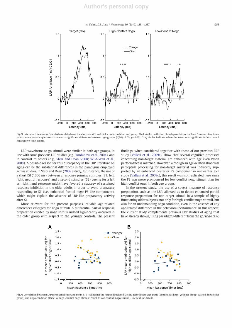
LRP waveforms can be better appreciated in Fig. 3. The LRPwaveforms were more pronounced in the older group than in theyounger group for the high-conflict nogo condition in the followingtime-windows between 236 and 404 ms: 236–256, 304–320, 352–372, 388–404 ms; and for the low-conflict nogo condition in an earlytime-window (208–308 ms), and in a later one (656–684 ms). Therewere no significant differences in the LRP for go stimuli.
The amplitude of the P2 component in CB1 was morepronounced for the low-conflict nogo stimuli than for the high-conflict ones [F(1,26)=49, pb0.001]. This component was morepronounced in the younger than in the older subjects [F(1,26)=11.7,pb0.01], probably due to the fact that the previous N1 component inthe older group was almost twice the size of the N1 component in theyounger group (see Fig. 1). However, in contrast with our previousresults (Vallesi et al., 2009c), there was no interaction between groupand nogo condition (p=0.37). The P3 component for low-conflictnogo stimuli was instead more pronounced in the older group than inthe younger controls [t(26)=−2.69, p=0.012], thus replicatingprevious findings (Vallesi et al., 2009c).
The correlationbetweenLRP andP3amplitude for low-conflict nogocondition was positive and significant in the older group (r=0.62,p=0.018) but not in the younger group (r=−0.01, p=0.99). This
pattern suggests that these twoERP components are functionally linkedin the older group. On the other hand, the correlation between theposterior P2 and LRP was not significant for either group (bothpsN0.09).
In the older group, the correlation between LRP and go-RTs wassignificantly negative for both high-conflict (r=−0.54, p=0.046)and low-conflict (r=−66, p=0.01) nogo conditions (see Fig. 4). Thispattern indicates that the faster elderly subjects were those whoprepared more sub-threshold response for nogo stimuli. Thesecorrelations were not significant in the young group (both psN0.51).Similar correlations between LRP and accuracy were never significantin either group (all psN0.63).
Discussion
The present study tested how the motor processing of non-targetmaterial changes in normal aging during a go/nogo task. Althougholder subjects were slower than their younger controls in the RTs togo stimuli by 36 ms on average (see Table 1), this difference was notsignificant. An age-related response slowing may have been expectedbased on previous literature, but this pattern is more likely to occurwith more complex task conditions (e.g., Yordanova et al., 2004;Vallesi, McIntosh and Stuss, under review). Performance was alsomatched in terms of accuracy. This result provides a good experi-mental context to investigate the neural mechanisms by means ofwhich the aging brain maintains a good level of performance.
Fig. 2. The topographic distribution of the event-related lateralisations (ERLs) according to age group, go/nogo condition and LRP peak latency in the older group is shown bymeansof normalized isovoltage maps. The circles on the head models indicate how the geometry of the map is related to the electrode sites. Since the LRP measures voltage differencesbetween homologous electrodes over the right and left hemispheres, the left hemispheric projection of the maps is arbitrary. The black circles show electrode C3 (and C4),representing the locations from which the LRP was computed. The vertical lines in these waveforms indicate the latencies where the LRP peaked in the older group. The isovoltagemaps refer to those LRP peak latencies (A: go stimuli; B: high-conflict nogo stimuli; C and D: early and late LRP peaks for low-conflict nogo stimuli, respectively).
1254 A. Vallesi, D.T. Stuss / NeuroImage 50 (2010) 1251–1257
Author's personal copy
LRP waveforms to go stimuli were similar in both age groups, inline with some previous LRP studies (e.g., Yordanova et al., 2004), andin contrast to others (e.g., Sterr and Dean, 2008; Wild-Wall et al.,2008). A possible reason for this discrepancy in the LRP literature onaging can be the substantial differences in the paradigms employedacross studies. In Sterr and Dean (2008) study, for instance, the use ofa short ISI (1300 ms) between a response priming stimulus (S1: left,right, neutral response) and a second stimulus (S2) cueing for a leftvs. right hand response might have favored a strategy of sustainedresponse inhibition in the older adults in order to avoid prematureresponding to S1 (i.e., enhanced frontal nogo P3-like component),which might explain the absence of LRP-like preparatory activityafter S1.
More relevant for the present purposes, reliable age-relateddifferences emerged for nogo stimuli. A differential partial responsepreparation elicited by nogo stimuli indeed significantly occurred inthe older group with respect to the younger controls. The present
findings, when considered together with those of our previous ERPstudy (Vallesi et al., 2009c), show that several cognitive processesconcerning non-target material are enhanced with age even whenperformance is matched. However, although an age-related abnormalperceptual processing for non-target material was indirectly sup-ported by an enhanced posterior P2 component in our earlier ERPstudy (Vallesi et al., 2009c), this result was not replicated here sincethe P2 was more pronounced for low-conflict nogo stimuli than forhigh-conflict ones in both age groups.
In the present study, the use of a covert measure of responsepreparation, such as the LRP, allowed us to detect enhanced partialresponse preparation for non-target stimuli in a sample of highlyfunctioning older subjects, not only for high-conflict nogo stimuli, butalso for an undemanding nogo condition, even in the absence of anyage-related difference in the behavioral performance. In this respect,the current study complements previous LRP studies of aging thathave already shown, using paradigms different from the go/nogo task,
Fig. 4. Correlation between LRPmean amplitude andmean RTs (collapsing the responding hand factor) according to age group (continuous lines: younger group; dashed lines: oldergroup) and nogo condition (Panel A: high-conflict nogo stimuli; Panel B: low-conflict nogo stimuli). See text for details.
Fig. 3. Lateralized Readiness Potential calculated over the electrodes C3 and C4 for each condition and group. Black circles on the top of each panel denote at least 5 consecutive time-points when two-sample t-tests showed a significant difference between age-groups [t(26)N2.05, pb0.05]. Gray circles indicate when the t-test was significant in less than 5consecutive time-points.
1255A. Vallesi, D.T. Stuss / NeuroImage 50 (2010) 1251–1257

Author's personal copy
abnormal activation of the wrong response side following conflictinginformation in older individuals (e.g., Zeef et al., 1996). In addition,the present study shows that the inappropriate partial responsepreparation may occur in the older group regardless of the degree ofconflict, although it can last longer for the high-conflict nogocondition than for the low-conflict one.
While there was no correlation between the LRP and the P2component, the positive correlation between the LRP and the P3amplitude for low-conflict nogo condition in the older group suggeststhat these two ERP components are functionally linked. Since the P3component (peak at 440 ms in the older group) followed aninappropriate partial response preparation (LRP peak at 216 ms) forlow-conflict nogo conditions, it might indicate a higher need for thecompensatory inhibition of a partial response in the older group, alsoin line with previous studies that link the nogo P3 to responseinhibition (Roberts et al., 1994; Smith et al., 2007, 2008; but seeFalkenstein et al., 1999).
This pattern suggests that response suppression declines withadvancing age and, even when it is not possible to detect age-relateddeficits with overt performance measures such as false alarms, thisdeficit can still be tracked using more sensitive covert measures ofcortical response preparation, such as the LRP. More generally, thesefindings support the view that suppressing cognitive and neuralprocessing of non-target information becomes less efficient withaging (Fabiani et al., 2006; Gazzaley et al., 2005; Hasher et al., 1988,1999; Wild-Wall and Falkenstein, 2010).
It is worth noting that while the present study shows anincreased reactivity of the preparatory system after the onset ofinterfering information in the older group, previous findings haveshown that the anticipatory frontally-based preparation following apreparatory or warning signal decreases with aging (Sterr and Dean,2008; Vallesi et al., 2009b; Wild-Wall and Falkenstein, 2010). Thisdissociation suggests a shift from a top-down to a stimulus-drivenregulation of motor control (see Paxton et al., 2008, for similar fMRIevidence).
The correlation analyses between nogo-LRPs and go-RTs helpunderstanding the functional meaning of the partial responsepreparation to nogo stimuli in the older group. These correlationswere negative (i.e., the faster subjects were also those with higherpreparatory activity to nogo stimuli), suggesting that the preparationof a response as soon as a stimulus (either go or nogo) appears mightrepresent a strategy to maintain a reasonable response speed to gostimuli, thus explaining the lack of a statistical difference in the go-RTsof the two age groups. Although in this experiment the false alarmrate was not different in the two age groups, it would be interesting toinvestigate whether increasing go/nogo conflict or time pressurewould also enhance inappropriate response preparation for nogostimuli above the response threshold level.
The LRP for low-conflict nogo stimuli showed a biphasic patternin the older group. However, the late LRP increase was notexpected. We acknowledge this effect but may only speculate onits possible functional meaning since it has never been reported inthe literature before. This late component may be related to themotor efferences and somatosensory afferences associated toslightly lifting the finger from the response key after older peoplerealized that an easy nogo stimulus had been presented. Unfortu-nately, the present study did not video-record hand movements oruse electromyography (EMG) to confirm this possibility. However,one experimenter recalled that this behavior was prominent in twoolder subjects. That this possible behavior is unlikely to have alsogenerated the earlier low-conflict nogo LRP peak is suggested bythe short latency (peak at 216 ms), that occurs much earlier thanthe average RTs (700 ms). Moreover, the topographic maps (seeFig. 2D) show that this age-related lateralized component had adifferent scalp distribution with respect to the other ones (slightlymore posterior, with another smaller positivity in ventro-lateral
frontal electrodes), which suggests a different functional meaning.Further studies should investigate the functional role of this lateLRP component.
A possible limit of the present study is the fact that it did not useforce-sensitive response devices or EMG recording. Future studiesshould investigate, by means of these measures, whether abnormalsub-threshold responses to nogo stimuli can also be detectedperipherally in the effector muscles with aging, although a dissoci-ation between LRP and these measures is possible (e.g., Praamstraet al., 1999).
In conclusion, the current LRP study suggests an age-relateddecline in the efficiency of response suppression for non-targetmaterial even when behavioral performance is matched between agegroups and at ceiling. This decline is probably due to disruptivechanges in frontal functionality (West, 1996) or, more generally, infronto-striatal dopaminergic systems (Beste et al., 2010; see Cropleyet al., 2006, for a review) with advancing aging. However, inefficientresponse preparation for non-target stimuli is probably a cost thatolder subjects had to pay in order to maintain a reasonable responsespeed, as suggested by the negative correlation between nogo-LRPand go-RTs. Future studies should further investigate the potentialbehavioral consequences of this excessive age-related motor prepa-ration for distracting material, whether it comes from an endogenouscompensatory strategy or from external demands (e.g., excessive timepressure), and the possible prognostic value of LRP as a covert index ofresponse suppression failure in both normal aging and subclinicalconditions such as incipient Parkinson's disease.
Acknowledgments
This research was partially supported by a postdoctoral fellowshipfunding from Canadian Institute of Health Research (CIHR, MFE-87658) to AV, by a grant from Canadian Foundation for Innovation(CFI) and Ontario Innovation Trust to University of Toronto FunctionalImaging Network (#1226) to DTS, and by the Heart and StrokeFoundation Centre for Stroke Recovery and Posluns Centre for Strokeand Cognition. We thank Sam Chan for his help in data collection andtwo anonymous reviewers for useful suggestions in an earlier versionof the manuscript.
References
Beste, C., Willemssen, R., Saft, C., Falkenstein, M., 2010. Response inhibitionsubprocesses and dopaminergic pathways: Basal ganglia disease effects. Neurop-sychologia 48, 366–373.
Carlson, M.C., Hasher, L., Zacks, R.T., Connelly, S.L., 1995. Aging, distraction, and thebenefits of predictable location. Psychol. Aging 10, 427–436.
Connelly, S.L., Hasher, L., Zacks, R.T., 1991. Age and reading: the impact of distraction.Psychol. Aging 6, 533–541.
Cropley, V.L., Fujita, M., Innis, R.B., Nathan, P.J., 2006. Molecular imaging of thedopaminergic system and its association with human cognitive function. Biol.Psychiatry 59, 898–907.
De Jong, R., Wierda, M., Mulder, G., Mulder, L.J.M., 1988. Use of partial stimulusinformation in response processing. J. Exp. Psychol.: Hum. Percept. Perform. 14,682–692.
Duchek, J.M., Balota, D.A., Faust, M.E., Ferraro, F.R., 1995. Inhibitory processes in youngand older adults in a picture word task. Aging Cogn. 2, 156–167.
Eimer, M., 1998. The lateralized readiness potential as an on-line measure of centralresponse activation processes. Behav. Res. Methods, Instrum. Comput. 30, 146–156.
Eimer, M., Schlaghecken, F., 1998. Effects of masked stimuli on motor activation:behavioral and electrophysiological evidence. J. Exp. Psychol. Hum. Percept.Perform. 24, 1737–1747.
Fabiani, M., Low, K.A.,Wee, E., Sable, J.J., Gratton, G., 2006. Reduced suppression or labilememory? Mechanisms of inefficient filtering of irrelevant information in olderadults. J. Cogn. Neurosci. 18, 637–650.
Falkenstein, M., Hoormann, J., Hohnsbein, J., 1999. ERP components in Go/Nogo tasksand their relation to inhibition. Acta Psychol. 101, 267–291.
Fox, A.M., Michie, P.T., Wynne, C.D., Maybery, M.T., 2000. ERP correlates of responseinhibition to elemental and configural stimuli in a negative patterning task. Clin.Neurophysiol. 111, 1045–1053.
Gazzaley, A., Cooney, J.W., Rissman, J., D'Esposito, M., 2005. Top–down suppressiondeficit underlies working memory impairment in normal aging. Nat. Neurosci. 8,1298–1300.
1256 A. Vallesi, D.T. Stuss / NeuroImage 50 (2010) 1251–1257

Author's personal copy
Gratton, G., Coles, M.G.H., Sirevaag, E.J., Eriksen, C.W., Donchin, E., 1988. Pre- and post-stimulus activation of response channels: a psychophysiological analysis. J. Exp.Psychol.: Hum. Percept. Perform. 14, 331–344.
Hasher, L., Zacks, R.T., 1988.Working memory, comprehension, and aging: a review anda new view. In: Bower, G.H. (Ed.), The Psychology of Learning and Motivation, Vol.22. Academic Press, New York, pp. 193–225.
Hasher, L., Zacks, R.T., May, C.P., 1999. Inhibitory control, circadian arousal, and age. In:Gopher, D., Koriat, A. (Eds.), Attention and Performance XVII, Cognitive Regulationof Performance: Interaction of Theory and Application. MIT Press, Cambridge, MA,pp. 653–675.
Juncos-Rabadan, O., Pereiro, A.X., Facal, D., 2008. Cognitive interference and aging:insights from a spatial stimulus-response consistency task. Acta Psychol.(Amst.)127, 237–246.
Leuthold, H., Sommer, W., Ulrich, R., 1996. Partial advance information and responsepreparation: inferences from the lateralized readiness potential. J Exp. Psychol.Gen. 125, 307–323.
Li, K.Z., Hasher, L., Jonas, D., Rahhal, T.A., May, C.P., 1998. Distractibility, circadianarousal, and aging: a boundary condition. Psychol. Aging 13, 574–583.
Madden, D.J., Langley, L.K., 2003. Age-related changes in selective attention andperceptual load during visual search. Psychol. Aging 18, 54–67.
Paxton, J.L., Barch, D.M., Racine, C.A., Braver, T.S., 2008. Cognitive control, goalmaintenance, and prefrontal function in healthy aging. Cereb. Cortex 18,1010–1028.
Picton, T.W., Bentin, S., Berg, P., Donchin, E., Hillyard, S.A., Johnson Jr, R., et al., 2000.Guidelines for using human event-related potentials to study cognition: recordingstandards and publication criteria. Psychophysiology 37, 127–152.
Praamstra, P., Stegeman, D.F., Horstink, M.W., Cools, A.R., 1996. Dipole source analysissuggests selective modulation of the supplementary motor area contribution to thereadiness potential. Electroencephalogr. Clin. Neurophysiol 98, 468–477.
Praamstra, P., Plat, E.M., Meyer, A.S., Horstink, M.W., 1999. Motor cortex activation inParkinson's disease: dissociation of electrocortical and peripheral measures ofresponse generation. Mov. Disord. 14, 790–799.
Roberts, L.E., Rau, H., Lutzenberger, W., Birbaumer, N., 1994. Mapping P300 waves ontoinhibition: Go/No-Go discrimination. Electroencephalogr. Clin. Neurophysiol. 92,44–55.
Scialfa, C.T., Esau, S.P., Joffe, K.M., 1998. Age, target-distractor similarity, and visualsearch. Exp. Aging Res. 24, 337–358.
Shin, E., Fabiani, M., Gratton, G., 2004. Evidence of partial response activation in amemory-search task. Brain Res.Cogn Brain Res. 20, 281–293.
Smith, J.L., Johnstone, S.J., Barry, R.J., 2007. Response priming in the Go/NoGo task: theN2 reflects neither inhibition nor conflict. Clin. Neurophysiol. 118, 343–355.
Smith, J.L., Johnstone, S.J., Barry, R.J., 2008. Movement-related potentials in theGo/NoGo task: the P3 reflects both cognitive andmotor inhibition. Clin. Neurophysiol.119, 704–714.
Sterr, A., Dean, P., 2008. Neural correlates of movement preparation in healthy ageing.Eur. J. Neurosci. 27, 254–260.
Sweeney, J.A., Rosano, C., Berman, R.A., Luna, B., 2001. Inhibitory control of attentiondeclines more than working memory during normal aging. Neurobiol. Aging 22,39–47.
Tun, P.A., O'Kane, G., Wingfield, A., 2002. Distraction by competing speech in young andolder adult listeners. Psychol. Aging 17, 453–467.
Vallesi, A., Mapelli, D., Schiff, S., Amodio, P., Umiltà, C., 2005. Horizontal and verticalSimon effect: different underlying mechanisms. Cognition 96, B33–B43 Erratum in:Cognition (2005), 96 (3), p. B115.
Vallesi, A., McIntosh, A.R., Alexander, M.P., Stuss, D.T., 2009a. FMRI evidence of afunctional network setting the criteria for withholding a response. Neuroimage 45,537–548.
Vallesi, A., McIntosh, A.R., Stuss, D.T., 2009b. Temporal preparation in aging: afunctional MRI study. Neuropsychologia 47, 2876–2881.
Vallesi, A., Stuss, D.T., McIntosh, A.R., Picton, T.W., 2009c. Age-related differences inprocessing irrelevant information: evidence from event-related potentials.Neuropsychologia 47, 577–586.
Verleger, R., Paehge, T., Kolev, V., Yordanova, J., Jaśkowski, P., 2006. On the relation ofmovement-related potentials to the go/no-go effect on P3. Biol. Psychol. 73,298–313.
West, R.L., 1996. An application of prefrontal cortex function theory to cognitive aging.Psychol. Bull. 120, 272–292.
Wild-Wall, N., Falkenstein, M., 2010. Age-dependent impairment of auditory proces-sing under spatially focused and divided attention: An electrophysiological study.Biol. Psychol. 83, 27–36.
Wild-Wall, N., Falkenstein, M., Hohnsbein, J., 2008. Flanker interference in young andolder participants as reflected in event-related potentials. Brain Res. 1211, 72–84.
Yordanova, J., Kolev, V., Hohnsbein, J., Falkenstein, M., 2004. Sensorimotor slowing withageing is mediated by a functional dysregulation of motor-generation processes:evidence from high-resolution event-related potentials. Brain 127, 351–362.
Zeef, E.J., Sonke, C.J., Kok, A., Buiten, M.M., Kenemans, J.L., 1996. Perceptual factorsaffecting age-related differences in focused attention: performance and psycho-physiological analyses. Psychophysiology 33, 555–565.
1257A. Vallesi, D.T. Stuss / NeuroImage 50 (2010) 1251–1257