Conscious Perception of Emotional Stimuli: Brain Mechanisms
13
The Neuroscientist XX(X) 1–13 © The Author(s) 2011 Reprints and permission: http://www. sagepub.com/journalsPermissions.nav DOI: 10.1177/1073858411416515 http://nro.sagepub.com Review Emotional stimuli carry special weight both in terms of altering our behavior and in shaping the contents of our awareness. Sometimes, this tendency can be advanta- geous, as when one is acutely aware of the location and direction of movement of a bear passing through the campsite. Other times, however, it can be disruptive, such as when the motorist stopped in front of us momentarily escapes our awareness in favor of the accident at the side of the highway. Perspectives on awareness of emotional visual stimuli are shaped by two views. One is that sub- cortical regions, particularly the amygdala, “automati- cally” (i.e., independently of awareness) process basic threat-related stimuli such as fearful facial expressions subconsciously to facilitate reflexive actions (Tamietto and de Gelder 2010). Another is that a frontoparietal net- work, in contrast, plays a generic role in bringing stimuli into consciousness, by providing a global workspace (Baars 2002) or by amplifying stimulus representations in other category-specific neural regions to allow these stimuli to reach awareness (Dehaene and others 2006). The purpose of this review is to examine current findings concerning the functional neuroanatomy of the aware- ness (i.e., conscious perception) of emotional visual stimuli. Specifically, we present a “multiple-mechanisms” view of emotional awareness that suggests that conscious perception of emotional stimuli occurs as a function of a complex interplay between three subsystems: cortical areas traditionally associated with attention, subcortical structures implicated in the rapid detection of emotional stimuli, and medial regions of the prefrontal cortex impli- cated in emotion regulation. According to this perspec- tive, rather than playing a generic role in consciousness, we argue that frontoparietal contributions are crucial in activating representations of classes of stimuli that are less salient, allowing them to reach awareness. Second, amygdala activity, whether initiated primarily through subcortical or re-entrant cortical input, does not occur as a separate phenomenon alongside higher-order processes 416515NRO XX X 10.1177/10738584114165 15Mitchell and GreeningThe Neuroscientist 1 Department of Psychiatry and Department of Anatomy & Cell Biology, Schulich School of Medicine & Dentistry, University of Western Ontario, London, Ontario, Canada 2 Centre for Brain and Mind, Department of Psychology, University of Western Ontario, London, Ontario, Canada Corresponding Author: Derek G.V. Mitchell, University Hospital, University of Western Ontario, 339 Windermere Road, London, Ontario, Canada Email: [email protected] Conscious Perception of Emotional Stimuli: Brain Mechanisms Derek G.V. Mitchell 1,2 and Steven G. Greening 1,2 Abstract Emotional stimuli are thought to gain rapid and privileged access to processing resources in the brain. The structures involved in this enhanced access are thought to support subconscious, reflexive processes. Whether these pathways contribute to the phenomenological experience of emotional visual awareness (i.e., conscious perception) is unclear. In this review, it is argued that subcortical networks associated with the rapid detection of emotionally salient stimuli also play a key role in shaping awareness. This proposal is based on the idea that awareness of visual stimuli should be considered along a continuum, having intermediate levels, rather than as an all-or-none construct. It is also argued that awareness of emotional stimuli requires less input from frontoparietal structures that are often considered crucial for visual awareness. Evidence is also presented that implicates a region of the medial prefrontal cortex, involved in emotion regulation, in modulating amygdala output to determine awareness of emotional visual stimuli; when emotional stimuli are present, the conscious perception of alternative stimuli requires greater regulatory influences from cortical structures. Thus, emotional stimuli are privileged not only for neuronal representation and impact on subconscious processes, but also for awareness, allowing humans to deal flexibly rather than merely reflexively to biologically significant stimuli. Keywords emotion, consciousness, attention, emotional awareness, visual awareness, emotion regulation, amygdala, prefrontal cortex at UNIV OF WESTERN ONTARIO on February 24, 2012 nro.sagepub.com Downloaded from
-
Upload
khangminh22 -
Category
Documents
-
view
4 -
download
0
Transcript of Conscious Perception of Emotional Stimuli: Brain Mechanisms
The NeuroscientistXX(X) 1 –13© The Author(s) 2011Reprints and permission: http://www. sagepub.com/journalsPermissions.navDOI: 10.1177/1073858411416515http://nro.sagepub.com
Review
Emotional stimuli carry special weight both in terms of altering our behavior and in shaping the contents of our awareness. Sometimes, this tendency can be advanta-geous, as when one is acutely aware of the location and direction of movement of a bear passing through the campsite. Other times, however, it can be disruptive, such as when the motorist stopped in front of us momentarily escapes our awareness in favor of the accident at the side of the highway. Perspectives on awareness of emotional visual stimuli are shaped by two views. One is that sub-cortical regions, particularly the amygdala, “automati-cally” (i.e., independently of awareness) process basic threat-related stimuli such as fearful facial expressions subconsciously to facilitate reflexive actions (Tamietto and de Gelder 2010). Another is that a frontoparietal net-work, in contrast, plays a generic role in bringing stimuli into consciousness, by providing a global workspace (Baars 2002) or by amplifying stimulus representations in other category-specific neural regions to allow these stimuli to reach awareness (Dehaene and others 2006). The purpose of this review is to examine current findings concerning the functional neuroanatomy of the aware-ness (i.e., conscious perception) of emotional visual stimuli. Specifically, we present a “multiple-mechanisms” view
of emotional awareness that suggests that conscious perception of emotional stimuli occurs as a function of a complex interplay between three subsystems: cortical areas traditionally associated with attention, subcortical structures implicated in the rapid detection of emotional stimuli, and medial regions of the prefrontal cortex impli-cated in emotion regulation. According to this perspec-tive, rather than playing a generic role in consciousness, we argue that frontoparietal contributions are crucial in activating representations of classes of stimuli that are less salient, allowing them to reach awareness. Second, amygdala activity, whether initiated primarily through subcortical or re-entrant cortical input, does not occur as a separate phenomenon alongside higher-order processes
416515 NROXXX10.1177/1073858411416515Mitchell and GreeningThe Neuroscientist
1Department of Psychiatry and Department of Anatomy & Cell Biology, Schulich School of Medicine & Dentistry, University of Western Ontario, London, Ontario, Canada2Centre for Brain and Mind, Department of Psychology, University of Western Ontario, London, Ontario, Canada
Corresponding Author:Derek G.V. Mitchell, University Hospital, University of Western Ontario, 339 Windermere Road, London, Ontario, Canada Email: [email protected]
Conscious Perception of Emotional Stimuli: Brain Mechanisms
Derek G.V. Mitchell1,2 and Steven G. Greening 1,2
Abstract
Emotional stimuli are thought to gain rapid and privileged access to processing resources in the brain. The structures involved in this enhanced access are thought to support subconscious, reflexive processes. Whether these pathways contribute to the phenomenological experience of emotional visual awareness (i.e., conscious perception) is unclear. In this review, it is argued that subcortical networks associated with the rapid detection of emotionally salient stimuli also play a key role in shaping awareness. This proposal is based on the idea that awareness of visual stimuli should be considered along a continuum, having intermediate levels, rather than as an all-or-none construct. It is also argued that awareness of emotional stimuli requires less input from frontoparietal structures that are often considered crucial for visual awareness. Evidence is also presented that implicates a region of the medial prefrontal cortex, involved in emotion regulation, in modulating amygdala output to determine awareness of emotional visual stimuli; when emotional stimuli are present, the conscious perception of alternative stimuli requires greater regulatory influences from cortical structures. Thus, emotional stimuli are privileged not only for neuronal representation and impact on subconscious processes, but also for awareness, allowing humans to deal flexibly rather than merely reflexively to biologically significant stimuli.
Keywords
emotion, consciousness, attention, emotional awareness, visual awareness, emotion regulation, amygdala, prefrontal cortex
at UNIV OF WESTERN ONTARIO on February 24, 2012nro.sagepub.comDownloaded from

2 The Neuroscientist XX(X)
that dictate awareness. Instead, we argue that subcortical structures may be activated in conditions of limited awareness and, in doing so, act to bias the content of awareness to emotional stimuli through interactions with the ventral visual system. Thus, activity in phylogeneti-cally older structures, such as the amygdala, can augment contributions from higher cortical structures to increase the odds that biologically significant stimuli will enter into our consciousness. Accordingly, in the context of modulating the threshold for awareness of emotional items, the amygdala operates in much the same way that frontoparietal structures do for more mundane stimuli. Third, we argue that emotional awareness represents a special case for consciousness in that it is not only modu-lated by attention but also by neural regions implicated in emotion regulation, particularly perigenual regions of the prefrontal cortex. Thus, also implicated in modulating awareness of emotional visual stimuli are areas of the medial prefrontal cortex associated with extinguishing conditioned associations (Milad and Quirk 2002) and controlling the deleterious impact of emotional distract-ers on behavior (Etkin and others 2006). This “multiple-mechanisms” view of emotional awareness addresses some of the apparent contradictions concerning the auto-maticity of emotional processing and the extent to which consciousness is best considered a dimensional rather than a discrete construct.
Operationalizing ConsciousnessThe overarching approach to consciousness adopted by cognitive neuroscience is evolutionary in nature; it acknowledges that while many adaptive actions to sen-sory input are reflexive and do not require prior thought or planning (e.g., the initial fight-or-flight response), these reflexes fail to meet the challenges that arise in more complex environments. According to this perspec-tive, consciousness evolved to facilitate less stereotyped actions that allow the organism to flexibly evaluate a situation, plan a course of action, and carry out more sophisticated behavioral responding (Crick and Koch 2003). Despite the growing interest and research on the topic of consciousness, cognitive neuroscience has not yet embraced a gold standard for its experimental mea-surement or manipulation. Multiple perspectives on the definition of consciousness exist, and the functional neu-rocognitive systems considered critically involved in consciousness vary according to the perspective adopted. Whereas some models consider a mental state conscious when it is capable of influencing choice behavior or per-ceptual discriminations at above-chance levels, others consider this to be insufficient evidence (for a review, see Seth and others [2008]). The most widely adopted per-spectives consider a mental state conscious when it is not
just available for self-report but also capable of influenc-ing a variety of cognitive processes (Baars 2002; Seth and others 2008). This enhanced cognitive availability is thought to assist a variety of complex operations such as voluntary action, working memory, visual imagery, long-term learning, and representations of the self (Baars 2002).
One factor that has critical implications for under-standing visual awareness is whether consciousness should be viewed in a discrete or dimensional fashion. The majority of research in cognitive neuroscience has considered awareness in a binary fashion; subjects are either aware or unaware of the stimuli in question. Unfortunately, the tendency to consider consciousness in this way may have resulted in some of the apparent contradictory findings concerning the role of subcortical structures in visual awareness. More recently, evidence from multiple areas has raised the possibility that con-sciousness is not an all-or-none phenomenon but instead occurs along a spectrum that ranges from a vague feel-ing of something happening to astute image awareness and meta-awareness (i.e., the ability to reflect on the nature or quality of a conscious state). One critique of the binary approach is that when participants are forced to indicate whether they are aware or unaware of a visual stimulus, a response bias exists towards answer-ing in the negative, even in situations where some level of awareness exists. Studies that have incorporated sig-nal detection theory, which provides a measure of sensi-tivity that is independent of a subject’s response bias, support the idea that visual awareness is best conceptu-alized in a graded fashion (Pessoa and others 2005a; Szczepanowski and Pessoa 2007). Such studies show that participants’ judgments of their performance remain high even under conditions that were typically assumed to eliminate awareness.
In considering the threshold for, and nature of, con-sciousness, an important distinction must be made between subjective and objective measures of visual awareness (Szczepanowski and Pessoa 2007). Objective awareness is defined as above-chance performance on a forced-choice discrimination task. In contrast, subjective awareness requires not only intact discrimination but also a subjective sense of being aware of the stimulus. Recent empirical evi-dence suggests that the threshold for subjective awareness is higher, and consequently, whether one adopts a subjec-tive or objective view of awareness will significantly shape neurocognitive models of visual awareness (Szczepanowski and Pessoa 2007). The relative merit of objective versus subjective perspectives is still debated. Some argue that subjective awareness, because it engenders greater calcula-tion and flexibility in responding, more closely reflects the core features of consciousness (Merikle and others 2001). Others have argued that subjective measures are flawed because forcing introspection about awareness alters
at UNIV OF WESTERN ONTARIO on February 24, 2012nro.sagepub.comDownloaded from
Mitchell and Greening 3
the target process, instead providing an index of meta-awareness (Persaud and others 2007).
In an attempt to circumvent this problem, a growing number of researchers provide additional measures of confidence in their studies of perceptual choice (Persaud and others 2007; Overgaard and others 2008). In these paradigms, participants first make a forced-choice judg-ment about the presence or nature of stimulus presenta-tions in which awareness is varied. Next, participants provide a rating concerning the quality of the percept or confidence in the accuracy of their judgment (e.g., a con-fidence rating or wager). The rationale is that evidence of some level of awareness is present if a significant rela-tionship exists between confidence in the response accu-racy of performance or perceptual quality of the stimulus and actual choice performance. This technique offers the advantage of combining elements of objective and sub-jective measures of consciousness while avoiding some of the difficulties of each (e.g., a focus on introspection about awareness). As is reviewed below, such measures have been found to be more sensitive to awareness than objective or subjective measures alone.
Many early studies of visual awareness considered consciousness subjectively, in an all-or-none fashion, which tended to underestimate awareness in subjects. More recent work supports the idea that awareness is bet-ter measured along a continuum. Dimensional measures of awareness appear to fulfill elements of both objective and subjective criteria for awareness, perhaps therefore better reflecting the more widely held view of conscious-ness as involving a mental state that influences a variety of cognitive processes. As we will argue below, adopting this more naturalistic view of consciousness has impor-tant implications for neurocognitive models concerning awareness of emotional visual stimuli.
The Relationship between Consciousness and AttentionThe nature of the relationship between attention and con-sciousness remains debated. It is clear that selective attention enhances the neural representation of target stimuli (Desimone and Duncan 1995). However, whereas it is considered a critical precursor for awareness by some (Driver and Mattingley 1998; Dehaene and others 2006), it is thought to be a distinct and potentially con-founding process by others (Koch and Tsuchiya 2007; Kentridge and others 2008). Like directed attention, awareness involves interactions between visual-cortical regions involved in stimulus representation and frontopa-rietal cortical regions (Fig. 1). Similarly, just as attention augments the strength of representations in visual and temporal cortices, visual awareness is associated with increased activity in these regions (Leopold and Logothetis 1996; Tong and others 1998; Rees and others
2000; Vuilleumier and others 2001b). Thus, attention appears able to both enhance the neural representation of target stimuli during perceptual conflict (Desimone and Duncan 1995; Reynolds and others 1999) and reduce the threshold necessary for perceptual input to reach aware-ness (Sumner and others 2006). Furthermore, changes in perceptual awareness are associated with activity in frontoparietal regions (Lumer and Rees 1999), and func-tional impairments within either the frontal or parietal cortex disrupt awareness (Pascual-Leone and Walsh 2001; Vuilleumier and others 2008; Del Cul and others 2009). Together, these results are consistent with the sug-gestion that normal awareness, if not critically dependent on attention, certainly benefits from it.
In addition to its effects on perceptual representation, there is also evidence that attention can augment cogni-tive processes without affecting awareness. For example, cueing attention towards a masked prime can lead to advantages in reaction times without affecting awareness of the prime (Kentridge and others 2008). Similarly, Sumner and others (2006) used a sensorimotor priming task in which a negative (response-slowing) effect occurred when the prime (an arrow cue pointing left or
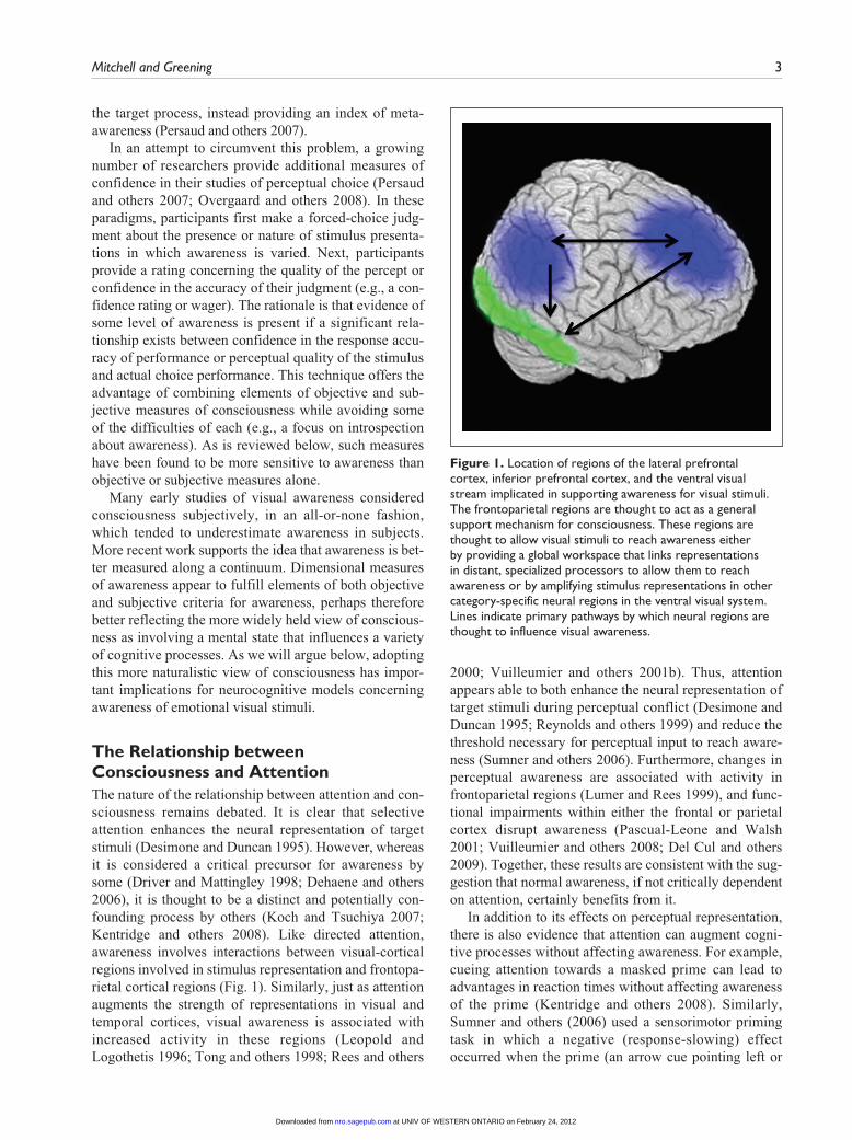
Figure 1. Location of regions of the lateral prefrontal cortex, inferior prefrontal cortex, and the ventral visual stream implicated in supporting awareness for visual stimuli. The frontoparietal regions are thought to act as a general support mechanism for consciousness. These regions are thought to allow visual stimuli to reach awareness either by providing a global workspace that links representations in distant, specialized processors to allow them to reach awareness or by amplifying stimulus representations in other category-specific neural regions in the ventral visual system. Lines indicate primary pathways by which neural regions are thought to influence visual awareness.
at UNIV OF WESTERN ONTARIO on February 24, 2012nro.sagepub.comDownloaded from

4 The Neuroscientist XX(X)
right that precedes the target stimulus) was invisible and a positive (response-speeding) effect occurred when the prime was visible. They further demonstrated that atten-tional cueing of the prime (a box appearing at the location of the prime) increased the existing behavioral impact of that prime on behavior. Thus, cueing the location of the subliminal prime resulted in an enhancement of the nega-tive effect rather than a reduction of the negative effect, as would be expected if attention made an unconscious prime more like a conscious one. Thus, the authors showed that attentional cueing can modulate the behav-ioral response to invisible stimuli without influencing awareness. Other studies suggest that frontoparietal areas may also augment representations in other contexts, such as in reward-related decision making (Mitchell and others 2009; Greening and others 2011), and the extent to which these processes mirror those involved in consciousness remains unclear (Mitchell 2011).
Although attention can be manipulated independent of awareness, attention also appears to be facilitated by awareness. For example, Kentridge and others (1999) demonstrated in a blindsight patient that attention can lead to improved detection of a stimulus presented at a cued location even though the patient was not aware of the cue. However, when the patient was aware of the cue, the observed behavioral advantage was even more robust. Similarly, Tsushima and others (2006) showed that only once a task-irrelevant stimulus reached a detectable threshold was its negative impact on behavior attenuated, leading the authors to suggest that awareness facilitates top-down inhibitory control over distracting stimuli. Lastly, it has been shown that the involvement of the anterior cingulate cortex in resolving perceptual conflict occurs only once a stimulus passes a detectable threshold (Dehaene and others 2003).
In this section, we reviewed evidence that although attention facilitates visual awareness when it is deployed to strengthen the perceptual representation of visual stimuli in sensory areas, attention can be greatly enhanced in some circumstances without increasing the likelihood of aware-ness. For example, attention can enhance sensorimotor pro-cesses without generating awareness regardless of the degree of enhancement. It was also noted that although attention can be manipulated without affecting awareness, selective attention is more effective when awareness is present. Together, these results suggest that attention and awareness operate in a synergistic fashion, adding value to one another. However, it also suggests that attention and awareness can operate with a degree of independence. As will be argued below, the fact that awareness can be uncou-pled from neurocognitive systems involved in top-down attention and that attention draws on a finite processing capacity place the amygdala in a position to provide a key adaptive role in bringing biologically significant stimuli into consciousness.
Does Emotional Visual Processing Occur Independently of Awareness?
One dominant perspective on emotion is that threat-related stimuli are so important that channels exist that allow subcortical emotion-related brain areas to process these stimuli independently of attention or awareness (LeDoux and others 1984; Vuilleumier and others 2002). It is suggested that, at the neural level, automatic emotional processing is subserved by a direct subcorti-cal route through the superior colliculus and pulvinar to the amygdala (Morris and others 1999; Morris and oth-ers 2001). This automatic subcortical response is thought to precipitate a series of adaptive reflexes to prepare the organism to respond to threat. It is further thought that nonconscious activation of the amygdala is not merely a degraded form of conscious processing but rather a qualitatively distinct mode of visual processing that depends on a partially segregated network of brain regions (Tamietto and de Gelder 2010). Evidence for this perspective comes from studies involving healthy subjects, patients with hemifield neglect, and patients with “affective blindsight” following lesions of the stri-ate cortex. Below, we will review evidence involving each of these populations that support the notion of automaticity. It will be noted that the implicit assump-tion in many of these studies is that consciousness is a binary phenomenon. Following this, we will reconsider these data by approaching visual awareness as a dimen-sion.
Evidence for Automatic Processing of Emotional ExpressionsA number of studies in healthy individuals are cited as evidence for the automaticity of emotional processing. Neuroimaging studies show enhanced amygdala activity to fearful faces even when attention is diverted elsewhere (Vuilleumier and others 2001a; Williams and others 2005). Furthermore, similar activity has been observed under conditions in which awareness to stimuli is dimin-ished through a process of backward masking. Backward masking attempts to render a target stimulus invisible by presenting it at a very short duration (e.g., a fearful face presented for 17 milliseconds) and then replacing it with a second irrelevant visual stimulus (the mask) of a longer duration. Studies employing this technique have noted significant amygdala activity to masked fear-conditioned stimuli (Morris and others 1999), masked fearful facial expressions (Whalen and others 1998), and even masked fearful eye whites (Whalen and others 2004). Because these studies show robust amygdala activity in the appar-ent absence of awareness, they suggest emotional encod-ing occurs independently of awareness.
at UNIV OF WESTERN ONTARIO on February 24, 2012nro.sagepub.comDownloaded from
Mitchell and Greening 5
Additional support for automaticity has also been gleaned from lesion studies (Dolan and Vuilleumier 2003) including those with parietal neglect and affective blindsight (e.g., Driver and Mattingley 1998). Patients with neglect following lesions to the right parietal cortex show reduced awareness of stimuli located in the contral-esional side of space, particularly in the presence of com-peting (right-sided) stimuli (a phenomenon known as “extinction”). Neglect differs from cortical blindness in that input to the early visual cortex is intact and the defi-cits are not retinotopic in nature. Interestingly, patients with parietal neglect are more likely to notice emotional stimuli relative to neutral stimuli presented to their affected hemifield (Vuilleumier and Schwartz 2001a). A subsequent fMRI study involving a patient with parietal lesions showed that even neglected emotional stimuli reliably activated the amygdala (Vuilleumier and others 2002). Because they also found emotion-related increases in activity in the fusiform gyrus to neglected stimuli, the authors concluded that feedback influences from the amygdala to such visual areas occurred indepen-dently of awareness and attention.
Blindsight is a condition resulting from a lesion to the striate cortex or its neural projections that leaves a patient subjectively blind while preserving the ability to discrim-inate stimulus features at above-chance (often high) lev-els of accuracy (Weiskrantz and others 1995; Dienes and Scott 2005). In their seminal study, de Gelder and others (1999) presented a case of “affective blindsight,” show-ing that cortically blind patient G.Y. was able to discrimi-nate emotional facial expressions of fear, anger, sadness, and happiness presented to his blind visual field. Blindsight patients have also shown potentiated startle reflexes to unseen fear-conditioned stimuli (Hamm and others 2003), and increased amygdala activity to blind-field presentations of fearful faces (Morris and others 2001). Similarly, a patient with complete bilateral lesions to the striate cortex has shown increased amygdala activ-ity to emotional facial expressions despite the apparent absence of awareness (Pegna and others 2005). One inter-pretation of the above studies is that because the amyg-dala is robustly activated in conditions of unconscious processing, activation of this automatic circuit occurs independently of, and therefore does not contribute to, consciousness. Below, we discuss two issues that compli-cate this interpretation.
Is Awareness Absent or Merely Degraded in Studies of Automaticity?Studies that reveal robust amygdala activity to subliminal emotional stimuli seem at odds with the argument that the amygdala plays a role in increasing the likelihood that stimuli enter into awareness. However, recent evidence suggests that the level of awareness elicited in many
studies involving healthy and brain lesion populations may have been underestimated. According to this per-spective, above-chance performance on forced-choice discrimination tasks where participants report that aware-ness is absent may actually reflect partial conscious perception. Accordingly, it has been argued that when subjective ratings of awareness are reduced due to degraded perceptions, participants show a response bias towards indicating that they are “guessing” despite empir-ical evidence of residual awareness (Pessoa and others 2006; Kouider and Dehaene 2007). Indeed, using sophis-ticated signal detection theory techniques, Pessoa and others (2005a) show that stimulus durations that were previously assumed to reflect unconscious processing produce visual awareness in the majority of healthy par-ticipants. Indeed, some individuals are able to detect stimuli in masking paradigms with presentations as short as 17 milliseconds, raising questions about the extent to which awareness was eliminated in many previous stud-ies utilizing masking procedures.
Similar concerns exist for studies involving patients with acquired lesions. Although consciousness is clearly altered in patients who show blindsight following lesions to the visual cortex, there is also evidence that residual awareness is present. For example, while denying aware-ness of stimuli presented in the blind field, patients often experience a “feeling that something happened” (Morris and others 2001; Stoerig and Barth 2001). Accordingly, a distinction has been made between type I blindsight, which involves a complete lack of awareness, and type II blindsight, which involves some feeling that an event has occurred (Weiskrantz 1997). It has also been noted that stimuli can transition between categories based on prac-tice effects or other considerations (Weiskrantz 1997; Sahraie and others 2010). For example, repeated expo-sure to a stimulus can boost discrimination performance to above-chance levels; with additional exposure, aware-ness can transition from type I to type II. In addition, although awareness remained absent during presentations of sine-wave gratings, blindsight patient D.B. reported being aware of afterimages triggered by these same stim-uli (Weiskrantz and others 2002). Lastly, it has been argued that G.Y.’s blindsight sensations can be re-created by altered presentations to the intact visual field, suggest-ing that the condition is better conceptualized as low-level conscious vision (Stoerig and Barth 2001).
Studies that incorporate more sensitive measures of awareness provide additional evidence that residual awareness capacities exist in patients with blindsight. Overgaard and others (2008) conducted two experiments with blindsight patient G.R. involving shape discrimina-tions. In the first experiment, they used the traditional binary coding for awareness (yes/no) and replicated previous findings that there is no relationship between accuracy and awareness. However, when using a more
at UNIV OF WESTERN ONTARIO on February 24, 2012nro.sagepub.comDownloaded from
6 The Neuroscientist XX(X)
sensitive four-point scale (including “weak glimpse” and “almost clear image” as intermediate awareness catego-ries), they found a significant linear relationship between perceptual awareness score and accuracy. Using a similar four-point scale with patient G.Y., Zeki and Ffytche (1998) demonstrated that a significant correlation was observed between subjective awareness and the accuracy in detecting the direction of moving stimuli. Lastly, Persaud and others (2007) used wagering as an indirect estimate of awareness in G.Y. during grating pattern judgments. Although G.Y. showed no tendency to place higher wagers on the trials in which he was correct, he was significantly more likely to wager highly on the sub-sequent trial despite receiving no feedback. The authors suggest this is indicative of occasional awareness of cor-rect decision making on the part of G.Y. Evidence of awareness was later corroborated through a reanalysis of the data using a log-linear approach (Szczepanowski 2010). Thus, evidence that residual awareness is associ-ated with blindsight exists in three separate patients (D.B., G.R., and G.Y.), in more than one-dimensional mea-sure of awareness, and under multiple task conditions.
Does the Amygdala Activate at Similar Levels to “Seen” and “Unseen” Stimuli?Contrary to strict accounts of automaticity, a number of studies have now found that activity within the amygdala is reduced when sufficient attentional demands are devoted
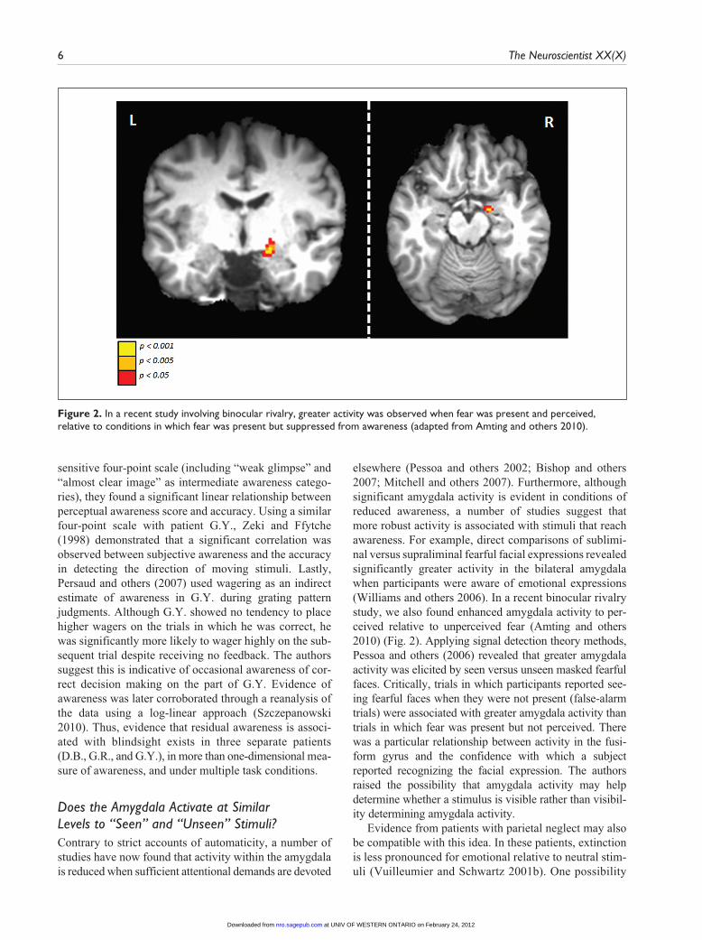
elsewhere (Pessoa and others 2002; Bishop and others 2007; Mitchell and others 2007). Furthermore, although significant amygdala activity is evident in conditions of reduced awareness, a number of studies suggest that more robust activity is associated with stimuli that reach awareness. For example, direct comparisons of sublimi-nal versus supraliminal fearful facial expressions revealed significantly greater activity in the bilateral amygdala when participants were aware of emotional expressions (Williams and others 2006). In a recent binocular rivalry study, we also found enhanced amygdala activity to per-ceived relative to unperceived fear (Amting and others 2010) (Fig. 2). Applying signal detection theory methods, Pessoa and others (2006) revealed that greater amygdala activity was elicited by seen versus unseen masked fearful faces. Critically, trials in which participants reported see-ing fearful faces when they were not present (false-alarm trials) were associated with greater amygdala activity than trials in which fear was present but not perceived. There was a particular relationship between activity in the fusi-form gyrus and the confidence with which a subject reported recognizing the facial expression. The authors raised the possibility that amygdala activity may help determine whether a stimulus is visible rather than visibil-ity determining amygdala activity.
Evidence from patients with parietal neglect may also be compatible with this idea. In these patients, extinction is less pronounced for emotional relative to neutral stim-uli (Vuilleumier and Schwartz 2001b). One possibility
Figure 2. In a recent study involving binocular rivalry, greater activity was observed when fear was present and perceived, relative to conditions in which fear was present but suppressed from awareness (adapted from Amting and others 2010).
at UNIV OF WESTERN ONTARIO on February 24, 2012nro.sagepub.comDownloaded from

Mitchell and Greening 7
is that this emotion-related resistance to neglect may be conferred by interactions between the amygdala and intact ventral visual areas. However, a subsequent fMRI study of parietal neglect showed enhanced amygdala activity to both seen and unseen fearful faces and no interaction between emotion and awareness when neutral faces were also considered (Vuilleumier and others 2002). It is important to note that this study treated aware-ness in a binary fashion, and so it is unclear whether residual awareness may have been evident had a dimen-sional approach been utilized. Even so, although the criti-cal direct comparison between seen versus unseen fearful faces was not performed, greater effect sizes within the left amygdala were observed to seen versus unseen fear-ful faces. The available evidence from parietal neglect should not be taken as incompatible with the idea that the amygdala plays a role in modulating the threshold for awareness of emotional visual stimuli.
Many current models of emotional awareness are dominated by the idea that emotional awareness of visual stimuli is categorical, being either present or absent. Studies that have examined awareness in this fashion generally come to the conclusion that the amygdala oper-ates independently of awareness to precipitate a series of reflexive actions. We reviewed evidence in support of the idea that consciousness is best measured along a dimen-sion. Further, that when it is measured in this fashion, the possibility is raised that the amygdala, along with other areas not traditionally associated with consciousness, may play a role in biasing the content of awareness. This idea is further developed in the remaining sections.
A Multiple-Mechanisms Model of Emotional AwarenessHere, we present a model of emotional awareness that implicates interactions between structures involved in top-down attentional control, bottom-up emotional salience, and emotion regulation. According to this con-ceptualization, the amygdala not only detects innate bio-logically and socially relevant information but may also facilitate awareness of such stimuli by interacting with phylogenetically newer systems. The view rests on the assumption that although it is true that the amygdala can show activation in conditions of diminished awareness, this activation may still influence the threshold for con-sciousness. This formulation reconciles the seemingly contradictory findings regarding the automaticity of amygdala activation noted above. Furthermore, while not ruling out a necessary contribution from top-down struc-tures to facilitate normal conscious perception, we raise the possibility that the degree of top-down involvement varies as a function of the stimuli that either reach, or are suppressed from, consciousness.
A Role for a Frontoparietal Network in Awareness
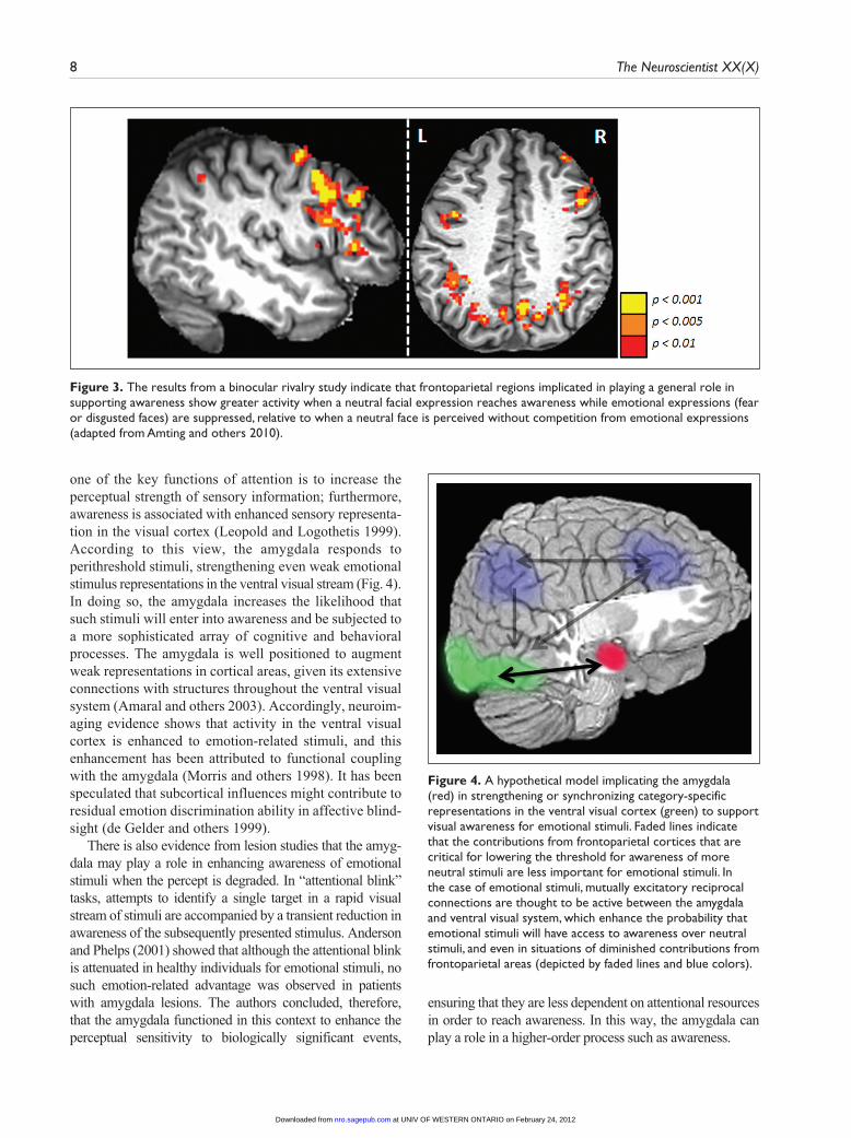
At a functional level, it has been suggested that the pari-etal cortex facilitates visual awareness by integrating retinal information with more sophisticated levels of processing from the ventral (object properties) and dorsal (location- and action-related information) streams (Driver and Mattingley 1998). Similarly, other models of con-sciousness suggest that frontoparietal areas act as a “global workspace,” linking representations in distant, specialized processors to allow them to reach awareness (Kouider and Dehaene 2007). In line with this idea, it has been shown that lesions to the left lateral prefrontal cor-tex reduce the likelihood that masked stimuli reach awareness (Del Cul and others 2009). A key means by which a frontoparietal network is thought to support awareness is by augmenting the strength of stimulus rep-resentations in sensory areas (Dehaene and others 2001). Whereas ventral areas of the occipital and temporal cor-tices facilitate category-specific processing, frontoparie-tal areas are thought to play a more generic role in consciousness. For example, it has been suggested that parietal contributions are equally important for the con-scious perception of emotional and nonemotional stimuli (Vuilleumier and others 2002). An alternate view, favored here, is that contributions from frontoparietal regions vary according to the nature of the stimulus. For example, studies involving patients with hemifield neglect suggest that inputs from parietal areas are less critical for emotional stimuli. Thus, even following severe lesions affecting the right parietal cortex, neglect is less pronounced for emotional relative to neutral facial expressions (Vuilleumier and Schwartz 2001b) or body language (Tamietto and others 2007). Similarly, we have recently shown that increased frontoparietal activity is associated with awareness of neutral relative to fearful or disgusted stimuli, despite the fact that emotional expres-sions were more likely to reach awareness overall (Amting and others 2010) (Fig. 3). If frontoparietal regions are less critically involved in amplifying or broadcasting the contents of emotional stimuli for visual awareness, what other mechanisms might be behind pre-paring this class of stimuli for awareness?
Amygdala Activity Amplifies Stimulus RepresentationsIt has been argued that the amygdala plays a role in shap-ing the threshold for consciousness (Anderson and Phelps 2001; Dehaene and others 2006; Pessoa and others 2006; Amting and others 2010). One means by which a phylo-genetically older system like the amygdala might influ-ence awareness is through attention. As we have noted,
at UNIV OF WESTERN ONTARIO on February 24, 2012nro.sagepub.comDownloaded from
8 The Neuroscientist XX(X)
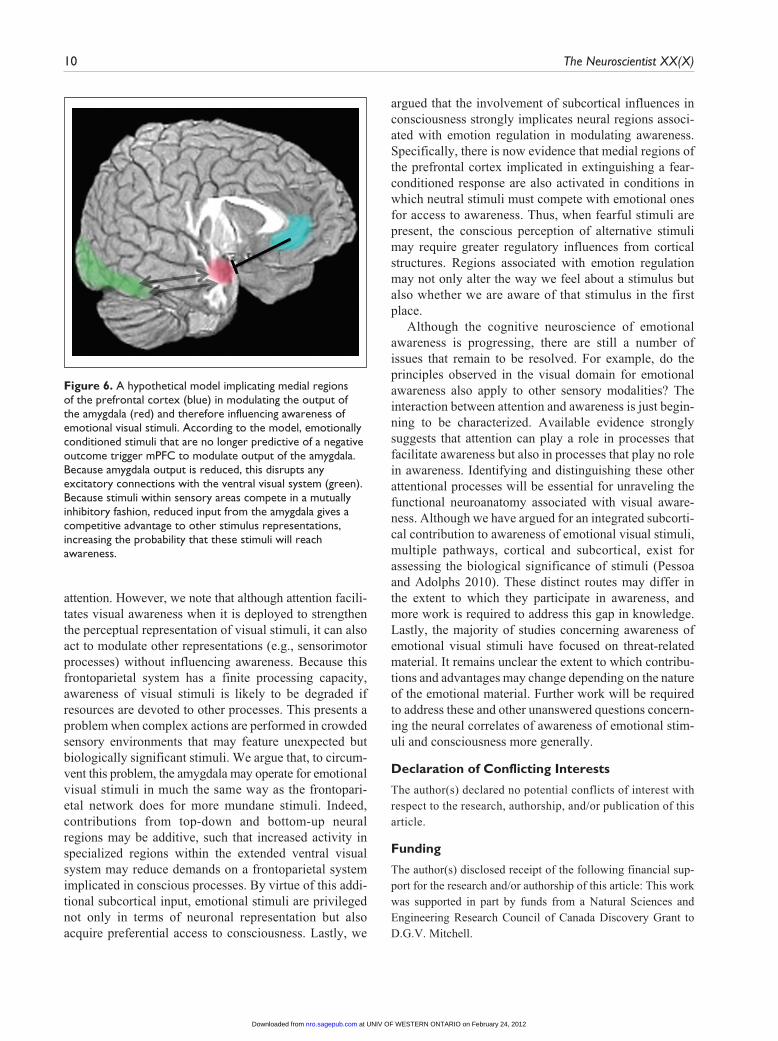
one of the key functions of attention is to increase the perceptual strength of sensory information; furthermore, awareness is associated with enhanced sensory representa-tion in the visual cortex (Leopold and Logothetis 1999). According to this view, the amygdala responds to perithreshold stimuli, strengthening even weak emotional stimulus representations in the ventral visual stream (Fig. 4). In doing so, the amygdala increases the likelihood that such stimuli will enter into awareness and be subjected to a more sophisticated array of cognitive and behavioral processes. The amygdala is well positioned to augment weak representations in cortical areas, given its extensive connections with structures throughout the ventral visual system (Amaral and others 2003). Accordingly, neuroim-aging evidence shows that activity in the ventral visual cortex is enhanced to emotion-related stimuli, and this enhancement has been attributed to functional coupling with the amygdala (Morris and others 1998). It has been speculated that subcortical influences might contribute to residual emotion discrimination ability in affective blind-sight (de Gelder and others 1999).
There is also evidence from lesion studies that the amyg-dala may play a role in enhancing awareness of emotional stimuli when the percept is degraded. In “attentional blink” tasks, attempts to identify a single target in a rapid visual stream of stimuli are accompanied by a transient reduction in awareness of the subsequently presented stimulus. Anderson and Phelps (2001) showed that although the attentional blink is attenuated in healthy individuals for emotional stimuli, no such emotion-related advantage was observed in patients with amygdala lesions. The authors concluded, therefore, that the amygdala functioned in this context to enhance the perceptual sensitivity to biologically significant events,
ensuring that they are less dependent on attentional resources in order to reach awareness. In this way, the amygdala can play a role in a higher-order process such as awareness.
Figure 3. The results from a binocular rivalry study indicate that frontoparietal regions implicated in playing a general role in supporting awareness show greater activity when a neutral facial expression reaches awareness while emotional expressions (fear or disgusted faces) are suppressed, relative to when a neutral face is perceived without competition from emotional expressions (adapted from Amting and others 2010).
Figure 4. A hypothetical model implicating the amygdala (red) in strengthening or synchronizing category-specific representations in the ventral visual cortex (green) to support visual awareness for emotional stimuli. Faded lines indicate that the contributions from frontoparietal cortices that are critical for lowering the threshold for awareness of more neutral stimuli are less important for emotional stimuli. In the case of emotional stimuli, mutually excitatory reciprocal connections are thought to be active between the amygdala and ventral visual system, which enhance the probability that emotional stimuli will have access to awareness over neutral stimuli, and even in situations of diminished contributions from frontoparietal areas (depicted by faded lines and blue colors).
at UNIV OF WESTERN ONTARIO on February 24, 2012nro.sagepub.comDownloaded from

Mitchell and Greening 9
Other Neural Mechanisms that Influence Awareness of Emotional Visual Stimuli
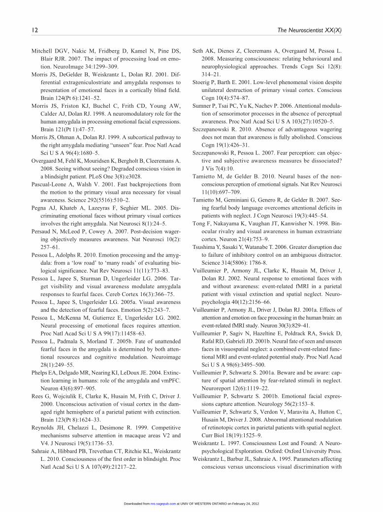
The perspective adopted here is that amygdala activity, whether arising primarily from subcortical or cortical inputs, can increase the likelihood that perithreshold emotional stimuli reach awareness. Regulating the impact of emotional distracters on behavior is thought to involve areas implicated in cognitive control, including the fron-toparietal cortex (Pessoa and others 2005b; Mitchell and others 2007; Mitchell and others 2008) and anterior cin-gulate cortex (Bishop and others 2004; Amting and oth-ers 2009). This raises the possibility that neural regions that modulate amygdala output may also have an impact on consciousness, particularly in conditions where mul-tiple stimuli compete for awareness. Recently, we used fMRI and a binocular rivalry task to delineate the neural correlates of visual awareness of competing emotional facial expressions. Participants watched as two congruent or discordant facial expressions were presented in a bin-ocular rivalry paradigm. At the behavioral level, fearful faces were significantly more likely to be consciously perceived than disgusted or neutral faces. Critically, tri-als in which fear was present and consciously perceived generated greater amygdala activity than trials in which fear was present but suppressed from awareness. Strikingly, evidence was also uncovered that prefrontal regions of the cortex implicated in regulating the impact of emo-tional stimuli on behavior are also involved in modulat-ing the contents of consciousness; awareness of neutral faces, and suppression of fearful faces, was associated with increased activity in regions associated with top-down control, including the perigenual prefrontal cortex, dorsolateral prefrontal cortex, and inferior parietal cor-tex. Furthermore, a connectivity analysis showed that whereas increased functional connectivity between the amygdala and ventral visual processing regions occurred during awareness of fearful faces, increased connectivity with the perigenual prefrontal cortex (BA 32/10) was evident when fear was present but unperceived (Fig. 5). Similar regions of the medial prefrontal cortex have been implicated in modulating emotion-related output from the amygdala in studies examining emotional learning (Phelps and others 2004; Milad and others 2007; Delgado and others 2008) and emotional distraction (Bishop and others 2004; Etkin and others 2006; Amting and others 2009). Figure 6 illustrates how this mechanism might operate in the context of awareness of emotional visual stimuli. The results are consistent with the idea that emotional awareness is influenced not only as a function of category-selective activity in the ventral visual system, but also as a function of emotion-dependent contribu-tions from top-down cortical areas implicated in attention and emotional regulation.
ConclusionsCurrent theories of the awareness of emotional visual stimuli are dominated by the idea that activity in the amygdala occurs independently of awareness. Here, we argued that subcortical networks associated with the rapid detection of emotionally salient stimuli are not segregated from those that modulate emotional aware-ness but are part of an integrated circuit. This proposal is based on now increasing evidence that awareness should be considered along a continuum, having intermediate levels, rather than as a categorical, all-or-none construct. Previous studies that have utilized a categorical approach have underestimated the level of awareness in healthy and brain lesion populations. This has resulted in a ten-dency to segregate pathways associated with rapid detec-tion of emotional material from those associated with awareness. We also acknowledge the considerable evi-dence for a critical role for frontoparietal regions in consciousness and that this role likely involves selective
Figure 5. Evidence from a recent context-dependent connectivity analysis investigating neural regions showing significantly altered functional connectivity with the right amygdala as a function of whether a fearful face was consciously perceived. Medial regions of the prefrontal cortex shown in blue show reduced connectivity with the amygdala in conditions in which fear was present and perceived relative to when fear was present but suppressed from awareness. The results implicate this region, previously associated with emotion regulation (extinguishing a fear-conditioned association and resolving emotional conflict), as playing a role in modulating awareness of emotional visual stimuli (adapted from Amting and others 2010).
at UNIV OF WESTERN ONTARIO on February 24, 2012nro.sagepub.comDownloaded from
10 The Neuroscientist XX(X)
attention. However, we note that although attention facili-tates visual awareness when it is deployed to strengthen the perceptual representation of visual stimuli, it can also act to modulate other representations (e.g., sensorimotor processes) without influencing awareness. Because this frontoparietal system has a finite processing capacity, awareness of visual stimuli is likely to be degraded if resources are devoted to other processes. This presents a problem when complex actions are performed in crowded sensory environments that may feature unexpected but biologically significant stimuli. We argue that, to circum-vent this problem, the amygdala may operate for emotional visual stimuli in much the same way as the frontopari-etal network does for more mundane stimuli. Indeed, contributions from top-down and bottom-up neural regions may be additive, such that increased activity in specialized regions within the extended ventral visual system may reduce demands on a frontoparietal system implicated in conscious processes. By virtue of this addi-tional subcortical input, emotional stimuli are privileged not only in terms of neuronal representation but also acquire preferential access to consciousness. Lastly, we
argued that the involvement of subcortical influences in consciousness strongly implicates neural regions associ-ated with emotion regulation in modulating awareness. Specifically, there is now evidence that medial regions of the prefrontal cortex implicated in extinguishing a fear-conditioned response are also activated in conditions in which neutral stimuli must compete with emotional ones for access to awareness. Thus, when fearful stimuli are present, the conscious perception of alternative stimuli may require greater regulatory influences from cortical structures. Regions associated with emotion regulation may not only alter the way we feel about a stimulus but also whether we are aware of that stimulus in the first place.
Although the cognitive neuroscience of emotional awareness is progressing, there are still a number of issues that remain to be resolved. For example, do the principles observed in the visual domain for emotional awareness also apply to other sensory modalities? The interaction between attention and awareness is just begin-ning to be characterized. Available evidence strongly suggests that attention can play a role in processes that facilitate awareness but also in processes that play no role in awareness. Identifying and distinguishing these other attentional processes will be essential for unraveling the functional neuroanatomy associated with visual aware-ness. Although we have argued for an integrated subcorti-cal contribution to awareness of emotional visual stimuli, multiple pathways, cortical and subcortical, exist for assessing the biological significance of stimuli (Pessoa and Adolphs 2010). These distinct routes may differ in the extent to which they participate in awareness, and more work is required to address this gap in knowledge. Lastly, the majority of studies concerning awareness of emotional visual stimuli have focused on threat-related material. It remains unclear the extent to which contribu-tions and advantages may change depending on the nature of the emotional material. Further work will be required to address these and other unanswered questions concern-ing the neural correlates of awareness of emotional stim-uli and consciousness more generally.
Declaration of Conflicting Interests
The author(s) declared no potential conflicts of interest with respect to the research, authorship, and/or publication of this article.
Funding
The author(s) disclosed receipt of the following financial sup-port for the research and/or authorship of this article: This work was supported in part by funds from a Natural Sciences and Engineering Research Council of Canada Discovery Grant to D.G.V. Mitchell.
Figure 6. A hypothetical model implicating medial regions of the prefrontal cortex (blue) in modulating the output of the amygdala (red) and therefore influencing awareness of emotional visual stimuli. According to the model, emotionally conditioned stimuli that are no longer predictive of a negative outcome trigger mPFC to modulate output of the amygdala. Because amygdala output is reduced, this disrupts any excitatory connections with the ventral visual system (green). Because stimuli within sensory areas compete in a mutually inhibitory fashion, reduced input from the amygdala gives a competitive advantage to other stimulus representations, increasing the probability that these stimuli will reach awareness.
at UNIV OF WESTERN ONTARIO on February 24, 2012nro.sagepub.comDownloaded from

Mitchell and Greening 11
References
Amaral DG, Behniea H, Kelly JL. 2003. Topographic organiza-tion of projections from the amygdala to the visual cortex in the macaque monkey. Neuroscience 118(4):1099–120.
Amting JM, Greening SG, Mitchell DGV. 2010. Multiple mechanisms of consciousness: the neural correlates of emo-tional awareness. J Neurosci 30(20):10039–47.
Amting JM, Miller J, Chow M, Mitchell DGV. 2009. Getting mixed messages: the impact of conflicting social signals on the brain’s target emotional response. NeuroImage 47:1950–9.
Anderson AK, Phelps EA. 2001. Lesions of the human amygdala impair enhanced perception of emotionally salient events. Nature 411(6835):305–9.
Baars BJ. 2002. The conscious access hypothesis: origins and recent evidence. Trends Cogn Sci 6(1):47–52.
Bishop SJ, Duncan J, Lawrence AD. 2004. State anxiety modu-lation of the amygdala response to unattended threat-related stimuli. J Neurosci 24(46):10364–8.
Bishop SJ, Jenkins R, Lawrence AD. 2007. Neural processing of fearful faces: effects of anxiety are gated by perceptual capacity limitations. Cereb Cortex 17(7):1595–603.
Crick F, Koch C. 2003. A framework for consciousness. Nat Neurosci 6(2):119–26.
de Gelder B, Vroomen J, Pourtois G, Weiskrantz L. 1999. Non-conscious recognition of affect in the absence of striate cortex. Neuroreport 10(18):3759–63.
Dehaene S, Artiges E, Naccache L, Martelli C, Viard A, Schurhoff F, and others. 2003. Conscious and subliminal conflicts in normal subjects and patients with schizophrenia: the role of the anterior cingulate. Proc Natl Acad Sci U S A 100(23):13722–7.
Dehaene S, Changeux JP, Naccache L, Sackur J, Sergent C. 2006. Conscious, preconscious, and subliminal processing: a testable taxonomy. Trends Cogn Sci 10(5):204–11.
Dehaene S, Naccache L, Cohen L, Bihan DL, Mangin JF, Poline JB, Riviere D. 2001. Cerebral mechanisms of word masking and unconscious repetition priming. Nat Neurosci 4(7):752–8.
Del Cul A, Dehaene S, Reyes P, Bravo E, Slachevsky A. 2009. Causal role of prefrontal cortex in the threshold for access to consciousness. Brain 132(Pt 9):2531–40.
Delgado MR, Nearing KI, Ledoux JE, Phelps EA. 2008. Neural circuitry underlying the regulation of conditioned fear and its relation to extinction. Neuron 59(5):829–38.
Desimone R, Duncan J. 1995. Neural mechanisms of selective visual attention. Annu Rev Neurosci 18:193–222.
Dienes Z, Scott R. 2005. Measuring unconscious knowledge: distinguishing structural knowledge and judgment knowl-edge. Psychol Res 69(5–6):338–51.
Dolan RJ, Vuilleumier P. 2003. Amygdala automaticity in emo-tional processing. Ann N Y Acad Sci 985:348–55.
Driver J, Mattingley JB. 1998. Parietal neglect and visual awareness. Nat Neurosci 1(1):17–22.
Etkin A, Egner T, Peraza DM, Kandel ER, Hirsch J. 2006. Resolving emotional conflict: a role for the rostral anterior
cingulate cortex in modulating activity in the amygdala. Neuron 51(6):871–82.
Greening SG, Finger EC, Mitchell DG. 2011. Parsing decision making processes in prefrontal cortex: response inhibition, overcoming learned avoidance, and reversal learning. Neu-roimage 54(2):1432–41.
Hamm AO, Weike AI, Schupp HT, Treig T, Dressel A, Kessler C. 2003. Affective blindsight: intact fear conditioning to a visual cue in a cortically blind patient. Brain 126(Pt 2):267–75.
Kentridge RW, Heywood CA, Weiskrantz L. 1999. Atten-tion without awareness in blindsight. Proc Biol Sci 266(1430):1805–11.
Kentridge RW, Nijboer TC, Heywood CA. 2008. Attended but unseen: visual attention is not sufficient for visual aware-ness. Neuropsychologia 46(3):864–9.
Koch C, Tsuchiya N. 2007. Attention and consciousness: two distinct brain processes. Trends Cogn Sci 11(1):16–22.
Kouider S, Dehaene S. 2007. Levels of processing during non-conscious perception: a critical review of visual masking. Philos Trans R Soc Lond B Biol Sci 362(1481):857–75.
LeDoux JE, Sakaguchi A, Reis DJ. 1984. Subcortical efferent projections of the medial geniculate nucleus mediate emo-tional responses conditioned to acoustic stimuli. J Neurosci 4(3):683–98.
Leopold DA, Logothetis NK. 1996. Activity changes in early visual cortex reflect monkeys’ percepts during binocular rivalry. Nature 379(6565):549–53.
Leopold DA, Logothetis NK. 1999. Multistable phenomena: changing views in perception. Trends Cogn Sci 3(7):254–64.
Lumer ED, Rees G. 1999. Covariation of activity in visual and prefrontal cortex associated with subjective visual percep-tion. Proc Natl Acad Sci U S A 96(4):1669–73.
Merikle PM, Smilek D, Eastwood JD. 2001. Perception without awareness: perspectives from cognitive psychology. Cogni-tion 79(1-2):115–34.
Milad MR, Quirk GJ. 2002. Neurons in medial prefron-tal cortex signal memory for fear extinction. Nature 420(6911):70–4.
Milad MR, Wright CI, Orr SP, Pitman RK, Quirk GJ, Rauch SL. 2007. Recall of fear extinction in humans activates the ventromedial prefrontal cortex and hippocampus in concert. Biol Psychiatry 62(5):446–54.
Mitchell DG. 2011. The nexus between decision making and emotion regulation: a review of convergent neurocognitive substrates. Behav Brain Res 217(1):215–31.
Mitchell DG, Luo Q, Avny SB, Kasprzycki T, Gupta K, Chen G, and others. 2009. Adapting to dynamic stimulus-response values: differential contributions of inferior frontal, dorso-medial, and dorsolateral regions of prefrontal cortex to deci-sion making. J Neurosci 29(35):10827–34.
Mitchell DG, Luo Q, Mondillo K, Vythilingam M, Finger EC, Blair RJ. 2008. The interference of operant task perfor-mance by emotional distracters: an antagonistic relationship between the amygdala and frontoparietal cortices. Neuroim-age 40(2):859–68.
at UNIV OF WESTERN ONTARIO on February 24, 2012nro.sagepub.comDownloaded from
12 The Neuroscientist XX(X)
Mitchell DGV, Nakic M, Fridberg D, Kamel N, Pine DS, Blair RJR. 2007. The impact of processing load on emo-tion. NeuroImage 34:1299–309.
Morris JS, DeGelder B, Weiskrantz L, Dolan RJ. 2001. Dif-ferential extrageniculostriate and amygdala responses to presentation of emotional faces in a cortically blind field. Brain 124(Pt 6):1241–52.
Morris JS, Friston KJ, Buchel C, Frith CD, Young AW, Calder AJ, Dolan RJ. 1998. A neuromodulatory role for the human amygdala in processing emotional facial expressions. Brain 121(Pt 1):47–57.
Morris JS, Ohman A, Dolan RJ. 1999. A subcortical pathway to the right amygdala mediating “unseen” fear. Proc Natl Acad Sci U S A 96(4):1680–5.
Overgaard M, Fehl K, Mouridsen K, Bergholt B, Cleeremans A. 2008. Seeing without seeing? Degraded conscious vision in a blindsight patient. PLoS One 3(8):e3028.
Pascual-Leone A, Walsh V. 2001. Fast backprojections from the motion to the primary visual area necessary for visual awareness. Science 292(5516):510–2.
Pegna AJ, Khateb A, Lazeyras F, Seghier ML. 2005. Dis-criminating emotional faces without primary visual cortices involves the right amygdala. Nat Neurosci 8(1):24–5.
Persaud N, McLeod P, Cowey A. 2007. Post-decision wager-ing objectively measures awareness. Nat Neurosci 10(2): 257–61.
Pessoa L, Adolphs R. 2010. Emotion processing and the amyg-dala: from a ‘low road’ to ‘many roads’ of evaluating bio-logical significance. Nat Rev Neurosci 11(11):773–83.
Pessoa L, Japee S, Sturman D, Ungerleider LG. 2006. Tar-get visibility and visual awareness modulate amygdala responses to fearful faces. Cereb Cortex 16(3):366–75.
Pessoa L, Japee S, Ungerleider LG. 2005a. Visual awareness and the detection of fearful faces. Emotion 5(2):243–7.
Pessoa L, McKenna M, Gutierrez E, Ungerleider LG. 2002. Neural processing of emotional faces requires attention. Proc Natl Acad Sci U S A 99(17):11458–63.
Pessoa L, Padmala S, Morland T. 2005b. Fate of unattended fearful faces in the amygdala is determined by both atten-tional resources and cognitive modulation. Neuroimage 28(1):249–55.
Phelps EA, Delgado MR, Nearing KI, LeDoux JE. 2004. Extinc-tion learning in humans: role of the amygdala and vmPFC. Neuron 43(6):897–905.
Rees G, Wojciulik E, Clarke K, Husain M, Frith C, Driver J. 2000. Unconscious activation of visual cortex in the dam-aged right hemisphere of a parietal patient with extinction. Brain 123(Pt 8):1624–33.
Reynolds JH, Chelazzi L, Desimone R. 1999. Competitive mechanisms subserve attention in macaque areas V2 and V4. J Neurosci 19(5):1736–53.
Sahraie A, Hibbard PB, Trevethan CT, Ritchie KL, Weiskrantz L. 2010. Consciousness of the first order in blindsight. Proc Natl Acad Sci U S A 107(49):21217–22.
Seth AK, Dienes Z, Cleeremans A, Overgaard M, Pessoa L. 2008. Measuring consciousness: relating behavioural and neurophysiological approaches. Trends Cogn Sci 12(8): 314–21.
Stoerig P, Barth E. 2001. Low-level phenomenal vision despite unilateral destruction of primary visual cortex. Conscious Cogn 10(4):574–87.
Sumner P, Tsai PC, Yu K, Nachev P. 2006. Attentional modula-tion of sensorimotor processes in the absence of perceptual awareness. Proc Natl Acad Sci U S A 103(27):10520–5.
Szczepanowski R. 2010. Absence of advantageous wagering does not mean that awareness is fully abolished. Conscious Cogn 19(1):426–31.
Szczepanowski R, Pessoa L. 2007. Fear perception: can objec-tive and subjective awareness measures be dissociated? J Vis 7(4):10.
Tamietto M, de Gelder B. 2010. Neural bases of the non-conscious perception of emotional signals. Nat Rev Neurosci 11(10):697–709.
Tamietto M, Geminiani G, Genero R, de Gelder B. 2007. See-ing fearful body language overcomes attentional deficits in patients with neglect. J Cogn Neurosci 19(3):445–54.
Tong F, Nakayama K, Vaughan JT, Kanwisher N. 1998. Bin-ocular rivalry and visual awareness in human extrastriate cortex. Neuron 21(4):753–9.
Tsushima Y, Sasaki Y, Watanabe T. 2006. Greater disruption due to failure of inhibitory control on an ambiguous distractor. Science 314(5806): 1786-8.
Vuilleumier P, Armony JL, Clarke K, Husain M, Driver J, Dolan RJ. 2002. Neural response to emotional faces with and without awareness: event-related fMRI in a parietal patient with visual extinction and spatial neglect. Neuro-psychologia 40(12):2156–66.
Vuilleumier P, Armony JL, Driver J, Dolan RJ. 2001a. Effects of attention and emotion on face processing in the human brain: an event-related fMRI study. Neuron 30(3):829–41.
Vuilleumier P, Sagiv N, Hazeltine E, Poldrack RA, Swick D, Rafal RD, Gabrieli JD. 2001b. Neural fate of seen and unseen faces in visuospatial neglect: a combined event-related func-tional MRI and event-related potential study. Proc Natl Acad Sci U S A 98(6):3495–500.
Vuilleumier P, Schwartz S. 2001a. Beware and be aware: cap-ture of spatial attention by fear-related stimuli in neglect. Neuroreport 12(6):1119–22.
Vuilleumier P, Schwartz S. 2001b. Emotional facial expres-sions capture attention. Neurology 56(2):153–8.
Vuilleumier P, Schwartz S, Verdon V, Maravita A, Hutton C, Husain M, Driver J. 2008. Abnormal attentional modulation of retinotopic cortex in parietal patients with spatial neglect. Curr Biol 18(19):1525–9.
Weiskrantz L. 1997. Consciousness Lost and Found: A Neuro-psychological Exploration. Oxford: Oxford University Press.
Weiskrantz L, Barbur JL, Sahraie A. 1995. Parameters affecting conscious versus unconscious visual discrimination with
at UNIV OF WESTERN ONTARIO on February 24, 2012nro.sagepub.comDownloaded from

Mitchell and Greening 13
damage to the visual cortex (V1). Proc Natl Acad Sci U S A 92(13):6122–6.
Weiskrantz L, Cowey A, Hodinott-Hill I. 2002. Prime-sight in a blindsight subject. Nat Neurosci 5(2):101–2.
Whalen PJ, Kagan J, Cook RG, Davis FC, Kim H, Polis S, and others. 2004. Human amygdala responsivity to masked fearful eye whites. Science 306(5704):2061.
Whalen PJ, Rauch SL, Etcoff NL, McInerney SC, Lee MB, Jenike MA. 1998. Masked presentations of emotional facial expressions modulate amygdala activity without explicit knowledge. J Neurosci 18(1):411–8.
Williams LM, Liddell BJ, Kemp AH, Bryant RA, Meares RA, Peduto AS, Gordon E. 2006. Amygdala-prefrontal dissocia-tion of subliminal and supraliminal fear. Hum Brain Mapp 27(8):652–61.
Williams MA, McGlone F, Abbott DF, Mattingley JB. 2005. Differential amygdala responses to happy and fearful facial expressions depend on selective attention. Neuroimage 24(2):417–25.
Zeki S, Ffytche DH. 1998. The Riddoch syndrome: insights into the neurobiology of conscious vision. Brain 121 (Pt 1):25–45.
at UNIV OF WESTERN ONTARIO on February 24, 2012nro.sagepub.comDownloaded from