Endovascular treatment of cerebrovascular diseases and intracranial neoplasms
Upload
independentCategory
view
1download
0
Converging Intracranial Markersof Conscious AccessRaphael Gaillard
1,2,3, Stanislas Dehaene
1,4,5, Claude Adam
6, Stephane Clemenceau
6, Dominique Hasboun
6,7,
Michel Baulac6,7
, Laurent Cohen1,6,7
, Lionel Naccache1,6,7*
1 INSERM, Cognitive Neuro-imaging Unit, Institut Federatif de Recherche (IFR) 49, Gif sur Yvette, France, 2 Centre Hospitalier Sainte Anne, Service Hospitalo-Universitaire de
Sante Mentale et de Therapeutique, Paris, France, 3 Universite Paris Descartes, Paris, France, 4 CEA, I2BM, NeuroSpin center, Gif sur Yvette, France, 5 College de France, Paris,
France, 6 Assistance Publique Hopitaux de Paris, Hopital de la Pitie-Salpetriere, Pole des Maladies du Systeme Nerveux, Paris, France, 7 Universite Pierre et Marie Curie Paris 6,
Departement de Physiologie, Paris, France
We compared conscious and nonconscious processing of briefly flashed words using a visual masking procedure whilerecording intracranial electroencephalogram (iEEG) in ten patients. Nonconscious processing of masked words wasobserved in multiple cortical areas, mostly within an early time window (,300 ms), accompanied by induced gamma-band activity, but without coherent long-distance neural activity, suggesting a quickly dissipating feedforward wave.In contrast, conscious processing of unmasked words was characterized by the convergence of four distinctneurophysiological markers: sustained voltage changes, particularly in prefrontal cortex, large increases in spectralpower in the gamma band, increases in long-distance phase synchrony in the beta range, and increases in long-rangeGranger causality. We argue that all of those measures provide distinct windows into the same distributed state ofconscious processing. These results have a direct impact on current theoretical discussions concerning the neuralcorrelates of conscious access.
Citation: Gaillard R, Dehaene S, Adam C, Clemenceau S, Hasboun D, et al. (2009) Converging intracranial markers of conscious access. PLoS Biol 7(3): e1000061. doi:10.1371/journal.pbio.1000061
Introduction
The neural correlates of consciousness (NCC) still remainhighly controversial. Indeed, the precise timing, location, anddynamics of neural events causing conscious access are notclearly and unequivocally determined. Do the NCC corre-spond to late [1,2] or early brain events [3–10]? Are theysystematically associated with reentrant ‘‘top down’’ process-ing [5,9,11–15]? If so, do they necessarily involve long-rangecoherent activity [16–21], including prefrontal cortex as anessential node [22–25], or can they be restricted to localpatterns of reverberating activity [3–6,8,11,13,15,26–29]? Is theconcept of ‘‘integrated information’’ relevant, rather than thespecific localization of the underlying cerebral network [21]?
In addition to such fundamental questions, an importantmethodological issue also remains open. Neural data relevantto conscious access originate from a diversity of techniquesincluding hemodynamic blood oxygen level dependent(BOLD) functional magnetic resonance imaging (fMRI) orpositron emission tomography (PET) responses and electro-physiological measures using scalp and intracranial event-related potentials (iERPs), event-related spectral perturba-tions (ERSPs), and phase synchrony parameters. How arethese distinct measures of conscious access related to eachother? Do they reflect common facets of the same underlyingphenomenon?
In this work, we address some of these issues usingintracerebral electrophysiological recordings of neural activ-ity in a group of implanted epileptic patients presented withvisually masked and unmasked printed words. This methodoffers a unique opportunity to measure neural correlates ofconscious access with both millisecond time resolution andcentimetric spatial resolution. Its high signal-to-noise ratioallowed us to compute several neurophysiological measures
from the intracerebral signal in order to unravel the relationsprevailing between iERPs, ERSPs, interelectrode phase syn-chrony, and a recently proposed estimate of causality(Granger causality).
The Global Workspace Model of ConsciousnessWe adopted a theory-driven approach, trying to test
experimentally a set of explicit predictions derived fromthe global workspace model of conscious access. This model,in part inspired from Bernard Baars’ theory [30], proposesthat at any given time, many modular cerebral networks areactive in parallel and process information in an unconsciousmanner [22,23,31,32]. Incoming visual information becomesconscious, however, if and only if the three followingconditions are met [23]: Condition 1: information must beexplicitly represented by the neuronal firing of perceptualnetworks located in visual cortical areas coding for thespecific features of the conscious percept. Condition 2: thisneuronal representation must reach a minimal threshold ofduration and intensity necessary for access to a second stageof processing, associated with a distributed cortical networkinvolved in particular parietal and prefrontal cortices.
Academic Editor: Leslie Ungerleider, National Institutes of Health, United States ofAmerica
Received August 4, 2008; Accepted February 2, 2009; Published March 17, 2009
Copyright: � 2009 Gaillard et al. This is an open-access article distributed underthe terms of the Creative Commons Attribution License, which permits unrestricteduse, distribution, and reproduction in any medium, provided the original authorand source are credited.
Abbreviations: EEG, electroencephalogram; ERP, event-related potential; ERSP,event-related spectral perturbation; iERP, intracranial event-related potential; ITC,intertrial phase coherence; MEG, magnetoencephalography; RT, response time
* To whom correspondence should be addressed. E-mail: [email protected]
PLoS Biology | www.plosbiology.org March 2009 | Volume 7 | Issue 3 | e10000610472
PLoS BIOLOGY

Condition 3: through joint bottom-up propagation and top-down attentional amplification, the ensuing brain-scaleneural assembly must ‘‘ignite’’ into a self-sustained reverber-ant state of coherent activity that involves many neuronsdistributed throughout the brain.
Why would this ignited state correspond to a consciousstate? The key idea behind the workspace model is thatbecause of its massive interconnectivity, the active coherentassembly of workspace neurons can distribute its contents toa great variety of other brain processors, thus making thisinformation globally available. The global workspace modelpostulates that this global availability of information is whatwe subjectively experience as a conscious state. Neuro-physiological, anatomical, and brain-imaging data stronglyargue for a major role of prefrontal cortex, anteriorcingulate, and the associative areas that connect to them, increating the postulated brain-scale workspace.
Scope and Limits of Our Experimental ParadigmIn the present work, we measured the neural correlates of
visually masked words and contrasted them with those ofconsciously visible unmasked words. On each trial, patientswere randomly presented with a masked word, a visible word,or with corresponding control stimuli in which the wordswere replaced by blank screens. In the masked condition,words or blank screens were presented for 29 ms, preceded bya forward mask and followed by a backward mask. In theunmasked conditions, words or blank screens were madevisible by simply removing the backward mask (see Materialsand Methods and Figure 1 for details). In order to discardactivations induced by the masks, we always subtracted fromword-present conditions the corresponding blank condition.This subtraction allowed us to isolate the entire processingpath evoked by the masked or unmasked word.
Advantages of the visual masking paradigm. Variants of themasking paradigm have been extensively used in behavioraland brain-imaging studies over the last 30 years. In humans, acumulative set of data demonstrated that a masked visualstimulus (e.g., word, number, or image) that cannot bereported can nevertheless be processed from low-level visualstages up to abstract cognitive processes, including semanticcontent, and eventually up to motor response preparation
[33]. From a neural point of view, masked stimuli activate alarge set of cortical structures, from occipital to anteriorfrontal regions [34–37]. The interpretation of these observa-tions is informed by recent studies of the mechanisms ofvisual masking. Recordings of single neurons in nonhumanprimates revealed that masking acts by reducing or inter-rupting the late activity evoked by stimuli while leaving theinitial feedforward activation largely unaffected [1,14,27,38–43]. Recent scalp–event-related potential (ERP) studiessuggest that the same mechanism may prevail in humans[1,44]. Finally, among the various methods that can be used tocompare conscious and nonconscious processes, the maskingparadigm allows for a comparison between two very stableand clear-cut states of perception: (1) a strong maskingcondition in which subjective report and objective discrim-ination are impossible, and (2) an unmasked condition inwhich undisputed subjective and objective measures ofconscious access can be gathered.Limits of the visual masking paradigm. When comparing
the processing of a clearly visible unmasked stimulus and of avisually degraded masked stimulus, one should be aware oftwo potential shortcomings. First, the masking procedureinduces an inescapable degradation of stimulus processing ata visual stage. Therefore, comparing masked and unmaskedstimuli does not amount to a pure comparison betweenconscious and nonconscious perception, but rather to thecomparison between nondegraded conscious informationand degraded nonconscious visual information. In otherwords, one has to keep in mind that any observed differencebetween the two conditions may include low-level processingdifferences due to visual degradation itself, upstream fromconscious access. Note, however, that in our experimentaldesign, we systematically subtracted activity evoked by thevisual masks, in order to isolate the correlates of masked andunmasked words processing.A second shortcoming of the masking method is that
subjects can perform the instructed task only when con-sciously perceiving the stimuli. Therefore, activation differ-ences between conscious and nonconscious stimuli may relateto the execution of the task downstream from consciousaccess, rather than to conscious access per se. However, thisconcern is moderated by evidence that the processing ofmasked stimuli is sensitive to task instructions and moregenerally to top-down strategical effects [45–53]. Note alsothat this issue is not restricted to visual masking, but that itequally applies to most paradigms used for the study ofconscious access, including paradigms without visual degra-dation such as the attentional blink, attentional blindness, ormasking at threshold. It is generally not feasible to equateperformance with conscious and nonconscious stimuli,probably because improved performance is an integralconsequence of the greater availability of information madepossible by conscious access. The few paradigms that haveattempted to compare conscious and nonconscious process-ing with equated performance levels have used various meansof degrading conscious performance down to nonconsciouslevels [24].
Neurophysiological Predictions Derived from the GlobalWorkspace ModelIn the light of our model, the masked–unmasked contrast
corresponds to a comparison between a visual representation
PLoS Biology | www.plosbiology.org March 2009 | Volume 7 | Issue 3 | e10000610473
Electrophysiology of Conscious Access
Author Summary
What is the neural signature of the conscious perception of a visualstimulus? To address this question, we recorded neural activitydirectly from the brains of human subjects (who were undergoingneural surgery for medical reasons). This rare opportunity affordedgreater spatial and temporal resolution than noninvasive methodsused previously to probe the neural basis of consciousness. Wecompared neural activity concomitant with conscious and noncon-scious processing of words by using a visual masking procedure thatallowed us to manipulate the conscious visibility of briefly maskedwords. Nonconscious processing of words elicited short-lastingactivity across multiple cortical areas, including parietal and visualareas. In sharp contrast, only consciously perceived words wereaccompanied by long-lasting effects (.200 ms) across a greatvariety of cortical sites, with a special involvement of the prefrontallobes. This sustained pattern of neural activity was characterized bya specific increase of coherence between distant areas, suggestingconscious perception is broadcasted widely across the cortex.

satisfying only condition 1 and a representation satisfying allthree conditions for conscious access listed above. The globalworkspace model therefore leads to the following fourpredictions.
Prediction 1: a common early stage of processing. Bothmasked and unmasked words should evoke similar neuralactivity within an early time window, reflecting a fastfeedforward sweep propagating from posterior to anteriorcortices. In particular, invisible masked words should inducetransient event-related responses along the ventral visualpathway, as assessed by iERPs and ERSP.
Prediction 2: a temporal divergence. Following this initialcommon stage, only unmasked words should be associatedwith sustained effects. We thus predict a divergence in corticalactivation for unmasked and masked words. Given that wecontrasted heavily masked stimuli with unmasked stimuli, weexpect a progressive buildup of the divergence between thesetwo conditions. In the light of recent high-resolution scalpelectroencephalogram (EEG) studies in visual masking andattentional blink paradigms, this temporal divergence isexpected to occur within a 200–500-ms window [1,2].
Prediction 3: an anatomical divergence. The activation offrontal and parietal areas, which are allegedly dense in globalworkspace neurons, should be particularly sensitive toconsciously perceived words (see [32] and Figure 1 of [22]for explicit simulations of this property). Although maskedwords may cause a small, transient and local activation withinthese regions, we predict that unmasked words should elicit aglobal and long-lasting activation of these regions, corre-sponding to a broadcasting process.Prediction 4: phase synchrony and causality. During this
late time window, the long-lasting and long-distance neuronalassembly specific to conscious processing should be associ-ated with an intense increase in bidirectional interelectrodecommunication. Measures of phase synchrony and Grangercausality should be particularly apt at capturing thisphenomenon. The global workspace theory is not yet fullyexplicit about the exact pattern of neuronal oscillationsinvolved in conscious access. In our initial modeling andtheoretical articles, no prediction was stated about whichfrequencies would be affected by conscious access [22,31].Increased high-frequency power and long-distance neural
Figure 1. Electrode Locations and Experimental Design
(A) Sagittal (left), coronal (middle), and axial (right) normalized glass brain showing all 176 electrodes after normalization in Talairach’s anatomical space.(B) Experimental paradigm used to present masked and unmasked words, with d9 measures in the forced-choice semantic task.doi:10.1371/journal.pbio.1000061.g001
PLoS Biology | www.plosbiology.org March 2009 | Volume 7 | Issue 3 | e10000610474
Electrophysiology of Conscious Access

synchrony were merely thought to be necessary, but notsufficient, conditions for conscious access, contrasting withthe local synchrony induced within modular processorsduring nonconscious processing [22]. In a recent theoreticalreview, we further pinpointed an important distinction to bedrawn between preconscious and conscious processing, asdefined by occipitotemporal loops with local synchrony in theformer, and long-distance synchronous loops in the latter[23]—but again without specifying the actual frequency bandsinvolved. Explicit neuronal network simulations of globalworkspace activity during the attentional blink paradigmdemonstrated a link between increased gamma-range oscil-lations (20–100 Hz) and conscious access [32]. In simulationsof inattentional blindness, this rather broad gamma bandnarrowed down to a predominant band at 40–45 Hz [54].Those frequency ranges, however, were only determined bythe particular choice of simulation parameters for cortico-cortical and thalamocortical propagation latencies, whosevalues in humans are not known in detail.
In the light of these considerations, the present study ofneuronal oscillations should be considered as an explorationof a broad theoretical proposal, rather than an assessment ofa precisely articulated prediction. Actually, theoretical con-siderations by other groups [55,56], as well as recentexperimental measurements [57–62], suggest that for distantcortical areas, synchrony cannot be easily achieved in thehigh-frequency range, where the oscillation period is shortrelative to corticocortical transmission delays. Thus, thesearticles would predict that long-distance synchrony associ-ated with conscious access should preferentially occur in thebeta frequency range (13–30 Hz), whereas local recurrencewould mostly concern the gamma range (.30 Hz), even formasked stimuli [7].
Results
Behavioral Measures of Word VisibilityUnmasked words were consciously reportable, and were
categorized better than chance level in a forced-choicecategorization task on the emotional valence of words (meandiscriminability index d9¼ 2.24 (þ1.14 toþ3.04), all individualv2 p-values and group analysis Student t-test p-value ,
0.0001). In sharp contrast, masked words were not consciouslyvisible, and forced-choice performance was at chance levelfor each of the implanted patients (mean d9 ¼ 0.02 (�0.18 toþ0.27), all p-values .0.2). Response times (RTs) were similaracross the two masking conditions (p . 0.38 in Student t-testperformed on mean RTs; masked mean RT ¼ 1,640 ms,unmasked mean RT¼ 1,300 ms).
Intracranial ERPsWe defined masked effects by subtracting the voltages
measured on masked blank trials from those associated withmasked word trials. This subtraction allowed us to isolate, ona sample-by-sample basis, activations associated with maskedword processing (see Figure 1 and Materials and Methods forour detailed three-step statistical procedure). Unmaskedeffects were defined similarly by subtraction of the unmaskedword and unmasked blank conditions.
Figure 1 shows the anatomical distribution of the 176reconstructed bipolar montages (‘‘electrodes’’) from which weobtained valid data across the ten patients. The bipolar
subtraction of nearby recording sites reduced distantinfluences, including those from the reference electrode,and resulted in a signal tightly localized to the implantedstructure. Although measures were obtained for all fourlobes, it should be kept in mind that major sectors ofdorsolateral and polar prefrontal cortex as well as parietalcortex were not sampled.Among the 176 electrodes, 24.4% (43 electrodes) showed a
significant effect for masked words. These effects wereobserved across all implanted structures but with a predom-inance of effects on occipital electrodes: 22/55 (40%) withinthe occipital lobe, 11/78 (14.1%) within the temporal lobe, 4/24 (16.7%) within the parietal lobe and 6/19 (31.6%) withinthe frontal lobe (v2 p-value ¼ 0.004).Concerning unmasked words, 68.8% of all electrodes (121
electrodes) showed a significant effect of word presence—aremarkably high percentage, given that electrodes had beenplaced at clinically relevant sites without consideration oftheir relevance to our visual stimuli. Unmasked effects wereobserved across all implanted structures but with a particularemphasis on the frontal lobe: 42/55 (76.4%) within theoccipital lobe, 49/78 (62.8%) within the temporal lobe, 12/24(50%) with the parietal lobe, and 18/19 (94.7%) within thefrontal lobe (v2 p-value ¼ 0.005). The frontal lobe showed amajor difference between trials containing masked andunmasked words: almost all contacts were systematicallyactivated during conscious processing of unmasked words(;95%), whereas this was not the case during unconsciousprocessing of masked words (;32%).In order to better assess the specificity of this last result, we
ran an ANOVA to directly compare the impact of masking onthe proportion of activated electrodes between occipital andfrontal lobes. A main effect of masking was observed (86%versus 36%; p , 10�4), confirming the larger spatial extensionof unmasked activations as compared to masked activations.No main effect was observed between frontal and occipitalelectrodes (58% versus 63%, p . 0.5). Crucially, we observed asignificant interaction between frontal and occipital corticesand masking condition (p ¼ 0.05), assessing the largerdifferential activation of frontal lobe between masked andunmasked conditions, as compared to the pattern observed inposterior visual cortex.Note that this spatial analysis is affected by the non-
homogenous sampling of brain regions, minimizing thecontribution of cortical structures that were less frequentlyimplanted. Nevertheless, masked effects were more frequenton posterior than on anterior electrodes, whereas unmaskedeffects were homogeneously distributed. To demonstrate thispoint, we examined the distribution of the anterior-posterior(y) coordinate, in Talairach space, of the electrodes showing asignificant effect, and compared it to the spatial distributionof all 176 recorded electrodes (see Figure S1). For maskedwords, the spatial distribution of significant electrodes wasstrongly shifted towards posterior sites (p , 10�6, Kolmogor-ov-Smirnov test, relative to the distribution of either the wholeset of 176 electrodes or to those showing an unmasked effect).The same analysis conducted on the cumulative distribution ofunmasked effects showed a spatial distribution statisticallyindistinguishable from that of the whole set of electrodes.Masked and unmasked words were also distinguished by the
temporal extension of their activation. A crude analysis,averaging across all electrodes, revealed that masked effects
PLoS Biology | www.plosbiology.org March 2009 | Volume 7 | Issue 3 | e10000610475
Electrophysiology of Conscious Access

had a mean duration of 60 ms, much shorter than the mean of378 ms for unmasked effects (p , 10�6). Masked effects alsoshowed an earlier onset (mean¼366ms;median¼301ms) thanunmasked effects (mean¼522 ms; median¼497 ms; t-test, p ,
10�5). A more relevant analysis focusing on the first significanteffect within the subset of electrodes with both masked andunmasked effects showed similar latencies between these twoconditions (299 ms and 348 ms, respectively, for masked andunmasked conditions; t-test, p ¼ 0.30). Indeed, up toapproximately 200 ms after word onset, glass brain visual-ization of the spatiotemporal dynamics of masked andunmasked effects showed a strikingly similar pattern ofactivations within posterior occipitotemporal cortical regions(see Figure 2). This dynamic pattern is very comparable to the‘‘feedforward sweep’’ described by Lamme in the nonhumanprimate visual cortex using multiunit recordings for latenciesup to 100 ms after visual stimulus onset [5]. Clear differencesbetween masked and unmasked effects appeared after 150 ms,with a progressive increase in the intensity and spatialextension of unmasked effects, while masked effects decayedand did not show a similar spatial extension (see Videos S1 andS2).This general pattern was observed on individual electrodes
(see Figure 3). The initial response was often indistinguish-able between masked and unmasked effects. This initialcommon response was usually followed by later effectsspecifically for the unmasked condition. Out of 14 electrodesshowing this pattern with our statistical criteria, 11 of themalso showed a polarity inversion of the late sustained effectsrelative to the polarity of the initial effect.A cortical lobe analysis focusing on the proportion of
electrodes showing a significant effect over time (Figure 4)showed a similar proportion of electrodes activated bymasked and unmasked words at short latencies, whereas atlater latencies, the effects were increasingly specific to theunmasked condition. An analysis of the mean voltage power,averaged across electrodes within one lobe, showed a similartemporal dynamics, and additionally allowed us to detect aprogressive time delay in the peak of the initial activationcommon to masked and unmasked words. The time point atwhich the first significant divergence between masked andunmasked effects occurred, as estimated by a t-test (p , 0.05),progressively increased from 215 ms to 275 ms and 347 ms,respectively, for the occipital, temporal, and frontal lobes (seeFigure 4). The divergence did not reach significance for thesmall set of 14 parietal lobe electrodes tested (those showingat least one significant effect, masked or unmasked).
Event-Related Spectral PerturbationsWe then turned to frequency-domain analyses of the
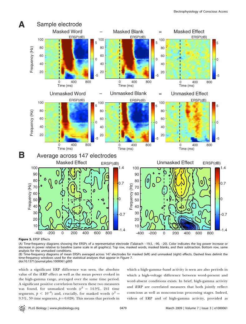
intracranial signals. Figure 5A shows a typical single-electrode example of the time-frequency transform appliedto our data. The masks alone evoked a very strong sequenceof event-related increase in the beta and gamma bandsaccompanied by alpha decrease, followed by a reversal of thispattern. Subtraction of each mask-only condition from thecorresponding word-present condition, however, isolated the
Figure 2. Spatiotemporal Dynamics of iERP Effects
Absolute values of iERPs difference (microVolts) between word-presentand word-absent in masked (left) and unmasked (right) conditions. Onlyelectrodes showing a significant effect are displayed as red squares.Square size and color intensity are proportional to the absolute voltage
difference between the word and blank conditions. Six different timeslices are displayed, ranging from 130 to 600 ms (see Videos S1 and S2for complete videos provided as supplementary on-line material).doi:10.1371/journal.pbio.1000061.g002
PLoS Biology | www.plosbiology.org March 2009 | Volume 7 | Issue 3 | e10000610476
Electrophysiology of Conscious Access

ERSP induced by the word alone, as a function of whether itwas masked or unmasked. As can be seen in this example,masked words induced a slight increase in gamma power 100–200 ms after the stimulus, whereas unmasked words induced amuch bigger effect that lasted throughout the epoch and wasaccompanied by alpha suppression.
To evaluate the generality and significance of such effects,we averaged the time-frequency diagrams across all elec-trodes (Figure 5B). Statistical comparisons over time-fre-quency regions of interest, with Bonferroni correction (seeMethods and Materials), identified several significant effects.In the 100–200-ms time window, masked words evoked highlysignificant power changes (beta suppression: p¼ 0.0004; high-
gamma increase: p¼ 0.0005). In this time period, there was nosignificant difference with unmasked words, confirming thata volley of activation, reflected primarily in a gammaincrease, can propagate nonconsciously while being largelyunaffected by masking [7].In the next time window (200–300 ms), whereas unmasked
words created an even larger power increase in the high-gamma band (p , 10�11) and decreases in alpha (p , 10�8) andbeta bands (p ¼ 10�5), masked words induced only smalleffects of alpha suppression (p ¼ 0.0014) and high-gammaincrease (p ¼ 0.038). Beta and high-gamma bands showedsignificantly stronger changes for unmasked compared tomasked words (all p , 0.0007). In the subsequent time window
Figure 3. iERP Effects on Three Representative Electrodes
(A) Maximum size of significant masked and unmasked effects across the 0–800-ms time window are displayed as red squares, whose size and colorintensity are proportional to peak absolute voltage amplitude.(B) Mean iERPs of three representative electrodes in occipital, fusiform, and frontal cortex (location shown in [A]). Shadowed areas indicate significanteffects (difference between word and blank conditions). The bottom graphs (blue traces) show the time course of the ‘‘word minus blank’’ subtractionseparately from the masked and unmasked conditions. All three sites exhibit an initial common peak, followed by a polarity reversal and delayed activityspecific to the unmasked condition.doi:10.1371/journal.pbio.1000061.g003
PLoS Biology | www.plosbiology.org March 2009 | Volume 7 | Issue 3 | e10000610477
Electrophysiology of Conscious Access

(300–500), alpha suppression, beta suppression, and high-gamma increase were very strong in the unmasked condition(all p , 0.0003), but altogether absent in the maskedcondition, creating a significant difference (all p , 0.0002).
In summary, masked words induced significant changes inthe power spectrum, particularly increases in the high-gamma band, but these induced oscillations quickly dissi-pated with time, whereas the ERSPs evoked by unmaskedwords exhibited a greater power and lasted significantlylonger. Note that the above analysis was based on the poolingof ERSP results from all electrodes, regardless of theirlocation. We also replicated this ERSP analysis while separat-ing the electrodes as a function of their lobe of origin. Boththe high-gamma increase and late alpha and beta suppressionspecific to the unmasked condition were replicated withineach of the four lobes (see Figure S2). Interestingly, the high-gamma increase peaked earlier in occipital cortex than in
temporal, parietal, or frontal, following an approximateposterior to anterior progression (see Videos S3 and S4).Furthermore, the lobar analysis showed that the earlynonconscious effects were confined to the occipital andtemporal lobes: the only significant effects were a high-gamma power increase in occipital cortex in the 100–200-msand 200–300-ms windows (respectively, p ¼ 0.013 and p ¼0.016), and decreases in alpha (200–300 ms, p ¼ 0.007) andbeta (100–200 ms, p , 10�3) in temporal cortex. In brief, theearly ERPS evoked by nonconscious stimuli originated onlyfrom occipitotemporal regions, whereas conscious percep-tion was associated with stronger and longer-lasting powerchanges spreading towards anterior cortical regions.In this respect, analyses of induced high-gamma power
yielded conclusions very similar to those derived from iERPanalyses. To better evaluate the relation between inducedgamma activity and iERPs, we calculated for each segment for
Figure 4. Lobar Analysis of iERPs
(A) For each lobe, proportions of electrodes showing a significant effect over time for masked (cyan) and unmasked (blue) conditions, respectively.(B) Voltage power, averaged across electrodes showing at least one significant effect, for masked (cyan) and unmasked (blue) conditions, respectively.Black dashed lines indicate latencies of the first significant differences (p , 0.05) between conditions.doi:10.1371/journal.pbio.1000061.g004
PLoS Biology | www.plosbiology.org March 2009 | Volume 7 | Issue 3 | e10000610478
Electrophysiology of Conscious Access
which a significant ERP difference was seen, the absolutevalue of the iERP effect as well as the mean power evoked inthe high-gamma range, averaged over the same time period.A significant positive correlation between these two measureswas found, for unmasked words (r2 ¼ 14.9%, 241 timesegments, p , 10�8) and, crucially, for masked words (r2 ¼9.3%, 59 time segments, p¼0.028). This means that periods in
which a high-gamma–band activity is seen are also periods in
which a high-voltage difference between word-present and
word-absent conditions exists. In brief, high-gamma activity
and iERP are correlated measures that both jointly reflect
conscious as well as nonconscious processing stages. Indeed,
videos of ERP and of high-gamma activity, provided as
Figure 5. ERSP Effects
(A) Time-frequency diagrams showing the ERSPs of a representative electrode (Talairach �19,5, �90, �20). Color indicates the log power increase ordecrease in power relative to baseline (same scale in all graphics). Top row, masked words, masked blanks, and their subtraction. Bottom row, sameanalysis for the unmasked conditions.(B) Time-frequency diagrams of mean ERSPs averaged across 147 electrodes for masked (left) and unmasked (right) effects. Dashed lines delimit thetime-frequency windows used for the statistical analyses that appear in Figure 7.doi:10.1371/journal.pbio.1000061.g005
PLoS Biology | www.plosbiology.org March 2009 | Volume 7 | Issue 3 | e10000610479
Electrophysiology of Conscious Access

supplementary online material, present high similar profiles(see Videos S1, S2, S3, and S4).
Phase Coherence
Spectral changes are complex phenomena that can besensitive to local as well as global neuronal synchronization of
thalamocortical networks [56]. To evaluate the global work-space model’s prediction that access to consciousness isassociated with long-distance synchronization, we measuredthe phase synchrony between all electrode pairs. Phasesynchrony can occur independently of changes in inducedpower: it solely evaluates whether oscillations are reprodu-
Figure 6. Phase Synchrony Effects
(A) Phase synchrony analyses of a representative pair of electrodes (Talairach�12,�97,�12 and�28.5,�77.5, 6). Top row, masked condition; bottomrow, unmasked condition. Each picture shows a time-frequency diagram of intertrial phase coherence across the two electrodes (ranging from 0 to 1)for the word condition, the blank condition, and their subtraction (different scale, including negative values).(B) Time-frequency diagrams of ITC averaged across all 1,283 electrode pairs, separately for masked (left) and unmasked (right) effects. Dashed linesdelimit the time-frequency windows used for the analyses that appear in Figure 7.doi:10.1371/journal.pbio.1000061.g006
PLoS Biology | www.plosbiology.org March 2009 | Volume 7 | Issue 3 | e10000610480
Electrophysiology of Conscious Access

cibly synchronized across two distant sites in the sense thatacross trials, they exhibit a systematic phase relationship.
Figure 6 shows a time-frequency diagram of the intertrialphase coherence (ITC) changes induced by the masked andunmasked words, both in an example electrode and in themean overall electrodes. All statistics were Bonferronicorrected. Statistical analyses revealed no significant coher-ence changes induced by the masked words. For unmaskedwords, an increase in beta synchrony in the 300–500-ms timewindow was highly significant (t(1,282 df) ¼ 7.12, p , 10�10;difference with masked condition, t(1,282 df) ¼ 5.43, p ,
10�6). It is particularly interesting to note that (1) this phasesynchrony increase was concomitant with a decrease ininduced spectral power (ERSP) within the same frequencyband (see Figure 7A and 7B); (2) no phase synchronyincrease was detected in the high-gamma band, although inthis band, a highly significant increase in induced power hadbeen detected by ERSP analysis. Thus, ERSP and phasesynchrony appear to double dissociate, and beta synchronyappears as a highly selective marker of the late phase ofconscious access.
Figure 8 shows in graphic form the value of the betacoherence increase due to word presence in the critical timewindow 300–500 ms, separately for unmasked and maskedwords. Clearly, unmasked words create a more globallysynchronous brain state than masked words. The figuremakes apparent that this phase coherence analysis isimportantly limited by the available electrodes: we can onlyanalyze coherences between electrodes within a given patient,and these tend to be regrouped within a cortical area, thuspreventing a thorough analysis of how coherence evolvesacross distant anatomical sites. For instance, it was notpossible to evaluate the prediction that frontal electrodesshould cohere more with posterior sites during consciousprocessing, because in our sample, these two regions werevery rarely recorded simultaneously. Still, to probe long-distance connections, we could analyze a subset consisting ofelectrode pairs in which the two electrodes lie in differenthemispheres, thus imposing a long-distance transfer acrossthe corpus callosum. As predicted by global workspacetheory, we observed an increase in long-distance interhemi-spheric beta coherence selective to unmasked words (t(71) ¼2.50, uncorrected p ¼ 0.015). In fact, interhemispheric betacoherence actually decreased when masked words werepresented (t(71) ¼ 3.14, uncorrected p ¼ 0.003), thus creatinga strong difference between visible and invisible conditions(t(71) ¼ 3.83, p ¼ 0.0003).
Conversely, Figure 8 suggests that in the masked condition,there might have been a small local increase in betacoherence within posterior occipitotemporal cortices, whichmight have been missed in our analysis pooling across allelectrode pairs. Indeed, when we restricted only to intra-hemispheric electrodes lying within occipital cortex or withintemporal cortex posterior to y¼�20, we detected a significantincrease in beta coherence for masked words during the 200–300-ms time window (t(734) ¼ 2.34, uncorrected p ¼ 0.02),which ceased to be significant in the 300–500-ms time window(t ¼ 0.41, not significant [n.s.]). No such increase was seen inother frequency bands, or in other regions (e.g., withinfrontal electrodes). Thus, nonconscious word processingresulted in only small and barely detectable transientincreases in phase coherence within visual cortex, whereas
conscious words yielded a massive increase in long-distancebeta coherence
Granger CausalityA final measure of conscious processing that we evaluated
is Granger causality [60,63–65], a mathematical tool that canestimate the causal influence that one electrode site exerts onanother. Global neuronal workspace theory predicted thataccess to consciousness for unmasked words would beaccompanied by a massive web of causal relations amongdistant cortical sites, not seen in the masked condition.Granger causality and phase coherence are similar in thatboth estimate the correlations among pairs of electrodes, butGranger causality looks for temporal contingencies inacces-sible to coherence analyses. In a nutshell, the methodestimates whether past samples of electrode j account for asignificant amount of variance in electrode i, over and abovea simpler ‘‘autoregressive’’ model using only past samples ofelectrode i (see [63] for details). It is possible for two timeseries to be strongly phase coherent, yet not causally related(for instance, two sine waves with constant phase lag andindependent noise). Thus, Granger causality analysis is notredundant with phase coherence analysis: finding thatGranger causality increases during conscious perception,perhaps simultaneously with the beta coherence increase,would provide additional evidence in favor of a large-scalereverberating neuronal assembly linking distant sites. Fur-thermore, unlike phase coherence, Granger causality is adirectional measure: it is possible for electrode j to causallyinfluence i without i causally influencing j (although it is alsopossible for two signals to exert mutual causal influences oneach other). This analysis therefore provided an opportunityto examine the top-down versus bottom-up propagation ofactivation during conscious and nonconscious processing.As a concrete example, Figure 9 illustrates the causality
analysis of a sample electrode pair consisting of one frontaland one occipital electrode. At the time of stimuluspresentation, a massive increase in Granger causality is seenin the feedforward, occipitofrontal direction (Figure 9A, leftpanel) and, to a smaller extent in the top-down, fronto-occipital direction (Figure 9A, right panel). Importantly, thecurves showing the evolution of the F-test for causality as afunction of time exhibit two successive peaks: one early peakis evoked by the masks alone (146 ms after mask onset),whereas a second peak (325 ms after word onset) is seen onlywhen a word is present and unmasked. As illustrated in Figure9B, a strong ‘‘causal gain’’ is therefore observed, approx-imately 200–450 ms after word onset, when the word-presentcondition is contrasted to the word-absent condition. Thiseffect is seen mostly in the feedforward direction, thusengendering a ‘‘causal imbalance’’ (higher causal gain in onedirection than in the other).Similar increases in causal gain and causal imbalance in the
unmasked condition were seen in a large set of electrodepairs. To evaluate their statistical significance, we firstaveraged the causal gains across all electrode pairs and bothcausal directions, separately for masked and unmaskedconditions, and used t-tests to evaluate the significance ofchanges within three temporal windows (100–200, 200–300,and 300–500 ms, similar to the ERSP and phase coherenceanalyses, with Bonferroni correction over the number ofwindows tested) (see Figure 9C). A massive increase in mean
PLoS Biology | www.plosbiology.org March 2009 | Volume 7 | Issue 3 | e10000610481
Electrophysiology of Conscious Access

causal gain was observed during the 300–500-ms window inthe unmasked condition (t(1805)¼ 7.60, p , 10�13), but not inthe masked condition (t ¼ 1.47, n.s.), resulting in asignificantly larger causal gain during conscious than duringnonconscious processing (t(1805)¼ 5.46, p , 10�8). The effectwas already perceptible in the 200–300-ms window, though itwas much smaller (unmasked: t(1805) ¼ 3.03, p ¼ 0.0075;masked, t¼�0.86, n.s.; difference: t(1805)¼2.89, p¼0.012). Noeffect reached significance in the 100–200-ms window.
Figure 8B illustrates the anatomical distribution of themean causal gains during the 300–500-ms window. In theunmasked condition, causal relations increased massivelyamong many distant sites, both within the occipitotemporalcortex, between occipitotemporal cortex and distant frontaland insular sites, and across the corpus callosum. By contrast,increases were very scarce in the masked condition and neverreached significance even when restricted to posteriorelectrodes only.
Figure 7. ERSP and Phase Synchrony across Three Time Windows
Twelve time-frequency regions of interest were defined on ERSP and phase synchrony–averaged analyses (see Figures 5B and 6B). For each region,mean ERSPs (A) and mean ITC (B) are plotted for three different time windows (abscissa axis: 100–200, 200–300, and 300–500 ms) and for four frequencybands (ordinate axis: alpha¼ 8–13 Hz; beta¼ 13–30 Hz; low gamma¼ 30–50 Hz; and high gamma¼ 50–100 Hz), separately for the masked (M) and forthe unmasked (UM) conditions. Bars represent one standard error of the mean.doi:10.1371/journal.pbio.1000061.g007
PLoS Biology | www.plosbiology.org March 2009 | Volume 7 | Issue 3 | e10000610482
Electrophysiology of Conscious Access

With similar methods, we evaluated the statistical signifi-cance of changes in the variable of ‘‘causal imbalance,’’ whichis the subtraction of forward causal gain minus backwardcausal gain in the same electrode pair. This variable evaluatedthe dominant directionality of causality (posterior to anterior¼ feedforward, or anterior to posterior ¼ feedback). Duringthe 300–500-ms time window, in the unmasked condition,there was a small imbalance with a higher causality gain in thefeedforward compared to the feedback direction (t(1,850) ¼2.07, p ¼ 0.039 before Bonferroni correction). Althoughmarginally significant, this finding occurred in the predictedlate time window and fits with our prior hypothesis that
during this time period, and only in the unmasked condition,perceptual information gains access to consciousness and istherefore able to invade anterior areas in a feedforwardmanner. Indeed, in this time window, the imbalance was notsignificant for masked targets (t ¼ �0.90, n.s.), creating adifference for unmasked as opposed to masked targets(t(1,805) ¼ 2.09, p ¼ 0.037 before Bonferroni correction).Quite surprisingly, however, in the preceding time window
(200–300 ms, see Figures 9D and S3), there was a significantimbalance in the converse direction (higher causality in thetop-down or feedback). This was true only for the maskedcondition (t(1,805) ¼�2.66, p ¼ 0.024, Bonferroni corrected),
Figure 8. Phase Synchrony and Granger Causal Gain between 300 and 500 ms after Word Onset
Each figure depicts three orthogonal views of a transparent ‘‘glass brain,’’ with segments linking, for each patient, all pairs of electrodes. Segments arecolored and sized according to the intensity of the increase or decrease in phase coherence in the beta frequency band (A), and in Granger causal gain(B) during the 300–500-ms time window. Superimposed lines are plotted in increasing order of the absolute value of the depicted parameter, so thatlarger values override smaller ones. Left two columns, masked effects; right two columns, unmasked effects.doi:10.1371/journal.pbio.1000061.g008
PLoS Biology | www.plosbiology.org March 2009 | Volume 7 | Issue 3 | e10000610483
Electrophysiology of Conscious Access

Figure 9. Granger Causality Analysis
(A and B) Illustration of Granger causality analysis for a representative pair of electrodes located respectively in the frontal and occipital lobes.For each of the four experimental conditions, an F-test evaluates, over a sliding timing window, the causal influence of occipital activity onfrontal electrode activity and vice versa (A). Note that this F-test is not directly comparable across conditions (because of smaller number of trialsin the blank control conditions), nor can it be taken directly as a test of significance (because of inflation due to auto-correlation [63]).Furthermore, masks alone obviously induce increases in causality. To evaluate how words and their conscious perception affect Grangercausality, causal gain was then computed as the difference in the percentage of word-absent (blank) condition (B). Here, an obvious imbalance isseen, with a massive increase in causality only in the occipital-to-frontal direction and in the unmasked condition. For statistical analysis, wedistinguished the mean causal gain (averaged across the two directions of causality) and the causal imbalance (difference in causal gain over thetwo directions of causality).
PLoS Biology | www.plosbiology.org March 2009 | Volume 7 | Issue 3 | e10000610484
Electrophysiology of Conscious Access

not the unmasked condition (t ¼ 1.11, n.s.), a significantdifference (t(1,805) ¼ 2.70, p ¼ 0.021 Bonferroni corrected).This unexpected finding, further discussed below, mayindicate that in the masked condition, there is a top-downcomponent of attentional amplification, perhaps relating toan unsuccessful effort to identify the masked word.
Discussion
We first summarize and discuss our results in the light ofthe four theoretical predictions listed in the introduction. Wethen focus on the nonpredicted results and discuss twoimportant limitations of this study. Finally, we propose adescription of the neural signature of conscious accesscombining the four neural measures that we could gatherhere within the same subjects.
Discussion of the Four PredictionsAn early stage of processing, common to masked and
unmasked stimuli. Masked iERP effects shared commontemporal properties: most of them (75%) exhibited a shortlatency and a transient nature. As indicated in Figures 2, 3,and 4, masked and unmasked iERPs were strikingly similar atshort latencies (see glass brain at 130 ms on Figure 2, andinitial parts of the curves in Figure 4). Masked words alsoinduced early (100–200 ms) spectral power effects corre-sponding to significant beta suppression and high-gammaband increases. Again, in this early time period, no significantdifference was found between masked and unmasked ERSPs.iERP correlates of the processing of masked words wereobserved in almost 25% of the 176 electrodes, across allcortical lobes. These effects were not exclusively confined toposterior areas, but reached the most-anterior implantedstructures, including prefrontal cortex.
Taken together, these results are highly suggestive of afeedforward mode of processing that propagates noncon-sciously and is initially largely unaffected by masking. Thisexperimental validation of our prediction 1 also correspondsto an extension to the human brain of the concept of‘‘feedforward sweep’’ proposed by Lamme and Roelfsema forthe macaque brain [11].
A temporal divergence. We observed a major divergence, aspredicted, between the processing of masked and unmaskedwords. This divergence occurred around 200 ms for the most-posterior implanted electrodes. Although both masked andunmasked words evoked similar initial cortical activity, onlyconsciously visible words evoked long-lasting effects. iERPsevoked by masked words decayed rapidly around 200 ms foroccipital electrodes and around 300 ms for frontal electrodes.Accordingly, clear differences in spectral power were alsoobserved. Initially (100–200 ms), increases in spectral powerin the high-gamma band were jointly elicited by masked andunmasked words, but in the 200–300-ms window, a cleardifference occurred and only unmasked words still inducedsignificant effects within the late temporal window (300–500ms).
Recordings of single-unit activity in other epilepticpatients indicate that single neurons within the human
temporal lobe encode an invariant representation of stimulusidentity [66]. A recent study using a backward-maskingparadigm has shown that these neurons are strongly activatedduring conscious perception [67]. The onset latencies of theseneurons are mostly around 400 ms [68], supporting ourpresent observation that neural representations of consciouspercepts are activated during the late phase (300–500 ms) ofstimulus processing.The observation of a common level of activation followed
by a clear divergence between masked and unmaskedconditions bears upon the debated issue of qualitative versusquantitative models of conscious perception (for discussionsof the quantitative activation strength theory, see [69–72]).Indeed, a fine temporal description of the brain activityassociated with masked and unmasked stimuli is essential toresolve this theoretical issue. If we just compared the netactivation summed across time, as might be reflected in thefMRI BOLD signal, we would find a difference of intensitybetween the two conditions, compatible with both qualitativeand quantitative models (e.g., see occipital electrodes inFigure 4B). However, the fine temporal description reportedhere refutes purely quantitative models of conscious percep-tion, by showing an initial common intensity followed by aclear divergence, in line with the predictions of nonlinearmodels of conscious access [1,2,32,54].Although we noted above that our observations fit with
global workspace theory, they are also largely in line with thefeedforward/feedback model advocated by Lamme andRoelfsema [11]. According to this model, an initial feedfor-ward sweep does not differentiate nonconscious and con-scious processing, but recurrent feedback loops uniquelycharacterize conscious processing. Yet a major differencebetween our results and those of Lamme and Roelfsemaconcerns the timing of feedback processing. Lamme andRoelfsema observed an early divergence in V1, around 100 msin nonhuman primates [4,26] and 120 ms in humans [9], andthey suggested that virtually any recurrent loop, even locallywithin visual cortex, might suffice to cause conscious-levelprocessing [5]. Zeki [3] and Pins and Ffytche [6] alsoemphasize early visual events as the main determinants ofconscious visual experience. On the other hand, we essentiallyobserved late differences between masked and unmaskedeffects, and our theory proposes that it is only the late andglobal patterns of recurrent activity, particularly thoseinvolving prefrontal cortex, that correspond to conscious-level processing. None of our four analyses (iERP, ERSP,synchrony, and causality) ever detected any event specificallyassociated with conscious reportability before 150 ms, and inmost of them, the main differences were found after 300 ms.One possible explanation for our observation of a late
divergence may originate in the complexity of the stimuli andin the areas being recorded. Visual word comprehensionnecessitates at least 250 ms in humans, and occurs in areaswell beyond V1 in the hierarchy of cortical visual pathways.With simpler stimuli (e.g., line gratings or Gabor patches), thedivergence may occur earlier. Yet, we consider this possibilityunlikely given that our main contrast simply asked about the
(C and D) show the mean results, averaged over all electrode pairs (bars indicate one standard error of the mean). Mean causal gain and mean causalimbalance were calculated separately across three time windows (100–200 ms, 200–300 ms, and 300–500 ms) are plotted separately for the masked (M)and for the unmasked (UM) conditions.doi:10.1371/journal.pbio.1000061.g009
PLoS Biology | www.plosbiology.org March 2009 | Volume 7 | Issue 3 | e10000610485
Electrophysiology of Conscious Access

conscious or nonconscious coding of word presence (bycontrasting word versus blank trials), a distinction that couldhave been seen as early as in V1.
It could also be argued that we did observe a difference inmasked versus unmasked iERPs at some occipital sites around150 ms (Figure 4), and that only this early divergence iscritical for conscious perception, whereas all the other latermeasures only reflect differences in subsequent wordprocessing. Yet this possibility is made unlikely by the resultsof other paradigms such as the attentional blink [73] orinattentional blindness [74]. During attentional blink, whenthe very same stimuli are classified subjectively as beingconsciously or nonconsciously perceived, only a late diver-gence is observed by EEG [2] or by magnetoencephalography(MEG) [61]. Early visual events such as the P1 and N1 arestrictly undiagnostic of conscious reports [2].
Anatomical divergence and involvement of prefrontalcortex. In close accordance with our broadcasting prediction,frontal lobe electrodes were characterized by a differentpattern for consciously and nonconsciously perceived words.During conscious processing of unmasked words, iERPsshowed a significant deviation on almost all electrodes (18/19), whereas the voltage differences of these same electrodesremained remarkably low for masked words (see Figure 4).Electrodes on other lobes did not show such a dichotomy(from 50% to 76% of active sites with unmasked words). Asimilar anatomical divergence was seen with ERSPs: in themasked condition, high-gamma increases were only seen inposterior occipital and temporal electrodes, whereas theyoccurred in all four lobes in the unmasked condition.
Those observations fit with the global workspace model,which postulates that once a representation is consciouslyaccessed, a broad distributed network, involving in particularprefrontal cortex, ignites and broadcasts its content. Com-puter simulations of this ignition process within thalamo-cortical networks have shown strong recurrent interactionsoccurring exclusively during conscious access, implying thatthe relevant neurons are suddenly coactivated in a cooper-ative manner and emit increased and synchronized high-frequency oscillations in the gamma range [32,54].
An important qualification, however, is that the 19 frontalelectrodes were recorded in only two patients, and that thepresent study did not sample homogeneously from frontaland parietal structures. Indeed, frontal electrodes werealmost confined to mesiofrontal and peri-insular regions,whereas dorsolateral prefrontal cortex was simply notimplanted in our sample. In the future, obtaining a bettersampling will be important in order to determine whether theignition property is presented in specific subareas ofprefrontal cortex and in which order they activate.
Beyond the sudden and massive involvement of prefrontalcortex in conscious access, the hypothesis that consciousinformation is ‘‘broadcasted’’ widely in the cortex is alsosupported by the incidental observation that althoughelectrode location was only determined by clinical consid-erations, up to 68% of electrodes showed an unmasked effect.This proportion is remarkably high and indicates that a pieceof visual information (here, the presence or absence of awritten word), although initially coded only locally withinoccipital cortex, can eventually influence a great variety ofdistant cortical areas. This distributed influence was exerted
only in the conscious case, since only 24% of electrodes wereaffected by masked words.Naturally, the observation of a voltage deviation to a visible
word does not imply the existence, within each of thesampled sites, of neurons specifically tuned to processing ofthat word. Rather, the global workspace model postulates thatactivation coding for conscious-level information is distrib-uted broadly by pyramidal neurons with long and branchingaxons arising from layer II/III neurons, which are particularlydense in prefrontal and cingulate cortex [75]. Diffusion ofworkspace activation along those axons would lead to ameasurable depolarization in the dendritic trees of manytarget neurons, as observed here, even if many of those targetneurons need not fire in response to this ‘‘broadcast.’’According to Shanahan and Baars [76], the broadcastingproperty of the workspace architecture, by allowing manyareas to simultaneously receive the same signal and, inparallel, determine its relevance to them, may provide thefirst elements of a solution to the ‘‘frame problem,’’ i.e., thefast shifting of information relevant to the organism’s goals.Whatever its ultimate functional interpretation, the presentobservation shows clearly that conscious information isindeed made available simultaneously to many cortical sites.In the future, replications studies in epileptic patientsimplanted with both iEEG electrodes and with microelec-trodes appropriate for single-unit recording could, inprinciple, dissociate action potentials from postsynapticdendritic integration, as was observed in macaques [77,78].Recent single-unit recordings in epileptic patients supportthe feasibility of such developments [66–68,79].Phase synchrony and causality. In our data, only the
unmasked stimuli were associated with a significant increaseof phase synchrony between distant pairs of electrodes in thebeta range of stereoelectroencephalographic (SEEG) frequen-cies (13–30 Hz, peaking around 20 Hz). This effect occurredin the late 300–500-ms time window and was simply absentfor masked words. This result supports our prediction thatconscious access involves an improved long-distance ex-change of information across a very broad cortical network.Interestingly, this increase of coherence between distantbrain regions occurred during the same temporal windowwhere long-lasting iERP effects and late ERSP effects wereseen (alpha and beta suppression and high-gamma increase).This finding suggests that all three electrophysiologicalmeasures may reflect different aspects of a single phenom-enon. Long-distance coherence between distant regionsappears to co-occur with the active maintenance of locallycoded representations.The last measure that we used to assess long-distance
communication between distant cortical sites examined thecausal relations between pairs of electrodes. We observed anincreased causal gain exclusively for the conscious processingof unmasked stimuli during the 200–500-ms time window.Again, late causal changes (.300 ms) were simply absent formasked words. The conscious increase in causal gain wasbidirectional, but with a small and marginally significantcausal imbalance suggesting that the dominant direction ofcausality was from posterior to anterior electrodes. Thisobservation is consistent with the global workspace theory’sprediction that conscious access occurs during a late timeperiod and is dominated by an inflow of perceptual
PLoS Biology | www.plosbiology.org March 2009 | Volume 7 | Issue 3 | e10000610486
Electrophysiology of Conscious Access

information from posterior visual areas into hierarchy higherfrontal and parietal cortices [22,23,31,32].
Given that all recordings were obtained from patients withfocal epilepsy, it could be argued that Granger causalityresults were affected by the epileptic focus driving surround-ing brain regions. However, several considerations minimizethe impact of this potential physiological artifact. First,electrodes providing abnormally ample voltage in more than5% of trials, corresponding mostly to epileptic events, weresystematically excluded from our analyses. Second, all trialswere submitted to stringent rejection criterion, based bothon voltage amplitude and on spectral time-frequency plots.Third, observing a strong occipital to frontal causal imbal-ance across all electrodes gathered from all patients suggeststhat this pattern is independent from the specific localizationof their various epileptic foci. Fourth, this hypothesis wouldrequire a replicable time-locking of epileptic activity withrespect to the onset of visual stimulation across patients, aninfrequent and unlikely possibility. Still, it will be importantto replicate the causality analysis in normal controls, usingeither MEG or high-density EEG measures.
Nonpredicted ResultsBeta-band rather than gamma-band phase synchrony. As
noted in the introduction, neuronal simulations of the globalworkspace architecture predicted that conscious access wouldcorrelate with increases in phase synchrony at frequencieswithin a large gamma band ranging from 20–100 Hz, centeredaround 40 Hz [32,54]. Actually, such increases in synchronyappeared in the beta frequency range (13–30 Hz), wherespectral power decreased, rather than in the gamma rangewhere the largest increases in induced power (ERSP) wereobserved.
The present finding, however, is not an isolated one. In themacaque monkey, Buschman and Miller, in their study of top-down versus bottom-up shifts in attention [62], found agreater increase in a low-frequency band (22–34 Hz) for top-down shifts of attention and a greater increase of synchronyin a higher band (35–55 Hz) for bottom-up shifts. Mostdirectly relevant to conscious access, Gross et al. [61], usingMEG in humans, observed that the main correlate of targetvisibility during the attentional blink paradigm was a massivechange in beta-band synchrony across distant frontal andparietal sites. Intracranial recordings in macaques andepileptic humans showed a similar pattern, with localgamma-range synchrony and long-distance beta-range syn-chrony [57–59]. Note also that several studies dedicated to thebinding problem in perception [80] seem compatible withthis explanatory framework. The first experimental contri-bution to the hypothesis of assembly coding signaled bygamma oscillatory synchrony came from recordings in area17 of the anesthetized cat [81]. Recordings in distant areas inthe cat, and scalp EEG studies in humans showed long-rangecoherence in low-frequency bands (theta and alpha, 4–12 Hz),whereas local coherence effects were predominantly observedwithin the gamma band [82,83]. Likewise, von Stein andcolleagues suggested, on the basis of local field potential (LFP)data in cats, that long-distance top-down effects could bemediated by middle band activities (4–30 Hz), whereas localbottom-up effects would affect gamma-band frequencies [82].
At the theoretical level, a systematic relation may indeedexist between the size of a neuronal assembly and the
frequency at which it can sustain synchronized oscillations.As suggested by Pascal Fries, gamma oscillations in corticalareas, with periods of 10 to 30 ms, may synchronize only inthe face of short conduction delays (1–3 ms) and maytherefore contribute to locally coded thalamocortical repre-sentations [55]. When the recorded neuronal groups are moredistant, phase-coherent oscillations are often found in thelower-frequency beta range [57–62], probably because theirslower period (30–80 ms) allows them to resist the longerconduction delays (5–15 ms) needed to bridge large corticaldistances. Thus, beta-coherent oscillations may preferentiallysubserve long-distance synchronization and broadcasting. Inthe future, it will be important to incorporate this effect inthe global workspace simulations, as well as to account for theobserved decrease in power that accompanies the increase inbeta synchrony in our data.Late effects during nonconscious processing.Most (75%) of
the iERP effects elicited by masked words were confined to anearly time window (,500 ms), in accordance with the usualfleeting lifetime of unconscious processes. However, a ratherhigh proportion (25%) of these unconscious iERP effectswere still observable and significant at latencies around 600–700 ms after word onset. This result replicates and extendsour previous observation of late unconscious modulations ofamygdala activity by the emotional valence of masked wordsin a close but distinct paradigm [43]. These late unconsciouseffects call for theoretical refinements, as the global work-space model and many other proposals predicted a fast decayof subliminal activity [22,33,84]. We may conceive of at leasttwo nonexclusive explanations. First, these late effects mayreflect a reverberation of activation within local cortical orthalamic loops, enabling their maintenance over an unusuallylong time window. Note, however, that neither phasesynchrony nor Granger causality measures could detect anylocal reverberations for masked stimuli beyond 300 ms (seeResults), raising the issue of their level of sensitivity. Second,subjects were actively engaged in the forced-choice discrim-ination task and tried to overcome visual masking by focusingtheir attention on the stimuli, as reflected in their long RTs(see Results). Such top-down attentional amplification ofperceptual processors may improve their ability to maintainthe fleeting stimulus information and thus cause a late iERPeffect for masked words. Indeed, the existence of such top-down effects on subliminal processing was inferred frombehavioral measures in other masking paradigms [45].Reverse pattern of causality during nonconscious process-
ing. Interestingly, a reversed pattern of causality was observedfor masked words during the intermediate-latency timewindow (200–300 ms), with causal relations now flowing fromanterior towards posterior electrodes. This observation wasunpredicted. However, it may again be explained by aneffortful top-down attentional amplification of the maskedstimuli, thus creating a temporary flow of activity betweenanterior regions and posterior visual processors. Severalstudies have previously noted how attention and conscious-ness differ, in that attention can be oriented towardsnonconscious stimuli and influence them without yet allow-ing them to cross the threshold for conscious access[23,45,46,53,85,86]. In this respect, our results bear similarwith those of Buschman and Miller [62], who used single-cellrecordings in the awake monkey to demonstrate a top-downfrontoparietal sequence of activity accompanied by beta
PLoS Biology | www.plosbiology.org March 2009 | Volume 7 | Issue 3 | e10000610487
Electrophysiology of Conscious Access

frequency increases during a difficult visual search task(although their study did not evaluate consciousness orGranger causality).
Note that the calculation of causality requires computing atemporally shifted regression over a relatively large timewindow (here, 320 ms). Thus, it is likely that only temporallysustained causal effects can be detected with this index. Thisfeature may explain why the initial nonconscious feedforwardsweep that we inferred from iERPs did not lead to ameasurable causal gain with a posterior to anterior causalimbalance. Our causal gain measure seems sensitive only tolong-lasting causal effects, and largely blind to fast andtransient propagations of neural activity.
Limits of the Present ParadigmIt is important to keep in mind the limits of the present
paradigm. First, when comparing the neural activity elicitedrespectively by a masked word and by an unmasked word, aswas done here, one is actually comparing the correlates of aconscious representation with those of a degraded noncon-scious representation. Early differences are therefore notguaranteed to isolate a neural signature of consciousprocessing per se [23]. Indeed, Del Cul et al. [1] demonstratedthat early visual components such as the P1b, N1, or N2 areaffected by masking, yet do not meet the criteria for a strongone-to-one correlation with subjective reports of consciousexperience. It therefore seems likely that the early iERPdifferences that we observed at occipital sites only reflectedthe early and not-yet conscious effects of masking (see, forinstance, the peak of activity observed on occipital lobeelectrodes on Figure 4, bottom right panel). The threeadditional markers of conscious processing that occurredlater on, around 300 ms, synchronous to the activation offrontal regions, appear to be more consistent with theexisting literature on electrophysiological correlates ofconscious access [1,2,61]. Indeed, they concur nicely withERP results obtained in the attentional blink [2,61], aparadigm that may be more appropriate for answering theearly versus late debate because it involves an undegradedstimulus that is only made invisible by central competition.
A second limitation relates to task confounds. In thepresent work, some of the observed differences mightcorrespond to differences between a task being performed(in the conscious condition) and a task not being performed(in the nonconscious condition). Once a word was consciouslyperceived, subjects were able to complete the requestedsemantic emotional categorization task, a complex decisionthat presumably requires high-level coordination of multiplebrain areas. In the masked condition, on the other hand,although subjects still responded to the task, objectiveperformance was by chance. Some of the neurophysiologicaldifferences that we reported between masked and unmaskedwords might therefore be related to task performance ratherthan to conscious access itself.
Mitigating this criticism, however, is the fact that our taskappeared to be more difficult for masked words than forunmasked ones, as indicated by longer RTs and by informalsubject reports. Indeed, the observed imbalance in the top-down direction for masked words not observed for unmaskedwords in the 200–300-ms window suggests that nonconsciousprocessing was not free of attentional effects.
Nevertheless, an important direction for future research
will be to minimize task differences while still comparingneural activity associated with conscious and nonconsciousperception. Passive viewing of stimuli could be a valuableinstruction, but does not guarantee an absence of taskdifferences (indeed, it seems obvious that subjects wouldcontinue to read and to memorize the conscious words, butnot the nonconscious words). The task problem is notoriouslydifficult because performance levels are very rarely identicalunder nonconscious and conscious conditions. Nevertheless,using ‘‘blindsight’’ conditions with unseen but better thanchance performance, matched with seen but degradedperformance, Lau and Passingham [24] used fMRI to identifyan activation of dorsolateral prefrontal cortex that wasspecifically associated with conscious experience. This typeof paradigm may be usefully complemented with intracranialrecordings.We close this issue with a general consideration, initially
put forward by phenomenological philosophers [87]: when-ever a subject is conscious, he is necessarily conscious of agiven mental content. Consciousness is an transitive or‘‘intentional’’ process (it is ‘‘about’’ a certain content), andtherefore it may be illusory to look for a ‘‘pure’’ form ofconsciousness independent of its particular contents and ofthe tasks that it affords. Applied to neuroscientific experi-ments, this property of consciousness implies that whenimaging a brain having some conscious experience, we willnecessarily observe activations corresponding to a specificconscious content. Nevertheless, satisfactory solutions toovercome this major limitation may exist. For instance, itcould be particularly relevant to replicate the present resultswhile manipulating the subjects’ task. This track of researchmay look for the common and invariant correlates ofconscious access, irrespective of the task being performedand of the specific mental content being probed. Plausiblecandidates include late brain-scale activation and betasynchrony, since they were observed in the present resultsas well as other EEG and MEG studies of conscious perception[1,2,61], with various tasks of letter perception, word reading,or digit comparison.
Toward a Neural Signature of Conscious AccessThe main motivation of our study was to probe the
convergence of multiple neurophysiological measures ofbrain activity in order to define candidate neural signaturesof conscious access. Conscious word processing was associ-ated with the following four markers: (1) sustained iERPswithin a late time window (.300 ms after stimulus presenta-tion); (2) sustained and late spectral power changes, combin-ing a high-gamma increase, beta suppression, and alphablockage; (3) sustained and late increases in long-range phasecoherence in the beta range; and (4) sustained and lateincreases in long-range causal relations.Our results suggest that in the search for neural correlates
of consciousness, time-domain, frequency-domain, and cau-sality-based electrophysiological measures should not be seenas competing possibilities. Rather, all of these measures mayprovide distinct glimpses into the same distributed state oflong-distance reverberation. Indeed, it seems to be theconvergence of these measures in a late time window, ratherthan the mere presence of any single one of them, that bestcharacterizes conscious trials. For instance, masked wordsalso elicited significant iERPs and significant increases in
PLoS Biology | www.plosbiology.org March 2009 | Volume 7 | Issue 3 | e10000610488
Electrophysiology of Conscious Access

spectral power in the gamma band, contemporary with short-range synchronies in the beta range during the 200–300-mswindow. Yet, those words were not consciously accessed,which implies that neither iERPs (even those recorded fromfrontal cortex), nor gamma-band activity or beta synchronyper se are unique markers of conscious experience. Ourresults suggest that only late sustained long-distance syn-chrony and late amplification (.300 ms) may be causallyrelated to conscious-level processing.
There are yet other mathematical measures derived fromnonlinear dynamics that could have been applied to ourdataset, such as dimensional activation [88] or neuralcomplexity [21,89], although some of them remain to bemade operational in a computationally tractable manner. Weconsider it likely that these measures would also show apattern unique to conscious perception. The present worksuggests that, rather than hoping for a putative uniquemarker (the neural correlate of consciousness), a more matureview of conscious processing should consider that it relates toa distributed pattern of brain activation that occurs at aspecific level within a complex anatomical and functionalarchitecture, and that it can therefore be reflected by manypartially overlapping physiological measures.
Materials and Methods
Ethics statement. Experiments were approved by the EthicalCommittee for Biomedical Research of Pitie-Salpetriere Hospital inParis (agreement #99–04 issued on 15 December 2004), participantsgave informed consent, and all clinical investigation have beenconducted according to the principles expressed in the Declarationof Helsinki.
Patients. Ten patients (five men) suffering from drug-refractoryepilepsy were stereotactically implanted with depth electrodes as partof a presurgical evaluation. One patient was implanted twice. Patientages ranged from 18 to 47 y. Eight patients were right-handed; oneman was left-handed; one woman was ambidextrous. Neuropsycho-logical assessment revealed normal or mild impairment in generalcognitive functioning: verbal IQ ranged from 65 to 97 and perform-ance IQ from 64 to 120.
Experimental protocol. On each trial, patients were randomlypresented either with a masked word (33%), a masked blank (17%), avisible word (33%), or a visible blank (17%). A total number of 548trials were presented for each patient. Words or blanks werepresented for 29 ms and were preceded by a visual forward mask(string of hash signs [#]) presented for 71 ms, and were followed by abackward mask (string of ampersands [&]) presented for 400 ms (seeFigure 1). Words or blanks were made visible by simply removing thebackward mask. Although mask removal in itself affected brainactivity, in order to discard the activation induced by the masks, wesystematically subtracted each word-present condition with itscorresponding blank condition, thus isolating the processing pathof the masked or unmasked word.
In order to maximize attentional engagement and word process-ing, participants were engaged in a forced-choice task of categorizingeach word as threatening or nonthreatening, even on the maskedtrials. Subjects responded by manually pressing one of two responsebuttons with the left and right index fingers, and hand responseinstructions were inverted halfway through the experiment. Toprevent automatic stimulus–response learning, we used two distinctsets of 92 French words each for the masked trials and the visibletrials, so that the masked words were never seen consciously. In eachlist, half of the words were threatening (e.g., danger, kill), with variablefrequencies, lengths (three to eight letters) and lexical categories(verbs and nouns). The other half included nonthreatening, emo-tionally neutral words (e.g., cousin, see), matched for frequency, length,and category.
Behavioral measures. RTs of less than 250 ms or more than 5,000ms were discarded. Median RTs were calculated for each patient andfor each condition. RTs were compared across conditions usinganalysis of variance (ANOVA) F-tests. For each patient, we alsocomputed objective discriminability (d9) separately for each of the
two masking conditions. We then assessed better-than-chanceperformance on d9 using both individual and group statisticalcriteria. First, individual v2 statistics were calculated for eachcondition: proportions of correct and incorrect responses toemotional and neutral words were compared with those in theexpected random distribution. Second, group analysis was per-formed using a Z-test testing distributions of d9 against a zero-centered Gaussian.
Intracranial recordings. Patients were implanted intracerebrallywith depth electrodes, each bearing four to eight recording sites (Ad-Tech Medical Instruments). Recording sites were 2.3-mm long, 1-mmdiameter cylinders, separated by a distance of 10 mm. The structuresto be explored were defined on the basis of ictal manifestations,electroencephalography (EEG), and neuroimaging studies. For eachrecording site, the Cartesian coordinates (x, y, z) were calculated afternormalization of the anatomical three-dimensional spoiled gradientrecalled (SPGR) anatomical cerebral MRI into Talairach space usingSPM2 (Matlab).
iERPs processing. iERPs were digitized at 400 Hz, referenced to thevertex (Nicolet-BMSI). Epochs were then extracted (�500 ms plus1,000 ms from word onset), submitted to automatic artifact rejection(6300-mV threshold), visually inspected, and notch filtered (50 Hz)using EEGLAB software (Matlab) [90]. Recording sites with more than5% of rejected trials were discarded from analysis. To maximallyprevent the measurement of the same electrical signals by multiplesites, for instance, through the common vertex reference, bipolarmontages were calculated by subtracting the signals recorded fromadjacent sites belonging to the same-depth electrode. This calculationresulted in a total of 176 bipolar recordings (here called ‘‘electrodes’’for simplicity) across ten patients (corresponding to 11 distinctimplantations). For each bipolar montage, the Cartesian coordinates(x, y, z) were calculated as the medium location of the two adjacentrecording sites, resulting in the following anatomical repartition: 55electrodes in the occipital lobe, 78 in the temporal lobe, 24 in theparietal lobe, and 19 within frontal lobes. For simplicity, in theremnant of this paper, we refer to these recomputed bipolarmontages as ‘‘electrodes.’’ Eighty-two electrodes were recordedwithin the left hemisphere and 94 in the right hemisphere. Baselinecorrection (from �500 to 0 ms before word onset) was applied, andpotentials were averaged from�500 up to 800 ms in order to keep thesame temporal windows for ERPs and time-frequency measures.
We used a three-step strategy to assess the statistical significance ofour results. First, the ERPs of masked words or of unmasked wordswere compared with those of masked or unmasked blanks by usingsample-by-sample t-tests, with a criterion of significance being set at p, 0.001 for a minimum of five consecutive samples. Second, wefurther checked the statistical significance of the observed effects(number of consecutive samples with p , 0.001 on t-test) throughMonte Carlo permutations. This method provides an estimation oftype I error rate (false positives) by using resampling procedures.Precisely, for each patient and for each electrode showing asignificant effect, we computed 20,000 random permutations of theobserved trials in two surrogate conditions: trials were randomlyassigned to one of the two groups, and then for each permutation, wecounted the number of surrogate effects satisfying the observed effectanywhere within a time window of 0–1,000 ms after stimulus onset.Third, statistical values obtained in the Monte Carlo procedure werecorrected for multiple comparisons across electrodes using the falsediscovery rate procedure [91]. The false discovery rate of a test isdefined as the expected proportion of false positives among thedeclared significant results.
Spectral analyses. Spectral analysis is exquisitely sensitive to smallsubcritical epileptic events of small amplitude, which are often hardto detect on conventional voltage time series yet can severely impacton time-frequency diagrams. For spectral analysis, our first stage wastherefore a visual inspection of the individual time-frequencydiagrams of all electrodes. Artifacts, in the form of large broad-bandexcursions on occasional trials, were detected in 19 additionalelectrodes that were therefore eliminated from further analysis. Forthe remaining 147 electrodes, we used the ‘newtimef’ and ‘newcrossf’functions of EEGLAB software [90] to estimate and plot, respectively,the mean event-related (log) spectral perturbation (ERSP) and ITCaround word onset. As illustrated in Figures 5 and 6, ERSP and ITCwere first calculated separately within each of the four conditions,then the word-present and word-absent conditions within eachmasking condition were subtracted from each other to yield a time-frequency plot of masked and unmasked effects. We used aprestimulus baseline correction over �400 to 0 ms, and all resultsare expressed as increases or decreases relative to this baseline (inlogarithmic decibel scale for ERSP, in coherence units ranging from 0
PLoS Biology | www.plosbiology.org March 2009 | Volume 7 | Issue 3 | e10000610489
Electrophysiology of Conscious Access

to 1 for ITC). Calculation was based on successive windows of 128samples (¼ 320 ms) centered on time points�500 to 1,000 ms aroundstimulus onset. In order to avoid windowing artifacts, we only reportresults over a window of�400 to 800 ms. We used EEGLAB’s defaultalgorithm (fast Fourier transform with Hanning window tapering), afrequency range of 0–100 Hz, and a padding ratio of 1. Prior tocomputation, individual trials were linearly detrended, and the meanERP in each condition was removed. Thus, our analysis was aimed atdetecting ‘‘induced’’ oscillatory activity, temporally associated withthe stimuli but not phase-synchronized with them, while discardingany phase-synchronous ‘‘evoked’’ activity [92–96].
Because of the larger number of available electrodes, times, andfrequencies, calculating a corrected-level statistic that evaluates thelevel of significance of a given time-frequency change in a givenelectrode poses a severe statistical problem. For simplicity, wetherefore only tested significance of effects across groups of electro-des and time-frequency regions of interest. For statistical purposes,we distinguished three time periods (100–200, 200–300, and 300–500ms relative to word onset) and four frequency bands (alpha ¼ 8–13Hz; beta¼ 13–30 Hz; low gamma¼ 30–50 Hz; and high gamma¼ 50–100 Hz), thus defining 12 time-frequency regions of interest. For eachregion, we then computed the mean ERSP for each of the 147electrodes, and then performed t-tests across electrodes to evaluatethe significance of the activity induced by masked and unmaskedwords relative to zero (i.e., relative to prestimulus baseline) andrelative to each other. Unless otherwise stated, p-values areBonferroni corrected for the 12 regions tested, and we only reportBonferroni-corrected effects at p , 0.05. Identical analyses wereperformed on mean phase synchrony, except that there were nowmeasures from 1,283 electrode pairs.
Granger causality. We adapted a Matlab software packagedeveloped by Anil K. Seth [63]. Because our goal was to establishwhether relations of causality between electrodes change in thecourse of the trial, we performed our analyses on successive timewindows of 320-ms width, spaced every 25 ms, with Hanning windowtapering. For each such window, for each of the four experimentalconditions, and for each pair of distinct electrodes (i, j), the softwarecomputed two linear regressions. The first is an autoregressive modelthat predicts the signal observed on electrode i at time t based on thesignal of the same electrode i at previous times t�dt, t�2dt,. . . t�ndt.The second model is a causal model that adds as regressors to theabove regression the signals of the other electrode j measured atprevious times t� dt, t�2dt,. . . t�ndt. Here the value of n was fixed ateight retrospective samples (¼20-ms retrospective time window) afterpiloting showed essentially similar results with n ¼ 4 or n ¼ 16. Anassociated F-value then probes whether the second causal regressionaccounted for significantly more variance than the first autoregres-sive model, indicating a putative causal influence of electrode j onelectrode i (see Figure 9 for an example). Seth (2005) suggests usingthe logarithm of the F-test as an index of causality strength, but aproblem with this index is that it varies with the number of trials,which differed in the word-present and word-absent conditions [63].We therefore used a causal index independent of run length: theamount of residual variance that was gained by the second modelcompared to the first, expressed as a percentage of the residualvariance in the second model.
A general finding (see, e.g., Figure 9) was that the presentation ofthe masks alone already created a considerable increase in Grangercausality relative to the baseline. Because we were interested in thefurther increases due to the presence of the words, over and abovethe effect of the masks, we calculated a ‘‘causal gain’’ by subtractingthe causal index in the word-absent condition from the causal indexin the word-present condition.
Finally, note that the causal gain is still a directional measure: thereare distinct causal gains for the effects of electrode j onto electrode iand vice-versa. In the results section, we therefore perform theanalysis in two steps: (1) analysis of the mean causal gain, averagedacross the two directions of causality, thus probing the existence ofcausal relations independently of their direction; (2) analysis of the‘‘causal imbalance,’’ obtained by subtracting the causal gains in thefeedforward and feedback directions, thus probing the existence of apreferred direction of causality. For the latter analysis, we defined thefeedforward direction as a causal influence of the more posteriorelectrode onto the more anterior electrode (in case their ycoordinates were identical, we used the z coordinate to define asfeedforward an effect of the lowest onto the highest electrode). Apositive causal imbalance, therefore, indicated stronger causality inthe feedforward or bottom-up direction, and a negative causalimbalance indicated a stronger feedback or top-down causality.Statistical significance was evaluated by t-tests over the 1,806
electrode pairs, on causal gains averaged over time windows of100–200, 200–300, and 300–500 ms. Unless otherwise state, p-valuesare Bonferroni corrected for the three windows tested.
Supporting Information
Figure S1. Spatial Distribution of Masked and Unmasked Effects
Cumulative distributions of ERP effects are plotted against theanterior–posterior axis of Talairach’s referential system (y coordi-nate) for the masked (red) and for the unmasked (green) conditions,respectively. The blue curve represents the electrode distribution.Unmasked effects are distributed homogeneously across the elec-trodes, whereas the distribution of the masked effects is skewedtowards posterior electrodes.
Found at doi:10.1371/journal.pbio.1000061.sg001 (74 KB PPT).
Figure S2. ERSP Effects Separated by Lobes
Time-frequency diagrams of mean ERSPs averaged by lobes formasked (left) and unmasked (right) effects. Color indicates log ofpower variation relative to baseline.
Found at doi:10.1371/journal.pbio.1000061.sg002 (650 KB PPT).
Figure S3. Granger Causal Imbalance in Two Time Windows
Glass brains are superimposed with segments linking, for eachpatient, all pairs of implanted electrodes. Segments are colored andsized according to the intensity of the feedforward (red) or feedback(blue) causal imbalance, calculated respectively in the 200–300 ms (A)and in the 300–500 ms (B) time windows after word onset. The samescale was used for masked (left column) and unmasked effects (rightcolumn).
Found at doi:10.1371/journal.pbio.1000061.sg003 (714 KB PPT).
Video S1. Absolute Values of iERPs Difference (microVolts) betweenWord-Present and Word-Absent in the Masked Condition
Only electrodes showing a significant effect are displayed as redsquares. Square size and color intensity are proportional to theabsolute voltage difference between the word and blank conditions.Time ranges from 4 ms after word onset to 799 ms, with 5-msincrements.
Found at doi:10.1371/journal.pbio.1000061.sv001 (9.38 MB AVI).
Video S2. Absolute Values of iERPs Difference (microVolts) betweenWord-Present and Word-Absent in the Unmasked Condition
Only electrodes showing a significant effect are displayed as redsquares. Square size and color intensity are proportional to theabsolute voltage difference between the word and blank conditions.Time ranges from 4 ms after word onset to 799 ms, with 5-msincrements.
Found at doi:10.1371/journal.pbio.1000061.sv002 (9.77 MB AVI).
Video S3. ERSP Difference (dB) in the High-Gamma Band (50–100Hz) in the Masked Condition
Only electrodes showing a significant ERSP increase are displayed asred squares. Square size and color intensity are proportional to theabsolute ERSP difference between the word and blank conditions.Time ranges from 2 ms after word onset to 800 ms, with 8-msincrements.
Found at doi:10.1371/journal.pbio.1000061.sv003 (5.70 MB AVI).
Video S4. ERSP Difference (dB) in the High-Gamma Band (50–100Hz) in the Unmasked Condition
Only electrodes showing a significant ERSP increase are displayed asred squares. Square size and color intensity are proportional to theabsolute ERSP difference between the word and blank conditions.Time ranges from 2 ms after word onset to 800 ms, with 8-msincrements.Found at doi:10.1371/journal.pbio.1000061.sv004 (5.97 MB AVI).
Acknowledgments
We thank the patients for participating in this study.Author contributions. RG, SD, and LN conceived and designed the
experiments. RG and LN performed the experiments. RG, SD, LC,and LN analyzed the data. CA, SC, DH, and MB contributed reagents/materials/analysis tools. RG, SD, LC, and LN wrote the paper.
PLoS Biology | www.plosbiology.org March 2009 | Volume 7 | Issue 3 | e10000610490
Electrophysiology of Conscious Access

Funding. This work was supported by the Institut pour le Cerveauet la Moelle epiniere (ICM Institute, Paris, France), by INSERM, andby the Federation Francaise pour la Recherche sur l’Epilepsie and theAcademie Nationale de Medecine (RG). The funders had no role in
study design, data collection and analysis, decision to publish, orpreparation of the manuscript.
Competing interests. The authors have declared that no competinginterests exist.
References1. Del Cul A, Baillet S, Dehaene S (2007) Brain dynamics underlying the
nonlinear threshold for access to consciousness. PLoS Biol 5: e260. doi:10.1371/journal.pbio.0050260
2. Sergent C, Baillet S, Dehaene S (2005) Timing of the brain eventsunderlying access to consciousness during the attentional blink. NatNeurosci 8: 1391–1400.
3. Pins D, Ffytche D (2003) The neural correlates of conscious vision. CerebCortex 13: 461–474.
4. Super H, Spekreijse H, Lamme VA (2001) Two distinct modes of sensoryprocessing observed in monkey primary visual cortex (V1). Nat Neurosci 4:304–310.
5. Lamme VA (2006) Towards a true neural stance on consciousness. TrendsCogn Sci 10: 494–501.
6. Zeki S (2003) The disunity of consciousness. Trends Cogn Sci 7: 214–218.7. Melloni L, Molina C, Pena M, Torres D, Singer W, et al. (2007)
Synchronization of neural activity across cortical areas correlates withconscious perception. J Neurosci 27: 2858–2865.
8. Tong F (2003) Primary visual cortex and visual awareness. Nat Rev Neurosci4: 219–229.
9. Boehler CN, Schoenfeld MA, Heinze HJ, Hopf JM (2008) Rapid recurrentprocessing gates awareness in primary visual cortex. Proc Natl Acad Sci U SA 105: 8742–8747.
10. Koivisto M, Revonsuo A, Lehtonen M (2006) Independence of visualawareness from the scope of attention: an electrophysiological study. CerebCortex 16: 415–424.
11. Lamme VA, Roelfsema PR (2000) The distinct modes of vision offered byfeedforward and recurrent processing. Trends Neurosci 23: 571–579.
12. Bullier J (2001) Feedback connections and conscious vision. Trends CognSci 5: 369–370.
13. Pascual-Leone A, Walsh V (2001) Fast backprojections from the motion tothe primary visual area necessary for visual awareness. Science 292: 510–512.
14. Lamme V (2003) Why attention and awareness are different. Trends CognSci 7: 12–18.
15. Silvanto J, Cowey A, Lavie N, Walsh V (2005) Striate cortex (V1) activitygates awareness of motion. Nat Neurosci 8: 143–144.
16. Rees G, Kreiman G, Koch C (2002) Neural correlates of consciousness inhumans. Nat Rev Neurosci 3: 261–270.
17. Vuilleumier P, Sagiv N, Hazeltine E, Poldrack RA, Swick D, et al. (2001)Neural fate of seen and unseen faces in visuospatial neglect: a combinedevent-related functional MRI and event-related potential study. Proc NatlAcad Sci U S A 98: 3495–3500.
18. Beck DM, Rees G, Frith CD, Lavie N (2001) Neural correlates of changedetection and change blindness. Nat Neurosci 4: 645–650.
19. Marois R, Yi DJ, Chun MM (2004) The neural fate of consciously perceivedand missed events in the attentional blink. Neuron 41: 465–472.
20. Lumer ED, Rees G (1999) Covariation of activity in visual and prefrontalcortex associated with subjective visual perception. Proc Natl Acad Sci U SA 96: 1669–1673.
21. Tononi G (2004) An information integration theory of consciousness. BMCNeurosci 5: 42.
22. Dehaene S, Naccache L (2001) Towards a cognitive neuroscience ofconsciousness: basic evidence and a workspace framework. Cognition 79: 1–37.
23. Dehaene S, Changeux JP, Naccache L, Sackur J, Sergent C (2006) Conscious,preconscious, and subliminal processing: a testable taxonomy. Trends CognSci 10: 204–211.
24. Lau HC, Passingham RE (2006) Relative blindsight in normal observers andthe neural correlate of visual consciousness. Proc Natl Acad Sci U S A 103:18763–18768.
25. Sahraie A, Weiskrantz L, Barbur JL, Simmons A, Williams SCR, et al. (1997)Pattern of neuronal activity associated with conscious and unconsciousprocessing of visual signals. Proc Natl Acad Sci U S A 94: 9406–9411.
26. Lamme V, Zipser K, Spekreijse H (2002) Masking interrupts figure-groundsignals in V1. J Cognitive Neurosci 14: 1044–1053.
27. Tse PU, Martinez-Conde S, Schlegel AA, Macknik SL (2005) Visibility, visualawareness, and visual masking of simple unattended targets are confined toareas in the occipital cortex beyond human V1/V2. Proc Natl Acad Sci U SA 102: 17178–17183.
28. Ress D, Backus BT, Heeger DJ (2000) Activity in primary visual cortexpredicts performance in a visual detection task. Nat Neurosci 3: 940–945.
29. Ress D, Heeger DJ (2003) Neuronal correlates of perception in early visualcortex. Nat Neurosci 6: 414–420.
30. Baars BJ (1989) A cognitive theory of consciousness. Cambridge (UnitedKingdom): Cambridge University Press. 424 p.
31. Dehaene S, Kerszberg M, Changeux JP (1998) A neuronal model of a globalworkspace in effortful cognitive tasks. Proc Natl Acad Sci U S A 95: 14529–14534.
32. Dehaene S, Sergent C, Changeux JP (2003) A neuronal network modellinking subjective reports and objective physiological data duringconscious perception. Proc Natl Acad Sci U S A 100: 8520–8525.
33. Kouider S, Dehaene S (2007) Levels of processing during non-consciousperception: a critical review of visual masking. Philos Trans R Soc Lond BBiol Sci 362: 857–875.
34. Lau HC, Passingham RE (2007) Unconscious activation of the cognitivecontrol system in the human prefrontal cortex. J Neurosci 27: 5805–5811.
35. Pessiglione M, Schmidt L, Draganski B, Kalisch R, Lau H, et al. (2007) Howthe brain translates money into force: a neuroimaging study of subliminalmotivation. Science 316: 904–906.
36. van Gaal S, Ridderinkhof KR, Fahrenfort JJ, Scholte HS, Lamme VA (2008)Frontal cortex mediates unconsciously triggered inhibitory control. JNeurosci 28: 8053–8062.
37. Dehaene S, Naccache L (2006) Can one suppress subliminal words? Neuron52: 397–399.
38. Macknik SL, Livingstone MS (1998) Neuronal correlates of visibility andinvisibility in the primate visual system. Nat Neurosci 1: 144–149.
39. Rolls ET, Tovee MJ, Panzeri S (1999) The neurophysiology of backwardvisual masking: information analysis. J Cogn Neurosci 11: 300–311.
40. Kovacs G, Vogels R, Orban GA (1995) Cortical correlate of patternbackward masking. Proc Natl Acad Sci U S A 92: 5587–5591.
41. Thompson KG, Schall JD (1999) The detection of visual signals by macaquefrontal eye field during masking. Nat Neurosci 2: 283–288.
42. Dehaene S, Naccache L, Cohen L, Bihan DL, Mangin JF, et al. (2001)Cerebral mechanisms of word masking and unconscious repetitionpriming. Nat Neurosci 4: 752–758.
43. Naccache L, Gaillard R, Adam C, Hasboun D, Clemenceau S, et al. (2005) Adirect intracranial record of emotions evoked by subliminal words. ProcNatl Acad Sci U S A 102: 7713–7717.
44. Fahrenfort JJ, Scholte HS, Lamme VA (2007) Masking disrupts reentrantprocessing in human visual cortex. J Cogn Neurosci 19: 1488–1497.
45. Naccache L, Blandin E, Dehaene S (2002) Unconscious masked primingdepends on temporal attention. Psychol Sci 13: 416–424.
46. Kiefer M, Brendel D (2006) Attentional modulation of unconscious‘‘automatic’’ processes: evidence from event-related potentials in a maskedpriming paradigm. J Cogn Neurosci 18: 184–198.
47. Fabre L, Lemaire P, Grainger J (2007) Attentional modulation of maskedrepetition and categorical priming in young and older adults. Cognition105: 513–532.
48. Lachter J, Forster KI, Ruthruff E (2004) Forty-five years after Broadbent(1958): still no identification without attention. Psychol Rev 111: 880–913.
49. Marzouki Y, Grainger J, Theeuwes J (2007) Exogenous spatial cueingmodulates subliminal masked priming. Acta Psychol (Amst) 126: 34–45.
50. Sumner P, Tsai PC, Yu K, Nachev P (2006) Attentional modulation ofsensorimotor processes in the absence of perceptual awareness. Proc NatlAcad Sci U S A 103: 10520–10525.
51. Nakamura K, Dehaene S, Jobert A, Le Bihan D, Kouider S (2007) Task-specific change of unconscious neural priming in the cerebral languagenetwork. Proc Natl Acad Sci U S A 104: 19643–19648.
52. Kunde W, Kiesel A, Hoffmann J (2003) Conscious control over the contentof unconscious cognition. Cognition 88: 223–242.
53. Kentridge RW, Heywood CA, Weiskrantz L (2004) Spatial attention speedsdiscrimination without awareness in blindsight. Neuropsychologia 42: 831–835.
54. Dehaene S, Changeux JP (2005) Ongoing spontaneous activity controlsaccess to consciousness: a neuronal model for inattentional blindness. PLoSBiol 3: e141. doi:10.1371/journal.pbio.0030141
55. Fries P (2005) A mechanism for cognitive dynamics: neuronal communi-cation through neuronal coherence. Trends Cogn Sci 9: 474–480.
56. Buzsaki G (2006) Rhythms of the brain. New York: Oxford University Press.448 p.
57. Lachaux JP, George N, Tallon-Baudry C, Martinerie J, Hugueville L, et al.(2005) The many faces of the gamma band response to complex visualstimuli. Neuroimage 25: 491–501.
58. Tallon-Baudry C, Bertrand O, Fischer C (2001) Oscillatory synchronybetween human extrastriate areas during visual short-term memorymaintenance. J Neurosci 21: RC177.
59. Tallon-Baudry C, Mandon S, Freiwald WA, Kreiter AK (2004) Oscillatorysynchrony in the monkey temporal lobe correlates with performance in avisual short-term memory task. Cereb Cortex 14: 713–720.
60. Brovelli A, Ding M, Ledberg A, Chen Y, Nakamura R, et al. (2004) Betaoscillations in a large-scale sensorimotor cortical network: directionalinfluences revealed by Granger causality. Proc Natl Acad Sci U S A 101:9849–9854.
61. Gross J, Schmitz F, Schnitzler I, Kessler K, Shapiro K, et al. (2004)Modulation of long-range neural synchrony reflects temporal limitations ofvisual attention in humans. Proc Natl Acad Sci U S A 101: 13050–13055.
PLoS Biology | www.plosbiology.org March 2009 | Volume 7 | Issue 3 | e10000610491
Electrophysiology of Conscious Access

62. Buschman TJ, Miller EK (2007) Top-down versus bottom-up control ofattention in the prefrontal and posterior parietal cortices. Science 315:1860–1862.
63. Seth AK (2005) Causal connectivity of evolved neural networks duringbehavior. Network 16: 35–54.
64. Hesse W, Moller E, Arnold M, Schack B (2003) The use of time-variant EEGGranger causality for inspecting directed interdependencies of neuralassemblies. J Neurosci Methods 124: 27–44.
65. Granger CWJ (1969) Investigating causal relations by econometric modelsand cross-spectral methods. Econometrica 37: 424–438.
66. Quiroga RQ, Reddy L, Kreiman G, Koch C, Fried I (2005) Invariant visualrepresentation by single neurons in the human brain. Nature 435: 1102–1107.
67. Quiroga RQ, Mukamel R, Isham EA, Malach R, Fried I (2008) Human single-neuron responses at the threshold of conscious recognition. Proc Natl AcadSci U S A 105: 3599–3604.
68. Mormann F, Kornblith S, Quiroga RQ, Kraskov A, Cerf M, et al. (2008)Latency and selectivity of single neurons indicate hierarchical processingin the human medial temporal lobe. J Neurosci 28: 8865–8872.
69. Moutoussis K, Zeki S (2002) The relationship between cortical activationand perception investigated with invisible stimuli. Proc Natl Acad Sci U S A99: 9527–9532.
70. Farah MJ (2000) The cognitive neuroscience of vision. Oxford (UnitedKingdom): Blackwell Publishers. 380 p.
71. Bar M, Tootell RB, Schacter DL, Greve DN, Fischl B, et al. (2001) Corticalmechanisms specific to explicit visual object recognition. Neuron 29: 529–535.
72. Grill-Spector K, Kushnir T, Hendler T, Malach R (2000) The dynamics ofobject-selective activation correlate with recognition performance inhumans. Nat Neurosci 3: 837–843.
73. Raymond JE, Shapiro KL, Arnell KM (1992) Temporary suppression ofvisual processing in an RSVP task: an attentional blink? J Exp Psychol HumPercept Perform 18: 849–860.
74. Mack A, Rock I (1998) Inattentional blindness. Cambridge (Massachusetts):MIT Press. 273 p.
75. Allman JM, Hakeem A, Erwin JM, Nimchinsky E, Hof P (2001) The anteriorcingulate cortex. The evolution of an interface between emotion andcognition. Ann N Y Acad Sci 935: 107–117.
76. Shanahan M, Baars B (2005) Applying global workspace theory to the frameproblem. Cognition 98: 157–176.
77. Logothetis NK (2002) The neural basis of the blood-oxygen-level-depend-ent functional magnetic resonance imaging signal. Philos Trans R Soc LondB Biol Sci 357: 1003–1037.
78. Logothetis NK (2008) What we can do and what we cannot do with fMRI.Nature 453: 869–878.
79. Reddy L, Quiroga RQ, Wilken P, Koch C, Fried I (2006) A single-neuroncorrelate of change detection and change blindness in the human medialtemporal lobe. Curr Biol 16: 2066–2072.
80. Singer W, Gray CM (1995) Visual feature integration and the temporalcorrelation hypothesis. Annu Rev Neurosci 18: 555–586.
81. Gray CM, Singer W (1989) Stimulus-specific neuronal oscillations inorientation columns of cat visual cortex. Proc Natl Acad Sci U S A 86:1698–1702.
82. von Stein A, Chiang C, Konig P (2000) Top-down processing mediated byinterareal synchronization. Proc Natl Acad Sci U S A 97: 14748–14753.
83. von Stein A, Sarnthein J (2000) Different frequencies for different scales ofcortical integration: from local gamma to long range alpha/thetasynchronization. Int J Psychophysiol 38: 301–313.
84. Rossetti Y (1998) Implicit short-lived motor representations of space inbrain damaged and healthy subjects. Conscious Cogn 7: 520–558.
85. Koch C, Tsuchiya N (2007) Attention and consciousness: two distinct brainprocesses. Trends Cogn Sci 11: 16–22.
86. Kentridge RW, Heywood CA, Weiskrantz L (1999) Attention withoutawareness in blindsight. Proc R Soc Lond B Biol Sci 266: 1805–1811.
87. Husserl E (1992) Meditations cartesiennes: introduction aI la pheınomeı-nologie. Paris: J. Vrin. 256 p.
88. Velly LJ, Rey MF, Bruder NJ, Gouvitsos FA, Witjas T, et al. (2007)Differential dynamic of action on cortical and subcortical structures ofanesthetic agents during induction of anesthesia. Anesthesiology 107: 202–212.
89. Tononi G, Edelman GM (1998) Consciousness and complexity. Science 282:1846–1851.
90. Delorme A, Makeig S (2004) EEGLAB: an open source toolbox for analysisof single-trial EEG dynamics including independent component analysis. JNeurosci Methods 134: 9–21.
91. Benjamini Y, Hochberg Y (1995) Controlling the false discovery rate: apractical and powerful approach to multiple testing. J R Statist Soc 57: 289–300.
92. Lachaux JP, Rodriguez E, Martinerie J, Varela FJ (1999) Measuring phasesynchrony in brain signals. Hum Brain Mapp 8: 194–208.
93. Mormann F, Lehnertz K, David P, Elger CE (2000) Mean phase coherence asa measure for phase synchronization and its application to the EEG ofepilepsy patients. Physica D: >358–369.
94. Makeig S, Westerfield M, Jung T, Enghoff S, Townsend J, et al. (2002)Dynamic brain sources of visual evoked responses. Science 295: 690–694.
95. Tallon-Baudry C (2003) Oscillatory synchrony and human visual cognition.J Physiol Paris 97: 355–363.
96. Makeig S, Debener S, Onton J, Delorme A (2004) Mining event-relatedbrain dynamics. Trends Cogn Sci 8: 204–210.
PLoS Biology | www.plosbiology.org March 2009 | Volume 7 | Issue 3 | e10000610492
Electrophysiology of Conscious Access
Copyright © 2022 FDOKUMEN