Recurrent inhibitory circuitry as a mechanism for grid formation

Evidence for an Intramedullary Prostaglandin-DependentMechanism in the Activation of Stress-Related NeuroendocrineCircuitry by Intravenous Interleukin-1
A. Ericsson,2 C. Arias,1 and P. E. Sawchenko1
1Laboratory of Neuronal Structure and Function, The Salk Institute, La Jolla, California 92037, and 2Unit of Rheumatology,The Karolinska Hospital, S-171 76 Stockholm, Sweden
We have provided evidence that the stimulatory effects of in-travenous interleukin-1 (IL-1) on neurosecretory neurons in theparaventricular nucleus (PVH) that express corticotropin-releasing factor (CRF) depend specifically on the integrity ofcatecholaminergic projections originating in caudal medulla.Here we report on experiments designed to test alternativemeans by which circulating IL-1 might access medullary amin-ergic neurons, including mechanisms involving sensory com-ponents of the vagus, the area postrema, or perivascular cellsbearing IL-1 receptors. Neither abdominal vagotomy nor areapostrema lesions reliably altered Fos expression induced in themedulla or PVH in response to a moderately suprathresholddose of IL-1b. Cytokine-stimulated increases in CRF mRNA inthe PVH were also unaffected by either ablation. By contrast,systemic administration of the cyclooxygenase inhibitor indo-methacin resulted in parallel dose-related attenuations of IL-1
effects in hypothalamus and medulla. Microinjections of pros-taglandin E2 (PGE2; $10 ng) in rostral ventrolateral medulla, theprincipal seat of IL-1-sensitive neurons that project to the PVH,provoked discrete patterns of cellular activation in hypothala-mus and medulla that mimicked those seen in response tointravenous IL-1. We interpret these findings as supporting thehypothesis that paracrine effects of PGE2 released fromperivascular cells in the medulla as a consequence of IL-1stimulation and, acting through prostanoid receptors on or nearlocal aminergic neurons that project to the PVH, contributeto the stimulatory effects of increased circulating IL-1 on neu-rons constituting the central limb of the hypothalamo–pituitary–adrenal axis.
Key words: catecholamine neurons; corticotropin-releasingfactor; hypothalamo–pituitary–adrenal axis; interleukin-1; neu-roimmune interactions; paraventricular nucleus; prostaglandins
Representative of the bidirectional nature of interactions be-tween the immune nervous system and the CNS are phenomenainvolving the hypothalamo–pituitary–adrenal (HPA) axis. Thecapacity of glucocorticoids, the end products of the HPA cascade,to inhibit immune and inflammatory responses broadly (Stern-berg and Wilder, 1993) has been exploited clinically for decades(Hench et al., 1949). More recently it has been established thatcertain cytokines, particularly interleukin-1 (IL-1), can potentlystimulate HPA activity (Besedovsky et al., 1975; Berkenbosch etal., 1987; Sapolsky et al., 1987; Harbuz et al., 1992). This has beentaken as indicating a co-opting of a neuroendocrine mechanismby the immune system to regulate its own activity negatively, thatis, to restrain excess cytokine production and immune cell pro-liferation after infectious or inflammatory episodes (Besedovskyet al., 1986). Dysfunction of this regulatory arm has been impli-cated in certain autoimmune disorders (Wick et al., 1993).
Stimulation of HPA output by IL-1 is mediated principallythrough parvocellular neurosecretory neurons that expresscorticotropin-releasing factor (CRF) and impart central drive to
pituitary–adrenal output (Berkenbosch et al., 1987; Sapolsky etal., 1987; Rivest and Rivier, 1991). But the fact that IL-1 is a 17.5kDa protein, which would not be expected to traverse the blood–brain barrier freely, poses questions as to how it might accesscentral HPA regulatory systems. Mechanisms involving entryat circumventricular organs, transduction by peripheral nerves,cytokine–receptor interactions at one or more of the brain–fluidinterfaces with consequent release of local signaling molecules,and facilitated transport across the barrier, which might allowdirect or afferent-mediated access to the endocrine hypothala-mus, have been considered as a basis for such interactions (forreview, see Dantzer, 1994; Watkins et al., 1995; Ericsson et al.,1996).
We have used immediate-early gene (IEG) technology (Mor-gan and Curran, 1991) to characterize the time- and dose-dependent patterns of cellular activation in the rat CNS afterintravenous administration of IL-1b. A moderately suprathresh-old dose of this cytokine preferentially activates CRF-producingneurosecretory neurons and medullary catecholaminergic neu-rons that project to them (Ericsson et al., 1994). The integrity ofthis pathway was shown to be specifically required for the IL-1-mediated activation of CRF neurons and upregulation of CRFmRNA (Ericsson et al., 1994; Li et al., 1996). Complementarystudies revealed a preferential expression of the type 1 IL-1receptor (IL-1R1) in non-neuronal cells associated with barrierfunctions and in the area postrema (Cunningham et al., 1992;Wong and Licinio, 1994; Yabuuchi et al., 1994; cf. Ericsson et al.,1995a). The only CNS sites found to exhibit both IL-1 sensitivityand IL-1R1 expression were perivascular cells and the area
Received March 6, 1997; revised July 1, 1997; accepted July 7, 1997.This work was supported by National Institutes of Health Grant NS-21182 and was
conducted in part by the Foundation for Medical Research. P.E.S is an Investigatorof the Foundation for Medical Research. During the initial phases of this work, A.E.was supported by a Fogarty Foundation Fellowship and now is supported by grantsfrom the Swedish Medical Research Council and the Swedish Society of Medicine.We thank Kris Trulock and Belle Wamsley for excellent assistance in the prepara-tion of the illustrations and manuscript, respectively.
Correspondence should be addressed to Dr. P. E. Sawchenko, The Salk Institute,P.O. Box 85800, San Diego, CA 92186-5800.Copyright © 1997 Society for Neuroscience 0270-6474/97/177166-14$05.00/0
The Journal of Neuroscience, September 15, 1997, 17(18):7166–7179
postrema (Ericsson et al., 1995a). In view of the established roleof medullary catecholaminergic neurons in processing visceralsensory information, these findings suggested three possiblemechanisms by which increased circulating IL-1 may influenceaminergic neurons, and, consequently, their hypothalamic targets:(1) peripheral transduction by visceral sensory nerves, particu-larly the vagus, which has been implicated as mediating endotoxinor IL-1 effects on HPA output under certain conditions (Wan etal., 1994; Gaykema et al., 1995; Kapcala et al., 1996); (2) trans-duction by the area postrema, a circumventricular structure, themajor projections of which access medullary aminergic cellgroups quite directly (Cunningham et al., 1994); or (3) transduc-tion by perivascular cells, which can synthesize and release localsignaling molecules, such as prostaglandins, with the capacity tomodulate neuronal transmission via paracrine influences (e.g.,Katsuura et al., 1988; Van Dam et al., 1993; Scammell et al., 1996;Elmquist et al., 1997). We report here the results of experimentsdesigned to test these alternative mechanisms.
MATERIALS AND METHODSAnimalsAdult male Sprague Dawley albino rats (280–350 gm) were used through-out. Rats were housed individually in a temperature-controlled room ona 12 hr light /dark cycle (lights on at 6 A.M.), with food and water freelyavailable, and were adapted to handling for 1 week before anymanipulation.
Intravenous administration of IL-1bRecombinant human IL-1b was generously provided by Dr. S. Gillis(Immunex, Seattle, WA). This protein corresponds to the 152 residuemature form of IL-1b and had an original specific biological activity inexcess of 1 3 10 5 U/mg protein [A375 assay (Nakano et al., 1988); 17 pgof endotoxin/mg of protein]. On receipt, the material was thawed on ice,diluted 1:1 in 200 mM Tris-HCl buffer, pH 7.4, at 25°C, containing 0.2%BSA, aliquoted in 1.5 mg batches, and refrozen at 270°C. Thawedaliquots of IL-1b were stored at 4°C for no longer than 3 d.
The procedure for systemic administration of IL-1b to rats has beendescribed (Ericsson and Sawchenko, 1993). Briefly, methoxyflurane-anesthetized rats were implanted with indwelling jugular venous cathe-ters (PE-50) containing sterile, pyrogen-free heparin–saline (500 U/ml);the sealed catheter was positioned with its internal SILASTIC (DowCorning) tip at the atrium and was exteriorized at an interscapularposition. After 2 d recovery, each experimental animal was connected inits home cage to a remote saline-filled catheter between 6 and 7 A.M. and1.87 mg/kg IL-1b, 1.87 mg/kg heat-inactivated IL-1b (70°C for 30 min),or vehicle alone was delivered 2 hr later in a total volume of 300 ml over3 min at 3 hr before anesthetization and perfusion. The final compositionof the vehicle was kept constant at 0.01% BSA, 0.01% ascorbic acid, 10mM Tris-HCl, 36 mM sodium phosphate buffer, pH 7.4, at 25°C. Brainswere harvested after perfusion fixation performed between 12:30 and 2P.M., to minimize any complicating effects of diurnal variations in HPAaxis activity.
Tissue processing and histologyAnimals were deeply anesthetized with chloral hydrate (35 mg/kg, i.p.)and perfused via the ascending aorta with saline followed by 500–700 mlof 4% paraformaldehyde in 0.1 M borate buffer, pH 9.5, at 10°C. Brainswere post-fixed for 3 hr and then cryoprotected in 10% sucrose in 0.1 Mphosphate buffer overnight at 4°C. Five one-in-five series of 30-mm-thickfrozen frontal sections through either whole brain or medulla and hypo-thalamus were collected in cold cryoprotectant (0.05 M sodium phosphatebuffer, 30% ethylene glycol, and 20% glycerol) and stored at 220°C untilhistochemical processing. Series of sections destined for in situ hybrid-ization histochemistry alone were further post-fixed in phosphate-buffered 4% paraformaldehyde overnight at 4°C before being transferredto cryoprotectant for storage.
ImmunohistochemistryImmunohistochemical detection of Fos immunoreactivity (Fos-ir) wasperformed using an affinity-purified polyclonal antiserum raised in rabbit
against a synthetic peptide corresponding to residues 4–17 of theN-terminal portion of the human Fos protein (Oncogene Sciences). Thisantiserum displays no known cross-reactivity with any identified Fos-related antigens. Analysis for Fos-ir in was performed on free-floatingsections using a conventional avidin–biotin–immunoperoxidase tech-nique (Sawchenko et al., 1990). This included pretreating sections for 10min each in 0.3% (v/v) hydrogen peroxide and in 1.0% (w/v) borohy-dride, with interposed rinses. Sections were incubated with the primaryantiserum at a dilution of 1:7500. The primary antiserum was localizedusing Vectastain Elite reagents, and the reaction product was developedon ice using a nickel-enhanced glucose oxidase method (Shu et al., 1988).The specificity of the primary antiserum was confirmed in control exper-iments in which preadsorption of the antiserum overnight at 4°C with 50mM synthetic immunogen eliminated basal and induced nuclear staining.The number of Fos-ir nuclear profiles was counted visually under 2503magnification in complete series of coronal sections through cell groupsof interest, including the nucleus of the solitary tract (NTS), the ventro-lateral medullary reticular formation (VLM) from the level of the caudalpole of the facial motor nucleus to the spinal-medullary transition area,and the medial parvocellular part of the PVH (Swanson and Kuypers,1980), where hypophysiotropic CRF-expressing neurons are concen-trated. Estimates were corrected for double-counting errors using themethod of Abercrombie (1946).
Dual staining for Fos-ir and markers for the catecholamine phenotypeand/or glial fibrillary acidic protein (GFAP) was performed by initiallypreparing free-floating sections through the medulla for immunoperox-idase localization of Fos-ir as outlined above, except that the hydrogenperoxide pretreatment was omitted. Sections were subsequently incu-bated in a mouse-derived monoclonal antibody against dopamine-b-hydroxylase (DBH, a marker for adrenergic and noradrenergic neurons)and phenylethanolamine-N-methyltransferase (PNMT, a specific markerfor adrenergic neurons) or GFAP (an astroglial marker). The anti-DBHserum (Chemicon, Temecula, CA; diluted 1:1000) was raised against ratadrenal DBH. The anti-PNMT serum (diluted at 1:2000), generouslyprovided by Dr. M. C. Bohn (Rochester University), was raised in rabbitsagainst purified rat adrenal PNMT (Bohn et al., 1987). Anti-GFAP(Chemicon, 1:2000) was a mouse-derived monoclonal antibody raisedagainst porcine GFAP. Primary antisera were localized using a mixtureof FITC-labeled goat anti-rabbit IgG (1:200; Tago, Burlingame, CA) andrhodamine-labeled goat anti-mouse IgG (1:100, American Qualex).Comparison with the distinctive cellular and regional labeling patternsknown to be exhibited by these markers, and the results of controlexperiments involving omission of one or both primary antisera, sup-ported the specificity of each antiserum.
In situ hybridizationCRF mRNA was detected using a 35S-labeled antisense cRNA probetranscribed from a 1.2 kb full-length cDNA (Dr. K. Mayo, NorthwesternUniversity). Control sections were hybridized with sense strand runoffsgenerated from the same cDNA clone and labeled to similar specificactivities. Hybridization histochemical localization was performed asdescribed previously (Simmons et al., 1989). Briefly, sections weremounted on gelatin- and poly-L-lysine-coated slides and desiccated undervacuum overnight. Sections were then post-fixed with neutral-buffered4% paraformaldehyde for 30 min, digested in 10 mg/ml proteinase K at37°C for 30 min, acetylated for 10 min, and then dehydrated and vacuumdried overnight. Hybridization of the radioactively labeled antisensecRNA probes (10 6 cpm/100 ml per slide, 5–10 3 10 8 dpm/mg) wasperformed in 247 mM NaCl, 8.2 mM Tris-HCl, pH 8.0 at 25°C, 41% (v/v)formamide, 0.823 Denhardt’s solution (13 Denhardt’s solution is0.002% BSA, Ficoll 400, and polyvinylpyrrolidone), 8.2% (w/v) dextransulfate, 411 mg/ml yeast tRNA, and 8.2 mM DTT on coverslipped slidesat 60°C for 16 hr. After initial rinses in 43 SSC, slides were incubated inRNase A (20 mg/ml, 37°C, 30 min), washed in 0.13 SSC and 10 mM DTTat 75°C for 30 min, and finally dehydrated and dried. Slides were exposedto Amersham (Arlington Heights, IL) b-Max autoradiography film for1–4 d, defatted in graded ethanols and xylene, and dipped in Kodak(Rochester, NY) NTB-2 nuclear track emulsion. Slides were exposed for10–14 d and developed in D-19 developer (Kodak) for 5 min at 14°C.Sections were counterstained with thionin, dehydrated, and coverslipped.
Semiquantitative comparisons of relative levels of CRF mRNA in-volved preparation of brain paste standards containing serial dilutions of35S-uridine triphosphate, which were sectioned in a cryostat at the samethickness as the experimental material, collected on slides, and fixed withparaformaldehyde in the vapor phase at 60°C. Unfixed sections adjacent
Ericsson et al. • Cytokine Effects on Hypothalamus J. Neurosci., September 15, 1997, 17(18):7166–7179 7167

to these were counted in a liquid scintillation counter. A standard curvewas generated by selecting the curve of best fit that related the opticaldensity of the brain paste standards (which were exposed along with thetissue sections to be used for analysis) to the amount of radioactivity perunit area of the standard. Densitometric analysis of autoradiographicimages was performed using Macintosh-driven Image software (version1.55; W. Rasband, National Institutes of Health, Bethesda, MD). Theanalysis was performed on slides coded to obscure the treatments towhich the animals were exposed. The medial parvocellular subdivision ofthe PVH (Swanson and Kuypers, 1980) was defined from Nissl stainingpatterns and aligned with corresponding dark-field images of hybridizedsections using redirected sampling. Optical density readings, correctedfor background, were taken at regularly spaced (150 mm) intervals, andaverage values were determined for three to five sections though thesecell groups for each animal. Data were compared using ANOVA withScheffe’s test for individual pairwise comparisons.
ProceduresIn all experiments, surgery was performed under ketamine/xylazine/acepromazine anesthesia (25:5:1 mg/kg, s.c.). Animals were processedthrough each of the paradigms described below in cohorts in which atleast one member from each group in the design was represented.Sections were saved in cryoprotectant at 220°C until final group sizeswere achieved, allowing tissue from animals in a given experiment to beprocessed in tandem, using a single batch or radiolabeled probe, or aminimum number of separate incubations and reactions forimmunohistochemistry.
Abdominal vagotomy. Rats were anesthetized and submitted to com-plete subdiaphragmatic vagotomy (Vag-X) or sham operations. Vagot-omy was performed as described (Sawchenko and Gold, 1981) andinvolved laparotomy, followed by independent identification and section-ing of the major branches (anterior and posterior gastric, hepatic, coeliac,and accessory coeliac) and stripping of the esophagus of both trunks toas near the level of the diaphragm as possible. Sham operations wereperformed similarly, and the vagal branches were isolated but not sev-ered. At the time of surgery, both Vag-X and controls received a ring ofsix to eight 100 nl injections of the retrogradely transported fluorochrometrue blue into the stomach wall just distal to the level of the gastroesoph-ageal junction, to provide an independent index of the effectiveness ofvagotomy. Postoperatively rats were offered wet and palatable foods (inaddition to lab chow) until body weight stabilized, typically 7–14 d aftersurgery. Animals were then reinstated on lab chow until rates of weightgain stabilized and implanted with jugular cannulae, and 2 d laterapproximately half of each group received intravenous injections of 1.87mg/kg IL-1b or vehicle. After 3 hr, they were anesthetized with chloralhydrate and perfused, and their brains were prepared for histology.
Area postrema lesions. A similar two-factor design was used, withseparate groups of animals receiving either lesions of the area postrema(AP-X) or sham operations and injections of IL-1b or vehicle. Animalswere anesthetized and mounted in a stereotaxic device, and the cister-num magnum was exposed after reflection of the atlanto-occipital mem-brane. Area postrema lesions were performed under visual (microscopic)guidance by aspiration, using a Pasteur pipette drawn to outer and innerdiameters of about 0.5 and 0.15 mm, respectively, connected to a vacuumpump drawing 10–12 psi. Sham operations were similar to the point ofaspiration. After 2 weeks’ recovery, animals were then treated andchallenged with IL-1 as described above.
Pretreatment with indomethacin. Rats were implanted with jugularcatheters, and 2 d later, 2 hr after connecting their intravenous lines, theywere injected intravenously with indomethacin at 0.25, 0.5, or 1.0 mg/kg,in 0.04 M PBS with 10% ethanol and 0.1% ascorbic acid, pH 6, or withvehicle, followed 15 min later by 1.87 mg/kg IL-1b or vehicle. Threehours after IL-1 infusion they were anesthetized and perfused forhistology as above.
Intramedullary PGE2 injection. Rats were anesthetized and stereotaxi-cally implanted with guide cannulae aimed to terminate 1.0 mm dorsal tothe C1 adrenergic cell group in the rostral VLM at the level of the rostralpole of the lateral reticular nucleus. Cannulae (Plastics One) were affixedto the skull with dental acrylic adhering to jeweler’s screws partiallydriven into the parietal and interparietal bones and were sealed withstylets cut to terminate flush with the tip. Seven to 10 d later, the styletswere removed and replaced with 30 ga injectors that extended 1.0 mmbeyond the tip of the guide, through which they received local injectionsof 1, 10, 100, or 1000 ng of PGE2 (Cayman Chemical) in 200 nl of sterile,pyrogen-free saline with 5% DMSO, or vehicle alone, over 3 min using a
pressure ejection device (WPI). Two hours later, the time point at whichmaximal Fos-ir induction was detected in preliminary experiments, ratswere anesthetized and perfused for histology.
ImagingAll images were captured on Ilford XP-2 negative film, imported intoAdobe Photoshop (version 3.0) using a Kodak RS-3570 film scanner,cropped, adjusted to balance brightness and contrast, exported to Canvas(version 3.54) for assembly, and rendered at 300 dots per inch using aKodak PS-8600 dye sublimation printer.
RESULTSAbdominal vagotomyThere exists substantial literature indicating that the mitigatingeffects of subdiaphragmatic vagotomy on endotoxin-induced IEGexpression in cell groups of interest here, and/or on HPA secre-tory activity, are most profound when the cytokine is adminis-tered intraperitoneally (e.g., Wan et al., 1994; Gaykema et al.,1995). Although compatible findings have been reported in IL-1challenge paradigms using HPA hormonal output as an end point(Katsuura et al., 1988; Kapcala et al., 1996), analyses at a cellularlevel have not yet been undertaken using the IL-1 model.
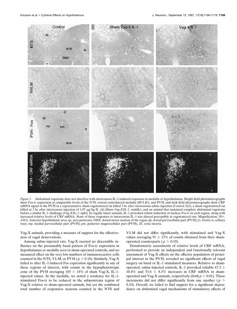
Fos-ir expression in control (sham Vag-X, saline-injected) ratswas generally low to nonexistent (Fig. 1). Labeled cells in theNTS and VLM were scattered widely and not observed consis-tently from section to section. A slightly higher level of basalexpression was observed in the PVH in some cases, concentratedin the autonomic-related dorsal, ventral–medial and lateral partsof the parvocellular division. Adjoining aspects of the zona in-certa and anterior hypothalamic area displayed more consistent,although still low, levels of nuclear Fos-ir. Injection of IL-1b (1.87mg/kg, i.v.) provoked robust and reliable Fos induction in each ofthe regions of interest (Fig. 1), the strength and distribution ofwhich were fully compatible with that described previously usingidentical parameters (Ericsson et al., 1994). In the dorsal vagalcomplex, activated neurons were heavily concentrated in thecaudal two-thirds of the medial subnucleus of the NTS, withsecondary accumulations in the dorsal and commissural parts.Only widely scattered Fos-ir neurons were detected in the areapostrema. IL-1-responsive neurons in the VLM were heavilyconcentrated in the rostral, C1, region, extending through theC1–A1 transition area at the level of the area postrema andtapered sharply in the A1 region, caudal to the level of the apexof the calamus scriptorus. In the PVH, Fos-ir neurons werepreferentially concentrated in the CRF-rich dorsal aspect of themedial parvocellular part of the nucleus. Fos expression in theautonomic-related dorsal, ventral–medial, and lateral parvocellu-lar parts was also reliably elevated over control levels. In themagnocellular division, Fos-ir was low to moderate and over-whelmingly concentrated in regions that preferentially expressoxytocin. These included subsets of the anterior, medial, andanteromedial aspects of the posterior magnocellular part of thePVH, as well as the anterior and dorsal portions of the supraopticnucleus. Counts of the number of Fos-ir neurons in the vagallyintact, IL-1-injected group revealed means 6 SEM (n 5 5) of716 6 73 labeled nuclei in the NTS, 770 6 108 in the VLM, and1126 6 148 in the medial parvocellular part of the PVH.
Sham Vag-X animals displayed extensive retrograde labelingfrom tracer injections in the forestomach throughout the rostro-caudal extent of the dorsal motor nucleus of the vagus, bilaterally(Fig. 2), and of isolated clusters of neurons in the rostral part ofthe principal column of ambiguual complex (data not shown). Noretrograde labeling was observed in any aspect of the medulla of
7168 J. Neurosci., September 15, 1997, 17(18):7166–7179 Ericsson et al. • Cytokine Effects on Hypothalamus
Vag-X animals, providing a measure of support for the effective-ness of vagal denervations.
Among saline-injected rats, Vag-X exerted no discernible in-fluence on the presumably basal pattern of Fos-ir expression inhypothalamus or medulla seen in sham-operated controls, and nomeasured effect on the very low numbers of immunoreactive cellscounted in the NTS, VLM, or PVH ( p . 0.10). Similarly, Vag-Xfailed to alter IL-1-induced Fos expression significantly in any ofthese regions of interest, with counts in the hypophysiotropiczone of the PVH averaging 105 6 14% of sham Vag-X, IL-1-injected values. In the medulla, we noted a tendency for IL-1-stimulated Fos-ir to be reduced in the subpostrema region ofVag-X relative to sham-operated animals, but yet the combinedtotal number of responsive neurons counted in the NTS and
VLM did not differ significantly, with stimulated and Vag-Xvalues averaging 91 6 13% of counts obtained from their sham-operated counterparts ( p . 0.10).
Densitometric assessments of relative levels of CRF mRNA,performed to provide an independent and functionally relevantassessment of Vag-X effects on the effector population of princi-pal interest in the PVH, revealed no significant effects of vagalsurgery on basal or IL-1 stimulated measures. Relative to sham-operated, saline-injected controls, lL-1 provoked reliable 67.3 610.4% and 53.6 6 8.3% increases in CRF mRNA in sham-operated and Vag-X animals, respectively (both p , 0.01). Theseincrements did not differ significantly from one another ( p .0.10). Overall, we failed to find support for a significant depen-dence on abdominal vagal mechanisms of stimulatory effects of
Figure 1. Abdominal vagotomy does not interfere with intravenous IL-1-induced responses in medulla or hypothalamus. Bright-field photomicrographsshow Fos-ir expression at comparable levels of the NTS, rostral ventrolateral medulla (RVLM ), and PVH, and dark-field photomicrographs show CRFmRNA signal in the PVH in a representative sham-vagotomized rat killed 3 hr after intravenous saline injection (Control; lef t), a sham-vagotomized ratkilled at 3 hr after intravenous injection of 1.87 mg/kg IL-1b (Sham Vag-X/IL-1; middle), and an animal that sustained complete abdominal vagotomybefore a similar IL-1 challenge (Vag-X/IL-1; right). In vagally intact animals, IL-1 provoked robust induction of nuclear Fos-ir in each region, along withincreased relative levels of CRF mRNA. None of these responses to intravenous IL-1 was altered perceptibly in vagotomized rats. Magnification, 503.AHA, Anterior hypothalamic area; ap, area postrema; DMX, dorsal motor nucleus of the vagus; dp, dorsal parvocellular part (PVH); fx, fornix; ts, solitarytract; mp, medial parvocellular part (PVH); pm, posterior magnocellular part (PVH), ZI, zona incerta.
Ericsson et al. • Cytokine Effects on Hypothalamus J. Neurosci., September 15, 1997, 17(18):7166–7179 7169

Figure 2. Neither abdominal vagotomy nor lesion of the area postrema modifies Fos expression in the medulla or hypothalamus induced by intravenousIL-1. Bar graphs show the effects of subdiaphragmatic vagotomy (Vag-X; top) or aspiration lesions of the area postrema (AP-X; bottom) on the numberof cells displaying IL-1-induced Fos-ir in the VLM and NTS (lef t) and PVH (right). Data are expressed as mean 6 SEM percentage of counts obtainedin sham Vag-X or sham AP-X animals treated with intravenous IL-1. n 5 5 or 6 per group. ns, Nonsignificant versus stimulated control values ( p . 0.10).Photomicrographs to the lef t of each graph provide documentation of the effectiveness or extent of the ablations. Top, Fluorescence photomicrographsshowing the robust retrograde labeling displayed by vagally intact rats of cells in the dorsal motor nucleus of the vagus as a consequence of fluorescenttracer injections placed in the wall of the stomach at the time of surgery and the complete lack of such labeling in a Vag-X animal. Magnification, 503.Bottom, Bright-field photomicrographs of Nissl-stained sections through the level of the maximal development of the area postrema, showing theappearance of the region in a sham-operated control and in rats that incurred minimal (min) or maximal (max) damage to underlying aspects of the NTS.ap, Area postrema; cc, central canal; DMX, dorsal motor nucleus of the vagus; ts, solitary tract; XII, hypoglossal nucleus. Magnification, 353.
7170 J. Neurosci., September 15, 1997, 17(18):7166–7179 Ericsson et al. • Cytokine Effects on Hypothalamus

increased circulating levels of IL-1 at the level of either themedulla or the hypothalamus under the conditions in force in thisexperiment.
Lesions of the area postrema and medial NTSThe area postrema, the circumventricular component of the dor-sal vagal complex, was identified previously as the only site apartfrom perivascular cells that exhibited both constitutive IL-1R1-and IL-1-induced IEG expression, under the dosage and treat-ment conditions used here (Ericsson et al., 1995a). In view of thefact that the major projections issued by the area postrema permitits influences to be exerted directly on the NTS, and at leastindirectly on the VLM (Cunningham et al., 1994), it is a viablecandidate for participating in IL-1-mediated effects on HPAcontrol systems.
Aspiration lesions were effective in removing all recognizableremnants of the area postrema in six IL-1-injected rats and fiveanimals challenged subsequently with vehicle. The lesioned areaalso consistently included portions of the underlying, and rela-tively cell-sparse, subpostrema region (Fig. 2). Three animals(two of which were subsequently injected with IL-1 and one withsaline) sustained more extensive damage, which included substan-tial portions of the dorsal and medial subnuclei of the NTS,extending in the most extreme case nearly to the medial margin ofthe solitary tract.
In sham-lesioned rats, both IL-1-stimulated and nonstimulatedpatterns of Fos-ir expression in hypothalamus, medulla, and else-where were in all respects similar to those described above,although the strength of Fos induction in this experiment wassomewhat less than that seen in the previous experiment. In fivenonlesioned, IL-1-injected rats, cell counts provided estimates of615 6 82 labeled nuclei in the NTS, 543 6 51 in the VLM, and844 6 68 in the medial parvocellular part of the PVH. As was thecase with vagotomy, lesions of the area postrema failed to reduceIL-1-stimulated Fos-ir expression significantly in either the PVHor in the NTS and VLM, where counts in AP-X, IL-1-treated ratsaveraged 82 6 13 and 94 6 9% of stimulated control values ( p .0.10) (Fig. 2). Similarly, relative levels of CRF mRNA in thesetwo groups were both reliably elevated (56 6 13%, p , 0.01; and42 6 11%, p , 0.05, respectively) over sham-lesioned, vehicle-injected control values, and did not differ significantly from oneanother ( p . 0.10).
It is of interest to note that even the two lesioned, IL-1-treatedanimals that sustained more extensive damage to the medial NTSdisplayed Fos induction with its normal topography in aspects ofthe NTS that were spared by the lesion, and that counts of thenumber of labeled cells in the VLM (593 and 641) and PVH (687and 811) from these two animals fell comfortably within the rangeof those derived from rats sustaining more discrete damage.
Prostaglandin synthesis inhibitionProstaglandin-dependent mechanisms have been implicated inthe IL-1 stimulation of the secretory activity of the HPA axis(Watanabe et al., 1990; Rivier and Rivest, 1993; Tilders et al.,1994; Watanobe et al., 1995). If prostaglandins and medullarycatecholamine-containing neurons are both involved in this acti-vation, then both aminergic neurons and their hypothalamic tar-gets would be expected to respond similarly to graded levels ofprostaglandin synthesis blockade. This notion was tested by pre-treating rats with systemic injections of the cyclooxygenase inhib-itor indomethacin 15 min before an intravenous IL challenge.
Rats pretreated with the vehicle used for indomethacin admin-
istration and subsequently challenged with IL-1 displayed veryprominent activation of Fos expression in the NTS, VLM, andPVH, fully compatible with that described above (Figs. 3, 4). Thiswas accompanied by a reliable, 59 6 7%, increase in relativelevels of CRF transcripts in the medial parvocellular part of thePVH ( p , 0.01 vs vehicle/vehicle-treated controls).
Although intravenous treatment with the higher dose of indo-methacin (1.0 mg/kg) alone (i.e., in the absence of subsequentIL-1 treatment) tended to reduce the low levels of Fos-ir expres-sion in all areas analyzed relative to controls, none of thesedifferences was reliable (all p . 0.10). Particularly significant isthe fact that relative levels of CRF mRNA, which is constitutivelyexpressed, were also not significantly altered as a consequence ofhigher-dose indomethacin treatment, averaging 85 6 4% of val-ues derived from vehicle/vehicle-treated rats.
Among IL-1-injected animals, indomethacin pretreatment pro-duced clear dose-related decrements in the number of Fos-irneurons counted in both regions of the medulla, as well as in thePVH, that were statistically reliable at the lowest (0.25 mg/kg)dose and that were not significantly elevated above control valuesat the higher of the three doses used (1.0 mg/kg; see Figs. 3, 4).Although only the medial parvocellular part of the PVH wasanalyzed quantitatively, IL-1-stimulated Fos induction in themagnocellular and autonomic-related subdivision of the nucleusappeared clearly to be similarly responsive to systemic treatmentwith the cyclooxygenase inhibitor. IL-1-stimulated increases inrelative levels of CRF mRNA in the PVH responded in a less-graded manner to indomethacin pretreatment, being nonsignifi-cantly affected, relative to the vehicle/IL-1-treated group, at thelower dose, and not differing reliably from vehicle/vehicle-treatedrats at both higher ones. Overall, these results provide strongsupport for an involvement of a prostaglandin-dependent mech-anism in IL-1-stimulated cellular responses at the levels of themedulla and hypothalamus. The comparable sensitivities exhib-ited by cell groups at both levels to indomethacin treatment isconsistent with the view that they constitute a unified system thatlies distal to the prostaglandin-dependent step.
Local prostaglandin microinjectionRecent findings localizing a prostaglandin E2 receptor subtype(EP3) in the region of medullary aminergic neurons (Ericsson etal., 1995b), but not in hypothalamus, raises the possibility thatprostanoids released by the perivascular cells as a consequence ofIL-1 stimulation might act locally to stimulate nearby aminergicneurons. Such a view would predict that microinjections of PGE2into the region of the C1 catecholamine cell group, the major seatof IL-1-responsive medullary aminergic cells that project to thePVH (Ericsson et al., 1994), should provoke IEG responses in C1cells and at least partially recapitulate the effects of intravenousIL-1 injection at the level of the PVH.
Examples of injection cannula placements above the C1 regionof the rostral VLM are shown in Figure 5. Immunoperoxidaselocalization of Fos revealed a mixture of both large (presumablyneuronal) and smaller (presumably astrocytic) labeled nucleiclosely associated with the cannula track and extending longitu-dinally in a 300–450 mm radius from the center of the injectionsite. Material stained for combined immunofluorescence localiza-tion of Fos-ir and GFAP-ir revealed numerous astrocytes in-tensely labeled for GFAP within and around the region invadedby the tip of the injector; this was surrounded by a band of largerFos-ir nuclei, beyond which GFAP-ir tapered sharply to levelscomparable to those observed at a corresponding locus on the
Ericsson et al. • Cytokine Effects on Hypothalamus J. Neurosci., September 15, 1997, 17(18):7166–7179 7171

contralateral side (Fig. 5). This basic pattern of Fos and GFAPlabeling near the tip of the injector did not vary systematically asa function of PGE2 dose or between PGE2- and vehicle-treatedanimals, and we interpret it as being indicative of nonspecificconsequences of the injections. Beyond this, however, rats in-jected with 100 ng of PGE2 and, to a lesser extent, those receiving10 ng displayed remarkably discrete Fos-ir expression concen-trated in the C1 region of the rostral VLM (Fig. 6). This extendedwell beyond the zone at which reactive astrocytes were observedand throughout most of the rostrocaudal extent of the C1 cellgroup in animals treated with 100 ng doses. No other recognizedcell group or field of the ventral medulla displayed consistentPGE2-stimulated Fos expression. Co-staining of series of sectionsthrough the medulla for DBH-ir and PNMT-ir revealed that asubstantial majority of all Fos-ir cells beyond the zone of reactiveastrocytosis surrounding the injection site were adrenergic. Inter-estingly, higher doses of PGE2 also provoked Fos-ir expression inthe medial NTS, albeit with lesser consistency than in the VLM;it is not clear whether this may occur as a secondary consequence
of activation more ventrally or directly as a result of injectatediffusing along the guide cannula tracks, which invariably im-pinged on the NTS. A lesser, although still substantial, fraction ofFos-ir neurons in the NTS displayed PNMT-ir and/or DBH-ir.
At the level of the PVH, intramedullary injections of 100 ng ofPGE2 induced prominent Fos induction, the distribution of whichclosely mirrored that seen in response to intravenous IL-1 injec-tions (Figs. 6, 7). Thus, the response was most pronounced in theCRF-rich dorsal–medial parvocellular subdivision, with onlyslightly less prominent foci in the autonomic-related dorsal, ven-tral–medial, and lateral parvocellular parts and in all parts of themagnocellular division. Fos-ir was heavily concentrated in as-pects of the magnocellular neurosecretory system in whichoxytocin-expressing neurons are concentrated, including the an-terior, medial, and discrete aspects of the posterior magnocellularparts of the PVH (including the ring-like array seen at the levelillustrated in Fig. 6), and with a crisp anterior and dorsal empha-sis in the supraoptic nucleus. Aspects of this pattern, principallyactivation in the parvocellular division of the PVH, were weakly
Figure 3. Effects of graded levels of prostaglandin synthesis blockade on IL-1-stimulated Fos-ir and CRF mRNA expression. Bright-field photomi-crographs show sections through similar levels of the PVH (top) and rostral ventrolateral medulla (bottom) stained for Fos-ir, and dark-fieldphotomicrographs show PVH sections hybridized with probes for CRF mRNA (middle) from animals pretreated intravenously with either vehicle ofvarying doses of indomethacin (Indo) 15 min before an intravenous challenge with 1.87 mg/kg IL-1b. Indomethacin pretreatment produces a dose-relateddecrease in the expression of Fos-ir and CRF mRNA in the PVH and of Fos-ir in the region of the C1 catecholamine cell group. The patterns andstrength of expression of both markers in the PVH and of Fos-ir in the VLM at the higher indomethacin doses are not distinguishable from those seenin rats injected with vehicle in lieu of IL-1. Note that Fos-ir expression in the regions immediately adjoining the PVH is ostensibly unaffected by anydosage of indomethacin. Magnification, 603.
7172 J. Neurosci., September 15, 1997, 17(18):7166–7179 Ericsson et al. • Cytokine Effects on Hypothalamus

apparent in most animals receiving 10 ng doses of PGE2. Injec-tions of 1 ng uniformly failed to elicit Fos expression in any aspectof the hypothalamus that was distinguishable from the very lowlevels seen in vehicle-injected controls.
Quantitative analyses revealed that although intramedullaryinjection of 1 ng of PGE2 failed to elicit a significant increase inthe number of Fos-ir neurons estimated in the medial parvocel-lular part of the PVH, relative to vehicle-treated controls (51 6
Figure 4. Dose-related inhibition by systemic indomethacin of IL-1 effects in hypothalamus and medulla. Mean 6 SEM number of Fos-ir neurons inthe NTS and VLM combined (top), the PVH (middle), and relative levels of CRF mRNA (bottom) of rats as a function of treatment condition. Dataare expressed as a percentage of values counts derived from vehicle-pretreated rats that were challenged with IL-1. n 5 4–7 per group. The key at thebottom indicates the status of the group: 2, vehicle-injected; 1, intravenous injection of 1.87 mg/kg IL-1b; 0.25, 0.5, and 1.0, doses of indomethacinadministered 15 min before treatment with IL-1 or its vehicle. IL-1 provokes a robust induction of Fos expression in both hypothalamus and medullathat are antagonized in parallel, dose-related manners by indomethacin. Cytokine-mediated upregulation of CRF mRNA is eliminated at both higherdoses of indomethacin but is not significantly affect by the lowest one. *p , 0.05; **p , 0.01; ***p , 0.005; ****p , 0.001 versus control(saline/saline-treated) rats; ns, nonsignificant. Additional comparisons are indicated by p values.
Ericsson et al. • Cytokine Effects on Hypothalamus J. Neurosci., September 15, 1997, 17(18):7166–7179 7173

11 vs 39 6 9; n 5 4 and 5, respectively; p . 0.10), 10 ng dosesprovoked a weak, but reliable, effect (87 6 16; n 5 6; p , 0.05),and 100 ng stimulated a strong activation (1211 6 138; n 5 4; p ,0.001) exceeding that commonly observed in the experimentsdescribed above in response to intravenous IL-1 treatment. Com-parisons of relative levels of CRF mRNA in animals receiving thehigher (100 ng) dose with controls revealed a marked, 1.9-fold,increase ( p , 0.001).
DISCUSSIONNeither abdominal vagotomy nor lesions of the area postremareliably altered Fos-ir induced in the NTS, VLM, and PVH byintravenous administration of a moderately suprathreshold doseof IL-1b. Cytokine-stimulated increases in relative levels of CRFmRNA in the hypophysiotropic zone of the PVH were alsounaffected by either ablation. By contrast, systemic administrationof the cyclooxygenase inhibitor indomethacin resulted in paralleldose-related attenuations of IL-1 effects in hypothalamus andmedulla. Microinjections of PGE2 in the region of the C1 cate-cholamine cell group, the principal seat of IL-1-sensitive neuronsthat project to the PVH, provoked surprisingly discrete patternsof cellular activation in the hypothalamus and medulla thatclosely mimicked those seen in response to intravenous IL-1.Coupled with previous work on this topic, we take these data as
suggesting a mechanism for stimulation of CRF-expressing par-vocellular neurosecretory neurons by circulating IL-1 that in-cludes (1) IL-1 binding of its type 1 receptor on cells lining themedullary vasculature with consequent local release of prostan-oids, most likely PGE2, into the extracellular space; (2) PGE2interacting with cognate receptors expressed on or near medul-lary aminergic neurons, resulting in activation of this population;and (3) consequent synaptic excitation of hypophysiotropic CRFneurons, by way of well documented direct axonal projections(see Fig. 8).
VagotomyMedullary catecholamine-containing neurons are acknowledgedas playing pivotal roles in dispersing visceral sensory informationto relevant effectors, prominently including ones in the endocrinehypothalamus. Support for a role of vagal sensory mechanisms inmediating central effects of immune challenges may be found inthe observations that glomus cells of abdominal vagal paragangliabind biotinylated IL-1 receptor antagonist (Goehler et al., 1994),suggesting the existence vagal IL-1 receptors, and that adminis-tration of IL-1 into the hepatic portal vein results in increasedelectrical activity of the hepatic branch of the vagus (Nijima,1992). Our failure to observe reliable effects of subdiaphragmaticvagotomy on cellular responses in the medulla and hypothalamus
Figure 5. Cannula placements and local effects of intramedullary microinjection of PGE2. Top, Bright-field photomicrographs of Nissl-stained sectionsshowing cannula placements in the rostral ventrolateral medulla. Magnification, 153. Bottom, Fluorescence photomicrographs of a single field near acannula tip in the ventrolateral medulla stained concurrently for GFAP-ir and Fos-ir. Surrounding the area of tissue damage (*) is a region of intenseGFAP labeling of reactive astrocytes. At the margin of this zone lies a band of Fos-ir neurons, which we take to be nonspecifically induced as aconsequence of microinjection. Beyond this, GFAP labeling tapers precipitously to levels indistinguishable from those seen at a comparable locus on thecontralateral side, and Fos-ir is seen in the C1 region of the rostral ventrolateral medulla in rats injected with suprathreshold doses of PGE2. XII,Hypoglossal nucleus; stv, spinal trigeminal tract; SpV, spinal trigeminal nucleus; py, pyramidal tract; IO, inferior olivary complex. Magnification, 1503.
7174 J. Neurosci., September 15, 1997, 17(18):7166–7179 Ericsson et al. • Cytokine Effects on Hypothalamus

to an intravenous IL-1 challenge is in line with the consensus ofprevious work on this topic, which supports a much more impor-tant role for the abdominal vagi in mediating central responses tocytokines or endotoxin administered intraperitoneally than itdoes to similar doses of the same agents given intravenously(Katsuura et al., 1988; Wan et al., 1994; Gaykema et al., 1995;Kapcala et al., 1996). It is important to point out, however, thatbecause rats do not survive cervical vagotomy, the possible in-volvement of thoracic receptor mechanisms that may be inner-vated by sensory elements of the cervical vagus and/or glossopha-ryngeal nerves remains to be considered experimentally.
Area postrema and NTSA potential role for the area postrema in IL-1 stimulation of thesystems of interest here was suggested by our observation that thearea postrema was the only site, apart from perivascular cells, atwhich we observed both IL-1R1 expression and IL-1-stimulatedIEG expression (of NGFI-B, but not Fos), using the same cyto-kine injection parameters used in the present experiments (Eric-sson et al., 1995a). The major projections issued by the areapostrema ramify in the aspects of the NTS that harbor catechol-amine neurons of the A2 and C2 cell groups (Cunningham et al.,
1994). Influences of the area postrema on the VLM may bemediated directly, via projections that traverse the regions of theA1 and C1 cell groups en route to the parabrachial nucleus or,more likely, indirectly, through NTS–VLM interactions. Despitethe fact that it is well positioned to participate in cytokine-mediated activation of HPA control systems and may well do sounder certain circumstances, the results of our lesion study of-fered no support for an involvement of the area postrema inmediating IL-1 effects at the level of the medulla or hypothala-mus, under the treatment conditions in force in our experiments.
ProstaglandinsSystemic injection of IL-1 provokes increased levels of PGE2 inthe general circulation (Rotondo et al., 1988; Watanobe et al.,1995), and pretreatment of rats with inhibitors of cyclooxygenase,the rate-limiting enzyme in prostaglandin synthesis, blocks orsignificantly attenuates ACTH secretion to intravenous IL-1(Watanabe et al., 1990). Such findings have commonly been takenas supporting an intermediary role for circulating PGE2 in con-veying cytokine signals to central or peripheral receptors, butrecent evidence is at least equally as strong in suggesting that theprostaglandin-dependent mechanism may reside within the brain
Figure 6. Microinjections of PGE2 in the ros-tral ventrolateral medulla mimic the effects ofintravenous IL-1. Top and middle, Bright-fieldphotomicrographs showing immunoperoxidaselocalization of Fos-ir in the rostral ventrolat-eral medulla (RVLM; top) and PVH (middle)from animals that received intramedullary in-jections of 200 nl of vehicle (Control ) or 100 ngof PGE2 in the same volume (PGE2). Medul-lary sections are taken from levels 300 mmrostral to center of the injection site. PGE2 butnot vehicle injection stimulates Fos inductionstrongly and discretely in both medulla andhypothalamus. The pattern in hypothalamus,primarily involving cells in the CRF-rich as-pects of the medial parvocellular part (mp), andsecondarily cells in the dorsal parvocellularpart and at the periphery of the posterior mag-nocellular part ( pm), conforms closely to thepattern elicited by intravenous IL-1 injections.Magnification, 753. Bottom, fluorescence pho-tomicrographs of a single section through theA1–C1 transition area of the ventrolateral me-dulla from an animal that received intramedul-lary injection of 100 ng of PGE2 and processedfor concurrent immunoperoxidase demonstra-tion of Fos-ir (dark nuclei) and immunofluores-cence demonstration of DBH-ir (lef t) andPNMT-ir (right). Examples of cells displayingall three markers are indicated (arrows). Mag-nification, 2503.
Ericsson et al. • Cytokine Effects on Hypothalamus J. Neurosci., September 15, 1997, 17(18):7166–7179 7175

parenchyma. Our observation that graded levels of prostaglandinsynthesis inhibition provoke comparable dose-related blockadesof IL-1-induced cellular activation responses in the NTS, VLM,and PVH is consistent with the view that the prostanoid-dependent step in the IL-1-mediated drive to the central limb ofthe HPA axis lies distal to the level of medullary aminergicneurons. In addition to expressing IL-1R1 (Cunningham et al.,1992; Yabuuchi et al., 1994; Ericsson et al., 1995a), cells liningaspects of the cerebral vasculature express cyclooxygenase-immunoreactive material in vivo (Breder et al., 1992), and sys-temic administration of bacterial endotoxins stimulate release ofPGE2-like immunoreactivity from the brain microvasculature(Van Dam et al., 1993). Ligand-binding studies have identifiedPGE2 receptor-like activity in the rat CNS, with the highest levelsobserved in the medial preoptic area and in the NTS (Matsumuraet al., 1992).
A number of structurally related PGE2 receptors have beenidentified and designated EP1–4 (Coleman et al., 1989), andcDNAs encoding each have been cloned in rat and/or mouse(Sugimoto et al., 1992; Honda et al., 1993; Takeuchi et al., 1993;Watabe et al., 1993; Nishigaki et al., 1995). All are members ofthe superfamily of G-protein-coupled receptors characterized byseven transmembrane domains. Northern analyses have identifiedthe EP3 subtype as the predominant form expressed in rodentbrain (Sugimoto et al., 1992; Honda et al., 1993; Watabe et al.,1993). We have isolated an EP3 cDNA from rat kidney and havefound this transcript to display a restricted central distributionthat includes regions of the NTS and VLM in which IL-1-
sensitive catecholamine neurons are massed. Expression in hypo-thalamus is largely limited to the medial preoptic area; specificlabeling has not been observed over the PVH (also see Sugimotoet al., 1994; Ericsson et al., 1995b).
These observations provide a basis for understanding the sur-prisingly discrete local patterns of Fos induction observed in themedulla after PGE2 microinjection. The fact that these injectionsfaithfully recapitulated the effects of intravenous IL-1 adminis-tration on the pattern of Fos induction and CRF mRNA expres-sion in the PVH fulfills a prediction that necessarily follows fromour model. Evidence supporting alternative mechanisms is avail-able. Intrahypothalamic injections of PGE2 (25 ng) have beenshown to stimulate ACTH secretion (Watanabe et al., 1990), andevidence that the PVH expresses the EP1 prostanoid receptorhas been provided (Båtshake et al., 1995). In addition, cells in themedial preoptic area identified as projecting to the PVH havebeen shown to display lipopolysaccharide (LPS)-induced Fosexpression (Elmquist and Saper, 1996), and injections of PGE2into the preoptic area provoke Fos induction in the PVH with asensitivity greater than that observed at our placements in themedulla (Scammell et al., 1996). Lacking a systematic and directcomparison, the primacy of these alternative means of accessingHPA control mechanisms cannot currently be assessed. Scammelland colleagues (1996) used smaller volumes to target a discretecell group, whereas we used larger ones in an effort to involve asmuch of the more diffuse and longitudinally organized C1 cellgroup as possible. Our working hypothesis emphasizing the roleof medullary afferents, as opposed to more direct effects or
Figure 7. Dose-related Fos-ir expression in the PVH after intramedullary PGE2 injections. Bright-field photomicrographs of section throughcomparable levels of the PVH show immunoperoxidase localization of Fos-ir seen in response to microinjections of various doses of PGE2 or vehicle.A modest effect is noted at the 10 ng dose, and a much more robust one is noted at 100 ng. dp, Dorsal parvocellular part; mp, medial parvocellular part;pm, posterior magnocellular part; pv, periventricular part; ZI, zona incerta. Magnification, 753.
7176 J. Neurosci., September 15, 1997, 17(18):7166–7179 Ericsson et al. • Cytokine Effects on Hypothalamus

influences mediated via forebrain inputs, is based on our previousfindings that transections of ascending projections from the me-dulla disrupt IL-1-induced responses in the PVH, and that sim-ilar lesions designed to disrupt descending projections from thelamina terminalis are ineffective in this regard (Ericsson et al.,1994). Although we suspect that these latter transections would beapt to disrupt ventral–medial preoptic projections to the PVH,this remains to be determined experimentally. A mediating, asopposed to a merely permissive, influence of ascending aminergicafferents is supported by the starkly differential dependence onthe integrity of ascending aminergic inputs of PVH responses tointravenous IL-1 and foot shock stress, two challenges that pro-voke patterns of Fos induction in the PVH and medullary amin-ergic cell groups that are essentially indistinguishable (Li et al.,1996). Similar findings have been reported by others using HPAsecretory responses as end points (Weidenfeld et al., 1989; Chu-luyan et al., 1992).
Thus, the general region of medullary catecholaminergic cellgroups and the proximate vasculature contain all essential com-ponents of the transduction and paracrine signaling mechanismswe propose (Fig. 8). Challenges to this model are posed by
uncertainties about precisely how these components may be dis-tributed, or co-distributed, across cell types in the area. Forexample, although it has been widely suggested that endothelialcells are the principal seat of perivascular IL-1R1 expression, thishas not been established experimentally, and recent evidence hasidentified perivascular microglia as a major locus of LPS-inducedcyclooxygenase-2 expression (Elmquist et al., 1997). Similarly,although the distribution of EP3 receptor message in the medullasuggests overlap with that of IL-1-sensitive catecholaminergicneurons, this, too, remains to be scrutinized directly. This isparticularly important, because activation of this receptor is com-monly associated with inhibition of adenylate cyclase activity,raising questions of how it might support a stimulatory effect onany aminergic neurons that might express it. It should be recalledthat perhaps the best established physiological role of medullaryaminergic cells, that of aspects of the C1 cell group in mediatingthe baroreceptor inhibition of sympathetic outflow to the periph-eral vasculature, is effected via local inhibitory neurons (see Reiset al., 1994), and recent work indicates that such GABAergicneurons are closely interdigitated among aminergic cells in theVLM as well as the NTS (R. K. W. Chan and P. E. Sawchenko,
Figure 8. Possible mechanism for intravenous IL-1-mediated stimulation of central HPA control systems. A polarized epifluorescence illuminationimage of a section through the rostral ventrolateral medulla shows combined hybridization histochemical localization of perivascular cells displayingIL-1R1 mRNA and immunoperoxidase detection of nuclear Fos-ir in the region of the C1 catecholamine cell group. We suggest that circulating IL-1binds its cognate receptor on perivascular cells in the region, inducing them to synthesize PGE2, which in turn diffuses through the extracellular spaceto (directly or indirectly) stimulate nearby aminergic neurons and, consequently, CRF-expressing targets of their axonal projections in the endocrinehypothalamus. Listed at the right are markers of potentially relevant components of this signaling cascade that have been localized in the requisiteregions, either under basal conditions or in response to intravenous IL-1 or endotoxin. Markers that have been co-localized to date are bracketed. Itremains to be determined how the others are distributed with respect to the key perivascular (i.e., IL-1R1-expressing) and neuronal (IL-1-sensitive,hypothalamically projecting, and catecholaminergic) cell types.
Ericsson et al. • Cytokine Effects on Hypothalamus J. Neurosci., September 15, 1997, 17(18):7166–7179 7177

unpublished data). Expression of the EP3 receptor by an inhibi-tory interneuron, with consequent activation of aminergic cells bydisinhibition, would be more consistent with the known pharma-cology of this receptor subtype than would expression by amin-ergic neurons themselves.
As discussed elsewhere (Ericsson et al., 1994), initial plasmalevels of IL-1 achieved after intravenous injection of IL-1b at thethreshold required to elicit IEG responses in hypothalamus andmedulla are similar to the peak concentrations seen after systemictreatment with LPS, a model for systemic bacterial infection(Zuckerman et al., 1989). IL-1 titers would be expected to fallprecipitously after acute injection (half-life, ;2.9 min; Reimers etal., 1991) but in LPS-treated animals are sustained for hours andreinforced by elevated levels of other proinflammatory cytokinesthat are capable of independently stimulating HPA output (Naitoet al., 1988; Bernardini et al., 1990). Thus, the intravenous IL-1injection paradigm is relevant to an established animal model ofsepsis, at least. In closing, it is important to emphasize that theavailable evidence is quite clear in indicating that central path-ways and mechanisms that are responsive to challenges posed bytreatment with individual cytokines, or to more complex immunestimuli, may vary markedly as a function of the nature of thestimulating agent(s), dose, and route of administration. The dataprovided here simply offer initial support for the mechanismoutlined above as a component of the minimum essential circuitryrequired for the activation of the central limb of the HPA axis byincreased circulating IL-1.
REFERENCESAbercrombie M (1946) Estimation of nuclear populations from mic-
rotome populations from microtome sections. Anat Rec 94:239–247.Båtshake B, Nilsson C, Sundelin J (1995) Molecular characterization of
the mouse prostanoid EP1 receptor gene. Eur J Biochem 231:809–814.Berkenbosch F, van Oers J, del Rey A, Tilders F, Besedovsky H (1987)
Corticotropin-releasing factor-producing neurons in the rat activatedby interleukin-1. Science 238:524–526.
Bernardini R, Kamilaris TC, Calogero AE, Johnson EO, Gomez MT,Gold PW, Chrousos GP (1990) Interactions between tumor necrosisfactor-alpha, hypothalamic corticotropin-releasing hormone, and adre-nocorticotropin secretion in the rat. Endocrinology 126:2876–2881.
Besedovsky H, Sorkin E, Keller M, Muller J (1975) Changes in bloodhormone levels during the immune response. Proc Soc Exp Biol Med150:466–470.
Besedovsky H, del Rey A, Sorkin E, Dinarello CA (1986) Immunoregu-latory feedback between interleukin-1 and glucocorticoid hormones.Science 233:652–654.
Bohn MC, Dreyfus CF, Friedman WJ, Markey KA (1987) Glucocorti-coid effects on phenylethanolamine N-methyltransferase (PNMT) inexplants of embryonic rat medulla oblongata. Brain Res 465:257–266.
Breder CD, Smith WL, Raz A, Masferrer J, Seibert K, Needleman P,Saper CB (1992) Distribution and characterization of cyclooxygenaseimmunoreactivity in the ovine brain. J Comp Neurol 322:409–438.
Chuluyan HE, Saphier D, Rohn WM, Dunn AJ (1992) Noradrenergicinnervation of the hypothalamus participates in adrenocortical re-sponses to interleukin-1. Neuroendocrinology 56:106–111.
Coleman RA, Kennedy I, Humphrey PPA, Bunce K, Lumley P (1989)Prostanoids and their receptors. In: Comprehensive medical chemistry:membranes and receptors (Taylor JB, Emmett JC, eds), pp 643–714.Oxford: Pergamon.
Cunningham Jr ET, Wada E, Carter DB, Tracey DE, Battey JF, DeSouzaEB (1992) In situ histochemical localization of type 1 interleukin-1receptor RNA in the central nervous system, pituitary and adrenalgland of the mouse. J Neurosci 12:1101–1114.
Cunningham Jr ET, Miselis RR, Sawchenko PE (1994) The relationshipof efferent projections from the area postrema to vagal motor and brainstem catecholamine-containing cell groups: an axonal transport andimmunohistochemical study in the rat. Neuroscience 58:635–648.
Dantzer R (1994) How do cytokines say hello to the brain? Neuralversus humoral mediation. Eur Cytokine Netw 5:271–273.
Elmquist JK, Saper CB (1996) Activation of neurons projecting to theparaventricular hypothalamic nucleus by intravenous lipopolysaccha-ride. J Comp Neurol 374:315–331.
Elmquist JK, Breder CD, Sherin JE, Scammell TE, Hickey WF, DeWittD, Saper CB (1997) Intravenous lipopolysaccharide induces cycloox-ygenase 2-like immunoreactivity in rat brain perivascular microglia andmeningeal macrophages. J Comp Neurol 381:119–129.
Ericsson A, Sawchenko PE (1993) c-fos-based functional mapping ofcentral pathways subserving effects of interleukin 1 on thehypothalamo-pituitary-adrenal axis. Methods Neurosci 16:155–172.
Ericsson A, Kovacs KJ, Sawchenko PE (1994) A functional anatomicalanalysis of central pathways subserving the effects of interleukin-1 onstress-related neuroendocrine neurons. J Neurosci 14:897–913.
Ericsson A, Liu C, Hart RP, Sawchenko PE (1995a) Type 1interleukin-1 receptor in the rat brain: distribution, regulation, andrelationship to sites of IL-1-induced cellular activation. J Comp Neurol361:681–698.
Ericsson A, Ek M, Lindefors N (1995b) Distribution of prostaglandin E2receptor (EP3 subtype) mRNA containing cells in the rat centralnervous system. Soc Neurosci Abstr 21:98.
Ericsson A, Ek M, Wahlstrom I, Kovacs K, Liu C-L, Hart R, SawchenkoPE (1996) Pathways and mechanisms for interleukin-1 mediated reg-ulation of the hypothalamo-pituitary-adrenal axis. In: Stress: moleculargenetic and neurobiological advances (McCarty R, Aguilera G, SabbanEL, Kvetnansky R, eds), pp 101–120. New York: Gordon and Breach.
Gaykema RPA, Dijkstra I, Tilders FJH (1995) Subdiaphragmatic vagot-omy suppresses endotoxin-induced activation of hypothalamiccorticotropin-releasing hormone neurons and ACTH secretion. Endo-crinology 136:4717–4720.
Goehler LE, Relton J, Maier SF, Watkins LR (1994) Biotinylatedinterleukin-1 receptor antagonist (IL-1RA) labels paraganglia in therat liver hilus and hepatic vagus. Soc Neurosci Abstr 20:956.
Harbuz MS, Stephanou A, Sarlis N, Lightman SL (1992) The effects ofrecombinant human interleukin (IL)-1a, IL-1b or IL-6 onhypothalamo-pituitary-adrenal axis activation. J Endocrinol133:349–355.
Hench TR, Kendall EC, Slocumb CH, Polley HF (1949) The effect of ahormone of the adrenal cortex (17-hydroxy-11-dehydrocorticosterone;compound) and of pituitary adrenocorticotropic hormone on rheuma-toid arthritis. Staff Proc Mayo Clin 24:181.
Honda A, Sugimoto Y, Namba T, Watabe A, Irie A, Negishi M, Naru-miya S, Ichikawa A (1993) Cloning and expression of a cDNA formouse prostaglandin E receptor EP2 subtype. J Biol Chem268:7759–7762.
Kapcala LP, He JR, Gao Y, Pieper JO, DeTolla LJ (1996) Subdiaphrag-matic vagotomy inhibits intra-abdominal interleukin-1b stimulation ofadrenocorticotropin secretion. Brain Res 728:247–254.
Katsuura G, Gottschall PE, Dahl RR, Arimura A (1988) Adrenocorti-cotropin release induced by intracerebroventricular injection of recom-binant human interleukin-1 in rats: Possible involvement of prostaglan-din. Endocrinology 122:1773–1779.
Li H-Y, Ericsson A, Sawchenko PE (1996) Distinct mechanisms under-lie activation of hypothalamic neurosecretory neurons and their med-ullary catecholaminergic afferents in categorically different stress par-adigms. Proc Natl Acad Sci USA 93:2359–2364.
Matsumura K, Watanabe Y, Imai-Matsumura K, Connolly M, KoyamaY, Onoe H, Watanabe Y (1992) Mapping of prostaglandin E2 bindingsites in rat brain using quantitative autoradiography. Brain Res581:292–298.
Morgan JL, Curran T (1991) Stimulus-transcription coupling in the ner-vous system: involvement of the inducible proto-oncogenes fos and jun.Annu Rev Neurosci 14:421–451.
Naito Y, Fukata J, Tominaga T, Nakai Y, Tamai S, Mori K, Imura H(1988) Interleukin-6 stimulates the secretion of adrenocorticotropichormone in conscious, freely-moving rats. Biochem Biophys Res Com-mun 155:1459–1463.
Nakano K, Okugawa K, Hayashi H, Abe S, Sohmura Y, Tsuboi T (1988)Establishment of dye-uptake method (A375 assay) for quantitativemeasurement of IL-1: correlation with LAF assay. Dev Biol 69:93–101.
Nijima A (1992) The afferent discharges from sensors for interleukin 1bin the hepato-portal system in the rat. J Physiol (Lond.) 446:236P.
Nishigaki N, Negishi M, Honda A, Sugimoto Y, Namba T, Narumiya S,Ichikawa A (1995) Identification of prostaglandin E receptor “EP2”cloned from mastocytoma cells as EP4 subtype. FEBS Lett364:339–341.
7178 J. Neurosci., September 15, 1997, 17(18):7166–7179 Ericsson et al. • Cytokine Effects on Hypothalamus

Reimers J, Wogensen LD, Welinder B, Hejnaes KR, Poulsen SS, NilssonP, Nerup J (1991) The pharmacokinetics, distribution and degradationof human recombinant interleukin-1b in normal rats. Scand J Immunol34:597–617.
Reis DJ, Golanov EV, Ruggiero DA, Sun MK (1994) Sympatho-excitatory neurons of the rostral ventrolateral medulla are oxygensensors and essential elements in the tonic and reflex control of thesystemic and cerebral circulations. J Hypertens 12:S159–S180.
Rivest S, Rivier C (1991) Influence of the paraventricular nucleus of thehypothalamus in the alteration of neuroendocrine functions induced byintermittent footshock or interleukin. Endocrinology 129:2049–2057.
Rivier C, Rivest S (1993) Mechanisms mediating the effects of cytokineson neuroendocrine functions in the rat. Ciba Found Symp172:204–220.
Rotondo D, Abul HT, Milton AS, Davidson J (1988) Pyrogenic immu-nomodulators increase the levels of prostaglandin E2 in the bloodsimultaneously with the onset of fever. Eur J Pharmacol 154:145–152.
Sapolsky R, Rivier C, Yamamoto G, Plotsky P, Vale W (1987)Interleukin-1 stimulates the secretion of hypothalamic corticotropin-releasing factor. Science 238:522–524.
Sawchenko PE, Gold RM (1981) Effects of complete subdiaphragmaticvs. selective gastric vagotomy on hypothalamic hyperphagia and obe-sity. Physiol Behav 26:281–292.
Sawchenko PE, Cunningham Jr ET, Mortrud MT, Pfeiffer SW, GerfenCR (1990) Phaseolus vulgaris-leucoagglutinin (PHA-L) anterogradeaxonal transport technique. Methods Neurosci 3:247–260.
Scammell TE, Elmquist JK, Griffin JD, Saper CB (1996) Ventromedialpreoptic prostaglandin E2 activates fever-producing autonomic path-ways. J Neurosci 16:6246–6254.
Shu S, Ju G, Fan L (1988) The glucose oxidase-DAB-nickel method inperoxidase histochemistry of the nervous system. Neurosci Lett85:169–171.
Simmons DM, Arriza JL, Swanson LW (1989) A complete protocol forin situ hybridization of messenger RNAs in brain and other tissues withradiolabeled single-stranded RNA probes. J Histotechnol 12:169–181.
Sternberg EM, Wilder, RL (1993) Corticosteroids. In: Arthritis and al-lied conditions: a textbook of rheumatology, Ed 12 (McCarthy DJ,Koopman DJ, eds), pp 665–682. Philadelphia: Lea & Febiger.
Sugimoto Y, Namba T, Honda A, Hayashi Y, Negishi M, Ichikawa A,Narumiya S (1992) Cloning and expression of a cDNA for mouseprostaglandin E receptor EP3 subtype. J Biol Chem 267:6463–6466.
Sugimoto Y, Shigemoto R, Namba T, Negishi M, Mizuno N, Narumiya S,Ichikawa A (1994) Distribution of the messenger RNA for the pros-taglandin E receptor subtype EP3 in the mouse nervous system. Neu-roscience 62:919–928.
Swanson LW, Kuypers HGJM (1980) The paraventricular nucleus of thehypothalamus: cytoarchitectonic subdivisions and the organization of
projections to the pituitary, dorsal vagal complex and spinal cord asdemonstrated by retrograde fluorescence double labeling methods.J Comp Neurol 194:555–575.
Takeuchi K, Abe T, Takahashi N, Abe K (1993) Molecular cloning andintrarenal localization of rat prostaglandin E2 receptor EP3 subtype.Biochem Biophys Res Commun 194:885–891.
Tilders FJ, DeRijk RH, Van Dam AM, Vincent VA, Schotanus K,Persoons JH (1994) Activation of the hypothalamus-pituitary-adrenalaxis by bacterial endotoxins: routes and intermediate signals. Psycho-neuroendocrinology 19:209–232.
Van Dam A-M, Brouns M, Man-A-Hing W, Berkenbosch F (1993) Im-munocytochemical detection of prostaglandin E2 in microvasculatureand in neurons of rat brain after administration of bacterial endotoxin.Brain Res 613:331–336.
Wan W, Wetmore L, Sorensen CM, Greenberg AH, Nance DM (1994)Neural and biochemical mediators of endotoxin and stress-inducedc-fos expression in the rat brain. Brain Res Bull 34:7–14.
Watabe A, Sugimoto Y, Honda A, Irie A, Namba T, Negishi M, Ito S,Narumiya S, Ichikawa A (1993) Cloning and expression of cDNA fora mouse EP1 subtype of prostaglandin E receptor. J Biol Chem268:20175–20178.
Watanabe T, Morimoto A, Sakata Y, Murakami N (1990) ACTH re-sponse induced by interleukin-1 is mediated by CRF secretion stimu-lated by hypothalamic PGE. Experientia 46:481–484.
Watanobe H, Nasushita R, Takebe K (1995) A study on the role ofcirculating prostaglandin E2 in the adrenocorticotropin response tointravenous administration of interleukin-1beta in the rat. Neuroendo-crinology 62:596–600.
Watkins LR, Maier SF, Goehler LE (1995) Cytokine-to-brain commu-nication: a review and analysis of alternative mechanisms. Life Sci57:1011–1026.
Weidenfeld J, Abramsky O, Ovadia H (1989) Evidence for the involve-ment of the central adrenergic system in interleukin 1-induced adreno-cortical response. Neuropharmacology 28:1411–1414.
Wick G, Hu Y, Schwartz S, Kroemer G (1993) Immunoendocrine com-munication via the hypothalamo-pituitary-adrenal axis in autoimmunediseases. Endocr Rev 14:539–563.
Wong M-L, Licinio J (1994) Localization of interleukin-1 type 1 recep-tor mRNA in rat brain. Neuroimmunomodulation 1:110–115.
Yabuuchi K, Minami M, Katsumata S, Satoh M (1994) Localization oftype I interleukin-1 receptor mRNA in the rat brain. Mol Brain Res27:27–36.
Zuckerman SH, Shellhaas J, Butler LD (1989) Differential regulation oflipopolysaccharide-induced interleukin 1 and tumor necrosis factorsynthesis: effects of endogenous and exogenous glucocorticoids and therole of the pituitary-adrenal axis. Eur J Immunol 19:301–305.
Ericsson et al. • Cytokine Effects on Hypothalamus J. Neurosci., September 15, 1997, 17(18):7166–7179 7179
Copyright © 2022 FDOKUMEN