Nitric oxide modulation of the Basal Ganglia circuitry: therapeutic implication for Parkinson's...
15
CNS & Neurological Disorders - Drug Targets, 2011, 10, 000-000 1 1871-5273/11 $58.00+.00 © 2011 Bentham Science Publishers Nitric Oxide Modulation of the Basal Ganglia Circuitry: Therapeutic Implication for Parkinson’s Disease and Other Motor Disorders Massimo Pierucci *,1,2 , Salvatore Galati 3 , Mario Valentino 1 , Vincenzo Di Matteo 4 , Arcangelo Benigno 5 , Alessandro Pitruzzella 2,5 , Richard Muscat 1 and Giuseppe Di Giovanni 1,2,5 1 Department of Physiology & Biochemistry, Faculty of Medicine and Surgery, University of Malta. Msida MSD 2080, Malta 2 IEMEST, Palermo, Italy 3 Neurocenter (EOC) of Southern Switzerland, Neurology Department, Ospedale Civico, Via Tesserete 46 CH-6903 Lugano, Switzerland 4 Istituto di Ricerche Farmacologiche “Mario Negri”, Consorzio “Mario Negri” Sud, 66030 Santa Maria Imbaro (Chieti), Italy 5 Dipartimento di BioMedicina Sperimentale e Neuroscienze Cliniche, Palermo Italy Abstract: Several recent studies have emphasized a crucial role for the nitrergic system in movement control and the pathophysiology of the basal ganglia (BG). These observations are supported by anatomical evidence demonstrating the presence of nitric oxide synthase (NOS) in all the basal ganglia nuclei. In fact, nitrergic terminals have been reported to make synaptic contacts with both substantia nigra dopamine-containing neurons and their terminal areas such as the striatum, the globus pallidus and the subthalamus. These brain areas contain a high expression of nitric oxide (NO)-producing neurons, with the striatum having the greatest number, together with important NO afferent input. In this paper, the distribution of NO in the BG nuclei will be described. Furthermore, evidence demonstrating the nitrergic control of BG activity will be reviewed. The new avenues that the increasing knowledge of NO in motor control has opened for exploring the pathophysiology and pharmacology of Parkinson’s disease and other movement disorders will be discussed. For example, inhibition of striatal NO/guanosine monophosphate signal pathway by phosphodiesterases seems to be effective in levodopa-induced dyskinesia. However, the results of experimental studies have to be interpreted with caution given the complexities of nitrergic signalling and the limitations of animal models. Nevertheless, the NO system represents a promising pharmacological intervention for treating Parkinson’s disease and related disorders. Keywords: Nitric oxide, basal ganglia, Parkinson’s disease, motor disorders, dyskinesia, selective nitrergic drugs. 1. INTRODUCTION Nitric oxide (NO) has been associated with a variety of physiological and pathological processes in the human body since it was identified as a novel signal molecule by Furchgott and Zawadzki [1]. NO is synthesized from L-arginine (L-ARG) by a nitric oxide synthase (NOS) using nicotinamide adenine dinucleotide phosphate (NADPH) and molecular oxygen [2]. To date, 3 isoforms of NOS, that is, neuronal NOS (nNOS), endothelial NOS (eNOS), and inducible NOS (iNOS), have been identified. While nNOS and eNOS are constitutively expressed, the expression of iNOS is induced through the inflammatory response process of cells to infections or injuries [3]. The characteristics of neurotransmitter NO are: (i) it is synthesized postsynaptically, (ii) it is not stored in vesicles being a diffusible gas, (iii) it does not act at conventional receptors on the surface of adjacent neurons, (iv) it can act as a retrograde messenger diffusing to the presynaptic terminal. Based on this evidence we can affirm that NO in the nervous system works as an unorthodox neurotransmitter. A major biochemical function of NO is to activate the soluble form of guanylyl cyclase (sGC), inducing the accumulation of cyclic guanosine monophosphate (cGMP) in target cells. cGMP subsequently acts via protein kinases, phosphodiesterases, and perhaps directly on ion channels [4, 5]. Furthermore, NO can exert its biological effects through other mechanisms, such as modulating the function of monoamine transporters and S-nitrosylation of receptors. It has been demonstrated that NO can S-nitrosylate the *Address correspondence to this author at the Department of Physiology & Biochemistry, Faculty of Medicine and Surgery, University of Malta. Msida MSD 2080, Malta; Tel: +356 23402776, +356 21316655; Fax: +356 21310577; E-mail: [email protected] N-methyl-D-aspartate (NMDA) receptor leading to its down- regulation [6]. We will review the compelling evidence showing a pivotal role for NO in motor behaviour through the modulation of the basal ganglia circuitry. 2. OVERVIEW OF BASAL GANGLIA ANATOMY AND FUNCTIONS The basal ganglia (BG) are the largest subcortical nuclei of the vertebrate brain including human forebrain, and they are placed in a key position to influence motor behaviour, emotions, and cognition [7]. Our understanding of the BG circuits remains incomplete, although knowledge has grown rapidly during the last decades. The overview presented here is simplified and mainly limited to the aspects most relevant to the discussion. The BG in the vertebrate brain consist of several different nuclei, the striatum, the external segment of the globus pallidus (GPe), the internal segment of the globus pallidus (GPi) and its equivalent in rodents, the entopeduncular nucleus, the subthalamic nucleus (STN) and the substantia nigra (SN), each of these being profoundly important clinically [8, 9]. Recently, it has been suggested that the pedunculopontine nucleus of the brainstem should be considered as part of the BG as well [10]. Indeed, it is anatomically and physiologically associated with them and affects the function of several nuclei in the BG circuits [10]. The striatum (or caudate- putamen) is the main input nucleus, which receives topographical excitatory projections from almost the entire cerebral cortex, especially from the sensorimotor and frontal cortex [11]. The striatum and the downstream structures in the BG are organised in
-
Upload
wwwuniroma1 -
Category
Documents
-
view
0 -
download
0
Transcript of Nitric oxide modulation of the Basal Ganglia circuitry: therapeutic implication for Parkinson's...

CNS & Neurological Disorders - Drug Targets, 2011, 10, 000-000 1
1871-5273/11 $58.00+.00 © 2011 Bentham Science Publishers
Nitric Oxide Modulation of the Basal Ganglia Circuitry: Therapeutic Implication for Parkinson’s Disease and Other Motor Disorders
Massimo Pierucci*,1,2
, Salvatore Galati3, Mario Valentino
1, Vincenzo Di Matteo
4,
Arcangelo Benigno5, Alessandro Pitruzzella
2,5, Richard Muscat
1 and Giuseppe Di Giovanni
1,2,5
1Department of Physiology & Biochemistry, Faculty of Medicine and Surgery, University of Malta. Msida MSD 2080,
Malta
2IEMEST, Palermo, Italy
3Neurocenter (EOC) of Southern Switzerland, Neurology Department, Ospedale Civico, Via Tesserete 46 CH-6903
Lugano, Switzerland
4Istituto di Ricerche Farmacologiche “Mario Negri”, Consorzio “Mario Negri” Sud, 66030 Santa Maria Imbaro
(Chieti), Italy
5Dipartimento di BioMedicina Sperimentale e Neuroscienze Cliniche, Palermo Italy
Abstract: Several recent studies have emphasized a crucial role for the nitrergic system in movement control and the pathophysiology of
the basal ganglia (BG). These observations are supported by anatomical evidence demonstrating the presence of nitric oxide synthase
(NOS) in all the basal ganglia nuclei. In fact, nitrergic terminals have been reported to make synaptic contacts with both substantia nigra
dopamine-containing neurons and their terminal areas such as the striatum, the globus pallidus and the subthalamus. These brain areas
contain a high expression of nitric oxide (NO)-producing neurons, with the striatum having the greatest number, together with important
NO afferent input. In this paper, the distribution of NO in the BG nuclei will be described. Furthermore, evidence demonstrating the
nitrergic control of BG activity will be reviewed. The new avenues that the increasing knowledge of NO in motor control has opened for
exploring the pathophysiology and pharmacology of Parkinson’s disease and other movement disorders will be discussed. For example,
inhibition of striatal NO/guanosine monophosphate signal pathway by phosphodiesterases seems to be effective in levodopa-induced
dyskinesia. However, the results of experimental studies have to be interpreted with caution given the complexities of nitrergic signalling
and the limitations of animal models. Nevertheless, the NO system represents a promising pharmacological intervention for treating
Parkinson’s disease and related disorders.
Keywords: Nitric oxide, basal ganglia, Parkinson’s disease, motor disorders, dyskinesia, selective nitrergic drugs.
1. INTRODUCTION
Nitric oxide (NO) has been associated with a variety of physiological and pathological processes in the human body since it was identified as a novel signal molecule by Furchgott and Zawadzki [1]. NO is synthesized from L-arginine (L-ARG) by a nitric oxide synthase (NOS) using nicotinamide adenine dinucleotide phosphate (NADPH) and molecular oxygen [2]. To date, 3 isoforms of NOS, that is, neuronal NOS (nNOS), endothelial NOS (eNOS), and inducible NOS (iNOS), have been identified. While nNOS and eNOS are constitutively expressed, the expression of iNOS is induced through the inflammatory response process of cells to infections or injuries [3]. The characteristics of neurotransmitter NO are: (i) it is synthesized postsynaptically, (ii) it is not stored in vesicles being a diffusible gas, (iii) it does not act at conventional receptors on the surface of adjacent neurons, (iv) it can act as a retrograde messenger diffusing to the presynaptic terminal. Based on this evidence we can affirm that NO in the nervous system works as an unorthodox neurotransmitter. A major biochemical function of NO is to activate the soluble form of guanylyl cyclase (sGC), inducing the accumulation of cyclic guanosine monophosphate (cGMP) in target cells. cGMP subsequently acts via protein kinases, phosphodiesterases, and perhaps directly on ion channels [4, 5]. Furthermore, NO can exert its biological effects through other mechanisms, such as modulating the function of monoamine transporters and S-nitrosylation of receptors. It has been demonstrated that NO can S-nitrosylate the
*Address correspondence to this author at the Department of Physiology &
Biochemistry, Faculty of Medicine and Surgery, University of Malta. Msida
MSD 2080, Malta; Tel: +356 23402776, +356 21316655; Fax: +356
21310577; E-mail: [email protected]
N-methyl-D-aspartate (NMDA) receptor leading to its down-regulation [6]. We will review the compelling evidence showing a pivotal role for NO in motor behaviour through the modulation of the basal ganglia circuitry.
2. OVERVIEW OF BASAL GANGLIA ANATOMY AND FUNCTIONS
The basal ganglia (BG) are the largest subcortical nuclei of the vertebrate brain including human forebrain, and they are placed in a key position to influence motor behaviour, emotions, and cognition [7]. Our understanding of the BG circuits remains incomplete, although knowledge has grown rapidly during the last decades. The overview presented here is simplified and mainly limited to the aspects most relevant to the discussion. The BG in the vertebrate brain consist of several different nuclei, the striatum, the external segment of the globus pallidus (GPe), the internal segment of the globus pallidus (GPi) and its equivalent in rodents, the entopeduncular nucleus, the subthalamic nucleus (STN) and the substantia nigra (SN), each of these being profoundly important clinically [8, 9]. Recently, it has been suggested that the pedunculopontine nucleus of the brainstem should be considered as part of the BG as well [10]. Indeed, it is anatomically and physiologically associated with them and affects the function of several nuclei in the BG circuits [10]. The striatum (or caudate-putamen) is the main input nucleus, which receives topographical excitatory projections from almost the entire cerebral cortex, especially from the sensorimotor and frontal cortex [11]. The striatum and the downstream structures in the BG are organised in

2 CNS & Neurological Disorders - Drug Targets, 2011, Vol. 10, No. 7 Pierucci et al.
topographically and functionally segregated pathways. The cortical inputs to the striatum are convergent, in such a way, for example, that sensory and motor cortex areas converge into single striatal zones [12]. Close to the striatum is located the GPi and the substantia nigra pars reticulata (SNr), the main output nuclei of the BG [13]. They project, via various thalamic nuclei, to most cortical areas of the frontal lobe [14]. This architecture means that the BG are part of extensive loops, BG-thalamocortical circuits, which link almost the entire cerebral cortex to the frontal lobe. The GPi and the SNr also have descending output to the brain stem, especially with the pedunculopontine tegmental (PPT) nucleus.
The striatum can be divided into three main parts: the putamen, the caudate nucleus, and the ventral striatum. This division roughly corresponds to a functional division of BG-thalamocortical circuits: sensory-motor circuits of the putamen, with output to primary motor cortex, the supplementary motor cortex and the premotor cortex; associative circuits of the caudate nucleus, with output to the prefrontal cortex; and limbic circuits of the ventral striatum, with output to the anterior cingulate cortex and medial prefrontal cortex [13, 15]. The ventral (limbic) striatum also receives input from limbic structures, such as the amygdala and hippocampus [16]. The striatum projects to the output structures (GPi and SNr) by two pathways, the so-called direct and indirect pathways. The indirect pathway also includes the STN. All projections from the striatum, the GPe, the GPi and SNr release -aminobutyric acid (GABA) and are inhibitory, while the projections from the cortex, the STN and the thalamus are excitatory, and use glutamate (GLU) as their neurotransmitter. The GABA-containing neurons in the GPi and the SNr are tonically active, they project to the ventral tier of the thalamus (ventrolateral, ventromedial, ventral anterior nuclei) and form inhibitory synaptic contacts with thalamocortical neurons that project to the motor and premotor cortex. Activation of the direct pathway inhibits GPi/SNr neurons, which in turn disinhibits thalamic neurons, finally resulting in excitation of the cortical neurons. Activation of the indirect pathway has an opposite effect, activating the GPi/SNr and thereby inhibiting the cortex [13]. In this way, the two pathways balance each other, modulating cortical activity. Alexander and Crutcher [14] suggested a model where the indirect pathway provides a diffuse background inhibition of behavioural impulses, while the direct pathway gives a focused activation of the desired behavioural program. In this model, the BG play an important role in inhibiting potentially competing motor programs. This may be a general mechanism for action selection where “the winner takes all”, by facilitation of the strongest cortical signal and suppression of the rest [17]. Recently, it has been proposed by Wilkström and co-workers that the BG can also elicit a behaviourally meaningful and varied motor pattern without the involvement of the cerebral cortex [18].
Among the BG nuclei, the striatum seems to have a prominent role in determining when a given motor program should be selected and called into action. For the hypothesised function of the striatum in selection of motor programs, a certain level of tonic dopamine (DA) activity is required. DA projections from the substantia nigra pars compacta (SNc) to the striatum modulate the activity of striatal neurons in a complex way. According to a simplified model, the striatal neurons forming the direct pathway mainly express excitatory D1-receptors, while the striatal neurons in the indirect pathway mainly have inhibitory D2-receptors. This means that DA would facilitate motor behaviours through the activation of the direct pathway and conversely through the inhibition of the indirect one. Reduced DA innervation of the striatum results, indeed, in hypokinesia and difficulty in initiating different motor patterns, including facial expression [19]; enhanced striatal DA activity will instead give rise to hyperkinesia (i.e. premature or unintended activation of motor programs). DA also seems to be involved in BG learning processes, by strengthening or weakening the efficacy of corticostriatal synapses [8, 20, 21]. In this way the striatum may learn to respond to certain patterns of cortical activation.
3. NO DISTRIBUTION IN THE BG
NO signalling plays an important role in controlling motor behaviour modulating the integration of information processed by the BG nuclei. Most likely, it interacts with dopaminergic (DAergic), serotonergic, cholinergic and glutamatergic (GLUergic) neurotransmission at different levels of these nuclei. Consistently, mice mutant for nNOS have altered locomotor abilities and rats and mice treated with various NOS inhibitors show problems with fine motor control. NO, furthermore, antagonizes the increase in locomotor activity found after DA agonist administration. The pharmacological blocking of nNOS decreases locomotion and induces catalepsy in different animal species [22].
Although NOS neurons are present throughout all BG nuclei and in other regions involved in motor control such as the motor cortices and the PPT, their concentration varies significantly [5, 22-28]. For example, in comparison with other brain centers, the SNc may be considered a rather NOS cell-poor nucleus [29, 30]. Some studies even failed to detect any NADPH-diaphorase (NADPH-d) (+) cells in the SNc [31]. Indeed, most NOS is contained in afferents from the PPT nucleus [32, 33], while only a small NOS+ neuronal population has been identified within the ventral tegmental area (VTA) and the SNc [29, 34, 35].
The sequential staining for NOS and tyrosine hydroxylase (TH) indicated that these enzymes are colocalized in less than 1% of the neurons positive for either marker [29, 30, 35]. Contrasting results have been shown in VTA by Klejbor and co-workers, revealing a higher percentage of NOS
+ neurons ranging from 9 to 22% [34].
Similarly, some evidence seems to suggest that the SNc NOS+
neuron population is higher and has almost the same density as those present in the ventral pallidus and nucleus accumbens [36, 37]. Thus, such data provide grounds for a production of NO by DAergic cells, together with an afferent NO input that could affect DA neurons through diffusion from neighbouring sources. Strikingly, NOS
+ neuron numbers within the SNc and the BG in
general are subject to modification, for example nigral nitrergic neurons increase after intoxication with 1-methyl 4-phenyl 1,2,3,6-tetrahydropyridine (MPTP) [38] and rotenone [39] but decreases after 30 and 50 days from 6-hydroxydopamine (6-OHDA) administration [40].
The scenario appears different in the SNr; in fact many NADPH-d (+) dendrites and axon-like processes are present, some of which have close relationships with vessels [31]. The origin of these dendrites and axon-like processes may be local, constituted by an ample intrinsic subpopulation of SNr GABA/NOS neurons, and extrinsic, constituted by medium to large cholinergic/NOS neurons of the PPT and lateraldorsal tegmental nucleus [30]. Within the SNr two types of GABA-cells co-express NOS: one is represented by large cells in the rostrolateral part of the SNr containing parvalbumin and NOS and another population constituted by small GABA-cells located in the rostromedial portion of the SNr [30].
NOS expression in the striatum has been more extensively studied [23, 24, 26-28, 31, 41, 42]. NOS neurons are one type of the four different classes of interneurons present in rodent and human striatum. They populate the entire striatum, including the tail and gyrus of the caudate nucleus, and represent 1-2% of all striatal cells and are spiny, co-expressing neurokinin 1-receptors, somatostatin (SOM) and neuropeptide Y (NPY) [28]. The striatal nitrergic neurons are essentially small in diameter (12-25 m), slender, bipolar and fusiform with a long dendrite. The neuronal population of the human striatum seems to be more heterogeneous than previously thought. In fact, up to 12 different subtypes of NOS neurons have been described in humans and, strikingly, one of them is a large reticular NOS cell that resembles the characteristics of a projecting neuron [42]. This efferent nitrergic cell was

Nitric Oxide Modulation of the Basal Ganglia Circuitry CNS & Neurological Disorders - Drug Targets, 2011, Vol. 10, No. 7 3
demonstrated to project to the insular cortex. Thus, at least some of the NOS reticular neurons of the human striatum have direct cortical projections, though the existence of their axon collaterals in striatal tissues close to the maternal cells also demonstrates that they influence surrounding cells [26]. Moreover, the matrix is the striatal compartment with the densest NOS neuronal population that tends to be located at the boundaries between the striosomes and the matrix, as well as at the boundaries between the core and the peripheral region of the striosomes [41]. This finding has an important functional implication, because the NOS neurons that occur at the edges between the two compartments are thought to mediate interactions between the medium spiny neurons (MSNs) of the matrix and the striosomes.
Despite recent advances, the role of NOS interneurons in the striatum is still not clear. Among their postulated functions are: (i) to control local blood flow in the striatum by releasing NO acting directly on sGC in the vascular smooth-muscle and causing vasodilatation; (ii) to produce NO that acts as a neurotransmitter modulating striatal discharge and plasticity, either through direct interactions with ligand-gated channels or by influencing surrounding striatal MSNs via the stimulation of second messenger systems [43].
mRNA expression studies have revealed a scattered sub-population of NOS neurons in the internal segment and medial medullary lamina (MML) of the globus pallidus (GP) and in almost all neurons of the STN, which are presumed to be GLUergic and excitatory. Moreover, it is important to note that NOS neurons have not been detected in the GPe and this may have functional implications, since it is the GPi and not the GPe which relays the BG output to the motor nuclei of the thalamus. In the GP, NO is probably co-localized with GABA, which has previously been shown to be the neurotransmitter of virtually all pallidal neurons [24, 27].
4. NO MODULATION OF BG CIRCUITRY
4.1 NO MODULATION of the Activity of Daergic Nigrostriatal System
Experimental evidence from a wealth of studies have shown that the NO system plays a prominent role in the control of central nigrostriatal DA function [43-48]. This experimental evidence is corroborated by anatomical localization of NOS in the ventral midbrain (see the discussion above).
NO modulation of DA nigrostriatal system has been intensive studied, especially with a neurochemical approach producing confounding results [43]. Both excitatory and inhibitory control by NO of striatal DA release has been described. Indeed, the role of NO in striatal DA release is one of the most controversial in neuroscience and enhancement is the commonly accepted effect [43]. However, a reconciliatory hypothesis has been suggested that NO acts to decrease DA release when the biological system is not in a state of oxidative stress [49, 50]. In contrast, under physiological conditions, NO might facilitate DA release. Nevertheless, neurochemical data obtained in our laboratory concur with the electrophysiological recordings, also showing that variation of endogenous nitrergic tone does not influence basal striatal DA release [44, 46, 47]. On the other hand, electrophysiological [44, 47, 51] evidence, although scanty, is compelling and shows a lack of tonic nitrergic control over the DA neuronal discharge both in vivo [44, 47, 52] and in vitro [51, 52] (Fig. 1). These studies have instead indicated a possible state-dependent facilitatory control of NO on the nigrostriatal DA pathway. Indeed, N -nitro-L-arginine methyl ester (L-NAME), an unselective NOS inhibitor, and the NO precursor L-ARG, inhibits and potentiates, respectively, NMDA-induced [51] increase of DA firing and bursting rate and had no effect in basal conditions in
vitro. Similar results have been obtained in vivo: disruption of NO
levels by general treatment or local application by microiontophoresis with L-NAME, L-ARG and the NO donor molsidomine (MOL) produced consistent changes in the firing rate and burst firing of SNc DA neurons [44]. Consistent with a block of NMDA-induced bursts in vitro by NOS inhibition [51], disrupting NO endogenous tone counteracted the stimulation induced by nicotine in the SNc [44] and in VTA [52] (see [53] for the nicotine effect in the DA function). 7-nitro-indazolone (7-NI) and L-NAME pretreatment completely prevented the increase in DA neuronal firing rate and burst firing induced by nicotine administration in the SNc and attenuated nicotine-induced enhancement of the extracellular levels of DA and 3,4-dihydroxy-phenylacetic acid (DOPAC) in the striatum of awake freely moving rats [44]. Moreover, the critical role played by NO in nigral nicotine/DA interaction is further supported by the evidence of a complete restoration of nicotine effects in rats pre-treated with 7-NI and L-NAME, plus the NO donor MOL.
The electrophysiological and neurochemical effects of 7-NI on the nigrostrial system are more complex and deserve more attention owing to the peculiarity of the molecule. 7-NI is a common pharmacological tool used to study NO effect in the central nervous system (CNS) since it preferentially inhibits nNOS in vivo [54] and above all it does not have any appreciable confounding pressor effects as does L-NAME [55]. However, 7-NI shows a strong monoamine oxidase (MAO) type B inhibitory activity [56-59]. Indeed, the effects obtained with 7-NI may be due not only to inhibition of neuronal NOS, but also, at least in part, to MAO inhibition suggesting that in general the results obtained with this NOS inhibitors should be taken cautiously.
Differently from L-NAME that was ineffective, although not significant in the overall statistical analysis, 7-NI treatment slightly decreased discharge rate and 2 DA neurons out of 6 were clearly affected in their firing pattern, showing a long-lasting decrease in the number of spikes fired in bursts [44]. In a SNc cells-per-track study, 7-NI significantly decreased the percentage of action potentials fired in bursts while the number of spontaneously active nigral DA neurons and the mean firing rate of these cells were unaffected [47]. In addition, 7-NI-induced a decrease of DOPAC [44, 47, 59]. All these effects are likely to be independent of nNOS inhibition or more generally of NO production and instead are a consequence of MAO B inhibitory activity possessed by 7-NI.
Indeed, it has been shown that the MAO B inhibitor deprenyl decreases the spontaneous firing discharge of DA SNc and VTA cells in vitro [60] and striatal DOPAC [59].
Increasing NO levels within the SNc seems ineffective as well on the nigral neuron firing pattern. MOL, although capable of inducing a significant increase in the number of spontaneously active SNc neurons, did not modify nigral burst and firing in a population study [47], and did not change firing rate and pattern [44], confirming the L-ARG lack of effect seen in vivo [52] and in
vitro [51]. Therefore, NO may be necessary, but not sufficient, for the induction of burst firing.
Surprisingly, a single MOL injection instead decreased both striatal DA tissue levels and DA metabolism, although the latter not significantly [47]. This evidence would suggest that NO has an inhibitory effect on striatal DA efflux in accordance with some previous studies [48]. Indeed, we have revealed an exacerbated effect on DA tissue levels in the 6-OHDA model of Parkinson’s disease (PD) produced by MOL [46], a toxin that induces degeneration of nigrostriatal DA-containing neurons by producing reactive oxygen species [61].
Furthermore, an interesting observation arising from [47] was that repeated (4 days) 7-NI and MOL administration affected nigrostriatal neurotransmission differently. Sub-chronic MOL treatment failed to modify either the number of spontaneously active neurons or other electrophysiological parameters.

4 CNS & Neurological Disorders - Drug Targets, 2011, Vol. 10, No. 7 Pierucci et al.
Conversely, striatal DA and DOPAC levels were still decreased [47].
Fig. (1). Effect of nitric oxide drugs on the firing rate of dopaminergic
neurons in vitro. Recording from a DA cell shows enhancement of burst
firing by L-arginine (100 mM) (A). L-NAME (100 mM) selectively inhibits
NMDA-induced burst firing in DA neurons (B). D-NAME (100 mM), the
less potent enantiomer for inhibition of NOS, fails to inhibit NMDA-
induced burst firing (C). L-NAME-induced inhibition of burst firing is
reversed by the addition of L-arginine (100 mM) to the perfusate (D).
Broken lines indicate -50 mV. Modified from [51].
Strikingly, the inhibition of NOS by 7-NI produced an unexpected significant rise in the number of spontaneously active nigral neurons and a correlated slight increase of striatal DA tissue levels. Nevertheless, sub-chronic 7-NI treatment induced an even stronger regularization of temporal structure of nigral spiking activity reducing burst firing of about 60% when compared with the control group. These findings are in line with the evidence that NOS inhibitors induce catalepsy in rodents [62, 63] and decrease exploratory behaviour in rats [64] while both effects disappear after only 4 days of NOS inhibition [36, 64-67]. Therefore, the increase in DA neurotransmission after 7-NI sub-chronic treatment showed by Di Matteo et al. [47] might be the mechanism involved in the rapid tolerance development after chronic NOS inhibition. Activation of the nigrostriatal system after chronic NO inhibition is difficult to explain and is likely to be the final result of different
effects. For example, an increase in the number of nitrergic neurons in the striatum, nucleus accumbens and in the PPT occurred in rodents that developed tolerance to the cataleptic effect of the non-selective NOS inhibitor L-nitro-arginine (L-NOARG) [36, 67]. These plastic changes result in partial recovery of striatal NO formation [67] and might be involved in the rise in striatal DA levels that we observed in our study. Moreover, the increase in the number of spontaneously active nigral DA neurons after sub-chronic 7-NI treatment might be a consequence of the augmented excitatory PPT input to the SNc [36], likely mediated by acetylcholine (ACh) inasmuch as NOS largely co-localizes with it [68]. On the other hand, the decrease in bursting activity seen after sub-chronic NO inhibition [47] might depend on the reduced nigral NO levels. Indeed, nitrergic neurons are reduced within the SNc after L-NOARG sub-chronic treatment [36]. Furthermore, it is possible to rule out the involvement of changes in D2 receptors on the effects of chronic NOS inhibition inasmuch as it has been shown in mice and rats that repeated treatment with L-NOARG failed to change striatal D2 binding and D2-mRNA expression in the dorsal striatum and SNc [67].
The effect of striatal NO has been investigated on the responsiveness of SNc DA neurons to the intermittent electrical stimulation of the striatum and orbital prefrontal cortex [69]. Increasing NO tone in the striatum counteracted the decrease in firing rate of DA cells observed in control animals during intermittent stimulation. Additionally, removal of NO tone increased the proportion of DA neurons responding to striatal stimulation and increased the prevalence of the initial inhibitory responses [48, 69]. Thus, it has been proposed that NO may play a pivotal role in controlling the delicate homeostatic processes that normally provide stability to the DA-nigral system. Indeed, it may be capable of dynamically regulating the relative phasic DA responsivity via its action on tonic DA levels, in a manner dependent on the arousal state of the animal. Under rest, NO produced by GLUergic activation of NOS interneurons might increase DA release either by intensifying GLU release or by influencing the activity of DA transporter, decreasing DA uptake and possibly causing a DA reverse release. This increase in tonic DA would down-modulate spike-dependent phasic DA release via
stimulation of the very sensitive DA autoreceptors present on DA terminals. In contrast, during behavioural arousal, NO exerts an opposite effect on tonic extracellular DA levels that seems to be concentration-dependent. The strong production of NO, caused by intense GLUergic corticostriatal transmission, would result in the inhibition of NMDA receptor function and produce less inhibition of phasic DA release via disinhibition of the DA autoreceptors [43]. As striatal NO controls DA concentration mutually, striatal NO is also under a DAergic influence [70, 71]; indeed both electric and chemical stimulation of the SNc elicited a robust surge in striatal NO efflux. This release seems to be neuronally dependent, being blocked by pretreatment with nNOS inhibitors, and also evoked only by high-frequency stimulation that resembles the natural burst firing of DA SNc neurons. This last piece of evidence indicates that NO efflux occurs only when DA transmission is phasically increased and suggests that information transmitted via the nigrostriatal pathway during DA cell burst firing may be processed and/or amplified by NOS interneurons [70, 71]. DA within the striatum could directly modulate NO efflux, exciting NOS interneurons through the activation of DA1/5 receptors present on their somas and increasing the release of NO [71]. On the other hand, DA modulates striatal NO levels via D2 receptors in an opposing manner. This inhibitory control seems to be indirect; it is plausible that D2 receptors are in fact presynaptically localized on GLU and ACh fibres impinging on NOS interneurons [72].
NO might be involved in both effects modulating GLU and GABA release in an opposite way. Therefore, NO directly or via a modulation of GLU, ACh and GABA striatal levels excites a sub-population of MSNs projecting to SN exciting SNc DA cells by
Nitric Oxide Modulation of the Basal Ganglia Circuitry CNS & Neurological Disorders - Drug Targets, 2011, Vol. 10, No. 7 5
inhibiting SNr neurons as suggested by West and Grace [69]. At the same time, SNc neurons could be directly inhibited by another striatal input [73, 74] and indirectly by a proportion of SNr neurons, possibly impinging on DA cells that we have shown to be excited by NO [75], limiting the excitatory nicotine effect. Thus, striatal NO might be crucial in permitting and maintaining nicotine action.
It is possible that removal of endogenous NO tone by 7-NI or L-NAME treatment might decrease the indirect excitatory pathway through the SNc balancing the direct inhibitory one, reducing GLU release and leading the SNc neurons to a hypo-functional state. Therefore, we propose that the degree of activity of nigrostriatal DA neurons may constitute a key factor for the expression of the NO/DA interaction, in that enhanced DA synthesis and/or release would be required to permit the occurrence of a NO modulatory control. In line with this hypothesis is the evidence that striatal NO increases only when SNc neurons fire at high frequency and in bursts [70, 71, 76]. Indeed, both electric and chemical stimulation of the SNc elicited a robust surge in striatal NO efflux [71, 76]. This release seems to be neuronally dependent, being blocked by pre-treatment with nNOS inhibitors, and also evoked only by high-frequency stimulation that resembles the natural burst firing of DA SNc neurons. This last piece of evidence indicates that NO efflux occurs only when DA transmission is phasically increased and suggests that information transmitted via the nigrostriatal pathway during DA cell burst firing may be processed and/or amplified by striatal NOS interneurons [71, 76]. DA within the striatum could directly modulate NO efflux, exciting NOS interneurons through the activation of D1/5 receptors present on their somas causing an increased release of NO [71]. On the other hand, DA modulates striatal NO levels via D2 receptors in an opposing manner. This inhibitory control seems to be indirect; it is plausible that D2 receptors are in fact presynaptic on GLU and ACh fibres impinging on NOS interneurons [76].
Thus, NO seems to have a general role in controlling the DA brain reward and motivation circuitries even being implicated in the placebo effect [77].
In summary, neurochemical and electrophysiological results demonstrate that NO is involved in both physiological and in drugs of abuse processes in the nigrostriatal system. Noticeably, the evidence above reviewed indicates that endogenous NO positively modulates the efflux of DA in the striatum only when DA transmission is increased above basal levels.
Clearly, NO modulation of nigrostriatal DA neurotransmission is complex and far from being completely understood.
4.2. NO Modulation of Striatal Activity
Several reports have demonstrated two distinct neuron populations within the striatum: the interneurons and projecting neurons. This classification, obviously based on anatomical features, was formerly supported by electrophysiological observations showing that striatal neurons differently respond to cortical stimulation [78] and to antidromic stimulation [79, 80]. So far, by intracellular recordings coupled with intracellular staining it has been clearly demonstrated that the projecting neurons, anatomically defined as medium spiny neurons, fire at low frequencies [81] whilst large aspiny interneurons (about 2–5% of striatal neurons), have a sustained and irregular activity [82, 83].
Based on this experience, the extracellular firing analysis allows definition of the two classes of striatal neurons both in awake and in anesthetized rats [73, 84, 85]. In primates, phasically active striatal neurons exhibit clear-cut increases in discharge rate occurring in several distinctive forms at specific phases of a task [86-88]. Conversely, interneurons exhibit a short lasting decrease in response to conditioned stimuli [89]. A more detailed classification identifies amongst interneurons [90] parvalbumin-positive GABAergic fast-spiking ones (about < 1%), because of their brief
spike duration [91, 92] separated by GABA interneurons, expressing neuropeptides SOM, or NPY or NOS (SOM/NPY/NOS+, about 0.6%) and the large aspiny cholinergic cells (about 3%).
Nitrergic interneurons exert slower neuromodulatory effects on their postsynaptic targets rather than fast synaptic effects [93]. The lack of synaptic responses in MSNs might be due to a preferential release of NO rather than to GABA. D1-class agonists through D1 family DA receptors elicit depolarization and action potential firing in vitro in nitrergic interneurons [94]. Interestingly, as is the case with fast spiking interneurons, the excitatory effect of DA on persistent low-threshold spiking neurons was also absent in D1 receptor knockout mice, indicating the involvement of D5 receptors. In addition, indirect cholinergic effects through M2 muscarinic ACh receptors have also been reported [95].
Forebrain regions express high levels of cGMP-stimulated phosphodiesterase (PDE) [96, 97]. In particular, it has been found that the striatonigral MSNs express this enzyme, and it appears to provide a mechanism whereby NO from aspiny interneurons, acting via cGMP can regulate DA-stimulated cAMP production [98].
NO remains a fascinating but puzzling messenger in the nervous system. Although NADPH-d histochemistry, combined with immunohistochemistry and in situ hybridization, have clearly delineated the cells which synthesize NO and those which express its receptor sGC, much remains unknown regarding the significance of this signaling system in the CNS. NO actions may be long-acting and state dependent. Indeed, measurements of endogenous NO production in the brain support this sort of prolonged global action for NO in the nervous system.
A large amount of evidence has shown that NO, under the influence of DA and GLU, plays a role in striatal function by acting upon the neuron subtypes [99-101]. NOS-positive fibers synapse along with GLUergic and DAergic terminals at level of the MSN spines where high levels of sGC were found [102-104], with profound functional consequences. For instance, the generation of the long-term depression, strictly dependent on NO [100] or the spreading of a slow (at 1 Hz) and large amplitude tightly correlated activity amongst cortex and striatum [72, 90, 105]. On the other hand, NO affects also the activity of the cholinergic large aspiny interneurons through the activation of protein kinase G [106].
So far, some studies have addressed the NO-mediated electrophysiological response in rat striatum by means of in vivo recordings and local microiontophoretic drug administrations. The administration of a NO donor strongly inhibits GLU-induced excitation of the sporadically firing neurons whilst inhibition of the production of endogenous NO produced clear and reproducible excitation of glutamate evoked firing [74, 107] (Fig. 2). These observations were corroborated and extended by the demonstration that the NO/cGMP pathway exerts a powerful control upon the striatum by inhibiting the phasically and exciting the tonically active neurons [73].
4.3. NO Modulation Of Substantia Nigra Pars Reticulata
Along with the GPi, the SNr represents the main output structure of the basal ganglia directly influencing the activity of thalamus and cortex at the end of motor program information processing [108-111].
SNr neurons have a tonic firing discharge [112, 113] resulting from the inhibitory striatonigral GABAergic afferents [108-110, [114] nd the excitatory afferents form the STN [115-117]. The presence of NOS-positive neurons has been demonstrated in the SNr [118, 119]. Recent evidence shows that the nitrergic system is capable of modulating SNr neuronal discharges [120, 121]. The data support the idea that NO exerts a tonic excitatory effect upon the SNr cells. This conclusion was based on the inhibitory effect of

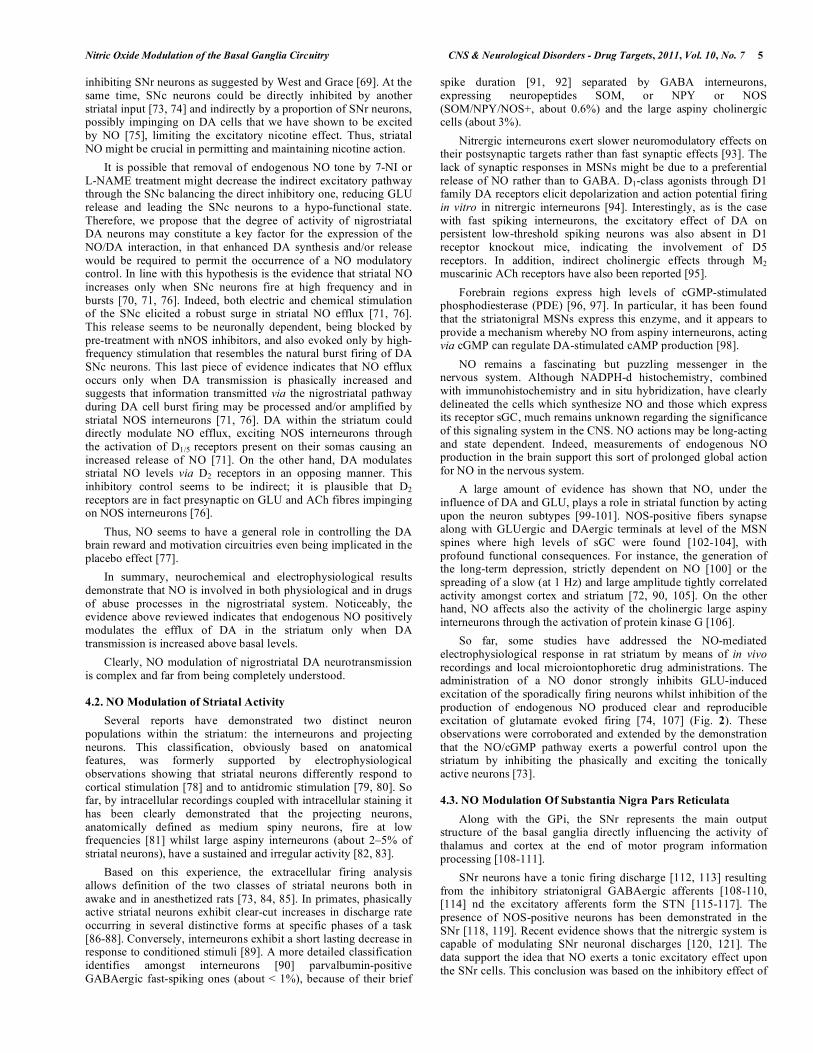
6 CNS & Neurological Disorders - Drug Targets, 2011, Vol. 10, No. 7 Pierucci et al.
Fig. (2). Effect of nitric oxide drugs on the firing rate of striatal neurons in
vivo. Representative rate histogram showing the typical inhibitory effect of
microiontophoretically applied SIN 1 (20–100 nA) (A) and L-NAME (20–
100 nA) (B) on GLU-induced activation of a striatal neuron. Lines and
numbers indicate iontophoretic ejection and currents (in nA). Representative
rate histogram showing that microiontophoretic application of methylene
blue (50 nA, continuous) (C) prevents the responses to SIN 1 (50–100 nA)
on GLU-induced activation of a striatal neuron. Lines and numbers indicate
iontophoretic ejection and currents (in nA). Dashed lines indicate onset and
offset of GLU pulses. GLU currents were constant throughout each
recording. Modified from [74].
the NOS inhibitor, L-NAME locally applied, suggesting that an NO tone is present upon SNr neurons. Furthermore, direct local release (by using an NO donor, i.e. 3-morpholino-syndnonimin-hydrocloride [SIN-1]) of NO caused a statistically significant increase in the firing rate of most of the responsive cells. Thus, based on these findings and on the peculiar physical characteristics of a gaseous neurotransmitter, NO could play a key role in the final stage of the so-called “pattern-model” of BG functioning. The sequenced involvement of the hyper-direct, direct and indirect pathway in the motor program out-flow, implies that at the end of the cycle a large part of the thalamus/cortex should be depressed in
order to reset the system for the other operation. As can be easily argued, the GLU-mediated augmentation of NO level within the SNr trough the STN, placed in the indirect pathway, can mediate the extensive excitation of the BG output, preparing the next step for a motor program selection.
This hypothesis seems to be corroborated by the finding that deep brain stimulation of the STN induced clinical amelioration in a PD patient was associated with a clear cut increase of NO tone within SNr [122].
4.4. NO Modulation of Substantia Subthalamic Nucleus
The NO is well known to play a role in modulating the neuronal activity of STN neurons. In situ hybridization studies have shown that NOS mRNA is expressed in the STN of rodents and humans [27, 123]. This neuro-anatomical evidence, suggesting a putative role for NO in the modulation of STN neuronal activity, has been successively supported by electrophysiological data, showing that NO-active drugs are able to alter the basal firing rates of recorded neurons in the STN. Thus, systemic injection of the selective NOS inhibitor, 7-NI, induced a decrease in basal firing rates of most of the recorded neurons in the STN. The same effect was observed following the local blockade of NOS activity with L-NAME, another NOS inhibitor, applied by microiontophoresis [124, 125]. Consistently, local administration by microiontophoresis of SIN-1 and S-nitroso-glutathione (SNOG), two NO donors, increased the discharge rate of the neurons recorded in vivo in the STN. Although this evidence might mainly indicate a functional involvement of NO in enhancing STN neuronal activity, the modalities through which NO modulates neurotransmission within the STN appear to be more complex. Indeed, the same authors described how a minority of the recorded cells showed an opposite response to the local application of NO-active compounds, both NOS inhibitors (L-NAME) and NO donors (SIN-1, SNOG) [124, 125]. Furthermore, when the drugs were tested on the same neuron, in most of the cases only one of them was able to elicit a response. According to the authors, these observations might indicate that NO regulates neuronal activity within the STN by acting as a neurotransmitter and interacting with other neurotransmitter systems (mainly GLU and GABA) at both pre and post-synaptic levels. This latter aspect of NO action on STN neuronal activity has been supported by some recently published data. Indeed, the magnitude of GABA-induced responses in the STN in vivo was reduced by local co-application of SNOG while, accordingly, it was enhanced by the NOS inhibitor L-NAME. Moreover, both excitatory and inhibitory responses to SNOG and L-NAME, respectively, were reduced by co-application of bicuculline, a selective GABAA receptors blocker, thus showing an involvement of this receptor subtype in the GABA-NO interaction within the STN [126]. Finally, GLUergic afferent fibers arising from the cortex represent an important input to this structure. A functional GLU-NO interaction within the STN has been suggested. Indeed, GLU-induced activation of STN neurons was enhanced by the local co-application of SNOG while it was attenuated by the NOS inhibitor L-NAME [126], thus showing that NO might exert a modulatory action of GLU opposite to GABA. The modulation of the GLUergic neurotransmission by this unorthodox neurotransmitter has long since been shown in other brain structures like the striatum [43]. For example, NO influences neuronal excitability by interacting with regulatory sites on the NMDA receptors, or by inducing calcium-independent release of GLU from synaptic terminals. Finally, NO has been recently shown to play an important role in mediating the activation of ATP sensitive K
+ channels induced by Ca
2+ influx through NMDA-gated
channels [127]. The hyperpolarizing currents activated by ATP sensitive K
+ channels channels opening appear to play a role in
counteracting the excessive neuronal activation induced by GLU release and regulating the firing pattern of STN neurons.

Nitric Oxide Modulation of the Basal Ganglia Circuitry CNS & Neurological Disorders - Drug Targets, 2011, Vol. 10, No. 7 7
4.5. NO Modulation of Substantia Globus Pallidus
The GP represents an important structure of the basal ganglia circuitry. So far, few data have been published showing a functional involvement of NO in modulating the neuronal activity of the neurons within the GP. Electrophysiological recordings in vivo of spontaneously active single neurons in the GP showed that the systemic injection of 7-NI was able to decrease the basal discharge rate in about half of the recorded cells [107]. Consistently, NOS inhibition induced by local administration of L-NAME resulted in a reduction of the firing activity in most of the neurons tested while, accordingly, the NO donor SIN-1 induced an increase of the basal neuronal activity following local ejection from the recording pipette [107]. Thus, these data clearly indicate a functional excitatory role of NO that is likely to be related to a nitrergic modulation of GLUergic neurotransmission within the GP. In fact, the increase of NO transmission has been shown to enhance GLU-induced excitation of GP neuron electrical activity while, consistently, it was reduced by the blockade of NOS activity in the GP [128].
5. INVOLVEMENT OF NO IN NEURODEGENERATION OF
DAERGIC NIGROSTRIATAL SYSTEM
NO is a Janus-faced molecule and, notwithstanding, the exact role (i.e. neuroprotective vs neurotoxic) it plays in neurodegenerative disorders is still ambiguous, with the effect depending on the redox state of the cellular environment. Substantial evidence demonstrates a causative role for NO in the degeneration of DAergic neurons of the nigrostriatal pathway in PD but the mechanism is still unknown and some data are controversial [24, 27, 45, 46, 129-132]. This hypothesis is also supported by studies reporting that NOS inhibitors or NOS gene knock-out significantly mitigate nigral cell loss in animal experimental models [133, 134].
Consistently, NOS polymorphisms which increase expression of astroglial iNOS and nNOS have been reported in PD patients and in different toxin-induced experimental models of PD. Evidence in animal models and in PD patients suggests that NOS is up-stimulated in the BG nuclei and enhanced NO formation takes place after partial injury of the nigrostriatal DAergic system. The exact mechanisms of how NO contributes to neurodegenerative diseases are not completely understood. Multiple lines of evidence indicate that NO is associated with excitotoxicity, DNA damage, and protein modifications, which are common pathogenic mechanisms involved in multiple neurodegenerative diseases [22]. Nevertheless NO, produced by either nNOS or iNOS, plays an important role in DA degeneration. iNOS once induced remains active for several hours to days and produces NO in 1000-fold greater quantities than the constitutive enzyme nNOS. A robust increase in iNOS mRNA levels has been observed after lipopolysaccharide, MPTP or 6-OHDA injection in striatum and SNc [2, 135]. Damage to striatal DAergic fibres seems to be mainly mediated by NO produced by nNOS, while the damage to nigral DAergic neurons is largely inflicted by NO generated by iNOS. Evidence from human post-mortem studies has revealed an increase of NOS mRNA expression only in the MML and in dorsal STN. Instead, a reduction has been shown to occur in the striatum although it was not statistically significant [24]. Such altered activity of NOS neurons of the MML and STN may play a role in the compensatory upregulation of nigrostriatal DAergic neurotransmission in PD, but might also exert an excitotoxic effect on striatal neurons and nigrostriatal terminals.
In animal 6-OHDA-models of PD nNOS expression is reduced while a proportion of nNOS nerve fibres in the striatum are apparently lost following DAergic deafferentation, resulting in a 50% decrease in NOS activity, and depression of the NO-cGMP pathway [136, 137]. In contrast, Gomes et al. [37] showed that 6-OHDA lesion induced a significant increase in NOS cell numbers in the ipsilateral dorsal striatum while a decrease was seen in the ipsilateral SNc and contralateral NAc.
A useful experimental approach to study PD in rats is to induce degeneration of nigrostriatal DA-containing neurons by perfusing 1-methyl-4-phenylpyridinium (MPP
+) into the striatum and to study
striatal DA release for two days by microdialysis, the so called 2-day test-challenge microdialysis method [138]. MPP
+ is
accumulated by DAergic terminals and then retrogradely transported in the cell bodies of DAergic neurons, causing cell degeneration and loss. Short perfusion of MPP
+ induced a
comparable impairment of DAergic striatal nerve terminals, associated with a massive increase in DA efflux 40 min after toxin injection, on day 1. A second challenge with MPP
+, 24 h later,
caused a limited output of extracellular DA in MPP+-lesioned rats,
and this is considered an index of neurotoxin-induced damage. We showed that the inhibition of the NO system by pretreatment with 7-NI can have a protective effect against neuronal damage induced by intrastriatal infusion of MPP
+ [45]. 7-NI given 1 h before
perfusion with the neurotoxins, on day 1, partially restored the cell’s ability to release DA after the second MPP
+ challenge,
showing a protective effect against MPP+ toxic effects on DAergic
neurons.
Inhibition of the NO system by pretreatment with 7-NI has also a protective effect against neuronal damage induced by intra-nigral infusion of 6-OHDA using a neurochemical assay on striatal rat tissues [46]. 7-NI given 1 h before perfusion with neurotoxin partially restored the DA content. Notably, DA levels after 7-NI pretreatment were three times higher than those revealed in the control 6-OHDA-lesioned rats suggesting an important protective effect against the toxin effects on DAergic neurons by NOS inhibition. Further confirming the involvement of NO in the DA SNc death mechanisms, pretreatment with MOL, halved DA levels in the lesioned striata and, most importantly, completely counteracted the neuroprotective effect of 7-NI when co-administrated with it [46]. From these findings, the result that 7-NI counteracts the neurotoxicity induced by 6-OHDA nigral-lesion while MOL worsens it, clearly indicates that the neuroprotective effect of nNOS inhibiton is mainly due to a block in the rise of toxic NO. It is likely that NO induces the formation of peroxynitrite (NO3¯) produced by its combination with hydroxyl radical (˙OH) induced by 6-OHDA [139-141]. Furthermore, NO produces other important mediators of neuronal degeneration in the 6-OHDA model such as semiquinones, peroxinitrite and 6-OHDA quinine, reacting with DA [142] and 6-OHDA [143], respectively.
It is known that 6-OHDA induces degeneration of nigrostriatal DA-containing neurons being transported in the cell bodies of DAergic neurons, causing cell degeneration in a way similar to that found in PD probably by the production of reactive oxygen species [61, 144]. 6-OHDA induced a compelling impairment of the DA nigrostriatal system, associated with a massive decrease of striatal DA and DOPAC levels measured 1 week after the surgery. Although the aetiology of PD is complex, a significant body of data from clinical and experimental models suggests a role for oxidative stress as a causative agent inducing DA neurodegeneration [2, 145] Thus, PD aetiology could be explained by a genetic susceptibility to environmental or endogenous agents, leading to oxidative damage in a neuronal population that is naturally under oxidative stress [146]. The role of oxidative stress in the pathogenesis of MPTP/MPP
+-induced DAergic degeneration, has been suggested
[56, 58, 61, 139-141, 147]. Numerous studies have proposed that nNOS inhibitors, including 7-NI, may reduce DAergic neuronal degeneration, both in vitro and in vivo through antioxidative mechanisms [148-150]. The neuroprotective effect of 7-NI against MPP
+-induced DA striatal depletion is probably due to a block in
the rise of the toxic NO3¯ produced by the combination of NO and ˙OH induced by MPP
+, as shown in previous studies [142, 151].
NO3¯ is a highly reactive molecule, a potent oxidizing agent known to initiate lipid peroxidation in biological membranes, hydroxylation, and nitration of aromatic amino acid residues, and sulfhydryl oxidation of proteins [143, 152]. Nevertheless, 7-NI did

8 CNS & Neurological Disorders - Drug Targets, 2011, Vol. 10, No. 7 Pierucci et al.
not modify extracellular DA output after the first perfusions with MPP
+, suggesting that it did not affect DA uptake or metabolism
[46], a piece of evidence in contrast with findings in other studies [153, 154].
These possible NOS inhibitor neuroprotective mechanisms are consistent with a significant body of data, from clinical and experimental models, suggesting a role for oxidative stress as a causative agent inducing DA neurodegeneration [150, 155-157]. In addition, DA catabolism by MAO induces the formation of hydrogen peroxide, thus rendering DA-containing neurons particularly liable to oxidative stress [152, 155, 158]. Increased nigral DA metabolism, seen in PD, is associated with the production of hydrogen peroxide which, together with iron, may be converted into ˙OH, reacting very rapidly with almost every molecule found in living cells, including DNA, membrane lipids and amino acids [153, 156]. The increased DA turnover could itself enhance basal production of hydrogen peroxide and cause a depletion of reduced glutathione stores, leading to further overproduction of toxic ˙OH, as a consequence of impaired glutathione scavenging activity. Thus, PD aetiology could be explained by a genetic susceptibility to environmental or endogenous agents leading to oxidative damage in a neuronal population that is naturally under oxidative stress [149]. However, other mechanisms by which 7-NI modifies the effects of 6-OHDA cannot be ruled out. For example, numerous studies have proposed that nNOS inhibitors, including 7-NI, may reduce DAergic neuronal degeneration, both in vitro and in vivo through NO-independent mechanisms [146, 154, 159]. Indeed, 7-NI might act as ˙OH scavenger and interfere with oxidative stress caused by MPTP [58]. Thus, this potent antioxidant action of 7-NI might be involved in its neuroprotective effects against 6-OHDA-induced neurotoxicity as well. Furthermore, NO can exert its biological effects through other mechanisms, such as apoptotic cell death induced by 6-OHDA via cGMP, modulating the function of monoamine transporters and S-nitrosylation of receptors [160].
In conclusion, NOS inhibitors have a neuroprotective effect on different toxin-induced nigrostriatal degeneration in rats, although its mechanism of action is still matter for debate. These results therefore provide further support that agents that inhibit the NO system may prove to be of therapeutic benefit against the ongoing loss of DA neurons and motor function that occurs in PD. Although these findings provide a potential link between NO activity and PD, they have to be interpreted with caution, as the PD models in rodents do not reproduce all the neurochemical abnormalities that may contribute to depression in PD. In this complex scenario, where the interaction of several pathogenic pathways concurs to PD, NO seems to represent an important downstream mediator and enhancer of molecular events leading to DAergic neuron death. NO overproduction appears to be an event that significantly contributes to death of DAergic neurons via oxidative damage on cellular lipids, proteins and DNA.
6. NO IMPLICATION ON BG DYSFUNCTION
PD is a disorder essentially characterized by a progressive slowing of movement (akinesia-bradykinesia) and an increased muscle tone (rigidity), frequently associated with resting tremor. Although this clinical description refers only to the motor features of the disease, ignoring the non-motor symptoms (commonly disclosing the disease course), it represents the clinical consequences of the degeneration of SNc DAergic neurons, the anatomo-pathological core of the PD [129, 161].
The DAergic axons of SNc reach the striatum, modulating at this level the processing of movement-related information. DAergic denervation triggers aberrant neuroplastic changes in the striatum influencing the direct and indirect projections to the output stations of the internal segment of the GPi and the SNr. Neurophysiological changes consist, as extensively described in the recent literature, of
abnormally synchronized oscillatory activity at multiple structures of the BG–cortical loop. The widespread BG neurons synchronization (largely in the beta band spectrum) is generally considered a PD hallmark of motor impairment in agreement with its high susceptibility to DAergic therapies [162-164].
It has been proposed that NO modulates motor behaviour [43, 66, 165] and considerable data indicate a direct NO-DA interaction either in normal BG function (as explained above) or in BG related movement disorders such as PD. For instance the DA agonists, cocaine, morphine, substance P or methamphetamine-induced hyperlocomotion could be impeded by NOS inhibitors in mice and rats [166-168]. Yet, inhibition of NOS also induces catalepsy after systemic or intrastriatal injection [65, 66], in agreement with the finding that striatal NOS activity is depressed in parkinsonian animal models [136, 137] and in human PD [24, 169]. NO synthesis seems to be under the control of the phasic/synaptic striatal DA transmission [70, 71] and extracellular increase of NO tone within the GPi, motor putamen and SNr are correlated with transient clinical transition in PD patients [122, 170, 171]. In parkinsonian 6-OHDA-treated rats different striatal responses to NO regarding the different subtypes of neurons can be observed [73]. Spontaneously active neurons are more susceptible to NO and less sensitive to endogenous NO inhibition, likely linked to a reduction of nNOS in DA-denervated striatum [136, 137]. The consequence of the decline of NOS-positive neurons following 6-OHDA denervation could be responsible for a decrease in tone upon spontaneously active neurons that subsequently, by a compensatory mechanism, leads to a more evident response to NO in 6-OHDA-lesioned animals. In agreement, both the expression and the activity of GC have been found to be augmented within striatum of parkinsonian mice [172].
On the other hand, MSNs of DA denervated striatum showed a peculiar dichotomy in relation to NO response, since about 40% MSNs showed a remarkable NO-mediated excitation. Parkinsonian state unmasks a decoupling effect of NO upon a subgroup of projection striatal neurons. This effect could be explained on the basis of a hypothetically different pattern of expression of PDE 1b/4/10 following DA-denervation or more consistently in the light of the topographical segregation of cholinergic interneurons [173, 174]. The latter hypothesis is centred on the increased activity of cholinergic interneurons in DA-lesioned striatum that might facilitate the activity of specific subclass of MSNs [175] by the activation of different subtypes of muscarinic receptors. In other words, parkinsonian state is associated with an abnormal NOergic activity that may also contribute to the pathogenesis of L-DOPA-induced dyskinesias (LID) [176-179].
6.1. Role of NO in L-DOPA-Induced Dyskinesia
The abnormal involuntary movements, or dyskinesia, generated by prolonged administration of L-DOPA represent one of the major challenges facing current therapy for PD [180, 181]. These debilitating motor disturbances are all the more problematic because L-DOPA, in spite of its introduction several decades ago [182], still represents the therapy of choice for the treatment of PD [183]. The discovery of pharmacological interventions able to counteract LID would therefore represent an important breakthrough in the therapy for PD. The design of novel agents for the prevention and treatment of LID requires the elucidation of the adaptive changes produced in the parkinsonian brain by repeated administration of L-DOPA and the assessment of their role in the development and expression of this condition [184]. The translation of antidyskinetic agents into clinically successful drugs has produced limited results yet. The new strategy for antidyskinetic drug discovery is based on non-DAergic adjuncts to L-DOPA. They should hypothetically be able to treat symptoms LID and PD, regulating the aberrant activity of the basal ganglia whilst maintaining the L-DOPA efficacy on motor function. Promising neurotransmitter targets are the noradrenergic, serotonergic, GLUergic, and adenosinergic systems [181, 185]. Recently, some

Nitric Oxide Modulation of the Basal Ganglia Circuitry CNS & Neurological Disorders - Drug Targets, 2011, Vol. 10, No. 7 9
evidence has been also produced in both animal models and in PD patients suggesting a contributory role of NO signalling in LID [178, 179, 186-189]. Hitherto, the study of NO in the effects of L-DOPA has produced conflicting results. Despite the fact that early evidence failed to reveal any L-DOPA-modulation of striatal NO levels [190, 191], a recent microdialysis study showed increased production of NO
3 although with no changes in either NO
3 or
total NO in mice striatum after L-DOPA administration [192].
Recently, more compelling evidence that an over-activity of NO system could contribute to the pathogenesis of LID has been obtained by using rodent-models of the disease [178, 179, 186, 187]. For example, chronic L-DOPA treatment induced FosB expression in the striatum D1 MSNs and in NOS-positive striatal interneurons in rodent PD-models [178, 193]. L-DOPA increased nNOS mRNA levels in the contra- and ipsilateral side to the DA lesion of the frontal cortex [193] but did not produce any further increases of nNOS protein in the striatum compared to the 6-OHDA-induced increase [193]. 7-NI and L-NOARG treatment prevented dyskinesia-induced by L-DOPA [179,186,193] and 7-NI improved motor performance in rats [179,186]. Especially noteworthy is also the evidence that 7-NI sub-chronic administration was devoid of tolerance to the anti-dyskinetic effect [179] differently from its cataleptic action [64], effects on SNc DA cell-population electrophysiological activity and DA striatal release [47]. The hypothesis that raises NO/GC/cGMP pathway activity in the CNS is also supported by some evidence in Parkinsonian patients. Indeed, increased levels of NO second messenger cGMP in serum [194] and NO3
in cerebrospinal fluid [195] have been
detected in PD patients receiving L-DOPA therapy. Nevertheless, the precise role of NO in the pathogenesis of such invalidating complications remains elusive and recent mounting evidence seems to indicate an inhibition of cGMP levels rather than an increase after L-DOPA. For instance, Stefani and colleagues [189] in a microdialysis study in advanced PD patients reported that acute L-DOPA administration in two subjects out of six produced a clear decrease of GPi cGMP levels. In the other four patients basal cGMP levels were not modified by L-DOPA treatment, probably because cGMP basal levels were close to the detection limit [189]. Moreover, L-DOPA did not affect the NO/sGC/cGMP pathway in the mouse MPTP PD model, being capable only of up-regulating the expression and activity of nNOS and GC in normal animal to the level seen in MPTP-treated mice [177]. Moreover, Giorgi and colleagues [196] observed very low levels of cGMP in the cortico-striatal-pallidal loop at the peak of LIDs in rats with experimental hemi-parkinsonism. Interestingly, especially for the possible new treatment applications, pretreatment of the dyskinetic animals with an unselective PDE inhibitor before L-DOPA treatment effectively reduced the severity of the dyskinesias, and partly prevented the decrease of cGMP levels in the GP, putamen and the sensorymotor cortex [196]. These findings have recently been confirmed by Picconi and co-workers [187] using more specific inhibitors for the PDE that specifically acts on cGMP metabolism. Given the above, PDE inhibitors might represent a new pharmacological target of benefit in correcting motor behaviour as well as their potential for reducing or ameliorating dyskinetic behaviour during long-term L-DOPA treatment. Additional experiments are needed to confirm these issues. We are optimistic and the future may disclose important new avenues for the treatment of LIDs. NO and its nucleotide cascade is a promising target that might act on different facets of dyskinesia, such as the impairment of striatal plasticity and non-DA alterations. The role that cGMP-regulated PDEs play in mediating other NO actions deserves further study. Hitherto, this attractive hypothesis has not been validated by either non-human primates or humans studies.
The whole spectrum of NO implication on parkinsonian BG activity strongly suggest that in the near future a new molecule able to modify NOergic pathway will be a new possible therapeutic approach either to motor impairment or in the control of
complications of long-term exposure to L-DOPA therapy in patients with PD.
CONCLUDING REMARKS
Abundant experimental evidence points to complex effects of NO in the BG circuits, both directly and via interactions with the DAergic, GLUergic and GABAergic systems and its dysfunction is involved in the pathophysiology of PD and other motor disorders. In recent years, NO and its reactive metabolites have been proposed as important actors in the processes leading to neuronal cell death in PD both in terms of pro-oxidants and mediators of inflammatory responses. Therefore, the blockage of NO synthesis or the scavenging of nitrogen reactive species could represent an efficient tool against PD progression and/or prevention. Moreover, numerous studies point to nitrergic system as a new potential pharmacologic approach for the motor manifestations of PD and adjuvant to L-DOPA therapy. In addition, NO at the levels of limbic structure may also contribute to the co-morbid depression in these patients.
It is reasonable to believe that in a few years, increased understanding regarding the role of NO in the physiopathology of circuitry changes in the BG will concur in order to set a rational basis for NO-based therapies that are designed not simply to manage disabling dyskinesia but to prevent its induction. Strategies intended to halt or at least reduce the pace of nigrostriatal loss would be likely to change the path that leads to PD, motor fluctuations and LID.
ACKNOWLEDGEMENTS
This study was supported in part by University of Malta research funding, coordinator G. Di Giovanni.
ABBREVIATIONS
DAT = Dopamine transporter
EP = Entopeduncular nucleus
GSH = Glutathione
LDTg = Lateraldorsal tegmental nucleus
LPS = Lipopolysaccharide
NAc = Nucleus accumbens
oPFC = Orbital prefrontal cortex
PV = Parvalbumine
PPN = Pedunculopontine nucleus
SMA = Supplementary motor cortex
MPTP = 1-Methyl 4-phenyl 1,2,3,6-tetrahydropyridine
MPP+ = 1-Methyl-4-phenilpyridinium ion
DOPAC = 3,4-Dihydroxy-phenylacetic acid
SIN-1 = 3-Morpholino-syndnonimin-hydrocloride
6-OHDA = 6-Hydroxydopamine
7-NI = 7-nitro-indazolone
ACh = Acetylcholine
BG = Basal ganglia
CNS = Central nervous system
cGMP = Cyclic guanosine monophosphate
DA = Dopamine
DAergic = Dopaminergic
eNOS = Endothelial nitric oxide synthase
GP = Globus pallidus
GPe = Globus pallidus/ external segment

10 CNS & Neurological Disorders - Drug Targets, 2011, Vol. 10, No. 7 Pierucci et al.
GPi = Globus pallidus/ internal segment
GLU = Glutamate
GLUergic = Glutamatergic
˙OH = Hydroxyl radical
iNOS = Inducible nitric oxide synthase
GPi = Internal segment of the globus pallidus
L-ARG = L-arginine
LID = L-DOPA-induced dyskinesia
L-NOARG = L-nitro-arginine
MML = Medial medullary lamina
MSNs = Medium spiny neurons
MOL = Molsidomine
MAO = Monoamine oxidase
NADPH-d = NADPH-diaphorase
nNOS = Neuronal nitric oxide synthase
NPY = Neuropeptide Y
NADPH = Nicotinamide adenine dinucleotide phosphate
NOS = Nitric oxide synthase
NO = Nitric oxide
NMDA = N-Methyl-D-aspartate
L-NAME = N -nitro-L-arginine methyl ester
PD = Parkinson’s disease
PPT = Pedunculopontine tegmental nucleus
NO3¯ = Peroxynitrite
PDE = Phosphodiesterase
SNOG = S-Nitroso-glutathione
sGC = Soluble guanylyl cyclase
SOM = Somatostatin
SNc = Substantia nNigra pars compacta
SNr = Substantia nigra pars reticulatereticulata
SN = Substantia nigra
STN = The subthalamic nucleus
TH = Tyrosine Hydroxylase
VTA = Ventral tegmental area
GABA = -aminobutyric acid
REFERENCES
[1] Furchgott, R.F.; Zawadzki, J.V. The obligatory role of endothelial
cells in the relaxation of arterial smooth muscle by acetylcholine. Nature, 1980, 288, 373-376.
[2] Bian, K.; Murad, F. Nitric oxide (NO)--biogeneration, regulation, and relevance to human diseases. Front. Biosci., 2003, 8, d264-278.
[3] Dawson, V.L.; Dawson, T.M. Nitric oxide in neurodegeneration. Prog. Brain Res., 1998, 118, 215-229.
[4] Arnold, W.P.; Mittal, C.K.; Katsuki, S.; Murad, F. Nitric oxide activates guanylate cyclase and increases guanosine 3':5'-cyclic
monophosphate levels in various tissue preparations. Proc. Natl. Acad. Sci. USA, 1977, 74, 3203-3207.
[5] Bredt, D.S.; Hwang, P.M.; Snyder, S.H. Localization of nitric oxide synthase indicating a neural role for nitric oxide. Nature, 1990,
347, 768-770. [6] Choi, Y.B.; Lipton, S.A. Redox modulation of the NMDA receptor.
Cell. Mol. Life Sci., 2000, 57, 1535-1541. [7] Graybiel, A.M.; Canales, J.J. The neurobiology of repetitive
behaviors: clues to the neurobiology of Tourette syndrome. Adv. Neurol., 2001, 85, 123-131.
[8] Mink, J.W.; Thach, W.T. Basal ganglia intrinsic circuits and their
role in behavior. Curr. Opin. Neurobiol., 1993, 3, 950-957. [9] Wichmann, T.; Delong, M.R. Anatomy and physiology of the basal
ganglia: relevance to Parkinson's disease and related disorders. Handb. Clin. Neurol., 2007, 83, 1-18.
[10] Mena-Segovia, J.; Bolam, J.P.; Magill, P.J. Pedunculopontine nucleus and basal ganglia: distant relatives or part of the same
family? Trends Neurosci., 2004, 27, 585-588. [11] DeLong, M.R.; Wichmann, T. Circuits and circuit disorders of the
basal ganglia. Arch. Neurol., 2007, 64, 20-24. [12] Flaherty, A.W.; Graybiel, A.M. Corticostriatal transformations in
the primate somatosensory system. Projections from physiologically mapped body-part representations. J.
Neurophysiol., 1991, 66, 1249-1263. [13] DeLong, M.R. Primate models of movement disorders of basal
ganglia origin. Trends Neurosci., 1990, 13, 281-285. [14] Alexander, G.E.; Crutcher, M.D.; DeLong, M.R. Basal ganglia-
thalamocortical circuits: parallel substrates for motor, oculomotor, "prefrontal" and "limbic" functions. Prog. Brain Res., 1990, 85,
119-146. [15] Parent, A.; Carpenter, M.B. Carpenter's Human Neuroanatomy;
9th ed.; Williams & Wilkins; Baltimore, 1996. [16] Joel, D.; Weiner, I. The connections of the dopaminergic system
with the striatum in rats and primates: an analysis with respect to the functional and compartmental organization of the striatum.
Neuroscience, 2000, 96, 451-474. [17] Kropotov, J.D.; Etlinger, S.C. Selection of actions in the basal
ganglia-thalamocortical circuits: review and model. Int. J. Psychophysiol., 1999, 31, 197-217.
[18] Grillner, S.; Hellgren, J.; Menard, A.; Saitoh, K.; Wikstrom, M.A. Mechanisms for selection of basic motor programs: roles for the
striatum and pallidum. Trends Neurosci., 2005, 28, 364-370. [19] Blair, R.J. Facial expressions, their communicatory functions and
neuro-cognitive substrates. Philos. Trans. R. Soc. Lond. B. Biol. Sci., 2003, 358, 561-572.
[20] Reynolds, J.N.; Hyland, B.I.; Wickens, J.R. A cellular mechanism of reward-related learning. Nature, 2001, 413, 67-70.
[21] Pisani, A.; Centonze, D.; Bernardi, G.; Calabresi, P. Striatal synaptic plasticity: implications for motor learning and Parkinson's
disease. Mov. Disord., 2005, 20, 395-402. [22] Del-Bel, E.A.; Bermúdez-Echeverry, M.; Salum, C.; Raisman-
Vozari, R. In: The Basal Ganglia Pathophysiology: Recent Advances. Di Giovanni, G., Ed.; Transworld Research Network:
Kerala, 2007, pp. 129-158. [23] Egberongbe, Y.I.; Gentleman, S.M.; Falkai, P.; Bogerts, B.; Polak,
J.M.; Roberts, G.W. The distribution of nitric oxide synthase immunoreactivity in the human brain. Neuroscience, 1994, 59, 561-
578. [24] Eve, D.J.; Nisbet, A.P.; Kingsbury, A.E.; Hewson, E.L.; Daniel,
S.E.; Lees, A.J.; Marsden, C.D.; Foster, O.J. Basal ganglia neuronal nitric oxide synthase mRNA expression in Parkinson's disease.
Brain Res. Mol. Brain Res., 1998, 63, 62-71. [25] Garthwaite, J.; Boulton, C.L. Nitric oxide signaling in the central
nervous system. Annu. Rev. Physiol., 1995, 57, 683-706. [26] Leontovich, T.A.; Mukhina, Y.K.; Fedorov, A.A. Neurons of the
basal ganglia of the human brain (striatum and basolateral amygdala) expressing the enzyme NADPH-d. Neurosci. Behav.
Physiol., 2004, 34, 277-286. [27] Nisbet, A.P.; Foster, O.J.; Kingsbury, A.; Lees, A.J.; Marsden,
C.D. Nitric oxide synthase mRNA expression in human subthalamic nucleus, striatum and globus pallidus: implications for
basal ganglia function. Brain Res. Mol. Brain Res., 1994, 22, 329-332.
[28] Vincent, S.R.; Kimura, H. Histochemical mapping of nitric oxide synthase in the rat brain. Neuroscience, 1992, 46, 755-784.
[29] Johnson, M.D.; Ma, P.M. Localization of NADPH diaphorase activity in monoaminergic neurons of the rat brain. J. Comp.
Neurol., 1993, 332, 391-406. [30] Gonzalez-Hernandez, T.; Rodriguez, M. Compartmental
organization and chemical profile of dopaminergic and GABAergic neurons in the substantia nigra of the rat. J. Comp. Neurol., 2000,
421, 107-135. [31] Govsa, F.; Kayalioglu, G. Relationship between nicotinamide
adenine dinucleotide phosphate-diaphorase-reactive neurons and blood vessels in basal ganglia. Neuroscience, 1999, 93, 1335-1337.

Nitric Oxide Modulation of the Basal Ganglia Circuitry CNS & Neurological Disorders - Drug Targets, 2011, Vol. 10, No. 7 11
[32] Niijima, K.; Yoshida, M. Activation of mesencephalic dopamine neurons by chemical stimulation of the nucleus tegmenti pedunculopontinus pars compacta. Brain Res., 1988, 451, 163-171.
[33] Vincent, S.R.; Satoh, K.; Armstrong, D.M.; Panula, P.; Vale, W.; Fibiger, H.C. Neuropeptides and NADPH-diaphorase activity in the ascending cholinergic reticular system of the rat. Neuroscience, 1986, 17, 167-182.
[34] Klejbor, I.; Domaradzka-Pytel, B.; Ludkiewicz, B.; WÃ jcik, S.; MoryÅ, J. The relationships between neurons containing dopamine and nitric oxide synthase in the ventral tegmental area. Folia Histochem. Cytobiol., 2004, 42, 83-87.
[35] Gonzalez-Hernandez, T.; Afonso-Oramas, D.; Cruz-Muros, I. Phenotype, compartmental organization and differential vulnerability of nigral dopaminergic neurons. J. Neural Transm. Suppl., 2009, 21-37.
[36] Del Bel, E.A.; Guimaraes, F.S. Sub-chronic inhibition of nitric-oxide synthesis modifies haloperidol-induced catalepsy and the number of NADPH-diaphorase neurons in mice. Psychopharmacology, 2000, 147, 356-361.
[37] Gomes, M.Z.; Del Bel, E.A. Effects of electrolytic and 6-hydroxydopamine lesions of rat nigrostriatal pathway on nitric oxide synthase and nicotinamide adenine dinucleotide phosphate diaphorase. Brain Res. Bull., 2003, 62, 107-115.
[38] Chalimoniuk, M.; Lukacova, N.; Marsala, J.; Langfort, J. Alterations of the expression and activity of midbrain nitric oxide synthase and soluble guanylyl cyclase in 1-methyl-4-phenyl-1,2,3,6-tetrahydropyridine-induced Parkinsonism in mice. Neuroscience, 2006, 141, 1033-1046.
[39] He, Y.; Imam, S.Z.; Dong, Z.; Jankovic, J.; Ali, S.F.; Appel, S.H.; Le, W. Role of nitric oxide in rotenone-induced nigro-striatal injury. J. Neurochem., 2003, 86, 1338-1345.
[40] Barthwal, M.K.; Srivastava, N.; Dikshit, M. Role of nitric oxide in a progressive neurodegeneration model of Parkinson's disease in the rat. Redox Rep., 2001, 6, 297-302.
[41] Bernacer, J.; Prensa, L.; Gimenez-Amaya, J.M. Morphological features, distribution and compartmental organization of the nicotinamide adenine dinucleotide phosphate reduced-diaphorase interneurons in the human striatum. J. Comp. Neurol., 2005, 489, 311-327.
[42] Johannes, S.; Reif, A.; Senitz, D.; Riederer, P.; Lauer, M. NADPH-diaphorase staining reveals new types of interneurons in human putamen. Brain Res., 2003, 980, 92-99.
[43] West, A.R.; Galloway, M.P.; Grace, A.A. Regulation of striatal dopamine neurotransmission by nitric oxide: effector pathways and signaling mechanisms. Synapse, 2002, 44, 227-245.
[44] Di Matteo, V.; Pierucci, M.; Benigno, A.; Esposito, E.; Crescimanno, G.; Di Giovanni, G. Critical role of nitric oxide on nicotine-induced hyperactivation of dopaminergic nigrostriatal system: electrophysiological and neurochemical evidence in rats. CNS Neurosci. Ther., 2010, 16, 127-136.
[45] Di Matteo, V.; Benigno, A.; Pierucci, M.; Giuliano, D.A.; Crescimanno, G.; Esposito, E.; Di Giovanni, G. 7-nitroindazole protects striatal dopaminergic neurons against MPP+-induced degeneration: an in vivo microdialysis study. Ann. NY Acad. Sci., 2006, 1089, 462-471.
[46] Di Matteo, V.; Pierucci, M.; Benigno, A.; Crescimanno, G.; Esposito, E.; Di Giovanni, G. Involvement of nitric oxide in nigrostriatal dopaminergic system degeneration: a neurochemical study. Ann. NY Acad. Sci., 2009, 1155, 309-315.
[47] Di Matteo, V.; Pierucci, M.; Benigno, A.; Orban, G.; Crescimanno, G.; Esposito, E.; Di Giovanni, G. Electrophysiological and neurochemical characterization of 7-nitroindazole and molsidomine acute and sub-chronic administration effects in the dopaminergic nigrostrial system in rats. J. Neural Transm. Suppl., 2009, 173-182.
[48] West, A.R.; Grace, A.A. Striatal nitric oxide signaling regulates the neuronal activity of midbrain dopamine neurons in vivo. J. Neurophysiol., 2000, 83, 1796-1808.
[49] Trabace, L.; Kendrick, K.M. Nitric oxide can differentially modulate striatal neurotransmitter concentrations via soluble guanylate cyclase and peroxynitrite formation. J. Neurochem., 2000, 75, 1664-1674.
[50] Buyukuysal, R.L. Effect of nitric oxide donors on endogenous dopamine release from rat striatal slices. I: Requirement to antioxidants in the medium. Fundam. Clin. Pharmacol., 1997, 11, 519-527.
[51] Cox, B.A.; Johnson, S.W. Nitric oxide facilitates N-methyl-D-aspartate-induced burst firing in dopamine neurons from rat midbrain slices. Neurosci. Lett., 1998, 255, 131-134.
[52] Schilstrom, B.; Mameli-Engvall, M.; Rawal, N.; Grillner, P.; Jardemark, K.; Svensson, T.H. Nitric oxide is involved in nicotine-induced burst firing of rat ventral tegmental area dopamine neurons. Neuroscience, 2004, 125, 957-964.
[53] Di Matteo, V.; Pierucci, M.; Di Giovanni, G.; Benigno, A.; Esposito, E. The neurobiological bases for the pharmacotherapy of nicotine addiction. Curr. Pharm. Des., 2007, 13, 1269-1284.
[54] Southan, G.J.; Szabo, C. Selective pharmacological inhibition of distinct nitric oxide synthase isoforms. Biochem. Pharmacol., 1996, 51, 383-394.
[55] Meyer, R.C.; Spangler, E.L.; Patel, N.; London, E.D.; Ingram, D.K. Impaired learning in rats in a 14-unit T-maze by 7-nitroindazole, a neuronal nitric oxide synthase inhibitor, is attenuated by the nitric oxide donor, molsidomine. Eur. J. Pharmacol., 1998, 341, 17-22.
[56] Castagnoli, K.; Palmer, S.; Anderson, A.; Bueters, T.; Castagnoli Jr, N. The neuronal nitric oxide synthase inhibitor 7-nitroindazole also inhibits the monoamine oxidase-B-catalyzed oxidation of 1 -methyl-4-phenyl- 1,2,3,6-tetrahydropyridine. Chem. Res. Toxicol., 1997, 10, 364-368.
[57] Boireau, A.; Dubedat, P.; Bordier, F.; Imperato, A.; Moussaoui, S. The protective effect of riluzole in the MPTP model of Parkinson's disease in mice is not due to a decrease in MPP+ accumulation. Neuropharmacology, 2000, 39, 1016-1020.
[58] Thomas, B.; Saravanan, K.S.; Mohanakumar, K.P. In vitro and in
vivo evidences that antioxidant action contributes to the neuroprotective effects of the neuronal nitric oxide synthase and monoamine oxidase-B inhibitor, 7-nitroindazole. Neurochem. Int., 2008, 52, 990-1001.
[59] Desvignes, C.; Bert, L.; Vinet, L.; Denoroy, L.; Renaud, B.; Lambas-Senas, L. Evidence that the neuronal nitric oxide synthase inhibitor 7-nitroindazole inhibits monoamine oxidase in the rat: in vivo effects on extracellular striatal dopamine and 3,4-dihydroxyphenylacetic acid. Neurosci. Lett., 1999, 261, 175-178.
[60] Mercuri, N.B.; Bonci, A.; Siniscalchi, A.; Stefani, A.; Calabresi, P.; Bernardi, G. Electrophysiological effects of monoamine oxidase inhibition on rat midbrain dopaminergic neurones: an in vitro study. Br. J. Pharmacol., 1996, 117, 528-532.
[61] Simola, N.; Morelli, M.; Carta, A.R. The 6-hydroxydopamine model of Parkinson's disease. Neurotox. Res., 2007, 11, 151-167.
[62] Del Bel, E.A.; da Silva, C.A.; Guimaraes, F.S. Catalepsy induced by nitric oxide synthase inhibitors. Gen. Pharmacol., 1998, 30, 245-248.
[63] Del Bel, E.A.; da Silva, C.A.; Guimaraes, F.S.; Bermudez-Echeverry, M. Catalepsy induced by intra-striatal administration of nitric oxide synthase inhibitors in rats. Eur. J. Pharmacol., 2004, 485, 175-181.
[64] Del Bel, E.A.; Souza, A.S.; Guimaraes, F.S.; da-Silva, C.A.; Nucci-da-Silva, L.P. Motor effects of acute and chronic inhibition of nitric oxide synthesis in mice. Psychopharmacology, 2002, 161, 32-37.
[65] Marras, R.A.; Martins, A.P.; Del Bel, E.A.; Guimaraes, F.S. L-NOARG, an inhibitor of nitric oxide synthase, induces catalepsy in mice. Neuroreport, 1995, 7, 158-160.
[66] Del Bel, E.A.; Guimaraes, F.S.; Bermudez-Echeverry, M.; Gomes, M.Z.; Schiaveto-de-souza, A.; Padovan-Neto, F.E.; Tumas, V.; Barion-Cavalcanti, A.P.; Lazzarini, M.; Nucci-da-Silva, L.P.; de Paula-Souza, D. Role of nitric oxide on motor behavior. Cell. Mol.
Neurobiol., 2005, 25, 371-392. [67] Del-Bel, E.A.; Guimaraes, F.S.; Joca, S.R.; Echeverry, M.B.;
Ferreira, F.R. Tolerance to the cataleptic effect that follows repeated nitric oxide synthase inhibition may be related to functional enzymatic recovery. J. Psychopharmacol. (Oxford, England), 2010, 24, 397-405.
[68] Sugaya, K.; McKinney, M. Nitric oxide synthase gene expression in cholinergic neurons in the rat brain examined by combined immunocytochemistry and in situ hybridization histochemistry. Brain Res. Mol. Brain Res., 1994, 23, 111-125.
[69] West, A.R.; Grace, A.A. Striatal nitric oxide signaling regulates the neuronal activity of midbrain dopamine neurons in vivo. J.
Neurophysiol., 2000, 83, 1796-1808. [70] Sammut, S.; Bray, K.E.; West, A.R. Dopamine D2 receptor-
dependent modulation of striatal NO synthase activity. Psychopharmacology, 2007, 191, 793-803.

12 CNS & Neurological Disorders - Drug Targets, 2011, Vol. 10, No. 7 Pierucci et al.
[71] Sammut, S.; Dec, A.; Mitchell, D.; Linardakis, J.; Ortiguela, M.;
West, A.R. Phasic dopaminergic transmission increases NO efflux in the rat dorsal striatum via a neuronal NOS and a dopamine
D(1/5) receptor-dependent mechanism. Neuropsychopharmacology, 2006, 31, 493-505.
[72] Sammut, S.; Park, D.J.; West, A.R. Frontal cortical afferents facilitate striatal nitric oxide transmission in vivo via a NMDA
receptor and neuronal NOS-dependent mechanism. J. Neurochem., 2007, 103, 1145-1156.
[73] Galati, S.; D'Angelo, V.; Scarnati, E.; Stanzione, P.; Martorana, A.; Procopio, T.; Sancesario, G.; Stefani, A. In vivo electrophysiology
of dopamine-denervated striatum: focus on the nitric oxide/cGMP signaling pathway. Synapse, 2008, 62, 409-420.
[74] Di Giovanni, G.; Ferraro, G.; Sardo, P.; Galati, S.; Esposito, E.; La Grutta, V. Nitric oxide modulates striatal neuronal activity via
soluble guanylyl cyclase: an in vivo microiontophoretic study in rats. Synapse, 2003, 48, 100-107.
[75] Di Giovanni, G.; Ferraro, G.; Sardo, P.; Di Maio, R.; Carletti, F.; La Grutta, V. Microiontophoretic evidence that nitric oxide alters
spontaneous activity of the substantia nigra pars reticulata neurons in the rat. Acta Physiol., 2006, 188, P184.
[76] Sammut, S.; Bray, K.E.; West, A.R. Dopamine D2 receptor-dependent modulation of striatal NO synthase activity.
Psychopharmacology, 2007, 191, 793-803. [77] Fricchione, G.; Stefano, G.B. Placebo neural systems: nitric oxide,
morphine and the dopamine brain reward and motivation circuitries. Med. Sci. Monit., 2005, 11, MS54-MS65.
[78] Nisenbaum, E.S.; Orr, W.B.; Berger, T.W. Evidence for two functionally distinct subpopulations of neurons within the rat
striatum. J. Neurosci., 1988, 8, 4138-4150. [79] Ryan, L.J.; Young, S.J.; Segal, D.S.; Groves, P.M. Antidromically
identified striatonigral projection neurons in the chronically implanted behaving rat: relations of cell firing to amphetamine-
induced behaviors. Behav. Neurosci., 1989, 103, 3-14. [80] Kimura, M.; Kato, M.; Shimazaki, H. Physiological properties of
projection neurons in the monkey striatum to the globus pallidus. Exp. Brain Res., 1990, 82, 672-676.
[81] Wilson, C.J.; Chang, H.T.; Kitai, S.T. Firing patterns and synaptic potentials of identified giant aspiny interneurons in the rat
neostriatum. J. Neurosci., 1990, 10, 508-519. [82] Wilson, C.J.; Groves, P.M. Spontaneous firing patterns of
identified spiny neurons in the rat neostriatum. Brain Res., 1981, 220, 67-80.
[83] Kitai, S.T.; Surmeier, D.J. Cholinergic and dopaminergic modulation of potassium conductances in neostriatal neurons. Adv.
Neurol., 1993, 60, 40-52. [84] Kish, L.J.; Palmer, M.R.; Gerhardt, G.A. Multiple single-unit
recordings in the striatum of freely moving animals: effects of apomorphine and D-amphetamine in normal and unilateral 6-
hydroxydopamine-lesioned rats. Brain Res., 1999, 833, 58-70. [85] Chen, M.T.; Morales, M.; Woodward, D.J.; Hoffer, B.J.; Janak,
P.H. In vivo extracellular recording of striatal neurons in the awake rat following unilateral 6-hydroxydopamine lesions. Exp. Neurol.,
2001, 171, 72-83. [86] DeLong, M.R. Putamen: activity of single units during slow and
rapid arm movements. Science, 1973, 179, 1240-1242. [87] Crutcher, M.D.; DeLong, M.R. Single cell studies of the primate
putamen. I. Functional organization. Exp. Brain Res., 1984, 53, 233-243.
[88] Alexander, G.E.; DeLong, M.R. Microstimulation of the primate neostriatum. II. Somatotopic organization of striatal microexcitable
zones and their relation to neuronal response properties. J. Neurophysiol., 1985, 53, 1417-1430.
[89] Kimura, M.; Rajkowski, J.; Evarts, E. Tonically discharging putamen neurons exhibit set-dependent responses. Proc. Natl.
Acad. Sci. USA, 1984, 81, 4998-5001. [90] Mallet, N.; Ballion, B.; Le Moine, C.; Gonon, F. Cortical inputs
and GABA interneurons imbalance projection neurons in the striatum of parkinsonian rats. J. Neurosci., 2006, 26, 3875-3884.
[91] Kawaguchi, Y. Physiological, morphological, and histochemical characterization of three classes of interneurons in rat neostriatum.
J. Neurosci., 1993, 13, 4908-4923. [92] Kawaguchi, Y.; Aosaki, T.; Kubota, Y. Cholinergic and
GABAergic interneurons in the striatum. Nihon Shinkei Seishin Yakurigaku Zasshi, 1997, 17, 87-90.
[93] Gittis, A.H.; Nelson, A.B.; Thwin, M.T.; Palop, J.J.; Kreitzer, A.C.
Distinct roles of GABAergic interneurons in the regulation of striatal output pathways. J. Neurosci., 2010, 30, 2223-2234.
[94] Centonze, D.; Bracci, E.; Pisani, A.; Gubellini, P.; Bernardi, G.; Calabresi, P. Activation of dopamine D1-like receptors excites LTS
interneurons of the striatum. Eur. J. Neurosci., 2002, 15, 2049-2052.
[95] Bernard, V.; Laribi, O.; Levey, A.I.; Bloch, B. Subcellular redistribution of m2 muscarinic acetylcholine receptors in striatal
interneurons in vivo after acute cholinergic stimulation. J. Neurosci., 1998, 18, 10207-10218.
[96] Repaske, D.R.; Corbin, J.G.; Conti, M.; Goy, M.F. A cyclic GMP-stimulated cyclic nucleotide phosphodiesterase gene is highly
expressed in the limbic system of the rat brain. Neuroscience, 1993, 56, 673-686.
[97] Van Staveren, W.C.; Steinbusch, H.W.; Markerink-Van Ittersum, M.; Repaske, D.R.; Goy, M.F.; Kotera, J.; Omori, K.; Beavo, J.A.;
De Vente, J. mRNA expression patterns of the cGMP-hydrolyzing phosphodiesterases types 2, 5, and 9 during development of the rat
brain. J. Comp. Neurol., 2003, 467, 566-580. [98] Lin, C.S. Phosphodiesterase type 5 regulation in the penile corpora
cavernosa. J. Sex. Med., 2009, 6(Suppl 3), 203-209. [99] Kawaguchi, Y. Neostriatal cell subtypes and their functional roles.
Neurosci. Res., 1997, 27, 1-8. [100] Calabresi, P.; Gubellini, P.; Centonze, D.; Sancesario, G.; Morello,
M.; Giorgi, M.; Pisani, A.; Bernardi, G. A critical role of the nitric oxide/cGMP pathway in corticostriatal long-term depression. J.
Neurosci., 1999, 19, 2489-2499. [101] West, A.R.; Grace, A.A. Opposite influences of endogenous
dopamine D1 and D2 receptor activation on activity states and electrophysiological properties of striatal neurons: studies
combining in vivo intracellular recordings and reverse microdialysis. J. Neurosci., 2002, 22, 294-304.
[102] Ariano, M.A. Distribution of components of the guanosine 3',5'-phosphate system in rat caudate-putamen. Neuroscience, 1983, 10,
707-723. [103] Morello, M.; Reiner, A.; Sancesario, G.; Karle, E.J.; Bernardi, G.
Ultrastructural study of nitric oxide synthase-containing striatal neurons and their relationship with parvalbumin-containing
neurons in rats. Brain Res., 1997, 776, 30-39. [104] Sancesario, G.; Morello, M.; Reiner, A.; Giacomini, P.; Massa, R.;
Schoen, S.; Bernardi, G. Nitrergic neurons make synapses on dual-input dendritic spines of neurons in the cerebral cortex and the
striatum of the rat: implication for a postsynaptic action of nitric oxide. Neuroscience, 2000, 99, 627-642.
[105] Mallet, N.; Le Moine, C.; Charpier, S.; Gonon, F. Feedforward inhibition of projection neurons by fast-spiking GABA
interneurons in the rat striatum in vivo. J. Neurosci., 2005, 25, 3857-3869.
[106] Centonze, D.; Pisani, A.; Bonsi, P.; Giacomini, P.; Bernardi, G.; Calabresi, P. Stimulation of nitric oxide-cGMP pathway excites
striatal cholinergic interneurons via protein kinase G activation. J. Neurosci., 2001, 21, 1393-1400.
[107] Sardo, P.; Ferraro, G.; Di Giovanni, G.; Galati, S.; La Grutta, V. Influence of nitric oxide on the spontaneous activity of globus
pallidus neurones in the rat. J. Neural Transm., 2002, 109, 1373-1389.
[108] Albin, R.L.; Aldridge, J.W.; Young, A.B.; Gilman, S. Feline subthalamic nucleus neurons contain glutamate-like but not
GABA-like or glycine-like immunoreactivity. Brain Res., 1989, 491, 185-188.
[109] Graybiel, A.M. Network-level neuroplasticity in cortico-basal ganglia pathways. Parkinsonism Relat. Disord., 2004, 10, 293-296.
[110] Smith, G.S.; Price, J.C.; Lopresti, B.J.; Huang, Y.; Simpson, N.; Holt, D.; Mason, N.S.; Meltzer, C.C.; Sweet, R.A.; Nichols, T.;
Sashin, D.; Mathis, C.A. Test-retest variability of serotonin 5-HT2A receptor binding measured with positron emission
tomography and [18F] altanserin in the human brain. Synapse, 1998, 30, 380-392.
[111] Wichmann, T.; DeLong, M.R. Pathophysiology of Parkinson's disease: the MPTP primate model of the human disorder. Ann. NY
Acad. Sci., 2003, 991, 199-213. [112] Atherton, J.F.; Bevan, M.D. Ionic mechanisms underlying
autonomous action potential generation in the somata and dendrites of GABAergic substantia nigra pars reticulata neurons in vitro. J.
Neurosci., 2005, 25, 8272-8281.

Nitric Oxide Modulation of the Basal Ganglia Circuitry CNS & Neurological Disorders - Drug Targets, 2011, Vol. 10, No. 7 13
[113] Richards, C.D.; Shiroyama, T.; Kitai, S.T. Electrophysiological and
immunocytochemical characterization of GABA and dopamine neurons in the substantia nigra of the rat. Neuroscience, 1997, 80,
545-557. [114] Smith, Y.; Bolam, J.P. Convergence of synaptic inputs from the
striatum and the globus pallidus onto identified nigrocollicular cells in the rat: a double anterograde labelling study. Neuroscience,
1991, 44, 45-73. [115] Iribe, Y.; Moore, K.; Pang, K.C.; Tepper, J.M. Subthalamic
stimulation-induced synaptic responses in substantia nigra pars compacta dopaminergic neurons in vitro. J. Neurophysiol., 1999,
82, 925-933. [116] Nakanishi, H.; Kita, H.; Kitai, S.T. Intracellular study of rat
substantia nigra pars reticulata neurons in an in vitro slice preparation: electrical membrane properties and response
characteristics to subthalamic stimulation. Brain Res., 1987, 437, 45-55.
[117] Robledo, P.; Feger, J. Excitatory influence of rat subthalamic nucleus to substantia nigra pars reticulata and the pallidal complex:
electrophysiological data. Brain Res., 1990, 518, 47-54. [118] Ibanez-Sandoval, O.; Carrillo-Reid, L.; Galarraga, E.; Tapia, D.;
Mendoza, E.; Gomora, J.C.; Aceves, J.; Bargas, J. Bursting in substantia nigra pars reticulata neurons in vitro: possible relevance
for Parkinson disease. J. Neurophysiol., 2007, 98, 2311-2323. [119] Beurrier, C.; Congar, P.; Bioulac, B.; Hammond, C. Subthalamic
nucleus neurons switch from single-spike activity to burst-firing mode. J. Neurosci., 1999, 19, 599-609.
[120] Di Giovanni, G.; Ferraro, G.; Sardo, P.; Di Maio, R.; Carletti, F.; La Grutta, V. Microiontophoretic Evidence that Nitric Oxide Alters
Spontaneous Activity of the Substantia Nigra Pars Reticulata Neurons in the Rat. Acta Physiol., 2006, 188, P184.
[121] Carletti, F.; Ferraro, G.; Rizzo, V.; D'Agostino, S.; Lonobile, G.; Sardo, P. Nitric oxide- and cGMP-active compounds affect the
discharge of substantia nigra pars reticulata neurons: in vivo evidences in the rat. J. Neural Transm., 2009, 116, 539-549.
[122] Galati, S.; Mazzone, P.; Fedele, E.; Pisani, A.; Peppe, A.; Pierantozzi, M.; Brusa, L.; Tropepi, D.; Moschella, V.; Raiteri, M.;
Stanzione, P.; Bernardi, G.; Stefani, A. Biochemical and electrophysiological changes of substantia nigra pars reticulata
driven by subthalamic stimulation in patients with Parkinson's disease. Eur. J. Neurosci., 2006, 23, 2923-2928.
[123] Endoh, M.; Maiese, K.; Wagner, J.A. Expression of the neural form of nitric oxide synthase by CA1 hippocampal neurons and other
central nervous system neurons. Neuroscience, 1994, 63, 679-689. [124] Sardo, P.; Carletti, F.; D'Agostino, S.; Rizzo, V.; Ferraro, G.
Effects of nitric oxide-active drugs on the discharge of subthalamic neurons: microiontophoretic evidence in the rat. Eur. J. Neurosci.,
2006, 24, 1995-2002. [125] Sardo, P.; Ferraro, G.; Carletti, F.; D'Agostino, S.; La Grutta, V.
The discharge of subthalamic neurons is modulated by inhibiting the nitric oxide synthase in the rat. Neurosci. Lett., 2006, 396, 252-
256. [126] Sardo, P.; Carletti, F.; D'Agostino, S.; Rizzo, V.; La Grutta, V.;
Ferraro, G. Intensity of GABA-evoked responses is modified by nitric oxide-active compounds in the subthalamic nucleus of the
rat: a microiontophoretic study. J. Neurosci. Res., 2009, 87, 2340-2350.
[127] Shen, K.-Z.; Johnson, S.W. Ca2+ Influx through NMDA-Gated Channels Activates ATP-Sensitive K+ Currents through a Nitric
Oxideâ “cGMP Pathway in Subthalamic Neurons. J. Neurosci., 2010, 30, 1882-1893.
[128] Sardo, P.; Carletti, F.; Rizzo, V.; Lonobile, G.; Friscia, S.; Ferraro, G. Nitric oxide-active compounds modulate the intensity of
glutamate-evoked responses in the globus pallidus of the rat. Life Sci., 2011, 88, 1113-1120.
[129] Esposito, E.; Di Matteo, V.; Di Giovanni, G. Death in the substantia nigra: a motor tragedy. Expert Rev. Neurother., 2007, 7,
677-697. [130] Duncan, A.J.; Heales, S.J. Nitric oxide and neurological disorders.
Mol. Aspects Med., 2005, 26, 67-96. [131] Zhang, L.; Dawson, V.L.; Dawson, T.M. Role of nitric oxide in
Parkinson's disease. Pharmacol. Ther., 2006, 109, 33-41. [132] Hunot, S.; Boissiere, F.; Faucheux, B.; Brugg, B.; Mouatt-Prigent,
A.; Agid, Y.; Hirsch, E.C. Nitric oxide synthase and neuronal vulnerability in Parkinson's disease. Neuroscience, 1996, 72, 355-
363.
[133] Schulz, J.B.; Matthews, R.T.; Muqit, M.M.; Browne, S.E.; Beal,
M.F. Inhibition of neuronal nitric oxide synthase by 7-nitroindazole protects against MPTP-induced neurotoxicity in mice. J.
Neurochem., 1995, 64, 936-939. [134] Hantraye, P.; Brouillet, E.; Ferrante, R.; Palfi, S.; Dolan, R.;
Matthews, R.T.; Beal, M.F. Inhibition of neuronal nitric oxide synthase prevents MPTP-induced parkinsonism in baboons. Nat.
Med., 1996, 2, 1017-1021. [135] Dawson, V.L.; Dawson, T.M. Nitric oxide in neurodegeneration.
Prog. Brain Res., 1998, 118, 215-229. [136] de Vente, J.; Markerink-van Ittersum, M.; van Abeelen, J.; Emson,
P.C.; Axer, H.; Steinbusch, H.W. NO-mediated cGMP synthesis in cholinergic neurons in the rat forebrain: effects of lesioning
dopaminergic or serotonergic pathways on nNOS and cGMP synthesis. Eur. J. Neurosci., 2000, 12, 507-519.
[137] Sancesario, G.; Giorgi, M.; D'Angelo, V.; Modica, A.; Martorana, A.; Morello, M.; Bengtson, C.P.; Bernardi, G. Down-regulation of
nitrergic transmission in the rat striatum after chronic nigrostriatal deafferentation. Eur. J. Neurosci., 2004, 20, 989-1000.
[138] Di Giovanni, G.; Esposito, E.; Di Matteo, D. In Vivo Microdialysis in Parkinson’s Research. J. Neural Transm. Suppl., 2009, 73, 223-
243. [139] Kirik, D.; Rosenblad, C.; Bjorklund, A. Characterization of
behavioral and neurodegenerative changes following partial lesions of the nigrostriatal dopamine system induced by intrastriatal 6-
hydroxydopamine in the rat. Exp. Neurol., 1998, 152, 259-277. [140] Betarbet, R.; Sherer, T.B.; Greenamyre, J.T. Animal models of
Parkinson's disease. Bioessays, 2002, 24, 308-318. [141] Deumens, R.; Blokland, A.; Prickaerts, J. Modeling Parkinson's
disease in rats: an evaluation of 6-OHDA lesions of the nigrostriatal pathway. Exp. Neurol., 2002, 175, 303-317.
[142] Antunes, F.; Nunes, C.; Laranjinha, J.; Cadenas, E. Redox interactions of nitric oxide with dopamine and its derivatives.
Toxicology, 2005, 208, 207-212. [143] Riobo, N.A.; Schopfer, F.J.; Boveris, A.D.; Cadenas, E.; Poderoso,
J.J. The reaction of nitric oxide with 6-hydroxydopamine: implications for Parkinson's disease. Free Radic. Biol. Med., 2002,
32, 115-121. [144] Soto-Otero, R.; Mendez-Alvarez, E.; Hermida-Ameijeiras, A.;
Munoz-Patino, A.M.; Labandeira-Garcia, J.L. Autoxidation and neurotoxicity of 6-hydroxydopamine in the presence of some
antioxidants: potential implication in relation to the pathogenesis of Parkinson's disease. J. Neurochem., 2000, 74, 1605-1612.
[145] Haik, K.L.; Shear, D.A.; Hargrove, C.; Patton, J.; Mazei-Robison, M.; Sandstrom, M.I.; Dunbar, G.L. 7-nitroindazole attenuates 6-
hydroxydopamine-induced spatial learning deficits and dopamine neuron loss in a presymptomatic animal model of Parkinson's
disease. Exp. Clin. Psychopharmacol., 2008, 16, 178-189. [146] Barc, S.; Page, G.; Barrier, L.; Piriou, A.; Fauconneau, B.
Impairment of the neuronal dopamine transporter activity in MPP(+)-treated rat was not prevented by treatments with nitric
oxide synthase or poly(ADP-ribose) polymerase inhibitors. Neurosci. Lett., 2001, 314, 82-86.
[147] Boireau, A.; Dubedat, P.; Bordier, F.; Imperato, A.; Moussaoui, S. The protective effect of riluzole in the MPTP model of Parkinson's
disease in mice is not due to a decrease in MPP(+) accumulation. Neuropharmacology, 2000, 39, 1016-1020.
[148] Gomes, M.Z.; Raisman-Vozari, R.; Del Bel, E.A. A nitric oxide synthase inhibitor decreases 6-hydroxydopamine effects on
tyrosine hydroxylase and neuronal nitric oxide synthase in the rat nigrostriatal pathway. Brain Res., 2008, 1203, 160-169.
[149] Di Giovanni, G. Will it ever become possible to prevent dopaminergic neuronal degeneration? CNS Neurol. Disord. Drug
Targets, 2008, 7, 28-44. [150] Jellinger, K. Alzheimer pathology in Parkinson's disease.
Neurology, 1989, 39, 874-875. [151] Scherman, D.; Desnos, C.; Darchen, F.; Pollak, P.; Javoy-Agid, F.;
Agid, Y. Striatal dopamine deficiency in Parkinson's disease: role of aging. Ann. Neurol., 1989, 26, 551-557.
[152] Zigmond, M.J.; Hastings, T.G.; Perez, R.G. Increased dopamine turnover after partial loss of dopaminergic neurons: compensation
or toxicity? Parkinsonism Relat. Disord., 2002, 8, 389-393. [153] Lang, A.E.; Lozano, A.M. Parkinson's disease. First of two parts.
N. Engl. J. Med., 1998, 339, 1044-1053. [154] Kurosaki, R.; Muramatsu, Y.; Michimata, M.; Matsubara, M.;
Kato, H.; Imai, Y.; Itoyama, Y.; Araki, T. Role of nitric oxide

14 CNS & Neurological Disorders - Drug Targets, 2011, Vol. 10, No. 7 Pierucci et al.
synthase against MPTP neurotoxicity in mice. Neurol. Res., 2002,
24, 655-662. [155] Jenner, P.; Dexter, D.T.; Sian, J.; Schapira, A.H.; Marsden, C.D.
Oxidative stress as a cause of nigral cell death in Parkinson's disease and incidental Lewy body disease. The Royal Kings and
Queens Parkinson's Disease Research Group. Ann. Neurol., 1992, 32(Suppl), S82-S87.
[156] Simonian, N.A.; Coyle, J.T. Oxidative stress in neurodegenerative diseases. Annu. Rev. Pharmacol. Toxicol., 1996, 36, 83-106.
[157] Hornykiewicz, O. Ageing and neurotoxins as causative factors in idiopathic Parkinson's disease--a critical analysis of the
neurochemical evidence. Prog. Neuropsychopharmacol. Biol. Psychiatry, 1989, 13, 319-328.
[158] Dexter, D.T.; Carayon, A.; Javoy-Agid, F.; Agid, Y.; Wells, F.R.; Daniel, S.E.; Lees, A.J.; Jenner, P.; Marsden, C.D. Alterations in
the levels of iron, ferritin and other trace metals in Parkinson's disease and other neurodegenerative diseases affecting the basal
ganglia. Brain, 1991, 114(Pt 4), 1953-1975. [159] Watanabe, H.; Muramatsu, Y.; Kurosaki, R.; Michimata, M.;
Matsubara, M.; Imai, Y.; Araki, T. Protective effects of neuronal nitric oxide synthase inhibitor in mouse brain against MPTP
neurotoxicity: an immunohistological study. Eur. Neuropsychopharmacol., 2004, 14, 93-104.
[160] Choi, H.J.; Jang, Y.J.; Kim, H.J.; Hwang, O. Tetrahydrobiopterin is released from and causes preferential death of catecholaminergic
cells by oxidative stress. Mol. Pharmacol., 2000, 58, 633-640. [161] Galati, S.; Di Giovanni, G. Neuroprotection in Parkinson's disease:
a realistic goal? CNS Neurosci. Ther., 2010, 16, 327-329. [162] Levy, N.; Horn, D.; Meilijson, I.; Ruppin, E. Distributed synchrony
in a cell assembly of spiking neurons. Neural Netw., 2001, 14, 815-824.
[163] Galati, S.; Stanzione, P.; D'Angelo, V.; Fedele, E.; Marzetti, F.; Sancesario, G.; Procopio, T.; Stefani, A. The pharmacological
blockade of medial forebrain bundle induces an acute pathological synchronization of the cortico-subthalamic nucleus-globus pallidus
pathway. J. Physiol., 2009, 587, 4405-4423. [164] Galati, S.; D'Angelo, V.; Olivola, E.; Marzetti, F.; Di Giovanni, G.;
Stanzione, P.; Stefani, A. Acute inactivation of the medial forebrain bundle imposes oscillations in the SNr: a challenge for the 6-
OHDA model? Exp. Neurol., 2010, 225, 294-301. [165] Salum, C.; Raisman-Vozari, R.; Michel, P.P.; Gomes, M.Z.;
Mitkovski, M.; Ferrario, J.E.; Ginestet, L.; Del Bel, E.A. Modulation of dopamine uptake by nitric oxide in cultured
mesencephalic neurons. Brain Res., 2008, 1198, 27-33. [166] Sandi, C.; Venero, C.; Guaza, C. Decreased spontaneous motor
activity and startle response in nitric oxide synthase inhibitor-treated rats. Eur. J. Pharmacol., 1995, 277, 89-97.
[167] Dzoljic, E.; De Vries, R.; Dzoljic, M.R. New and potent inhibitors of nitric oxide synthase reduce motor activity in mice. Behav. Brain
Res., 1997, 87, 209-212. [168] Dzoljic, E.; Nesic, Z.; Stojanovic, R.; Divac, N.; Todorovic, Z.;
Vuckovic, S.; Kostic, V.; Prostran, M. Nitric oxide, neurodegeneration, and Parkinson's disease. Vojnosanit. Pregl.,
2005, 62, 751-756. [169] Bockelmann, R.; Wolf, G.; Ransmayr, G.; Riederer, P. NADPH-
diaphorase/nitric oxide synthase containing neurons in normal and Parkinson's disease putamen. J. Neural Transm. Park. Dis. Dement.
Sect., 1994, 7, 115-121. [170] Stefani, A.; Fedele, E.; Galati, S.; Pepicelli, O.; Frasca, S.;
Pierantozzi, M.; Peppe, A.; Brusa, L.; Orlacchio, A.; Hainsworth, A.H.; Gattoni, G.; Stanzione, P.; Bernardi, G.; Raiteri, M.;
Mazzone, P. Subthalamic stimulation activates internal pallidus: evidence from cGMP microdialysis in PD patients. Ann Neurol.,
2005, 57, 448-452. [171] Stefani, A.; Fedele, E.; Galati, S.; Raiteri, M.; Pepicelli, O.; Brusa,
L.; Pierantozzi, M.; Peppe, A.; Pisani, A.; Gattoni, G.; Hainsworth, A.H.; Bernardi, G.; Stanzione, P.; Mazzone, P. Deep brain
stimulation in Parkinson's disease patients: biochemical evidence. J. Nneural Transm., 2006, 401-408.
[172] Chalimoniuk, M.; Langfort, J.; Lukacova, N.; Marsala, J. Upregulation of guanylyl cyclase expression and activity in
striatum of MPTP-induced parkinsonism in mice. Biochem. Biophys. Res. Commun., 2004, 324, 118-126.
[173] Bolam, J.P. Synapses of identified neurons in the neostriatum. Ciba Found. Symp., 1984, 107, 30-47.
[174] Izzo, P.N.; Bolam, J.P. Cholinergic synaptic input to different parts
of spiny striatonigral neurons in the rat. J. Comp. Neurol., 1988, 269, 219-234.
[175] Pakhotin, P.; Bracci, E. Cholinergic interneurons control the excitatory input to the striatum. J. Neurosci., 2007, 27, 391-400.
[176] Chalimoniuk, M.; Glowacka, J.; Zabielna, A.; Eckert, A.; Strosznajder, J.B. Nitric oxide alters arachidonic acid turnover in
brain cortex synaptoneurosomes. Neurochem. Int., 2006, 48, 1-8. [177] Chalimoniuk, M.; Langfort, J. The effect of subchronic,
intermittent L-DOPA treatment on neuronal nitric oxide synthase and soluble guanylyl cyclase expression and activity in the striatum
and midbrain of normal and MPTP-treated mice. Neurochem. Int., 2007, 50, 821-833.
[178] Pavon, N.; Martin, A.B.; Mendialdua, A.; Moratalla, R. ERK phosphorylation and FosB expression are associated with L-
DOPA-induced dyskinesia in hemiparkinsonian mice. Biol. Psychiatry, 2006, 59, 64-74.
[179] Novaretti, N.; Padovan-Neto, F.E.; Tumas, V.; da-Silva, C.A.; Del Bel, E.A. Lack of tolerance for the anti-dyskinetic effects of 7-
nitroindazole, a neuronal nitric oxide synthase inhibitor, in rats. Braz. J. Med. Biol. Res., 2010, 43, 1047-1053.
[180] Cenci, M.A.; Lindgren, H.S. Advances in understanding L-DOPA-induced dyskinesia. Curr. Opin. Neurobiol., 2007, 17, 665-671.
[181] Stefani, A.; Pierantozzi, M.; Koch, G.; Galati, S.; Stanzione, P. Therapy for Dyskinesias in Parkinson's Disease Patients. Future
Neurol., 2010, 5, 277-299. [182] Cotzias, G.C.; Papavasiliou, P.S.; Gellene, R. L-dopa in parkinson's
syndrome. N. Engl. J. Med., 1969, 281, 272. [183] Marsden, C.D. Parkinson's disease. Lancet, 1990, 335, 948-952.
[184] Cenci, M.A.; Ohlin, K.E.; Rylander, D. Plastic effects of L-DOPA treatment in the basal ganglia and their relevance to the
development of dyskinesia. Parkinsonism Relat. Disord., 2009, 15(Suppl 3), S59-S63.
[185] Buck, K.; Ferger, B. L-DOPA-induced dyskinesia in Parkinson's disease: a drug discovery perspective. Drug Discov. Today, 2010,
15, 867-875. [186] Padovan-Neto, F.E.; Echeverry, M.B.; Tumas, V.; Del-Bel, E.A.
Nitric oxide synthase inhibition attenuates L-DOPA-induced dyskinesias in a rodent model of Parkinson's disease. Neuroscience,
2009, 159, 927-935. [187] Picconi, B.; Bagetta, V.; Ghiglieri, V.; Paille, V.; Di Filippo, M.;
Pendolino, V.; Tozzi, A.; Giampa, C.; Fusco, F.R.; Sgobio, C.; Calabresi, P. Inhibition of phosphodiesterases rescues striatal long-
term depression and reduces levodopa-induced dyskinesia. Brain, 2011, 134, 375-387.
[188] Del-Bel, E.; Padovan-Neto, F.E.; Raisman-Vozari, R.; Lazzarini, M. Role of nitric oxide in motor control: implications for
Parkinson's disease pathophysiology and treatment. Curr. Pharm. Des., 2011, 17, 471-488.
[189] Stefani, A.; Fedele, E.; Vitek, J.; Pierantozzi, M.; Galati, S.; Marzetti, F.; Peppe, A.; Bassi, M.S.; Bernardi, G.; Stanzione, P.
The clinical efficacy of L-DOPA and STN-DBS share a common marker: reduced GABA content in the motor thalamus. Cell Death
Dis., 2011, 2, e154. [190] Kashihara, K.; Sakai, K.; Marui, K.; Shohmori, T. L-DOPA does
not facilitate nitric oxide production in the rat striatum and substantia nigra: in vivo microdialysis study. Life Sci., 1998, 63,
PL59-64. [191] Smith, T.S.; Parker, W.D., Jr.; Bennett, J.P., Jr. L-dopa increases
nigral production of hydroxyl radicals in vivo: potential L-dopa toxicity? Neuroreport, 1994, 5, 1009-1011.
[192] Itokawa, K.; Ohkuma, A.; Araki, N.; Tamura, N.; Shimazu, K. Effect of L-DOPA on nitric oxide production in striatum of freely
mobile mice. Neurosci. lett., 2006, 402, 142-144. [193] Padovan-Neto, F.E.; Echeverry, M.B.d.; Chiavegatto, S.; Del Bel,
E. Nitric oxide synthase inhibitor improves de novo and long-term L-DOPA-induced dyskinesia in hemiparkinsonian rats. Front. Syst.
Neurosci., 2011, 5, [194] Chalimoniuk, M.; Stepien, A. Influence of the therapy with
pergolide mesylate plus L-DOPA and with L-DOPA alone on serum cGMP level in PD patients. Pol. J. Pharmacol., 2004, 56,
647-650. [195] Qureshi, G.A.; Baig, S.; Bednar, I.; Sodersten, P.; Forsberg, G.;
Siden, A. Increased cerebrospinal fluid concentration of nitrite in Parkinson's disease. Neuroreport, 1995, 6, 1642-1644.

Nitric Oxide Modulation of the Basal Ganglia Circuitry CNS & Neurological Disorders - Drug Targets, 2011, Vol. 10, No. 7 15
[196] Giorgi, M.; D'Angelo, V.; Esposito, Z.; Nuccetelli, V.; Sorge, R.;
Martorana, A.; Stefani, A.; Bernardi, G.; Sancesario, G. Lowered cAMP and cGMP signalling in the brain during levodopa-induced
dyskinesias in hemiparkinsonian rats: new aspects in the
pathogenetic mechanisms. Eur. J. Neurosci., 2008, 28, 941-950.
Received: September 8, 2011 Revised: September 26, 2011 Accepted: September 27, 2011