Essential Role for Caspase-8 in Toll-like Receptors and NF B Signaling
13
Essential Role for Caspase-8 in Toll-like Receptors and NFB Signaling * □ S Received for publication, July 14, 2006, and in revised form, November 29, 2006 Published, JBC Papers in Press, January 9, 2007, DOI 10.1074/jbc.M606721200 Be ´ne ´ dicte Lemmers ‡§¶ , Leonardo Salmena ‡§¶ , Nicolas Bide `re , Helen Su , Elzbieta Matysiak-Zablocki ‡§¶ , Kiichi Murakami ‡§¶ , Pamela S. Ohashi ‡§¶ , Andrea Jurisicova**, Michael Lenardo , Razqallah Hakem ‡§¶1 , and Anne Hakem ‡§¶2 From the ‡ Ontario Cancer Institute, § The Advanced Medical Discovery Institute, and the ¶ Department of Medical Biophysics, University of Toronto, Toronto, Ontario M5G 2C1, Canada, the Laboratory of Immunology, NIAID, National Institutes of Health, Bethesda, Maryland 20892, and the **Samuel Lunenfeld Research Institute, Mount Sinai Hospital, Toronto, Ontario M5G 2X1, Canada In addition to its pro-apoptotic function in the death receptor pathway, roles for caspase-8 in mediating T-cell proliferation, maintaining lymphocyte homeostasis, and suppressing immu- nodeficiency have become evident. Humans with a germline point mutation of CASPASE-8 have multiple defects in T cells, B cells, and NK cells, most notably attenuated activation and immunodeficiency. By generating mice with B-cell-specific inactivation of caspase-8 (bcasp8 / ), we show that caspase-8 is dispensable for B-cell development, but its loss in B cells results in attenuated antibody production upon in vivo viral infection. We also report an important role for caspase-8 in maintaining B-cell survival following stimulation of the Toll-like receptor (TLR)2, -3, and -4. In response to TLR4 stimulation, caspase-8 is recruited to a complex containing IKK, and its loss resulted in delayed NFB nuclear translocation and impaired NFB transcriptional activity. Our study supports dual roles for caspase-8 in apoptotic and nonapoptotic functions and demon- strates its requirement for TLR signaling and in the regulation of NFB function. Caspase-8 has been best characterized as a cysteine protease that cleaves specific substrates to transmit apoptotic signals downstream of death receptors (DR) 3 (1– 4). During DR signal- ing, procaspase-8 is recruited to the death-inducing signaling complex (DISC), where oligomerization drives its own activa- tion by self-cleavage to form an activated caspase-8 tetramer complex in a process termed “proximity-induced activation” (5). Once activated, caspase-8 mediates apoptosis through a cell-specific type I or type II DR pathway. In type I cells, active caspase-8 at the DISC can directly process and activate caspase-3, leading to apoptosis. In type II cells, caspase-8 pro- cesses the BH3-only molecule Bid to form a truncated Bid mol- ecule that translocates to the mitochondrial membrane and triggers cell death through the mitochondria (6). In addition to their apoptotic roles, a new paradigm for caspases in non-apoptotic roles has become clear. In various studies, activation of caspases was detected in non-apoptotic- activated lymphocytes (7–10). Additionally, lymphocyte activa- tion and proliferation was defective in the presence of selective caspases inhibitors (11–14). T-cell-specific deletion or overex- pression of a dominant negative form of Fas-associated death domain protein (FADD), another DISC component, demon- strated its requirement for proper T-cell activation and IL2-de- pendent proliferation (15–18). Together these findings impli- cate caspases and other DISC components in non-apoptotic functions. Until recently, embryonic lethality prevented the generation and phenotypic characterization of adult mice missing caspase-8, necessitating tissue-specific inactivation (19 –21). We recently reported the conditional inactivation of caspase-8 in the T-cell lineage (tcasp8 / ) and identified important novel non-apoptotic roles for caspase-8 in maintaining peripheral T-cell homeostasis, in mediating expansion of T-cells upon in vitro activation, and in inducing an effective in vivo T-cell immune response against viral infections (20). Studies of human patients with a caspase-8 point mutation also support a non-apoptotic role for caspase-8. These patients are immuno- deficient, show reduced activation of B, T, and NK cells in vitro, exhibit decreased levels of circulating antibodies and impaired immunoglobulin production in response to antigenic stimula- tion (22). Impaired activation of T, B, and NK cells from these patients was associated with defective NFB nuclear transloca- tion (23). In response to TCR stimulation of T cells, caspase-8 contributes to the formation of the activation receptor-induced signalosome (ARIS) that contains CARMA1-BCL10-MALT1 and the IB kinase (IKK) complexes. Loss of caspase-8 impairs ARIS formation leading to defective NFB signaling in response to TCR stimulation. Toll-like receptors are important components of innate immunity and provide a first-line of * This work was supported in part by Canadian Institute of Health Research Grant MOP 36537 and National Cancer Institute of Canada Grant TFPP 12000. The costs of publication of this article were defrayed in part by the payment of page charges. This article must therefore be hereby marked “advertisement” in accordance with 18 U.S.C. Section 1734 solely to indi- cate this fact. □ S The on-line version of this article (available at http://www.jbc.org) contains supplemental Figs. S1–S3. 1 Supported by a salary award from the Canadian Institute of Health Research. To whom correspondence may be addressed: Ontario Cancer Inst., Advanced Medical Discovery Inst., 620 University Ave., Suite 706, Toronto, Ontario M5G 2C1, Canada. Tel.: 416-946-2398; Fax: 416-204-2277; E-mail: [email protected]. 2 To whom correspondence may be addressed: Ontario Cancer Inst., Advanced Medical Discovery Inst., 620 University Ave., Suite 706, Toronto, Ontario M5G 2C1, Canada. E-mail: [email protected]. 3 The abbreviations used are: DR, death receptor; LPS, lipopolysaccharide; FACS, fluorescent-activated cell sorter; VSV, vesicular stomatitis virus; LTA, lipoteichoic acid; DISC, death-inducing signaling complex; FADD, Fas-as- sociated death domain protein; FLIP, FLICE-like inhibitory protein); RIP-1, receptor-interacting protein-1; LN, lymph node; BM, bone marrow; TLR, toll-like receptor; NK, natural killer cells; PFU, plaque-forming units. THE JOURNAL OF BIOLOGICAL CHEMISTRY VOL. 282, NO. 10, pp. 7416 –7423, March 9, 2007 Printed in the U.S.A. 7416 JOURNAL OF BIOLOGICAL CHEMISTRY VOLUME 282 • NUMBER 10 • MARCH 9, 2007 by guest on July 2, 2016 http://www.jbc.org/ Downloaded from by guest on July 2, 2016 http://www.jbc.org/ Downloaded from by guest on July 2, 2016 http://www.jbc.org/ Downloaded from by guest on July 2, 2016 http://www.jbc.org/ Downloaded from by guest on July 2, 2016 http://www.jbc.org/ Downloaded from by guest on July 2, 2016 http://www.jbc.org/ Downloaded from
-
Upload
independent -
Category
Documents
-
view
0 -
download
0
Transcript of Essential Role for Caspase-8 in Toll-like Receptors and NF B Signaling
Essential Role for Caspase-8 in Toll-like Receptors andNF�B Signaling*□S
Received for publication, July 14, 2006, and in revised form, November 29, 2006 Published, JBC Papers in Press, January 9, 2007, DOI 10.1074/jbc.M606721200
Benedicte Lemmers‡§¶, Leonardo Salmena‡§¶, Nicolas Bidere�, Helen Su�, Elzbieta Matysiak-Zablocki‡§¶,Kiichi Murakami‡§¶, Pamela S. Ohashi‡§¶, Andrea Jurisicova**, Michael Lenardo�, Razqallah Hakem‡§¶1,and Anne Hakem‡§¶2
From the ‡Ontario Cancer Institute, §The Advanced Medical Discovery Institute, and the ¶Department of Medical Biophysics,University of Toronto, Toronto, Ontario M5G 2C1, Canada, the �Laboratory of Immunology, NIAID, National Institutes of Health,Bethesda, Maryland 20892, and the **Samuel Lunenfeld Research Institute, Mount Sinai Hospital, Toronto, Ontario M5G 2X1, Canada
In addition to its pro-apoptotic function in the death receptorpathway, roles for caspase-8 in mediating T-cell proliferation,maintaining lymphocyte homeostasis, and suppressing immu-nodeficiency have become evident. Humans with a germlinepointmutation ofCASPASE-8havemultiple defects inT cells, Bcells, and NK cells, most notably attenuated activation andimmunodeficiency. By generating mice with B-cell-specificinactivation of caspase-8 (bcasp8�/�), we show that caspase-8 isdispensable for B-cell development, but its loss in B cells resultsin attenuated antibody production upon in vivo viral infection.We also report an important role for caspase-8 in maintainingB-cell survival following stimulation of the Toll-like receptor(TLR)2, -3, and -4. In response toTLR4 stimulation, caspase-8 isrecruited to a complex containing IKK��, and its loss resultedin delayed NF�B nuclear translocation and impaired NF�Btranscriptional activity. Our study supports dual roles forcaspase-8 in apoptotic and nonapoptotic functions and demon-strates its requirement forTLRsignaling and in the regulationofNF�B function.
Caspase-8 has been best characterized as a cysteine proteasethat cleaves specific substrates to transmit apoptotic signalsdownstream of death receptors (DR)3 (1–4). During DR signal-ing, procaspase-8 is recruited to the death-inducing signalingcomplex (DISC), where oligomerization drives its own activa-
tion by self-cleavage to form an activated caspase-8 tetramercomplex in a process termed “proximity-induced activation”(5). Once activated, caspase-8 mediates apoptosis through acell-specific type I or type II DR pathway. In type I cells,active caspase-8 at the DISC can directly process and activatecaspase-3, leading to apoptosis. In type II cells, caspase-8 pro-cesses the BH3-only molecule Bid to form a truncated Bidmol-ecule that translocates to the mitochondrial membrane andtriggers cell death through the mitochondria (6).In addition to their apoptotic roles, a new paradigm for
caspases in non-apoptotic roles has become clear. In variousstudies, activation of caspases was detected in non-apoptotic-activated lymphocytes (7–10). Additionally, lymphocyte activa-tion and proliferation was defective in the presence of selectivecaspases inhibitors (11–14). T-cell-specific deletion or overex-pression of a dominant negative form of Fas-associated deathdomain protein (FADD), another DISC component, demon-strated its requirement for proper T-cell activation and IL2-de-pendent proliferation (15–18). Together these findings impli-cate caspases and other DISC components in non-apoptoticfunctions.Until recently, embryonic lethality prevented the generation
and phenotypic characterization of adult mice missingcaspase-8, necessitating tissue-specific inactivation (19–21).We recently reported the conditional inactivation of caspase-8in the T-cell lineage (tcasp8�/�) and identified important novelnon-apoptotic roles for caspase-8 in maintaining peripheralT-cell homeostasis, in mediating expansion of T-cells upon invitro activation, and in inducing an effective in vivo T-cellimmune response against viral infections (20). Studies ofhuman patients with a caspase-8 point mutation also support anon-apoptotic role for caspase-8. These patients are immuno-deficient, show reduced activation of B, T, andNK cells in vitro,exhibit decreased levels of circulating antibodies and impairedimmunoglobulin production in response to antigenic stimula-tion (22). Impaired activation of T, B, and NK cells from thesepatients was associated with defective NF�B nuclear transloca-tion (23). In response to TCR stimulation of T cells, caspase-8contributes to the formation of the activation receptor-inducedsignalosome (ARIS) that contains CARMA1-BCL10-MALT1and the I�B kinase (IKK) complexes. Loss of caspase-8 impairsARIS formation leading to defective NF�B signaling inresponse to TCR stimulation. Toll-like receptors are importantcomponents of innate immunity and provide a first-line of
* This work was supported in part by Canadian Institute of Health ResearchGrant MOP 36537 and National Cancer Institute of Canada Grant TFPP12000. The costs of publication of this article were defrayed in part by thepayment of page charges. This article must therefore be hereby marked“advertisement” in accordance with 18 U.S.C. Section 1734 solely to indi-cate this fact.
□S The on-line version of this article (available at http://www.jbc.org) containssupplemental Figs. S1–S3.
1 Supported by a salary award from the Canadian Institute of Health Research.To whom correspondence may be addressed: Ontario Cancer Inst.,Advanced Medical Discovery Inst., 620 University Ave., Suite 706, Toronto,Ontario M5G 2C1, Canada. Tel.: 416-946-2398; Fax: 416-204-2277; E-mail:[email protected].
2 To whom correspondence may be addressed: Ontario Cancer Inst.,Advanced Medical Discovery Inst., 620 University Ave., Suite 706, Toronto,Ontario M5G 2C1, Canada. E-mail: [email protected].
3 The abbreviations used are: DR, death receptor; LPS, lipopolysaccharide;FACS, fluorescent-activated cell sorter; VSV, vesicular stomatitis virus; LTA,lipoteichoic acid; DISC, death-inducing signaling complex; FADD, Fas-as-sociated death domain protein; FLIP, FLICE-like inhibitory protein); RIP-1,receptor-interacting protein-1; LN, lymph node; BM, bone marrow; TLR,toll-like receptor; NK, natural killer cells; PFU, plaque-forming units.
THE JOURNAL OF BIOLOGICAL CHEMISTRY VOL. 282, NO. 10, pp. 7416 –7423, March 9, 2007Printed in the U.S.A.
7416 JOURNAL OF BIOLOGICAL CHEMISTRY VOLUME 282 • NUMBER 10 • MARCH 9, 2007
by guest on July 2, 2016http://w
ww
.jbc.org/D
ownloaded from
by guest on July 2, 2016
http://ww
w.jbc.org/
Dow
nloaded from
by guest on July 2, 2016http://w
ww
.jbc.org/D
ownloaded from
by guest on July 2, 2016
http://ww
w.jbc.org/
Dow
nloaded from
by guest on July 2, 2016http://w
ww
.jbc.org/D
ownloaded from
by guest on July 2, 2016
http://ww
w.jbc.org/
Dow
nloaded from
defense against microbial infections. Binding of specific TLRsby their appropriate antigens activates B cells, induces theirantigen-presenting ability, and stimulates the production ofneutralizing antibodies (24, 25).At least 10 members of the Toll-like receptor (TLR1–10)
family have been identified in human, and 13 in mice (26).Examples of such antigens activating specific TLRs are: bacte-rial lipoprotein for TLR2, double-stranded viral RNA (or ana-log, poly(I:C)) for TLR3, bacterial lipopolysaccharide (LPS) forTLR4, flagellin for TLR5, single-stranded DNA for TLR7, andunmethylated CpG DNA for TLR9 (13, 27, 28).A link between TLR pathways and DISC components has
been identified in recent studies of caspase-8, FADD, Flip(FLICE-like inhibitory protein), and RIP-1 (receptor-interact-ing protein-1) (29–32). Flip is recruited to the DISC and func-tions as an endogenous dominant-negative molecule by inhib-iting FADD-caspase-8 interactions and thereby inhibits DRapoptosis (33). Deletion of FADD or FLIP in mouse embryonicfibroblats (MEFs) enhanced NF�B activation in response toLPS stimulation (29, 30), whereas RIP-1 deficiency resulted indecreased NF�B activation or survival following TLR3 or TLR4stimulation, respectively (31, 32). The molecular mechanismslinking FADD, Flip, and RIP-1 with TLR pathways remain to beidentified.In this study, we have investigated the roles of caspase-8 in
development, apoptosis, and proliferation of B lymphocytes. Asexpected, caspase-8-deficient B cells were resistant to CD95-mediated killing; however unlike T lymphocytes (20), we foundthat caspase-8 is not required for development andhomeostasisof B lymphocytes. Our study identifies an impaired response ofcaspase-8-deficient B cells to TLR4 and links this defect to adelayed nuclear translocation ofNF�B, and defective transcrip-tion of its target genes in the absence of caspase-8. We alsoreport that loss of caspase-8 leads to reduced phosphorylationof p65 at serine 536, which is known to regulate p65 nucleartranslocation (34). Finally, in response to TLR4 stimulation,caspase-8 was found to be transiently recruited to a complexthat contains IKK�� thereby placing this caspase in a signalingcomplex important for cell survival and immune responses.
EXPERIMENTAL PROCEDURES
Mice—Casp8-conditional mutant mice (20) were inter-crossed with CD19Cre transgenic mice (35) to obtain bcasp8mice. All mice studied were in a mixed 129/J � C57BL/6genetic background and were genotyped by PCR (primersequences and PCR conditions available upon request). Con-trols used in the different experiments correspond to eitherwild-type, casp8fl/wt CD19-Cre, or CD19-Cremice. The formertwo genotypes produced similar results compared with thewild-type controls. Mice used for each experiment were lit-termates. All experiments were performed in compliancewithin the Ontario Cancer Institute Animal Care CommitteeGuidelines.Flow Cytometry Analysis—Single cell suspensions prepared
from thymus, spleen, lymph lodes (cervical, inguinal, axillary,brachial, and inguinal lymph nodes were pooled), and bonemarrow from mice 8–12 weeks of age were stained with anti-body at 4 °C in phosphate-buffered saline, 10% fetal calf serum
(Invitrogen). The following antibodies were used to analyze cellpopulations: anti-CD4, anti-CD8, anti-Thy.1, anti-B220, anti-CD43, anti-IgM, anti-IgD, anti-CD21/35, and anti-CD23. Lym-phocytes were analyzed by flow cytometry (FACScalibur; Bec-ton Dickinson) with CellQuest software (Applied Biosystems).Cells were sorted using a FACSVantage flow cytometer (BectonDickinson).B-cell Purification and Thymidine Incorporation—For all the
experiments, B cells were purified by negative selection orselected by activated cell sorting (FACS). For negative selection,splenocytes were subjected to red blood cell lysis, depleted ofmacrophages using anti-rat IgG magnetic beads (Dynabeads,Dynal) following anti-CD11b and anti-Gr-1 (eBioscience)labeling. T cells were eliminated using anti-Thy 1.2-boundmagnetic beads (Dynabeads, Dynal). All B-cell experimentswere performed with purified B cells with a purity �90%, LPS(10, 1 and 0.1�g/ml) (serotype 0111:B4; 055:B5; Sigma), CPG (1�g/ml) (Coley Pharmaceutical ODN 1826/2138), and poly(IC)(100 �g/ml) (invivoGen). Cells were harvested at 24 or 48 hafter an 18-h pulse with [3H]thymidine (1 �Ci/well) (Amer-sham Biosciences), and incorporation of [3H]thymidine wasmeasured with a Matrix 96 direct �-counter system (CanberraPackard). Lipoteichoic acid (LTA) and ultrapure LPS (invivo-Gen) were used at 10 �g/ml and 5 �g/ml, respectively.Cell Cycle and CFSE Analysis—For cell cycle analysis, puri-
fied B cells were fixed in 70% ethanol and stained with 5 �g/mlof propidium iodide as previously described (20). Purified sple-nic B cells (106) were collected in serum-freemedium and incu-bated with CFSE (Molecular Probes-5 �M) at 37 °C for 10 min.Following three washes in Iscove’s medium, 10% fetal calfserum, the CFSE-labeled cells were stimulated for 24, 48, and72 h with LPS (10 �g/ml). For each time point, cells werestained for survival using 7-amino-actinomycin D (7-ADD),and the levels of CFSE incorporation determined by FACSanalysis.Apoptosis—Purified B cells were stimulated with anti-IgM
�IL4 (20 �g/ml, 5 ng/ml) for 3 days and then with CD95L(recombinant hCD8-mCD95L fusion protein 1 �g/ml) with orwithout cycloheximide (0.1 �g/ml) for 24 h. Apoptosis and celldeath were measured after treatment using Annexin-V-FITC/PI Apoptosis Detection kit (R&D Systems) or 7-AAD(Sigma).Vesicular Stomatitis Virus Infections—Mice were immu-
nized with VSV-Indiana (2 � 106 PFU, intravenously). After 4,8, and 12 days post-infection, sera were collected, and neutral-izing IgM and IgG immunoglobulin titers determined asdescribed (36).Immunofluorescence—Cells (105) were used for cytospin
centrifugation. Cytospin specimens of B cells were fixed with4% PFA for 30 min at room temperature. Cells were incubatedwith 1:50 dilution of anti-p65Ab (C-20, SantaCruz Biotechnol-ogy) for 45 min at room temperature. Labeling was revealedusing anti-rabbit-conjugated to fluorescein isothiocyanate(Jackson Immunoresearch). Cells were stained with Hoeschtfor 5 min and then mounted with Fluoromount (Southern Bio-tec). Images were collected on a Leica TCS-NT/SP confocalmicroscope (Leica Microsystems) using a �63 oil immersionobjective NA 1.32, zoom X.
Caspase-8 Role in TLR and NF�B Signaling
MARCH 9, 2007 • VOLUME 282 • NUMBER 10 JOURNAL OF BIOLOGICAL CHEMISTRY 7417
by guest on July 2, 2016http://w
ww
.jbc.org/D
ownloaded from
Immunoprecipitation and Western Blot Analysis—Totalprotein extracts from purified B cells were prepared as pre-viously described (20). Immunoprecipitation was performedusing protein A-Sepharose following incubation of cellextracts with anti-IKK�� (H-470, Santa Cruz Biotechnol-ogy) and anti-caspase-8 (epitope SNKDDRRNKGKQMP;amino acids 453–466 of murine caspase-8) antibodies. Opti-mal Ab concentrations and conditions for immunoprecipi-tation were determined in pilot studies. The separation ofcytosol and nucleus was performed as previously described(23). Proteinswere separatedon4–20%Tris-glycinegels (Novex).The following antibodies were used for Western blotting in 5%powdered milk (Carnation) in PBS-T: rabbit polyclonal anti-caspase-8 (epitope SNKDDRRNKGKQMP; amino acids 453–466 of murine caspase-8), rat monoclonal anti-caspase-8 anti-body (kindly provided by Dr. A Strasser), anti-phospho-p38MAPK, anti-phospho-IKB�, anti-IKB�, anti-PARP, anti-phos-pho-p65 (S536), and anti-�-actin (Sigma), and anti-NFkB-p65(Santa Cruz Biotechnology, C-20).Real Time PCR—Cell-sorted B cells were stimulated with
LPS (10 �g/ml) for 0, 2, 4, and 7 h. Cells were harvested, andtotal RNA was made using the TRIzol reagent (Invitrogen).cDNA was made using the Invitrogen kit. The SYBR green kitfrom Applied Biosystems was used for real time PCR. The fol-lowing oligonucleotides were used for real time PCR: GADPH(5�-AACAGGAAGCCCATCACCATCTT-3� and 5�-GCCCT-TCCACAATGCCAAAGTT-3�); TNF-� (5�-CCCTCACACT-CAGATCATCTTCT-3� and 5�-GCTACGACGTGGGCTA-CAG-3�); IL6 (5�-TAGTCCTTCCTACCCCAATTTCC-3� and5�-TTGGTCCTTAGCCACTCCTTC-3�), INF-� (5�-CAGCT-CCAAGAAAGGACGAAC-3�and 5�-GGCAGTGTAACTCT-TCTGCAT-3�) and IP10 (5�-CCAAGTGCTGCCGTCAT-TTTC-3�and 5�-GGCTCGCAGGGATGATTTCAA-3�). AllPCR was performed on the Applied Biosystems 7900HT FastReal Time PCR System.
RESULTS
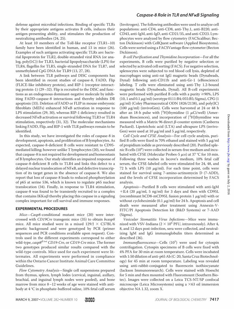
To study the function of caspase-8 in B lymphocytes, micecarrying the caspase-8 mutation targeted to the B-cell line-age (casp8fl/fl;CD19Cre, also called bcasp8�/�) were generatedby crossing mice homozygous for the caspase-8-floxed allele(casp8fl/fl) (20) with mice expressing the Cre recombinaseunder the control of the B-cell-specific CD19 promoter(CD19Cre) (35). Southern blot analysis indicated the highlyefficient deletion of caspase-8 gene in purified peripheral Bcells from bcasp8�/� mice (Fig. 1A). Western blot analysis ofserial dilutions of protein extracts prepared from purified Bcells confirmed the loss of caspase-8 protein in bcasp8�/�
cells (Fig. 1B).Analysis by flow cytometry of cell subpopulations in bone
marrow (BM), spleen, and lymph nodes (LN) did not show anydifferences between bcasp8�/� mice and control littermates(Fig. 1C and data not shown). The proportion of T and B cells inLN and spleens and total lymphocyte numbers in thymus,spleen, LN, andBMwere similar in control and bcasp8�/�mice(Fig. 1D and data not shown). Therefore, unlike its function inthe T-cell lineage, caspase-8 is not essential for the mainte-nance of B-lymphocyte homeostasis.
To confirm the functional deletion of caspase-8, we assessedthe sensitivity of casp8�/� B cells to CD95-induced apoptosis.Purified peripheral B cells were activated in vitro for 3 days withanti-IgM/IL4, and then treated with Fas ligand, in the presenceor absence of cycloheximide, an inhibitor of protein biosynthe-sis known to potentiate Fas killing (37). Annexin-V/PI stainingdemonstrated that while B cells from control mice were sensi-tive to CD95-induced killing, bcasp8�/� cells were resistant(Fig. 1E). These results confirm the essential role for caspase-8in DR-mediated cell death.Vesicular stomatitis virus (VSV) infection in mice induces a
primary infection phase, which consists of a T-cell-independ-ent response characterized by the production of neutralizingIgM antibodies by B cells. The second phase of the infection,
FIGURE 1. Caspase-8 is dispensable for B-cell homeostasis but requiredfor Fas killing. A, Southern blot analysis of caspase-8 deletion in B cells.Caspase-8 floxed allele (casp8fl) and mutant caspase-8 allele lacking exons 3and 4 (casp8�) are indicated. B, Western blot of caspase-8 and actin (loadingcontrol) using 2-fold serial dilutions of cell extracts from purified B cells fromcontrol and casp8fl/fl CD19-Cre (bcasp8�/�) mice. C, FACS analysis of B lympho-cytes populations in spleen (Spl) and LN of bcasp8�/� and control mice. D, cellnumbers in thymus, Spl, LN, and BM. Data are presented as the mean � S.D. of12 mice for each genotype (p values are 0.32 for thymus, 0.48 for spleen, 0.7for LN, and 0.5 for BM). E, Fas ligand (FasL) induced cell death in control andbcasp8�/� B lymphocytes. The percent of surviving fraction is shown. UT,untreated; CHX, cycloheximide.
Caspase-8 Role in TLR and NF�B Signaling
7418 JOURNAL OF BIOLOGICAL CHEMISTRY VOLUME 282 • NUMBER 10 • MARCH 9, 2007
by guest on July 2, 2016http://w
ww
.jbc.org/D
ownloaded from
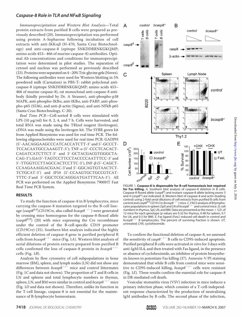
which is T-cell-dependent, leads to the production of IgGimmunoglobulin (36, 38).We investigated the role of caspase-8in the in vivo B-cell response to VSV infection. Control andbcasp8�/� mice were subjected to intravenous immunizationwith 2 � 106 PFU of VSV, and the production of VSV-neutral-izing IgM and IgG antibodies was measured 4, 8, and 12 dayslater (39). While the levels of IgM antibodies directed againstVSV were not affected in bcasp8�/� mice, the production ofVSV-neutralizing IgG was reduced in bcasp8�/� mice (Fig. 2).This finding is consistent with the increased susceptibility ofhuman patients deficient for caspase-8 to viral infections (22).These findings suggest that caspase-8 deficiency in B cells leadsto impaired in vivo responses to VSV infection.B cells are essential components of the innate immune
response against microbial infection (24). TLR signaling isimportant for the activation of B cells and other components ofthe immune response by microbes (40). Given the connectionbetween DISC components and TLR signaling (29–32), weinvestigated the effect of caspase-8 deficiency onTLR signaling.This was accomplished by treating purified B-cell populationswith antigens specific for different TLRs. B-cell expansion wasmeasured by [3H]thymidine incorporation following treatmentwith increasing doses of different Escherichia coli-derived LPS,ultrapure LPS, CpG, LTA, or poly (I:C) to identify TLR4, TLR9,TLR2, and TLR3 responses, respectively. Whereas the stimula-tion by CpG consistently produced similar B-cell expansion inthe control and bcasp8�/� backgrounds, responses of casp8�/�
B cells to poly(I:C), LPS (Fig. 3A and supplemental Fig. S1), andLTA (supplemental Fig. S2) were reduced compared with con-trols, demonstrating that caspase-8 does indeed play a role inTLR2, TLR3, andTLR4 signaling. Similarly, CFSE labeling indi-cated defective expansion of casp8�/� B cells in response toLPS and poly(I:C) stimulation (Fig. 3B and data not shown).These defects could not be attributed to reduceTLR3 andTLR4protein levels in bcaps8�/� cells as their cell surface expressionlevels comparable to controls (data not shown). No majordefect in proliferation of bcasp8�/� B cells was observed inresponse to anti-IgM, anti-IgM, and IL4, or anti-IgM and anti-CD40 compared with wild-type controls (data not shown).
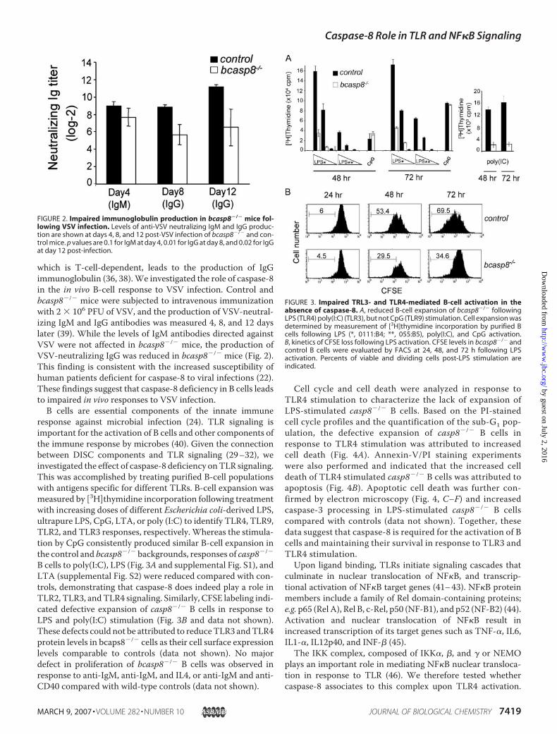
Cell cycle and cell death were analyzed in response toTLR4 stimulation to characterize the lack of expansion ofLPS-stimulated casp8�/� B cells. Based on the PI-stainedcell cycle profiles and the quantification of the sub-G1 pop-ulation, the defective expansion of casp8�/� B cells inresponse to TLR4 stimulation was attributed to increasedcell death (Fig. 4A). Annexin-V/PI staining experimentswere also performed and indicated that the increased celldeath of TLR4 stimulated casp8�/� B cells was attributed toapoptosis (Fig. 4B). Apoptotic cell death was further con-firmed by electron microscopy (Fig. 4, C–F) and increasedcaspase-3 processing in LPS-stimulated casp8�/� B cellscompared with controls (data not shown). Together, thesedata suggest that caspase-8 is required for the activation of Bcells and maintaining their survival in response to TLR3 andTLR4 stimulation.Upon ligand binding, TLRs initiate signaling cascades that
culminate in nuclear translocation of NF�B, and transcrip-tional activation of NF�B target genes (41–43). NF�B proteinmembers include a family of Rel domain-containing proteins;e.g. p65 (Rel A), Rel B, c-Rel, p50 (NF-B1), and p52 (NF-B2) (44).Activation and nuclear translocation of NF�B result inincreased transcription of its target genes such as TNF-�, IL6,IL1-�, IL12p40, and INF-� (45).The IKK complex, composed of IKK�, �, and � or NEMO
plays an important role in mediating NF�B nuclear transloca-tion in response to TLR (46). We therefore tested whethercaspase-8 associates to this complex upon TLR4 activation.
FIGURE 2. Impaired immunoglobulin production in bcasp8�/� mice fol-lowing VSV infection. Levels of anti-VSV neutralizing IgM and IgG produc-tion are shown at days 4, 8, and 12 post-VSV infection of bcasp8�/� and con-trol mice. p values are 0.1 for IgM at day 4, 0.01 for IgG at day 8, and 0.02 for IgGat day 12 post-infection.
FIGURE 3. Impaired TRL3- and TLR4-mediated B-cell activation in theabsence of caspase-8. A, reduced B-cell expansion of bcasp8�/� followingLPS (TLR4) poly(I:C) (TLR3), but not CpG (TLR9) stimulation. Cell expansion wasdetermined by measurement of [3H]thymidine incorporation by purified Bcells following LPS (*, 0111:B4; **, 055:B5), poly(I:C), and CpG activation.B, kinetics of CFSE loss following LPS activation. CFSE levels in bcasp8�/� andcontrol B cells were evaluated by FACS at 24, 48, and 72 h following LPSactivation. Percents of viable and dividing cells post-LPS stimulation areindicated.
Caspase-8 Role in TLR and NF�B Signaling
MARCH 9, 2007 • VOLUME 282 • NUMBER 10 JOURNAL OF BIOLOGICAL CHEMISTRY 7419
by guest on July 2, 2016http://w
ww
.jbc.org/D
ownloaded from
Immunoprecipitation was performed using anti-IKK�� and/oranti-caspase-8 antibodies on protein extracts derived fromuntreated or LPS-stimulated casp8�/� or control B cells. Thesestudies indicated that caspase-8 co-immunoprecipitates withIKK�� following 30 min of LPS stimulation of control B cells(Fig. 5A). The specificity of the IKK�� immunoprecipitationswas shown by Western blot using anti-IKK�� antibodies. Therecruitment of caspase-8 to a complex containing IKK�� inresponse to LPS stimulation was confirmed in 3T3-immortal-ized fibroblasts. As shown in Fig. 5B, IKK�� association tocaspase-8 in 3T3 was detected as early as 4 min following TLR4activation and appeared to be optimal at 7-min post-LPS stim-ulation. Thus, our data identify that TLR4 stimulation drives
caspase-8 transient recruitment to IKK��, an important regu-lator of NF�B functions.NF�B is an important downstream target of IKK�� and its
activation, in response to TLR4 stimulation, leads to transcrip-tional activation of a subset of target genes. Thus, the effect ofcaspase-8 mutation on TLR4-induced NF�B transcriptionalactivation was assessed using real time PCR to quantify thetranscription levels of the NF�B target genes including IL6,TNF-�, IFN-�, and IP-10. cDNA, representative of total cellu-larmRNA,was prepared from cell-sorted control and casp8�/�
B cells activated with LPS for 0, 2, 4, and 7 h. As expected,increased expression of these geneswas observed in B cells aftertreatmentwith LPS; however, the level of transcriptional induc-
FIGURE 4. Increased apoptosis of caspase-8-deficient B cells in response to LPS stimulation. A, cell cycle progression and sub-G1 populations weredetermined using propidium iodide staining and FACS analysis of LPS-stimulated (10 �g/ml) casp8�/� and control B cells. Increased cell death but no defectsin cell cycle were observed. B, increased apoptosis in casp8�/� B-cells in response to LPS stimulation (10 �g/ml). Analysis was performed by FACS usingAnnexin-V/propidium iodide staining. C–F, electron microscopy of casp8�/� B cells before (C and D) or 24-h post-stimulation with LPS (E and F). Nuclearcondensation, a feature of apoptosis is seen in E and F.
Caspase-8 Role in TLR and NF�B Signaling
7420 JOURNAL OF BIOLOGICAL CHEMISTRY VOLUME 282 • NUMBER 10 • MARCH 9, 2007
by guest on July 2, 2016http://w
ww
.jbc.org/D
ownloaded from
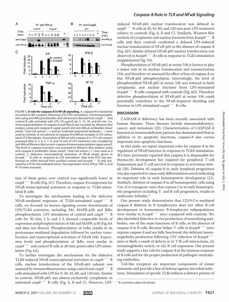
tion of these genes over control was significantly lower incasp8�/�B cells (Fig. 5C). Therefore, caspase-8 is important forNF�B transcriptional activation in response to TLR4-stimu-lated B cells.To investigate the mechanisms leading to the defective
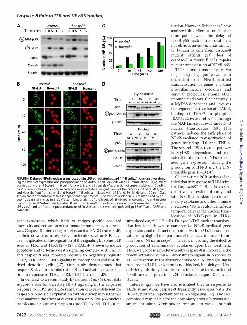
NF�B-mediated responses of TLR4-stimulated casp8�/� Bcells, we focused on known signaling events downstream ofLPS/TLR4 activation, including Akt, MAPK-p38, and IkB�phosphorylation. LPS stimulation of control and casp8�/� Bcells for 30 min, 1 h, and 2 h showed comparable levels ofexpression andphosphorylation ofAkt andMAPK-p38 (Fig. 6Aand data not shown). Phosphorylation of I�B� results in itsproteasome-mediated degradation followed by nuclear trans-location and transcriptional activation of NF�B (44). Expres-sion levels and phosphorylation of IkB� were similar incasp8�/� and control B cells at all time points after LPS stimu-lation (Fig. 6A).To further investigate the mechanisms for the defective
TLR4-induced NF�B transcriptional activation in casp8�/� Bcells, nuclear translocation of the NF�B-p65 subunit wasassessed by immunofluorescence using control and casp8�/� Bcells stimulated with LPS for 0, 30, 45, 60, and 120 min. Similarto controls, NF�B-p65 was almost exclusively cytoplasmic inuntreated casp8�/� B cells (Fig. 6, B and C). However, LPS-
induced NF�B-p65 nuclear translocation was delayed incasp8�/� B cells at 30, 45, 60, and 120 min post-LPS treatmentrelative to controls (Fig. 6, B and C). Similarly, Western blotanalysis of cytoplasmic and nuclear fractions from bcasp8�/� Bcells and their controls confirmed a delayed LPS-inducednuclear translocation of NF�B-p65 in the absence of caspase-8(Fig. 6D). Similar delayed NF�B-p65 nuclear translocation wasobserved in bcasp8�/� B cells in response to TLR3 stimulation(supplemental Fig. S3).Phosphorylation of NF�B-p65 at serine 536 is known to play
a major role in its nuclear translocation and transactivation(34), and therefore we assessed the effect of loss of caspase-8 onthis NF�B-p65 phosphorylation. Interestingly, the level ofphosphorylated NF�B-p65 at serine 536 was reduced in bothcytoplasmic and nuclear fractions from LPS-stimulatedbcasp8�/� B cells compared with controls (Fig. 6D). Thereforedefective phosphorylation of NF�B-p65 at serine 536 couldpotentially contribute to the NF�B-impaired shuttling andfunction in LPS-stimulated casp8�/� B cells.
DISCUSSION
CASPASE-8 deficiency has been recently associated withhuman diseases. These diseases include immunodeficiency,cancer, and metastasis (22). Characterization of CASPASE-8function in immunodeficient patients has demonstrated that inaddition to its apoptotic functions, caspase-8 also carriesimportant non-apoptotic functions.In this study, we report important roles for caspase-8 in the
regulation of NF�B function in response to TLR4 stimulation.We have previously reported that caspase-8 is dispensable forthymocyte development but required for peripheral T-cellhomeostasis and T-cell survival in response to activation stim-uli (20). Deletion of caspase-8 in early lymphoid progenitorswas also reported to cause early differentiation arrest indicatingits important role in early hematopoietic development (21).Similarly, deletion of caspase-8 in all hematopoietic cells usingVav-iCre transgenic mice that express Cre in early hematopoi-etic progenitors including T- and B-cell progenitors, results inembryonic lethality.4Our present study demonstrates that CD19-Cre-mediated
caspase-8 deletion in B lymphocytes does not affect B-celldevelopment or homeostasis: B-lymphocyte subpopulationswere similar in bcasp8�/� mice compared with controls. Wealso identified defective in vivo production of neutralizing anti-bodies, one of the main functions of B cells, in the absence ofcaspase-8 in B cells. Because helper T cells in bcasp8�/� miceexpress caspase-8 and are fully functional, the deficient immu-noglobulin production following VSV infection of bcasp8�/�
mice is likely a result of defects in (i) T/B-cell interactions, (ii)immunoglobulin switch, or (iii) B-cell expansion. Our presentstudy supports a key role for caspase-8 in the immune responseof B cells and for the proper production of pathogen-neutraliz-ing antibodies.Toll-like receptors are important components of innate
immunity and provide a line of defense against microbial infec-tions. Stimulation of specific TLRs induces a distinct pattern of
4 B. Lemmers, data not shown.
FIGURE 5. A role for caspase-8 in NF�B signaling. A, caspase-8 is transientlyrecruited to IKK complex following LPS/TLR4 stimulation. Immunoprecipita-tion using anti-IKK�� antibodies and cell extracts derived from casp8�/� andcontrol B cells activated with LPS (10 �g/ml) for 5, 15, 30, and 60 min. Co-immunoprecipitation of caspase-8 and IKK�� was revealed by Western blotanalysis using anti-caspase-8 (upper panel) and anti-IKK�� antibodies (lowerpanel). Total cell extract (�) and an irrelevant polyclonal antibody (�) wereused as controls. B, recruitment of caspase-8 to IKK�� complex in LPS-stimu-lated 3T3 fibroblasts. Association of IKK�� with caspase-8 in 3T3 cell extractsprepared after 0, 3, 4, 5, 7, 8, and 10 min of LPS treatment was revealed byanti-IKK�� Western blot on anti-caspase-8 immunoprecipitates (upper panel).The level of caspase-8 protein was assessed by Western blot analysis usinganti-caspase-8 antibodies (lower panel). Total cell extract (�) was used as acontrol. C, defective transcriptional activation of NF�B target genes inbcasp8�/� B cells in response to LPS stimulation. Real time PCR was per-formed on cDNA derived from purified control and bcasp8�/� B cells acti-vated by LPS for the indicated times. The expression of IL6, IFN-�, TNF-�, andIP-10 were assessed.
Caspase-8 Role in TLR and NF�B Signaling
MARCH 9, 2007 • VOLUME 282 • NUMBER 10 JOURNAL OF BIOLOGICAL CHEMISTRY 7421
by guest on July 2, 2016http://w
ww
.jbc.org/D
ownloaded from
gene expression, which leads to antigen-specific acquiredimmunity and activation of the innate immune response path-way. Caspase-8-interacting proteins such as FADD and c-FLIP,or their downstream responsive molecules such as RIP, havebeen implicated in the regulation of the signaling by some TLRsuch as TLR3 and TLR4 (29–32). TRAIL-R, known to induceapoptosis and to form a death signaling complex with FADD,and caspase-8 was reported recently to negatively regulateTLR2, TLR3, and TLR4 signaling in macrophages and BM-de-rived dendritic cells (47). Our study demonstrates thatcaspase-8 plays an essential role in B-cell activation and expan-sion in response to TLR2, TLR3, TLR4, but not TLR9.In contrast to a recent study by Beisner et al. (48), our data
support a role for defective NF�B signaling in the impairedresponse to TLR3 and TLR4 stimulation of B cells deficient forcaspase-8. A possible explanation for this discrepancy is that wehave analyzed the effect of caspase-8 loss on NF�B-p65 nucleartranslocation at earlier time points post-TLR3 and -TLR4 stim-
ulation.However, Beisner et al.haveanalyzed this effect at much latertime points when the delay ofNF�B-p65 nuclear translocation isnot obvious anymore. Thus, similarto human B cells from caspase-8mutant patients (23), loss ofcaspase-8 in mouse B cells impairsnuclear translocation of NF�B-p65.TLR4 stimulations activate two
major signaling pathways, bothdependent on NF�B-mediatedtransactivation of genes encodingpro-inflammatory cytokines andsurvival molecules, among otherimmune mediators. One pathway isa MyD88-dependent and involvesthe sequential activation of IRAK-4,binding of TRAF6 to phospho-IRAK1, activation of AP-1 throughtheMAP kinase pathway, andNF�Bnuclear translocation (49). Thispathway induces the early phase ofNF�B-mediated transactivation ofgenes including IL6 and TNF-�.The second LPS-activated pathwayis MyD88-independent, and acti-vates the late phase of NF�B-medi-ated gene expression, driving theproduction of IFN-� and the IFN-inducible gene IP-10 (50).Our real-time PCR analysis iden-
tified that in response to TLR4 stim-ulation, casp8�/� B cells exhibitdefective expression of early andlate NF�B-dependent pro-inflam-matory cytokines and other immunemediators.We have also identified atemporal delay in the nuclear trans-location of NF�B-p65 in TLR4-
stimulated casp8�/� B cells. Delayed NF�B nuclear transloca-tion has been shown to compromise NF�B-mediated geneexpression, and cell function upon activation (51). These obser-vations highlight the importance of the delayed nuclear trans-location of NF�B in casp8�/� B cells, in causing the defectiveproduction of inflammatory cytokines upon LPS treatment.Thus, we propose a model whereby caspase-8 is involved in thetimely activation of NF�B downstream signals in response toTLR4 activation. In the absence of caspase-8, NF�B signaling inresponse to TLR4 activation is not blocked, but delayed. Nev-ertheless, this delay is sufficient to impair the transduction ofNF�B survival signals in TLR4-stimulated caspase-8-deficientB cells.Interestingly, we have also identified that in response to
TLR4 stimulation, caspase-8 transiently associates with theIKK��, proteins important for NF�B signaling (52). The IKKcomplex is responsible for the phosphorylation of various sub-strates including NF�B-p65 in response to various stimuli
FIGURE 6. Delayed NF�B nuclear translocation in LPS-stimulated bcasp8�/� B cells. A, Western blots show-ing the levels of expression and phosphorylation of MAPp38 and IkB� following LPS stimulation (10 �g/ml) ofpurified control and bcasp8�/� B cells for 0, 0.5, 1, and 2 h. Levels of expression of casp8 and �-actin (loadingcontrol) are shown. B, confocal microscope representative merged views of the p65 subunit of NF�B (green)and Hoescht (red) from control and bcasp8�/� B cells stimulated with LPS for 0, 30, 45, 60, and 120 min. Datashown are representative of five independent experiments. C, percent of nuclear NF�B as measured by anti-p65 nuclear staining as in B. D, Western blot analysis of the levels of NF�B-p65 in cytoplasmic and nuclearfractions from LPS-stimulated purified B cells from bcasp8�/� and control mice. B cells were stimulated withLPS as in A, and cell fractions prepared and used for Western blots with anti-p65, anti-p65 Ser536, anti-PARP, andanti-actin.
Caspase-8 Role in TLR and NF�B Signaling
7422 JOURNAL OF BIOLOGICAL CHEMISTRY VOLUME 282 • NUMBER 10 • MARCH 9, 2007
by guest on July 2, 2016http://w
ww
.jbc.org/D
ownloaded from
including pro-inflammatory signals (52). Interestingly, we haveidentified that in response to LPS/TLR4 stimulation, caspase-8transiently associates with the IKK complex. We also demon-strate that caspase-8 deficiency in TLR4-stimulated caspase-8-deficient B cells leads to decreased phosphorylation of NF�B-p65 at serine 536. This phosphorylation is mediated by the IKKcomplex, modulates NF�B nuclear translocation, and there-fore, its impairment could contribute to the defective NF�Bnuclear translocation and functions observed in TLR4-stimu-lated bcasp8�/� cells. Our findings support a role for defectiveNF�B signaling in the impaired responses to TLR4 activation inthe absence of caspase-8 and provide a molecular mechanismfor the immunodeficiency of caspase-8 mutant patients.
Acknowledgments—We thankW. C. Yeh, B. Au, andM.Woo for help-ful discussions. We also thank A. Strasser for kindly providing us withthe anti-caspase-8 monoclonal antibody.
REFERENCES1. Earnshaw, W. C., Martins, L. M., and Kaufmann, S. H. (1999) Annu. Rev.
Biochem. 68, 383–4242. Gross, A., McDonnell, J. M., and Korsmeyer, S. J. (1999) Genes Dev. 13,
1899–19113. Krammer, P. H. (2000) Nature 407, 789–7954. Wang, X. (2001) Genes Dev. 15, 2922–29335. Boatright, K. M., and Salvesen, G. S. (2003) Biochem. Soc. Symp. 233–2426. Barnhart, B. C., Alappat, E. C., and Peter,M. E. (2003) Semin Immunol. 15,
185–1937. Kennedy,N. J., Kataoka, T., Tschopp, J., andBudd, R. C. (1999) J. Exp.Med.
190, 1891–18968. Alam, A., Cohen, L. Y., Aouad, S., and Sekaly, R. P. (1999) J. Exp.Med. 190,
1879–18909. Miossec, C., Dutilleul, V., Fassy, F., and Diu-Hercend, A. (1997) J. Biol.
Chem. 272, 13459–1346210. Wilhelm, S., Wagner, H., and Hacker, G. (1998) Eur. J. Immunol. 28,
891–90011. Olson, N. E., Graves, J. D., Shu, G. L., Ryan, E. J., and Clark, E. A. (2003)
J. Immunol. 170, 6065–607212. Falk, M., Ussat, S., Reiling, N., Wesch, D., Kabelitz, D., and Adam-Klages,
S. (2004) J. Immunol. 173, 5077–508513. Krakauer, T. (2004) Clin. Diagn. Lab. Immunol. 11, 621–62414. Mack, A., and Hacker, G. (2002) Eur. J. Immunol. 32, 1986–199215. Newton, K., and Strasser, A. (1998) Curr. Opin. Genet. Dev. 8, 68–7516. Walsh, C. M., Wen, B. G., Chinnaiyan, A. M., O’Rourke, K., Dixit, V. M.,
and Hedrick, S. M. (1998) Immunity 8, 439–44917. Zornig, M., Hueber, A. O., and Evan, G. (1998) Curr. Biol. 8, 467–47018. Zhang, J., Cado, D., Chen, A., Kabra, N. H., andWinoto, A. (1998)Nature
392, 296–30019. Varfolomeev, E. E., Schuchmann,M., Luria, V., Chiannilkulchai, N., Beck-
mann, J. S., Mett, I. L., Rebrikov, D., Brodianski, V. M., Kemper, O. C.,Kollet, O., Lapidot, T., Soffer, D., Sobe, T., Avraham, K. B., Goncharov, T.,Holtmann, H., Lonai, P., and Wallach, D. (1998) Immunity 9, 267–276
20. Salmena, L., Lemmers, B., Hakem, A., Matysiak-Zablocki, E., Murakami,K., Au, P. Y., Berry, D.M., Tamblyn, L., Shehabeldin, A., Migon, E.,Wake-ham, A., Bouchard, D., Yeh, W. C., McGlade, J. C., Ohashi, P. S., andHakem, R. (2003) Genes Dev. 17, 883–895
21. Kang, T. B., Ben-Moshe, T., Varfolomeev, E. E., Pewzner-Jung, Y., Yogev,N., Jurewicz, A., Waisman, A., Brenner, O., Haffner, R., Gustafsson, E.,Ramakrishnan, P., Lapidot, T., and Wallach, D. (2004) J. Immunol. 173,2976–2984
22. Chun, H. J., Zheng, L., Ahmad, M., Wang, J., Speirs, C. K., Siegel, R. M.,Dale, J. K., Puck, J., Davis, J., Hall, C. G., Skoda-Smith, S., Atkinson, T. P.,Straus, S. E., and Lenardo, M. J. (2002) Nature 419, 395–399
23. Su, H., Bidere, N., Zheng, L., Cubre, A., Sakai, K., Dale, J., Salmena, L.,Hakem, R., Straus, S., and Lenardo, M. (2005) Science 307, 1465–1468
24. Vos, Q., Lees, A., Wu, Z. Q., Snapper, C. M., and Mond, J. J. (2000)Immunol. Rev. 176, 154–170
25. Hoebe, K., Janssen, E., and Beutler, B. (2004) Nat. Immunol. 5, 971–97426. Kawai, T., and Akira, S. (2006) Cell Death Differ. 13, 816–82527. Mukhopadhyay, S., Herre, J., Brown, G. D., and Gordon, S. (2004)
Immunology 112, 521–53028. Beutler, B. (2004) Nature 430, 257–26329. Bannerman, D. D., Tupper, J. C., Kelly, J. D.,Winn, R. K., and Harlan, J. M.
(2002) J. Clin. Investig. 109, 419–42530. Bannerman,D.D., Eiting, K. T.,Winn, R. K., andHarlan, J.M. (2004)Am. J.
Pathol. 165, 1423–143131. Vivarelli, M. S., McDonald, D., Miller, M., Cusson, N., Kelliher, M., and
Geha, R. S. (2004) J. Exp. Med. 200, 399–40432. Meylan, E., Burns, K., Hofmann, K., Blancheteau, V., Martinon, F.,
Kelliher, M., and Tschopp, J. (2004) Nat. Immunol. 5, 503–50733. Yeh,W.C., Itie, A., Elia, A. J., Ng,M., Shu,H. B.,Wakeham,A.,Mirtsos, C.,
Suzuki, N., Bonnard,M., Goeddel, D. V., andMak, T.W. (2000) Immunity12, 633–642
34. Mattioli, I., Sebald, A., Bucher, C., Charles, R. P., Nakano, H., Doi, T.,Kracht, M., and Schmitz, M. L. (2004) J. Immunol. 172, 6336–6344
35. Rickert, R. C., Roes, J., and Rajewsky, K. (1997) Nucleic Acids Res. 25,1317–1318
36. Fehr, T., Lopez-Macias, C., Odermatt, B., Torres, R. M., Schubart, D. B.,O’Keefe, T. L., Matthias, P., Hengartner, H., and Zinkernagel, R.M. (2000)Int. Immunol. 12, 1275–1284
37. Barnhart, B. C., Pietras, E. M., Algeciras-Schimnich, A., Salmena, L.,Sayama, K., Hakem, R., and Peter,M. E. (2005)Cell DeathDiffer. 12, 25–37
38. Fehr, T., Bachmann, M. F., Bluethmann, H., Kikutani, H., Hengartner, H.,and Zinkernagel, R. M. (1996) Cell. Immunol. 168, 184–192
39. Bertram, E.M., Tafuri, A., Shahinian, A., Chan, V. S., Hunziker, L., Recher,M., Ohashi, P. S., Mak, T.W., andWatts, T. H. (2002) Eur. J. Immunol. 32,3376–3385
40. Iwasaki, A., and Medzhitov, R. (2004) Nat. Immunol. 5, 987–99541. Zhang, G., and Ghosh, S. (2000) J. Endotoxin. Res. 6, 453–45742. Akira, S., and Takeda, K. (2004) Nat. Rev. Immunol. 4, 499–51143. Baldwin, A. S., Jr. (1996) Annu. Rev. Immunol. 14, 649–68344. Hayden, M. S., and Ghosh, S. (2004) Genes Dev. 18, 2195–222445. Takeda, K., and Akira, S. (2004) J. Dermatol. Sci. 34, 73–8246. Ghosh, S., and Karin, M. (2002) Cell 109, Suppl. 1, S81–S9647. Diehl, G. E., Yue,H.H., Hsieh, K., Kuang, A.A., Ho,M.,Morici, L. A., Lenz,
L. L., Cado, D., Riley, L.W., andWinoto, A. (2004) Immunity 21, 877–88948. Beisner, D. R., Ch’en, I. L., Kolla, R. V., Hoffmann, A., and Hedrick, S. M.
(2005) J. Immunol. 175, 3469–347349. Takeda, K., and Akira, S. (2005) Int. Immunol. 17, 1–1450. Akira, S., and Hoshino, K. (2003) J. Infect. Dis. 187, Suppl. 2, S356–S36351. Kaisho, T., Takeuchi, O., Kawai, T., Hoshino, K., and Akira, S. (2001)
J. Immunol. 166, 5688–569452. Doyle, S. L., Jefferies, C. A., and O’Neill L, A. (2005) J. Biol. Chem. 280,
23496–23501
Caspase-8 Role in TLR and NF�B Signaling
MARCH 9, 2007 • VOLUME 282 • NUMBER 10 JOURNAL OF BIOLOGICAL CHEMISTRY 7423
by guest on July 2, 2016http://w
ww
.jbc.org/D
ownloaded from
Supplemental Data
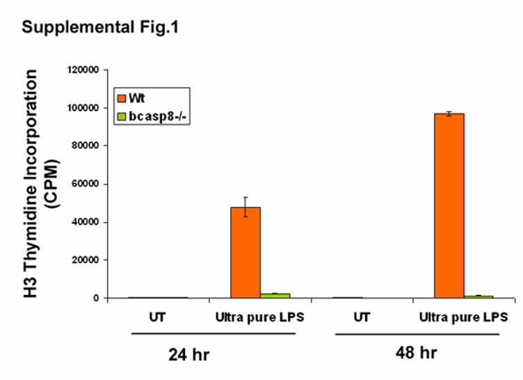
Supplemental Figure 1. Impaired TLR4 mediated B-cell activation in the absence of
caspase-8. Reduced B-cell expansion of bcasp8-/- following stimulation with 5µg/ml of ultra
pure LPS. Cell expansion was determined at 24 hr and 48 hr post stimulation of purified Wt and
bcasp8-/- B cells by measurement of their [3H] thymidine incorporation. Data shown is
representative of 3 independent experiments. UT: untreated.
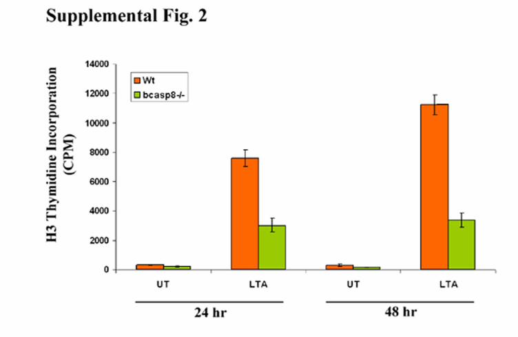
Supplemental Figure 2. Impaired TRL2 mediated B-cell activation in the absence of
caspase-8. Wt and bcasp8-/- B-cells were stimulated with 10µg/ml of lipoteichoic acid (LTA)
and their expansion was determined by measurement of their levels of [3H] thymidine
incorporation at 24 and 48 hr post stimulation. Data shown is representative of 2 independent
experiments. UT: untreated.
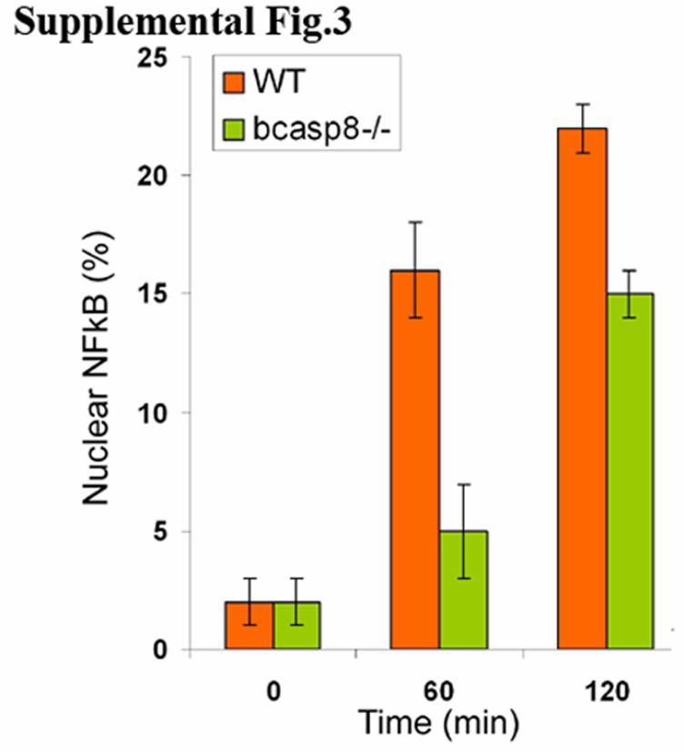
Supplemental Figure 3. Delayed NFκB nuclear translocation in TLR3 stimulated bcasp8-/-
B-cells. Confocal microscopy was used to determine the percent of nuclear NFκB as measured
by anti-p65 nuclear staining of Wt and bcasp8-/- stimulated with 100µg/ml of Poly-IC for 0, 1
and 2 hr. Data shown is representative of 2 independent experiments.
Lenardo, Razqallah Hakem and Anne HakemMatysiak-Zablocki, Kiichi Murakami, Pamela S. Ohashi, Andrea Jurisicova, Michael
Bénédicte Lemmers, Leonardo Salmena, Nicolas Bidère, Helen Su, ElzbietaB SignalingκEssential Role for Caspase-8 in Toll-like Receptors and NF
doi: 10.1074/jbc.M606721200 originally published online January 9, 20072007, 282:7416-7423.J. Biol. Chem.
10.1074/jbc.M606721200Access the most updated version of this article at doi:
Alerts:
When a correction for this article is posted•
When this article is cited•
to choose from all of JBC's e-mail alertsClick here
Supplemental material:
http://www.jbc.org/content/suppl/2007/01/10/M606721200.DC1.html
http://www.jbc.org/content/282/10/7416.full.html#ref-list-1
This article cites 48 references, 19 of which can be accessed free at
by guest on July 2, 2016http://w
ww
.jbc.org/D
ownloaded from