Autoproteolysis of PIDD marks the bifurcation between pro-death caspase-2 and pro-survival NF-κB...
12
Autoproteolysis of PIDD marks the bifurcation between pro-death caspase-2 and pro-survival NF-jB pathway Antoine Tinel 1,3 , Sophie Janssens 1,4 , Saskia Lippens 1 , Solange Cuenin 1 , Emmanuelle Logette 1 , Bastienne Jaccard 1,2 , Manfredo Quadroni 1,2 and Ju ¨ rg Tschopp 1, * 1 Department of Biochemistry, University of Lausanne, Epalinges, Switzerland and 2 Protein Analysis Facility, University of Lausanne, Epalinges, Switzerland Upon DNA damage, a complex called the PIDDosome is formed and either signals NF-jB activation and thus cell survival or alternatively triggers caspase-2 activation and apoptosis. PIDD (p53-induced protein with a death do- main) is constitutively processed giving rise to a 48-kDa N-terminal fragment containing the leucine-rich repeats (LRRs, PIDD-N) and a 51-kDa C-terminal fragment con- taining the death domain (DD, PIDD-C). The latter under- goes further cleavage resulting in a 37-kDa fragment (PIDD-CC). Here we show that processing occurs at S446 (generating PIDD-C) and S588 (generating PIDD-CC) by an auto-processing mechanism similar to that found in the nuclear pore protein Nup98/96 and inteins. Auto-cleavage of PIDD determines the outcome of the downstream signaling events. Whereas initially formed PIDD-C medi- ates the activation of NF-jB via the recruitment of RIP1 and NEMO, subsequent formation of PIDD-CC causes cas- pase-2 activation and thus cell death. A non-cleavable PIDD mutant is unable to translocate from the cytoplasm to the nucleus and loses both activities. In this way, auto- proteolysis of PIDD might participate in the orchestration of the DNA damage-induced life and death signaling pathways. The EMBO Journal (2007) 26, 197–208. doi:10.1038/ sj.emboj.7601473; Published online 7 December 2006 Subject Categories: proteins; differentiation & death Keywords: auto-proteolysis; caspase-2; DNA damage; NF-kB; PIDD Introduction PIDD (p53-induced protein with a death domain), also known as LRDD (leucine-rich repeat and death domain- containing protein), was discovered during a bioinformatics screen for novel death domain (DD)-containing proteins using the DD of RIP1 as bait (Telliez et al, 2000). Independently, PIDD was identified as a novel p53-inducible gene in an erythroleukemia cell line expressing a tempera- ture-sensitive p53 mutant allele, and shown to be implicated in p53-dependent apoptosis (Lin et al, 2000). PIDD contains seven leucine-rich repeats (LRRs) at the N-terminus, followed by two ZU-5 domains and a C-terminal DD. Recent studies have shown that PIDD plays a critical role in the activation of caspase-2 (Tinel and Tschopp, 2004; Berube et al, 2005; Ren et al, 2005; Seth et al, 2005). As caspase-2 shares characteristics of both effector and initiator caspases, its activation mechanism remained enigmatic for a long time. An important breakthrough was made by showing that caspase-2 was activated in a high-molecular-weight complex, reminiscent of the apoptosome and other initiator caspase- activating platforms (Read et al, 2002). The recruitment and activation of caspase-2 within this complex is entirely depen- dent on the presence of an adaptor protein called RAIDD and on PIDD, which together with caspase-2 constitute the so- called PIDDosome (Tinel and Tschopp, 2004; Berube et al, 2005). These results were particularly intriguing as PIDD, just like caspase-2, had been suspected to participate in DNA damage-induced apoptosis (Robertson et al, 2002; Castedo et al, 2004; Giagkousiklidis et al, 2005; Zhivotovsky and Orrenius, 2005; Vakifahmetoglu et al, 2006). PIDD also plays an essential role in NF-kB activation in- duced by DNA damage. Indeed, genotoxic stress triggers the formation of an alternative PIDDosome, consisting of PIDD, RIP1 and NEMO (Hur et al, 2003; Janssens et al, 2005). NEMO is recruited to the PIDDosome via RIP1 and appears to be modified immediately upon recruitment to the activated PIDDosome by sumoylation (Janssens et al, 2005). This is the beginning of a sequential series of modifications on NEMO, in which the nuclear protein kinase ATM plays an essential role, and which alter the protein’s subcellular locali- zation, providing a means to link a nuclear signal (DNA damage) to the activation of a cytosolic NF-kB-activating complex (the IKK complex) (Huang et al, 2003; Wu et al, 2006). Regulation of protein activation can be achieved in many different ways. Apart from a tightly controlled transcriptional response as a DNA damage-dependent response gene, PIDD activation appears to be controlled by a second, unusual mechanism of post-translational cleavage. Full-length PIDD has a predicted molecular mass of 100kDa, but is constitu- tively processed at two sites giving rise to an N-terminal fragment of 48 kDa and a C-terminal fragment of 51 kDa, which is further cleaved, resulting in a 37 kDa fragment (Telliez et al, 2000; Tinel and Tschopp, 2004). The current study aims at characterizing the molecular mechanisms underlying PIDD cleavage and determining its functional consequences. Received: 29 June 2006; accepted: 6 November 2006; published online: 7 December 2006 *Corresponding author. Department of Biochemistry, University of Lausanne, Ch. des Boveresses 155, CH-1066 Epalinges, Switzerland. Tel.: þ 41 21 692 5738; Fax: þ 41 21 692 6705; E-mail: [email protected] 3 Present address: Department of Pathology, Harvard Medical School, 77 avenue Louis Pasteur, Boston, MA 02115, USA 4 Present address: Department of Molecular Biomedical Research, University of Ghent-VIB, Technologiepark 927, 9052 Gent, Belgium The EMBO Journal (2007) 26, 197–208 | & 2007 European Molecular Biology Organization | All Rights Reserved 0261-4189/07 www.embojournal.org & 2007 European Molecular Biology Organization The EMBO Journal VOL 26 | NO 1 | 2007 EMBO THE EMBO JOURNAL THE EMBO JOURNAL 197
-
Upload
independent -
Category
Documents
-
view
0 -
download
0
Transcript of Autoproteolysis of PIDD marks the bifurcation between pro-death caspase-2 and pro-survival NF-κB...
Autoproteolysis of PIDD marks the bifurcationbetween pro-death caspase-2 and pro-survivalNF-jB pathway
Antoine Tinel1,3, Sophie Janssens1,4,Saskia Lippens1, Solange Cuenin1,Emmanuelle Logette1, BastienneJaccard1,2, Manfredo Quadroni1,2
and Jurg Tschopp1,*1Department of Biochemistry, University of Lausanne, Epalinges,Switzerland and 2Protein Analysis Facility, University of Lausanne,Epalinges, Switzerland
Upon DNA damage, a complex called the PIDDosome is
formed and either signals NF-jB activation and thus cell
survival or alternatively triggers caspase-2 activation and
apoptosis. PIDD (p53-induced protein with a death do-
main) is constitutively processed giving rise to a 48-kDa
N-terminal fragment containing the leucine-rich repeats
(LRRs, PIDD-N) and a 51-kDa C-terminal fragment con-
taining the death domain (DD, PIDD-C). The latter under-
goes further cleavage resulting in a 37-kDa fragment
(PIDD-CC). Here we show that processing occurs at S446
(generating PIDD-C) and S588 (generating PIDD-CC) by
an auto-processing mechanism similar to that found in the
nuclear pore protein Nup98/96 and inteins. Auto-cleavage
of PIDD determines the outcome of the downstream
signaling events. Whereas initially formed PIDD-C medi-
ates the activation of NF-jB via the recruitment of RIP1
and NEMO, subsequent formation of PIDD-CC causes cas-
pase-2 activation and thus cell death. A non-cleavable
PIDD mutant is unable to translocate from the cytoplasm
to the nucleus and loses both activities. In this way, auto-
proteolysis of PIDD might participate in the orchestration
of the DNA damage-induced life and death signaling
pathways.
The EMBO Journal (2007) 26, 197–208. doi:10.1038/
sj.emboj.7601473; Published online 7 December 2006
Subject Categories: proteins; differentiation & death
Keywords: auto-proteolysis; caspase-2; DNA damage; NF-kB;
PIDD
Introduction
PIDD (p53-induced protein with a death domain), also
known as LRDD (leucine-rich repeat and death domain-
containing protein), was discovered during a bioinformatics
screen for novel death domain (DD)-containing proteins
using the DD of RIP1 as bait (Telliez et al, 2000).
Independently, PIDD was identified as a novel p53-inducible
gene in an erythroleukemia cell line expressing a tempera-
ture-sensitive p53 mutant allele, and shown to be implicated
in p53-dependent apoptosis (Lin et al, 2000). PIDD contains
seven leucine-rich repeats (LRRs) at the N-terminus, followed
by two ZU-5 domains and a C-terminal DD.
Recent studies have shown that PIDD plays a critical role in
the activation of caspase-2 (Tinel and Tschopp, 2004; Berube
et al, 2005; Ren et al, 2005; Seth et al, 2005). As caspase-2
shares characteristics of both effector and initiator caspases,
its activation mechanism remained enigmatic for a long time.
An important breakthrough was made by showing that
caspase-2 was activated in a high-molecular-weight complex,
reminiscent of the apoptosome and other initiator caspase-
activating platforms (Read et al, 2002). The recruitment and
activation of caspase-2 within this complex is entirely depen-
dent on the presence of an adaptor protein called RAIDD and
on PIDD, which together with caspase-2 constitute the so-
called PIDDosome (Tinel and Tschopp, 2004; Berube et al,
2005). These results were particularly intriguing as PIDD, just
like caspase-2, had been suspected to participate in DNA
damage-induced apoptosis (Robertson et al, 2002; Castedo
et al, 2004; Giagkousiklidis et al, 2005; Zhivotovsky and
Orrenius, 2005; Vakifahmetoglu et al, 2006).
PIDD also plays an essential role in NF-kB activation in-
duced by DNA damage. Indeed, genotoxic stress triggers the
formation of an alternative PIDDosome, consisting of PIDD,
RIP1 and NEMO (Hur et al, 2003; Janssens et al, 2005). NEMO
is recruited to the PIDDosome via RIP1 and appears to be
modified immediately upon recruitment to the activated
PIDDosome by sumoylation (Janssens et al, 2005). This is
the beginning of a sequential series of modifications on
NEMO, in which the nuclear protein kinase ATM plays an
essential role, and which alter the protein’s subcellular locali-
zation, providing a means to link a nuclear signal (DNA
damage) to the activation of a cytosolic NF-kB-activating
complex (the IKK complex) (Huang et al, 2003; Wu et al, 2006).
Regulation of protein activation can be achieved in many
different ways. Apart from a tightly controlled transcriptional
response as a DNA damage-dependent response gene, PIDD
activation appears to be controlled by a second, unusual
mechanism of post-translational cleavage. Full-length PIDD
has a predicted molecular mass of 100 kDa, but is constitu-
tively processed at two sites giving rise to an N-terminal
fragment of 48 kDa and a C-terminal fragment of 51 kDa,
which is further cleaved, resulting in a 37 kDa fragment
(Telliez et al, 2000; Tinel and Tschopp, 2004). The current
study aims at characterizing the molecular mechanisms
underlying PIDD cleavage and determining its functional
consequences.Received: 29 June 2006; accepted: 6 November 2006; publishedonline: 7 December 2006
*Corresponding author. Department of Biochemistry, University ofLausanne, Ch. des Boveresses 155, CH-1066 Epalinges, Switzerland.Tel.: þ 41 21 692 5738; Fax: þ 41 21 692 6705;E-mail: [email protected] address: Department of Pathology, Harvard Medical School,77 avenue Louis Pasteur, Boston, MA 02115, USA4Present address: Department of Molecular Biomedical Research,University of Ghent-VIB, Technologiepark 927, 9052 Gent, Belgium
The EMBO Journal (2007) 26, 197–208 | & 2007 European Molecular Biology Organization | All Rights Reserved 0261-4189/07
www.embojournal.org
&2007 European Molecular Biology Organization The EMBO Journal VOL 26 | NO 1 | 2007
EMBO
THE
EMBOJOURNAL
THE
EMBOJOURNAL
197

Results
PIDD is constitutively processed at S446 and S588
As previously reported, full-length PIDD (PIDD-FL, 910 aa)
with an apparent molecular weight of 100 kDa is constitu-
tively cleaved into three main fragments, an N-terminal
fragment of approximately 48 kDa (PIDD-N) and two
C-terminal fragments of 51 kDa (PIDD-C) and 37 kDa (PIDD-
CC) (Tinel and Tschopp, 2004). This processing is well
documented for cells in which PIDD is transiently or stably
overexpressed. In order to exclude an artifact of overexpres-
sion, it was however important to determine whether these
cleavage events also occurred at the level of the endogenous
protein. A monoclonal antibody directed against the DD of
PIDD was generated, which was able to detect endogenous
PIDD. In all cell lines tested, the PIDD-C and the PIDD-CC
fragments formed the major species, whereas full-length
endogenous PIDD was hardly detectable or even absent
(Figure 1A and Supplementary Figure 1A). Interestingly,
several species were detected around 51 kDa, possibly reflect-
ing the expression of different isoforms, as all species dis-
appeared when the cells were transfected with siRNA directed
against PIDD (Supplementary Figure 1B). The ratio between
the PIDD-C and PIDD-CC fragments varied from cell line to
A
B
Moc
k
Fla
g-P
IDD
PIDD-FL
PIDD-C
PIDD-CC
S446
910778 873124 285 320 420 455 554
DDZU-5ZU-5
LRR
1 PIDD-FL
910DDZU-5
S588
PIDD-C446
910DD PIDD-CC588
ZU-51 445 PIDD-N
WB: αPIDD
ID
D
C
PIDD-C
PIDD-FL
PIDD-CC
Moc
k
PID
D
PID
DS
446A
PID
DS
588A
PID
DS
446A
/S58
8A
WB: αFlag
WB: αRAIDD
PIDD-FL
Raj
i B c
ells
Ram
os B
cel
ls
Jurk
at T
cel
ls
293T
HC
T11
6
U2O
S
83 -62 -
47 -
HCT116 p53
+/+ –/–
WB: αPIDD
WB: αRAIDD
WB: αCasp2
PIDD-C
PIDD-CC
83 -
62 -
47 -
32 -
Moc
k
PID
D
IP: αVSV
WB: αVSV
Moc
k
PID
D
Lysates
83 -
62 -
47 -
32 -
Moc
k
PID
D
IP: αFlag
IP: αVSV
IP: αVSV
WB: αFlag
WB: αFlag
WB: αVSV
WB: αFlag
Moc
k
PID
D
Lysates
PIDD-C
PIDD-FL
PIDD-CC
PIDD-N
E
PIDD-C
PIDD-CC
PIDD-CCPIDD-CS588A
PIDD-CPIDD-N
Cell extracts
+ ++
+
+++
+
PIDD-C
PIDD-CC
Figure 1 PIDD is constitutively processed. (A) Detection of endogenous PIDD. Western blot analysis of cell extracts of various cell lines probedwith a monoclonal anti-PIDD antibody recognizing the death domain of PIDD. (B) Schematic representation of the PIDD fragments andcleavage sites. PIDD is processed at Ser446 giving rise to the PIDD-C fragment and at Ser588 resulting in the PIDD-CC fragment. FL, full-length;DD, death domain; ZU-5, ZU-5 domain (domain present in ZO-1 and Unc5-like netrin receptors); ID: intermediate domain; LRR, leucine-richrepeat. Immunoblot analysis of extracts of HEK293Tcells stably expressing PIDD shows the various fragments. (C) Mutants at the cleavage siteare no longer processed. Mutant forms of PIDD were stably expressed in HEK293T cells and cell extracts were analyzed by Western blotting.(D) HEK293T cells stably expressing PIDD with an N-terminal VSV and a C-terminal Flag tag were lysed and anti-VSV and anti-Flagimmunoprecipitates analyzed by Western blots. (E) HEK293T cells were transiently transfected with the indicated constructs and an anti-VSVimmunoprecipitation was performed. PIDD-N is VSV-tagged and PIDD-C and PIDD-CC constructs are Flag-tagged.
Autoproteolysis of PIDD shapes its functionA Tinel et al
The EMBO Journal VOL 26 | NO 1 | 2007 &2007 European Molecular Biology Organization198

cell line, but was approximately 2:1 in most cases. As PIDD is
a p53-inducible gene, we investigated whether p53 influences
the extent of PIDD processing. Although both fragments were
generated in the presence and absence of p53, the amount
of PIDD-C appeared to be slightly higher in p53-proficient
cells (Figure 1A).
In order to map the two processing sites, C-terminally Flag-
tagged PIDD was immunoprecipitated from human embryo-
nic kidney 293T (HEK293T) cells stably expressing PIDD. The
purified protein was trypsinized and subjected to tandem
mass spectrometry. Protein fragments detected in the digests
of PIDD-C and PIDD-CC and not in PIDD-FL were identified as
SWFLVVSR and SWYWLWYTTK, respectively. This allowed
us to map the sites as F445/S446 (PIDD-C) and F587/S588
(PIDD-CC) (Figure 1B). The mapping of the cleavage sites
was confirmed by mutational analysis, as S446A and S588A
mutations inhibited the generation of the different fragments
(Figure 1C). PIDD S446A was still cleaved at the S588 site and
PIDD S588A was also cleaved at the S446 site. However,
a substantial amount of PIDD-FL was detected in cells stably
expressing the PIDD S446A mutant, but not PIDD S588A
mutant, suggesting that the processing was not as efficient
in the PIDD S446A mutant as in PIDD wildtype (wt) or PIDD
S588A mutant (see also further).
Importantly, PIDD fragments remained associated after
processing (Figure 1D). Immunoprecipitates of a PIDD con-
struct containing both an N-terminal VSV and a C-terminal
Flag tag revealed that a considerable portion of the respective
fragments remained bound to each other (Figure 1D). PIDD-N
appears to bind equally to PIDD-C and PIDD-CC (Figure 1E).
Additional mapping experiments indicated that the intra-
molecular interaction was mediated by the regions flanking
the two ZU-5 domains (Supplementary Figure 1C–E).
PIDD cleavage occurs by auto-processing
A close examination of the sequence surrounding the two
cleavage sites revealed high sequence similarity, suggesting
that one distinct protease was responsible for both PIDD
processing events (Figure 2A). Interestingly, the two surroun-
ding sequences also showed strong similarity to a sequence
found in Nup98 (Figure 2A), a component of the nuclear pore
(Fabre et al, 1994; Fontoura et al, 1999). Nup98 contains a
highly conserved HFS motif, which corresponds to the site of
auto-processing that is essential for the assembly of Nup98
in the nuclear pore complex (Rosenblum and Blobel, 1999;
Hodel et al, 2002). The proposed self-cleavage mechanism for
Nup98 is similar to that of other auto-proteolytic enzymes
including the self-splicing protein inteins, hedgehog, EMR2
and the N-terminal nucleophile (NTN) family of enzymes
(Krasnoperov et al, 1997; Paulus, 2000; Perler and Adam,
2000; Nechiporuk et al, 2001; Chang et al, 2003). All classes
of enzymes utilize acyl shift chemistry, with the side chains
of cysteine, threonine or serine acting as the nucleophile.
In addition, the inteins and the NTN enzymes contain a
conserved histidine residue positioned two residues N-terminal
from the catalytic C/T/S side chain (Figure 2A). Analysis of
the Nup98 structure revealed that the HFS motif resides in a
hydrophobic pocket, which is closed by a loop composed of
residues N-terminal to the HFS motif (Hodel et al, 2002). This
results in the requirement of a sequence of approximately
120 aa at the N-terminus of the HFS motif but only a short
sequence (approximately 8 aa) at the C-terminus of Nup98 for
successful auto-processing. Mutants of PIDD with deletions
leaving around 40–60 aa on each side from the S446
site (PIDDD1–379 and PIDDD490–554) were generated and
stably expressed in HEK293T cells (Figure 2B). Only mutant
PIDDD490–554 was processed at S446, indicating that for
auto-processing, a longer sequence N-terminal to the HFS
motif and a shorter C-terminal region are required, as is the
case with Nup98. In agreement with this result, PIDD-CC was
not generated in mutant PIDDD490–554, strengthening the fact
that both sites are processed by the same unusual mechanism.
The sequence resemblance between Nup98 and the two
PIDD cleavage sites therefore suggested that auto-processing
of PIDD was a more likely mechanism than cleavage by
exogenous proteases. Self-proteolysis reactions preceding
serines, cysteines or threonines involve a nucleophilic attack
by the hydroxyl or thiol group of the respective amino acids
on the preceding peptide bond (Rosenblum and Blobel,
1999), resulting in the replacement of the peptide bond by
an ester or a thioester bond (Figure 2C). These bonds are
more reactive than peptide bonds and can then be attacked
by a second nucleophile and broken. This model implies that
serine, cysteine or threonine (in the case of PIDD, a serine) is
essential for the reaction, and that they are interchangeable
with only limited effects on catalytic activity. In contrast,
nonhydroxyl-containing amino acids are predicted to inacti-
vate the enzymatic activity. As expected, mutating the active
site S446 or S588 to Ala completely inhibited the generation
of the PIDD-C and PIDD-CC fragments, respectively, whereas
mutating S446 and S588 to cysteine still permitted cleavage
and almost equivalent amounts of the PIDD-C or PIDD-CC
fragment were detectable (Figure 2D). The importance of the
conserved HFS motif was further investigated by mutating
F445 to Trp or His. Both mutations led to the ablation of the
activity, indicating delicate structural requirements (the ana-
logous Phe-Trp change in Nup98 conserves the activity). In
agreement with the proposed role of the His in the HFS motif,
acting to deprotonate the OH group of Ser (Figure 2C),
replacement of H444 with Gln resulted in inactivation of
the proteolytic activity (Figure 2D, left panel). Analogous
mutations in the second HFS motif also led to the disappear-
ance of the PIDD-CC fragment (Figure 2D, right panel). To
definitively prove that cleavage of the PIDD precursor is a
self-catalyzed process, we purified PIDD from HEK293T cells
that stably expressed PIDD mutants unable to spontaneously
generate the PIDD-C fragment. On the basis of mutations in
Nup98 shown to hydrolyze very slowly in the absence of
exogenously added nucleophiles (Rosenblum and Blobel,
1999), the purified non-cleavable mutants H444Q/S446C
and F445H were exposed to hydroxylamine (NH2OH),
which in both cases caused auto-processing as evidenced
by the appearance of the PIDD-C fragment (Figure 2E and
Supplementary Figure 2). Processing was not observed in the
presence of denaturing sodium dodecyl sulfate (SDS) or with
the S446A mutant, indicating that cleavage induced by
NH2OH indeed occurred at the S446 site. Taken together,
the above results indicate that PIDD is one of the few known
human proteins where auto-processing occurs in an intein-
like manner.
PIDD-N, a regulatory fragment
In order to investigate the functional consequences, if at all,
of PIDD processing, we expressed the individual fragments
Autoproteolysis of PIDD shapes its functionA Tinel et al
&2007 European Molecular Biology Organization The EMBO Journal VOL 26 | NO 1 | 2007 199

on their own and measured their capacity to interact with
molecules known to be present in the PIDDosome. We first
concentrated on the PIDD-N fragment. Unlike PIDD-FL
or PIDD-C, PIDD-N did not interact with RAIDD, and thus
did not activate caspase-2 (Figure 3A). Similarly, PIDD-N-
expressing cells did not significantly alter NF-kB activation
in response to DNA damage (Figure 3B). In contrast, cells
expressing a PIDD mutant lacking the N-terminal LRR
showed enhanced activation of NF-kB (Figure 3B). These
data suggest that PIDD-N somehow represses PIDD functions
and that its deletion leads to increased activation of PIDD.
This is reminiscent of proteins of other initiator caspase-acti-
vating platforms, such as the apoptosome (Hu et al, 1998;
Srinivasula et al, 1998), and the NOD-like receptor family,
A
E
PIDD S446PIDD S588
Nup98
435
577
454
596
853 872
R L W A H C Q V P H F S W F L V V S R PH L Y A R F Q V T H F S W Y W L W Y
DT T
T G S W V F K V S H F S K Y G L Q S D
h / 2 2
+NH2OH
SDS
/ 2 2
+ +++++++
PIDDH444Q S446C
PIDDS446A
PIDDF445H
WB: αPIDD
PIDD-C
PIDD-FL
PIDD-CC
Induction ofPIDD mutant
5 hIP: αFlag Elution Flag
peptideAdd NH2OH± SDS 2%
Incubation RT
2 2 / 2 22
B
D
C
WB: αFlag
WB: αRAIDD
WT
S44
6A
S44
6C
F44
5H
F44
5W
H44
4Q
Moc
k
∆428
–43
4
PIDD-C
PIDD-FL
PIDD-CC
WT
S58
8A
S58
8C
F58
7H
F58
2A
S44
6A/S
588A
WB: αFlag
WB: αRAIDD
Moc
k
PID
D
PID
D∆1
–379
PID
D∆4
90–5
54
83 -
47 -
32 -
Unprocessed PIDD∆1–379
Processed PIDD∆490–554
62 -
PIDD
PIDDPIDD
PIDD
PIDDPIDD
His444
His444
Ser446
Ser446
Phe445
Phe445
His444Ser446
Phe445
O
O
OO
OO
O
O
O
O
HOHO
H2N
H2N
OH2
OH
N N
N
N
N
N
NN
N
N
H
NH
NNH
H
H
H
Figure 2 PIDD undergoes auto-processing at Ser446 and Ser588. (A) Alignment of amino-acid sequences surrounding the two PIDD cleavagesites together with the Nup98 cleavage site. The arrow indicates the cleavage site. (B) Deletion mutants of PIDD (D1–379 and D490–554) or thefull-length protein were stably expressed in HEK293T cells and processing was analyzed by Western blotting. (C) Proposed mechanism for theautoproteolytic process of PIDD at the PIDD-C cleavage site, based on the mechanism proposed by Rosenblum and Blobel (1999). An identicalmechanism is also proposed for the PIDD-CC cleavage site. (D) PIDD-FL containing mutations in amino acids preceding or following the PIDD-C (left panel) or PIDD-CC (right panel) cleavage sites was analyzed for its capacity to auto-process. (E) In vitro processing of inactive H444Q/S446C, S446A and F445H mutants induced by the nucleophile NH2OH. Expression of the Flag-tagged PIDD mutants was induced bydoxocycline treatment of HEK293Trex during 5 h and purified on an anti-Flag affinity column. Immunoprecipitates were eluted using Flagpeptides. The various purified PIDD mutants were then incubated with NH2OH in the presence or absence of denaturing SDS. PIDD cleavagewas analyzed by Western blotting using the monoclonal anti-PIDD antibody.
Autoproteolysis of PIDD shapes its functionA Tinel et al
The EMBO Journal VOL 26 | NO 1 | 2007 &2007 European Molecular Biology Organization200

such as the NODs or NALPs, where the LRRs exert a strong
auto-inhibitory effect (Poyet et al, 2001; Martinon and
Tschopp, 2005). PIDD-N, however, does not seem to act as
a dominant negative interfering with PIDD’s ability to induce
caspase-2 (Supplementary Figure 3A) or NF-kB activation
(Supplementary Figure 3B).
Cleavage at the S588 site is required for caspase-2
activation
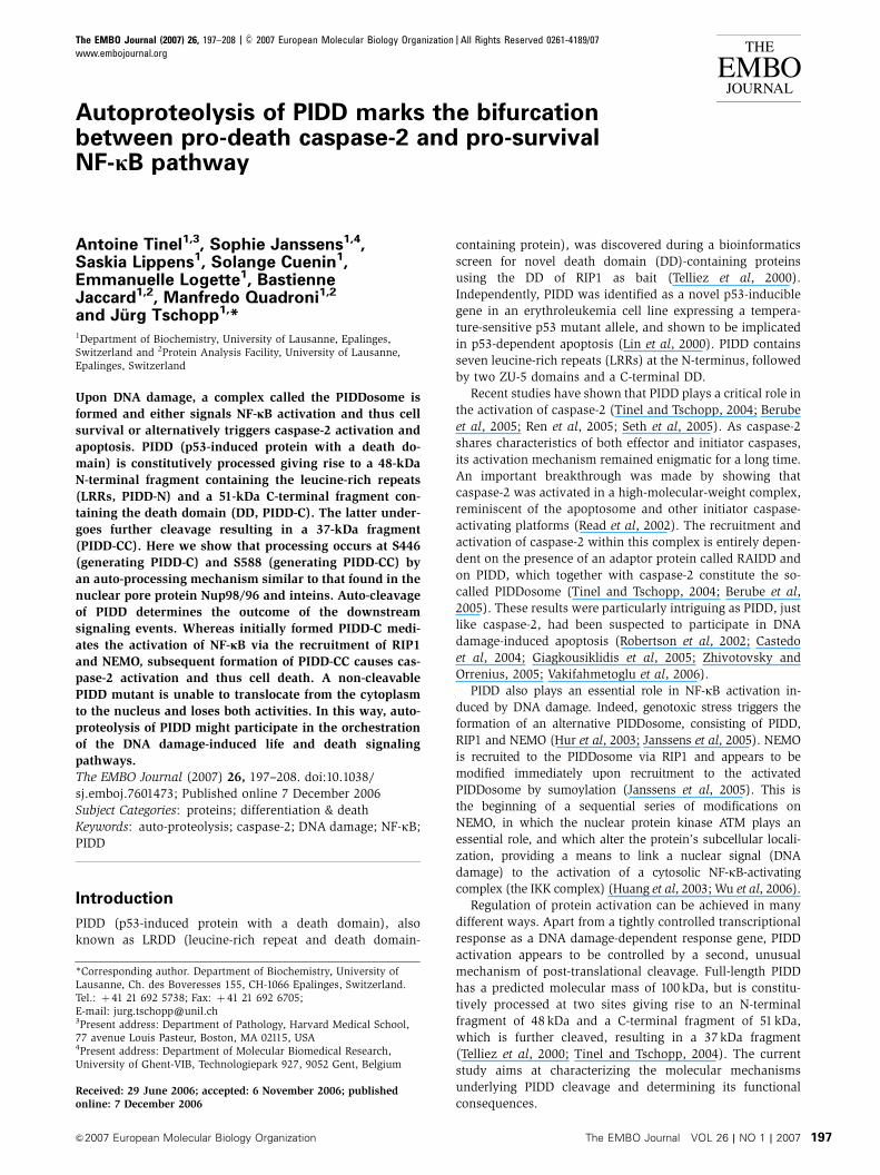
We next evaluated the functions of the PIDD-C and PIDD-CC
fragments. We first compared the caspase-2-activating capa-
city of wt PIDD with the different mutant forms of PIDD
that are unable to generate PIDD-C, PIDD-CC or both of
these fragments. Interestingly, transient overexpression of
the PIDD-CC-deficient S588A mutant failed to activate
caspase-2, whereas the PIDD-C-deficient S446A mutant was
still competent (Figure 4A). As expected from these data, the
double-PIDD-C/PIDD-CC-deficient S446A/S588A mutant also
lost its caspase-2-activating capacity. Caspase-2 activation by
PIDD is mediated by RAIDD. Whereas wt and PIDD-C-defi-
cient S446A mutant bound to RAIDD, the PIDD-CC-deficient
S588A mutant or the double mutant did not, explaining at
the molecular level the absence of caspase-2 activation
(Figure 4B). These data suggest that the generation of the
PIDD-CC fragment is required for PIDD-induced activation of
caspase-2. To further strengthen this finding, we established
populations of HeLa cells stably expressing the different
mutants and treated them with doxorubicin (Figure 4C).
Only wt PIDD and PIDD-C-deficient S446A mutant sensitized
the cells to doxorubicin-induced apoptosis. The low level of
sensitization observed with the PIDD-CC-deficient S588A
mutant is probably owing to overexpression and residual
caspase-2 activation. In order to determine which fragment
of PIDD is the active fragment involved in caspase-2 activa-
tion, HEK293T cells stably expressing both Flag-PIDD and
VSV-RAIDD were generated. These cells exhibited constitu-
tive caspase-2 activation owing to elevated expression of
PIDD (Tinel and Tschopp, 2004). When RAIDD was immuno-
precipitated, the PIDD-CC fragment, but not PIDD-C, was
detectable in the complex (Figure 4D and Supplementary
Figure 4A), confirming the importance of PIDD-CC genera-
tion for caspase-2 activation. The PIDD-CC fragment require-
ment for activation of the caspase-2 pathway was also seen
in HEK293Trex cell lines, which express PIDD-wt, PIDD-C or
PIDD-CC in an inducible manner. Induction of expression of
the various PIDD fragments showed that caspase-2 was most
efficiently activated in the clone expressing preformed PIDD-
CC, whereas in cells expressing wt and PIDD-C, caspase-2
activation was slower and followed the appearance of the
PIDD-CC fragment (Figure 4E). Immunoprecipitation studies
performed on these cells allowed us to correlate increased
caspase-2 activation with increased RAIDD and caspase-2
recruitment (Supplementary Figure 4B). Thus, PIDD-CC
expression is necessary and sufficient for the activation of
caspase-2. Importantly, stable expression of PIDD-DD (PIDD
776–910) failed to activate caspase-2 (Supplementary Figure
4C), indicating that yet another region of PIDD is required for
caspase-2 activation in addition to PIDD-DD.
Cleavage at S446 is required for NF-jB activation
PIDD constitutes not only a platform that, upon DNA da-
mage, results in caspase-2 activation, but is also an essential
protein for the DNA-damage-triggered NF-kB response
(Janssens et al, 2005). We previously showed that, in res-
ponse to DNA damage, PIDD translocates to the nucleus
(Janssens et al, 2005). To address how PIDD processing
affects nuclear translocation, we analyzed the localization
of several processing-defective PIDD mutants upon treatment
with camptothecin (CPT), a DNA-damaging drug. When DNA
damage was induced, a substantial portion of PIDD was
detectable in the nucleus (Figure 5A and B), whereas in
non-treated cells, PIDD was mostly cytoplasmic, in agree-
ment with previous results (Janssens et al, 2005). In contrast,
a PIDD-C-deficient S446A or double S446A/S588A mutant
no longer translocated to the nucleus, whereas the lack of
cleavage before S588 in the PIDD-CC-deficient S588A mutant
only slightly affected accumulation in the nucleus upon
genotoxic stress (Figure 5A and B). These results therefore
indicate that generation of the PIDD-C fragment is a pre-
requisite for PIDD nuclear translocation upon DNA damage.
Indeed, expression of the different PIDD fragments confirmed
that PIDD-C was able to translocate to the nucleus, whereas
PIDD-CC alone showed an exclusively cytoplasmic localiza-
tion (see Supplementary Figure 5).
Moc
k
PID
D
PID
D-N
PID
D-C
Moc
k
PID
D
PID
D-N
PID
D-C
WB: αRAIDD
WB: αFlag
WB: αCasp2 Procasp2
p31
p19
PIDD-CPIDD-NPIDD-CC
No treatment Etoposide
Fol
d in
duct
ion
MockPIDDPIDD∆LRRPIDD-NPIDD-C
1
2
3
4
5
6
7
8
9
10
0
PIDD-CC
IP: αFlag LysatesA
B
Figure 3 PIDD-N is a regulatory fragment of PIDD. (A) PIDD-N isnot required for RAIDD interaction. FL PIDD, PIDD-N and PIDD-Cwere stably expressed in HEK293T cells and their interaction withRAIDD and their capacity to activate caspase-2 assayed. (B)HEK293T cells stably expressing the indicated constructs weretested for their NF-kB-activating capacity induced by DNA damage(40 mM etoposide) using a luciferase-based reporter gene assay.
Autoproteolysis of PIDD shapes its functionA Tinel et al
&2007 European Molecular Biology Organization The EMBO Journal VOL 26 | NO 1 | 2007 201
To check if the altered translocation capacity also affected
PIDD’s ability to activate NF-kB, we analyzed HEK293T cells
stably expressing the various PIDD mutants using an NF-kB-
dependent luciferase reporter gene assay (Figure 5C). We
previously demonstrated that in comparison to mock-trans-
fected cells, cells stably expressing PIDD showed a moderate
but significant enhancement of the NF-kB response upon
DNA damage (Janssens et al, 2005). In keeping with our
translocation data, we observed that the enhanced NF-kB
response was only detectable for the wt PIDD and PIDD-CC-
deficient mutant (S588A), whereas the PIDD-C-deficient
mutants (S446A and S446A/S588A) were inactive. An identical
Moc
k
PID
D
PID
DS
446A
PID
DS
588A
PID
DS
446A
/S58
8A
Moc
k
Moc
k
PID
D
PID
D
PID
DS
446A
PID
DS
446A
PID
DS
588A
PID
DS
588A
PID
DS
446A
/S58
8A
PID
DS
446A
/S58
8A
PIDD-C
PIDD-CC
PIDD-FLPIDD-C
PIDD-CC
PIDD-FL
PIDD-C
PIDD-CC
Moc
k
RA
IDD
PID
D
PID
D+R
AID
D
VSV-RAIDDFlag-PIDD
PIDD-C
PIDD-CC
PIDD-C
PIDD-CC
No treatment Doxorubicin
% C
ell d
eath
10
20
30
40
50
60
70
0
WB: αRAIDD
WB: αRAIDD
WB: αTubulin
WB: αRAIDD
WB: αPIDD
WB: αPIDD
WB: αVSV
WB: αFADD
WB: αFlag
WB: αFlag
WB: αFlag
WB: αCasp2
WB: αCasp2
Procasp2
p31
p19
MockPIDDPIDDS446APIDDS588APIDDS446A /S588A
IP: αFlag Lysates
IP: αVSV
IP: αVSV
Lysates
WB: αVSVLysates
Procasp2
p31
p19
0h 5 8 24 0 05 58 824 24
PIDD PIDD-C PIDD-CC
A B
C
D
E
Figure 4 PIDD-CC recruits RAIDD and activates caspase-2. (A) The different PIDD mutants were transiently expressed in HEK293T cells andcaspase-2 activation determined by monitoring cleavage of caspase-2. (B) Flag-tagged PIDD mutants stably expressed in HEK293T cells andimmunoprecipitated using anti-Flag antibodies were analyzed for their capacity to interact with RAIDD. (C) HeLa cells stably expressing theindicated constructs were treated with doxorubicin (5mg/ml) and the dead cells were counted by Trypan blue. Representative of threeindependent experiments is shown. (D) HEK293T cells, stably expressing wt Flag-PIDD, VSV-RAIDD or both, were lysed and anti-VSV-RAIDDimmunoprecipitates analyzed for the presence of PIDD fragments. (E) HEK293Trex cells expressing FL-PIDD, PIDD-C or PIDD-CC were inducedby doxycycline for the indicated times and caspase-2 activation was monitored.
Autoproteolysis of PIDD shapes its functionA Tinel et al
The EMBO Journal VOL 26 | NO 1 | 2007 &2007 European Molecular Biology Organization202

tendency could be observed when phosphorylation of IkB
was used as a readout for NF-kB activation (Supplementary
Figure 6).
We next determined which fragment of PIDD interacted
with the downstream adaptor RIP1 (Figure 5D). RIP1 medi-
ates recruitment of NEMO to PIDD, resulting in NEMO
ANontreated NontreatedCPT CPT
PIDD
PIDDS588A
PIDDS446A
PIDDS446A S588A
Flag+DRAQ5
PIDD-C
PIDD-FLPIDD∆LRR
PIDD-CC
VSV-RIP1 + + +
Flag-PIDD∆LRR +
Flag-PIDD-C +
Flag-PIDD +++
VSV-RAIDD ++ + +
+
++++
+
PIDD-C
PIDD-FL
PIDD-CC
cy nu cy nu cy nu cy nu cy cy cy cy cy cynu nu nu nu nu nu
Moc
k
Moc
k
PID
D
PID
D
PID
DS
446A
PID
DS
446A
PID
DS
588A
PID
DS
588A
PID
DS
446A
S58
8A
PID
DS
446A
S58
8A
DMSO CPT
Flag
WB: αFlag
WB: αPARP
WB: αCasp3
WB: αPIDD
WB: αVSV
IP: αVSV Lysates
7
6
5
4
3
2
1
0No treatment
Fol
d in
duct
ion
Etoposide
MockPIDDPIDDS446APIDDS588APIDDS446A/S588A
B
C D
Figure 5 PIDD-C recruits RIP1 and activates NF-kB in response to DNA damage. (A) PIDD mutants unable to generate PIDD-C do nottranslocate to the nucleus in response to genotoxic stress. PIDD- or mutant PIDD-expressing HEK293Tcells were left untreated or stimulated for2 h with 50mM CPT. Confocal images show nuclear (blue, DRAQ5) or PIDD (green, Alexa 488) staining. (B) Subcellular fractionation of HEK293Tcells stably expressing the different PIDD mutants. The cells were treated with DMSO or with 50mM CPT. (C) PIDD-FL and PIDD mutant proteinsstably expressed in HEK293Tcells were tested for their NF-kB-activating capacity induced by DNA damage (40mM etoposide) using a luciferase-based reporter gene assay. (D) HEK293T cells were cotransfected with VSV-RAIDD or VSV-RIP1 in combination with different Flag-tagged PIDDconstructs and immunoprecipitated with an anti-VSV antibody. The presence of the different PIDD fragments in the eluates was monitored.
Autoproteolysis of PIDD shapes its functionA Tinel et al
&2007 European Molecular Biology Organization The EMBO Journal VOL 26 | NO 1 | 2007 203
sumoylation and subsequent NF-kB activation (Janssens
et al, 2005). In contrast to RAIDD that interacted with
PIDD-CC, RIP1 recruited exclusively the PIDD-C fragment.
Thus, the generation of the PIDD-C fragment is crucial
for the activation of NF-kB but not of caspase-2. Moreover,
cleavage at S446 is a prerequisite for PIDD translocation to
the nucleus in response to DNA damage, showing an intimate
link between these two processes.
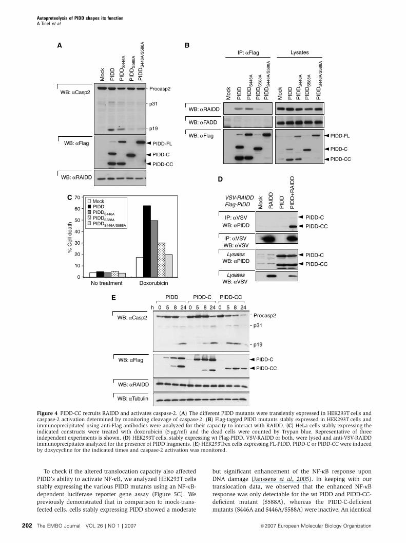
The two PIDD fragments are formed in a sequential
manner
The results of Figure 1C, in which we evaluated which
fragments were formed in the different PIDD cleavage mut-
ants, already suggested that the cleavage of the two fragments
occurred in a sequential manner. Thus, the generation of
PIDD-CC appeared much more efficient in molecules that
were initially cut at S446. We confirmed this sequential
order of events by monitoring the kinetics of PIDD fragment
formation in stable cell lines expressing PIDD in an inducible
manner (Figure 6A). HEK293 Flp-In TRex (HEK293Trex) cells
were induced to express PIDD by addition of doxycycline.
Four hours following the induction, only PIDD-C was detect-
able, whereas the PIDD-CC form was absent. Minor amounts
of PIDD-FL were also detected, but PIDD-FL levels did not
increase, confirming that PIDD-FL was directly processed
after expression. PIDD-CC was detectable only after 6–8 h.
The ratio of PIDD-CC to PIDD-C then steadily increased to
attain comparable levels 16 h after induction. The notion that
PIDD processing occurs constitutively in our stable cells was
further supported by an experiment in which protein synth-
esis was blocked with cycloheximide (CHX) (Figure 6B).
Together with PIDD-FL, the amount of PIDD-C fragment
steadily diminished, whereas the relative amount of PIDD-
CC slightly increased. That the disappearance of PIDD-C
fragment represented a genuine conversion to PIDD-CC and
not proteasome-dependent degradation was apparent based
on the observation that (1) it also disappeared in the presence
of the proteasome inhibitor MG-132 and (2) in the case of the
PIDD-CC-deficient S588A mutant, the PIDD-C fragment was
stable up to 16 h following CHX addition. Of note, PIDD-FL
was rapidly and completely processed into PIDD-C and PIDD-
N. No intermediary PIDD-N fragment (1–587) was detected
(Figure 1D and Supplementary Figure 6), suggesting that
PIDD-CC does not normally arise from PIDD-FL processing.
Addition of CHX did not result in further processing of
PIDD-N (Supplementary Figure 7). A sequential processing
of PIDD-FL to PIDD-N and PIDD-C and then to PIDD-CC
is therefore likely to occur.
That this mechanism is of physiological relevance was
supported by monitoring PIDD expression in response to
DNA damage. In agreement with previous reports (Lin et al,
2000), we found that DNA damage such as that induced by
etoposide or mitomycin C treatment led to a general increase
of PIDD expression in HCT116 cells (Figure 6C). Interestingly,
PIDD-C levels increased before PIDD-CC levels in response
to both treatments.
We then investigated the possibility that distinct doses
of DNA damage insults would differentially induce the two
PIDD fragments upon de novo synthesis. To this end, we
exposed the cells to a single dose of g-irradiation, which, in
contrast to continuous treatment with topoisomerase inhibi-
tors, allows the cells to recuperate from the DNA damage
insult. We exposed HEK293T cells to different amounts of
g-irradiation, ranging from 0.1 to 10 Gy (only the results for
0.1 and 10 Gy are depicted in the figure). As can be seen in
Figure 6D, PIDD-C was the first fragment to be induced and
was present from the 8 h time point onwards. We have never
been able to detect PIDD-FL, suggesting that the first cleavage
event is indeed constitutive. PIDD-CC increase occurred
much more slowly, the actual kinetics being strongly depen-
0 8 16 16+
Hours of CHX
PIDD-C
PIDD-FL
PIDD-CC
MG-132
PIDD
0 8 16 16+
PIDDS446A
0 8 16 16+
PIDDS588AA B
PIDD-C
PIDD-CC
Etoposide
72 0
Mitomycin C
12 24 480 12 24 48h
C
WB: αFlag WB: αFlag
0 4 6 8 10 16h
PIDD-C
PIDD-FL
PIDD-CC
WB: αRAIDD
WB: αRAIDD
WB: αRAIDD
WB: αPIDD
WB: αPARP
WB: αPIDD
WB: αRAIDD
D0
0.1 Gy
8 24 48 72
PIDD-C
PIDD-CC
h 0
10 Gy
8 24 48 72
PARPCleaved PARP
Figure 6 PIDD-C and PIDD-CC are induced with differential kinetics. (A) Induction of PIDD expression in HEK293Trex cells by doxycyclineduring the indicated times. Processing of PIDD was monitored by Western blot analysis of the cell extracts. (B) HEK293Tcells stably expressingthe indicated PIDD mutants were treated for 0, 8 and 16 h with 10mg/ml CHX in the presence or absence of the proteasome inhibitor MG-132(10mM). Processing of PIDD was monitored by Western blot. (C) HCT116 cells were treated with 10 mM etoposide or 10mM mitomycin C for theindicated times and PIDD expression levels were monitored. (D) HCT116 cells were exposed to a single dose of g-irradiation (0.1 or 10 Gy),whereafter the cells could recuperate. Endogenous PIDD expression levels and PARP cleavage were monitored.
Autoproteolysis of PIDD shapes its functionA Tinel et al
The EMBO Journal VOL 26 | NO 1 | 2007 &2007 European Molecular Biology Organization204

dent on the dose of treatment used. Whereas low doses of
g-irradiation, with only a mild effect on cell death, induced
significant levels of PIDD-CC after 48 h, high doses, which
induced strong apoptosis, evoked an immediate induction of
both PIDD-C and PIDD-CC (Figure 6D). Interestingly, at 72 h,
PIDD-C was no longer present, suggesting that all PIDD-C
was converted into PIDD-CC. These data therefore indicate
that both PIDD fragments are not induced with the same
kinetics upon DNA damage, and may reflect a regulated
activity of the second cleavage site in response to this
stimulus.
Discussion
Protein splicing is a form of post-translational processing that
is the protein equivalent of RNA splicing. There is now formal
evidence that these reactions exist in several protein matura-
tion pathways, including hedgehog protein maturation
(Porter et al, 1996), nucleoporin processing (Rosenblum
and Blobel, 1999; Hodel et al, 2002) and GPCR processing
(Lin et al, 2004). PIDD is constitutively cleaved into three
main fragments: PIDD-N (1–445), PIDD-C (446–910) and
PIDD-CC (588–910). Although our results do not provide
direct evidence for the exact reaction mechanism involved
in PIDD processing, they are consistent with auto-proteolysis,
similar to the previously described mechanisms, as they
include: (i) the cleavage site before a serine residue, (ii)
requirement for a hydroxyl or thiol group at the cleavage
position, and (iii) the sensitivity of in vitro cleavage of the
H444Q/S446C mutant to hydroxylamine. Thus, PIDD is auto-
processed at two sites, F445/S446 and F587/S588, that are
highly conserved and contain the HFS tripeptide.
An almost identical auto-processing site is found in Nup98,
for which the three-dimensional structure has been deter-
mined (Rosenblum and Blobel, 1999; Hodel et al, 2002).
Auto-processing of precursor Nup98 into Nup98 and the
8-kDa tail fragment results in only a small molecular
rearrangement and the interaction of the mature Nup98
with the 8-kDa tail fragment persists (Hodel et al, 2002).
Non-covalent interaction between the different fragments
generated appears to be a rule for proteins that undergo
auto-processing (Brannigan et al, 1995; Hodel et al, 2002;
Chang et al, 2003; Hsieh et al, 2003). Similarly, we found
that fragments generated by both cleavage events in PIDD
remained associated in a complex containing PIDD-N and
PIDD-C or PIDD-CC. The observation that RAIDD preferen-
tially immunoprecipitates PIDD-CC and not PIDD-C suggests
that PIDD-C and PIDD-CC may not be present simultaneously
in the same complex, or that dissociation of the different
fragments may occur upon activation. The fate of the 14-kDa
fragment (446–587) generated upon cleavage at S588 is
currently unknown.
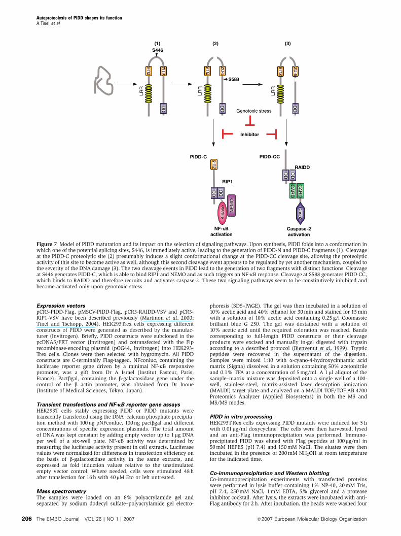
Our data unambiguously demonstrate an important role
of PIDD processing with respect to the initiation of down-
stream signaling events. We previously demonstrated that
PIDD is implicated in the activation of both caspase-2 and
NF-kB upon DNA damage. Intriguingly, both functions
appear to be controlled by different PIDD fragments gener-
ated by autoproteolysis (Figure 7). PIDD-C is able to interact
with RIP1, thereby initiating the NEMO modifications that
are required for NF-kB activation (Janssens et al, 2005).
Interestingly, the PIDD-C-deficient S446A mutant does not
translocate into the nucleus in response to DNA damage,
which is a prerequisite for NEMO modification to occur
(Huang et al, 2003; Janssens et al, 2005; Wu et al, 2006).
Theoretically, processing at S446 may unmask a nuclear
localization site or a site that allows binding to a nuclear
shuttling protein, thereby explaining the need for the first
processing. Although the PIDD-C fragment and the PIDD-CC-
deficient S588A mutant have the capacity to translocate and
induce NF-kB activation in response to DNA damage, they
neither recruit RAIDD nor activate caspase-2. The latter
functions are apparently triggered only by the RAIDD-
interacting PIDD-CC fragment, which is generated at a later
stage. It is noteworthy that the mere presence of PIDD-C and
PIDD-CC fragments found to be constitutively expressed in
a number of cell lines is insufficient to trigger caspase-2
or NF-kB activation in the absence of DNA damage. This
suggests that under normal conditions, the activities asso-
ciated with PIDD-C and PIDD-CC fragments are suppressed
by an unknown inhibitor.
We have previously shown that PIDD can act as a switch
allowing to either activate an NF-kB-dependent cell survival
pathway or trigger apoptosis by activation of caspase-2
(Janssens et al, 2005). The strength with which these two
signaling pathways are engaged will certainly be modulated
by the presence of other regulators. However, we also pro-
pose that the regulation of these alternate responses to
different stimuli might result from modulation of PIDD
processing. Indeed, the relative ratio of PIDD-C and PIDD-
CC varies in cells exposed to different DNA-damaging stimuli.
Whereas the introduction of a limited amount of DNA breaks
initially only leads to the induction of the NF-kB-activating
fragment PIDD-C, more severe DNA damage concomitantly
induced PIDD-C and PIDD-CC, correlating with a strong
apoptotic response of the cells. Thus, the temporarily distinct
generation of the PIDD-C and PIDD-CC fragments might
govern the fate of the cell, leading by default to the activation
of a cell survival pathway and, only when the cell repair
process fails, to the activation of the pro-apoptotic caspase-2
pathway.
In conclusion, we have shown that auto-processing of
PIDD generates PIDD-C and PIDD-CC fragments that are
involved in the activation of NF-kB and caspase-2, respec-
tively. Induction of PIDD upon DNA damage together with
regulated auto-processing may function to selectively direct
signaling pathways.
Materials and methods
Cell culture and biological reagentsHEK293T cells, HEK293Trex cells (Invitrogen), U2OS humanosteosarcoma cells, HCT116 p53þ /þ and p53�/� cells (gift fromDr B Vogelstein, John Hopkins University, Baltimore, MD, USA) and293T cells stably expressing different constructs of PIDD weregrown in DMEMþGlutamax (Life Technologies), supplementedwith 10% fetal calf serum, 100 U/ml penicillin and 100mg/mlstreptomycin. Raji B cells, Ramos B cells and Jurkat T cells weregrown in RPMI (Life Technologies) containing the same supple-ments. The generation of cells stably expressing PIDD or RAIDDwas described elsewhere (Tinel and Tschopp, 2004). Camptothecin(CPT), mitomycin C, MG-132, etoposide (Eto) and Ratjadone Cwere purchased from Alexis. Cells were g-irradiated with a 137Cssource in a single pulse. Doxycyline, hydroxylamine, CHX, and Flagpeptides were obtained from Sigma. Hygromycin was purchasedfrom Invitrogen.
Autoproteolysis of PIDD shapes its functionA Tinel et al
&2007 European Molecular Biology Organization The EMBO Journal VOL 26 | NO 1 | 2007 205
Expression vectorspCR3-PIDD-Flag, pMSCV-PIDD-Flag, pCR3-RAIDD-VSV and pCR3-RIP1-VSV have been described previously (Martinon et al, 2000;Tinel and Tschopp, 2004). HEK293Trex cells expressing differentconstructs of PIDD were generated as described by the manufac-turer (Invitrogen). Briefly, PIDD constructs were subcloned in thepcDNA5/FRT vector (Invitrogen) and cotransfected with the Flprecombinase-encoding plasmid (pOG44, Invitrogen) into HEK293-Trex cells. Clones were then selected with hygromycin. All PIDDconstructs are C-terminally Flag-tagged. NFconluc, containing theluciferase reporter gene driven by a minimal NF-kB responsivepromoter, was a gift from Dr A Israel (Institut Pasteur, Paris,France). Pactbgal, containing the b-galactosidase gene under thecontrol of the b actin promoter, was obtained from Dr Inoue(Institute of Medical Sciences, Tokyo, Japan).
Transient transfections and NF-jB reporter gene assaysHEK293T cells stably expressing PIDD or PIDD mutants weretransiently transfected using the DNA–calcium phosphate precipita-tion method with 100 ng pNFconluc, 100 ng pactbgal and differentconcentrations of specific expression plasmids. The total amountof DNA was kept constant by adding empty vector up to 1mg DNAper well of a six-well plate. NF-kB activity was determined bymeasuring the luciferase activity present in cell extracts. Luciferasevalues were normalized for differences in transfection efficiency onthe basis of b-galactosidase activity in the same extracts, andexpressed as fold induction values relative to the unstimulatedempty vector control. Where needed, cells were stimulated 48 hafter transfection for 16 h with 40mM Eto or left untreated.
Mass spectrometryThe samples were loaded on an 8% polyacrylamide gel andseparated by sodium dodecyl sulfate–polyacrylamide gel electro-
phoresis (SDS–PAGE). The gel was then incubated in a solution of10% acetic acid and 40% ethanol for 30 min and stained for 15 minwith a solution of 10% acetic acid containing 0.25 g/l Coomassiebrilliant blue G 250. The gel was destained with a solution of10% acetic acid until the required coloration was reached. Bandscorresponding to full-length PIDD constructs or their cleavageproducts were excised and manually in-gel digested with trypsinaccording to a described protocol (Bienvenut et al, 1999). Trypticpeptides were recovered in the supernatant of the digestion.Samples were mixed 1:10 with a-cyano-4-hydroxycinnamic acidmatrix (Sigma) dissolved in a solution containing 50% acetonitrileand 0.1% TFA at a concentration of 5 mg/ml. A 1 ml aliquot of thesample–matrix mixture was deposited onto a single well of a 100-well, stainless-steel, matrix-assisted laser desorption ionization(MALDI) target plate and analyzed on a MALDI TOF/TOF AB 4700Proteomics Analyzer (Applied Biosystems) in both the MS andMS/MS modes.
PIDD in vitro processingHEK293T-Rex cells expressing PIDD mutants were induced for 5 hwith 0.01mg/ml doxycycline. The cells were then harvested, lysedand an anti-Flag immunoprecipitation was performed. Immuno-precipitated PIDD was eluted with Flag peptides at 100mg/ml in50 mM HEPES (pH 7.4) and 150 mM NaCl. The eluates were thenincubated in the presence of 200 mM NH2OH at room temperaturefor the indicated time.
Co-immunoprecipitation and Western blottingCo-immunoprecipitation experiments with transfected proteinswere performed in lysis buffer containing 1% NP-40, 20 mM Tris,pH 7.4, 250 mM NaCl, 1 mM EDTA, 5% glycerol and a proteaseinhibitor cocktail. After lysis, the extracts were incubated with anti-Flag antibody for 2 h. After incubation, the beads were washed four
DD
ZU
-5ZU
-5
LRR
LRR
LRR
S446
S588
Caspase-2activation
NF-κB activation
Kin
ase
NE
MO
DD
PIDD-C
RIP1 CA
RD
Casp2
CA
RD
PIDD-CC
RAIDD
Genotoxic stress
Inhibitor
(1) (2) (3)
DD
DD
DD
DD
DD
ZU
-5
ZU
-5
ZU
-5
ZU
-5
ZU
-5
Figure 7 Model of PIDD maturation and its impact on the selection of signaling pathways. Upon synthesis, PIDD folds into a conformation inwhich one of the potential splicing sites, S446, is immediately active, leading to the generation of PIDD-N and PIDD-C fragments (1). Cleavageat the PIDD-C proteolytic site (2) presumably induces a slight conformational change at the PIDD-CC cleavage site, allowing the proteolyticactivity of this site to become active as well, although this second cleavage event appears to be regulated by yet another mechanism, coupled tothe severity of the DNA damage (3). The two cleavage events in PIDD lead to the generation of two fragments with distinct functions. Cleavageat S446 generates PIDD-C, which is able to bind RIP1 and NEMO and as such triggers an NF-kB response. Cleavage at S588 generates PIDD-CC,which binds to RAIDD and therefore recruits and activates caspase-2. These two signaling pathways seem to be constitutively inhibited andbecome activated only upon genotoxic stress.
Autoproteolysis of PIDD shapes its functionA Tinel et al
The EMBO Journal VOL 26 | NO 1 | 2007 &2007 European Molecular Biology Organization206
times with lysis buffer and analyzed by SDS–PAGE and immuno-blotting. The antibodies used for Western blotting were anti-phospho-IkB (Cell Signalling), anti-caspase-2 11B4 (Alexis), rabbitanti-RAIDD (Alexis) and anti-FADD (Transduction Labs). A mono-clonal anti-PIDD antibody (Anto-1) was raised against PIDD-DD(776–910) according to standard procedures. AL249, a rabbit anti-PIDD polyclonal antibody raised against a peptide covering aminoacids 148–174, was purchased from Alexis.
Confocal microscopyHEK293T cells stably expressing the different PIDD mutants werecultured on sterile glass coverslips in six-well plates and fixed with3% paraformaldehyde for 15 min. Cells were permeabilized with0.3% saponin for 10 min and treated with 2% normal goat serum(NGS)/0.5% BSA/0.1% saponin as a blocking reagent. Flagantibody (M2, Sigma) was used at a dilution of 1/500 in 0.1%saponin/0.1%BSA/PBS; a secondary Alexa 488 anti-mouse IgG1(Molecular Probes) was used at a dilution of 1/300 in 0.1%saponin/0.1%BSA/PBS. Cells were mounted in FluorSave (Calbio-chem) containing a 1/1000 dilution of DRAQ5 (Alexis) for nuclearcounterstain. Images from immunostaining were acquired by usinga Zeiss inverted laser scanning confocal microscope LSM 510 witha � 63 oil-immersion objective.
Biochemical fractionationCells were lysed in a cytosolic lysis buffer containing 10 mM HEPES,pH 7.9, 1.5 mM MgCl2, 300 mM sucrose, 0.5% NP-40, 10 mM KCl,supplemented with DTT and protease inhibitors. The lysates werecentrifuged and the supernatant constituted the cytosolic fraction.The pellet was then lysed in RIPA buffer and subsequentlysonicated. The supernatant constituted the nuclear-enrichedfraction.
Supplementary dataSupplementary data are available at The EMBO Journal Online(http://www.embojournal.org).
Acknowledgements
We thank S Hertig for technical support and H Everett, E Meylan,D Muruve, M Thome and P Schneider and the whole Tschopp groupfor discussions and critical reading of the manuscript. This workwas supported by grants from the Swiss National ScienceFoundation. SJ is supported by an EMBO fellowship and EL by anFRM fellowship.
References
Berube C, Boucher LM, Ma W, Wakeham A, Salmena L, Hakem R,Yeh WC, Mak TW, Benchimol S (2005) Apoptosis caused byp53-induced protein with death domain (PIDD) depends onthe death adapter protein RAIDD. Proc Natl Acad Sci USA 102:14314–14320
Bienvenut WV, Sanchez JC, Karmime A, Rouge V, Rose K, Binz PA,Hochstrasser DF (1999) Toward a clinical molecular scanner forproteome research: parallel protein chemical processing beforeand during Western blot. Anal Chem 71: 4800–4807
Brannigan JA, Dodson G, Duggleby HJ, Moody PC, Smith JL,Tomchick DR, Murzin AG (1995) A protein catalytic frameworkwith an N-terminal nucleophile is capable of self-activation.Nature 378: 416–419
Castedo M, Perfettini JL, Roumier T, Valent A, Raslova H, YakushijinK, Horne D, Feunteun J, Lenoir G, Medema R, Vainchenker W,Kroemer G (2004) Mitotic catastrophe constitutes a special case ofapoptosis whose suppression entails aneuploidy. Oncogene 23:4362–4370
Chang GW, Stacey M, Kwakkenbos MJ, Hamann J, Gordon S, LinHH (2003) Proteolytic cleavage of the EMR2 receptor requiresboth the extracellular stalk and the GPS motif. FEBS Lett 547:145–150
Fabre E, Boelens WC, Wimmer C, Mattaj IW, Hurt EC (1994)Nup145p is required for nuclear export of mRNA and bindshomopolymeric RNA in vitro via a novel conserved motif. Cell78: 275–289
Fontoura BM, Blobel G, Matunis MJ (1999) A conserved biogenesispathway for nucleoporins: proteolytic processing of a 186-kilo-dalton precursor generates Nup98 and the novel nucleoporin,Nup96. J Cell Biol 144: 1097–1112
Giagkousiklidis S, Vogler M, Westhoff MA, Kasperczyk H, DebatinKM, Fulda S (2005) Sensitization for gamma-irradiation-inducedapoptosis by second mitochondria-derived activator of caspase.Cancer Res 65: 10502–10513
Hodel AE, Hodel MR, Griffis ER, Hennig KA, Ratner GA, Xu S,Powers MA (2002) The three-dimensional structure of the auto-proteolytic, nuclear pore-targeting domain of the human nucleo-porin Nup98. Mol Cell 10: 347–358
Hsieh JJ, Cheng EH, Korsmeyer SJ (2003) Taspase1: a threonineaspartase required for cleavage of MLL and proper HOX geneexpression. Cell 115: 293–303
Hu Y, Ding L, Spencer DM, Nunez G (1998) WD-40 repeat regionregulates Apaf-1 self-association and procaspase-9 activation.J Biol Chem 273: 33489–33494
Huang TT, Wuerzberger-Davis SM, Wu ZH, Miyamoto S (2003)Sequential modification of NEMO/IKKgamma by SUMO-1 andubiquitin mediates NF-kappaB activation by genotoxic stress. Cell115: 565–576
Hur GM, Lewis J, Yang Q, Lin Y, Nakano H, Nedospasov S, Liu ZG(2003) The death domain kinase RIP has an essential role in
DNA damage-induced NF-kappa B activation. Genes Dev 17:873–882
Janssens S, Tinel A, Lippens S, Tschopp J (2005) PIDD mediatesNF-kappaB activation in response to DNA damage. Cell 123:1079–1092
Krasnoperov VG, Bittner MA, Beavis R, Kuang Y, Salnikow KV,Chepurny OG, Little AR, Plotnikov AN, Wu D, Holz RW, PetrenkoAG (1997) alpha-Latrotoxin stimulates exocytosis by the interactionwith a neuronal G-protein-coupled receptor. Neuron 18: 925–937
Lin HH, Chang GW, Davies JQ, Stacey M, Harris J, Gordon S(2004) Autocatalytic cleavage of the EMR2 receptor occurs ata conserved G protein-coupled receptor proteolytic site motif.J Biol Chem 279: 31823–31832
Lin Y, Ma W, Benchimol S (2000) Pidd, a new death-domain-containing protein, is induced by p53 and promotes apoptosis.Nat Genet 26: 122–127
Martinon F, Holler N, Richard C, Tschopp J (2000) Activation ofa pro-apoptotic amplification loop through inhibition of NF-kappaB-dependent survival signals by caspase-mediated inactiva-tion of RIP. FEBS Lett 468: 134–136
Martinon F, Tschopp J (2005) NLRs join TLRs as innate sensors ofpathogens. Trends Immunol 26: 447–454
Nechiporuk T, Urness LD, Keating MT (2001) ETL, a novel seven-transmembrane receptor that is developmentally regulated in theheart. ETL is a member of the secretin family and belongs to theepidermal growth factor-seven-transmembrane subfamily. J BiolChem 276: 4150–4157
Paulus H (2000) Protein splicing and related forms of proteinautoprocessing. Annu Rev Biochem 69: 447–496
Perler FB, Adam E (2000) Protein splicing and its applications. CurrOpin Biotechnol 11: 377–383
Porter J, Ekker S, Park W, von Kessler D, Young K, Chen C, Ma Y,Woods A, Cotter R, Koonin E (1996) Hedgehog patterning acti-vity: role of a lipophilic modification mediated by the carboxy-terminal autoprocessing domain. Cell 86: 21–34
Poyet JL, Srinivasula SM, Tnani M, Razmara M, Fernandes-AlnemriT, Alnemri ES (2001) Identification of Ipaf, a human caspase-1-activating protein related to Apaf-1. J Biol Chem 276:28309–28313
Read SH, Baliga BC, Ekert PG, Vaux DL, Kumar S (2002) A novelApaf-1-independent putative caspase-2 activation complex. J CellBiol 159: 739–745
Ren J, Shi M, Liu R, Yang QH, Johnson T, Skarnes WC, Du C (2005)The Birc6 (Bruce) gene regulates p53 and the mitochondrialpathway of apoptosis and is essential for mouse embryonicdevelopment. Proc Natl Acad Sci USA 102: 565–570
Robertson JD, Enoksson M, Suomela M, Zhivotovsky B, Orrenius S(2002) Caspase-2 acts upstream of mitochondria to promotecytochrome c release during etoposide-induced apoptosis. J BiolChem 277: 29803–29809
Autoproteolysis of PIDD shapes its functionA Tinel et al
&2007 European Molecular Biology Organization The EMBO Journal VOL 26 | NO 1 | 2007 207

Rosenblum JS, Blobel G (1999) Autoproteolysis in nucleoporinbiogenesis. Proc Natl Acad Sci USA 96: 11370–11375
Seth R, Yang C, Kaushal V, Shah SV, Kaushal GP (2005) p53-dependent caspase-2 activation in mitochondrial release of apop-tosis-inducing factor and its role in renal tubular epithelial cellinjury. J Biol Chem 280: 31230–31239
Srinivasula SM, Ahmad M, Fernandes-Alnemri T, Alnemri ES (1998)Autoactivation of procaspase-9 by Apaf-1-mediated oligomeriza-tion. Mol Cell 1: 949–957
Telliez JB, Bean KM, Lin LL (2000) LRDD, a novel leucine richrepeat and death domain containing protein. Biochim BiophysActa 1478: 280–288
Tinel A, Tschopp J (2004) The PIDDosome, a protein compleximplicated in activation of caspase-2 in response to genotoxicstress. Science 304: 843–846
Vakifahmetoglu H, Olsson M, Orrenius S, Zhivotovsky B(2006) Functional connection between p53 and caspase-2 isessential for apoptosis induced by DNA damage. Oncogene 25:5683–5692
Wu ZH, Shi Y, Tibbetts RS, Miyamoto S (2006) Molecular linkagebetween the kinase ATM and NF-kappaB signaling in response togenotoxic stimuli. Science 311: 1141–1146
Zhivotovsky B, Orrenius S (2005) Caspase-2 function in response toDNA damage. Biochem Biophys Res Commun 331: 859–867
Autoproteolysis of PIDD shapes its functionA Tinel et al
The EMBO Journal VOL 26 | NO 1 | 2007 &2007 European Molecular Biology Organization208