Epistatic interactions between loci of one-carbon metabolism modulate susceptibility to breast...
11
1 23 Molecular Biology Reports An International Journal on Molecular and Cellular Biology ISSN 0301-4851 Volume 38 Number 8 Mol Biol Rep (2011) 38:4893-4901 DOI 10.1007/s11033-010-0631-z Epistatic interactions between loci of one- carbon metabolism modulate susceptibility to breast cancer Shaik Mohammad Naushad, Addepalli Pavani, Raghunadha Rao Digumarti, Suryanarayana Raju Gottumukkala & Vijay Kumar Kutala
Transcript of Epistatic interactions between loci of one-carbon metabolism modulate susceptibility to breast...
1 23
Molecular Biology ReportsAn International Journal on Molecularand Cellular Biology ISSN 0301-4851Volume 38Number 8 Mol Biol Rep (2011) 38:4893-4901DOI 10.1007/s11033-010-0631-z
Epistatic interactions between loci of one-carbon metabolism modulate susceptibilityto breast cancer
Shaik Mohammad Naushad, AddepalliPavani, Raghunadha Rao Digumarti,Suryanarayana Raju Gottumukkala &Vijay Kumar Kutala

1 23
Your article is protected by copyright and
all rights are held exclusively by Springer
Science+Business Media B.V.. This e-offprint
is for personal use only and shall not be self-
archived in electronic repositories. If you
wish to self-archive your work, please use the
accepted author’s version for posting to your
own website or your institution’s repository.
You may further deposit the accepted author’s
version on a funder’s repository at a funder’s
request, provided it is not made publicly
available until 12 months after publication.
Epistatic interactions between loci of one-carbon metabolismmodulate susceptibility to breast cancer
Shaik Mohammad Naushad • Addepalli Pavani •
Raghunadha Rao Digumarti • Suryanarayana Raju Gottumukkala •
Vijay Kumar Kutala
Received: 14 September 2010 / Accepted: 3 December 2010 / Published online: 14 December 2010
� Springer Science+Business Media B.V. 2010
Abstract In view of growing body of evidence substanti-
ating the role of aberrations in one-carbon metabolism in the
pathophysiology of breast cancer and lack of studies on
gene–gene interactions, we investigated the role of dietary
micronutrients and eight functional polymorphisms of one-
carbon metabolism in modulating the breast cancer risk in
244 case–control pairs of Indian women and explored pos-
sible gene–gene interactions using Multifactor dimension-
ality reduction analysis (MDR). Dietary micronutrient status
was assessed using the validated Food Frequency Ques-
tionnaire. Genotyping was done for glutamate carboxypep-
tidase II (GCPII) C1561T, reduced folate carrier (RFC)1
G80A, cytosolic serine hydroxymethyltransferase (cSHMT)
C1420T, thymidylate synthase (TYMS) 50-UTR tandem
repeat, TYMS 30-UTR ins6/del6, methylenetetrahydrofolate
reductase (MTHFR) C677T, methyltetrahydrofolate-homo-
cysteine methyltransferase (MTR) A2756G, methylte-
trahydrofolate-homocysteine methyltransferase reductase
(MTRR) A66G polymorphisms by using the PCR-RFLP/
AFLP methods. Low dietary folate intake (P \ 0.001),
RFC1 G80A (OR: 1.38, 95% CI 1.06–1.81) and MTHFR
C677T (OR: 1.74 (1.11–2.73) were independently associ-
ated with the breast cancer risk whereas cSHMT C1420T
conferred protection (OR: 0.72, 95% CI 0.55–0.94). MDR
analysis demonstrated a significant tri-variate interaction
among RFC1 80, MTHFR 677 and TYMS 50-UTR loci
(Ptrend \ 0.02) with high-risk genotype combination show-
ing inflated risk for breast cancer (OR 4.65, 95% CI
1.77–12.24). To conclude, dietary as well as genetic factors
were found to influence susceptibility to breast cancer.
Further, the current study highlighted the importance of
multi-loci analyses over the single-locus analysis towards
establishing the epistatic interactions between loci of one-
carbon metabolism modulate susceptibility to the breast
cancer.
Keywords Breast cancer � One-carbon metabolism �Dietary micronutrients � Epistasis � Multifactor
dimensionality reduction
Introduction
Breast cancer is the most common malignancy among
Indian women with a steady increase in its incidence [1].
Highly penetrant mutations account for a small proportion
of the breast cancer cases [2]. Several studies were con-
ducted globally, to explore etiological factors contributing
to majority of the breast cancer cases [3]. The importance
of low penetrant genetic polymorphisms as predictive as
well as prognostic markers for multi-factorial disorders has
become evident after the elucidation of human genome
sequence [4]. Genetic polymorphisms and dietary micro-
nutrients of one-carbon metabolism were studied exten-
sively in different cancers as these factors influence DNA
synthesis, repair, and methylation. The principal mecha-
nisms of carcinogenesis by these factors are uracil misin-
corporation in DNA causing DNA damage [5] or aberrant
S. M. Naushad � A. Pavani � V. K. Kutala (&)
Department of Clinical Pharmacology & Therapeutics, Nizam’s
Institute of Medical Sciences, Panjagutta, Hyderabad 500082,
Andhra Pradesh, India
e-mail: [email protected]
R. R. Digumarti
Department of Medical Oncology, Nizam’s Institute of Medical
Sciences, Panjagutta, Hyderabad 500082, India
S. R. Gottumukkala
Department of Surgical Oncology, Nizam’s Institute of Medical
Sciences, Panjagutta, Hyderabad 500082, India
123
Mol Biol Rep (2011) 38:4893–4901
DOI 10.1007/s11033-010-0631-z
Author's personal copy
methylation (focal hypermethylation and global hypome-
thylation) that triggers the inactivation of tumor suppressor
genes and inactivation of proto-oncogenes [6].
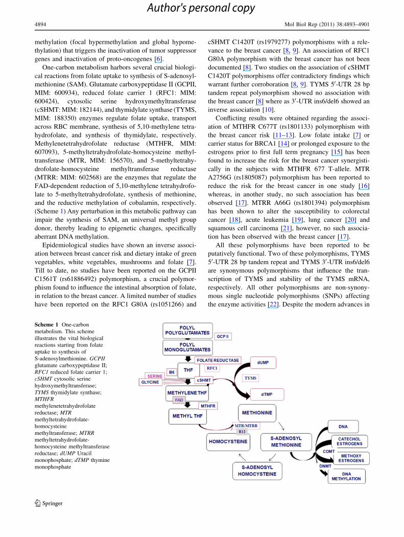
One-carbon metabolism harbors several crucial biologi-
cal reactions from folate uptake to synthesis of S-adenosyl-
methionine (SAM). Glutamate carboxypeptidase II (GCPII,
MIM: 600934), reduced folate carrier 1 (RFC1: MIM:
600424), cytosolic serine hydroxymethyltransferase
(cSHMT: MIM: 182144), and thymidylate synthase (TYMS,
MIM: 188350) enzymes regulate folate uptake, transport
across RBC membrane, synthesis of 5,10-methylene tetra-
hydrofolate, and synthesis of thymidylate, respectively.
Methylenetetrahydrofolate reductase (MTHFR, MIM:
607093), 5-methyltetrahydrofolate-homocysteine methyl-
transferase (MTR, MIM: 156570), and 5-methyltetrahy-
drofolate-homocysteine methyltransferase reductase
(MTRR: MIM: 602568) are the enzymes that regulate the
FAD-dependent reduction of 5,10-methylene tetrahydrofo-
late to 5-methyltetrahydrofolate, synthesis of methionine,
and the reductive methylation of cobalamin, respectively.
(Scheme 1) Any perturbation in this metabolic pathway can
impair the synthesis of SAM, an universal methyl group
donor, thereby leading to epigenetic changes, specifically
aberrant DNA methylation.
Epidemiological studies have shown an inverse associ-
ation between breast cancer risk and dietary intake of green
vegetables, white vegetables, mushrooms and folate [7].
Till to date, no studies have been reported on the GCPII
C1561T (rs61886492) polymorphism, a crucial polymor-
phism found to influence the intestinal absorption of folate,
in relation to the breast cancer. A limited number of studies
have been reported on the RFC1 G80A (rs1051266) and
cSHMT C1420T (rs1979277) polymorphisms with a rele-
vance to the breast cancer [8, 9]. An association of RFC1
G80A polymorphism with the breast cancer has not been
documented [8]. Two studies on the association of cSHMT
C1420T polymorphisms offer contradictory findings which
warrant further corroboration [8, 9]. TYMS 50-UTR 28 bp
tandem repeat polymorphism showed no association with
the breast cancer [8] where as 30-UTR ins6/del6 showed an
inverse association [10].
Conflicting results were obtained regarding the associ-
ation of MTHFR C677T (rs1801133) polymorphism with
the breast cancer risk [11–13]. Low folate intake [7] or
carrier status for BRCA1 [14] or prolonged exposure to the
estrogens prior to first full term pregnancy [15] has been
found to increase the risk for the breast cancer synergisti-
cally in the subjects with MTHFR 677 T-allele. MTR
A2756G (rs1805087) polymorphism has been reported to
reduce the risk for the breast cancer in one study [16]
whereas, in another study, no such association has been
observed [17]. MTRR A66G (rs1801394) polymorphism
has been shown to alter the susceptibility to colorectal
cancer [18], acute leukemia [19], lung cancer [20] and
squamous cell carcinoma [21], however, no such associa-
tion has been observed with the breast cancer [17].
All these polymorphisms have been reported to be
putatively functional. Two of these polymorphisms, TYMS
50-UTR 28 bp tandem repeat and TYMS 30-UTR ins6/del6
are synonymous polymorphisms that influence the tran-
scription of TYMS and stability of the TYMS mRNA,
respectively. All other polymorphisms are non-synony-
mous single nucleotide polymorphisms (SNPs) affecting
the enzyme activities [22]. Despite the modern advances in
Scheme 1 One-carbon
metabolism. This scheme
illustrates the vital biological
reactions starting from folate
uptake to synthesis of
S-adenosylmethionine. GCPIIglutamate carboxypeptidase II;
RFC1 reduced folate carrier 1;
cSHMT cytosolic serine
hydroxymethyltransferase;
TYMS thymidylate synthase;
MTHFRmethylenetetrahydrofolate
reductase; MTRmethyltetrahydrofolate-
homocysteine
methyltransferase; MTRRmethyltetrahydrofolate-
homocysteine methyltransferase
reductase; dUMP Uracil
monophosphate; dTMP thymine
monophosphate
4894 Mol Biol Rep (2011) 38:4893–4901
123
Author's personal copy
human genetics, no comprehensive multi-locus studies on
Indian women in relation to the breast cancer have been
documented. Results on the single locus investigations are
also sparse [23]. Furthermore, wide variations at the ethnic
and population levels have been reported in the distribution
of these polymorphisms [22]. In view of this, we conducted
a case–control study on South Indian subjects to examine
the individual and combined effects of eight genetic
polymorphisms such as GCPII C1561T, RFC1 G80A,
cSHMT C1420T, TYMS 50-UTR tandem repeat, TYMS 30-UTR ins6/del6, MTHFR C677T, MTR A2756G and
MTRR A66G, and the dietary status of four B vitamins in
modulating susceptibility to the breast cancer.
Materials and methods
Study population
Eligible cases were a consecutive series of female patients
aged 18–80 years with newly diagnosed and histologically
confirmed breast cancer. Cases were recruited during the
period of January, 2009 to July, 2010 at the Nizam’s
Institute of Medical Sciences (NIMS), Hyderabad, India. A
total of 244 cases met these criteria. Eligible controls were
subjects without any history of cancer, selected from a
group of 270 healthy women volunteers. These volunteers
comprised mostly of hospital staff or their relatives who
had no history of benign or malignant breast disease. Their
recruitment was done at NIMS, Hyderabad after careful
evaluation of their personal health and family history. The
selection of controls was done by matching each control to
each case by age (±5years), ethnicity, region, linguistic
origin and menopausal status. Ultimately, 244 matched
case–control pairs were enrolled in the study. Informed
consent was obtained from all the subjects. This study was
approved by the Institutional Ethical committee of Nizam’s
Institute of Medical Sciences, Hyderabad (EC/NIMS/767/
2007, dated 05.09.2008).
Demographic data collection and dietary assessment
Personal interviews were conducted by trained interviewers
using a standardized questionnaire which included demo-
graphic characteristics, life style (exercise, smoking, alco-
hol intake, tobacco chewing), reproductive history (age of
menarche, age at the time of first full term pregnancy,
breast feeding history, number of live births, number of
miscarriages, menopausal status, use of oral contraceptives,
hormone replacement therapy) and family history (breast
cancer or any other malignancies). Measurements of height
and weight were taken at the time of recruitment from all
the subjects to calculate body mass index (BMI). Clinical
details were obtained from medical records of the patients.
Personal interviews were conducted during their first visit
to the oncologist after confirmed diagnosis (for cases). The
blood samples were collected on the same day following
the interview in order to minimize the impact of surgical or
therapeutic intervention on biochemical parameters.
For food frequency questionnaire, all the subjects were
requested to keep a log on the type of food item consumed,
quantity of food consumed, and frequency of food con-
sumption (times per day/week/month/three months or
never) over a period of 1 week. Daily nutrient intakes were
calculated as grams of food multiplied by the amount of
each nutrient in the food and the frequency of consump-
tion, summing over all the foods consumed. The compo-
sition of raw and cooked food items was determined from
the 2007 report on the Nutritive value of Indian foods [24].
For further information on the composition of other food
items, McCance and Widdowson’s The composition of
Foods [25] and the United States Department of Agricul-
ture’s National Nutrient Database for Standard Reference
release 18 (USDA, Washington, DC, USA) were consulted
[26]. Subjects who are on vitamin supplementation were
not included as no comprehensive data were available.
The validity of micronutrient intake estimated from the
FFQ was established by two approaches. A subgroup of 62
women, who visited the hospital for a follow-up, com-
pleted one-week dietary record twice. The dietary micro-
nutrient status at two different time intervals was correlated
with one another. Spearman rank correlation coefficients
for folate, B2, B6 and B12 were 0.35, 0.30, 0.34 and 0.32,
respectively. We validated this data further by the bio-
chemical determination of folate in the plasma of subjects
who are not on anti-folate medication/folate supplementa-
tion. Spearman rank correlation coefficient was 0.29 for
dietary folate versus plasma folate levels.
Genetic analysis
Whole blood samples were collected in EDTA from all the
subjects and plasma was separated immediately and stored
at -80�C for further biochemical analyses. The buffycoat
was used for the genomic DNA isolation by the method
described by Salazar et al. [27]. The extracted DNA was
analyzed for eight genetic polymorphisms using the poly-
merase chain reaction-restriction fragment length poly-
morphism (PCR-RFLP) and PCR-amplified fragment
length polymorphism (PCR-AFLP) methods. Cases and
matched controls were analyzed in the same set of PCR by
the analyst blinded to case–control status. For all the
genetic analyses, each PCR set was accompanied by a
negative control without genomic DNA in order to check
contamination of components. Activities of all the restric-
tion enzymes were checked by digesting the plasmids with
Mol Biol Rep (2011) 38:4893–4901 4895
123
Author's personal copy
known restriction sites. For RFLP analysis, a positive
control was included in each set, which ensures complete
digestion. Genotypes in few specimens were randomly
rechecked to rule out the genotyping errors and 100%
concordance was observed.
Plasma folate estimation
Plasma folate was determined on the AxSYM automated
analyzer (Abbott Laboratories) according to the manufac-
turer’s instructions using the AxSYM folate assay kits.
Plasma folate analysis was performed only in the subjects
who were not on any anti-folate medication or vitamin
supplementation at the time of specimen collection. The
intra assay CV was 3.8% where as inter assay CV was
4.2%.
Statistical analysis
Demographic characteristics distributed as categorical
variables were subjected to simple logistic regression to
assess the trend associated with each characteristic. For
other demographic data where only absence or presence of
the variable was assessed, Fisher’s exact test was
employed. All the genetic analysis data were computed in
0, 1 and 2 model representing homozygous wild, hetero-
zygous and homozygous mutant genotypes, respectively.
Since the representative samples for homozygous mutant
genotype are less for some polymorphisms, the compari-
sons were made based 0 and 1 ? 2 combinations. Condi-
tional logistic regression analysis was done on the control
data for confounding effects and adjusted odds ratios (ORs)
were obtained. Potential confounders considered for the
multivariate analysis were body mass index, age at men-
arche, age at first full term pregnancy, parity, and the
family history of breast cancer. They were treated as
continuous variables for logistic regression analysis.
Wherever the information was in the format of yes/no, it
was labeled as 1/0 respectively. The Hardy–Weinberg
equilibrium was checked for cases and controls by adopt-
ing the chi-squared test.
All the continuous variables were subjected to the stu-
dent’s t-test to obtain mean, standard deviation (SD) and
the P value. In order to validate the food frequency ques-
tionnaire, the plasma folate levels were correlated with the
dietary folate levels by using the Spearman rank correlation
coefficient. Multifactor dimensionality reduction analysis
(MDR, version 2.0 beta 6) was used to reduce the data
dimension and to get a set of single nucleotide polymor-
phisms (SNPs) that can interpret the results best. The data
obtained was again subjected to the Fisher’s exact test for
each genotype combination to ascertain the trend across
different combinations. Dietary micronutrient status was
stratified into three tertiles. Each variant allele frequency in
cases and controls was computed across three tertiles and a
trend test was performed to assess gene-nutrient interac-
tions. For all the above statistical analysis, the statistical
web page ‘‘www.statpages.org’’ was used. For all associ-
ations, a P value of \0.05/n (n = number of variables
tested) was considered as statistically significant. All the
observed statistically significant genetic associations
exhibited C80% power with type I-alpha error of 0.05.
Results
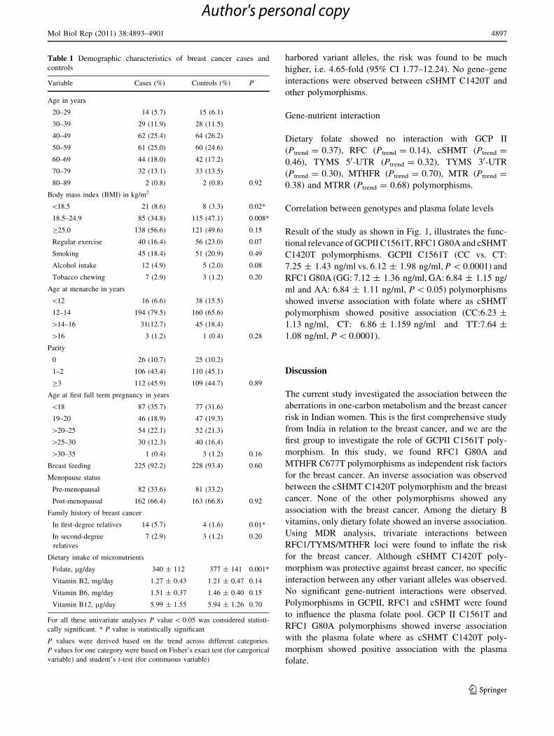
Demographic characteristics of cases and controls at
baseline are shown in Table 1. Women with low body mass
index (BMI) were found to have increased risk. However, a
careful evaluation revealed that risk is due to low plasma
folate, not directly due to low BMI. However, a significant
reduction in breast cancer risk was observed in women who
had optimal BMI. No statistically significant association
was observed for regular exercise, smoking, alcohol intake,
tobacco chewing, age at menarche, parity, age at first full
term pregnancy and breast feeding. Family history of breast
cancer and low dietary folate intake were associated with
the breast cancer risk (Table 1).
Individual genetic effects
The genotype distribution for all the polymorphisms
([0.29) except for MTR A2756G and MTRR A66G was in
accordance with the Hardy–Weinberg equilibrium in cases
and controls. Among the eight putatively functional poly-
morphisms tested, RFC1 G80A (OR: 1.38, 95% CI
1.06–1.81) and MTHFR C677T (OR: 1.74 (1.11–2.73)
were independently associated with the breast cancer risk,
whereas cSHMT C1420T conferred protection (OR: 0.72,
95% CI 0.55–0.94). Other polymorphisms showed no
independent effects (Table 2).
Gene–gene interactions
The multifactor dimensionality reduction as well as the
logistic regression indicated significant epistatic interac-
tions between three allelic variants i.e. RFC1 80A-MTHFR
677T-TYMS 2R in modulating the breast cancer risk in
dose-dependent manner (Ptrend \ 0.02) (Table 3). When
the different genotype combinations at RFC1, TYMS and
MTHFR loci were examined, with reference to the triple
wild genotype, presence of TYMS 2R allele alone showed
2.17-fold risk (95% CI 1.06–4.43) for the breast cancer.
Co-segregation of RFC1 80A and MTHFR 677T-variant
alleles was associated with 2.89-fold risk (95% CI
1.13–7.42) for the breast cancer. When all the three loci
4896 Mol Biol Rep (2011) 38:4893–4901
123
Author's personal copy
harbored variant alleles, the risk was found to be much
higher, i.e. 4.65-fold (95% CI 1.77–12.24). No gene–gene
interactions were observed between cSHMT C1420T and
other polymorphisms.
Gene-nutrient interaction
Dietary folate showed no interaction with GCP II
(Ptrend = 0.37), RFC (Ptrend = 0.14), cSHMT (Ptrend =
0.46), TYMS 50-UTR (Ptrend = 0.32), TYMS 30-UTR
(Ptrend = 0.30), MTHFR (Ptrend = 0.70), MTR (Ptrend =
0.38) and MTRR (Ptrend = 0.68) polymorphisms.
Correlation between genotypes and plasma folate levels
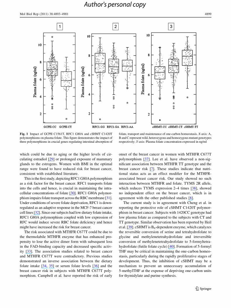
Result of the study as shown in Fig. 1, illustrates the func-
tional relevance of GCPII C1561T, RFC1 G80A and cSHMT
C1420T polymorphisms. GCPII C1561T (CC vs. CT:
7.25 ± 1.43 ng/ml vs. 6.12 ± 1.98 ng/ml, P \ 0.0001) and
RFC1 G80A (GG: 7.12 ± 1.36 ng/ml, GA: 6.84 ± 1.15 ng/
ml and AA: 6.84 ± 1.11 ng/ml, P \ 0.05) polymorphisms
showed inverse association with folate where as cSHMT
polymorphism showed positive association (CC:6.23 ±
1.13 ng/ml, CT: 6.86 ± 1.159 ng/ml and TT:7.64 ±
1.08 ng/ml, P \ 0.0001).
Discussion
The current study investigated the association between the
aberrations in one-carbon metabolism and the breast cancer
risk in Indian women. This is the first comprehensive study
from India in relation to the breast cancer, and we are the
first group to investigate the role of GCPII C1561T poly-
morphism. In this study, we found RFC1 G80A and
MTHFR C677T polymorphisms as independent risk factors
for the breast cancer. An inverse association was observed
between the cSHMT C1420T polymorphism and the breast
cancer. None of the other polymorphisms showed any
association with the breast cancer. Among the dietary B
vitamins, only dietary folate showed an inverse association.
Using MDR analysis, trivariate interactions between
RFC1/TYMS/MTHFR loci were found to inflate the risk
for the breast cancer. Although cSHMT C1420T poly-
morphism was protective against breast cancer, no specific
interaction between any other variant alleles was observed.
No significant gene-nutrient interactions were observed.
Polymorphisms in GCPII, RFC1 and cSHMT were found
to influence the plasma folate pool. GCP II C1561T and
RFC1 G80A polymorphisms showed inverse association
with the plasma folate where as cSHMT C1420T poly-
morphism showed positive association with the plasma
folate.
Table 1 Demographic characteristics of breast cancer cases and
controls
Variable Cases (%) Controls (%) P
Age in years
20–29 14 (5.7) 15 (6.1)
30–39 29 (11.9) 28 (11.5)
40–49 62 (25.4) 64 (26.2)
50–59 61 (25.0) 60 (24.6)
60–69 44 (18.0) 42 (17.2)
70–79 32 (13.1) 33 (13.5)
80–89 2 (0.8) 2 (0.8) 0.92
Body mass index (BMI) in kg/m2
\18.5 21 (8.6) 8 (3.3) 0.02*
18.5–24.9 85 (34.8) 115 (47.1) 0.008*
C25.0 138 (56.6) 121 (49.6) 0.15
Regular exercise 40 (16.4) 56 (23.0) 0.07
Smoking 45 (18.4) 51 (20.9) 0.49
Alcohol intake 12 (4.9) 5 (2.0) 0.08
Tobacco chewing 7 (2.9) 3 (1.2) 0.20
Age at menarche in years
\12 16 (6.6) 38 (15.5)
12–14 194 (79.5) 160 (65.6)
[14–16 31(12.7) 45 (18.4)
[16 3 (1.2) 1 (0.4) 0.28
Parity
0 26 (10.7) 25 (10.2)
1–2 106 (43.4) 110 (45.1)
C3 112 (45.9) 109 (44.7) 0.89
Age at first full term pregnancy in years
\18 87 (35.7) 77 (31.6)
19–20 46 (18.9) 47 (19.3)
[20–25 54 (22.1) 52 (21.3)
[25–30 30 (12.3) 40 (16.4)
[30–35 1 (0.4) 3 (1.2) 0.16
Breast feeding 225 (92.2) 228 (93.4) 0.60
Menopause status
Pre-menopausal 82 (33.6) 81 (33.2)
Post-menopausal 162 (66.4) 163 (66.8) 0.92
Family history of breast cancer
In first-degree relatives 14 (5.7) 4 (1.6) 0.01*
In second-degree
relatives
7 (2.9) 3 (1.2) 0.20
Dietary intake of micronutrients
Folate, lg/day 340 ± 112 377 ± 141 0.001*
Vitamin B2, mg/day 1.27 ± 0.43 1.21 ± 0.47 0.14
Vitamin B6, mg/day 1.51 ± 0.37 1.46 ± 0.40 0.15
Vitamin B12, lg/day 5.99 ± 1.55 5.94 ± 1.26 0.70
For all these univariate analyses P value \ 0.05 was considered statisti-
cally significant. * P value is statistically significant
P values were derived based on the trend across different categories.
P values for one category were based on Fisher’s exact test (for categorical
variable) and student’s t-test (for continuous variable)
Mol Biol Rep (2011) 38:4893–4901 4897
123
Author's personal copy
The age distribution as depicted in Table 1, showed
doubling of the breast cancer risk in women with each
decade of life after the age of 20 years till the age of
50 years, suggesting the role of reproductive hormones in
the etiology of breast cancer [28]. Furthermore, post-
menopausal women had higher risk for breast cancer,
Table 2 Distribution of genetic polymorphisms in one-carbon metabolism among the breast cancer cases and controls
Polymorphism WW (%) WM (%) MM (%) MAF (%) Adjusted OR (95% CI)
GCPII C1561T CC CT TT T-allele
Cases 208 (85.2) 36 (14.8) 0 (0.0) 36/488 (7.4) 0.74 (0.46–1.22)
Controls 195 (80.6) 47 (19.4) 0 (0.0) 47/484 (9.7)
RFC1 G80A GG GA AA A-allele
Cases 87 (35.7) 107 (43.9) 50 (20.5) 207/488 (42.4) 1.38 (1.06–1.81)*
Controls 96 (39.3) 122 (50.0) 26 (10.7) 174/488 (35.7)
cSHMT C1420T CC CT TT T-allele
Cases 56 (23.0) 128 (52.5) 60 (24.6) 248/488 (50.8) 0.72 (0.55–0.94)*
Controls 43 (17.6) 119 (48.8) 82 (33.6) 283/488 (58.0)
TYMS 50-UTR 3R/2R 3R3R 2R3R 2R2R 2R-allele
Cases 99 (40.6) 110 (45.1) 35 (14.3) 180/488 (36.9) 1.08 (0.83–1.42)
Controls 101 (41.4) 117 (48.0) 26 (10.7) 169/488 (34.6)
TYMS 30-UTR Ins6/del6 Ins6/Ins6 Del6/Ins6 Del6/Del6 Del6-allele
Cases 55 (22.5) 122 (50.0) 67 (27.5) 256/488 (52.5) 1.06 (0.81–1.40)
Controls 58 (23.8) 130 (53.3) 56 (23.0) 242/488 (49.6)
MTHFR C677T CC CT TT T-allele
Cases 185 (75.8) 56 (23.0) 3 (1.2) 62/488 (12.7) 1.74 (1.11–2.73)*
Controls 205 (84.0) 39 (16.0) 0 (0.0) 39/488 (8.0)
MTR A2756G AA AG GG G-allele
Cases 117 (48.0) 115 (47.1) 12 (4.9) 139/488 (28.5) 1.06 (0.77–1.45)
Controls 122 (50.0) 112 (45.9) 10 (4.1) 132/488 (27.0)
MTR A2756G GG AG AA A-allele
Cases 61(25.0) 172 (70.5) 11 (4.5) 194/488 (39.8) 1.32 (0.93–1.88)
Controls 78 (32.0) 154 (63.1) 12 (4.9) 178/488 (36.5)
WW wild genotype; WM heterozygous genotype; MM homozygous mutant genotype; MAF minor allele frequency; Adjusted OR Odds ratio
adjusted for age, body mass index, age at menarche, age at first full term pregnancy, parity, family history of breast cancer; CI confidence
interval. * P value statistically significant (\0.05)
The association of each genetic variant with breast cancer was elucidated by conditional logistic regression analysis. The potential effect of
confounding variables was controlled at each level
Table 3 Epistatic interactions between three loci of one-carbon metabolism modulating susceptibility to breast cancer
RFC-MTHFR-TYMS Cases (%) Case/control (%) Adjusted OR (95% CI)
W-W-W 25 (10.2) 40 (16.4) 1.00 (ref)
W-W-M 45 (18.4) 36 (14.8) 2.17 (1.06–4.43)*
M-W-W 46 (18.9) 44 (18.0) 1.86 (0.92–3.75)
M-W-M 69 (28.3) 85 (34.8) 1.29 (0.70–2.40)
W-M-W 9 (3.7) 7 (2.9) 2.17 (0.66–7.11)
W-M-M 8 (3.3) 13 (5.3) 1.00 (0.35–2.85)
M-M-W 19 (7.8) 10 (4.1) 2.89 (1.13–7.42)*
M-M-M 23 (9.4) 9 (3.7) 4.65 (1.77–12.24)*
W wild genotype; M heterozygous and homozygous mutant genotypes; adjusted OR Odds ratio adjusted for age, body mass index, age of
menarche, parity and family history of breast cancer; CI confidence interval; ref reference genotype combination. * P value statistically
significant
The data obtained by multifactor dimensionality reduction analysis was reconfirmed using multiple logistic regression to demonstrate the risk
pattern across different genotype combinations
4898 Mol Biol Rep (2011) 38:4893–4901
123
Author's personal copy
which could be due to aging or the higher levels of cir-
culating estradiol [29] or prolonged exposure of mammary
glands to the estrogens. Women with BMI in the optimal
range were found to have reduced risk for breast cancer,
consistent with established literature.
This is the first study, depicting RFC1 G80A polymorphism
as a risk factor for the breast cancer. RFC1 transports folate
into the cells and hence, is crucial in maintaining the intra-
cellular concentrations of folate [30]. RFC1 G80A polymor-
phism impairs folate transport across the RBC membrane [31].
Under conditions of severe folate deprivation, RFC1 is down-
regulated as an adaptive response in the MCF-7 breast cancer
cell lines [32]. Since our subjects had low dietary folate intake,
RFC1 G80A polymorphism coupled with low expression of
RFC would induce severe RBC folate deficiency and hence
might have increased the risk for breast cancer.
The risk associated with MTHFR C677T could be due to
the thermolabile MTHFR enzyme that has enhanced pro-
pensity to lose the active dimer form with subsequent loss
in the FAD-binding capacity and decreased specific activ-
ity [33]. The association studies related to breast cancer
and MTHFR C677T were contradictory. Previous studies
demonstrated an inverse association between the dietary
folate intake [34, 35] or serum folate levels [36] and the
breast cancer risk in subjects with MTHFR C677T poly-
morphism. Campbell et al. have reported the risk of early
onset of the breast cancer in women with MTHFR C677T
polymorphism [37]. Lee et al. have observed a non-sig-
nificant association between MTHFR TT genotype and the
breast cancer risk [7]. These studies indicate that nutri-
tional status acts as an effect modifier for the MTHFR-
associated breast cancer risk. Our study showed no such
interaction between MTHFR and folate. TYMS 2R allele,
which reduces TYMS expression 2–4 times [38], showed
no independent effect on the breast cancer, which is in
agreement with the other published studies [8].
The current study is in agreement with Cheng et al. in
reporting the protective role of cSHMT C1420T polymor-
phism in breast cancer. Subjects with 1420CC genotype had
low plasma folate as compared to the subjects with CT and
TT genotype. Similar observation has been reported by Heil
et al. [39]. cSHMT is B6-dependent enzyme, which catalyzes
the reversible conversion of serine and tetrahydrofolate to
glycine and methylenetetrahydrofolate and irreversible
conversion of methylenetetrahydrofolate to 5-formyltetra-
hydrofolate (futile folate cycle) [40]. Formation of 5-formyl
THF may be critical in maintaining the one-carbon homeo-
stasis, particularly during the rapidly proliferative stages of
development. Thus, the inhibition of cSHMT may be a
mechanism to prevent an unnecessary accumulation of
5-methylTHF at the expense of depriving one-carbon units
for thymidylate and purine synthesis.
Fig. 1 Impact of GCPII C1561T, RFC1 G80A and cSHMT C1420T
polymorphisms on plasma folate. This figure demonstrates the impact of
three polymorphisms in crucial genes regulating intestinal absorption of
folate, transport and maintenance of one-carbon homeostasis. X-axis: A,
B and C represent wild, heterozygous and homozygous mutant genotypes
respectively; Y-axis: Plasma folate concentration expressed in ng/ml
Mol Biol Rep (2011) 38:4893–4901 4899
123
Author's personal copy
Although, TYMS 2R is not an independent risk allele, it
was found to play an additive role in combination with high
risk alleles at RFC1 and MTHFR loci in inflating the breast
cancer risk, which could be due to the cumulative effect of
uracil misincorporation into DNA and defective remethy-
lation, which in turn correlated with two hallmarks of
cancer i.e. genomic instability and aberrant methylation
profile (global hypomethylation and focal hypermethyla-
tion). The interactions between these three variants are
biologically plausible because RBC folate is the main form
of functional folate, which is needed for 5,10-methylene-
tetrahydrofolate synthesis. MTHFR and TYMS compete
for this substrate for the conversion to 5-methyltetrahy-
drofolate and for the synthesis of thymidylate, respectively.
The risk associated with low dietary folate intake might
be due to the decrease in the reaction velocities of MTHFR,
SHMT and TS enzymes. This is supported by the reported
reduction in methylation rate, decrease in methionine and
SAM levels, and increase in plasma homocysteine as
observed in the mathematical model of Reeds et al. [41].
The overall data suggested the role of dietary as well as
genetic factors of one-carbon metabolism in influencing
breast cancer risk probably by affecting the synthesis of S-
adenosylmethionine (SAM). Global hypomethylation of
DNA [42] or decreased methylation of catechol estrogens
to methoxy estrogens [43] might be the consequences of
reduced availability of SAM, which can trigger breast
carcinogenesis.
The major hallmark of this study was the application of
MDR analysis apart from the conventional statistical tests
to explore the best combination of genotypes from a given
set of data, which facilitated the better understanding of
complex biological interactions. By using matched case–
control pairs, potential confounders such as age, ethnicity,
geographical location and menopause status were con-
trolled at the time of enrollment of the subjects. Other
confounders such as body mass index, age at menarche,
age at first full term pregnancy, parity, and family history
of breast cancer were controlled using the logistic regres-
sion models. All the significant genetic associations had
C80% power with type I a error.
The limitations of this study were: (i) The assessment of
dietary intake of B vitamins after the breast cancer diag-
nosis sensitive to recall bias. (ii), The assessment of risk for
heterozygous and homozygous mutants separately could
not be done as limited number of subjects of the study were
homozygous mutants for certain SNPs. (iii) As we recrui-
ted the subjects from only one hospital in Hyderabad,
India, they all might not necessarily represent the general
population, and thus any extrapolation of these results to
the general population should be judged cautiously. (iv)
Studies on different ethnic groups and populations warrant
further corroboration.
To conclude, in this case–control study conducted on
Indian women, we found low dietary folate intake, RFC
G80A and MTHFR C677T as independent risk factors for
the breast cancer, whereas cSHMT was found to be pro-
tective. TYMS 50-UTR 2R allele, although exhibited no
independent risk for the breast cancer, it was found to
inflate the risk in presence of RFC1 80A and MTHFR 677T
variants.
Acknowledgments This work was supported by the grant funded by
Indian Council of Medical Research (ICMR), New Delhi (Ref No. 5/
13/32/2007). We thank Miss Y. Rupasree, Mrs. S.V. Vijaya Lakshmi,
Miss P. Shree Divyya and Mr. E. Chandra Sekhar for providing
technical support.
References
1. Murthy NS, Chaudhry K, Nadayil D, Agarwal UK, Saxena S
(2009) Changing trends in incidence of breast cancer: Indian
scenario. Indian J Cancer 46:73–74
2. Rebbeck TR, Walker AH, Phelan CM, Godwin AK, Buetow KH,
Garber JE, Narod SA, Weber BL (1997) Defining etiologic het-
erogeneity in breast cancer using genetic biomarkers. Prog Clin
Biol Res 396:53–61 Review
3. Halapi E, Hakonarson H (2002) Advances in the development of
genetic markers for the diagnosis of disease and drug response.
Expert Rev Mol Diagn 2(5):411–421 Review
4. Ford D, Easton DF (1995) The genetics of breast and ovarian
cancer. Br J Cancer 72:805–812
5. Melnyk S, Pogribna M, Miller BJ, Basnakian AG, Pogribny IP,
James SJ (1999) Uracil misincorporation, DNA strand breaks,
and gene amplification are associated with tumorigenic cell
transformation in folate deficient/repleted Chinese hamster ovary
cells. Cancer Lett 146(1):35–44
6. Christman JK, Sheikhnejad G, Dizik M, Abileah S, Wainfan E
(1993) Reversibility of changes in nucleic acid methylation and
gene expression induced in rat liver by severe dietary methyl
deficiency. Carcinogenesis 14(4):551–557
7. Lee SA, Kang D, Nishio H, Lee MJ, Kim DH, Han W, Yoo KY,
Ahn SH, Choe KJ, Hirvonen A, Noh DY (2004) Methylenete-
trahydrofolate reductase polymorphism, diet, and breast cancer in
Korean women. Exp Mol Med 36(2):116–121
8. Xu X, Gammon MD, Zhang H, Wetmur JG, Rao M, Teitelbaum
SL, Britton JA, Neugut AI, Santella RM, Chen J (2007) Poly-
morphisms of one-carbon-metabolizing genes and risk of breast
cancer in a population-based study. Carcinogenesis 28(7):
1504–1509
9. Cheng CW, Yu JC, Huang CS, Shieh JC, Fu YP, Wang HW, Wu
PE, Shen CY (2008) Polymorphism of cytosolic serine hydrox-
ymethyltransferase, estrogen and breast cancer risk among Chi-
nese women in Taiwan. Breast Cancer Res Treat 111(1):145–155
10. Akisik E, Dalay N (2007) Functional polymorphism of thymi-
dylate synthase, but not of the COMT and IL-1B genes, is
associated with breast cancer. J Clin Lab Anal 21(2):97–102
11. Chen J, Gammon MD, Chan W, Palomeque C, Wetmur JG,
Kabat GC, Teitelbaum SL, Britton JA, Terry MB, Neugut AI,
Santella RM (2005) One-carbon metabolism, MTHFR polymor-
phisms, and risk of breast cancer. Cancer Res 65(4):1606–1614
12. Grieu F, Powell B, Beilby J, Iacopetta B (2004) Methylenete-
trahydrofolate reductase and thymidylate synthase polymor-
phisms are not associated with breast cancer risk or phenotype.
Anticancer Res 24(5B):3215–3219
4900 Mol Biol Rep (2011) 38:4893–4901
123
Author's personal copy
13. Lissowska J, Gaudet MM, Brinton LA, Chanock SJ, Peplonska B,
Welch R, Zatonski W, Szeszenia-Dabrowska N, Park S, Sherman
M, Garcia-Closas M (2007) Genetic polymorphisms in the one-
carbon metabolism pathway and breast cancer risk: a population-
based case-control study and meta-analyses. Int J Cancer
120(12):2696–2703
14. Pepe C, Guidugli L, Sensi E, Aretini P, D’Andrea E, Montagna
M, Manoukian S, Ottini L, Radice P, Viel A, Bevilacqua G,
Caligo MA (2007) Methyl group metabolism gene polymor-
phisms as modifier of breast cancer risk in Italian BRCA1/2
carriers. Breast Cancer Res Treat 103(1):29–36
15. Lin WY, Chou YC, Wu MH, Huang HB, Jeng YL, Wu CC, Yu
CP, Yu JC, You SL, Chu TY, Chen CJ, Sun CA (2004) The
MTHFR C677T polymorphism, estrogen exposure and breast
cancer risk: a nested case-control study in Taiwan. Anticancer
Res 24(6):3863–3868
16. Shrubsole MJ, Gao YT, Cai Q, Shu XO, Dai Q, Jin F, Zheng W
(2006) MTR and MTRR polymorphisms, dietary intake, and breast
cancer risk. Cancer Epidemiol Biomarkers Prev 15(3):586–588
17. Justenhoven C, Hamann U, Pierl CB, Rabstein S, Pesch B, Harth
V, Baisch C, Vollmert C, Illig T, Bruning T, Ko Y, Brauch H
(2005) One-carbon metabolism and breast cancer risk: no asso-
ciation of MTHFR, MTR, and TYMS polymorphisms in the
GENICA study from Germany. Cancer Epidemiol Biomarkers
Prev 14(12):3015–3018
18. Matsuo K, Hamajima N, Hirai T, Kato T, Inoue M, Takezaki T,
Tajima K (2002) Methionine synthase reductase gene A66G
polymorphism is associated with risk of colorectal cancer. Asian
Pac J Cancer Prev 3(4):353–359
19. Gemmati D, Ongaro A, Scapoli GL, Della Porta M, Tognazzo S,
Serino ML, Di Bona E, Rodeghiero F, Gilli G, Reverberi R,
Caruso A, Pasello M, Pellati A, De Mattei M (2004) Common
gene polymorphisms in the metabolic folate and methylation
pathway and the risk of acute lymphoblastic leukemia and non-
Hodgkin’s lymphoma in adults. Cancer Epidemiol Biomarkers
Prev 13(5):787–794
20. Shi Q, Zhang Z, Li G, Pillow PC, Hernandez LM, Spitz MR, Wei
Q (2005) Polymorphisms of methionine synthase and methionine
synthase reductase and risk of lung cancer: a case-control anal-
ysis. Pharmacogenet Genomics 15(8):547–555
21. Zhang Z, Shi Q, Liu Z, Sturgis EM, Spitz MR, Wei Q (2005)
Polymorphisms of methionine synthase and methionine synthase
reductase and risk of squamous cell carcinoma of the head and
neck: a case-control analysis. Cancer Epidemiol Biomarkers Prev
14(5):1188–1193
22. Carr DF, Whiteley G, Alfirevic A, Pirmohamed M, FolATED
study team (2009) Investigation of inter-individual variability of
the one-carbon folate pathway: a bioinformatic and genetic
review. Pharmacogenomics J 9(5):291–305 Review
23. Kumar K, Kaiser J (2006) Methylene tetrahydrofolate reductase
(MTHFR) C677T and A1298C polymorphisms and breast cancer
in South Indian population. Int J Cancer Res 2(2):143–151
24. Gopalan C, Rama Sastri BV, Balasubramanian SC (2007)
Nutritive value of Indian foods. National Institute of Nutrition,
Indian Council of Medical Research, India
25. Krebs J (2002) McCance and Widdowson’s the composition of
foods: summary edition, 6th summary ed. The Royal Society of
Chemistry/Food Standards Agency, Cambridge/London
26. U.S. Department of Agriculture, Agricultural Research Service
(2006) USDA National Nutrient Database for Standard Refer-
ence, Release 18. Nutrient Data Laboratory Home Page.
http://www.ars.usda.gov/ba/bhnrc/ndl
27. Salazar LA, Hirata MH, Cavalli SA, Machado MO, Hirata RD
(1998) Optimized procedure for DNA isolation from fresh and
cryopreserved clotted human blood useful in clinical molecular
testing. Clin Chem 44:1748–1750
28. Pike MC, Spicer DV, Dahmoush L, Press MF (1993) Estrogens,
progestogens, normal breast cell proliferation, and breast cancer
risk. Epidemiol Rev 15(1):17–35
29. Ursin G, London S, Stanczyk FZ, Gentzschein E, Paganini-Hill
A, Ross RK, Pike MC (1999) Urinary 2-hydroxyestrone/16alpha-
hydroxyestrone ratio and risk of breast cancer in postmenopausal
women. J Natl Cancer Inst 91(12):1067–1072
30. Jansen G, Mauritz R, Drori S, Sprecher H, Kathmann I, Bunni M,
Priest DG, Noordhuis P, Schornagel JH, Pinedo HM, Peters GJ,
Assaraf YG (1998) A structurally altered human reduced folate
carrier with increased folic acid transport mediates a novel
mechanism of antifolate resistance. J Biol Chem 273(46):
30189–30198
31. Chango A, Emery-Fillon N, de Courcy GP, Lambert D, Pfister M,
Rosenblatt DS, Nicolas JP (2000) A polymorphism (80 G ? A)
in the reduced folate carrier gene and its associations with folate
status and hyperhomocysteinemia. Mol Genet Metab 70:310–315
32. Ifergan I, Jansen G, Assaraf YG (2008) The reduced folate carrier
(RFC) is cytotoxic to cells under conditions of severe folate
deprivation. RFC as a double edged sword in folate homeostasis.
J Biol Chem 283(30):20687–20695
33. Yamada K, Chen Z, Rozen R, Matthews RG (2001) Effects of
common polymorphisms on the properties of recombinant human
methylenetetrahydrofolate reductase. Proc Natl Acad Sci USA
98(26):14853–14858
34. Sharp L, Little J, Schofield AC, Pavlidou E, Cotton SC, Mied-
zybrodzka Z, Baird JO, Haites NE, Heys SD, Grubb DA (2002)
Folate and breast cancer: the role of polymorphisms in methy-
lenetetrahydrofolate reductase (MTHFR). Cancer Lett 181(1):
65–71
35. Shrubsole MJ, Gao YT, Cai Q, Shu XO, Dai Q, Hebert JR, Jin F,
Zheng W (2004) MTHFR polymorphisms, dietary folate intake,
and breast cancer risk: results from the Shanghai Breast Cancer
Study. Cancer Epidemiol Biomarkers Prev 13(2):190–196
36. Beilby J, Ingram D, Hahnel R, Rossi E (2004) Reduced breast
cancer risk with increasing serum folate in a case-control study of
the C677T genotype of the methylenetetrahydrofolate reductase
gene. Eur J Cancer 40(8):1250–1254
37. Campbell IG, Baxter SW, Eccles DM, Choong DY (2002)
Methylenetetrahydrofolate reductase polymorphism and suscep-
tibility to breast cancer. Breast Cancer Res 4(6):R14
38. Horie N, Aiba H, Oguro K, Hojo H, Takeishi K (1995) Functional
analysis and DNA polymorphism of the tandemly repeated
sequences in the 50-terminal regulatory region of the human gene
for thymidylate synthase. Cell Structr Funct 20(3):191–197
39. Heil SG, Van der Put NM, Wass ET, den Heijer M, Trijbels FJ,
Blom HJ (2001) Is mutated serine hydroxymethyltransferase
(SHMT) involved in the etiology of neural tube defects? Mol
Genet Metab 73(2):164–172
40. Fu TF, Hunt S, Schirch V, Safo MK, Chen BH (2005) Properties
of human and rabbit cytosolic serine hydroxymethyltransferase
are changed by single nucleotide polymorphic mutations. Arch
Biochem Biophys 442(1):92–101
41. Reed MC, Nijhout HF, Neuhouser ML, Gregory JF III, Shane B,
James SJ, Boynton A, Ulrich CM (2006) A mathematical model
gives insights into nutritional and genetic aspects of folate-
mediated one-carbon metabolism. J Nutr 136(10):2653–2661
42. Dizik M, Christman JK, Wainfan E (1991) Alterations in
expression and methylation of specific genes in livers of rats fed a
cancer promoting methyl-deficient diet. Carcinogenesis
12(7):1307–1312
43. Butterworth M, Lau SS, Monks TJ (1996) 17 beta-Estradiol
metabolism by hamster hepatic microsomes. Implications for the
catechol-O-methyl transferase-mediated detoxication of catechol
estrogens. Drug Metab Dispos 24(5):588–594
Mol Biol Rep (2011) 38:4893–4901 4901
123
Author's personal copy