Enhancement of learning and memory by elevating brain magnesium
13
Neuron Article Enhancement of Learning and Memory by Elevating Brain Magnesium Inna Slutsky, 3,6,7 Nashat Abumaria, 1,7 Long-Jun Wu, 5 Chao Huang, 1 Ling Zhang, 1 Bo Li, 1 Xiang Zhao, 1 Arvind Govindarajan, 2,3,4 Ming-Gao Zhao, 5 Min Zhuo, 5 Susumu Tonegawa, 2,3,4 and Guosong Liu 1,3,4, * 1 Center for Learning and Memory, School of Medicine, Tsinghua University, Beijing 100084, China 2 Howard Hughes Medical Institute 3 Department of Brain and Cognitive Sciences 4 Department of Biology Massachusetts Institute of Technology, Cambridge, MA 02139, USA 5 Department of Physiology, Faculty of Medicine, University of Toronto, Toronto, ON M5S 1A8, Canada 6 Department of Physiology and Pharmacology, Faculty of Medicine, Tel Aviv University, Tel Aviv 69978, Israel 7 These authors contributed equally to this work *Correspondence: [email protected] DOI 10.1016/j.neuron.2009.12.026 SUMMARY Learning and memory are fundamental brain func- tions affected by dietary and environmental factors. Here, we show that increasing brain magnesium using a newly developed magnesium compound (magnesium-L-threonate, MgT) leads to the en- hancement of learning abilities, working memory, and short- and long-term memory in rats. The pattern completion ability was also improved in aged rats. MgT-treated rats had higher density of synaptophy- sin-/synaptobrevin-positive puncta in DG and CA1 subregions of hippocampus that were correlated with memory improvement. Functionally, magne- sium increased the number of functional presynaptic release sites, while it reduced their release proba- bility. The resultant synaptic reconfiguration enabled selective enhancement of synaptic transmission for burst inputs. Coupled with concurrent upregulation of NR2B-containing NMDA receptors and its down- stream signaling, synaptic plasticity induced by cor- related inputs was enhanced. Our findings suggest that an increase in brain magnesium enhances both short-term synaptic facilitation and long-term poten- tiation and improves learning and memory functions. INTRODUCTION The pattern and strength of synaptic connections are widely believed to code memory traces. Long-term potentiation of synaptic strength (LTP) is correlated with behaviorally relevant memory function: reductions in LTP cause memory impairments (Barnes, 1979; Morris et al., 1986), whereas increases in LTP are associated with enhancement of learning and memory (for reviews, see Lee and Silva, 2009; Martin et al., 2000; Nakazawa et al., 2004). However, the ability to store new information in neural networks depends on the degree of plasticity of synaptic connec- tions, as well as the number of available connections. Therefore, number of synapses should be critical for learning and memory too. Indeed, loss of synapses is correlated with age-dependent memory decline in rats (for review, see Burke and Barnes, 2006; Chen et al., 1995; Smith et al., 2000; Wilson et al., 2006), while hormones and neuropeptides, such as estrogen (Li et al., 2004), neurotophins (Vicario-Abejo ´ n et al., 2002), insulin/IGF (Lichten- walner et al., 2001; O’Kusky et al., 2000), and ghrelin (Diano et al., 2006), increase synaptic density and improve memory. Diet, in conjunction with environmental factors, has a crucial role in shaping brain cognitive capacity (for review, see Go ´ mez- Pinilla, 2008). Therefore, searching for dietary components that can increase the number and plasticity of synapses might yield new strategies to enhance learning and memory functions. Magnesium (Mg 2+ ), the fourth most abundant ion in body and a cofactor for more than 300 enzymes, is essential for the proper functioning of many tissues and organs, including the cardiovas- cular, neuromuscular, and nervous systems. In brain, one major action of Mg 2+ is modulating the voltage-dependent block of NMDA receptors (NMDAR), controlling their opening during coincidence detection that is critical for synaptic plasticity (Mayer et al., 1984; Nowak et al., 1984). Our previous study suggests that Mg 2+ is a positive regulator of synaptic plasticity; increasing Mg 2+ concentration in the extracellular fluid ([Mg 2+ ] o ) within the physiological range leads to permanent enhancement of synaptic plasticity in networks of cultured hippocampal neurons in vitro (Slutsky et al., 2004). Therefore, it is tempting to investigate whether the increase in brain Mg 2+ content will enhance cognitive function in vivo. Mg 2+ concentration is higher in the cerebrospinal fluid than in plasma. Thisconcentration gradient is maintained by active trans- port process, which appears to regulate and limit the amount of Mg 2+ that can be loaded into the brain. In fact, increasing plasma [Mg 2+ ] by 3-fold via intravenous infusion of MgSO 4 for 5 days fails to elevate brain Mg 2+ content in rats (Kim et al., 1996). In human, dramatic increase (100%–300%) in blood [Mg 2+ ] via intravenous infusion of MgSO 4 corresponds to elevation in cerebrospinal fluid [Mg 2+ ] only by 10%–19% (McKee et al., 2005). Therefore, boost- ing brain Mg 2+ via chronic oral magnesium supplement, the necessary condition for testing the influence of elevating brain Neuron 65, 165–177, January 28, 2010 ª2010 Elsevier Inc. 165
-
Upload
independent -
Category
Documents
-
view
2 -
download
0
Transcript of Enhancement of learning and memory by elevating brain magnesium
Neuron
Article
Enhancement of Learning and Memoryby Elevating Brain MagnesiumInna Slutsky,3,6,7 Nashat Abumaria,1,7 Long-Jun Wu,5 Chao Huang,1 Ling Zhang,1 Bo Li,1 Xiang Zhao,1
Arvind Govindarajan,2,3,4 Ming-Gao Zhao,5 Min Zhuo,5 Susumu Tonegawa,2,3,4 and Guosong Liu1,3,4,*1Center for Learning and Memory, School of Medicine, Tsinghua University, Beijing 100084, China2Howard Hughes Medical Institute3Department of Brain and Cognitive Sciences4Department of Biology
Massachusetts Institute of Technology, Cambridge, MA 02139, USA5Department of Physiology, Faculty of Medicine, University of Toronto, Toronto, ON M5S 1A8, Canada6Department of Physiology and Pharmacology, Faculty of Medicine, Tel Aviv University, Tel Aviv 69978, Israel7These authors contributed equally to this work
*Correspondence: [email protected]
DOI 10.1016/j.neuron.2009.12.026
SUMMARY
Learning and memory are fundamental brain func-tions affected by dietary and environmental factors.Here, we show that increasing brain magnesiumusing a newly developed magnesium compound(magnesium-L-threonate, MgT) leads to the en-hancement of learning abilities, working memory,and short- and long-term memory in rats. The patterncompletion ability was also improved in aged rats.MgT-treated rats had higher density of synaptophy-sin-/synaptobrevin-positive puncta in DG and CA1subregions of hippocampus that were correlatedwith memory improvement. Functionally, magne-sium increased the number of functional presynapticrelease sites, while it reduced their release proba-bility. The resultant synaptic reconfiguration enabledselective enhancement of synaptic transmission forburst inputs. Coupled with concurrent upregulationof NR2B-containing NMDA receptors and its down-stream signaling, synaptic plasticity induced by cor-related inputs was enhanced. Our findings suggestthat an increase in brain magnesium enhances bothshort-term synaptic facilitation and long-term poten-tiation and improves learning and memory functions.
INTRODUCTION
The pattern and strength of synaptic connections are widely
believed to code memory traces. Long-term potentiation of
synaptic strength (LTP) is correlated with behaviorally relevant
memory function: reductions in LTP cause memory impairments
(Barnes, 1979; Morris et al., 1986), whereas increases in LTP
are associated with enhancement of learning and memory (for
reviews, see Lee and Silva, 2009; Martin et al., 2000; Nakazawa
et al., 2004). However, the ability to store new information in neural
networks depends on the degree of plasticity of synaptic connec-
tions, as well as the number of available connections. Therefore,
number of synapses should be critical for learning and memory
too. Indeed, loss of synapses is correlated with age-dependent
memory decline in rats (for review, see Burke and Barnes, 2006;
Chen et al., 1995; Smith et al., 2000; Wilson et al., 2006), while
hormones and neuropeptides, such as estrogen (Li et al., 2004),
neurotophins (Vicario-Abejon et al., 2002), insulin/IGF (Lichten-
walner et al., 2001; O’Kusky et al., 2000), and ghrelin (Diano
et al., 2006), increase synaptic density and improve memory.
Diet, in conjunction with environmental factors, has a crucial
role in shaping brain cognitive capacity (for review, see Gomez-
Pinilla, 2008). Therefore, searching for dietary components that
can increase the number and plasticity of synapses might yield
new strategies to enhance learning and memory functions.
Magnesium (Mg2+), the fourth most abundant ion in body and
a cofactor for more than 300 enzymes, is essential for the proper
functioning of many tissues and organs, including the cardiovas-
cular, neuromuscular, and nervous systems. In brain, one major
action of Mg2+ is modulating the voltage-dependent block of
NMDA receptors (NMDAR), controlling their opening during
coincidence detection that is critical for synaptic plasticity
(Mayer et al., 1984; Nowak et al., 1984). Our previous study
suggests that Mg2+ is a positive regulator of synaptic plasticity;
increasing Mg2+ concentration in the extracellular fluid
([Mg2+]o) within the physiological range leads to permanent
enhancement of synaptic plasticity in networks of cultured
hippocampal neurons in vitro (Slutsky et al., 2004). Therefore, it
is tempting to investigate whether the increase in brain Mg2+
content will enhance cognitive function in vivo.
Mg2+ concentration is higher in the cerebrospinal fluid than in
plasma. Thisconcentration gradient is maintained by active trans-
port process, which appears to regulate and limit the amount of
Mg2+ that can be loaded into the brain. In fact, increasing plasma
[Mg2+] by 3-fold via intravenous infusion of MgSO4 for 5 days fails
to elevate brain Mg2+ content in rats (Kim et al., 1996). In human,
dramatic increase (100%–300%) in blood [Mg2+] via intravenous
infusion of MgSO4 corresponds to elevation in cerebrospinal fluid
[Mg2+] only by 10%–19% (McKee et al., 2005). Therefore, boost-
ing brain Mg2+ via chronic oral magnesium supplement, the
necessary condition for testing the influence of elevating brain
Neuron 65, 165–177, January 28, 2010 ª2010 Elsevier Inc. 165
0 12 2480
90
100
110
120Control
MgCl2MgT
MgG+milk
Time (days)
***
[Mg]
CS
F(%
)
Control
2
MgCl
MgCitrate
MgT
MgG+m
ilk
0.5
1.0
1.5
2.0
2.5
*
Reco
gn
itio
n in
dex
Control
2
MgCl
MgCitrate
MgT
MgG+m
ilk
0.5
1.0
1.5
2.0
2.5
*
Reco
gn
itio
n in
dex
Control
NaT 2
NaT+M
gCl
MgT
0.5
1.0
1.5
2.0
2.5
*
Reco
gn
itio
n in
dex
Control
NaT 2
NaT+M
gCl
MgT
0.5
1.0
1.5
2.0
2.5 *
Reco
gn
itio
n in
dex
A
D
B
E
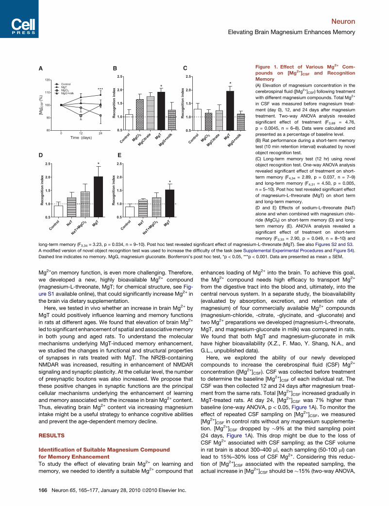
C Figure 1. Effect of Various Mg2+ Com-
pounds on [Mg2+]CSF and Recognition
Memory
(A) Elevation of magnesium concentration in the
cerebrospinal fluid ([Mg2+]CSF) following treatment
with different magnesium compounds. Total Mg2+
in CSF was measured before magnesium treat-
ment (day 0), 12, and 24 days after magnesium
treatment. Two-way ANOVA analysis revealed
significant effect of treatment (F3,69 = 4.76,
p = 0.0045, n = 6–8). Data were calculated and
presented as a percentage of baseline level.
(B) Rat performance during a short-term memory
test (10 min retention interval) evaluated by novel
object recognition test.
(C) Long-term memory test (12 hr) using novel
object recognition test. One-way ANOVA analysis
revealed significant effect of treatment on short-
term memory (F4,34 = 2.89, p = 0.037, n = 7–9)
and long-term memory (F4,31 = 4.50, p = 0.005,
n = 5–10). Post hoc test revealed significant effect
of magnesium-L-threonate (MgT) on short term
and long-term memory.
(D and E) Effects of sodium-L-threonate (NaT)
alone and when combined with magnesium chlo-
ride (MgCl2) on short-term memory (D) and long-
term memory (E). ANOVA analysis revealed a
significant effect of treatment on short-term
memory (F3,33 = 2.90, p = 0.049, n = 8–10) and
long-term memory (F3,34 = 3.23, p = 0.034, n = 9–10). Post hoc test revealed significant effect of magnesium-L-threonate (MgT). See also Figures S2 and S3.
A modified version of novel object recognition test was used to increase the difficulty of the task (see Supplemental Experimental Procedures and Figure S4).
Dashed line indicates no memory. MgG, magnesium gluconate. Bonferroni’s post hoc test, *p < 0.05, ***p < 0.001. Data are presented as mean ± SEM.
Neuron
Elevating Brain Magnesium Enhances Memory
Mg2+on memory function, is even more challenging. Therefore,
we developed a new, highly bioavailable Mg2+ compound
(magnesium-L-threonate, MgT; for chemical structure, see Fig-
ure S1 available online), that could significantly increase Mg2+ in
the brain via dietary supplementation.
Here, we tested in vivo whether an increase in brain Mg2+ by
MgT could positively influence learning and memory functions
in rats at different ages. We found that elevation of brain Mg2+
led to significant enhancement of spatial and associative memory
in both young and aged rats. To understand the molecular
mechanisms underlying MgT-induced memory enhancement,
we studied the changes in functional and structural properties
of synapses in rats treated with MgT. The NR2B-containing
NMDAR was increased, resulting in enhancement of NMDAR
signaling and synaptic plasticity. At the cellular level, the number
of presynaptic boutons was also increased. We propose that
these positive changes in synaptic functions are the principal
cellular mechanisms underlying the enhancement of learning
and memory associated with the increase in brain Mg2+ content.
Thus, elevating brain Mg2+ content via increasing magnesium
intake might be a useful strategy to enhance cognitive abilities
and prevent the age-dependent memory decline.
RESULTS
Identification of Suitable Magnesium Compoundfor Memory EnhancementTo study the effect of elevating brain Mg2+ on learning and
memory, we needed to identify a suitable Mg2+ compound that
166 Neuron 65, 165–177, January 28, 2010 ª2010 Elsevier Inc.
enhances loading of Mg2+ into the brain. To achieve this goal,
the Mg2+ compound needs high efficacy to transport Mg2+
from the digestive tract into the blood and, ultimately, into the
central nervous system. In a separate study, the bioavailability
(evaluated by absorption, excretion, and retention rate of
magnesium) of four commercially available Mg2+ compounds
(magnesium-chloride, -citrate, -glycinate, and -gluconate) and
two Mg2+ preparations we developed (magnesium-L-threonate,
MgT, and magnesium-gluconate in milk) was compared in rats.
We found that both MgT and magnesium-gluconate in milk
have higher bioavailability (X.Z., F. Mao, Y. Shang, N.A., and
G.L., unpublished data).
Here, we explored the ability of our newly developed
compounds to increase the cerebrospinal fluid (CSF) Mg2+
concentration ([Mg2+]CSF). CSF was collected before treatment
to determine the baseline [Mg2+]CSF of each individual rat. The
CSF was then collected 12 and 24 days after magnesium treat-
ment from the same rats. Total [Mg2+]CSF increased gradually in
MgT-treated rats. At day 24, [Mg2+]CSF was 7% higher than
baseline (one-way ANOVA, p < 0.05, Figure 1A). To monitor the
effect of repeated CSF sampling on [Mg2+]CSF, we measured
[Mg2+]CSF in control rats without any magnesium supplementa-
tion. [Mg2+]CSF dropped by �9% at the third sampling point
(24 days, Figure 1A). This drop might be due to the loss of
CSF Mg2+ associated with CSF sampling; as the CSF volume
in rat brain is about 300–400 ml, each sampling (50-100 ml) can
lead to 15%–30% loss of CSF Mg2+. Considering this reduc-
tion of [Mg2+]CSF associated with the repeated sampling, the
actual increase in [Mg2+]CSF should be �15% (two-way ANOVA,
0.25 1 2 5
60
80
100 Ctrl MgT
Cor
rect
cho
ice
(%)
Delay (min)0.25 1 2 5
Day 24
Delay (min)
*
Day 0
0 6 12 18 24 30 36 42 48 54 60
-5
0
5
10
15
***
***
*
*** ***
*** ******
***
***
Time (days)
Cha
nge
in %
of c
orre
ctch
oice
s
0.25 1 2 5
60
80
100 Ctrl MgT
Cor
rect
cho
ice
(%)
Delay (min)0.25 1 2 5
Delay (min)
*
22 month old
2 month old
A B
C D
MgT
0 6 12 18 24 30 36 42 48 54 60 66 72
-10
0
10
20
30
******
***
***
******
***
Time (days)
Cha
nge
in %
of c
orre
ctch
oice
s
MgT MgT
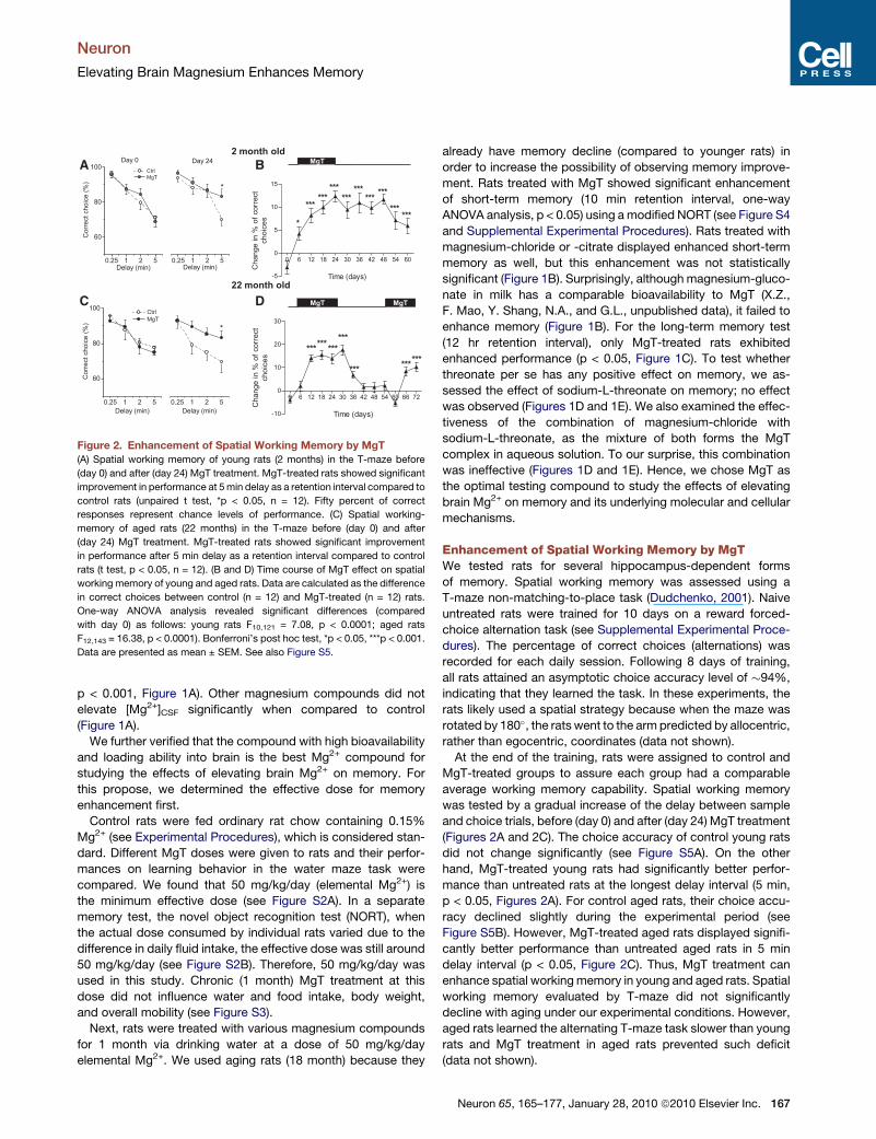
Figure 2. Enhancement of Spatial Working Memory by MgT
(A) Spatial working memory of young rats (2 months) in the T-maze before
(day 0) and after (day 24) MgT treatment. MgT-treated rats showed significant
improvement in performance at 5 min delay as a retention interval compared to
control rats (unpaired t test, *p < 0.05, n = 12). Fifty percent of correct
responses represent chance levels of performance. (C) Spatial working-
memory of aged rats (22 months) in the T-maze before (day 0) and after
(day 24) MgT treatment. MgT-treated rats showed significant improvement
in performance after 5 min delay as a retention interval compared to control
rats (t test, p < 0.05, n = 12). (B and D) Time course of MgT effect on spatial
working memory of young and aged rats. Data are calculated as the difference
in correct choices between control (n = 12) and MgT-treated (n = 12) rats.
One-way ANOVA analysis revealed significant differences (compared
with day 0) as follows: young rats F10,121 = 7.08, p < 0.0001; aged rats
F12,143 = 16.38, p < 0.0001). Bonferroni’s post hoc test, *p < 0.05, ***p < 0.001.
Data are presented as mean ± SEM. See also Figure S5.
Neuron
Elevating Brain Magnesium Enhances Memory
p < 0.001, Figure 1A). Other magnesium compounds did not
elevate [Mg2+]CSF significantly when compared to control
(Figure 1A).
We further verified that the compound with high bioavailability
and loading ability into brain is the best Mg2+ compound for
studying the effects of elevating brain Mg2+ on memory. For
this propose, we determined the effective dose for memory
enhancement first.
Control rats were fed ordinary rat chow containing 0.15%
Mg2+ (see Experimental Procedures), which is considered stan-
dard. Different MgT doses were given to rats and their perfor-
mances on learning behavior in the water maze task were
compared. We found that 50 mg/kg/day (elemental Mg2+) is
the minimum effective dose (see Figure S2A). In a separate
memory test, the novel object recognition test (NORT), when
the actual dose consumed by individual rats varied due to the
difference in daily fluid intake, the effective dose was still around
50 mg/kg/day (see Figure S2B). Therefore, 50 mg/kg/day was
used in this study. Chronic (1 month) MgT treatment at this
dose did not influence water and food intake, body weight,
and overall mobility (see Figure S3).
Next, rats were treated with various magnesium compounds
for 1 month via drinking water at a dose of 50 mg/kg/day
elemental Mg2+. We used aging rats (18 month) because they
already have memory decline (compared to younger rats) in
order to increase the possibility of observing memory improve-
ment. Rats treated with MgT showed significant enhancement
of short-term memory (10 min retention interval, one-way
ANOVA analysis, p < 0.05) using a modified NORT (see Figure S4
and Supplemental Experimental Procedures). Rats treated with
magnesium-chloride or -citrate displayed enhanced short-term
memory as well, but this enhancement was not statistically
significant (Figure 1B). Surprisingly, although magnesium-gluco-
nate in milk has a comparable bioavailability to MgT (X.Z.,
F. Mao, Y. Shang, N.A., and G.L., unpublished data), it failed to
enhance memory (Figure 1B). For the long-term memory test
(12 hr retention interval), only MgT-treated rats exhibited
enhanced performance (p < 0.05, Figure 1C). To test whether
threonate per se has any positive effect on memory, we as-
sessed the effect of sodium-L-threonate on memory; no effect
was observed (Figures 1D and 1E). We also examined the effec-
tiveness of the combination of magnesium-chloride with
sodium-L-threonate, as the mixture of both forms the MgT
complex in aqueous solution. To our surprise, this combination
was ineffective (Figures 1D and 1E). Hence, we chose MgT as
the optimal testing compound to study the effects of elevating
brain Mg2+ on memory and its underlying molecular and cellular
mechanisms.
Enhancement of Spatial Working Memory by MgTWe tested rats for several hippocampus-dependent forms
of memory. Spatial working memory was assessed using a
T-maze non-matching-to-place task (Dudchenko, 2001). Naive
untreated rats were trained for 10 days on a reward forced-
choice alternation task (see Supplemental Experimental Proce-
dures). The percentage of correct choices (alternations) was
recorded for each daily session. Following 8 days of training,
all rats attained an asymptotic choice accuracy level of �94%,
indicating that they learned the task. In these experiments, the
rats likely used a spatial strategy because when the maze was
rotated by 180�, the rats went to the arm predicted by allocentric,
rather than egocentric, coordinates (data not shown).
At the end of the training, rats were assigned to control and
MgT-treated groups to assure each group had a comparable
average working memory capability. Spatial working memory
was tested by a gradual increase of the delay between sample
and choice trials, before (day 0) and after (day 24) MgT treatment
(Figures 2A and 2C). The choice accuracy of control young rats
did not change significantly (see Figure S5A). On the other
hand, MgT-treated young rats had significantly better perfor-
mance than untreated rats at the longest delay interval (5 min,
p < 0.05, Figures 2A). For control aged rats, their choice accu-
racy declined slightly during the experimental period (see
Figure S5B). However, MgT-treated aged rats displayed signifi-
cantly better performance than untreated aged rats in 5 min
delay interval (p < 0.05, Figure 2C). Thus, MgT treatment can
enhance spatial working memory in young and aged rats. Spatial
working memory evaluated by T-maze did not significantly
decline with aging under our experimental conditions. However,
aged rats learned the alternating T-maze task slower than young
rats and MgT treatment in aged rats prevented such deficit
(data not shown).
Neuron 65, 165–177, January 28, 2010 ª2010 Elsevier Inc. 167

1 2 3 4 5 6 7 80
20
40
60
80
100
)ces( emiT epacsE
Trial number
Ctrl MgT
1 2 3 4 5 6 7 80
20
40
60
80
100 Ctrl MgT
)ces( emiT epacsE
Trial number
22 months old
2 months old
1 h 24 h0
20
40
60 Target Opposite
***
***
1 h 24 h0
20
40
60
***
Tim
e in
Qua
dran
t (%
)
1 h 24 h0
20
40
60
**
***
full partial full 0
20
40
60 Ctrl MgT
cue
)ces( emiT epacsE
full partial full0
20
40
60 Ctrl MgT
cue
)ces( emiT epacsE
*
A B DC
E F HG
Ctrl MgT
1 h 24 h0
20
40
60
Tim
e in
Qua
dran
t (%
)
Target Opposite***
Figure 3. Enhancement of Spatial Long-
Term Memory in Water Maze by MgT
(A) Escape time to find the hidden platform of
young rats during the water maze training trials.
MgT-treated (n = 15) rats learned faster than
controls (n = 14, two-way ANOVA, F1,216 = 7.85,
p = 0.006). ANOVA was followed by Bonferroni’s
post hoc test.
(B and C) Percentage of time spent in the target
versus opposite quadrant during the first (1 hr after
training) and the second (24 hr after training) probe
trials in control (n = 14, B) and MgT-treated (n = 15,
C) young rats. During the first probe trial, both
groups spent significantly more time in the target
quadrant (paired t test, p < 0.0001). On the other
hand, only MgT-treated rats spent significantly
more time in the target quadrant during the second
test trial (24 hr after training, p < 0.0001).
(D) Pattern completion test with partial extra maze
cues of young rats. Partial cues did not impair the
rats’ ability to find the platform.
(E) Escape time of aged rats. MgT-treated (n = 12)
rats learned faster than controls (n = 16, ANOVA,
F1,208 = 11.42, p = 0.0009).
(F and G) Performance of control (n = 16, F) and
MgT-treated (n = 12, G) aged rats during the first
(1 hr after training) and the second (24 hr after
training) memory probe trials. During the first trial,
both groups spent more time in the target quadrant (paired t test, p < 0.0001). Twenty-four hours later, only MgT-treated aged rats spent more time in the target
quadrant (p < 0.01).
(H) Partial removal of extra-maze cues impaired the ability of aged rats to find the hidden platform, while MgT-treated aged rats were still capable of locating the
platform (unpaired t test, p < 0.05). *p < 0.05, **p < 0.01, ***p < 0.001. Data are presented as mean ± SEM. See also Table S1.
Neuron
Elevating Brain Magnesium Enhances Memory
To monitor the time-course of MgT treatment on working
spatial memory, task performance was evaluated every sixth
day (Figures 2B and 2D). Since the largest difference in choice
accuracy between the treated versus control rats was obtained
at 5 min delay interval, we monitored choice accuracy only at
this delay interval for the remaining experiments. A significant
increase in choice accuracy of MgT-treated young rats was
apparent 6 days after the onset of treatment (one-way ANOVA,
p < 0.05), peaked on day 12 (p < 0.001), and did not decline
over 1 month after MgT treatment was stopped (days 30 to 60,
Figure 2B). For MgT-treated aged rats, a significant increase in
choice accuracy occurred 12 days after the onset of treatment
(p < 0.001) and remained stable until MgT was stopped (day 30).
In contrast to young rats, the working memory performance of
aged rats declined to the baseline value within 12 days following
interruption of MgT treatment (Figure 2D). Therefore, the on/off
kinetics of MgT-induced spatial memory enhancement seems
symmetric in aged rats. To test if MgT could re-enhance spatial
memory functions of MgT-treated aged rats, following 30 days of
drinking plain water (days 30–60), they drank water supple-
mented with MgT again. Strikingly, aged rat performance was
re-enhanced within 12 days of treatment (Figure 2D). Thus,
MgT consumption enhanced spatial working memory of young
and aged rats (For the detailed time course curves for each
group, see Figures S5C and S5D).
Enhancement of Spatial Long-Term Memory by MgTWe used the Morris water maze to perform further experiments
to determine whether MgT leads to the improvement of spatial
168 Neuron 65, 165–177, January 28, 2010 ª2010 Elsevier Inc.
long-term memory (Morris, 1984). Young rats underwent 8
trials of training within one day with a 1 hr intertrial interval.
For aged rats, the training protocol was spread over two
days: 5 trials on day 1, and 3 trials on day 2. This protocol
was adopted because aged rats are not able to perform 8 trials
within one day. During the training period, the performance of
all rats gradually improved (Figures 3A and 3E). However, MgT-
treated rats learned to find the hidden platform faster than
controls (two-way ANOVA, MgT-treated versus control young
rats, p < 0.01 and MgT-treated versus control aged rats,
p < 0.001). In addition, the degree of learning ability enhance-
ment by MgT was higher in aged (Figure 3E) than in young rats
(Figure 3A).
To evaluate memory functions, we performed two test trials
with the platform removed; the rats were allowed to search for
60 s. The first test trial commenced 1 hr after the end of training.
All rats showed a remarkable preference for the target versus
opposite quadrant (young, Figures 3B and 3C, paired t test,
p < 0.001; old, Figures 3F and 3G, p < 0.001), suggesting that
all, i.e., control and MgT-treated young and aged rats, could
remember the platform location. To test long-term spatial
memory, a second test trial was performed 24 hr later. Both
young and aged control rats lost their preference for the target
quadrant compared to other quadrants (Figures 3B and 3F). In
contrast, MgT-treated young (Figure 3B) and aged (Figure 3G)
rats retained their quadrant preference (p < 0.001; p < 0.01,
respectively). Visual and locomotor functions were equal in
both groups, as judged by latency of escape to a visible platform
(data not shown) and swimming speed (see Table S1). Thus,
p-C
aMK
II/C
aMK
II
p-C
RE
B/C
RE
B
0
40
80
120**
**
Cha
nge
(%)
MgT
NR2B
NR2A
NR1
0
20
40
60
80***
Cha
nge
(%)MgT
C
NR2B
NR2A
NR1
control
p-CamKII
CamKII
control
2B
2A
R1
0
20
40
60
Cha
nge
(%)MgTcontrol
p-CamKII
CamKII
CREB
p-CREB
β-actin
D
Control
MgT
0
20
40
60
*
BD
NF
(ng/
g)
A B
-80 -40 40
-20
20
40
600.8 (control)1.2 (acute Mg)1.2 (chronic Mg)
I NM
DA(
pA)
Vm (mV)
E
-80 -40 40
-80
-40
40
80acute 1.2 Mgchronic 1.2 Mg
V (mV)
% c
hang
e
homeostasis
enhancement
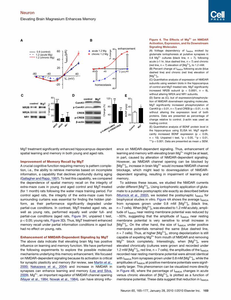
Figure 4. The Effects of Mg2+ on NMDAR
Activation, Expression, and Its Downstream
Signaling Molecules
(A) Voltage dependency of INMDA evoked by
glutamate iontophoresis at putative synapses in
0.8 Mg2+ cultures (black line, n = 7), following
acute (<1 hr, blue dashed line, n = 7) and chronic
(red line, n = 7) elevation of [Mg2+]o to 1.2 mM.
(B) Percent change of INMDA following acute (blue
dashed line) and chronic (red line) elevation of
[Mg2+]o.
(C) Quantitative analysis of expression of NMDAR
subunits using western blots in the hippocampus
of control and MgT-treated rats. MgT significantly
increased NR2B subunit (p < 0.0001, n = 8),
without altering NR2A and NR1 subunits.
(D) Same as (C), but of expression/phosphoryla-
tion of NMDAR downstream signaling molecules.
MgT significantly increased phosphorylation of
CamKII (p < 0.01, n = 7) and CREB (p < 0.01, n = 8)
without altering the expression level of both
proteins. Data are presented as percentage of
change relative to control. b-actin was used as
loading control.
(E) Quantitative analysis of BDNF protein level in
the hippocampus using ELISA kit. MgT signifi-
cantly increased BDNF expression (p < 0.05,
n = 10). Unpaired t test, *p < 0.05, **p < 0.01,
***p < 0.001. Data are presented as mean ± SEM.
Neuron
Elevating Brain Magnesium Enhances Memory
MgT treatment significantly enhanced hippocampus-dependent
spatial learning and memory in both young and aged rats.
Improvement of Memory Recall by MgTA crucial cognitive function requiring memory is pattern comple-
tion, i.e., the ability to retrieve memories based on incomplete
information, a capability that declines profoundly during aging
(Gallagher and Rapp, 1997). To test this capability, we compared
the dependence of spatial memory recall on the integrity of
extra-maze cues in young and aged control and MgT-treated
(for 1 month) rats following the water maze training period. For
control aged rats, the integrity of the extra-maze cues from
surrounding curtains was essential for finding the hidden plat-
form, as their performance significantly degraded under
partial-cue conditions. In contrast, MgT-treated aged rats, as
well as young rats, performed equally well under full- and
partial-cue conditions (aged rats, Figure 3H, unpaired t test,
p < 0.05; young rats, Figure 3D). Thus, MgT treatment enhanced
memory recall under partial information conditions in aged but
had no effect on young, rats.
Enhancement of NMDAR-Dependent Signaling by MgTThe above data indicate that elevating brain Mg has positive
influence on learning and memory function. We have performed
the following experiments to explore the possible molecular
mechanisms underlying this memory enhancement. We focused
on NMDAR-dependent signaling because its activation is critical
for synaptic plasticity and memory (for review, see Martin et al.,
2000; Nakazawa et al., 2004) and increase in NMDAR in
synapses can enhance learning and memory (Lee and Silva,
2009). Mg2+, an important regulator of NMDAR channel opening
(Mayer et al., 1984; Nowak et al., 1984), can have strong influ-
ence on NMDAR-dependent signaling. Thus, enhancement of
learning and memory with elevating brain Mg2+ might be at least,
in part, caused by alteration of NMDAR-dependent signaling.
However, as NMDAR channel opening can be blocked by
[Mg2+]o, increase in brain Mg2+ would increase NMDAR channel
blockage, which might lead to downregulation of NMDAR-
dependent signaling, resulting in impairment of learning and
memory.
To address these issues, we studied the NMDAR currents
under different [Mg2+]o. Using iontophoretic application of gluta-
mate to a putative postsynaptic site exactly as described before
(Murnick et al., 2002), we isolated the postsynaptic INMDA for
biophysical studies in vitro. Figure 4A shows the average INMDA
from synapses grown under 0.8 mM [Mg2+]o (black line,
n = 7 cells). When [Mg2+]o was elevated to 1.2 mM acutely, ampli-
tude of INMDA near resting membrane potential was reduced by
�50%, suggesting that the amplitude of INMDA near resting
membrane potential is very sensitive to small increase in
[Mg2+]o. On the other hand, the size of INMDA under positive
membrane potentials remained the same (blue dashed line,
n = 7 cells). Thus, at higher [Mg2+]o, strong depolarization is still
capable of expelling Mg2+ from mouth of NMDAR and removing
Mg2+ block completely. Interestingly, when [Mg2+]o were
elevated chronically (cultures were grown and recorded under
1.2 mM [Mg2+]o, red line, n = 7 cells), the amplitudes of the INMDA
recorded near resting membrane potential were almost identical
with INMDA from synapses grown under 0.8 mM [Mg2+]o, while the
amplitudes of INMDA at positive membrane potentials were signif-
icantly larger. This phenomenon can be visualized more directly
in Figure 4B, where the percentage of INMDA changes in acute
versus chronic elevation of [Mg2+]o is plotted as a function of
membrane potential. These data suggest that reduction in INMDA
Neuron 65, 165–177, January 28, 2010 ª2010 Elsevier Inc. 169
control
MgT
0
10
20
30**
NR
2B
co
mp
on
en
t (
%)
0
10
20
30
40 **
single burst
control
MgT
Am
plitu
de
pe
r A
P (
pA
)
-10 0 10 20 3050
100
150
200
250
time (min)
EP
SC
am
plitu
de (%
)
-10 0 10 20 3050
100
150
200
250
time (min)
EP
SC
am
plitu
de (%
)
control
MgT
100
125
150
175
200
***
No
rm
alize
d a
mp
litu
de (
%)
control MgT
A B
DC
100 ms
40 pA
50 ms
20 p
A
control MgT
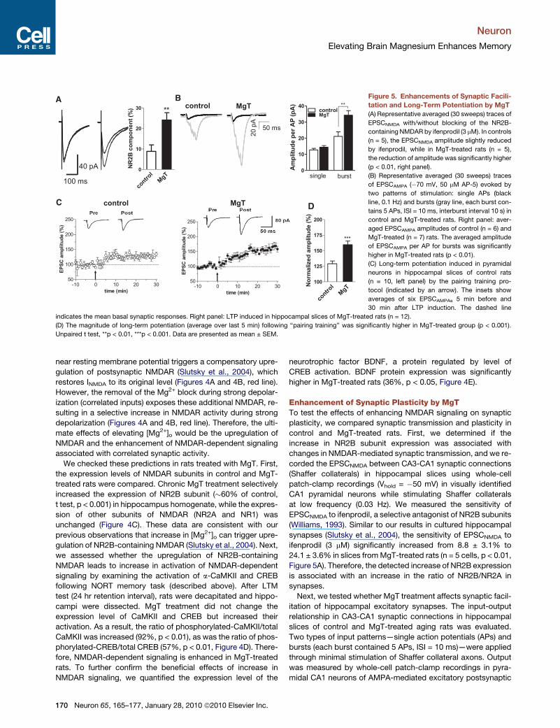
Figure 5. Enhancements of Synaptic Facili-
tation and Long-Term Potentiation by MgT
(A) Representative averaged (30 sweeps) traces of
EPSCNMDA with/without blocking of the NR2B-
containing NMDAR by ifenprodil (3 mM). In controls
(n = 5), the EPSCNMDA amplitude slightly reduced
by ifenprodil, while in MgT-treated rats (n = 5),
the reduction of amplitude was significantly higher
(p < 0.01, right panel).
(B) Representative averaged (30 sweeps) traces
of EPSCAMPA (�70 mV, 50 mM AP-5) evoked by
two patterns of stimulation: single APs (black
line, 0.1 Hz) and bursts (gray line, each burst con-
tains 5 APs, ISI = 10 ms, interburst interval 10 s) in
control and MgT-treated rats. Right panel: aver-
aged EPSCAMPA amplitudes of control (n = 6) and
MgT-treated (n = 7) rats. The averaged amplitude
of EPSCAMPA per AP for bursts was significantly
higher in MgT-treated rats (p < 0.01).
(C) Long-term potentiation induced in pyramidal
neurons in hippocampal slices of control rats
(n = 10, left panel) by the pairing training pro-
tocol (indicated by an arrow). The insets show
averages of six EPSCAMPAs 5 min before and
30 min after LTP induction. The dashed line
indicates the mean basal synaptic responses. Right panel: LTP induced in hippocampal slices of MgT-treated rats (n = 12).
(D) The magnitude of long-term potentiation (average over last 5 min) following ‘‘pairing training’’ was significantly higher in MgT-treated group (p < 0.001).
Unpaired t test, **p < 0.01, ***p < 0.001. Data are presented as mean ± SEM.
Neuron
Elevating Brain Magnesium Enhances Memory
near resting membrane potential triggers a compensatory upre-
gulation of postsynaptic NMDAR (Slutsky et al., 2004), which
restores INMDA to its original level (Figures 4A and 4B, red line).
However, the removal of the Mg2+ block during strong depolar-
ization (correlated inputs) exposes these additional NMDAR, re-
sulting in a selective increase in NMDAR activity during strong
depolarization (Figures 4A and 4B, red line). Therefore, the ulti-
mate effects of elevating [Mg2+]o would be the upregulation of
NMDAR and the enhancement of NMDAR-dependent signaling
associated with correlated synaptic activity.
We checked these predictions in rats treated with MgT. First,
the expression levels of NMDAR subunits in control and MgT-
treated rats were compared. Chronic MgT treatment selectively
increased the expression of NR2B subunit (�60% of control,
t test, p < 0.001) in hippocampus homogenate, while the expres-
sion of other subunits of NMDAR (NR2A and NR1) was
unchanged (Figure 4C). These data are consistent with our
previous observations that increase in [Mg2+]o can trigger upre-
gulation of NR2B-containing NMDAR (Slutsky et al., 2004). Next,
we assessed whether the upregulation of NR2B-containing
NMDAR leads to increase in activation of NMDAR-dependent
signaling by examining the activation of a-CaMKII and CREB
following NORT memory task (described above). After LTM
test (24 hr retention interval), rats were decapitated and hippo-
campi were dissected. MgT treatment did not change the
expression level of CaMKII and CREB but increased their
activation. As a result, the ratio of phosphorylated-CaMKII/total
CaMKII was increased (92%, p < 0.01), as was the ratio of phos-
phorylated-CREB/total CREB (57%, p < 0.01, Figure 4D). There-
fore, NMDAR-dependent signaling is enhanced in MgT-treated
rats. To further confirm the beneficial effects of increase in
NMDAR signaling, we quantified the expression level of the
170 Neuron 65, 165–177, January 28, 2010 ª2010 Elsevier Inc.
neurotrophic factor BDNF, a protein regulated by level of
CREB activation. BDNF protein expression was significantly
higher in MgT-treated rats (36%, p < 0.05, Figure 4E).
Enhancement of Synaptic Plasticity by MgTTo test the effects of enhancing NMDAR signaling on synaptic
plasticity, we compared synaptic transmission and plasticity in
control and MgT-treated rats. First, we determined if the
increase in NR2B subunit expression was associated with
changes in NMDAR-mediated synaptic transmission, and we re-
corded the EPSCNMDA between CA3-CA1 synaptic connections
(Shaffer collaterals) in hippocampal slices using whole-cell
patch-clamp recordings (Vhold = �50 mV) in visually identified
CA1 pyramidal neurons while stimulating Shaffer collaterals
at low frequency (0.03 Hz). We measured the sensitivity of
EPSCNMDA to ifenprodil, a selective antagonist of NR2B subunits
(Williams, 1993). Similar to our results in cultured hippocampal
synapses (Slutsky et al., 2004), the sensitivity of EPSCNMDA to
ifenprodil (3 mM) significantly increased from 8.8 ± 3.1% to
24.1 ± 3.6% in slices from MgT-treated rats (n = 5 cells, p < 0.01,
Figure 5A). Therefore, the detected increase of NR2B expression
is associated with an increase in the ratio of NR2B/NR2A in
synapses.
Next, we tested whether MgT treatment affects synaptic facil-
itation of hippocampal excitatory synapses. The input-output
relationship in CA3-CA1 synaptic connections in hippocampal
slices of control and MgT-treated aging rats was evaluated.
Two types of input patterns—single action potentials (APs) and
bursts (each burst contained 5 APs, ISI = 10 ms)—were applied
through minimal stimulation of Shaffer collateral axons. Output
was measured by whole-cell patch-clamp recordings in pyra-
midal CA1 neurons of AMPA-mediated excitatory postsynaptic
Contro
lMgT
0
200
400
600
800 **
Syn
punc
ta (D
G)/1
000µ
m2
Control MgT
Contro
lMgT
0
200
400
600
800
*
Syn
punc
ta (C
A1)
/100
0µm
2
A B C
Control MgT
D E F
Contro
lMgT
0
200
400
600
800
*
SNB-
1 pu
ncta
(DG
)/100
0µm
2
Contro
lMgT
0
200
400
600
800
*
SNB-
1 pu
ncta
(CA1
)/100
0µm
2
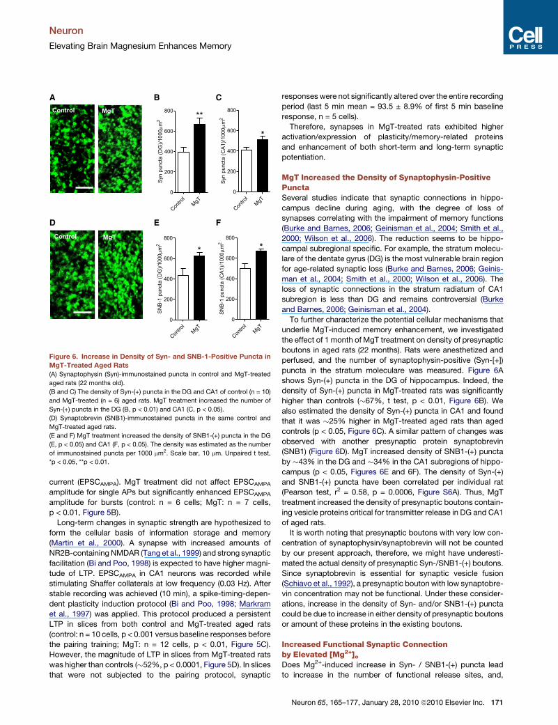
Figure 6. Increase in Density of Syn- and SNB-1-Positive Puncta in
MgT-Treated Aged Rats
(A) Synaptophysin (Syn)-immunostained puncta in control and MgT-treated
aged rats (22 months old).
(B and C) The density of Syn-(+) puncta in the DG and CA1 of control (n = 10)
and MgT-treated (n = 6) aged rats. MgT treatment increased the number of
Syn-(+) puncta in the DG (B, p < 0.01) and CA1 (C, p < 0.05).
(D) Synaptobrevin (SNB1)-immunostained puncta in the same control and
MgT-treated aged rats.
(E and F) MgT treatment increased the density of SNB1-(+) puncta in the DG
(E, p < 0.05) and CA1 (F, p < 0.05). The density was estimated as the number
of immunostained puncta per 1000 mm2. Scale bar, 10 mm. Unpaired t test,
*p < 0.05, **p < 0.01.
Neuron
Elevating Brain Magnesium Enhances Memory
current (EPSCAMPA). MgT treatment did not affect EPSCAMPA
amplitude for single APs but significantly enhanced EPSCAMPA
amplitude for bursts (control: n = 6 cells; MgT: n = 7 cells,
p < 0.01, Figure 5B).
Long-term changes in synaptic strength are hypothesized to
form the cellular basis of information storage and memory
(Martin et al., 2000). A synapse with increased amounts of
NR2B-containing NMDAR (Tang et al., 1999) and strong synaptic
facilitation (Bi and Poo, 1998) is expected to have higher magni-
tude of LTP. EPSCAMPA in CA1 neurons was recorded while
stimulating Shaffer collaterals at low frequency (0.03 Hz). After
stable recording was achieved (10 min), a spike-timing-depen-
dent plasticity induction protocol (Bi and Poo, 1998; Markram
et al., 1997) was applied. This protocol produced a persistent
LTP in slices from both control and MgT-treated aged rats
(control: n = 10 cells, p < 0.001 versus baseline responses before
the pairing training; MgT: n = 12 cells, p < 0.01, Figure 5C).
However, the magnitude of LTP in slices from MgT-treated rats
was higher than controls (�52%, p < 0.0001, Figure 5D). In slices
that were not subjected to the pairing protocol, synaptic
responses were not significantly altered over the entire recording
period (last 5 min mean = 93.5 ± 8.9% of first 5 min baseline
response, n = 5 cells).
Therefore, synapses in MgT-treated rats exhibited higher
activation/expression of plasticity/memory-related proteins
and enhancement of both short-term and long-term synaptic
potentiation.
MgT Increased the Density of Synaptophysin-PositivePunctaSeveral studies indicate that synaptic connections in hippo-
campus decline during aging, with the degree of loss of
synapses correlating with the impairment of memory functions
(Burke and Barnes, 2006; Geinisman et al., 2004; Smith et al.,
2000; Wilson et al., 2006). The reduction seems to be hippo-
campal subregional specific. For example, the stratum molecu-
lare of the dentate gyrus (DG) is the most vulnerable brain region
for age-related synaptic loss (Burke and Barnes, 2006; Geinis-
man et al., 2004; Smith et al., 2000; Wilson et al., 2006). The
loss of synaptic connections in the stratum radiatum of CA1
subregion is less than DG and remains controversial (Burke
and Barnes, 2006; Geinisman et al., 2004).
To further characterize the potential cellular mechanisms that
underlie MgT-induced memory enhancement, we investigated
the effect of 1 month of MgT treatment on density of presynaptic
boutons in aged rats (22 months). Rats were anesthetized and
perfused, and the number of synaptophysin-positive (Syn-[+])
puncta in the stratum moleculare was measured. Figure 6A
shows Syn-(+) puncta in the DG of hippocampus. Indeed, the
density of Syn-(+) puncta in MgT-treated rats was significantly
higher than controls (�67%, t test, p < 0.01, Figure 6B). We
also estimated the density of Syn-(+) puncta in CA1 and found
that it was �25% higher in MgT-treated aged rats than aged
controls (p < 0.05, Figure 6C). A similar pattern of changes was
observed with another presynaptic protein synaptobrevin
(SNB1) (Figure 6D). MgT increased density of SNB1-(+) puncta
by �43% in the DG and �34% in the CA1 subregions of hippo-
campus (p < 0.05, Figures 6E and 6F). The density of Syn-(+)
and SNB1-(+) puncta have been correlated per individual rat
(Pearson test, r2 = 0.58, p = 0.0006, Figure S6A). Thus, MgT
treatment increased the density of presynaptic boutons contain-
ing vesicle proteins critical for transmitter release in DG and CA1
of aged rats.
It is worth noting that presynaptic boutons with very low con-
centration of synaptophysin/synaptobrevin will not be counted
by our present approach, therefore, we might have underesti-
mated the actual density of presynaptic Syn-/SNB1-(+) boutons.
Since synaptobrevin is essential for synaptic vesicle fusion
(Schiavo et al., 1992), a presynaptic bouton with low synaptobre-
vin concentration may not be functional. Under these consider-
ations, increase in the density of Syn- and/or SNB1-(+) puncta
could be due to increase in either density of presynaptic boutons
or amount of these proteins in the existing boutons.
Increased Functional Synaptic Connectionby Elevated [Mg2+]o
Does Mg2+-induced increase in Syn- / SNB1-(+) puncta lead
to increase in the number of functional release sites, and,
Neuron 65, 165–177, January 28, 2010 ª2010 Elsevier Inc. 171
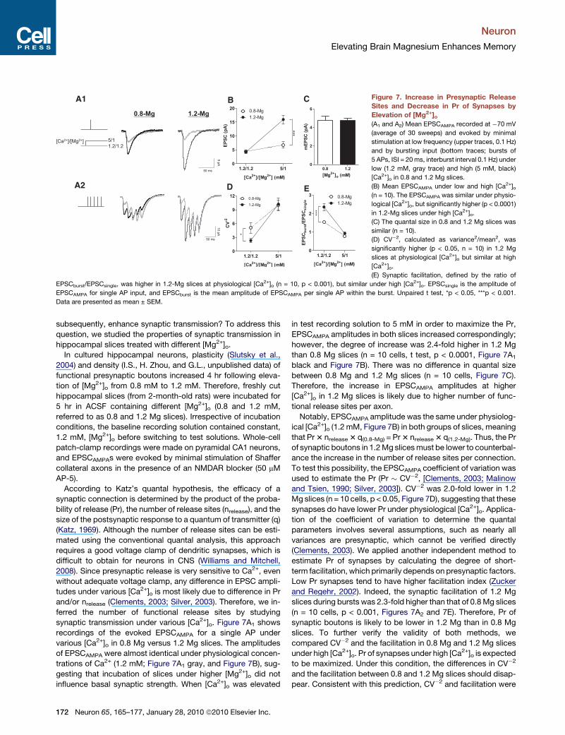
0.8 1.2
0
2
4
6
[Mg2+
]o (mM)
mE
PS
C (p
A)
0
5
10
15
20 0.8-Mg1.2-Mg
***
1.2/1.2 5/1
[Ca2+
]/[Mg2+
] (mM)
EP
SC
(p
A)
[Ca2+]/[Mg2+] 5/11.2/1.2
50 ms
5 pA
0
3
6
9
12 0.8-Mg1.2-Mg
*
1.2/1.2 5/1
[Ca2+
]/[Mg2+
] (mM)
CV
-2
A1
0.8-Mg
50 ms
10 pA
0
1
2
3 0.8-Mg1.2-Mg
***
1.2/1.2 5/1
[Ca2+
]/[Mg2+
] (mM)
EP
SC
bu
rst/E
PS
Csin
gle
A2
1.2-Mg
B
ED
C Figure 7. Increase in Presynaptic Release
Sites and Decrease in Pr of Synapses by
Elevation of [Mg2+]o(A1 and A2) Mean EPSCAMPA recorded at �70 mV
(average of 30 sweeps) and evoked by minimal
stimulation at low frequency (upper traces, 0.1 Hz)
and by bursting input (bottom traces; bursts of
5 APs, ISI = 20 ms, interburst interval 0.1 Hz) under
low (1.2 mM, gray trace) and high (5 mM, black)
[Ca2+]o in 0.8 and 1.2 Mg slices.
(B) Mean EPSCAMPA under low and high [Ca2+]o(n = 10). The EPSCAMPA was similar under physio-
logical [Ca2+]o, but significantly higher (p < 0.0001)
in 1.2-Mg slices under high [Ca2+]o.
(C) The quantal size in 0.8 and 1.2 Mg slices was
similar (n = 10).
(D) CV�2, calculated as variance2/mean2, was
significantly higher (p < 0.05, n = 10) in 1.2 Mg
slices at physiological [Ca2+]o but similar at high
[Ca2+]o.
(E) Synaptic facilitation, defined by the ratio of
EPSCburst/EPSCsingle, was higher in 1.2-Mg slices at physiological [Ca2+]o (n = 10, p < 0.001), but similar under high [Ca2+]o. EPSCsingle is the amplitude of
EPSCAMPA for single AP input, and EPSCburst is the mean amplitude of EPSCAMPA per single AP within the burst. Unpaired t test, *p < 0.05, ***p < 0.001.
Data are presented as mean ± SEM.
Neuron
Elevating Brain Magnesium Enhances Memory
subsequently, enhance synaptic transmission? To address this
question, we studied the properties of synaptic transmission in
hippocampal slices treated with different [Mg2+]o.
In cultured hippocampal neurons, plasticity (Slutsky et al.,
2004) and density (I.S., H. Zhou, and G.L., unpublished data) of
functional presynaptic boutons increased 4 hr following eleva-
tion of [Mg2+]o from 0.8 mM to 1.2 mM. Therefore, freshly cut
hippocampal slices (from 2-month-old rats) were incubated for
5 hr in ACSF containing different [Mg2+]o (0.8 and 1.2 mM,
referred to as 0.8 and 1.2 Mg slices). Irrespective of incubation
conditions, the baseline recording solution contained constant,
1.2 mM, [Mg2+]o before switching to test solutions. Whole-cell
patch-clamp recordings were made on pyramidal CA1 neurons,
and EPSCAMPAs were evoked by minimal stimulation of Shaffer
collateral axons in the presence of an NMDAR blocker (50 mM
AP-5).
According to Katz’s quantal hypothesis, the efficacy of a
synaptic connection is determined by the product of the proba-
bility of release (Pr), the number of release sites (nrelease), and the
size of the postsynaptic response to a quantum of transmitter (q)
(Katz, 1969). Although the number of release sites can be esti-
mated using the conventional quantal analysis, this approach
requires a good voltage clamp of dendritic synapses, which is
difficult to obtain for neurons in CNS (Williams and Mitchell,
2008). Since presynaptic release is very sensitive to Ca2+, even
without adequate voltage clamp, any difference in EPSC ampli-
tudes under various [Ca2+]o is most likely due to difference in Pr
and/or nrelease (Clements, 2003; Silver, 2003). Therefore, we in-
ferred the number of functional release sites by studying
synaptic transmission under various [Ca2+]o. Figure 7A1 shows
recordings of the evoked EPSCAMPA for a single AP under
various [Ca2+]o in 0.8 Mg versus 1.2 Mg slices. The amplitudes
of EPSCAMPA were almost identical under physiological concen-
trations of Ca2+ (1.2 mM; Figure 7A1 gray, and Figure 7B), sug-
gesting that incubation of slices under higher [Mg2+]o did not
influence basal synaptic strength. When [Ca2+]o was elevated
172 Neuron 65, 165–177, January 28, 2010 ª2010 Elsevier Inc.
in test recording solution to 5 mM in order to maximize the Pr,
EPSCAMPA amplitudes in both slices increased correspondingly;
however, the degree of increase was 2.4-fold higher in 1.2 Mg
than 0.8 Mg slices (n = 10 cells, t test, p < 0.0001, Figure 7A1
black and Figure 7B). There was no difference in quantal size
between 0.8 Mg and 1.2 Mg slices (n = 10 cells, Figure 7C).
Therefore, the increase in EPSCAMPA amplitudes at higher
[Ca2+]o in 1.2 Mg slices is likely due to higher number of func-
tional release sites per axon.
Notably, EPSCAMPA amplitude was the same under physiolog-
ical [Ca2+]o (1.2 mM, Figure 7B) in both groups of slices, meaning
that Pr 3 nrelease 3 q(0.8-Mg) = Pr 3 nrelease 3 q(1.2-Mg). Thus, the Pr
of synaptic boutons in 1.2 Mg slices must be lower to counterbal-
ance the increase in the number of release sites per connection.
To test this possibility, the EPSCAMPA coefficient of variation was
used to estimate the Pr (Pr � CV�2, [Clements, 2003; Malinow
and Tsien, 1990; Silver, 2003]). CV�2 was 2.0-fold lower in 1.2
Mg slices (n = 10 cells, p < 0.05, Figure 7D), suggesting that these
synapses do have lower Pr under physiological [Ca2+]o. Applica-
tion of the coefficient of variation to determine the quantal
parameters involves several assumptions, such as nearly all
variances are presynaptic, which cannot be verified directly
(Clements, 2003). We applied another independent method to
estimate Pr of synapses by calculating the degree of short-
term facilitation, which primarily depends on presynaptic factors.
Low Pr synapses tend to have higher facilitation index (Zucker
and Regehr, 2002). Indeed, the synaptic facilitation of 1.2 Mg
slices during bursts was 2.3-fold higher than that of 0.8 Mg slices
(n = 10 cells, p < 0.001, Figures 7A2 and 7E). Therefore, Pr of
synaptic boutons is likely to be lower in 1.2 Mg than in 0.8 Mg
slices. To further verify the validity of both methods, we
compared CV�2 and the facilitation in 0.8 Mg and 1.2 Mg slices
under high [Ca2+]o. Pr of synapses under high [Ca2+]o is expected
to be maximized. Under this condition, the differences in CV�2
and the facilitation between 0.8 and 1.2 Mg slices should disap-
pear. Consistent with this prediction, CV�2 and facilitation were
Contro
l
MgT O
N
MgT O
FF0
200
400
600
800
***
Syn
punc
ta (C
A1)
/100
0µm
2
Contro
l
MgT O
N
MgT O
FF0
200
400
600
800
**
Syn
punc
ta (D
G)/1
000µ
m2
0 200 400 600 800 10000
20
40
60
80
100
120ControlMgT
r2 = 0.50 p = 0.02
Syn puncta (DG)/1000µm2
Rec
ogni
tion
inde
x (%
)
0
20
40
60
80
100
Control MgT
Rec
ogni
tion
inde
x (%
)
A B
D E F
C
MgT
treatment
(2 weeks)
MgT ONRats perfused
MgT OFFRats perfused
Control
period
(2 weeks)
No MgT
(2 weeks)
Control MgT ON MgT OFF
0 200 400 600 800 10000
20
40
60
80
100
120
r2 = 0.41 p = 0.04
Syn puncta (CA1)/1000µm2
Rec
ogni
tion
inde
x (%
)
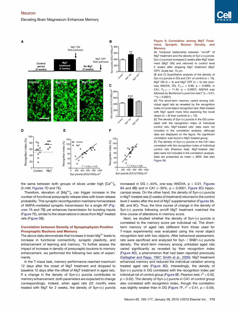
Figure 8. Correlation among MgT Treat-
ment, Synaptic Bouton Density, and
Memory
(A) Temporal relationship between ‘‘on/off’’ of
MgT treatment and the density of Syn-(+) puncta.
Syn-(+) puncta increased 2 weeks after MgT treat-
ment (MgT ON) and returned to control level
2 weeks after stopping MgT treatment (MgT
OFF). Scale bar, 10 mm.
(B and C) Quantitative analysis of the density of
Syn-(+) puncta in DG and CA1 of control (n = 10),
MgT ON (n = 5) and MgT OFF (n = 5) rats (one-
way ANOVA, DG, F2,17 = 6.88, p = 0.0065; in
CA1, F2,17 = 11.45, p = 0.0007). ANOVA was
followed by Bonferroni’s post hoc test (**p < 0.01;
***p < 0.0001).
(D) The short-term memory varied among indi-
vidual aged rats as revealed by the recognition
index of novel object recognition test. Rats treated
with MgT spent more time exploring the novel
object (n = 6) than controls (n = 10).
(E) The density of Syn-(+) puncta in the DG corre-
lated with the recognition index of individual
control rats. MgT-treated rats’ data were not
included in the correlation analysis, although
data are displayed on the figure. No significant
correlation was found in MgT-treated group.
(F) The density of Syn-(+) puncta in the CA1 also
correlated with the recognition index of individual
control rats (Pearson test). MgT-treated rats’
data were not included in the correlation analysis.
Data are presented as mean ± SEM. See also
Figure S6.
Neuron
Elevating Brain Magnesium Enhances Memory
the same between both groups of slices under high [Ca2+]o(5 mM, Figures 7D and 7E).
Therefore, elevation of [Mg2+]o can trigger increase in the
number of functional presynaptic release sites with lower release
probability. This synaptic reconfiguration maintains homeostasis
of AMPA-mediated synaptic transmission for a single AP (Fig-
ures 7A and 7B) yet enhances transmission for bursting inputs
(Figure 7E), similar to the observations in slices from MgT-treated
rats (Figure 5B).
Correlation between Density of Synaptophysin-PositivePresynaptic Boutons and MemoryThe above data demonstrate that increase in brain Mg2+ leads to
increase in functional connectivity, synaptic plasticity, and
enhancement of learning and memory. To further assess the
impact of increase in density of presynaptic boutons to memory
enhancement, we performed the following two sets of experi-
ments.
In the T-maze task, memory performance reached maximum
12 days after the onset of MgT treatment and dropped to
baseline 12 days after the offset of MgT treatment in aged rats.
If a change in the density of Syn-(+) puncta contributes to
memory enhancement, both parameters are expected to change
correspondingly. Indeed, when aged rats (22 month) were
treated with MgT for 2 weeks, the density of Syn-(+) puncta
increased in DG (�44%, one-way ANOVA, p < 0.01, Figures
8A and 8B) and in CA1 (�30%, p < 0.0001, Figure 8C) hippo-
campal areas. On the other hand, the density of Syn-(+) puncta
in MgT-treated rats (2 weeks of treatment) returned to the control
level 2 weeks after the end of MgT supplementation (Figures 8A,
8B, and 8C). Thus, the time course of change in the density of
Syn-(+) puncta following on/off MgT treatment matched the
time course of alterations in memory score.
Next, we studied whether the density of Syn-(+) puncta is
correlated to the memory score per individual rat. The short-
term memory of aged rats (different from those used for
T-maze experiments) was evaluated using the novel object
recognition test with two objects. After behavioral assessment,
rats were sacrificed and analyzed for Syn- / SNB1-(+) puncta
density. The short-term memory among untreated aged rats
varied significantly as revealed by their recognition index
(Figure 8D), a phenomenon that had been reported previously
(Gallagher and Rapp, 1997; Smith et al., 2000). MgT treatment
enhanced memory and reduced the individual variation among
treated aged rats (Figure 8D). Interestingly, the density of
Syn-(+) puncta in DG correlated with the recognition index per
individual rat of control group (Figure 8E, Pearson test, r2 = 0.50,
p = 0.02). The density of Syn-(+) puncta in CA1 of control group
also correlated with recognition index, though the correlation
was slightly weaker than in DG (Figure 7F, r2 = 0.41, p = 0.04).
Neuron 65, 165–177, January 28, 2010 ª2010 Elsevier Inc. 173
Neuron
Elevating Brain Magnesium Enhances Memory
The density of SNB1-(+) puncta in control group also correlated
to short-term memory score (see Figures S6B and S6C). No
significant correlation between density of presynaptic puncta
and memory score was found in MgT-treated group. This lack
of correlation is expected since MgT treatment reduced interin-
dividual variation of memory score within the group (Figure 8D).
Altogether, these data suggest that increasing the density of
synaptophysin-/synaptobrevin-containing presynaptic boutons
might be a key structural change underlying the MgT-induced
memory enhancement.
DISCUSSION
We found that increasing brain Mg2+ in both young and aged rats
can enhance different forms of learning and memory (Figures 1,
2, and 3). Chronic MgT treatment upregulated NR2B-containing
NMDAR and increased activation/expression of downstream
signaling molecules in the hippocampus (Figure 4). This was
associated with a dramatic increase in short-term synaptic facil-
itation and long-term potentiation that are critical for learning and
memory (Figure 5). At the cellular level, on the other hand, MgT
treatment also increased number of synaptophysin-/synapto-
brevin-containing presynaptic boutons (Figure 6). Therefore,
elevated Mg2+ induced reconfiguration of synaptic networks
from a small number of synapses with high release probability
to a larger number of synapses with low release probability
(Figure 7). Finally, increase in the density of synaptophysin-/ syn-
aptobrevin-containing presynaptic boutons correlated with
improvement of memory functions (Figure 8).
What are the potential molecular mechanisms translating
increase in brain Mg2+ level to enhancement of learning and
memory? One molecular target of brain Mg2+ might be the
NMDAR. NMDAR-dependent signaling plays a critical role in
synaptic plasticity and memory (Martin et al., 2000; Nakazawa
et al., 2004). Increase in NR2B-containing NMDAR via overex-
pression (Tang et al., 1999), augmentation of its membrane
transportation (Wong et al., 2002), or reduction of its degradation
(Hawasli et al., 2007) leads to enhancement of synaptic plasticity
and learning and memory (for review, see Lee and Silva, 2009).
In this study, we show that NR2B-containing NMDAR can be
upregulated by increase in [Mg2+]o in vitro and elevating brain
Mg2+ in vivo. Our biophysical studies suggest that this upregula-
tion might be due to a homeostatic regulatory mechanism
(Figures 4A and 4B) (also see Turrigiano, 2008), which increases
synaptic NMDAR to counterbalance the increase in blockage of
NMDAR opening associated with chronic increase in [Mg2+]o.
Under this condition, level of background NMDAR currents
remains constant, but NMDAR current during bursting activity
is enhanced.
One important effector downstream of NMDAR signaling is
CaMKII. Increased activation of CaMKII was shown to underlie
the enhancement of LTP and learning and memory observed in
mice lacking the nociceptin opioid receptor (Mamiya et al.,
2003; Manabe et al., 1998). In MgT-treated rats, upregulation
of NR2B is associated with enhancement of CaMKII activation
following memory task, suggesting that NMDAR signaling is
enhanced. CREB is another important downstream molecule
critical for learning and memory. Ca2+ influx through synaptic
174 Neuron 65, 165–177, January 28, 2010 ª2010 Elsevier Inc.
NMDAR triggers activation of CREB transcription factor, leading
to the expression of genes that promote cell survival and
synaptic plasticity such as the neurotrophic factor BDNF (Van-
houtte and Bading, 2003). Increased activation of CREB and/or
expression of BDNF enhance LTP in hippocampus and learning
and memory (Fukushima et al., 2008; Pang and Lu, 2004). Here,
we found increased activation of CREB and higher level of BDNF
in MgT-treated rats too. Because CREB can also be activated by
other molecular pathways such as cAMP pathway (Silva et al.,
1998), we cannot exclude the possibility that Mg2+ enhances
CREB activation by acting on other signaling pathways.
In addition to voltage-dependent inhibition of NMDAR, Mg2+
may act at other targets that, synergistically or independently,
might have led to the observed effects. For instance, in the
intracellular compartment, an increase in Mg2+ could compete
with Ca2+, altering Ca2+ signaling. Furthermore, increased
intracellular Mg2+ might influence Mg2+-dependent enzymatic
reactions, which might affect other cellular processes such as
cell excitability and/or cell metabolism that might contribute to
the observed enhancement of memory.
At the cellular level, we hypothesize that increase in bouton
density might be a key change underlying memory enhancement
by MgT. In support of this hypothesis, on one hand, is the
temporal correlation among the onset of MgT treatment, eleva-
tion of brain Mg2+, increase in density of synaptic boutons, and
enhancement of memory functions (12 days time-course,
Figures 1, 2 and 8). On the other hand, ending of MgT supple-
mentation leads to a reduction in bouton density and memory
performance back to the baseline in aged rats (Figures 2 and 8).
Although changing extracellular [Mg2+] in vitro can alter synaptic
configuration in hippocampal slices within 5 hr (Figure 7), slow
time-course of Mg2+ loading into the brain (Figure 1A) might be
the factor that delays the onset of MgT effect in vivo. In young
rats the enhanced memory functions persisted for 60 days after
the end of MgT treatment. This is possibly due to lower Mg2+
excretion rate in young animals (Corman and Michel, 1987).
The exact mechanisms underlying the prolonged effect of MgT
on memory functions in young rats remain to be investigated.
Diet, exercise, and environmental enrichment can affect brain
health and cognitive function (for review, see Gomez-Pinilla,
2008). Here, we introduce a new strategy to enhance learning
and memory and prevent age-related memory decline by
increasing brain Mg2+. It is worth noting that the control rats in
the present study had a normal diet, which is widely accepted
as containing a sufficient amount of Mg2+. The effects we
observed were due to elevation of body Mg2+ content to higher
levels than a normal diet. Improvement of memory functions in
aged rats by a high dosage of Mg2+ diet (2% elemental Mg2+)
has been reported before (Landfield and Morgan, 1984).
However, it triggered weight loss due to Mg2+-induced diarrhea,
hindering further mechanistic studies. Having studied the
biophysical effects of Mg2+ on synaptic plasticity in cultured
hippocampal neurons in vitro (Slutsky et al., 2004), and after
studying the homeostatic regulation of Mg2+ in intact rats, we
concluded that development of a new compound that efficiently
loads Mg2+ into the brain was essential. With this Mg2+ com-
pound (MgT), we are able to study the influences of long-term
elevation of brain magnesium on cognitive functions without
Neuron
Elevating Brain Magnesium Enhances Memory
disrupting other physiological functions. In the current study, we
did not test the effects of Mg2+ deficiency on synaptic plasticity
and memory function. A previous study already showed that
chronic reduction of dietary magnesium impairs memory (Bard-
gett et al., 2005). However, because Mg2+ is an essential ion for
normal cellular functions and body health, many physiological
functions are impaired with the reduction of body Mg2+. There-
fore, it is difficult to establish a casual relationship between
brain Mg2+ and memory functions by induction of Mg2+ defi-
ciency. Nonetheless, our ‘‘on/off’’ experiments in the T-maze
provide evidence for the possible causal relationship between
high Mg2+ intake and memory enhancement in aged rats
(Figure 2D).
A recent survey indicates that a significant portion of the
human population in industrialized countries do not take in a
sufficient amount of Mg2+. For example, only 32% of Americans
met the RDA-DRI criteria for daily Mg2+ intake (http://www.ars.
usda.gov/Services/docs.htm?docid=11046). Furthermore, Mg2+
intake in the aging population declines to 50% of the RDA (Ford
and Mokdad, 2003). One might speculate that inadequate Mg2+
intake might impair cognitive function and lead to faster deterio-
ration of memory during aging in human. Based on the data
presented here, further studies to investigate the relationship
between dietary Mg2+ intake, body, and brain Mg2+ status and
cognitive abilities in human are warranted.
EXPERIMENTAL PROCEDURES
Rats for In Vivo Studies
Male Sprague-Dawley young (2-month-old), aging (12- to 18-month-olds), and
aged (22- to 24-month-olds) rats were obtained from Vital River Laboratory
(Animal Technology Co. Ltd., Beijing, China) and Charles River Laboratories
(Boston). All rats were individually housed with free access to food and water
under a 12:12 hr reversed light-dark cycle, with light onset at 8:00 p.m. All
experiments involving animals were approved by Massachusetts Institute of
Technology, Tsinghua University, and University of Toronto Committees on
Animal Care.
Magnesium Compounds
The following magnesium preparations were used in the present study:
magnesium-L-threonate (Magceutics Inc., USA), magnesium chloride
(Modern Eastern Fine Chemicals, China), magnesium gluconate, and magne-
sium citrate (Sigma-Aldrich, Germany).
Magnesium-L-Threonate Treatment
Magnesium-L-threonate (604 mg/kg/day) was administered via drinking water
(50 mg/kg/day elemental Mg2+). The average drinking water per day was
determined (�30 ml/day), and the dose was dissolved in the daily drinking
amount. Rat chow contained 0.15% of elemental Mg2+. We also monitored
the food intake to determine the amount of Mg2+ intake from food.
Magnesium Content in the Cerebrospinal Fluid
The content of magnesium ion in cerebrospinal fluid (CSF) was estimated at
baseline (day 0), 12, and 24 days of treatment with different magnesium prep-
arations. Rats were treated with different magnesium preparations via drinking
water (dose, 50 mg/kg/day elemental magnesium). Before each sampling
point, rats were anesthetized with Chloral hydrate (400 mg/kg, i.p.) and then
the CSF was manually obtained from the cisterna magna by the interruption
of the atlanto-occipital membrane using a microneedle (diameter 450 mm).
CSF samples (50–100 ml/rat) were collected and stored at�20�C until magne-
sium measurement was performed. Magnesium level in CSF was determined
by Calmagite chromometry (Abernethy and Fowler, 1982).
Western Blot and ELISA Analyses
Samples of hippocampal homogenate from MgT-treated and control rats were
resolved on polyacrylamide gels. Protein was then transferred to PVDF
membrane and probed with anti-NR1 (Chemicon), NR2A (Upstate), NR2B
(Santa Cruz Biotech), P-CaMKII, CaMKII, P-CREB, CREB, and b-actin (Cell
signaling) antibodies followed by the appropriate HRP-coupled secondary
antibody in Tris-buffered saline (pH 7.3) containing 5% dry milk and 0.1%
Tween (BioRad). Visualization of immunoreactive bands was induced by
enhanced chemiluminescence (Perkin Elmer Biosciences) captured on autora-
diography film (Kodak Scientific). Standard curves were constructed to
establish that we operated within the linear range of the chemiluminescence
detection method. For the western blot analyses, digital images were quanti-
fied using GelPro Analyzer 3.1 software. The integrated optical density (IOD) of
each immunoreactive band was measured. IOD was normalized to the IOD of
actin band also in the same lane. Analysis of BDNF level in total homogenate of
hippocampus was performed using Chemikine BDNF ELISA kit (Millipore) with
complete adherence to manufacturer’s instructions.
Slice Preparation
Coronal slices of the hippocampus (300 mm thick) were prepared (Wei et al., 2002)
from 12-month-old rats (control and MgT-treated; Figure 5) and young rats
(2 months old; Figure 7). Slices were transferred to a submerged recovery
chamber containing oxygenated (95% O2 and 5% CO2) artificial cerebrospinal
fluid (ACSF, mM): 124 NaCl, 2.5 KCl, 1.2 CaCl2, 25 NaHCO3, 1 NaH2PO4,
10 glucose. [Mg2+]o was varied according to the experimental conditions. For
experiments in Figure 5, slices were incubated in ACSF ([Mg2+]o was matched
to [Mg2+]CSF in each group [1.06 to 1.2 mM]) at room temperature for 1 hr before
recordings. For experimentspresented inFigure 7, slices prepared from 2-month-
old rats were preincubated at32�C for 5 hr in two chambers containing ACSF with
0.8 and 1.2 mM [Mg2+]o, respectively, before recordings were performed.
Whole-Cell Recordings In Slice
Experiments were performed in a recording chamber on the stage of an
Axioskop 2FS microscope with infrared DIC optics for visualizing whole-cell
patch-clamp recordings. EPSCs were recorded from CA1 pyramidal neurons
using an Axon 200B amplifier (Molecular Devices, Union City, CA), and stimu-
lations were evoked in Schaffer collateral-commissural pathway. These proce-
dures are described in detail in Supplemental Experimental Procedures.
LTP Induction Protocol
The postsynaptic neurons were switched to current-clamp recording mode.
LTP was induced by 15 trains of presynaptic stimuli coupled with 15 trains
of postsynaptic action potentials (ISI = 33 ms, inter-burst-interval = 5 s for
both pre- and postsynaptic trains) delivered 10 ms after the onset of each
EPSP. After that, recording was switched back to voltage-clamp mode.
Tissue Preparation and Fluorescent Immunostaining
Rats were anesthetized with Chloral hydrate and perfused transcardially
with PBS, followed by 4% paraformaldehyde. The brain was postfixed in 4%
paraformaldehyde for 24 hr before being transferred to 30% sucrose in PBS
and left for 48 hr. Coronal sections from the hippocampus were cut at 5 mm
on a cryostat (Leica), immediately mounted on superfrost slides, and left to
dry overnight. On the following day, anatomically matched cryosections
were washed with PBS and blocked (3% rabbit serum, 0.2% TX-100 in PBS)
for 2 hr at 4�C. Sections were incubated with mouse anti-synaptophysin or
anti-synaptobrevin antibody (Chemicon) in blocking solution for 24 hr at 4�C.
On the second day, brain sections were rinsed with PBS and incubated with
Alexa 488-coupled rabbit anti-mouse IgG in PBS (Invitrogen) for 2 hr at room
temperature. Slides were then coverslipped with fluorescent mounting
medium (Vector Laboratories) and left for 48 hr at 4�C.
Estimation of Density of Syn-/SNB1-(+) Puncta
Slides were coded until the completion of data analysis. Stained brain sections
were imaged with Olympus IX-70 confocal microscope and Syn-/SNB1-(+)
puncta were estimated using Image Pro-Plus software (Media Cybernetics).
These procedures are described in detail in Supplemental Experimental
Procedures.
Neuron 65, 165–177, January 28, 2010 ª2010 Elsevier Inc. 175
Neuron
Elevating Brain Magnesium Enhances Memory
Novel Object Recognition Test
The novel object recognition test was used as described before with modifica-
tions (Ennaceur and Delacour, 1988). These procedures are described in detail
in Supplemental Experimental Procedures.
Elevated T-Maze
Spatial working memory was assessed using a T-maze non-matching-to-
place task. These procedures are described in detail in Supplemental Experi-
mental Procedures.
Morris Water Maze
The standard Morris water maze procedure was used with minor modifications
(Morris, 1984). These procedures are described in detail in Supplemental
Experimental Procedures.
Statistical Analysis
Data are presented as mean ± SEM (standard error of the mean). Statistical
significance was defined as p < 0.05.
SUPPLEMENTAL INFORMATION
The Supplemental Information includes six figures, one table, and Supple-
mental Experimental Procedures and can be found with this article online at
doi:10.1016/j.neuron.2009.12.026.
ACKNOWLEDGMENTS
We thank J. Feldman and D. Johnston for comments on the manuscript. This
work was supported by grants from the National Institutes of Health NS37342,
USA (to G.L.), National Basic Research Program of China Grants 2006CB3031
and 2009CB941303 (to G.L.), NSFC China 30630026 (to G.L.), The National
High Technology Research and Development Program of China
2007AA02Z443 (to G.L.), Tsinghua-Yue-Yuen Medical Sciences Fund (to
G.L.), China Scientific Foundation Grant for post doc 023205002 (to N.A.),
and the NSFC China 20091300830 (to N.A.). G.L. declares that he is
a cofounder of Magceutics, a company whose goal is to develop drugs to treat
age-dependent memory decline and Alzheimer’s disease.
Accepted: December 18, 2009
Published: January 27, 2010
REFERENCES
Abernethy, M.H., and Fowler, R.T. (1982). Micellar improvement of the calma-
gite compleximetric measurement of magnesium in plasma. Clin. Chem. 28,
520–522.
Bardgett, M.E., Schultheis, P.J., McGill, D.L., Richmond, R.E., and Wagge,
J.R. (2005). Magnesium deficiency impairs fear conditioning in mice. Brain
Res. 1038, 100–106.
Barnes, C.A. (1979). Memory deficits associated with senescence: a neuro-
physiological and behavioral study in the rat. J. Comp. Physiol. Psychol. 93,
74–104.
Bi, G.Q., and Poo, M.M. (1998). Synaptic modifications in cultured hippo-
campal neurons: dependence on spike timing, synaptic strength, and postsyn-
aptic cell type. J. Neurosci. 18, 10464–10472.
Burke, S.N., and Barnes, C.A. (2006). Neural plasticity in the ageing brain. Nat.
Rev. Neurosci. 7, 30–40.
Chen, K.S., Masliah, E., Mallory, M., and Gage, F.H. (1995). Synaptic loss in
cognitively impaired aged rats is ameliorated by chronic human nerve growth
factor infusion. Neuroscience 68, 19–27.
Clements, J.D. (2003). Variance-mean analysis: a simple and reliable approach
for investigating synaptic transmission and modulation. J. Neurosci. Methods
130, 115–125.
Corman, B., and Michel, J.B. (1987). Glomerular filtration, renal blood flow, and
solute excretion in conscious aging rats. Am. J. Physiol. 253, R555–R560.
176 Neuron 65, 165–177, January 28, 2010 ª2010 Elsevier Inc.
Diano, S., Farr, S.A., Benoit, S.C., McNay, E.C., da Silva, I., Horvath, B.,
Gaskin, F.S., Nonaka, N., Jaeger, L.B., Banks, W.A., et al. (2006). Ghrelin
controls hippocampal spine synapse density and memory performance. Nat.
Neurosci. 9, 381–388.
Dudchenko, P.A. (2001). How do animals actually solve the T maze? Behav.
Neurosci. 115, 850–860.
Ennaceur, A., and Delacour, J. (1988). A new one-trial test for neurobiological
studies of memory in rats. 1: Behavioral data. Behav. Brain Res. 31, 47–59.
Ford, E.S., and Mokdad, A.H. (2003). Dietary magnesium intake in a national
sample of US adults. J. Nutr. 133, 2879–2882.
Fukushima, H., Maeda, R., Suzuki, R., Suzuki, A., Nomoto, M., Toyoda, H., Wu,
L.J., Xu, H., Zhao, M.G., Ueda, K., et al. (2008). Upregulation of calcium/
calmodulin-dependent protein kinase IV improves memory formation and
rescues memory loss with aging. J. Neurosci. 28, 9910–9919.
Gallagher, M., and Rapp, P.R. (1997). The use of animal models to study the
effects of aging on cognition. Annu. Rev. Psychol. 48, 339–370.
Geinisman, Y., Ganeshina, O., Yoshida, R., Berry, R.W., Disterhoft, J.F., and
Gallagher, M. (2004). Aging, spatial learning, and total synapse number in
the rat CA1 stratum radiatum. Neurobiol. Aging 25, 407–416.
Gomez-Pinilla, F. (2008). Brain foods: the effects of nutrients on brain function.
Nat. Rev. Neurosci. 9, 568–578.
Hawasli, A.H., Benavides, D.R., Nguyen, C., Kansy, J.W., Hayashi, K.,
Chambon, P., Greengard, P., Powell, C.M., Cooper, D.C., and Bibb, J.A.
(2007). Cyclin-dependent kinase 5 governs learning and synaptic plasticity
via control of NMDAR degradation. Nat. Neurosci. 10, 880–886.
Katz, B. (1969). The Release of Neural Transmitter Substances (Liverpool:
Liverpool University Press).
Kim, Y.J., McFarlane, C., Warner, D.S., Baker, M.T., Choi, W.W., and Dexter, F.
(1996). The effects of plasma and brain magnesium concentrations on lido-
caine-induced seizures in the rat. Anesth. Analg. 83, 1223–1228.
Landfield, P.W., and Morgan, G.A. (1984). Chronically elevating plasma Mg2+
improves hippocampal frequency potentiation and reversal learning in aged
and young rats. Brain Res. 322, 167–171.
Lee, Y.S., and Silva, A.J. (2009). The molecular and cellular biology of
enhanced cognition. Nat. Rev. Neurosci. 10, 126–140.
Li, C., Brake, W.G., Romeo, R.D., Dunlop, J.C., Gordon, M., Buzescu, R.,
Magarinos, A.M., Allen, P.B., Greengard, P., Luine, V., and McEwen, B.S.
(2004). Estrogen alters hippocampal dendritic spine shape and enhances
synaptic protein immunoreactivity and spatial memory in female mice. Proc.
Natl. Acad. Sci. USA 101, 2185–2190.
Lichtenwalner, R.J., Forbes, M.E., Bennett, S.A., Lynch, C.D., Sonntag, W.E.,
and Riddle, D.R. (2001). Intracerebroventricular infusion of insulin-like growth
factor-I ameliorates the age-related decline in hippocampal neurogenesis.
Neuroscience 107, 603–613.
Malinow, R., and Tsien, R.W. (1990). Presynaptic enhancement shown by
whole-cell recordings of long-term potentiation in hippocampal slices. Nature
346, 177–180.
Mamiya, T., Yamada, K., Miyamoto, Y., Konig, N., Watanabe, Y., Noda, Y., and
Nabeshima, T. (2003). Neuronal mechanism of nociceptin-induced modulation
of learning and memory: involvement of N-methyl-D-aspartate receptors. Mol.
Psychiatry 8, 752–765.
Manabe, T., Noda, Y., Mamiya, T., Katagiri, H., Houtani, T., Nishi, M., Noda, T.,
Takahashi, T., Sugimoto, T., Nabeshima, T., and Takeshima, H. (1998).
Facilitation of long-term potentiation and memory in mice lacking nociceptin
receptors. Nature 394, 577–581.
Markram, H., Lubke, J., Frotscher, M., and Sakmann, B. (1997). Regulation of
synaptic efficacy by coincidence of postsynaptic APs and EPSPs. Science
275, 213–215.
Martin, S.J., Grimwood, P.D., and Morris, R.G. (2000). Synaptic plasticity and
memory: an evaluation of the hypothesis. Annu. Rev. Neurosci. 23, 649–711.
Neuron
Elevating Brain Magnesium Enhances Memory
Mayer, M.L., Westbrook, G.L., and Guthrie, P.B. (1984). Voltage-dependent
block by Mg2+ of NMDA responses in spinal cord neurones. Nature 309,
261–263.
McKee, J.A., Brewer, R.P., Macy, G.E., Phillips-Bute, B., Campbell, K.A.,
Borel, C.O., Reynolds, J.D., and Warner, D.S. (2005). Analysis of the brain
bioavailability of peripherally administered magnesium sulfate: A study in
humans with acute brain injury undergoing prolonged induced hypermagnese-
mia. Crit. Care Med. 33, 661–666.
Morris, R. (1984). Developments of a water-maze procedure for studying
spatial learning in the rat. J. Neurosci. Methods 11, 47–60.
Morris, R.G., Anderson, E., Lynch, G.S., and Baudry, M. (1986). Selective
impairment of learning and blockade of long-term potentiation by an
N-methyl-D-aspartate receptor antagonist, AP5. Nature 319, 774–776.
Murnick, J.G., Dube, G., Krupa, B., and Liu, G. (2002). High-resolution ionto-
phoresis for single-synapse stimulation. J. Neurosci. Methods 116, 65–75.
Nakazawa, K., McHugh, T.J., Wilson, M.A., and Tonegawa, S. (2004). NMDA
receptors, place cells and hippocampal spatial memory. Nat. Rev. Neurosci.
5, 361–372.
Nowak, L., Bregestovski, P., Ascher, P., Herbet, A., and Prochiantz, A. (1984).
Magnesium gates glutamate-activated channels in mouse central neurones.
Nature 307, 462–465.
O’Kusky, J.R., Ye, P., and D’Ercole, A.J. (2000). Insulin-like growth factor-I
promotes neurogenesis and synaptogenesis in the hippocampal dentate
gyrus during postnatal development. J. Neurosci. 20, 8435–8442.
Pang, P.T., and Lu, B. (2004). Regulation of late-phase LTP and long-term
memory in normal and aging hippocampus: role of secreted proteins tPA
and BDNF. Ageing Res. Rev. 3, 407–430.
Schiavo, G., Benfenati, F., Poulain, B., Rossetto, O., Polverino de Laureto, P.,
DasGupta, B.R., and Montecucco, C. (1992). Tetanus and botulinum-B neuro-
toxins block neurotransmitter release by proteolytic cleavage of synaptobre-
vin. Nature 359, 832–835.
Silva, A.J., Kogan, J.H., Frankland, P.W., and Kida, S. (1998). CREB and
memory. Annu. Rev. Neurosci. 21, 127–148.
Silver, R.A. (2003). Estimation of nonuniform quantal parameters with multiple-
probability fluctuation analysis: theory, application and limitations. J. Neurosci.
Methods 130, 127–141.
Slutsky, I., Sadeghpour, S., Li, B., and Liu, G. (2004). Enhancement of synaptic
plasticity through chronically reduced Ca2+ flux during uncorrelated activity.
Neuron 44, 835–849.
Smith, T.D., Adams, M.M., Gallagher, M., Morrison, J.H., and Rapp, P.R.
(2000). Circuit-specific alterations in hippocampal synaptophysin immunore-
activity predict spatial learning impairment in aged rats. J. Neurosci. 20,
6587–6593.
Tang, Y.P., Shimizu, E., Dube, G.R., Rampon, C., Kerchner, G.A., Zhuo, M.,
Liu, G., and Tsien, J.Z. (1999). Genetic enhancement of learning and memory
in mice. Nature 401, 63–69.
Turrigiano, G.G. (2008). The self-tuning neuron: synaptic scaling of excitatory
synapses. Cell 135, 422–435.
Vanhoutte, P., and Bading, H. (2003). Opposing roles of synaptic and extrasy-
naptic NMDA receptors in neuronal calcium signalling and BDNF gene regula-
tion. Curr. Opin. Neurobiol. 13, 366–371.
Vicario-Abejon, C., Owens, D., McKay, R., and Segal, M. (2002). Role of neuro-
trophins in central synapse formation and stabilization. Nat. Rev. Neurosci. 3,
965–974.
Wei, F., Qiu, C.S., Kim, S.J., Muglia, L., Maas, J.W., Pineda, V.V., Xu, H.M.,
Chen, Z.F., Storm, D.R., Muglia, L.J., and Zhuo, M. (2002). Genetic elimination
of behavioral sensitization in mice lacking calmodulin-stimulated adenylyl
cyclases. Neuron 36, 713–726.
Williams, K. (1993). Ifenprodil discriminates subtypes of the N-methyl-
D-aspartate receptor: selectivity and mechanisms at recombinant heteromeric
receptors. Mol. Pharmacol. 44, 851–859.
Williams, S.R., and Mitchell, S.J. (2008). Direct measurement of somatic
voltage clamp errors in central neurons. Nat. Neurosci. 11, 790–798.
Wilson, I.A., Gallagher, M., Eichenbaum, H., and Tanila, H. (2006). Neurocog-
nitive aging: prior memories hinder new hippocampal encoding. Trends
Neurosci. 29, 662–670.
Wong, R.W., Setou, M., Teng, J., Takei, Y., and Hirokawa, N. (2002). Overex-
pression of motor protein KIF17 enhances spatial and working memory in
transgenic mice. Proc. Natl. Acad. Sci. USA 99, 14500–14505.
Zucker, R.S., and Regehr, W.G. (2002). Short-term synaptic plasticity. Annu.
Rev. Physiol. 64, 355–405.
Neuron 65, 165–177, January 28, 2010 ª2010 Elsevier Inc. 177