Tunable Electroluminescence in Planar Graphene/SiO 2 Memristors
Upload
independentCategory
view
0download
0
Communication
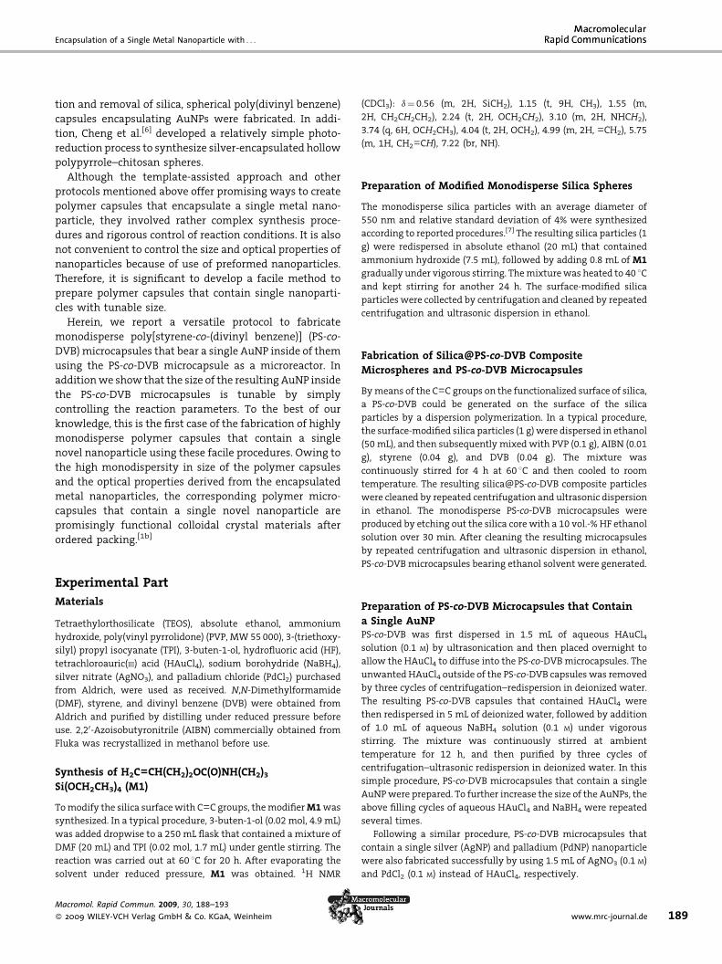
188
Encapsulation of a Single Metal Nanoparticlewith Tunable Size in a Monodisperse PolymerMicrocapsule
Haiqing Li, Chang-Sik Ha, Il Kim*
A versatile approach to fabricate monodisperse poly[styrene-co-(divinyl benzene)] (PS-co-DVB)microcapsules that contain a single gold nanoparticle (AuNP) has been demonstrated. Usingthe PS-co-DVB microcapsule as a microreactor, aqueous HAuCl4 and NaBH4 solutions aresubsequently infiltrated. The size of the result-ing AuNP inside of the PS-co-DVBmicrocapsulesis easily tunable by controlling the repeatedinfiltration cycles of aqueous HAuCl4 andNaBH4. PS-co-DVB microcapsules that containa single silver and palladium nanoparticle arealso obtained by following a similar protocol.
Introduction
Spherical polymer microcapsules have received consider-
able attention because of their diverse technological
applications as microreaction vessels, drug delivery
vehicles, photonic crystals, and biomedical implants.[1] A
variety of chemical and physicochemical methods, such as
the template approach, self-assembly of diblock copoly-
mers, the inverse water-in-oil emulsion process, and the
swelling–evaporation strategy, have been employed to
produce polymer capsules.[2]
In general, the as-prepared hollow polymer capsules
have relatively limited functionalization surfaces, which
limit their applications greatly. To further extend the
applications of the resulting polymer capsules, it is
significant to modify their surface with specific functions.
An important aspect concerning the modification methods
is that the functionalization of the interface of polymer
H. Li, C.-S. Ha, I. KimDepartment of Polymer Science and Engineering, Pusan NationalUniversity, Pusan 609-735, KoreaFax: þ82-51-513-7720; E-mail: [email protected]
Macromol. Rapid Commun. 2009, 30, 188–193
� 2009 WILEY-VCH Verlag GmbH & Co. KGaA, Weinheim
capsules can be achieved by encapsulating guest species,
which could endow these capsules with diverse properties.
As an important type of guest species, novel nanoparticles
such as metal nanoparticles, metal oxide nanoparticles,
and quantum dots have attracted interest because of their
remarkable optoelectronic, catalytic, and biomedical
properties.[3] To date, several studies concerning the
synthesis of polymer capsules that encapsulate these
novel nanoparticles have been explored. Most of the
reported protocols are based on a template-assistant
approach. First, the preformed nanoparticles (e.g., Au
nanoparticles (AuNPs)) are coated with a silica shell. This
sheath is further functionalized with certain reactive
groups to grow a polymer layer. Upon removal of the
middle silica layer using hydrofluoric acid, polymer
capsules that contain single nanoparticles are formed.
For instance, Kamata et al.[4] synthesized poly(benzyl
methacrylate) capsules that contained a single AuNP by
means of conformally coating a silica shell and a
poly(benzyl methacrylate) sheath by atom transfer radical
polymerization, followed by the removal of the silica shell.
Kim et al.[5] included divinyl benzene and initiator into the
mesoporous silica coating of the AuNP. After polymeriza-
DOI: 10.1002/marc.200800629
Encapsulation of a Single Metal Nanoparticle with . . .
tion and removal of silica, spherical poly(divinyl benzene)
capsules encapsulating AuNPs were fabricated. In addi-
tion, Cheng et al.[6] developed a relatively simple photo-
reduction process to synthesize silver-encapsulated hollow
polypyrrole–chitosan spheres.
Although the template-assisted approach and other
protocols mentioned above offer promising ways to create
polymer capsules that encapsulate a single metal nano-
particle, they involved rather complex synthesis proce-
dures and rigorous control of reaction conditions. It is also
not convenient to control the size and optical properties of
nanoparticles because of use of preformed nanoparticles.
Therefore, it is significant to develop a facile method to
prepare polymer capsules that contain single nanoparti-
cles with tunable size.
Herein, we report a versatile protocol to fabricate
monodisperse poly[styrene-co-(divinyl benzene)] (PS-co-
DVB) microcapsules that bear a single AuNP inside of them
using the PS-co-DVB microcapsule as a microreactor. In
addition we show that the size of the resulting AuNP inside
the PS-co-DVB microcapsules is tunable by simply
controlling the reaction parameters. To the best of our
knowledge, this is the first case of the fabrication of highly
monodisperse polymer capsules that contain a single
novel nanoparticle using these facile procedures. Owing to
the high monodispersity in size of the polymer capsules
and the optical properties derived from the encapsulated
metal nanoparticles, the corresponding polymer micro-
capsules that contain a single novel nanoparticle are
promisingly functional colloidal crystal materials after
ordered packing.[1b]
Experimental Part
Materials
Tetraethylorthosilicate (TEOS), absolute ethanol, ammonium
hydroxide, poly(vinyl pyrrolidone) (PVP, MW 55 000), 3-(triethoxy-
silyl) propyl isocyanate (TPI), 3-buten-1-ol, hydrofluoric acid (HF),
tetrachloroauric(III) acid (HAuCl4), sodium borohydride (NaBH4),
silver nitrate (AgNO3), and palladium chloride (PdCl2) purchased
from Aldrich, were used as received. N,N-Dimethylformamide
(DMF), styrene, and divinyl benzene (DVB) were obtained from
Aldrich and purified by distilling under reduced pressure before
use. 2,20-Azoisobutyronitrile (AIBN) commercially obtained from
Fluka was recrystallized in methanol before use.
Synthesis of H2C––CH(CH2)2OC(O)NH(CH2)3
Si(OCH2CH3)4 (M1)
To modify the silica surface with C––C groups, the modifier M1 was
synthesized. In a typical procedure, 3-buten-1-ol (0.02 mol, 4.9 mL)
was added dropwise to a 250 mL flask that contained a mixture of
DMF (20 mL) and TPI (0.02 mol, 1.7 mL) under gentle stirring. The
reaction was carried out at 60 8C for 20 h. After evaporating the
solvent under reduced pressure, M1 was obtained. 1H NMR
Macromol. Rapid Commun. 2009, 30, 188–193
� 2009 WILEY-VCH Verlag GmbH & Co. KGaA, Weinheim
(CDCl3): d¼0.56 (m, 2H, SiCH2), 1.15 (t, 9H, CH3), 1.55 (m,
2H, CH2CH2CH2), 2.24 (t, 2H, OCH2CH2), 3.10 (m, 2H, NHCH2),
3.74 (q, 6H, OCH2CH3), 4.04 (t, 2H, OCH2), 4.99 (m, 2H, ––CH2), 5.75
(m, 1H, CH2––CH), 7.22 (br, NH).
Preparation of Modified Monodisperse Silica Spheres
The monodisperse silica particles with an average diameter of
550 nm and relative standard deviation of 4% were synthesized
according to reported procedures.[7] The resulting silica particles (1
g) were redispersed in absolute ethanol (20 mL) that contained
ammonium hydroxide (7.5 mL), followed by adding 0.8 mL of M1
gradually under vigorous stirring. The mixture was heated to 40 8Cand kept stirring for another 24 h. The surface-modified silica
particles were collected by centrifugation and cleaned by repeated
centrifugation and ultrasonic dispersion in ethanol.
Fabrication of Silica@PS-co-DVB CompositeMicrospheres and PS-co-DVB Microcapsules
By means of the C––C groups on the functionalized surface of silica,
a PS-co-DVB could be generated on the surface of the silica
particles by a dispersion polymerization. In a typical procedure,
the surface-modified silica particles (1 g) were dispersed in ethanol
(50 mL), and then subsequently mixed with PVP (0.1 g), AIBN (0.01
g), styrene (0.04 g), and DVB (0.04 g). The mixture was
continuously stirred for 4 h at 60 8C and then cooled to room
temperature. The resulting silica@PS-co-DVB composite particles
were cleaned by repeated centrifugation and ultrasonic dispersion
in ethanol. The monodisperse PS-co-DVB microcapsules were
produced by etching out the silica core with a 10 vol.-% HF ethanol
solution over 30 min. After cleaning the resulting microcapsules
by repeated centrifugation and ultrasonic dispersion in ethanol,
PS-co-DVB microcapsules bearing ethanol solvent were generated.
Preparation of PS-co-DVB Microcapsules that Contain
a Single AuNPPS-co-DVB was first dispersed in 1.5 mL of aqueous HAuCl4
solution (0.1 M) by ultrasonication and then placed overnight to
allow the HAuCl4 to diffuse into the PS-co-DVB microcapsules. The
unwanted HAuCl4 outside of the PS-co-DVB capsules was removed
by three cycles of centrifugation–redispersion in deionized water.
The resulting PS-co-DVB capsules that contained HAuCl4 were
then redispersed in 5 mL of deionized water, followed by addition
of 1.0 mL of aqueous NaBH4 solution (0.1 M) under vigorous
stirring. The mixture was continuously stirred at ambient
temperature for 12 h, and then purified by three cycles of
centrifugation–ultrasonic redispersion in deionized water. In this
simple procedure, PS-co-DVB microcapsules that contain a single
AuNP were prepared. To further increase the size of the AuNPs, the
above filling cycles of aqueous HAuCl4 and NaBH4 were repeated
several times.
Following a similar procedure, PS-co-DVB microcapsules that
contain a single silver (AgNP) and palladium (PdNP) nanoparticle
were also fabricated successfully by using 1.5 mL of AgNO3 (0.1 M)
and PdCl2 (0.1 M) instead of HAuCl4, respectively.
www.mrc-journal.de 189

H. Li, C.-S. Ha, I. Kim
190
Sample Characterization
Samples for transmission electron microscopy (TEM) were
deposited onto carbon-coated copper electron microscope grids
and dried in air. TEM analysis was performed using a JEOL 1200 EX
at 120 keV. All the UV-vis absorption spectra of the PS-co-DVB
microcapsule suspensions (in absolute alcohol) were recorded at
room temperature on a UV-1650PC apparatus (Shimadzu).
Infrared spectra of the samples were obtained at a resolution of
1 cm�1 with a Bruker FT-IR spectrophotometer over a wavenum-
ber range of 4 800 to 400 cm�1. IR measurement of the powder
samples was performed in the form of KBr pellets.
Results and Discussion
The facile but efficient protocol developed in this study is
illustrated in Scheme 1. The first step involves the
modification of monodisperse silica microspheres with
polymerizable vinyl groups (M1), followed by the encap-
sulation of the modified silica surface by a PS-co-DVB shell
through a disperse polymerization. After the resultant PS-
co-DVB-coated silica (silica@PS-co-DVB) composites are
subjected to removal of the silica cores using hydrofluoric
acid (HF), monodisperse PS-co-DVB capsules are created.
The resulting PS-co-DVB capsules are dispersed in aqueous
HAuCl4 to allow Au3� ions to diffuse into the PS-co-DVB
capsules. Subsequent diffusion of aqueous NaBH4 into the
PS-co-DVB capsules produces a single AuNP (AuNP@PS-co-
DVB). By repeating the filling cycles of HAuCl4 and NaBH4,
the size of the resulting AuNPs can be controlled. This
protocol for making AuNP@PS-co-DVB has the following
advantages over the reported methods: 1) The preparation
Scheme 1. Schematic illustration of the preparation of poly[styrene-conanoparticles (AuNP@ PS-co-DVB). M1 is the surface-modifier of the
Macromol. Rapid Commun. 2009, 30, 188–193
� 2009 WILEY-VCH Verlag GmbH & Co. KGaA, Weinheim
procedures are simple and readily controllable, 2) the size
of the resultant AuNPs are tunable, which endows the
PCAs with controllable optical absorption, 3) the resulting
PCAs are highly monodisperse.
Figure 1a and 1b show TEM images of silica micro-
spheres and the corresponding silica@PS-co-DVB core–
shell microspheres, respectively. It is observed that the
size of the composite microspheres increases from
550 nm (neat silica) to 800 nm. In addition, the
silica@PS-co-DVB particles exhibit good monodispersity
that is around 4% in size polydispersity. The functional
silica surface and the PS-co-DVB coating, the as-prepared
surface-modified silica microspheres, and the correspond-
ing silica@PS-co-DVB composites were characterized by
infrared spectroscopy (Figure 1c). The absorption peak
centered at 1 645 cm�1 (curve i) is assigned to the
characteristic peak of hydrogen-bonded urethane carbo-
nyl groups,[8] which confirms M1 is grafted onto the silica
surface successfully. The characteristic peaks of polystyr-
ene at 1 600 and 1 450 cm�1, which belong to the C––C
bands of the phenyl ring (curve ii),[9] evidence the
formation of the PS-co-DVB shell. The monodisperse PS-
co-DVB microcapsules are produced by the removal of the
silica core using HF. The TEM image (Figure 1d) shows a
clear contrast between the dark ring and the pale center
of the spherical particles, which indicates the formation
of a hollow interior within the PS-co-DVB. By investigat-
ing over 100 microcapsules, it is found that most of the
microcapsules exhibit fairly regular shapes and high
monodispersity in both outer and inner diameter. The
average shell thickness of the PS-co-DVB microcapsules is
about 125 nm.
-(divinyl benzene)] (PS-co-DVB) microcapsules that contain single Ausilica microsphere, H2C––CH(CH2)2OC(O)NH(CH2)3Si(OCH2CH3)4.
DOI: 10.1002/marc.200800629

Encapsulation of a Single Metal Nanoparticle with . . .
Figure 2. TEM images of PS-co-DVB microcapsules that contain Au nanoparticles(AuNP@PS-co-DVB) of varied size prepared by repeat filling cycles of aqueous HAuCl4and NaBH4: a) one cycle, b) two cycles, and (c) three cycles. (d) UV-Vis spectra of theresulting AuNP@PS-co-DVBs obtained by repeated filling cycles of aqueous HAuCl4 andNaBH4: i) one cycle, ii) two cycles, and iii) three cycles.
Figure 1. TEM images of a) silica microspheres and b) poly[styrene-co-(divinyl benzene)](PS-co-DVB)-coated silica microspheres (silica@PS-co-DVB). c) FT-IR spectra of i) H2C––CH(CH2)2OC(O)NH(CH2)3Si(OCH2CH3)4-modified silica particles and ii) silica@PS-co-DVBmicrospheres. d) TEM image of PS-co-DVB microcapsules obtained by removal of thesilica core from silica@PS-co-DVB composite microspheres.
Macromol. Rapid Commun. 2009, 30, 188–193
� 2009 WILEY-VCH Verlag GmbH & Co. KGaA, Weinheim
Figure 2 shows the TEM images of PS-
co-DVB microcapsules with a diverse size
of AuNPs. When the PS-co-DVB micro-
capsules were subject to the first filling
cycle of HAuCl4 and NaBH4, AuNPs of
30 nm average diameter are formed
(Figure 2a). It is interesting to note that
more than 90% of the PS-co-DVB capsules
contain a single AuNP inside of them,
which demonstrates the versatility of
this protocol. When the filling cycle is
repeated two and three times, the
average diameter of the AuNPs increases
to 45 and 67 nm, respectively. No more
newly formed AuNPs are observed. In
addition, the spherical shape of the
capsules is maintained after the incor-
poration of AuNPs.
The mechanistic pathway for the for-
mation of a single AuNP within the PS-co-
DVB microcapsule might be explained in
terms of a confined nucleation and
growth process. When the reducing agent
NaBH4 diffuses into the cavity of the PS-
co-DVB microcapsules filled with HAuCl4,
the AuCl�4 ions are reduced and form
many Au nuclei, which distribute into the
solution within the cavity of the PS-co-
DVB capsule (see Scheme 1). These nuclei
tend to aggregate to form bigger particles.
Once the larger particles form, the remain-
ing nuclei within the cavity of the capsule
will be successively absorbed onto the
surface of the preformed particles, which
results in the growth of Au particles. Even
though several tiny AuNPs could form at
the same time, the confined space within
the capsule also allows them to aggregate,
which may also be responsible for the
formation of a single AuNP. The limited
amount of precursor (AuCl�4 ions) accom-
modated in the confined cavity of the PS-
co-DVB capsule avoids further growth of
the nanoparticle. In addition, the PS-co-
DVB shell also acts as a protective shell to
stabilize the resulting nanoparticles.
When HAuCl4 and NaBH4 solutions are
further introduced into the cavity of the
PS-co-DVB capsule, the preformed AuNPs
act as nuclei and keep growing into larger
nanoparticles.
AuNPs usually exhibit strong surface
plasmon resonance owing to the inter-
action with the external electromag-
www.mrc-journal.de 191

H. Li, C.-S. Ha, I. Kim
Figure 3. TEM images of PS-co-DVB microcapsules that contain single silver (a) andpalladium (b) nanoparticles. c) UV-vis spectrum of PS-co-DVB microcapsules thatcontain single silver (i) and palladium (ii) nanoparticles. The scale bars in (a) and (b)are 1 mm.
192
netic field induced by light.[10] Moreover, the plasmon
band of the AuNPs is tunable depending on the size of the
nanoparticles.[11] In our current study, the formation of
AuNPs with varied size encapsulated in the PS-co-DVB
capsules would endow the polymer capsules with
different optical absorptions. This can be evidenced by
investigating the resulting AuNP@PS-co-DVB by UV-vis
spectroscopy. Figure 2d shows that the AuNP@PS-co-DVB,
obtained from the first, the second, and the third filling
cycles of HAuCl4 and NaBH4, exhibit broad optical
absorption peaks centered at 525, 544, and 556 nm
(curves i, ii, and iii), respectively. It is evident that this
red-shift in the optical absorption is induced by the
increase of the size of the resulting AuNPs. It is also found
that the absorption intensity increases with increasing
AuNP size because of light scattering in the presence of
larger nanoparticles.
To extend the current protocol to fabricate PS-co-DVB
microcapsules that contain other metal nanoparticles, Ag
and Pd nanoparticles were chosen as candidate materials
because of their extensive applications in the field of
biomedicine and catalysis.[12] According to the TEM images
in Figure 3, more than 90% of the PS-co-DVB microcapsules
encapsulate a single Ag or Pd nanoparticle. The PS-co-DVB
microcapsules were subjected to three filling cycles of
metal precursor (AgNO3 or PdCl2) solutions and aqueous
NaBH4. The average diameters of the resulting AgNPs and
PdNPs are 60 and 62 nm, respectively. The optical
absorption of the resulting PS-co-DVB capsules that
contain Ag and Pd nanoparticles were also investigated.
As is shown in Figure 3c, the two composites, AgNP@PS-
co-DVB and PdNP@PS-co-DVB, exhibit absorption peaks
centered at 435 (curve i) and 337 nm (curve ii), which are
derived from the surface plasmon resonances of Ag and Pd
nanoparticles, respectively. These results demonstrate that
Macromol. Rapid Commun. 2009, 30, 188–193
� 2009 WILEY-VCH Verlag GmbH & Co. KGaA, Weinheim
the versatile protocol developed in this
study might be utilized to fabricate a
variety of polymer–metal nanoparticle
composites, by using different types of
polymers and metal nanoparticles or
quantum dots. Since this will endow
the polymer capsules with diverse prop-
erties and thus extend their applications
greatly, related studies that include
searching for suitable applications are
in progress.
Conclusion
In summary, we present a simple yet
efficient way to fabricate monodisperse
PS-co-DVB microcapsules that contain-
ing a single AuNP. The size of the
resulting AuNPs can be controlled by simply changing
the infiltration cycles of the aqueous HAuCl4 and NaBH4
solution. This tunability in the size of the AuNPs endows
the resulting monodiperse AuNP@PS-co-DVB with tunable
optical absorption. Following a similar procedure, PS-co-
DVB microcapsules that bear a single Ag or Pd nanoparticle
were also prepared successfully, which demonstrates the
versatility of the protocol developed in this study.
Acknowledgements: This work was supported by grants-in-aidfor the National Core Research Center Program from MOST/KOSEF(R15-2006-022-01001-0), Brain Korea 21 program (BK-21), and theCenter for Ultramicrochemical Process Systems (CUPS).
Received: October 7, 2008; Revised: November 11, 2008; Accepted:November 14, 2008; DOI: 10.1002/marc.200800629
Keywords: disperse polymerization; gold nanoparticles; micro-encapsulation; microreactors; monodisperse microcapsules;silicas
[1] [1a] D. G. Shchukin, G. B. Sukhorukov, Adv. Mater. 2004, 16,671; [1b] X. Xu, S. A. Asher, J. Am. Chem. Soc. 2004, 126,7940; [1c] H. Ai, S. A. Jones, Y. M. Lvov, Cell Biochem. Biophys.2003, 39, 1085.
[2] [2a] S. A. Johnson, P. J. Ollivier, T. E. Mallouk, Science 1999, 283,963; [2b] M. S. Wong, J. N. Cha, K. Choi, T. J. Deming, D.Stucky, Nano Lett. 2002, 2, 583. [2c] L. Song, M. Wang, Y.Cong, W. Liu, X. Ge, Z. Zhang, Polymer 2007, 48, 150. [2d] J.Han, G. Song, R. Guo, J. Polym. Sci., Part A: Polym. Chem. 2007,45, 2638.
[3] A. N. Shipway, E. Katz, I. Willner, CHEMPHYSCHEM 2000, 1, 18.[4] K. Kamata, Y. Lu, Y. Xia, J. Am. Chem. Soc. 2003, 125, 2384.[5] M. Kim, K. Sohn, H. B. Na, T. Hyeon, Nano Lett. 2002, 2, 1383.
DOI: 10.1002/marc.200800629

Encapsulation of a Single Metal Nanoparticle with . . .
[6] D. Cheng, X. Zhou, H. Xia, H. Z. O. Chan, Chem.Mater. 2005, 17,3578.
[7] H. Li, S. Abraham, W. Yan, C.-S. Ha, I. Kim, Macromol. RapidCommun. 2007, 28, 1534.
[8] C. Chen, S. A. Dai, H. Chang, W. Su, T. Wu, R. Jeng, Polymer2005, 46, 11849.
Macromol. Rapid Commun. 2009, 30, 188–193
� 2009 WILEY-VCH Verlag GmbH & Co. KGaA, Weinheim
[9] D. Yu, J. An, S. D. Ahn, S. Kang, K. S. Suh, Colloids Surf. A 2005,266, 62.
[10] A. C. Templeton, W. P. Wuelfing, R. W. Murray, Acc. Chem. Res.2000, 33, 27.
[11] P. Mulvaney, Langmuir 1996, 12, 788.[12] M. De, P. S. Ghosh, V. M. Rotello, Adv. Mater. 2008, 20, 1.
www.mrc-journal.de 193
Copyright © 2022 FDOKUMEN