Glass matrix/pyrochlore phase composites for nuclear wastes encapsulation
Upload
khangminh22Category
view
0download
0
HAL Id: tel-01834554https://tel.archives-ouvertes.fr/tel-01834554
Submitted on 10 Jul 2018
HAL is a multi-disciplinary open accessarchive for the deposit and dissemination of sci-entific research documents, whether they are pub-lished or not. The documents may come fromteaching and research institutions in France orabroad, or from public or private research centers.
L’archive ouverte pluridisciplinaire HAL, estdestinée au dépôt et à la diffusion de documentsscientifiques de niveau recherche, publiés ou non,émanant des établissements d’enseignement et derecherche français ou étrangers, des laboratoirespublics ou privés.
Silica based materials for the encapsulation ofβ-Galactosidase
Ileana-Alexandra Pavel-Licsandru
To cite this version:Ileana-Alexandra Pavel-Licsandru. Silica based materials for the encapsulation of β-Galactosidase.Chemical engineering. Université de Lorraine, 2017. English. �NNT : 2017LORR0322�. �tel-01834554�
AVERTISSEMENT
Ce document est le fruit d'un long travail approuvé par le jury de soutenance et mis à disposition de l'ensemble de la communauté universitaire élargie. Il est soumis à la propriété intellectuelle de l'auteur. Ceci implique une obligation de citation et de référencement lors de l’utilisation de ce document. D'autre part, toute contrefaçon, plagiat, reproduction illicite encourt une poursuite pénale. Contact : [email protected]
LIENS Code de la Propriété Intellectuelle. articles L 122. 4 Code de la Propriété Intellectuelle. articles L 335.2- L 335.10 http://www.cfcopies.com/V2/leg/leg_droi.php http://www.culture.gouv.fr/culture/infos-pratiques/droits/protection.htm

1
Collégium SCIENCES & TECHNOLOGIES Pôle scientifique Chimie Physique Moléculaires Ecole Doctorale SESAMES ED 412
Thèse de doctorat
Présentée pour l’obtention du titre de
Docteur de l’Université de Lorraine en Chimie
par
Ileana-Alexandra Pavel
Silica based materials for the encapsulation of
β-galactosidase
Soutenu le 29 Novembre 2017
Membres du jury:
Rapporteurs: Mihail Barboiu Directeur de Recherche Institut Européen des
Membranes, France Jordi Esquena Moret Directeur de Recherche
Institute of Advanced Chemistry of Catalonia, Espagne
Examinateurs: Vanessa Fierro Directrice de Recherche au CNRS
Institut Jean Lamour (IJL), France
Bruno Medronho Chargé de recherche Université de l'Algarve, Portugal Directrice de thèse: Andreea Pasc Maitre de conférences (HDR) Université de Lorraine, France Co-directeur de thèse: Nadia Canilho Maitre de conférences
Université de Lorraine, France

2

3
Abstract
The engineering of solid dietary supplements provides several
advantages in the industrial formulation of food products, in terms of its
production, storage and handling. Thereby, the goal of this doctoral work is to
design bio-responsive carriers for the encapsulation of an exogenous enzyme
able to catalyze the hydrolysis of lactose towards simple sugar molecules. In
fact, there is a consensus that the onset of symptoms characteristic of lactose
intolerance are associated with lactase deficiency in the small intestine.
Providing the organism with exogenous lactase is the underlying application
targeted by this work through the design of silicabased materials for
encapsulation.
The different types of bio-carriers developed had to overcome the
simulated gastric conditions in order to release active enzyme molecules in the
small intestine. Amorphous porous silica is a very good and non-toxic
component affording protection versus acidic conditions, while providing
controlled release. This inorganic material approved by the US Food and Drug
Administration (FDA) has a relatively low cost, and presents a controlled
structure (shape, size, pore diameter), as well as tunable surface chemistry.
In agreement with the main objectives, four bio-adapted encapsulation
strategies were investigated as potential routes to produce solid dietary
supplements for lactose intolerance treatment: (i) physical entrapment of the
enzyme in pre-synthesized meso-macroporous silica materials, (ii) physical
entrapment of the enzyme in low porosity silica particles coated by liposomes,
(iii) encapsulation of the enzyme into thermosensitive solid lipid nanoparticles
(SLNs) (iv) encapsulation of the enzyme into a biopolymer matrix coated in a
mesoporous silica shell.

4
Resume
L’ingénierie des compléments alimentaires solides offre plusieurs
avantages dans la formulation industrielle des produits alimentaires, en termes
de production, stockage, et manipulation. Pour ces raisons, l’objectif de cette
thèse était d’élaborer des ‘cargos’ bio-réactifs, permettant l’encapsulation d’une
enzyme exogène capable de réaliser la réaction d’hydrolyse des molécules de
lactose. Aujourd’hui il est établi que les symptômes caractéristiques de
l’intolérance au lactose sont associés à une carence en lactase dans le gros
intestine. Ainsi, fournir au corps humain de la lactase est l’application ciblée par
ce travail, par la conception de matériaux siliciques comme support
d’encapsulation.
En général, les types de cargos développés doivent surmonter les
conditions gastriques pour libérer l’enzyme dans le gros intestine. La silice
poreuse amorphe est un matériau inorganique non-toxique qui assure une
bonne protection dans des conditions acides et permet une libération contrôlée
au pH légèrement basique du colon. L’utilisation de silice amorphe poreuse
permet à coût réduit d’obtenir une structure intrinsèque contrôlée (forme, taille
particulaire, diamètre du pore) et une chimie de surface modifiable.
En accord avec les objectifs principaux, quatre stratégies
d’encapsulation bio-adaptées ont été étudiées comme de potentiels voies pour
la production de compléments alimentaires solides d’intérêt pour le traitement
de l’intolérance au lactose : (i) immobilisation de l’enzyme par adsorption dans
des matériaux siliciques meso-macroporeux pré-synthétises, (ii) immobilisation
de l’enzyme sur des particules de silice faiblement poreuses recouvertes par
des liposomes, (iii) encapsulation de l’enzyme dans des nanoparticules de
lipides solides (SLNs), (iv) encapsulation de l’enzyme dans une matrice de
biopolymère recouvert d’une coque de silice mésoporeuse.

5
Popularized Abstract
Today smart technologies are everywhere. Most of them are related with
electronics, right? But what about food? Can food be smart? Can food go
beyond the necessity? What about having food that can treat, or even avoid
allergies?
This PhD project is placed in the field of smart food designed to treat
lactose intolerance. This investigation has been focused on the encapsulation
of lactase, an enzyme or protein, that “cuts” lactose in glucose and galactose,
more digestible sugars for our organism. For this, we have been working in the
development of potential strategies of encapsulation of lactase in biocompatible
and bio-responsive carriers. These have to protect and transport active enzyme
macromolecules up to the small intestine where it can degrade the lactose
molecules. Thus, the key property of these carriers is to protect the lactase from
gastric pH conditions, while at the same time to release it in the small intestine.
Therefore, these four main strategies were investigated: the encapsulation of
enzyme in meso-macroporous silica, in thermo-responsive solid lipid
nanoparticles, in a bio-polymer matrix coated with mesoporous silica shell or
lactase immobilization on less porous silica beads protected by a liposome
coating. All of the four are compatible in designing a route towards solid dietary
supplements formulation.
This work has also perspective applications in dairy products
manufacturing. So, enjoy some ice cream or some cheese and forget about
lactose intolerance.

6
Résume vulgarisé
Aujourd’hui les technologies intelligentes sont partout et surtout dans le
domaine de l’électronique. Mais concernant la nourriture ? Peut-elle être un
aliment intelligent ? Et répondre plus qu’à un besoin nutritionnel ? Que dirait-
on d’un aliment traitant ou préventif des allergies ?
La thématique du sujet de cette thèse s’inscrit dans l’optique de
contribuer au développement d’aliments intelligents permettant de traiter ou de
soulager les personnes intolérantes au lactose. Ce travail de recherche a été
focalisé sur l’encapsulation d’une lactase, autrement dit une enzyme ou une
protéine, qui « coupe » le lactose en glucose et galactose qui sont des sucres
plus facilement digérables pour notre organisme. Pour atteindre cet objectif,
plusieurs stratégies d’encapsulation ont été étudiées afin d’obtenir des ‘cargos’
biocompatibles et bio-réactifs dans les conditions physiologiques. Néanmoins,
le principal rôle des ‘cargos’ est de protéger l’enzyme du pH gastrique et de la
transporter jusqu’à l’intestin petit pour qu’elle y dégrader les molécules de
lactose. Ainsi, le cargo doit arriver intègre à l’intestin puis s’y désintégrer pour
y libérer la lactase. C’est pourquoi, ces quatre stratégies d’encapsulation de
l’enzyme ont été étudiées afin de répondre au cahier des charges : (i)
encapsulation de l’enzyme dans un matériaux silicique méso et macroporeuse,
(ii) immobilisation dans des nanoparticules lipidiques solides, et (iii) dans une
matrice bio-polymérique couverte par une coque de silice mésoporeuse ou (iv)
immobilisation de la lactase sur des billes de silice faiblement poreuses
protégées par une couche de liposomes. Ces méthodes d’encapsulation
contribuent à l ‘élaboration de formulation de suppléments alimentaires solides.
Enfin, ce travail présente des perspectives d’application dans
l’industrialisation de produits laitiers. Alors, appréciez une glace ou du fromage
et oubliez les symptômes liés à l’intolérance au lactose.

7
Table of Contents
Acknowledgement ........................................................................................... 10
Abreviations ..................................................................................................... 12
General introduction ....................................................................................... 14
Introduction générale ...................................................................................... 17
Chapter 1. State-of-the-art .............................................................................. 21
1.1. β-Galactosidase ........................................................................................ 21 1.1.1. Sources of Beta-galactosidase ............................................................ 21
1.1.1.1. β-Galactosidases from bacteria ......................................................... 23 1.1.1.2. β-Galactosidases from fungi .............................................................. 23 1.1.1.3. β-Galactosidases from Plants ........................................................... 24 1.1.1.4. β-Galactosidases from yeast ............................................................. 25
1.1.2. Lactose .................................................................................................... 26 1.1.2.1. Lactose intolerance ........................................................................... 27 1.1.2.2. Hydrolysis of lactose ......................................................................... 29
1.1.3. Techniques and matrices for immobilization ..................................... 30 1.1.3.1. Methods of reversible immobilization ................................................. 31
1.1.3.1.1. Adsorption ..................................................................................... 32 1.1.3.1.2. Ionic binding .................................................................................. 34 1.1.3.1.3. Hydrophobic adsorption................................................................. 34 1.1.3.1.4. Affinity binding ............................................................................... 35 1.1.3.1.5. Chelation or metal binding ............................................................. 36 1.1.3.1.6. Disulfide bonds .............................................................................. 38
1.1.3.2. Methods of irreversible immobilization .............................................. 39 1.1.3.2.1. Covalent immobilization................................................................. 39 1.1.3.2.2. Entrapment.................................................................................... 41
1.1.4. Application of immobilized β-galactosidase ...................................... 45 1.1.4.1. Industrial application ......................................................................... 45
1.1.5. Conclusion ......................................................................................... 49
1. 2. Silica-based systems for oral and food delivery .............................. 54
1.2.1. Prerequisite for oral delivery systems and food applications ........ 55
1.2.2. Formation and origin of silica .............................................................. 57 1.2.2.1. Synthesis of non-porous silica nanoparticles ...................................... 59
1.2.2.1.1. Fumed silica nanoparticles ................................................................ 59 1.2.2.1.2. Stöber nanoparticles ......................................................................... 60
1.2.2.2. Synthesis of mesoporous silica ............................................................. 61 1.2.2.3. Synthesis of hybrid silica microparticles .............................................. 65 1.2.2.4. Biosilica (silica diatoms) ......................................................................... 67
1.2.3. Silica-based oral delivery systems and food applications .............. 70 1.2.3.1. Passive release delivery systems .......................................................... 72 1.2.3.2. Active release delivery systems ............................................................. 75
1.2.3.2.1. pH-controlled release ....................................................................... 75 1.2.3.2.2. Enzyme-triggered release ................................................................ 77 1.2.3.2.3. pH/Enzyme-triggered release .......................................................... 78
1.2.4. Silica health benefits and limitations ................................................. 79

8
1.2.5. Conclusion ............................................................................................. 80
Chapter 2. Materials and Methods ................................................................ 85
2.1. Materials ................................................................................................... 85 2.1.1. Enzyme ........................................................................................................ 85 2.1.2. Buffer solution ............................................................................................ 85 2.1.3. In vitro digestion solutions ....................................................................... 85 2.1.4. Substrate ..................................................................................................... 86
2.2. Preparation methods ............................................................................... 86 2.2.1. Modified meso-macroporous silica supports- β-Galx@SiO2 ............... 86
2.2.1.1. Solid Lipid Nanoparticles (SLN) synthesis ............................................ 86 2.2.1.2. Preparation of meso-macroporous silica supports .............................. 87 2.2.1.3. Preparation of modified meso-macroporous silica supports- β-Galx@SiO2 ...................................................................................................... 88
2.2.2. Liposomes coated silica particles (LCSP) ............................................... 89 2.2.2.1. Preparation of liposomes .................................................................... 89 2.2.2.2. Modification of porous silica particles (ESP) ....................................... 89 2.2.2.3. Preparation of liposomes coated silica particles (LCSP) ..................... 90
2.2.3. Double emulsion type Solid Lipid Nanoparticles (SLN) synthesis ...... 90 2.2.4. Preparation of hybrid materials ................................................................ 93
2.2.4.1. Preparation of hybrid alginate silica particles (ASP) ............................ 93 2.2.4.2. Preparation of alginate core silica shell materials (SAM) ..................... 93 2.2.4.3. Preparation of alginate particles (AP) .................................................. 93
2.3. Characterization methods ...................................................................... 94 2.3.1. Protein Quantification Assay .................................................................... 94 2.3.2. Detection of enzyme and material activity .............................................. 95 2.3.3. Enzyme release in simulated gastro-intestinal fluid ............................... 97 2.3.4. Dynamic light scattering ............................................................................ 98 2.3.5. SAXS measurements ................................................................................. 98 2.3.6. Nitrogen sorption analysis ........................................................................ 99 2.3.7. Microscopy .................................................................................................. 99 2.3.8. Fourier Transform Infrared spectroscopy – FTIR ................................. 100 2.3.9. Thermogravimetric analysis (TGA) ........................................................ 100 2.3.10. Zeta potential measurements................................................................ 101 2.3.11. DSC........................................................................................................... 101
Chapter 3. Physisorption on silica .............................................................. 103 3.1. Preferential adsorption of β-galactosidase regarding Hierarchical Meso-Macro porosity of a Silica Material ................................................................... 107
3.1.2. Morphology and texture of bare and enzyme-loaded silica supports ..... 108 3.1.3. Interaction of β-galactosidase with the meso-macroporous material ..... 113 3.1.4. Activity of free and immobilized enzyme into meso-macroporous silica materials ......................................................................................................... 119 3.1.5. Conclusion ............................................................................................ 120
3.2 Silica-coated liposomes for β-galactosidase delivery .............................. 121 3.2.2. Characterization of bare and enzyme-loaded silica ............................... 122 3.2.3. Characterization of liposomes coated silica particles (LCSP) ................ 126 3.2.4. Enzyme activity during the immobilization and incubation ..................... 127 3.2.5. Enzyme release .................................................................................... 128 3.2.5. Conclusion ............................................................................................ 132

9
Chapter 4. Encapsulation of β-galactosidase in responsive carriers allowing release triggered by either temperature or pH .......................... 137
4.1. Thermo-responsive food grade delivery system for the treatment of lactose intolerance ........................................................................................ 142
4.1.1. SLNs particle characterization ................................................................. 145 4.1.2. In situ UV-visible spectroscopy of enzyme activity ............................... 147 4.1.3. Conclusions and perspectives ................................................................ 157
4.2. pH-responsive hybrid silica-alginate carrier for lactose intolerance treatment ......................................................................................................... 159
4.2.1. Particle characterization ........................................................................... 160 4.2.2. Enzyme release in gastro intestinal simulated fluids ........................... 166
4.2.3. Conclusion and perspectives ................................................................ 168
General Conclusions and Perspectives ..................................................... 172
Conclusions générales et perspectives ..................................................... 176
Appendix 1 Techniques of characterization .............................................. 180 DLS ........................................................................................................................ 180 Small angle X-ray scattering (SAXS) ................................................................. 180 Nitrogen sorption analysis ................................................................................. 181
Appendix 2 Shea butter technical sheet ..................................................... 185
Appendix 3 Halactase technical sheet ........................................................ 186

10
Acknowledgement
Firstly, I would like to thank the director of my thesis Dr. Andreea Pasc.
For the three years I spent in her lab, NANO Group of Laboratoire Structure et
Réactivité des Systèmes Moléculaires Complexes(SRSMC), Unité Mixte de
Recherche n°7565 of CNRS and of University of Lorraine, she has guided my
work and has supported my ideas. Her true passion for science, for chemistry
and biochemistry are a true inspiration both for me and for her students. Her
native curiosity has transferred into my work, offering me an enriched
experience and allowing me to work on a variety of subjects, in a
interdisciplinary fashion. I would particularly like to thank Nadia Canilho, my co-
supervisor, for her patience and guidance that allowed me to grow as a
researcher. On both a professional, as well as on a personal level I can truly
say that I learned a lot from both of my supervisors. I am very grateful for the
chance to be their PhD student, to be part of the SRSMC laboratory and for
being included in the BIBAFOODS Marie Curie ITN. I thank Marie Curie actions,
the European Union’s Seventh Framework Program for research and the
BIBAFOODS project (ITN 606713) for financing my work.
I would like to thank the members of the jury: Mihail Barboiu, Research
Director at Institut Européen des Membranes and Jordi Esquena Moret,
Research Director at Institute of Advanced Chemistry of Catalonia, who
accepted to be rapporteurs, as well Bruno Medronho, Researcher (Investigador
FCT) at Algarve University who accepted to be examiner. Last but not least, I
would like to thank Vanessa Fierro, Research Director at CNRS, Institut Jean
Lamour (IJL) the president of the jury. I truly appreciate their presence for my
defense, as well as their constructive questions and comments on my work.
I thank Philippe Gros, professor at the University of Lorraine and director
of the SRSMC laboratory, and Xavier Assfeld, professor at the University of
Lorraine and director of the Doctoral School SESAMES for welcoming me to
their respective institutions, and for always having a positive attitude.
Many thanks are also due to the many people that helped me and whom
met during this project.
From the SESAMES school, my PhD fellows, Sijin, Maxime, Philippe,
Fernanda, Maciej, Hugo, Benjamin, Issam, Youssef, Timothé, Violetta, Audrey,

11
Sanghoon and Cheryl, I thank them for all the moments and memories we
shared.
From SRSMC, Stéphane Parant and Katalin Selmeczi for their direct
help in my research, and for their time, Paule Bazard for the efficiency in her
work, Jean-Bernard Regnouf De Vains for allowing me to use his lab’s
infrastructure. Eric Dumortier, Lionel Richaudeau, Dominique Dodin and others
who made me feel that I belonged to a big group (or in a big family).
Our many collaborators:
Surender Kumar Dhayal, Hans van den Brink and Martin Lund from Chr.
Hansen for providing the enzyme and being great conversation partners.
Sofia Prazeres from University of Alcala for brightening the lab during
her secondment. Gemma and Carnem, her supervisors, together with who we
had a very fruitful collaboration in the project concerning the meso-
macroporous silica.
Federico Amadei and his supervisor Dr. Tanaka from University of
Heidelberg for their insight knowledge on the project concerning liposomes
coted silica.
Yoran Beldengrün from IQAC-CSIC and his supervisor Jordi Esquena
together with who we had a very fruitful collaboration in the project concerning
pickering emulsions.
My BIBAFOODS fellows, Poonam, Maryam, Maria, Cigdem, Tomasz,
Racha, Federica and Davide, I thank them for all the knowledge and fun we
shared during our encounters.
I also thank to all senior researchers in the project, Tommy, Jens,
Marité, Dennis, Tom, Björn, Maria, Bruno, Filipe, Carmen, Stefan and Anna,
firstly for their entire contribution to the project and for always being a such
pleasant and insightful company.
Last but not least, I would also like to thank my family who made the trip to
see my presentation and to friends, who have been close to me and
supported me all these years. Particularly, I would like to thank my husband,
the chairman of ways and means, who has always been next to me, helped
me, and with whom I share the love for science.
12
Abreviations α-CD: α-CD cyclodextrine β-gal: β-galactosidase β-Galx@SiO2 :enzyme absorbed on meso-macroporous silica
A.oryzae: Aspergillus oryzae APTMS : aminopropyltriethoxysilane AP: alginate particles ASP: hybrid alginate silica particles BCA: bicinchoninic acid BCS: biopharmaceutical classification system BET: Brunauer−Emmett−Teller CALB: lipase B from Candida antarctica CLA: colloidal liquid aphron CLEA: Cross-linked enzyme aggregates Ch: cholesterol Con A: concanavalin A-enzyme CSA: cooperative self-assembly DDS: drug delivery systems DLS: dynamic light scattering DOPC:2-dioleoyl-sn-glycero-3-phosphocholine E. Coli: Escherichia coli EDTA: ethylene diamine tetraacetic acid FDA: US Food and Drug Administration EFSA: European Food Safety Authority EVA: ethylene vinyl acetate Gal: galacto residue GIT: gastrointestinal tract Glc: Galactosyl residue GMA: glycidyl methacrylate GOS: Galacto-oligosaccharides GRAS: generally regarded as safe IMA: Immobilized Metal-Ion Affinity IUPAC: International Union of Pure and Applied Chemistry K.lactis: Kluyveromyces lactis Kluyveromyces sp.: Kluyveromices species LAB: Lactic acid bacteria LCSP: liposomes coated silica particles LCT: transcription mechanism LDPE- low density polyethylene MCM-41: Mobil Composition of Matter series MPTS: Mercaptopropyl-trimethoxysilane MSN: mesoporous silica nanoparticles NBS: N-bromosuccinimide NP: silica nanoparticles NHP: Cetyl palmitate (n-hexadecylpalmitate) NLC: nanostructured lipid carriers NLU: neutral lactase units OC: octyl-agarose ONP: o-nitrophenol

13
ONPG: o-nitrophenyl-β-D-galactopyranoside PBS: buffer phosphate PC: phosphatidylcholine PEI: polyethylenimine PGPR: polyglycerol polyricinoleate PMES: undec-1-en-11-yltetra(ethylene glycol) phosphate mono-ester surfactant PsBGAL: pea seeds β-galactosidase PU: unit of protease RT: room temperature SAP: alginate core silica shell particle SAXS: Small angle X-ray scattering SBA-15: Santa Barbara type SBF: simulated body fluid SDS: sodium lauryl sulfate SGF: simulated gastric fluid SEM: scanning electronic microscopy SIF: simulated intestinal fluid SLN: solid lipid nanoparticles; subsp.: subspecies SP: enzyme modified low porosity silica particles TBG: tomato β -galactosidases TEAH3: tri-ethanolamine Thermus sp.-Thermus species TMOS: tetramethylortosilicate TEOS: tetraethylortosilicate TEM-: transmission electronic microscopy THP: trishydroxymethylphosphine Vmax: maximum velocity ZnO: Native zinc oxide ZnO-NP:Zinc oxide nanoparticles W1: water phase 1 W2: water phase 2 W/O emulsion: inverse water in oil emulsion

14
General introduction
β-galactosidase or lactase is an enzyme naturally present in the small
intestine that helps the organism to digest lactose molecules. The lactase acts
as catalyst for the reaction of lactose hydrolysis, that produces simple sugars,
glucose and galactose. It is now known that the majority of individuals with mild
to severe symptoms of lactose intolerance present a lactase deficiency inducing
an incomplete hydrolysis of the lactose. A significant fraction of global
populations, around 70%, presents symptoms related to lactose intolerance. In
fact, the natural production of lactase in human body gradually decreases after
weaning in infancy. That is why, apart from genetically affected individuals,
mainly adults develop this type of intolerance.
Consequently, lactose intolerant people avoid the consumption of dairy
products. Some of them elect to take lactase supplements or to eat lactose-free
foods, whose industrial production is still on the rise, notably through the use of
β-galactosidases. Nevertheless, in most manufacturing processes the enzyme
cannot be recovered and reused in another production cycle. On the other
hand, the cost of extracting and isolating a specific enzyme remains high. As a
result, the cost of producing lactose-free products is limited by the purchase
price of the enzyme. Thus, one strategy is to immobilize the enzyme inside a
solid support to facilitate catalyst recovery and to maintain the lactase activity
along the production process. Another approach, which is underlying this work,
is to introduce lactase directly into food. This method requires the encapsulation
of the enzyme in a specifically designed carrier, which can be introduced as an
additive during the production of any type of foods. The carrier must be
compatible and responsive physiologically. In fact, as additive laws mandate, it
should be food grade or above and as the oral delivery carrier, it must preserve
and to protect the enzyme from the gastric pH to release it in the small intestine.
This type of pH responsive carriers is usually prepared from biopolymers (e.g.
zein, shellac, alginate, chitosan), from inorganic compounds like silica, or from
a combination of both.
Mesoporous amorphous silica materials have been discovered in 1992.
Ten years later in 2001, they started being intensively used as drug delivery

15
carriers, since amorphous silica had been approved as biocompatible. In fact,
due to their tunable pore size, free volume and consequently large specific
surface area, such a porous material offers a high uptake capacity for
molecules with less than 50 nm diameter with the possibility to generate a
controlled drug release system. Besides organic drugs, the porous material can
also accomodate biomolecules such as enzymes. Since then, the development
of sustained enzymatic catalysts is still a topic of intense investigations in very
different applications (e.g food, energy-biodiesel, pharmaceutical synthesis).
The main challenges to be considered for designing supported enzyme
catalysts are: retaining the native properties of the biomolecule, choosing the
appropriate mode of immobilization for the intended application, and obtaining
a good efficiency for the catalyst. Usually, enzyme immobilization on porous
supports, either organic or inorganic, is achieved by physical adsorption
(hydrogen bonding, Van der Waals interactions) or chemical adsorption through
reactive linkers bonding directly the enzyme and the silica. In the perspective
of increasing the enzyme loading, the life time and efficiency of the catalyst,
amorphous meso-macroporous silica materials are of interest. This type of
support is synthesized through a colloidal formulation. Colloidal engineering is
a little explored route but in accordance with the results of this work, it appears
as a promising approach to answer the challenging criteria previously
announced.
In the present work, we focus on the design of biocompatible and bio-
responsive enzyme carriers. Among the lactases, β-galactosidase from
Kluyveromices lactis source was chosen and used in this work. This enzyme is
robust and a well-known catalyst for the hydrolysis of lactose in the dairy
industry.
The investigation work is presented in four scientific chapters. Chapter 1
presents an overview on β-galactosidase sources and its applications. Different
ways of immobilization to improve the enzyme performance are also presented
and detailed. In the second part of Chapter 1, an overview of porous silica used
as enzyme and drug delivery system for oral and food applications is detailed.

16
Chapter 2 entitled “Materials and methods” is describing in detail the
experimental protocols, technics and chemicals used in this investigation.
Chapter 3 focus on the elaboration of two types of supported enzyme
catalysts prepared from physical adsorption of the β-galactosidase on: (1) a
meso-macroporous silica obtained through colloidal engineering is described in
the first part of the chapter, and (2) a commercially available silica material,
presenting low porosity, coated in a lipid bilayer for protection. In the case of
the meso-macroporous material, the adsorption of the enzyme was
investigated as a function of pore size and related to the specific activity within
the material. The release and the catalytic efficiency, e.g. the activity of the
enzyme, has been studied for liposomes coted carriers in simulated gastro-
intestinal fluids.
Chapter 4 presents β-galactosidase encapsulation in two different types
of carriers: (1) solid lipid nanoparticles (SLNs) obtained from a double emulsion
of Water/Oil/Water (W/O/W), and (2) hybrid silica-alginate particles. The SLN
entrapment approach in was investigated to design a physiological thermo-
responsive carrier that would free the enzyme at a certain temperature. The
entrapment of β-galactosidase in hybrid silica-alginate particles strategy was
investigated as a controlled release pH stimuli carrier for intestinal delivery.
The general conclusions and perspectives of future work are compiled
in Chapter 5.
17
Introduction générale
La β-galactosidase ou la lactase est une enzyme est naturellement
présente dans l’intestin grêle et participe à la digestion de molécules de lactose.
La lactase agit comme un catalyseur pour la réaction d’hydrolyse du lactose, à
l’issue de laquelle des sucres simples comme le glucose et le galactose sont
formés. Il est maintenant connu que la majorité des individus avec des
symptômes faibles à sévères d’intolérance au lactose, présentent une carence
en lactase, induisant une hydrolyse incomplète du lactose. La fraction d’individu
présentant des symptômes liés à l’intolérance au lactose représente 70% de la
population globale. Il est avéré que la production naturelle de la lactase dans
le corps humain baisse dans l’enfance après le sevrage laitier. C’est pourquoi,
outre les individus affectés génétiquement, de nombreux adultes sont affectés
par ce type d’intolérance.
En conséquence, les personnes intolérantes au lactose évitent la
consommation des produits laitières, mais certaines préfèrent prendre des
suppléments de lactase ou manger des produits sans lactose, dont la
production industrielle est en plein essors, notamment par l’usage de β-
galactosidases. D’autre part, le coût d’extraction et d’isolation d’une enzyme
spécifique reste élevé. De ce fait, le coût de production des produits sans
lactose est limité par le prix d’achat de l’enzyme. Ainsi, une stratégie pour
réduire le coût est l’immobilisation d’enzyme dans un support solide pour
faciliter la récupération du catalyseur et pour maintenir l’activité de lactase
durant le processus de production. Une autre approche, sous-jacente à ce
travail, est l’introduction de la lactase directement dans la nourriture. Cette
méthode requiert l’encapsulation de l’enzyme dans un transporteur
spécialement conçu, qui peut être ajouté comme un additif durant la production
de tout type de nourriture. Le transporteur doit être biocompatible et sensible
au milieu physiologique du corps humain. Ainsi, comme les additifs
alimentaires, la composition du transporteur il doit également être de grade
alimentaire (« food grade »). Le transporteur doit aussi préserver et protéger
l’enzyme du pH gastrique, pour favoriser la libération de l’enzyme dans
l’intestine grêle. Un tel transporteur réactif au pH sont généralement préparés

18
à partir de biopolymères (e.g. zein, shellac, alginate, chitosan), de composés
inorganiques comme la silice ou d’une combinaison des deux.
Les matériaux de silice amorphe ont été découverts en 1992, et dix ans
plus tard, en 2001, ils ont été utilisés intensivement comme transporteurs pour
les médicaments, suite à l’approbation par l’‘European Food Safety Authority’
(EFSA) et la ‘US Food and Drug Administration’ (FDA). En effet, comme les
dimensions et le volume de ces pores sont adaptables, la surface spécifique
d’un matériau de silice amorphe poreux peut être importante. De ce fait, un tel
support poreux présent une grande capacité de chargement pour les molécules
de tailles à inférieures à 50 nm. Ces matériaux poreux offrent aussi la possibilité
d’une libération contrôlée des molécules encapsulées. Ainsi, le développement
de catalyseurs enzymatiques est un thème d’investigation intense pour des
applications très diverses (e.g. nourriture, énergie-biodiesel, synthèse
pharmaceutique). Les principaux défis à prendre en considération pour
fabriquer le catalyseur d’enzyme supporté sont les suivants : préserver les
propriétés natives de la biomolécule, choisir une stratégie adaptée à
l’application visée, et finalement obtenir une bonne efficacité catalytique.
Normalement, l’immobilisation de l’enzyme sur supports poreux, organiques ou
inorganiques, est effectuée par adsorption physique (liaisons d’hydrogène,
interactions Van der Waals), ou par adsorption chimique de l’enzyme sur le
support par liaison covalente. En perspective, pour augmenter le degré de
chargement de l’enzyme dans le support, la durée de vie et l’efficacité du
catalyseur formé, les matériaux de silice amorphe méso-macroporeuse
présentent un fort intérêt. Ce type de support silicique est synthétisé à partir
d’une émulsion qui est une dispersion colloïdale d’une phase huileuse dans
l’eau et vis-versa. L’ingénierie colloïdale dans ce domaine est une voie peu
explorée, mais d’après ce travail de thèse, cette approche est prometteuse pour
répondre aux défis annoncés précédemment.
Ce travail est focalisé sur la conception des transporteurs
biocompatibles et bio-sensibles. Parmi les lactases, la β-galactosidase extraite
de la levure Kluyveromices lactis, a été choisi dans ce travail. Cette enzyme

19
est robuste et bien connue dans l’industrie laitière comme catalyseur pour
l’hydrolyse de lactose.
Ce travail d’investigation est présenté en quatre chapitres de recherche
expérimentale. Le Chapitre 1 fait l’état de l’art des sources de β-galactosidase
et leurs applications. D’autre part, les différentes voies d’immobilisation de
l’enzyme sur des supports solides sont aussi présentées en détail. Dans la
deuxième partie du Chapitre 1, fait état de la littérature portant sur la silice
poreuse, utilisée comme systèmes de transport pour la libération de
médicaments et d’enzymes par voie orale.
Le Chapitre 2 intitulé « Matériaux et Méthodes » décrit en détail les
protocoles expérimentaux, les techniques et les composées utilisés pour mener
cette recherche.
Le chapitre 3 est dédié à l’élaboration de deux types de catalyseurs
supportés préparés par l’adsorption physique de β-galactosidase sur : (1) silice
méso-macroporeuse obtenue par ingénierie colloïdale et (2) des billes de silice
commerciales avec une faible porosité, protégées par une couche de
liposomes. Dans le cas du matériel méso-macroporeux, l’adsorption de
l’enzyme était étudiée en fonction de la dimension du pore et rapporté à
l’activité intrinsèque du matériel. La libération, l’efficacité catalytique et l’activité
de l’enzyme, ont été étudiés dans fluides gastrique et intestinal simulés pour
les transporteurs enrobés par les liposomes.
Le chapitre 4 présente l’encapsulation de β-galactosidase dans deux
types de transporteurs : (1) des nanoparticules lipidiques solides (NLS)
obtenues par une émulsion eau/huile/eau (w/o/w), (2) des particules hybrides
silice-alginate. L’approche d’emprisonnement dans des NLS était étudiées pour
concevoir un système transporteur répondant à la température physiologique
du corps humain facteur déclenchant la libération de l’enzyme. Quant au
second systèmes, l’emprisonnement de la β-galactosidase dans des particules
hybrides silice-alginate, a été étudié comme système de transport sensible au
pH pour permettre une libération contrôlée de l’enzyme dans l’intestin grêle.

20
Enfin, les conclusions générales et les perspectives de ce travail sont
exposées dans le chapitre 5.

21
Chapter 1. State-of-the-art
1.1. β-Galactosidase
β-Galatosidase (lactase, EC 3.2.1.23) is an enzyme that hydrolyses D-
galactosyl residues from oligosaccharides, polymers and secondary
metabolites. The enzyme can be used in dairy industry, in problems associated
with whey disposal and lactose crystallization (sweetened and frozen dairy
products) [1] or to produce prebiotics (by the production of
galactooligosaccharies) [2,3]. However, because free β -galactosidase is very
expensive and somewhat sensitive to external factors immobilisation is
required. This makes the enzyme more economically feasible by improving its
catalytic activity and allowing its reuse in batch reactors.
Enzyme identification is given by the EC number. It describes classes of
enzymes catalyzing similar reactions and is a numerical nomenclature that
groups enzymes based on the overall reaction that catalyzed. The EC number
of β-Galatosidase means that the reaction that the enzyme catalyzes is the
hydrolysis of terminal non-reducing β-D-galactose residues in β-D-
galactosides.
1.1.1. Sources of Beta-galactosidase
β-Galatosidases are found in plants (peaches, apricots, almonds [4],
apples, kiwis [5], tomatoes [5,6]), animal organs or in microorganisms (bacteria,
fungi [7,8] and yeasts Table 1.1 .1. The enzymes produced in large (industrial)
quantities are mainly obtained from Aspergillus sp., Kluyveromyces sp. and E.
Coli.
Table 1.1 .1 Sources of β-galactosidase (adapted after [9]).
Plants peach, apricot, kefir grains, almond, tips of wild roses, alfalfa
seeds, coffee berries, beans
[4,10–12]
Animals small intestine, brain and skin tissue [13]
Fungi
Kluyveromyces (Saccharomyces) lactis, Kluyveromyces (Saccharomyces) fragilis, Brettanomyces anomolus, Wingea
robersii [14,15]
Bacteria Escherichia coli, Streptococcus thermophilus, Bacillus circulans, Bacillus steorotherrphilus, Lactobacillus sporogenes
[16][17][18][19]

22
[20]
Yeast Aspergillus niger, Aspergillus oryzeae, Curvularia inoegualli [21][22][23]
The properties of β-Galactosidases, such as structure or size, varies with
the source, but their specificity (hydrolysis of D-galactosyl residues) remains
essentially the same. The characteristics of β-galactosidase from different
sources are shown in Table 1.1 2.
Table 1.1 2. Properties of β-galactosidases from different sources.
SOURCES OPTIMAL
PH
OPTIMAL
TEMPERATURE ACTIVATORS INHIBITORS REF
A. NIGER 3.0-4.0 55-60 none none
A.
ORYZAE 4-6 40-55 none
Hg2+, Cu2+,
NBS*, SDS* [24]
K.
FRAGILIS 6.9-7.3 37 Mn2+, K+, Mg2+ Ca2+, Na+ [25]
K.
LACTIS 6.5-7.3 35 K+, Mg2+ Ca2+, Na+ [14]
E. COLI 7.2 40 Na+, K+ [26]
*NBS: N-bromosuccinimide, SDS: sodium lauryl sulfate
Ca2+ ions are known to be an inhibitor for β-galactosidase. However, the
enzymatic activity of β-galactosidase is not affected by the calcium ions in milk
since they are bounded to casein [27]. Divalent cations, such as magnesium
and manganese, may enhance the β-galactosidase activity, while monovalent
cations may have a positive or negative effect depending of the origin of the
enzyme [27].
The β-galactosidase enzyme exists in three forms in human intestine:
1- lactase found in the edge membrane of the epithelium of the small
intestine (this enzyme is solely responsible for the hydrolysis of lactose);
2- lactase found in the lysosome of the epithelium cells of small intestine.
It is also called acid β-galactosidase;
3- the hetero-β-galactosidase found in the cytoplasm of the epithelium cells
of small intestine.

23
1.1.1.1. β-Galactosidases from bacteria
β-Galactosidases from bacterial sources have been widely used in food
industry due to their advantages, as high activity and stability of the enzyme
and ease of fermentation [28]. Lactic acid bacteria (LAB) have been the most
studied and consist of Lactobacillus delbrueckii subsp. bulgaricus and
Streptococcus thermophilusc (the strains of yogurt culture). The main reasons
of why they gained attention are: (i) little or no adverse effects in the
consumption of some fermented dairy products by lactose maldigestion (ii) the
enzyme derived from them may be used without extensive purification since
they are generally regarded as safe (GRAS), (iii) the probiotic activity found for
some strains, which improve the digestion of lactose [29,30].
The highest production and specific activity of β-Galactosidases was
attained with Bifidobacterium longum CCRC 15708 strain, compared with
Bifidobacterium infantis CCRC 14633 and Bifidobacterium longum B6 strains
[29]. These bacteria are also used as probiotics for their potential health benefits.
Bifidobacteria are present in the human and animal gut, and appear in new-
borns within days after birth [31]. β- galactosidase present in the colon of humans
catalyses the first step of lactose fermentation. Its activity is an indicator of the
capacity of colonic microbiota to ferment the lactose present in the intestine [32].
1.1.1.2. β-Galactosidases from fungi
Fungal β-galactosidases are effective in the hydrolysis of lactose present
in whey (an acidic product), their optimal pH being 2.5–5.4. Fungal β-
galactosidases are more sensitive to product inhibition but present the
advantage of thermal stability [33]. Thermophilic β-galactosidase are one of the
most robust enzymes. They present a built-in stability to temperature and other
inactivation agents. They are used in industrial processing of dairy products
along with heat treatment to lower the microbial contamination and obtain a
sterile product [34]. In the food industry, free and immobilized enzyme forms are
used. For example, β-Galactosidases from thermophile microorganisms such
as Thermus sp. strain T2 can be used for the simultaneous soft thermal
treatment and the hydrolysis of lactose [35,36].

24
β-galactosidase can be purified from Aspergillus oryzae (A. oryzae)
RT102 strain by 2-propanol fractional column chromatography on DEAE-
Sephadex A-50 and Sephadex G-200 [24]. The amino acid sequence of the
enzyme includes 1005 residues with an average molecular mass of 110 kDa.
The three dimensional model of A. Oryzae β-galactosidase shows that it is a
monomeric enzyme with the active site similar with Penicllium sp. and
Trichoderma reesei β-galactosiase [37]. Figure 1.1. 1. represents the three-
dimensional structure of the ribbon model (A) and the catalytic center of the
enzyme (B).
Figure 1.1. 1. The three dimensional structure of the ribbon model (A) and the catalytic center of the enzyme (B) of A. oryzae β-galactosidase (reproduced after [37]).
1.1.1.3. β-Galactosidases from Plants
-Galactosidases are widely distributed in plant tissues. β-
galactosidases from chickpea, radish, mung beans [38], barley, carrot [39], rice
shoots [40], lupins [41], and kidney beans [42] have also been isolated, purified and
characterized. Plant β-galactosidases are generally dimeric and much smaller
compared with other β-galactosidases. Such enzymes have an optimal pH in
the acidic range (3.5-7) [43]. Moreover, these enzymes are not only involved in
the hydrolysis of lactose but also in plant growth and fruit ripening and
development [38,44]. β-galactosidase activity has been reported during tomato
fruit ripening (Lycopersicon esculentum Mill.) The cDNAs of a family of seven
tomato β-galactosidase (TBG) was recognized [45]. Significant decrease in cell
wall galactosyl content and the associated pectin degradation that helps with
the ripening of fruit has been associated with the presence of the β-

25
galactosidases [46]. It has been established that β-galactosidase in papaya is
responsible for the hydrolysis of its cell wall and softening of the fruit during
ripening with sugar release, [47] as in the case of strawberries ripening [48]. For
comparison, -galactosidase from the cotyledons of germinated nasturtium
(Tropaeolum majus L.) seeds is involved in in vivo hydrolysis of stored
xyloglucan [49].
β-Galactosidase isolated and purified from plants can be used for the
hydrolysis of lactose from milk. Cicer arietinum (chickpeas) extracted enzyme
was immobilized on two different types of resins and evaluated as a cheap way
to remove the lactose from milk [50]. β-galactosidase isolated from almond
(Amygdalus communis) extracted by ammonium sulfate precipitation was used
in a stirred milk batch process. This enzyme could hydrolyse 90% of lactose in
milk and 94% lactose in buffer solution and whey [4]. β-galactosidase from pea
seeds (PsBGAL) proved to be very unstable at low concentrations at 4°C. To
work around this instability, Dwevedi et al. immobilized it on Amberlite MB-150
beads (5 μm diameter) with glutaraldehyde. The enzyme maintained its activity
for a period of 12 months at room temperature, and through its reusability cycles
in lactose hydrolysis [51]. The same research group optimized PsBGAL
immobilization on Sephadex and chitosan with glutaraldehyde. The new
obtained catalyst presented a broad optimal working temperature and large pH
intervals. The higher temperature stability and reusability propose it as suitable
for industrial applications [52].
1.1.1.4. β-Galactosidases from yeast
The natural habitat of the Kluyveromyces lactis (K. lactis) yeast can be
found in dairy. The β-galactosidase extracted from K. lactis yeast presents a
good lactose hydrolysis activity. For this reason β-galactosidase from this yeast
is commercially feasible and largely used in industry [53,54]. The K. lactis β-
galactosidase forms a homo-oligomer of four identical units, a tetrameric
enzyme, that was described as a dimer of dimers [55]. It is active in its tetrameric
and dimeric forms [14]. The enzyme is formed of 1024 residues and have a
molecular mass of 119 kDa. The monomer folds into five domains (Figure 1.1.
2. A), that contain two long insertions related to the oligomerization and

26
specificity. Each dimer contains two catalytic centres at the interface (Figure
1.1. 2 B).
Figure 1.1. 2. Stereo view of K.Lactis-β-Gal monomer (A) Surface representation of the K.Lactis- β -Gal tetramer (B) (reproduced after [55]).
1.1.2. Lactose
The principal constituents of milk are water, fat, protein, lactose,
minerals, as well as the intrace amounts pigments, enzymes, vitamins,
phospholipids and gases. The main carbohydrate in milk is lactose, a
disaccharide sugar with lower solubility compared to sucrose or dextrose (less
than 5%). A β-(1-4) glycosidic linker joins the two monosaccharides, -D-
galactose and -D-glucose, on the anomeric C1 of the β-D-galactose and the
C4 of D-glucose (Figure 1.1. 3.) [56].
Figure 1.1. 3. α and β lactose.
In solid phase, lactose can be crystalline or amorphous. Crystalline

27
lactose can exist in one of two distinct forms, β-lactose and α-lactose (as
monohydrate) (Figure 1.1. 3.). In milk, lactose is present in two isomeric forms
β-lactose and α-lactose that are in chemical equilibrium [57].
Lactose is largely used in food industry. In yogurt, acid-coagulated dairy
and some varieties of cheese the presence of lactose is crucial [58]. Isolated
lactose can be used in the production of food and pharmaceutical products [59].
Galacto-oligosacchrides, lactulose, lactitol, and lactobionic acid are obtained
from lactose [60].
Oligosaccharides are polymeric saccharides consisting of two to ten
monomer residues (simple sugars) joined through glycosidic bonds. They can
be obtained from different sources such as crops (onion, garlic) or lactose
present in milk and whey (galacto-oligosaccharides) [61]. Galacto-
oligosaccharides (GOS) are (galactosyl)nlactose oligomers (2≤n≤4),
synthesized by a transgalactosylation reaction from lactose catalyzed by β-
galactosidase [62]. The reaction mechanism for the producing GOS was first
proposed by Wallenfals and Malhotra, who used GOS as a growth factor for
Bifidobacterium spp strain (have advantageous physiological effects on the
host human). Owing to their roles in controlling pH in the large intestine (by
promoting the production of lactic and acetic acids which limit the growth of
pathogens and putrefactive bacteria [63]). The amount and composition of
galacto-oligosaccharides vary with the source of enzyme, lactose concentration
and the reaction conditions used in the process.
1.1.2.1. Lactose intolerance
A person unable to completely digest lactose is diagnosed with lactose
intolerance. A significant fraction of global populations, of around 70%,
presents symptoms related to lactose intolerance [64,65]. In fact, lactose
intolerance symptoms are specifically caused by the deficiency of the β-
galactosidase in the small intestine. Adults are mainly affected by lactose
intolerance because the natural production of lactase in human body gradually
decreases after weaning in infancy. As a result of the lack of lactase (or of the
low activity of the enzyme or a diminished quantity) the hydrolysis of lactose is

28
incomplete. The undigested sugar pulls fluids into the large intestine, where the
colonic bacteria digest the rest of the sugar, producing short chain fatty acids,
gases (hydrogen, CO2, methane). In the end, the combined osmotic effect
results in the passage of acidic diarrheal stools. Lactose intolerance is
described by the presence of gastrointestinal symptoms such as abdominal
pain and distension, abdominal colic, bloating, flatulence, nausea or diarrhea
[66].
Three types of lactose deficiency have been described: primary,
secondary or congenital. Primary lactase deficiency or Late Onset Lactase
Deficiency is the most common type, caused by the decline of the lactase
production from infancy into adulthood, in spite of a continuous intake of
exogenous lactase. Secondary lactose deficiency results from small intestine
resections and diseases damaging the intestinal epithelium. Congenital
occurrence is genetic and appears when two ineffective genes from parents are
inherited (inability of the newborn to produce lactase) [67].
Some common tests are used to diagnose lactose intolerance: the blood
test, the breath test and the endoscopy test. When the blood sugar rises above
a critical threshold, subsequently to drinking a lactose-based solution, the
person can be concluded as not lactose intolerant. In the breath test, the
presence of hydrogen is analyzed as the concentration of hydrogen in the
exhaled air increases when the lactase is fermented by the bacteria present in
intestine. In the endoscopy test, the lining (mucin) of the intestine is observed
and biopsied to check any damage caused by acid reflux or infection [68].
As lactose intolerant people are unable to digest milk and other dairy
products, a strategy to remove the lactose from these products is required.
Thus, an acidic or enzymatic lactose hydrolysis process can be applied to milk
and other dairy products. The acidic method can raise some problems such as
the formation of a brown colour product, protein denaturation and yield of
undesirable toxic by-products (like lysino-alanine) [69] . Milder conditions of
temperature and pH can be achieved by use of enzymes. However, the
industrial application of the process based on the hydrolysis of the lactose with
free β-galactosidase is limited due to the cost of soluble lactase.

29
1.1.2.2. Hydrolysis of lactose
Many research efforts have been developed to reduce or remove sugar
(lactose) from dairy products. The most common way to accomplish this
remains the use of β-galactosidase. Thanks to the improvements in processing
techniques, hydrolyzing the lactose before packaging certain dairy products
has become more prevalent. The lactose hydrolysis catalyzed by lactase
mechanism was first described by Wallenfels who used β-Galactosidase
extracted from Escherichia coli [62]. In the reaction mechanism proposed, the
cysteine and the histidine residues from the active site of β-galactosidase act
as proton donor and acceptor, respectively to the glycosidic linker. Cysteine
contains the sulfhydryl group while histidine residue contains imidazole group
acting as a proton acceptor and as nucleophile site to facilitate splitting of the
glycosidic bond. Compared with E.Coli extracted enzyme, β-galactosidase from
microbial sources presents two glutamic acid residues, Glu482 and Glu551
working as proton donor and nucleophile/base at the same time in the catalytic
reaction [70]. The positions of the two residues in the catalytic pocket are situated
in the center of each monomer of the tetrameric K. Lactis β-galactosidase as
can be observed in Figure 1.1. 4. Residues from the domains 1, 3 and 5
surround the catalytic pocket that shapes a very narrow cavity of about 20 Å
deep (see Figure 1.1. 4. A). In the dimeric arrangement, the cavities are located
face-to-face within the interface. As domain 3 folds the pockets become
accessible for the external substrate through a 10 Å width slot [55].
Figure 1.1. 4. (A)-Residues from 1, 3 and 5 domains building up the pocket entrance (zoomed view). (B)-Galactose bound to the active site (reproduced from [55]).
The reaction mechanism for K. Lactis β-galactosidase is shown in Figure

30
1.1. 5. In the first step of the reaction, the enzyme-galactosyl complex is formed
and glucose is released. In the second step, the complex enzyme-galactosyl is
transferred to a hydroxyl acceptor group (water or other saccharides). In a
diluted lactose solution, water is more competitive to be an acceptor, therefore,
galactose is formed and released from the active site. On the contrary, in a
concentrated solution, lactose is more competitive as acceptor and binds to the
enzyme-galactose complex to form oligosaccharides.
Figure 1.1. 5. Schematic mechanism of lactose hydrolysis by K. Lactis β-galactosidase (I) enzyme-galactosyl complex formation with the liberation of glucose, (II) enzyme-galactosyl complex transferred to an acceptor containing hydroxyl group (adapted
from [70]).
1.1.3. Techniques and matrices for immobilization
The major drawback of free enzymes is their limited lifetime. External
factors, such as pH, temperature, pressure, organic solvents, high ionic
strength or proteases can destabilize the structure of the enzyme, leading to a
decrease in their catalytic activity. Such drawbacks can be overcome by the
immobilization of the enzyme. Moreover, immobilization might bring other
advantages: the enzymes can be recovered and reused in a new catalytic
reaction, stand longer storage, or can be released under specific conditions.
OH
482
COO-551
O-
482
C=O551
OOH
OH
OH
CH2OH
O +
O-
482
C=O551
OOH
OH
OH
CH2OH
O
H
O
R
OH
482
COO-551

31
Supported enzymes might also became catalytically active in organic solvents
in which the native enzymes are insoluble (eg. lipase) [71–73]. Currently, the study
of solid supports suitable for enzyme immobilization is still a scientific challenge
constrained by enzyme nature and target application. Furthermore, the
immobilization process should also be mild, to prevent enzyme denaturation. In
lactose hydrolysis, for example, the industrial process must be economically
feasible, to address technical interests in milk industry. The extraction enzyme
technology is still expensive, thus immobilized enzymes in food industry are in
focus and the methods for the immobilization of various β-galactosidase
enzymes in different solid supports were envisaged. The scientific community
has also highlighted the benefit of pH and thermal stability of the biocatalyst [74].
Despite those advantages, it turns out that immobilization process can
have some drawbacks, such as the loss of the enzyme activity after
immobilization, mass transfer limitation (slow diffusion between the substrate in
a liquid phase and the biocatalyst phase-solid), leakage of the enzyme from the
matrix and cost aspect related to the implementation of the immobilization step
in an industrial process [74]. According to literature, immobilization techniques
can be classified in two major categories: reversible and irreversible.
Irreversible methods involve the formation of the biocatalyst, while the
components cannot be separated without destroying either the enzyme or the
support. Reversible methods do not involve covalent bonding with the enzyme,
and the enzyme can de detached from the support under gentle conditions.
Chemical and physical properties of the support material including particle size,
surface, porosity, functional group on the surface and morphology are important
in enzyme immobilization. All these have to be considered when choosing the
immobilization technique. The methods used for immobilization can also affect
the kinetic parameters of the immobilized enzyme [75].
1.1.3.1. Methods of reversible immobilization
Reversible immobilization involves weak forces between the enzyme and
the support, so the immobilized enzyme can be detached under gentle
conditions, without destroying one of the components. The methods used are
a) adsorption, b) ionic or hydrophobic binding, c) affinity binding, d) chelation

32
or metal binding, e) disulfite bonds. These are schematized in Figure 1.1. 6.
The reversible methods for enzyme immobilization are mostly used for
economic reasons:
• when the cost of the support plays an important role and the enzyme
can be regenerated
• when the cost of the enzyme is high and reversible immobilization is
used for the purification of the enzyme
Figure 1.1. 6. Methods of reversible immobilization: (A) adsorption, (B) ionic binding/ hydrophobic binding, (C) affinity binding, (D) chelation or metal binding, (E) disulfite
bonds (adapted from [75]).
1.1.3.1.1. Adsorption
This method is based on the physical adsorption of enzyme on the
surface of a solid support. The nature of the interactions of the enzyme with the
support is usually a combination of hydrogen bonding, van der Waals forces,
hydrophobic or/and electrostatic interactions depending on the chemistry of the
surface. One major advantage of this method is that, usually, no reagents or a
minimal modification step for the support are required, which make the
procedure simple and inexpensive. In any case, this method involves weak
bonds that do not prevent enzyme desorption by varying pH, temperature or in
the presence of substrate.
Investigating suitable solid supports for each enzyme and for each
industrial application immobilization is still a current scientific challenge. The
materials employed for enzyme adsorption can be organic or inorganic.
Bone powder was used for the immobilization of Kluyveromices fragilis
enzyme, for the removal of lactose from dairy [76]. The thermal stability of the
enzyme was improved, and the material showed a 90% conversion of the

33
lactose present in buffered solutions, whey, whey permeate (a substitute of
lactose, sweet whey powder, and/or demineralized whey powder) and skimmed
milk. K. lactis enzyme was adsorbed on a mixed-matrix membrane containing
zirconium dioxide. The maximal adsorption of the enzyme onto the membrane
could be achieved under extreme parameters (temperature and pH) but it would
lead to the loss of activity for the enzyme. Immobilized under the optimum
parameters, the enzyme increases its activity almost 8 times [77]. Native zinc
oxide (ZnO) and zinc oxide nanoparticles (ZnO-NP) were also used to
immobilize the A. Oryzae β-Gal by simple physical adsorption mechanism.
Thus, compared to the enzyme adsorbed on native ZnO, the enzyme adsorbed
on ZnO-NP showed better stability against pH, temperature, galactose
inhibition, better reusability and conversion of lactose in milk and whey [78]. The
increased stability of the enzyme is due to the multipoint attachment of enzyme
molecules to the nanomaterial that leads to limited protein unfolding.
To increase the interaction between the enzyme and the support, the
specific active groups on the surface of the support can be modified. K. lactis
enzyme was adsorbed on plasma modified cellulose acetate with
ethylenediamine and 2-mercaptoethanol. Although high enzyme loading was
achieved, only the thiolated membrane surface could keep a high enzymatic
activity [71]. The adsorption of enzymes onto composites based on covalent
coating of supports with polymers has also been proposed.
β-galactosidase from A. oryzae was adsorbed on with glutaraldehyde-
treated chitosan for the production of galactooligosaccharides (GOS) [79] in a
plug reactor, while K. lactis enzyme immobilized on glutaraldehyde-activated
chitosan was used in a packed-bed reactor for the continuous hydrolysis of
lactose and the synthesis of GOS [80]. K. lactis β-galactosidase was also
immobilized on glutaraldehyde modified silica nanoparticles (10-20 nm) and
showed an increase in the optimal pH, temperature and maximum velocity
(Vmax) in the hydrolysis of lactose [81] .

34
1.1.3.1.2. Ionic binding
Another approach to the reversible immobilization of enzymes is based
on ionic binding, the protein–ligand interactions, a principle employed in certain
types of chromatography and based on ionic-exchangers. Enzyme adsorption
on ion exchange supports is quick and simple method, that permits to reuse the
material and it is also applicable to enzyme purification [82]. Depending of the
ligand, the optimum pH and temperature of the enzyme can change.
Co-immobilization is another strategy that has been explored. For
example, Peirce et al immobilized lipase B from Candida antarctica (CALB) on
octyl-agarose (OC). β-galactosidase from A. oryzae was immobilized by ion
exchange after precoating with polyethylenimine (PEI). The adsorption and
desorption of β-galactosidase could be easily achieved and the OC-CALB was
reused as shown in the Figure 1.1. 7 [83].
Figure 1.1. 7. Strategy of co-immobilization of Lipase B and β-galactosidase
(reproduced after [83]).
1.1.3.1.3. Hydrophobic adsorption
Another chromatographic principle based on hydrophobic interactions,
can be used for enzyme immobilization. The strength of interaction relies on
both the hydrophobicity of the adsorbent and of the protein which varies with
the change of pH, salt concentration, or temperature.
β-Galactosidase from A. oryzae was immobilized on colloidal liquid
aphron (CLA) via hydrophobic and electrostatic interaction. The aphron is a
core/shell structure in which a gas is stabilized by a layer of polymer or
35
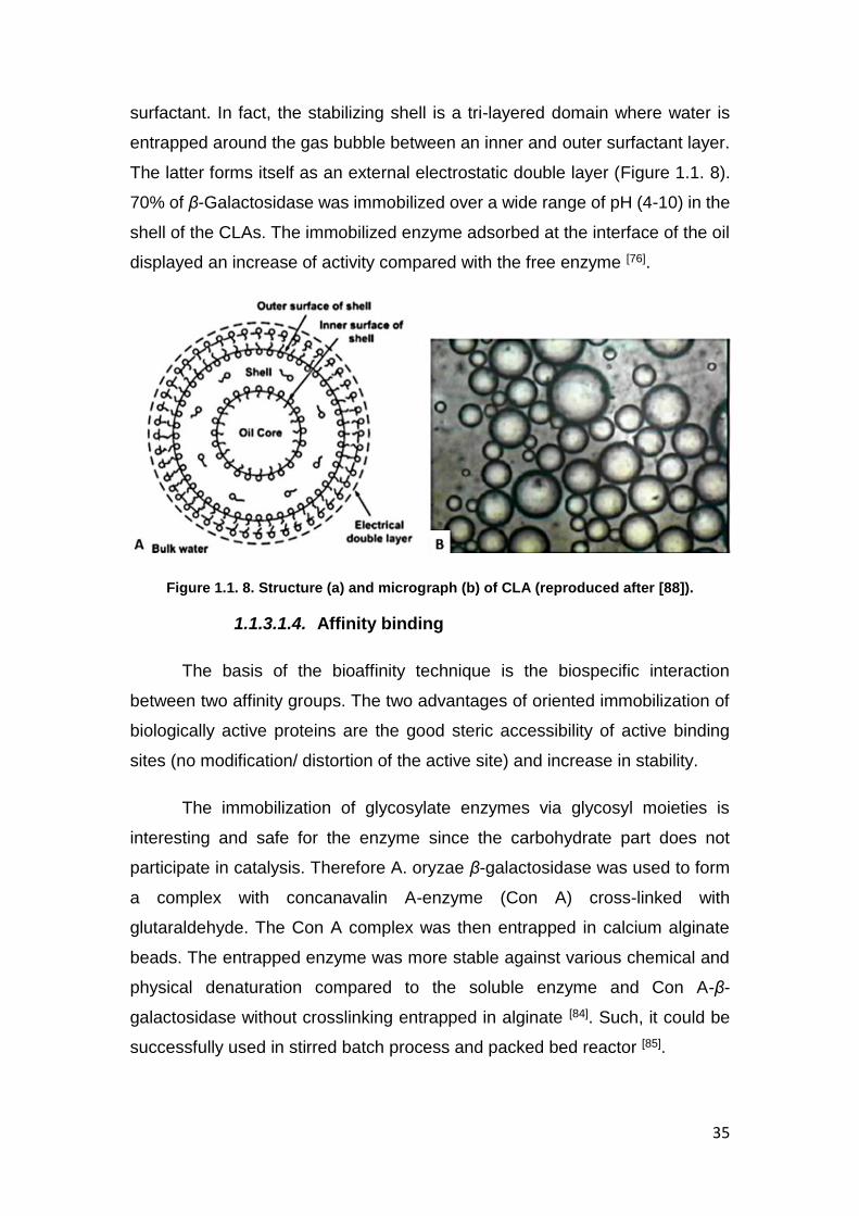
surfactant. In fact, the stabilizing shell is a tri-layered domain where water is
entrapped around the gas bubble between an inner and outer surfactant layer.
The latter forms itself as an external electrostatic double layer (Figure 1.1. 8).
70% of β-Galactosidase was immobilized over a wide range of pH (4-10) in the
shell of the CLAs. The immobilized enzyme adsorbed at the interface of the oil
displayed an increase of activity compared with the free enzyme [76].
Figure 1.1. 8. Structure (a) and micrograph (b) of CLA (reproduced after [88]).
1.1.3.1.4. Affinity binding
The basis of the bioaffinity technique is the biospecific interaction
between two affinity groups. The two advantages of oriented immobilization of
biologically active proteins are the good steric accessibility of active binding
sites (no modification/ distortion of the active site) and increase in stability.
The immobilization of glycosylate enzymes via glycosyl moieties is
interesting and safe for the enzyme since the carbohydrate part does not
participate in catalysis. Therefore A. oryzae β-galactosidase was used to form
a complex with concanavalin A-enzyme (Con A) cross-linked with
glutaraldehyde. The Con A complex was then entrapped in calcium alginate
beads. The entrapped enzyme was more stable against various chemical and
physical denaturation compared to the soluble enzyme and Con A-β-
galactosidase without crosslinking entrapped in alginate [84]. Such, it could be
successfully used in stirred batch process and packed bed reactor [85].

36
In another work of Haider et al, A. oryzae β-galactosidase was
immobilized by bioaffinity adsorption on the surface of a novel support:
concanavalin A layered calcium alginate–starch beads. The immobilized β-
galactosidase exhibited significantly higher stability against conditions of
digestive system such as pH and enzymes (salivary amylase, pepsin and
trypsin) [86] and against external factors such as heat, urea, MgCl2, and CaCl2.
It also presented a higher activity in the hydrolysis of lactose in whey and milk
compared to the free enzyme [87]. Another support that was analysed under the
same conditions is a polyclonal antibody bound cellulose support. The
immobilized enzyme was much more stable compared to the free enzyme, and
displayed a shift in pH and temperature [88].
K. lactis β-galactosidase was also immobilized by bioaffinity adsorption
on the surface of a concanavalin A layered aluminium oxide nanoparticles
support. The immobilized enzyme exhibited enhanced pH stability and broad
optimum spectrum temperature compared to the soluble β-galactosidase.
Immobilized galactosidase was stable against galactose inhibition and retained
85% activity after its sixth repeated use in continuous stirred tank bioreactors[89].
1.1.3.1.5. Chelation or metal binding
Metal binding is also employed to immobilize enzymes. On the surface
of organic carriers, transition metal salts or hydroxides are deposited and bound
onto the matrix by coordination through nucleophilic groups. The metal salt or
hydroxide is precipitated onto the support (e.g., cellulose, chitin, alginic acid,
and silica-based carriers) through heating or neutralization. A part of the
coordinative positions of the metals remain free to coordinate with enzyme
groups. Because of steric factors, it is impossible for the matrix to occupy all
coordination positions of the metal. The metal ions bounded on solid
chromatographic supports absorb the enzyme through the amino acid residues
that are exposed on the surface of the protein.
In order to improve the control over the formation of the adsorption sites,
and improve the reproducibility, chelator ligands can be immobilized on the
solid supports by means of stable covalent bonds. The metal ions are then

37
bound by coordination and the stable complexes formed can be used for the
retention of proteins. The release of the bound proteins can then be achieved
by decreasing the pH or by using competition with other, more soluble ligands.
The support is subsequently regenerated by washing with a strong chelating
agent such as ethylene diamine tetraacetic acid (EDTA) when desired. These
metal chelated supports were named Immobilized Metal-Ion Affinity (IMA)
adsorbents and have been used extensively in protein chromatography
separation. This approach, using E. coli β-galactosidase as a model was used
to test different IMA-gels with different chelated ligands (Cu2+, Ni2+ and Fe3+) as
supports for enzyme immobilization [90].
A strategy to absorb large proteins has been reported by Pessla et al.
Different activation degrees of amino group per gram of agarose were analysed
for the purification of β-galactosidase from Thermus sp. strain T2 from a crude
extract. The highly activated supports (40 μmol of ionic groups/g of agarose)
are capable to immobilize two large β-galactosidase, with molecular size of 465
kDa (E.coli) and 75 kDa (Thermus sp.) [91] and to specifically absorbed them in
the presence of 50 mM imidazole [82]. Due to the fact that large proteins have a
large surface that permits long distance interactions with groups dispersed on
a support, and that the interaction can be too strong, a Cu2+-chelate-
iminodiacetic acid-agarose support was considered. In this way, the number of
enzyme bounds with the support are relatively low and the enzyme can be
easily desorbed [92] –Figure 1.1. 9.
Figure 1.1. 9. Adsorption mechanism of large proteins on Cu2+-chelate-iminodiacetic acid-agarose support by (A) very intense multi-point ion exchange (reproduced after
[91]) and (B) mild adsorption ionic exchange (reproduced after [92])
38
The same group used heterofunctional epoxy Sepabeads (boronate-
epoxy-Sepabeads and chelate-epoxy-Sepabeads) used to immobilize β-
galactosidase from Thermus sp. T2 (Htag-BgaA) to decrease the inhibition. The
immobilization produced small changes in the conformation of the active center,
that allowed for more than a 99% hydrolysis of lactose [34].
A way to purify and immobilize the protein in a single step is to combine
two techniques the epoxy groups at the surface of the matrix for enzyme
immobilization and the purification by metal-chelate affinity chromatography.
The Thermus sp. strain T2 β-galactosidase overexpressed in E. coli poly-His-
tagged-β-galactosidase crude was immobilized with a low concentration of Co2+
chelating on a high epoxy groups density support. The enzyme was purified
and absorbed onto the support, resulting in a very high activity and stability [93].
1.1.3.1.6. Disulfide bonds
In the case of this method, a stable covalent disulfide bond (-S-S-) is
formed between the matrix and the enzyme. The reactivity of the thiol groups (-
SH) from the surface of both the enzyme and the support can be controlled by
pH modification. The absorption yield of this method is usually high (when the
appropriate thiol-reactive
For example, β-galactosidase from E. Coli was reversibly attached to
disulfide oxide groups introduced into thiol-containing agarose beads. The
enzyme was immobilized with a yield of 90% [94,95] and the activity increased
after the immobilization [96]. Due to reversibility of the disulfide bound, the
enzyme can be easily detached from the support [97] and this method can be
used for enzyme purification.
Introducing disulfide bonds in the enzyme structure, can increase the
absorption of the enzyme on the carrier [98] or can improve the stability of the
free enzyme against temperature and pH [99].

39
1.1.3.2. Methods of irreversible immobilization
Irreversible immobilization involves strong interactions between the
enzyme and the support. The enzyme cannot be detached without destroying
either the structure of the enzyme (and by default the activity), either the
support. The irreversible enzyme immobilization methods are: covalent
immobilizations (A), entrapment in gels (B) and fibers (C), microencapsulation
(D) and chemical aggregation (E) as schematized in Figure 1.1. 10.
Figure 1.1. 10. Irreversible methods of immobilization: covalent immobilizations (A),
entrapment in gels (B) and fibers (C), microencapsulation (D) and chemical aggregation (E) (adapted from [75]).
1.1.3.2.1. Covalent immobilization
Covalent immobilisation methods lead to irreversible and sustainable
biocatalysts and prevent the leakage of enzyme, covalently bonded to the
support. However, covalent coupling may induce drastic changes in the enzyme
conformation, when it occurs near the active site [100] or reduce enzyme
flexibility at high bonding density to the support [101]. Therefore, the support and
the spacer must be carefully chosen. The covalent immobilization can be
divided in two main classes: i) post functionalization of the matrix and ii) in situ
functionalization of the matrix during the synthesis.
Usually the covalent immobilization is used to increase the stability
[102,103] and the reusability of an enzyme, or to change the optimal pH and
temperature when the biocatalyst is used in a reactor (see more in subchapter
Industrial application)
For example, β-Galactosidase from A. oryzae was immobilized in Nylon
membranes grafted with glycidyl methacrylate (Nylon/GMA) via diazotization
(through tyrosine residues of the enzyme) and via condensation (through
A B C D E

40
multipoint attachment with arginine residues of the support). Both techniques
lead to the increase of the Km values [104]. It should be noticed that diazotization
method was more efficient in a bioreactor under non-isothermal conditions[105].
On the same support (Nylon/GMA), the influence of the spacer length between
the enzyme and the support (hexamethylenediamine, ethylenediamine or
hydrazine) was analyzed. With the increase of the spacer length, the optimum
pH, temperature and the apparent Km. decreased. But all membranes showed
good results in non-isothermal bioreactors [106].
Moreover, the same enzyme was also covalently attached to cotton cloth
activated by tosylchloride and used for the production of galacto-
oligossaccharides [107]. Thus, the enzyme changed its reactivity, from the
hydrolysis of lactose (into glucose and galactose) to the trans-glycosylation of
lactose to galacto-oligossaccharides.
To obtain double reactivity of the enzyme, Talaromyces thermophilus
CBS β-galactosidase was covalently attach to Eupergit C (macromolecular
beads of acrylic polymer) and the resulting material was used for the removal
of lactose at high temperature as well as for the formation of galacto-
oligosaccharides. [108]. For the same application K. lactis [109] and A. Oryzae
[110] were immobilized on magnetic polysiloxane-polyvinyl alcohol composite.
Immobilized enzymes might become resistant to glucose inhibitions.
This phenomenon was observed with a β-galactosidase from the thermophilic
microorganism, Thermus sp. strain T2, immobilized on
trishydroxymethylphosphine (THP) activated silica-alumina support [35].
For the reuse of the enzyme, Elnashar et al [111] covalently attached A.
Oryzae enzyme with glutaraldehyde on natural biopolymers i.e. carrageen
coated chitosan. The material showed thermal stability and conferred pH and
temperature improvements, also providing good reusability to the enzyme.
Crosslinking (or chemical aggregation) is based on the formation of
covalent bonds between enzymes molecules, leading to three-dimensional
structures. Thus, there is no requirement for a support. Crosslinking is
generally combined with other methods, mostly to stabilize and prevent the
enzyme leakage during the crosslinking. Glutaraldehyde is the most common

41
cross-linker. It can be used to chemically aggregate the enzyme [112] to be linked
with the support as aggregates, or to lock the enzyme onto the support after
absorption [113].
Gaur et al. compared aggregation by cross-linking with two other
techniques (adsorption on celite and covalent coupling to chitosan) for the
synthesis of GOS with A. oryzae β-gal. Cross-linked enzyme aggregates
(CLEA) were efficient in lactose hydrolysis with a yield of 78% monosaccharide
in 12h [114].
Crosslinked with glutaraldehyde, concanavalin A-Celite 545 immobilized
A. oryzae β-galactosidase was compared with the adsorbed enzyme and with
the free enzyme. The covalent immobilized enzyme showed better resistance
to product inhibition (glucose and galactose) and increased efficiency in
hydrolyzing lactose from milk and whey in batch processes [115]. N-terminal α-
amines and lysine ε-amines from K. lactis enzyme were covalently attached to
amino-modified polyethylene film with glutaraldehyde. The as-modified film with
stood heat treatment in the presence of an ionic denaturant with a high enzyme
retention, suggesting that the material can be used in food active packaging [116]
.
1.1.3.2.2. Entrapment
Entrapment is defined as physical confinement of enzymes or cells in an
environment where the substrate is able to penetrate, and the enzyme cannot
escape. This method differs from covalent binding or cross-linking since the
enzyme does not bind to the matrix. This entrapment mode can be classified in
five major types: lattice, microcapsule, liposome, membrane and reverse
micelle [117]. The entrapment can be made in organic, inorganic or hybrid
matrices.
For β-galactosidase immobilization, the lattice method
(microencapsulation) is mostly used. In that case, the enzyme is entrapped in
a matrix of natural or synthetic polymers. The most popular one is alginate, a
natural polysaccharide that forms hydrogels by ionotropic gelation with a
divalent metal cation such as Ca2+ -Figure 1.1. 11.

42
Figure 1.1. 11. Structure of alginate and its binding with calcium cations (reproduced after [118]).
Mammarella et al. entrapped K. fragilis β-Galactosidase in alginate-
carrageenan hydrogel beads of 2.4 mm [119]. It was observed compared with
other ions (Na+, Mn2+ , Mg2+, Ca2+ ) the K+ ions have the most beneficial effect
on enzyme activity [120].K-carrageenan, along with the K+ ions increased the
enzyme activity. Combined with gelatin, alginate was used to immobilize A.
oryzae in fibers hardened with glutaraldehyde. The fibers were formed by
pumping a solution of β-galactosidase, alginate, gelatine, glycerol and sodium
acetate buffer through a syringe into a solution of CaCl2 solution prepared in
calcium acetate buffer (pH 5.3) containing glutaraldehyde. The immobilized
enzyme showed good storage stability (for 35 days without activity decrease)
and was more stable at high pH and temperature, compared with the free
enzyme [121].
On the other hand, Taqieddin et al. [122] entrapped enzyme in a liquid or
solid core alginate and a chitosan shell microcapsule technology to encapsulate
β-galactosidase. Ba2+ crosslinked alginate was more efficient (100% enzyme
entrapped) compared with Ca2+ (only 60% efficiency). Ca2+ ions present an
inhibitor effect for the enzyme as shown in Table 1.1 2.
Gelatin-based films with β-galactosidase from A. oryzae were elaborated
to immobilized enzyme to extend the stability of the protein in dried films state.
The film and the enzyme activity were stables up to 36º C and a relative
humidity of 75% [123]. The resulting dry films might fit with industrial requests in

43
terms of stability, handling, storage, and transportation of functional proteins in
a cost-effective manner.
Synthetic polymers forming hydrogels are also attractive for enzyme
immobilization. For example, the polyvinylalcohol (PVA) gel is interesting for its
innocuous character, biocompatibility, low toxicity, good long-term and
mechanical stability and low biodegradability. Batsalova et al entrapped fungal
β-galactosidase in PVA cryogel beads and the enzyme gained thermal stability
compared to the free enzyme. The latest retaining 70% of activity at 50°C and
5% at 60°C [124]. β-galactosidase form A. oryzae was immobilized in lens-
shaped PVA capsules (LentiKats). The immobilized enzyme showed stability
after 35 repeated batch runs and during 14 months storage [125].
For the digestion of lactose in milk, dried liposomes containing β-
galatosidase were prepared. In the presence of bile salts, the lysis of liposomes
is efficient and the entrapped enzyme is released in the stomach for the “in situ”
digestion of the lactose [126]. Rodríguez-Nogales et al. [65] [127] optimized the
entrapment of β-Galactosidase from E. Coli in cholesterol-phosphatidylcholine
(Ch:PC) liposomes. The dehydration-rehydration vesicle method was used in
order to create the liposomes. The enzyme concentration, pH, type and time of
sonication, the ratio Ch:PC, and sucrose concentration (as cryoprotectant)
were optimized as well.
Another approach to encapsulate enzyme is the “fish-in-net” method.
This method implies a direct interaction between the silica precursor and
enzyme. The enzyme (fish) is trapped in the silica network (net). This method
is useful for the prevention of bacterial contamination when the β-galactosidase
and lysozyme enzymes are co-immobilization. A silica matrix that contained
entrapped β-galactosidase from A. oryzae was modified with covalently bound
lysozyme (Figure1.1.13). Both enzymes were active, and the materials were
used to treat milk. This resulted in bacterial cell viability dramatically decreased
when used in the industrial process. Thus, the rate of lactose hydrolysis was
improved and a good storage and operational stability was reached during the
reuse of the catalyst support in the industrial production of low lactose milk [128].
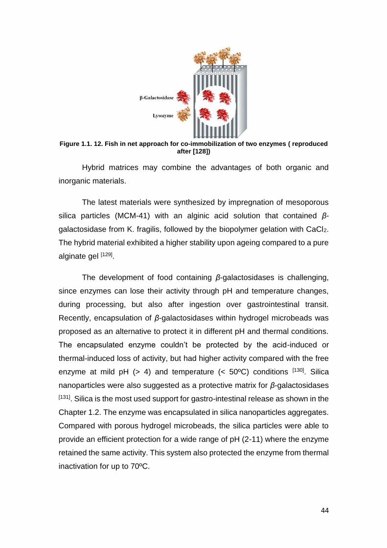
44
Figure 1.1. 12. Fish in net approach for co-immobilization of two enzymes ( reproduced
after [128])
Hybrid matrices may combine the advantages of both organic and
inorganic materials.
The latest materials were synthesized by impregnation of mesoporous
silica particles (MCM-41) with an alginic acid solution that contained β-
galactosidase from K. fragilis, followed by the biopolymer gelation with CaCl2.
The hybrid material exhibited a higher stability upon ageing compared to a pure
alginate gel [129].
The development of food containing β-galactosidases is challenging,
since enzymes can lose their activity through pH and temperature changes,
during processing, but also after ingestion over gastrointestinal transit.
Recently, encapsulation of β-galactosidases within hydrogel microbeads was
proposed as an alternative to protect it in different pH and thermal conditions.
The encapsulated enzyme couldn’t be protected by the acid-induced or
thermal-induced loss of activity, but had higher activity compared with the free
enzyme at mild pH (> 4) and temperature (< 50ºC) conditions [130]. Silica
nanoparticles were also suggested as a protective matrix for β-galactosidases
[131]. Silica is the most used support for gastro-intestinal release as shown in the
Chapter 1.2. The enzyme was encapsulated in silica nanoparticles aggregates.
Compared with porous hydrogel microbeads, the silica particles were able to
provide an efficient protection for a wide range of pH (2-11) where the enzyme
retained the same activity. This system also protected the enzyme from thermal
inactivation for up to 70ºC.

45
Enzyme immobilization has attracted a lot of attention. From the binding
to a support to encapsulation into a matrix, enzyme immobilization is used to
improve the enzymes’ performance. Organic and inorganic materials were
used, both with their own strengths and weaknesses. Hybrid materials are
promising materials since they can potentially have the advantages of both.
1.1.4. Application of immobilized β-galactosidase
Hydrolysis of lactose to glucose and galactose by β-galactosidase would
overcome some of the industrial application limitations of whey and milk,
lactose has a poor solubility, insufficient sweetness and generates lactose
intolerance problems. There are basically two ways to use the enzyme, in
soluble enzyme form for batch process or as an immobilized enzyme in
continuous operation. The possibility of reusing the enzyme in a continuous
industrial process could make the cost of enzyme immobilization profitable [68].
1.1.4.1. Industrial application
β-galactosidase is sometimes used in alcohol containing beverages, but
its main application is in the production of milk and fermented milk products
such as yoghurt and cheese. Low lactose milk and dairy products gives chance
to lactose intolerant people to consume these products. Another reason to
decrease the lactose content in dairy products is the prevention of lactose
crystallization in ice cream, frozen milks, whey and condensed milk. In respect
to the mentioned points, limiting lactose in food processing can improve some
technological and sensorial quality of dairy foods, by increasing the digestibility,
softness and creaminess.
In the cheese industry, the whey represents a waste product, which
causes several economic and environmental problems. Hydrolysis of lactose
present in whey converts whey into very useful sweet syrup, which can be used
in the dairy [132], baking and soft drinks industries [117]. After hydrolysis, the whey
can be used as cattle food resource or even to develop new free lactose
products [133] . This organic waste can also be used as an available substrate
for microbial cell cultivation [134,135].

46
Another important aspect to the dairy industry is that the hydrolysis of
lactose by β–galactosidase can lead to the synthesis of oligosaccharide and
galactooligosaccharides. They can be produced from D-lactose via by glycosyl
transfer catalyzed by the enzyme β- galactosidase. Galactooligosaccharides
are prebiotics, and are not usually digested in the small intestine, but fermented
by colonic bacteria in the large intestine. This could lead to changes in the
colonic ecosystem in favor of some bacteria, such as bifidobacteria, which may
have health benefits, including protection against certain cancers and lowering
of cholesterol levels [136]. The structures of some oligosaccharides obtained
from lactose are presented in Table 1.1 3.
Table 1.1 3. Structures of some oligosaccharides obtained from lactose [137] .
Disaccharides
β-D-Gal (1→6)-D-Glc β-D-Gal (1→6)-D-Gal
β-D-Gal (1→3)-D-Glc
β-D-Gal (1→2)-D-Glc β-D-Gal (1→3)-D-Gal
allactose galactobiose
Trisaccharides
β-D-Gal (1→6)- β-D-
Gal (1→6)-D-Glc
β-D-Gal (1→6)- β-D-Gal (1→4)-D-Glc
β-D-Gal (1→6)- β-D-
Gal (1→6)-D-Gal β-D-Gal (1→3)- β-D-Gal
(1→4)-D-Glc β-D-Gal
(1→4)- β-D-Gal (1→4)-D-Glc
6’digalactosyl-glucose 6’ galactosyl-lactose
6’ galactotriose 3’ galactosyl-lactose 4’ galactosyl-lactose
Tetrasaccharides
β-D-Gal (1→6)- β-D-
Gal (1→6)- β-D-Gal
(1→4)-D-Glc β-D-Gal (1→6)- β-D-Gal
(1→3)- β-D-Gal
(1→4)-D-Glc β-D-Gal (1→3)- β-D-Gal
(1→6)- β-D-Gal
(1→4)-D-Glc
6’digalactosyl-lactose
Pentasacchride
β-D-Gal (1→6)- β-D-
Gal (1→6)- β-D-Gal
(1→6)- β-D-Gal
(1→4)-D-Glc
6’trigalactosyl-lactose
Gal-galacto, Glc- Galactosyl
Various reactors with different configurations and varying the operation
conditions (like temperature and pH) were used for the enzymatic hydrolysis of
lactose from either milk/whey or pure lactose (Table 1.1.4.). As the price of the
process is determined by the cost of the enzyme, a continuous industrial
production that involves the reuse of a single batch of enzyme can be

47
considered [107,138]. The direct addition of the soluble enzyme by recycling it
through a membrane separation processes or the use of immobilized enzymes
are technically feasible. Compared to the free β-galactosidase, its
immobilization offers numerous advantages. The supported catalyst can be
used in batch or in a continuous process, it simplifies enzyme removal from the
reaction mixture, and affords a rapid termination of the reactions and a better
control on product formation [87,139,140]. The β-galactosidase enzyme
immobilized reactors have been extensively studied. Enzyme-membrane
systems (EMR) [141], hollow-fiber reactors (HFRs), fluidized-bed reactors (FBRs
)[142], packed-bed reactors (PBRs) [119,143,144], and stirred-tank reactors (STRs)
are the main reactors used in lactose hydrolysis [145]. They all present
advantages and disadvantages. Commonly, the stirred-tank reactors (batch
procedure) using free enzyme, in batch operation mode are used in commercial
applications[140]. Although, it is simple and easy to control, it has couple of
disadvantages related to high enzyme content and labour cost [146]. Compared
with the batch procedure, β-galactosidase immobilized onto highly activated
supports in packed-bed reactors, allows for the continuous reuse of the
biocatalyst improving the global yield. This technique also has some
drawbacks, the enzyme can lose the activity due to the multipoint
immobilization and the diffusion can be limited by the support. The bioreactor
configuration with a suitable membrane (MBR) displays some advantages: it is
facile, low relative costs and inexpensive adjusting hydronomics with a high
packing density (large specific surface per unit of mass) [147,148].The enzyme
can be absorbed [149] or chemisorbed [150] onto the bioreactor membrane and
the lactose conversion is accomplished in a single step with high yields . The
main advantages of MBR method are the preservation of the native kinetic
properties of the free enzyme and the possibility of working in homogeneous
solutions in presence of the substrate. The major disadvantages of this
configuration are the clogging of ultra-filtration membranes with milk proteins
and the increased risk of microbial contamination, especially during prolonged
operation times at ambient temperatures. By operating the system at relatively
high temperatures and using deproteinated substrates (whey permeate) these
drawbacks can be partially eliminated [151].

48
Numerous hydrolysis systems have been investigated (with some
examples shown in Table 1.1.4) using different types of reactors and different
immobilization strategies (as discussed previously). However, the scaling up
process has been applied only to a few of them and even fewer have been
applied on an industrial or semi-industrial level. For instance, only couple of
companies such as, Centrale del Latte of Milan, Cooperative Butter Factory and
Snow Brand are using immobilized β-galactosidase for the production of dairy
products [152].
Table 1.1 4. Hydrolysis of lactose by immobilized β-galactosidase under different operating conditions.
SOURCE OF Β-GALACTOSIDA
SE
SUPPORT USED FOR
IMMOBILIZATION
SOURCE OF
LACTOSE
OPERATING
CONDITIONS
REACTOR
COVERSION (%)
REF
K. FRAGILIS
Porous silanized glass modified
with glutaraldehyde
Whey permeate
pH 6, 50°C, reactor
batch and recycling packed-
bed
86-90 [134]
K. FRAGILIS Silica-alumina Milk
buffer pH 7, 40°C batch 99% [133]
A. ORYZAE
Cross-linked poly(vinyl
alcohol)/natural polysaccharide
chitosan
Lactose and milk
whey
pH 5, 50-60°C
Fixed bed 95% [153]
THERMUS SP T2
Sepa beads Novo buffer
pH 6.5, 50°C batch 99% [34]
K. FRAGILIS Cellulose beads Milk and
whey pH 6.6, 50°C
Batch and fluidized
bed
>80% in whey and 7% in milk
[142]
E. COLI Magnetic poly (GMA-MMA)
Lactose pH 7, 35°C
Fixed bed, flow rate 20
mL/h for 60 h
88% [154]
K.LATIS Magnetic
polysiloxane-polyvinyl alcohol
Skimmed milk
pH 6.5, 25°C Batch and
stirred 90% [109]
A. ORYZAE Lens-shaped
polyvinyl capsules
lactose pH 4.6, 45°C Batch and
stirred 100% [125]
A. NIGER
Concanavalin A layered hybrid beads calcium alginate-starch
Whey, milk
pH 4.8, pH 6.6, 37°C
≈80% [87]
E.COLI Polypropylene
spirally membrane
pH 6.8, 40°C Bioreacto
r 90% [155]

49
1.1.5. Conclusion
β-Galactosidase in one of the most used enzymes in food dairy
processing, with an array of applications, from technological and environmental
to nutritional and quality control. Mainly, the enzyme has two main function: the
hydrolysis of lactose from milk products for the production of free/low lactose
dairy products to be consumed by lactose intolerant people and the production
of galactosylated products by transgalactosylation reaction. β-Galactosidase
enzymes are extracted from various sources, from plants (peaches, apricots,
almonds, apples, kiwis, tomatoes), animal organs or from microorganisms
(bacteria, fungi, yeasts). Depending of the natural source, β-Galactosidase is
active at different pH levels and at various temperatures. β-Galactosidase
enzymes from plants have an optimal pH between 3.5-7, from fungi between
pH 2.5–5.4, from animal cells between 6-7, from bacterial and yeast sources
between pH 6.5–7.5.
To increase the enzyme reusability, thermal stability and pH tolerance,
β-Galactosidase enzymes have been immobilized on both organic (e.g.
alginate, chitosan, cotton) and inorganic (e.g. silica, carbon) supports. Different
immobilization methods have been used, from reversible immobilization
(adsorption, ionic or hydrophobic binding, affinity binding, chelation or metal
binding, disulfite bonds), that involves weak forces between the enzyme and
the support, to irreversible immobilization (covalent immobilizations,
entrapment in gels and fibers, microencapsulation, chemical aggregation), that
involves strong interactions between the enzyme and the support.
Moreover, immobilized systems can provide better enzyme activity and
confer a tuneable reactivity (either the hydrolysis of lactose to glucose and
galactose, either to the production or oligosaccharides).
Immobilized β-galactosidase enzymes have been researched in different
reactors for the continuous hydrolysis of lactose from whey, milk or different
buffers. Enzyme-membrane systems, hollow-fiber reactors, fluidized-bed
reactors, packed-bed reactors, and stirred-tank reactors are couple of reactors
examples used in lactose hydrolysis.

50
Bibliography [1] S. S. Asraf, P. Gunasekaran, 2016. [2] A. Gosling, G. W. Stevens, A. R. Barber, S. E. Kentish, S. L. Gras, Food Chem. 2010, 121,
307–318. [3] B. Rodriguez-Colinas, L. Fernandez-Arrojo, A. O. Ballesteros, F. J. Plou, Food Chem. 2014,
145, 388–394. [4] T. Haider, Q. Husain, J. Sci. Food Agric. 2007, 87, 1278–1283. [5] G. S. Ross, R. J. Redgwell, E. A. MacRae, Planta 1993, 189, 499–506. [6] A. T. Carey, K. Holt, S. Picard, R. Wilde, C. A. Tucker, C. R. Bird, W. Schuch, C. B. Seymour, U.
K. A. T. C, E. A. Macrae, et al., 2017, 1099–1107. [7] M. T. Flood, M. Kondo, Regul. Toxicol. Pharmacol. 2004, 40, 281–292. [8] Z. Nagy, T. Kiss, A. Szentirmai, S. Biró, Protein Expr. Purif. 2001, 21, 24–9. [9] M. L. Richmond, J. I. Gray, C. M. Stine, J. Dairy Sci. 1981, 64, 1759–1771. [10] P. Prunus, D. H. Lee, S. Kang, S. Suh, J. K. Byun, 2003, 15, 68–74. [11] S. Gulzar, Am. J. Plant Sci. 2012, 3, 636–645. [12] K. Itoh, T. Toba, T. Itoh, S. Adachi, Lett. Appl. Microbiol. 1992, 15, 232–234. [13] J. C. Ball, L. G. Puckett, L. G. Bachas, Anal. Chem. 2003, 75, 6932–6937. [14] M. Becerra, E. Cerdán, M. I. G. Siso, Biotechnol. Tech. 1998, 12, 253–256. [15] J. Fiedurek, J. Szczodrak, Acta Microbiol. Pol. 1994, 43, 57–65. [16] J. LEDERBERG, J. Bacteriol. 1950, 60, 381–392. [17] M. V. R. RAO, S. M. DUTTA, J. Food Sci. 1981, 46, 1419–1423. [18] R. a Daniel, J. Haiech, F. Denizot, J. Errington, J. Bacteriol. 1997, 179, 5636–5638. [19] H. Hirata, T. Fukazawa, S. Negoro, H. Okada, J. Bacteriol. 1986, 166, 722–727. [20] Y. M. Hanʼguk Sanop Misaengmul Hakhoe., J. C. (Ildong P. C. L. . S. (Korea R. R. L. Lee, Y. J.
Choi, H. C. (Korea U. S. (Korea R. D. of G. E. Yang, Sanop Misaengmul Hakhoe Chi., Hanʼguk Sanop Misaengmul Hakhoe, 2001.
[21] K. M. Nevalainen, 1981, 41, 593–596. [22] S. Nizamuddin, a Sridevi, G. Narasimha, African J. Biotechnol. 2008, 7, 1096–1100. [23] N. A. Zagustina, A. S. Tikhomirova, Biokhimiia 1976, 41, 1061–6. [24] Y. Tanaka, T. Horiuchi, J. Biochem. 1975, 77, 241–247. [25] R. R. MAHONEY, J. R. WHITAKER, J. Food Biochem. 1978, 1, 327–350. [26] M. C. Crescimbeni, V. Nolan, P. D. Clop, G. N. Marín, M. A. Perillo, Colloids Surf. B.
Biointerfaces 2010, 76, 387–96. [27] J. Garman, T. Coolbear, J. Smart, Appl. Microbiol. Biotechnol. 1996, 46, 22–27. [28] C. Picard, J. Fioramonti, A. Francois, T. Robinson, F. Neant, C. Matuchansky, Aliment.
Pharmacol. Ther. 2005, 22, 495–512. [29] C. G. Vinderola, J. A. Reinheimer, Food Res. Int. 2003, 36, 895–904. [30] T. Vasiljevic, P. Jelen, Innov. Food Sci. Emerg. Technol. 2002, 3, 175–184. [31] K. Arunachalam, Nutr. Res. 1999, 19, 1559–1597. [32] A. Jain, Y. Gupta, S. K. Jain, J. Pharm. Pharm. Sci. 2007, 10, 86–128. [33] M. . Boon, A. E. . Janssen, K. van ‘t Riet, Enzyme Microb. Technol. 2000, 26, 271–281. [34] B. C. C. Pessela, C. Mateo, M. Fuentes, A. Vian, J. L. García, A. V. Carrascosa, J. M. Guisán,
R. Fernández-Lafuente, Enzyme Microb. Technol. 2003, 33, 199–205. [35] M. Ladero, M. T. Perez, A. Santos, F. Garcia-Ochoa, Biotechnol. Bioeng. 2003, 81, 241–252. [36] M. Ladero, G. Ruiz, B. C. C. Pessela, A. Vian, A. Santos, F. Garcia-Ochoa, Biochem. Eng. J.
2006, 31, 14–24. [37] M. M. Maksimainen, A. Lampio, M. Mertanen, O. Turunen, J. Rouvinen, Int. J. Biol. Macromol.
2013, 60, 109–115. [38] S. C. Li, J. W. Han, K. C. Chen, C. S. Chen, Phytochemistry 2001, 57, 349–359. [39] H. Konno, K. Katoh, 1992. [40] H. Konno, H. Tsumuki, 1993, 40–47. [41] M. S. Buckeridge, J. S. G. Reid, 1993, 502–511. [42] S. Biswas, A. M. Kayastha, R. Seckler, J. Plant Physiol. 2003, 160, 327–37. [43] G. Simos, T. Giannakouros, J. G. Georgatsos, 1989, 28, 2587–2592. [44] A. Carrillo-López, A. Cruz-Hernéndez, A. Cárabez-Trejo, F. Guevara-Lara, O. Paredes-López,
J. Agric. Food Chem. 2002, 50, 1681–1685. [45] D. L. Smith, D. A. Starrett, K. C. Gross, Plant Physiol. 1998, 117, 417–423. [46] I. K. Kang, S. G. Suh, K. C. Gross, J. K. Byun, Plant Physiol. 1994, 105, 975–9. [47] H. Lazan, S. Y. Ng, L. Y. Goh, Z. M. Ali, Plant Physiol. Biochem. 2004, 42, 847–853. [48] D. L. Smith, K. C. Gross, Plant Physiol. 2000, 123, 1173–1184. [49] M. Edwards, J. L. Bowman, I. C. M. Deagll, J. S. G. R. Ii, J. Biol. Chem. 1988, 263, 4333–4337. [50] W. Tu, S. Sun, S. Nu, X. Li, Food Chem. 1999, 64, 495–500. [51] A. Dwevedi, A. M. Kayastha, J. Agric. Food Chem. 2009, 57, 682–688. [52] A. Dwevedi, A. M. Kayastha, Bioresour. Technol. 2009, 100, 2667–2675. [53] C. S. Kim, E.-S. Ji, D.-K. Oh, Biochem. Biophys. Res. Commun. 2004, 316, 738–43.

51
[54] S. R. Tello-Solís, J. Jiménez-Guzmán, C. Sarabia-Leos, L. Gómez-Ruíz, A. E. Cruz-Guerrero, G. M. Rodríguez-Serrano, M. García-Garibay, 2005, 10200–10204.
[55] A. Pereira-Rodríguez, R. Fernández-Leiro, M. I. González-Siso, M. E. Cerdán, M. Becerra, J. Sanz-Aparicio, J. Struct. Biol. 2012, 177, 392–401.
[56] G. D. Miller, J. K. Jarvis, L. D. McBean, National Dairy Council., Handbook of Dairy Foods and Nutrition, CRC Press, 2007.
[57] P. SCHUCK, A. DOLIVET, Brazilian J. Chem. Eng. 2002, 19, 397–402. [58] P. Walstra, T. J. (Tom J. . Geurts, J. T. M. Wouters, Dairy Science and Technology, CRC/Taylor
& Francis, 2006. [59] V. H. Holsinger, Physical and Chemical Properties of Lactose, 1997. [60] M. G. Gänzle, G. Haase, P. Jelen, Int. Dairy J. 2008, 18, 685–694. [61] † C. A. Hsu, ‡ and S. L. Lee, † C. C. Chou*, 2007, DOI 10.1021/JF063126+. [62] K. Wallenfels, O. Prakash Malhotra, Adv. Carbohydr. Chem. 1962, 239–298. [63] I. Y. S. Rustom, M. I. Foda, M. H. López-Leiva, Food Chem. 1998, 62, 141–147. [64] T. M. Bayless, E. Brown, D. M. Paige, Curr. Gastroenterol. Rep. 2017, 19, DOI 10.1007/s11894-
017-0558-9. [65] J. M. Rodríguez-Nogales, A. D. López, Int. Dairy J. 2006, 16, 354–360. [66] P. S. Panesar, R. Panesar, R. S. Singh, J. F. Kennedy, H. Kumar, J. Chem. Technol.
Biotechnol. 2006, 81, 530–543. [67] M. B. Heyman, Pediatrics 2006, 118, 1279–1286. [68] Q. Husain, Crit. Rev. Biotechnol. 2010, 30, 41–62. [69] R. Sinha, C. Radha, J. Prakash, P. Kaul, Food Chem. 2007, 101, 1484–1491. [70] Z. Quinn Z.K, C. Xiao Dong, Biochem. Eng. J. 2001, 9, 33–40. [71] H. A. Gulec, Colloids and Surfaces B-Biointerfaces 2013, 104, 83–90. [72] A. M. Klibanov, Nature 2001, 409, 241–246. [73] P. V. Iyer, L. Ananthanarayan, Process Biochem. 2008, 43, 1019–1032. [74] B. Brena, P. González-Pombo, F. Batista-Viera, Immobil. Enzym. Cells Third Ed. Methods Mol.
Biol. 2013, 1051, 1–11. [75] N. Carlsson, H. Gustafsson, C. Thörn, L. Olsson, K. Holmberg, B. Åkerman, Adv. Colloid
Interface Sci. 2014, 205, 339–60. [76] S. B. Lamb, D. C. Stuckey, Enzyme Microb. Technol. 1999, 26, 574–581. [77] P. Jochems, Y. Satyawali, S. Van Roy, W. Doyen, L. Diels, W. Dejonghe, Enzyme Microb.
Technol. 2011, 49, 580–8. [78] Q. Husain, S. A. Ansari, F. Alam, A. Azam, Int. J. Biol. Macromol. 2011, 49, 37–43. [79] D.-C. Sheu, S.-Y. Li, K.-J. Duan, C. W. Chen, CEUR Workshop Proc. 2014, 1225, 41–42. [80] M. P. Klein, L. P. Fallavena, J. da N. Schöffer, M. A. Z. Ayub, R. C. Rodrigues, J. L. Ninow, P. F.
Hertz, Carbohydr. Polym. 2013, 95, 465–70. [81] M. L. Verma, C. J. Barrow, J. F. Kennedy, M. Puri, Int. J. Biol. Macromol. 2012, 50, 432–7. [82] B. C. C. Pessela, C. Mateo, M. Filho, A. Carrascosa, R. Fernández-Lafuente, J. M. Guisan,
Enzyme Microb. Technol. 2007, 40, 242–248. [83] S. Peirce, J. J. Virgen-Ortíz, V. G. Tacias-Pascacio, N. Rueda, R. Bartolome-Cabrero, L.
Fernandez-Lopez, M. E. Russo, A. Marzocchella, R. Fernandez-Lafuente, RSC Adv. 2016, 6, 61707–61715.
[84] T. Haider, Q. Husain, Int. J. Biol. Macromol. 2007, 41, 72–80. [85] T. Haider, Q. Husain, Chem. Eng. Process. Process Intensif. 2009, 48, 576–580. [86] T. Haider, Q. Husain, Int. J. Pharm. 2008, 359, 1–6. [87] T. Haider, Q. Husain, Int. Dairy J. 2009, 19, 172–177. [88] T. Haider, Q. Husain, Biochem. Eng. J. 2009, 43, 307–314. [89] S. A. Ansari, Q. Husain, J. Mol. Catal. B Enzym. 2011, 70, 119–126. [90] B. Brena, L. Ryden, J. Porath, Biotechnol. Appl. Biochem. 1994, 19, 217–231. [91] B. C. C. Pessela, M. Fuentes, C. Mateo, R. Munilla, A. V. Carrascosa, R. Fernandez-Lafuente,
J. M. Guisan, Enzyme Microb. Technol. 2006, 39, 909–915. [92] B. C. C. Pessela, R. Torres, M. Fuentes, C. Mateo, R. Munilla, A. Vian, A. V. Carrascosa, J. L.
Garcia, J. M. Guisán, R. Fernandez-Lafuente, J. Chromatogr. A 2004, 1055, 93–98. [93] B. C. C. Pessela, C. Mateo, A. V Carrascosa, A. Vian, G. Rivas, C. Alfonso, M. Guisan, R.
Ferna, Biomacromolecules 2003, 4, 107–113. [94] F. Batista-Viera, M. Barbieri, K. Ovsejevi, C. Manta, J. Carlsson, Appl. Biochem. Biotechnol.
1991, 31, 175–195. [95] K. Ovsejevi, C. Manta, F. Batista-Viera, in Methods Mol. Biol., 2013, pp. 89–116. [96] K. Ovsejevi, B. Brena, F. Batista-Viera, J. Carlsson, Enzyme Microb. Technol. 1995, 17, 151–
156. [97] F. BATISTA-VIERA, M. CARMEN, J. CARLSSON, Appl. Biochem. Biotechnol. 1994, 44, 1–14. [98] K. Ovsejevi, V. Grazú, K. Cuadra, F. Batista-Viera, Enzyme Microb. Technol. 2004, 35, 203–
209. [99] A. Rico-Díaz, M.-E. Álvarez-Cao, J.-J. Escuder-Rodríguez, M.-I. González-Siso, M. E. Cerdán,
M. Becerra, Sci. Rep. 2017, 7, 45535. [100] M. Di Serio, C. Maturo, E. De Alteriis, P. Parascandola, R. Tesser, E. Santacesaria, Catal.

52
Today 2003, 79–80, 333–339. [101] J. Pedroche, M. del Mar Yust, C. Mateo, R. Fernández-Lafuente, J. Girón-Calle, M. Alaiz, J.
Vioque, J. M. Guisán, F. Millán, Enzyme Microb. Technol. 2007, 40, 1160–1166. [102] M. J. Hernaiz, D. H. G. Crout, Enzyme Microb. Technol. 2000, 27, 26–32. [103] P. Torres, F. Batista-Viera, Molecules 2017, 22, 284. [104] M. M. El-masry, a De Maio, P. L. Martelli, R. Casadio, a B. Moustafa, S. Rossi, D. G. Mita, J
Mol Catal B-Enzym 2001, 16, 175–189. [105] A. De Maio, M. M. El-Masry, Z. H. Abd El-Latif, M. Portaccio, U. Bencivenga, D. G. Mita, J. Mol.
Catal. - B Enzym. 2001, 16, 191–204. [106] A. De Maio, M. M. El-masry, P. De Luca, V. Grano, S. Rossi, N. Pagliuca, F. S. Gaeta, M.
Portaccio, D. G. Mita, 2003, 21, 253–265. [107] N. Albayrak, S.-T. Yang, Biotechnol. Bioeng. 2002, 77, 8–19. [108] P. Nakkharat, H. Dietmar, Appl. Biochem. Biotechnol. 2002, 98–100, 311–318. [109] D. F. M. Neri, V. M. Balcão, M. G. Carneiro-da-Cunha, L. B. Carvalho Jr., J. A. Teixeira, Catal.
Commun. 2008, 9, 2334–2339. [110] D. F. M. Neri, V. M. Balcão, R. S. Costa, I. C. A. P. Rocha, E. M. F. C. Ferreira, D. P. M. Torres,
L. R. M. Rodrigues, L. B. Carvalho, J. A. Teixeira, Food Chem. 2009, 115, 92–99. [111] M. M. M. Elnashar, M. A. Yassin, Appl. Biochem. Biotechnol. 2009, 159, 426–437. [112] Q. Z. K. Zhou, X. Dong Chen, J. Food Eng. 2001, 48, 69–74. [113] M. Portaccio, S. Stellato, S. Rossi, U. Bencivenga, M. S. Mohy Eldin, F. S. Gaeta, D. G. Mita,
Enzyme Microb. Technol. 1998, 23, 101–106. [114] R. Gaur, H. Pant, R. Jain, S. K. Khare, Food Chem. 2006, 97, 426–430. [115] S. A. Ansari, Q. Husain, Food Bioprod. Process. 2012, 90, 351–359. [116] J. M. Goddard, J. N. Talbert, J. H. Hotchkiss, J. Food Sci. 2007, 72, 1–6. [117] Z. Grosová, M. Rosenberg, M. Rebroš, Czech J. Food Sci. 2008, 26, 1–14. [118] K. Kashima, I. Masanao, in Adv. Desalin., 2012, pp. 3–36. [119] E. J. Mammarella, A. C. Rubiolo, J. Mol. Catal. B Enzym. 2005, 34, 7–13. [120] E. J. Mammarella, 2001, 142. [121] A. Tanriseven, Ş. Doǧan, Process Biochem. 2002, 38, 27–30. [122] E. Taqieddin, M. Amiji, Biomaterials 2004, 25, 1937–1945. [123] L. Zhang, A. Otte, M. Xiang, D. Liu, R. Pinal, Molecules 2015, 20, 17180–17193. [124] K. Batsalova, K. Kunchev, Y. Popova, A. Kozhukharova, N. Kirova, 1987, 227–230. [125] Z. Grosová, M. Rosenberg, M. Rebroš, M. Šipocz, B. Sedláčková, Biotechnol. Lett. 2008, 30,
763–767. [126] C. K. Kim, H. S. Chung, M. K. Lee, L. N. Choi, M. H. Kim, Int. J. Pharm. 1999, 183, 185–193. [127] J. M. Rodriguez-Nogales, A. Delgadillo, J. Mol. Catal. B Enzym. 2005, 33, 15–21. [128] H. Li, S. Li, P. Tian, Z. Wu, Z. Li, Molecules 2017, 22, DOI 10.3390/molecules22030377. [129] T. Coradin, J. Livage, Comptes Rendus Chim. 2003, 6, 147–152. [130] Z. Zhang, R. Zhang, L. Chen, D. J. McClements, Food Chem. 2016, 200, 69–75. [131] Z. Wu, Z. Wang, B. Guan, X. Wang, Y. Zhang, Y. Xiao, B. Zhi, Y. Liu, Z. Li, Q. Huo, New J.
Chem. 2013, 37, 3793. [132] L. Domingues, N. Lima, J. A. Teixeira, Process Biochem. 2005, 40, 1151–1154. [133] M. Ladero, A. Santos, F. García-Ochoa, Enzyme Microb. Technol. 2000, 27, 583–592. [134] J. Szczodrak, Acta Biotechnol. 2000, 10, 631–637. [135] R. Rech, M. A. Z. Ayub, Process Biochem. 2007, 42, 873–877. [136] S. Chockchaisawasdee, V. I. Athanasopoulos, K. Niranjan, R. A. Rastall, Biotechnol. Bioeng.
2005, 89, 434–443. [137] R. R. Mahoney, Food Chem. 1998, 63, 147–154. [138] A. Axelsson, G. Zacchi, Appl. Biochem. Biotechnol. 1990, 24–25, 679–693. [139] G. Matioli, F. F. De Moraes, G. M. Zanin, Acta Sci. - Heal. Sci. 2003, 25, 7–12. [140] X. Li, Q. Z. K. Zhou, X. D. Chen, Chem. Eng. Process. Process Intensif. 2007, 46, 497–500. [141] S. Novalin, W. Neuhaus, K. D. Kulbe, J. Biotechnol. 2005, 119, 212–218. [142] I. Roy, M. N. Gupta, Process Biochem. 2003, 39, 325–332. [143] A. R. Özdural, D. Tanyolaç, I. H. Boyaci, M. Mutlu, C. Webb, Biochem. Eng. J. 2003, 14, 27–36. [144] C. R. Carrara, E. J. Mammarella, A. C. Rubiolo, Chem. Eng. J. 2003, 92, 123–129. [145] E. Jurado, F. Camacho, G. Luzón, J. M. Vicaria, Bioprocess Biosyst. Eng. 2005, 28, 27–36. [146] G. M. Rios, M. P. Belleville, D. Paolucci, J. Sanchez, J. Memb. Sci. 2004, 242, 189–196. [147] A. N. Genari, F. V. Passos, F. M. L. Passos, J. Dairy Sci. 2003, 86, 2783–2789. [148] N. Vasileva, Y. Ivanov, S. Damyanova, I. Kostova, T. Godjevargova, Int. J. Biol. Macromol.
2016, 82, 339–346. [149] S. CURCIO, V. CALABRO, G. IORIO, J. Memb. Sci. 2006, 273, 129–142. [150] N. Diano, V. Grano, S. Rossi, U. Bencivenga, M. Portaccio, U. Amato, F. Carfora, M. Lepore, F.
S. Gaeta, D. G. Mita, Biotechnol. Prog. 2004, 20, 457–466. [151] D. G. Hatzinikolaou, E. Katsifas, D. Mamma, A. D. Karagouni, P. Christakopoulos, D. Kekos,
Biochem. Eng. J. 2005, 24, 161–172. [152] P. S. Panesar, S. Kumari, R. Panesar, Enzyme Res. 2010, 2010, 1–16. [153] S. Rejikumar, S. Devi, Int. J. Food Sci. Technol. 2001, 36, 91–98.

53
[154] G. Bayramoglu, Y. Tunali, M. Y. Arica, Catal. Commun. 2007, 8, 1094–1101. [155] N. Vasileva, Y. Ivanov, S. Damyanova, I. Kostova, T. Godjevargova, Int. J. Biol. Macromol.
2016, 82, 339–346.

54
1. 2. Silica-based systems for oral and food delivery 1
Silicon dioxide (SiO2) or silica, is abundantly distributed in the earth’s crust
in the form of silicate minerals, and is a component in plants, cereals and fruits
[1]. Human body also contains silicon in its skeleton, heart, muscles, blood
vessels, skin, hair and nails, ligaments of cartilage [2]. Living organisms such as
siliceous sponges have an amorphous skeleton generated by an enzyme, the
silicatein [3].
From robust industrial solids to soft biocompatible materials, silica has a
range of properties that can be tuned according to the design applications,
through wet or thermal processes. Mesoporous materials with high surface
areas and pore volumes [4] are obtained through sol-gel process, by far the most
used synthetic route. Highly ordered pores with hexagonal or cubic
arrangements can be obtained with tunable sizes in the range of 2 to 50 nm [5].
Moreover, the presence of silanol groups (Si-OH) on the inner and outer surface
of the material facilitates its chemical functionalization by specific groups
related to the desired application. Such chemical properties make those
materials suitable for adsorption of pollutants, polymer filler, and catalyst
applications [6]. Beyond those applications, nanomedecine is also of high
interest. Drug delivery and bio-imaging are topics in which the use of
amorphous silica is becoming popular especially because of its biocompatibility
and capacity to uptake poorly soluble drugs. There are more and more
sophisticated targeted silica carriers that are stimuli-responsive multifunctional
platforms to allow both diagnostic and therapy, known as theranostic approach.
In the food sector, silica can be employed as catalysts for the synthesis of
bioactive molecules and nutrients [7], as sensors [8] or as carriers for the
formation of functional food [9].”Functional food” refers to the food or food
component that can offer beyond basic nutrition some health benefits [10].
Nowadays, significant efforts are made in respect to developing smart
drug delivery systems (DDS) for different administration routes, including the
1 This subchapter is based on the review: R.Diab, N. Canilho, I.A.Pavel, F.B.Haffner, M.Girardon and A.
Pasc, “Silica-based systems for oral delivery of drugs, macromolecules and cells”-Advances in Colloid
and Interface Science-In press [26]

55
oral one. Indeed, the oral administration is still the most comfortable and
efficient for the patient, although the passage through the gastrointestinal
barrier remains challenging. Before being approved as nontoxic and
biodegradable, silica was already used as excipients in medicine and food
additives (E551). More recently, according to the US Food and Drug
Administration (FDA) and the European Food Safety Authority (EFSA),
amorphous forms of silica and silicates are generally recognized as safe for oral
delivery of ingredients in amounts up to 1500 mg per day [11].
In this context, the aim of this chapter is to give an extended overview
on the latest advances of the use of silica for oral delivery including drugs,
proteins, hormones, cells (probiotic bacteria) and enzyme. They can be used in
the elaboration of functional food or in oral pharmaceutical dosage forms.
1.2.1. Prerequisite for oral delivery systems and food applications
Noninvasive, accessible, simple and economical, the oral administration
remains at the top of the prescribed and over-the-counter medications over the
world. Therefore, it is not surprising that whenever possible, clinicians prefer to
prescribe oral dosage forms instead of parenteral ones which are not only
invasive but require medical interventions and occasionally hospitalization.
Several intravenous-to-oral route conversion programs were implemented in
hospitals aiming to reduce infection risks inherent to the intravenous
administration, to reduce medication costs and to improve patients’ compliance
[12–16].
Great research efforts have been dedicated to the development of oral
dosage forms that might substitute/limit the use of parenteral forms.
Nevertheless, the design of drug products for the oral route remains
challenging. Indeed, the passage through the gastrointestinal tract (GIT) is
harsh on drugs, which will encounter several barriers before reaching the
systemic circulation. Several factors are involved: i) the gastric pH that is as low
as 1 in fast conditions; ii) the presence of hydrolytic enzymes in the gastric and
intestinal juices; iii) the intestinal drug efflux by P-glycoprotein pumps localized

56
in enterocytes’ apical membranes; and iv) the premature drug catabolism by
the hepatic and/or intestinal cytochrome P450 [17].
The success of this passage depends partially on the drug’s own
physicochemical properties such as water solubility, molecular weight, partition
coefficient [18] and also on the delivery system. The design of ODDS could be
adjusted to ensure other functions beyond the protection of the payload from
its premature degradation. In this respect we can mention:
i) the gastro-retention sought for different purposes, e.g. to achieve a
local action in the stomach, to enhance the absorption of drugs within
a narrow window or even to enhance the stability of unstable drugs
in the intestinal juice [19];
ii) the gastro-resistance of drugs that are subjected to degradation in
the severe gastric environment or conversely, to protect the gastric
mucus against drugs’ irritant effects;
iii) the sustained release aiming a reduction in dosing frequency and
therefore improving the patients’ compliance;
iv) the triggered release to a specific site, e.g. targeting a high drug
amount directly on specific cells in the colon;
v) the mucoadhesion on the intestinal epithelium allowing the increase
of the intestinal residence time, and thus a longer time for drug
absorption;
vi) an enhancement of the dissolution rate of drugs of class II and IV
(according to the biopharmaceutical classification system).
The above-mentioned functions of ODDS could be reached by using
appropriate formulations from the wide range of ingredients nowadays
available. For instance, polysaccharides, e.g. chitosan, cellulose, starch and
their derivatives, alginates, pectin, and acacia gum; proteins, e.g. albumin,
gelatin and gliadin; polymethacrylates bearing quaternary ammonium groups
(Eudragit ® RS/ Eudragit ® RL) are all mucoadhesive polymers allowing the
close contact of payloads with the intestinal epithelium for extended periods of
time. Moreover, in the formulation of hydrophilic matrices, micro- and
nanoparticulate DDS, these polymers form swellable networks enabling the

57
sustained diffusion of drugs. Furthermore, these polymers are able to promote
paracellular transport through the intestinal epithelium by producing a transient
opening of the tight junctions between the enterocytes [20,21] .
The gastro-resistance becomes possible thanks to pH-sensitive
polymers such as, cellulose acetato-phthalate and Eudragit ® L or Eudragit ® S.
The unique feature of these polymers is the presence of carboxylic groups (–
COOH). In the acidic gastric juice where the pH is below the pKa of carboxylic
acid groups (≈4), the protonated form (water insoluble) is predominant, which
constitutes a physical barrier against drug release. In the intestinal juice, the pH
is higher than 4, which induces polymer deprotonation, dissolution and thus
allowing drug release.
The enhancement of the dissolution of poorly soluble drugs is carried out
using a variety of materials. For instance, lipids such as monoglycerides,
triglycerides, glycerophospholipids and middle- and short-chains fatty acids are
used in the formulation of lipophilic matrix tablets, self-microemulsified DDS,
solid lipid nanoparticulate DDS and nanostructured lipid carriers, among others.
It is noteworthy to mention that middle- and short-chains fatty acids are able to
reversibly destabilize the tight junctions of the epithelium, and thereby
enhancing intestinal permeability of the loaded drug [22,23].
On top of this properties, for food applications, the effect of silica on product
appearance, mouth feel, texture, flavor and shelf live should be minimum or
compatible with the food matrix [24].
1.2.2. Formation and origin of silica
Silica can be classified in two main categories, crystalline (i.e. quartz,
cristobalite, tridymite or calcinated diatomite) or amorphous, as a function of the
connectivity between the tetrahedral units and of the long-range periodicity in
the network. Figure 1.2. 1 illustrates typical differences of periodicity between
an amorphous silica bulk and cristobalite, chosen as example of crystalline
silica. In both cases, the bulk of silica is composed of SiO4 tetrahedral units that
form siloxane rings of different Si–O sizes. The size of the silica rings present
on the silica surface generally ranges from flexible 12-membered rings to

58
strained 4-member Si–O rings, and the distribution of such siloxane rings
generally depends on the calcination/activation temperature of silica [25]. At the
surface, various kinds of silanols can be found; they can be classified as
isolated (non-H-bonded), geminal, vicinal, and interacting (H-bonded) silanols
(Figure 1.2. 1). In the case of crystalline silica, such as cristobalite, only three
kind of silanols can be found at the surface: geminal (for 001 termination),
vicinal (for 101 termination) and isolated silanols (for 111 termination) while in
the case of amorphous silica, all kinds of silanols are present (Figure 1.2. 2).
This property explains the highest reactivity of amorphous silica surface vs
crystalline ones. Moreover, depending on the reaction conditions, such as
temperature or condensation degree, the -OH density at the surface of the
material can be tuned between less than 1 to 7 -OH/nm2.
Figure 1.2. 1. Unit cells of bulk structures of (A) crystalline (ex. β –cristobalite) 7.16 x 7.16 x 7.16 Å3 unit cell, containing 8 SiO2 units and (B) amorphous SiO2, 14.32 x 14.32 x 14.32 Å3 unit cell, containing 64 SiO2 units (reproduced from [25]).
Figure 1.2. 2. Types of silanol and silica bridges ( reproduced from [26]).
In oral drug delivery and food applications, only amorphous silica should
be considered, due to its lower toxicity and increased dissolution in biological

59
fluids. Amorphous silica can be classified in natural (biosilica, e.g. diatomite)
and synthetic (Figure 1.2. 3). In the last category, various forms of silica were
defined depending for example on the process: wet, i.e. silica-gel, precipitated
and colloidal silica and thermal, i.e. fused and fumed silica.
Figure 1.2. 3. Classification of various forms of silica.
There are four types of silica-based materials used as oral delivery
systems and smart foods: (1) non-porous silica nanoparticles (fumed and
Stöber nanoparticles), (2) mesoporous silica nanoparticles, (3) mesoporous
silica based materials, (4) biosilica, e.g. diatoms. Payload materials can be
obtained either by post silica synthesis or by one pot synthesis. These materials
can be likewise obtained at low temperatures, which is compatible with the
manipulation of drugs, biomolecules [27] or cells [28].
1.2.2.1. Synthesis of non-porous silica nanoparticles
1.2.2.1.1. Fumed silica nanoparticles
Amorphous fumed silica is manufactured through a flame-synthesis
technology in which silicon tetrachloride (SiCl4), playing the role of the silica
precursor, is vaporized in an oxygen-hydrogen flame. The spontaneous and
quantitative hydrolysis reactions with oxygen and hydrogen result in SiO2. The
flame temperature reaches between 1200 to 1600 °C allowing the formation of
viscous droplets of amorphous silicon dioxide, so-called primary particles,
which collide and fuse together building up stable aggregates. The primary
particle sizes are around 3 to 50 nm while the aggregates are of 200 to 500 nm.
Silica Amorphous
Crystallinequartz,cristobalite,tridymitecalcineddiatomite
Biosilica(diatomite)natural
Precipitatedsilicasynthe c
Silica-gel
Colloidalsilica
Fusedsilica
FumedSilica
wet(sol-gel)
thermal
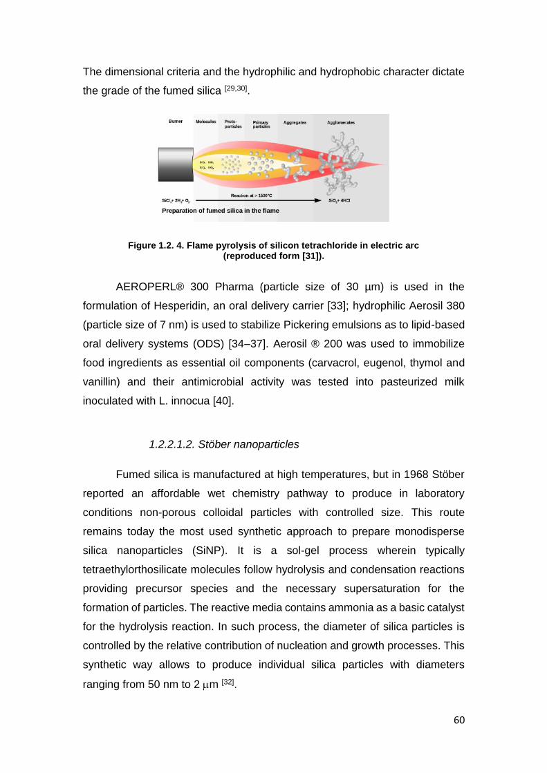
60
The dimensional criteria and the hydrophilic and hydrophobic character dictate
the grade of the fumed silica [29,30].
Figure 1.2. 4. Flame pyrolysis of silicon tetrachloride in electric arc (reproduced form [31]).
AEROPERL® 300 Pharma (particle size of 30 µm) is used in the
formulation of Hesperidin, an oral delivery carrier [33]; hydrophilic Aerosil 380
(particle size of 7 nm) is used to stabilize Pickering emulsions as to lipid-based
oral delivery systems (ODS) [34–37]. Aerosil ® 200 was used to immobilize
food ingredients as essential oil components (carvacrol, eugenol, thymol and
vanillin) and their antimicrobial activity was tested into pasteurized milk
inoculated with L. innocua [40].
1.2.2.1.2. Stöber nanoparticles
Fumed silica is manufactured at high temperatures, but in 1968 Stöber
reported an affordable wet chemistry pathway to produce in laboratory
conditions non-porous colloidal particles with controlled size. This route
remains today the most used synthetic approach to prepare monodisperse
silica nanoparticles (SiNP). It is a sol-gel process wherein typically
tetraethylorthosilicate molecules follow hydrolysis and condensation reactions
providing precursor species and the necessary supersaturation for the
formation of particles. The reactive media contains ammonia as a basic catalyst
for the hydrolysis reaction. In such process, the diameter of silica particles is
controlled by the relative contribution of nucleation and growth processes. This
synthetic way allows to produce individual silica particles with diameters
ranging from 50 nm to 2 m [32].

61
This type of synthetic non-porous silica is largely used in oral
applications due to their chemical stability and intrinsic hydrophilicity, which are
appropriate for biological environments. Andreani et al. associated insulin to
SiNP and they coated them with mucoadhesive polymers such as chitosan or
PEG aiming the oral delivery of insulin [33,34].
1.2.2.2. Synthesis of mesoporous silica
Since 1990, porous synthetic amorphous silica materials have been
widely used in various applications, especially as catalyst [35] or absorbents, but
also as drug carriers [9,36–40]. The porosity of materials can be classified
according to IUPAC into three categories: macroporous (> 50 nm), mesoporous
(2 to 50 nm) and microporous (< 2 nm). So far, mesoporous silica materials are
synthesized from either micelles of surfactants by cooperative self-assembly
(CSA) or from surfactant liquid crystals templates through the transcription
mechanism (LCT) [41,42] (Figure 1.2. 5). In both pathways, the silica source
hydrolyses and then polycondenses around the structure-directing agent
leading to a hybrid gel, called mesophase. Nevertheless, the two mechanisms
differ by the concentration of surfactant in the synthesis media. At high
surfactant concentration liquid crystals are formed, thus hydrolysis and
polycondensation of a silica source leads to a mesostructured material
templated by the liquid crystalline phase. In the case of the cooperative self-
assembly mechanism
Figure 1.2. 6, the surfactant concentration is low but usually above the critical
micellar concentration. During the polycondensation of the silica, the micelles
grow to finally form an inorganic mesostructured silica-surfactant composite,
also called mesophase. The mesoporosity is released after surfactant removal,
either by calcination or by solvent extraction [43].

62
Figure 1.2. 5. Formation of mesoporous materials by structure-directing agents: a) true liquid-crystal template mechanism, b) cooperative liquid crystal template mechanism
(reproduced from [41]).
Figure 1.2. 6. cooperative self-assembly mechanism ( reproduced from [44])
Nevertheless, obtaining mesoporous materials with highly ordered
porosity is not trivial. In fact, intimate interactions between the organic template
and the silica must take place during the hydrolysis and the polycondensation
of the silica mesophase as shown in Figure 1.2. 5 and in
Figure 1.2. 6. The main factors influencing the material structuring are
the experimental conditions (pH [45], temperature, aging time), the type of silica
precursor, the surfactant nature, its concentration and the molar ratio between
the surfactant and the silica source that have a direct impact on the hydrolysis,

63
condensation, and dissolution reactions [46].Pore size can also be tuned by the
diameter of the micelles that depends on the surfactant molecular weight, the
bigger the micelles the larger the pore sizes. The micelles can also be swollen
by organic solvents [47] . The porogens (or surfactant) can be either non-ionic
[48], cationic [49] or anionic [50,51]. Food grade directing agents, such as
polyglycerol esters of fatty acids [52], myristic acid ester of pentaglycerol [53], and
oleic acid [54] have also been employed. Each template has a specific interaction
with the silica source, which is in most of the cases an alkoxysilane, such as
tetramethylortosilicate (TMOS) or tetraethylortosilicate (TEOS). Alkoxysilanes
release methanol or ethanol as reaction by-products that might compromise the
hybrid silica/surfactant self-assembly or could be detrimental to proteins or
cells, if directly entrapped during the sol-gel synthesis [55]. The formation
mechanism of the silica scaffold and the release of ethanol during the
hydrolysis/condensation reaction from TEOS are outlined as an example
below.
To avoid possible toxic by-products, acidified aqueous sodium silicate
solutions can be used to perform the synthesis in appropriate pH conditions

64
with respect to the entrapped biocompound [56,57]. A food grade alternative is
rice husk ashes [58,59]. The pH and the salts content are drastically influencing
the charge density of silica and the ionic strength of the media during the
synthesis. The temperature also has an important effect either in helping the
formation of the hybrid structure or in dissolving it. However, the sol-gel process
can occur at temperatures below 40°C, which makes it compatible with
biomolecules or cells[28]. The aging time and pH are also influencing the
properties of the resulting silica material, in terms of texture, morphology and
mesopores structuring. During the aging step, the silica precursor is hydrolysed
and starts to condense to form a gel, which strength and stiffness increase with
the degree of siloxane crosslinking. At low pH, the hydrolysis of alkoxysilane is
faster than at high pH, but the condensation is slower. It should be also noted
that TMOS is hydrolysed faster than TEOS [43,60].
Since the 90’s, many new synthetic amorphous mesoporous materials
have been created with 1D, 2D or 3D pore channel arrays, different pore sizes,
specific areas and even with nanoparticular morphologies. In fact, silica
nanoparticles (SiNP) are an excellent candidate for their conjugation with drug,
biomolecules or cells [28] in the sense that (i) SiNP possess residual silanol
groups (Si-OH) at their surface which can be functionalized by different organic
groups [61] (ii) these materials can be synthesized at low temperature, which is
compatible with the manipulation of biomolecules [27], (iii) SiNP present large
surface areas which allows high interactions with drugs [62], (iv) SiNP can act as
drug reservoirs by possessing high porosity and allowing efficient drug loading
[63], and (v) the material is known to be biocompatible for in vivo applications [64],
(vi) SiNP are not subjected to microbial attack [65].
The most used mesoporous silica nanoparticles (MSN) in drug or bio-
encapsulation are MCM-41 and SBA-15. The synthesis of MCM-41 (Mobil
Composition of Matter series) involves liquid crystal templating using commonly
cetyl trimethylammonium bromide (CTAB) that leads to a 2D hexagonal pore
channel array with 3.6 nm in size. The diameters of MCM-41 nanoparticles can
be controlled ranging from 25 to 100-150 nm [66–70]

65
The SBA-15 (Santa Barbara type) is also largely used as biocarrier. This
type of mesoporous silica material is prepared by cooperative self-assembly
with a pluronic surfactant, P123, a non-ionic block co-polymer. The pore
channels adopt also a 2D hexagonal packing with a diameter varying from 6 to
10 nm depending on the synthesis conditions [5,71].
1.2.2.3. Synthesis of hybrid silica microparticles
The discovery of the previously described synthetic routes opened up
the frontiers for the preparation of nanoparticular or microparticular silica
materials with hierarchical porosity, such as meso-macroporous materials.
Our group prepared such materials by using a silica precursor and
dispersions of solid lipid nanoparticles in a micellar solution ( Figure 1.2. 7), [72–
74]. The formation of the silica matrix is based on a dual templating mechanism,
combining self-assembly mechanism of surfactant micelles (Tween 20, Tween
40 or Pluronic P123) with transcription mechanism of solid lipid nanoparticles.
Depending on the reaction conditions, the morphology of the final material can
be tuned to capsules or to block matter (Figure 1.2. 7, left). The size of the
mesopores is strongly dependent on the nature of the surfactant in excess, 3
nm (Tween 20), 5 nm (Tween 40) or 9 nm (Pluronic P123), whereas the size of
the macropores depends only on the size of SLN (250 150 nm). The
macroporous void was clearly evidenced by TEM ( Figure 1.2. 7 right). The
organization degree of the silica wall depends on the surfactant: only wormlike
mesoporous capsules were obtained with Tween 20, and hexagonally ordered
microdomains embedded in wormlike mesoporous silica capsules were
obtained with Tween 40. Hexagonally ordered silica with circularly ordered
mesoporosity could be achieved with Pluronic block copolymer P123.
Using the same strategy, core-shell microparticles were prepared with a
core of solid lipid nanoparticles and a mesostructured silica shell. By
encapsulating curcumin in the solid lipid nanoparticles before the silica
formation, hybrid core-shell materials were obtained after drying, without any
further washing step [75].

66
Figure 1.2. 7 (Left) Schematic representation of hierarchical meso-macroporous silica obtained through a dual templating mechanism combining self-assembly mechanism
with micelles of Tween 20, Tween 40 and P123 and SLN transcription; (Right) TEM micrographs of meso–macroporous silica obtained from SLN dispersions with P123 at various temperatures: 100°C (a and c), 70°C (b) and 40°C (d–f) (reproduced from [74]).
Core-shell microparticles containing an ionogel as core and a silica shell
could be also obtained in a two steps synthesis (Figure 1.2. 8). In the first step,
the probiotic bacteria Lactobacillus rhamnosus GG and sodium alginate were
electrosprayed over a calcium bath leading to alginate beads of about 22015
µm (Figure 1.2. 8). In a second step, the microgels were dispersed in an
aqueous solution containing Tween 40 as porogen, APTMS
(aminopropyltrimethoxysilane) and TMOS (tetramethoxysilane) as silica
sources. The hydrolysis of both precursors and their co-condensation resulted
in core–shell microparticles. Moreover, due to the presence of the surfactant,
mesoporosity could be introduced into the silica shell during the mineralization
process. When encapsulating living matter, the porosity is essential to allow
bidirectional diffusion of nutrients and metabolites in and out of the beads,
which in our case resulted in proliferation of the bacteria under confinement.
The mild synthesis conditions of the mineralization step were not detrimental to
bacteria and allowed their encapsulation while maintaining a good viability
(more than 8 log/mL). The release studies simulating the gastrointestinal
conditions showed that the bacteria encapsulated in the alginate-silica
microparticles had a superior survivability in comparison to free bacteria or even
with bacteria encapsulated only in alginate beads [76].
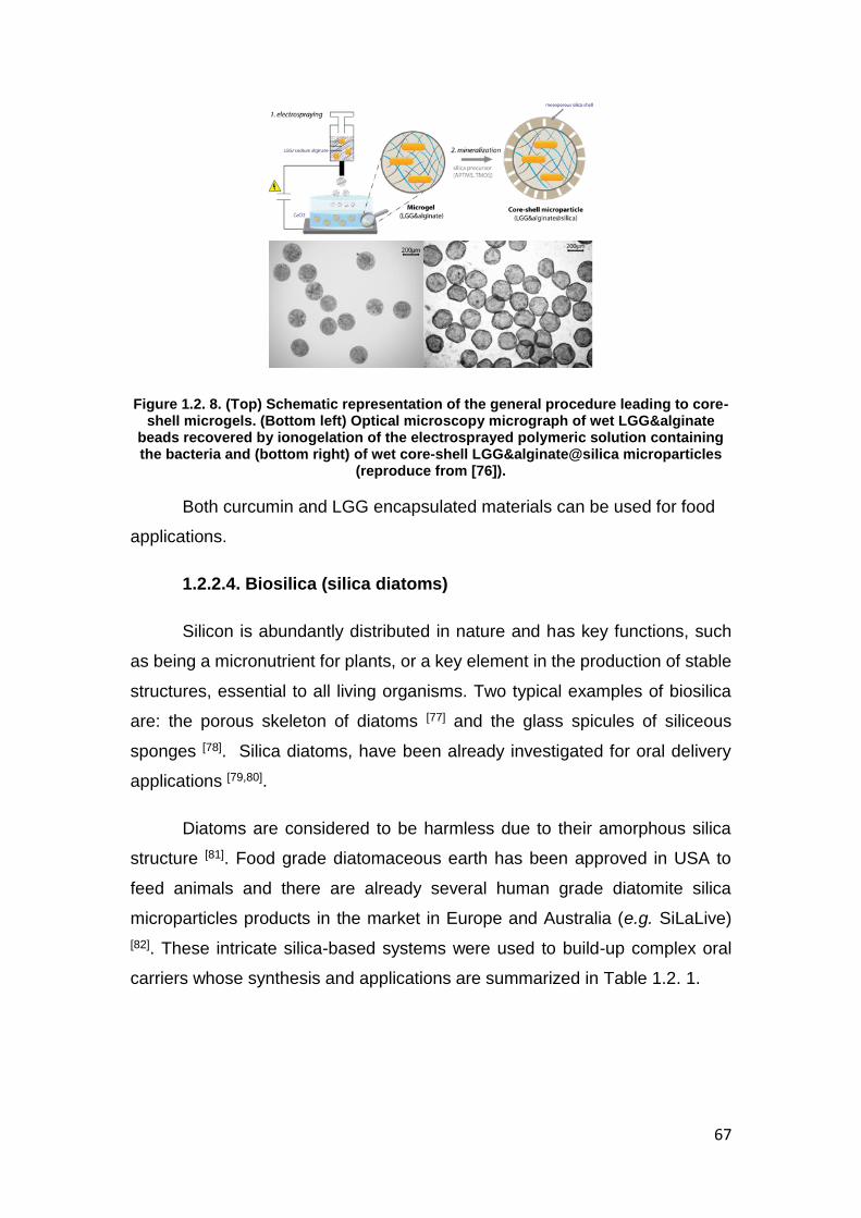
67
Figure 1.2. 8. (Top) Schematic representation of the general procedure leading to core-shell microgels. (Bottom left) Optical microscopy micrograph of wet LGG&alginate
beads recovered by ionogelation of the electrosprayed polymeric solution containing the bacteria and (bottom right) of wet core-shell LGG&alginate@silica microparticles
(reproduce from [76]).
Both curcumin and LGG encapsulated materials can be used for food
applications.
1.2.2.4. Biosilica (silica diatoms)
Silicon is abundantly distributed in nature and has key functions, such
as being a micronutrient for plants, or a key element in the production of stable
structures, essential to all living organisms. Two typical examples of biosilica
are: the porous skeleton of diatoms [77] and the glass spicules of siliceous
sponges [78]. Silica diatoms, have been already investigated for oral delivery
applications [79,80].
Diatoms are considered to be harmless due to their amorphous silica
structure [81]. Food grade diatomaceous earth has been approved in USA to
feed animals and there are already several human grade diatomite silica
microparticles products in the market in Europe and Australia (e.g. SiLaLive)
[82]. These intricate silica-based systems were used to build-up complex oral
carriers whose synthesis and applications are summarized in Table 1.2. 1.

68
Table 1.2. 1. Selected examples of silica-based oral delivery systems [26].
Delivery system Silica source Payload Coating Encapsulation method Release mechanism
In vitro/in vivo/ex vivo studies
Ref
Non porous silica nanoparticles
Stöber NPs TEOS Insulin PEG 6000 PEG 20000
Physisorption of insulin to pre-synthesized uncoated silica nanoparticles- subsequent coating with PEG
Passive diffusion
Ex vivo permeation studies using everted rat intestine
[33]
Stöber NPs TEOS Insulin Chitosan Physisorption of insulin in chitosan solution to pre-synthesized uncoated silica NPs
Passive diffusion
In vitro studies of NP interactions with porcine mucin
[34]
Mesoporous silica particles
MCM-48 Ia3d MSN
Ludox AS40 TEOS/MPTS
Ibuprofen Erythromycin
- Physisorption by immersion Passive diffusion
In vitro drug release in a simulated body fluid (pH 7.7-7.4)
[91]
SBA-15 silica nf Itraconazole - Physisorption by immersion Passive diffusion
In vitro drug release in a simulated gastric fluid (pH 1.2)
[89]
SBA-15 and MCM-41 functionnalized with amino group
nf Bisphosphonates - Electrostatic interaction between drug’s phosphate group and silica’s amine group at pH 4.8
Passive diffusion at pH 7.4
In vitro drug release in phosphate buffer pH 7.4
[92]
MCM41 nanoparticles nf Rhodamine B α-CD, adamantly ester
Physisorption Porcine liver esterase triggered
In vitro hydrolysis in HEPES buffer pH7.5
[93]
MCM48 TEOS/APTES Silfalazine Succinylated soy protein isolate
Physisorption and coating pH/enzyme triggered
In vitro drug release in simulated GIT fluid at pH 1.2, 5., 7.4
[94]
Hybrid silica microparticles
Core-shell (SLN-mesostrctured silica)
TMOS Curcumin - 1. encapsulation of curcumin in SLN by emulsification/sonication; 2. Sol-gel
Passive diffusion
In vitro drug release in a simulated GIT fluid (pH 1.2-7.4)
[75]
Core-shell (alginate silica)
TMOS/ APTMS
LGG - 1. preparation of LGG/alginate microgels by electrospraying, 2. mineralization
Erosion of silica shell
In vitro drug release in a simulated GIT fluid (pH 1.2-7.4)
[76]
Spherical mesocellular foam
hydrophilic fumed silica
Insulin Eudragit L30D-55 Eudragit L100
Physical adsorption by immersion pH triggered In vitro drug release in simulated GIT fluid at pH 1.2 and 6.8
[95]
Diatom silica microparticles
Diatom silica fossile Indomethacin/ Gentamicin
- Physisorption Passive diffusion
In vitro drug release in simulated intestinal fluid at pH 7.2
[96]
Diatom silica fossile Mesalamine/ prednisone
- Physisorption Passive diffusion
In vitro drug release in a simulated GIT fluid (pH 1.2-7.4)
[82]

69
Table 1.2. 2. Selected examples of silica-based food delivery systems.
Delivery system Silica source Payload Coating/Capped Encapsulation method Release mechanism
Release studies Ref
Non porous silica nanoparticles
Stöber NPs TEOS resveratrol PMES Physisorption Passive diffusion In vitro drug release in phosphate buffer pH 7.4
[97]
Mesoporous silica particles
MCM-41 TEOS resveratrol - Impregnation, solid-state method
Passive diffusion In vitro drug release in phosphate buffer pH 7.4
[98]
MCM-41 TEOS Vitamin B2 polyamine 3-[2-(2-aminoethylamino)ethylamino]propyl- trimethoxysilane).
Physisorption by immersion pH and salts triggered
In vitro release in a pH containing different anions
[39]
Hollow microspheres, SBA-15
TEOS Vitamin B3 precursor
- Covalent grafting method pH trigger In vitro release in a simulated gastric fluid (pH 1.2)
[52]
MSNs TEOS Vitamin C - Physisorption Burst release at basic pH
In vitro drug release in SGF(pH 1.2), SIF (7.4), SBF(7.4)
[99]
MCM41 micropart TEOS/ TEAH3 Folic acid - Impregnation pH triggered Yoghurt in vitro drug release in a simulated GIT fluid (pH 2, 4, 7.5)
[100]
Mesoporous silica mesoparticles
TEOS Curcumin Entrapment Passive diffusion In vitro release at physiological pH
[101]
Mesoporous silica mesoparticles
TEOS oligophenol Entrapment Passive diffusion In vitro release at physiological pH
[101]
Mesoporous silica mesoparticles
TEOS Trans- β-Carotene
- Entrapment Passive diffusion In vitro release at physiological pH
[101]
SBA-15 TEOS/APTES Vitamin E LDPE Impregnation Passive diffusion Migration tests in olive oil [102] MCM-41 SiO2 Quercetin - Kneading method Passive diffusion Diffusionthrough a semi-
permeable cellulose membrane
[103]
SBA-15, Syloblock TEOS α-tocopherol LPDE, EVA Physisorption Passive diffusion Migration tests in ethanol [104] MCM-41 TEOS/TEAH3 Garlic extract Polyamines and
hydrolyzed starch on nylon-6
Physisorption Pancreatic or pH triggered
Electrochemical studies in aqueous solution at ph 7 or 2
[105]
MPTS : Mercaptopropyl-trimethoxysilane ; APTMS : aminopropyltriethoxysilane,SLN: solid lipid nanoparticles; α-CD cyclodextrine PMES: undec-1-en-11-yltetra(ethylene glycol) phosphate mono- ester surfactant ,SGF: simulated gastric fluid , SIF: simulated intestinal fluid, SBF simulated body fluid TEAH3:tri-ethanolamine,LDPE: low density polyethylene, EVA: ethylene vinyl acetate

70
1.2.3. Silica-based oral delivery systems and food applications
Single or multiple drug substances can potentially be loaded in the same dosage
form, making mesoporous silica materials a versatile tool for combination of therapies [4].
The special architecture of mesoporous silica offers an efficient protection for
biomolecules which are subjected to lysis by the gastrointestinal juices [33]. The protection
related mechanism may consist in a confinement or a steric hindrance that would protect
the loaded biomolecule inside the pores from the action of catalytic enzymes [83].
The wide range of pore sizes offer the possibility to load in silica matrices a large
variety of drugs [84], macromolecules [33], genes [85,86], or even cells [87]. Furthermore, pore
size monitoring could be used in the obesity treatment[88] or in the adjust the release
kinetics adjustment [89,90].
The highly porous structure of silica makes it an ideal candidate as a DDS floating
matrix intended to achieve a certain gastro-retention. Mesoporous silica nanoparticles
mixed with sodium bicarbonate and cellulose derivative polymer were used for the
preparation of floating tablets of curcumin and captopril, as hydrophobic and hydrophilic
model drugs, respectively. The obtained tablets showed an extensive floating behavior
over 12 hours of gastro-retention [106]. Highly porous calcium silicate [107] and aluminum
silicate [108] were also successfully used for the preparation of floating DDS containing
repaglinide and methotrexate, a combination of a hypoglycemic agent with poor
absorption in the upper intestinal tract and an antineoplastic agent with a short half-life of
2 hours, respectively.
The principal issues related to oral drug delivery, i.e. solubility and intestinal
permeability [109], could be addressed by the implementation of mesoporous silica
materials in the development strategies of ODDS. The poor solubility of BCS class II,
such as celecoxib [110], fenofibrate [111] and telmisartan [112], and BCS class IV such as
furosemide [113], could also be reversed following their encapsulation in mesoporous silica
DDS. It is noteworthy that the design of ordered mesoporous silica into nanoparticulate

71
DDS result in a higher intestinal permeability due to enhanced cell uptake and reduced
drug efflux [112].
Mesoporous silica has been used not only for dugs delivery applications, but also
for the encapsulation of food ingredients to improve molecules stability and bioavailability.
Vitamins are nutrients essential for humans that are not produced by the body. They have
a low activity in the presence of metal ions, ultraviolet light and heat. Water-soluble
vitamins as riboflavin (vitamin B2) [39,114], niacin ( vitamin B3)[52], and ascorbic acid (vitamin
C) [99] or oil-soluble vitamins as β-carotene (precursor of vitamin A) [101] and α-tocopherol
(vitamin E) [102,115] were immobilized on silica for food applications. Phytochemicals as
polyphenols (from wine [116]), quercetin [103,117], resveratrol [97,98] or curcumin [101] that have
antioxidant activity, organosulfurs with antibiotic activity [105], glucosinolates that have
antibacterial efficacy [118] were immobilized in silica to improve their properties. Another
class of food ingredients that were encapsulated in silica are bioactive peptides [119–122].
Silica-based ODDS were classified according to their payloads release
mechanisms in the GIT, which in turn depends on the employed loading strategy [26].
Different methods (Figure 1.2. 9) such as one-step condensation (one-pot encapsulation),
immersion method, impregnation method or supercritical CO2 method, incipient wetness
impregnation method, hot melt can be used for the loading of porous silica. After the
loading, the pores can be capped with molecular ensembles to confer the carriers a stimuli
response [9].

72
Figure 1.2. 9. Schematic representation of different loading methods (reproduced from [123]).
Generally speaking, the physically adsorbed drugs are more likely to be released
by diffusion; whereas the covalently-linked counterparts to be released by interactions
with internal and external triggers, e.g. hydrolysis by digestive or gut enzymes, increased
ionic strength and/or pH variation along the GIT, external magnetic field action [26], among
others as showed in the Figure 1.2. 9 and summarized is Table 1.2. 1 and Table 1.2. 2.
1.2.3.1. Passive release delivery systems
As presented above, the synthesis of ordered-porous silica could be controlled, by
varying the reaction parameters and the structure directing agent (template), with the aim
to obtain materials with different morphologies, pore sizes, ordering and volume and
displaying various surface properties. These features were found to determine both drug
loading and passive release.
Several studies highlighted a relationship between release kinetics and pore sizes
[89,91]pores shape [124], particle size [112,125], or hydrophilicity [126].

73
Pore size is an important factor, not only to ensure the loading of the molecules
inside the pores but also to release it [127]. Mellaerts et al. studied the influence of four
different types of SBA-15, a mesoporous silica material having hexagonally-ordered
pores, on the release kinetics of itraconazole, an anti-fungal agent with a very poor
aqueous solubility [89]. The pore size varied from 4.5 to 9.0 nm, and the pore volume
ranged from 0.42 to 0.80 cm3·g− 1. The release performance of materials loaded with
10 wt% of itraconazole was assessed in a simulated gastric fluid at pH 1.2. It was found
that the larger the material pore size and volume, the faster the release rate. To explain
these findings, the authors suggested: i) more rapid influx and a competitive absorption
of water in the wider mesopores, and ii) breaking up intermolecular interactions between
in drug crystals due to their separation onto the SBA-15 surface [89]. Allyl isothio- cyanate
(AITC), an antimicrobial, used principally in foods as a flavoring agent, was encapsulated
in MCM-41 and SBA-15 [118,128,129]. The pore size and distribution influences the
desorption, as 65% of the compound was burst release form the SBA-15 system in the
first 12 hours.
On the other hand, Balas et al. developed a DDS based on mesoporous silica
intended for the oral delivery of bisphosphonates, an anti-osteoporotic drug with poor
bioavailability. For this purpose, they used two silica materials, with hexagonally ordered
mesopores, SBA-15 and MCM-41. Unexpectedly, it was found that the release rate from
SBA-15 (pore size 9.0 nm) was slower than that from MCM-41 (pore size 3.8 nm).
Additionally, the loading efficiency on SBA-15 was smaller than that on MCM-41. These
findings were explained by the higher surface area of MCM-41 (1157 m2 g− 1) with respect
to that of SBA-15 (719 m2 g− 1) and that the release was rather a surface-dependent
phenomenon [124].
The size of the DDS is also an important feature and has to be carefully
considered. Zhang et al. designed mesoporous silica micro- and nanoparticulate as
ODDS for telmisartan [112] . The performed release studies, in an enzyme-free simulated
intestinal fluid (SIF) pH 6.8, showed an increase in the release rate when the particle size
was decreased down to the nanometer range. The authors explained this finding by the
shorter channel length and then the shorter diffusion distance to be crossed by the guest

74
molecule before reaching the release medium. Similarly, Aerts et al. studied the influence
of particle size on the drug release behavior from amorphous microporous silica materials
[125] and they found that the smaller the particle size, the faster the release.
Alteration of the silica surface hydrophilicity (by methylation of Si-OH) is another
way to control the passive diffusion of the guest drug [126]. The digestive fluids influx could
be reduced inside the inner channels and this way, the drug dissolution/degradation could
be delayed.
In 2011, Aw et al. proposed silica microcapsules from diatoms as new carrier for
oral delivery of therapeutics, namely indomethacin and gentamicin[96]. The first one is a
widely used and poorly soluble anti-inflammatory drug and the second is a highly water-
soluble cationic aminoglycoside antibiotic widely used in therapeutic implants to prevent
bacterial infections. Typical structural features of diatom silica microparticles (DSMs)
used in this study are: diameters between 4 and 6 μm; length between 10 and 20 μm,
with regularly spaced rows of pores with a diameter of approximately 400–500 nm. Drug
molecules can be loaded on both internal and external surface of the material. Controlled
release over 6–7 h for both drugs were achieved in sink conditions and the results proved
that those DSM meet requirements for extended oral dosage.
More recently, Zhang et al. have evaluated the potential of DSMs for the delivery
of mesalamine and prednisone, two commonly prescribed drugs for gastrointestinal
diseases [82]. DSMs used in this study were cylindrical, with 10–20 μm in length
and ca.10 μm in diameter, with well-defined pores of 300–500 nm). In vitro release
studies showed no difference in release kinetics for free and encapsulated mesalamine.
However, prednisone showed a clear sustained release after loading in DSMs). In overall,
the study showed that DSMs have a low cytotoxicity on Caco-2, HT-29, HCT-116 cells
and Caco-2/HT-29 co-cultured cells, at concentrations up to 1000 mg/mL. Both drugs
undergo a controlled release under simulated gastro-intestinal conditions and an
enhanced permeability across Caco-2/HT-29 co-culture monolayers. The results thus
demonstrate that DSMs can be considered as a non-cytotoxic biomaterial with a high

75
potential to improve the mesalamine and prednisone bioavailability by sustaining the drug
release and enhancing drug permeability.
1.2.3.2. Active release delivery systems
The large silica surface covered by silanol group Si-OH offers a unique opportunity
to design smart DDS. Indeed, silanols can be easily functionalized leading to delivery
systems with programmed drug release as a function of either (i) internal signals, e.g. pH
variation [130], glucose [131–133] or catabolic or bacterial enzyme availability [93], [94]or (ii)
external stimuli, e.g. temperature [134], irradiation [135], magnetic field [136], etc. Herein, are
described and presented (Figure 1.2. 10) stimuli-responsive silica-based systems.
Figure 1.2. 10. Molecular gates and trigger responses for pore-capped silica materials (reproduced from [123]).
1.2.3.2.1. pH-controlled release
pH-sensitive silica-based carriers represent an interesting alternative for the oral
delivery of peptides and proteins, or for vitamins protecting them from the GIT proteolytic
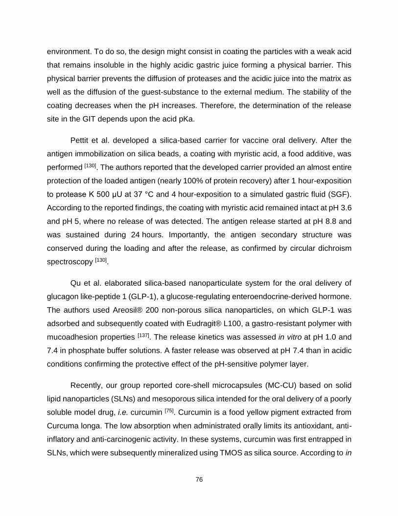
76
environment. To do so, the design might consist in coating the particles with a weak acid
that remains insoluble in the highly acidic gastric juice forming a physical barrier. This
physical barrier prevents the diffusion of proteases and the acidic juice into the matrix as
well as the diffusion of the guest-substance to the external medium. The stability of the
coating decreases when the pH increases. Therefore, the determination of the release
site in the GIT depends upon the acid pKa.
Pettit et al. developed a silica-based carrier for vaccine oral delivery. After the
antigen immobilization on silica beads, a coating with myristic acid, a food additive, was
performed [130]. The authors reported that the developed carrier provided an almost entire
protection of the loaded antigen (nearly 100% of protein recovery) after 1 hour-exposition
to protease K 500 μU at 37 °C and 4 hour-exposition to a simulated gastric fluid (SGF).
According to the reported findings, the coating with myristic acid remained intact at pH 3.6
and pH 5, where no release of was detected. The antigen release started at pH 8.8 and
was sustained during 24 hours. Importantly, the antigen secondary structure was
conserved during the loading and after the release, as confirmed by circular dichroism
spectroscopy [130].
Qu et al. elaborated silica-based nanoparticulate system for the oral delivery of
glucagon like-peptide 1 (GLP-1), a glucose-regulating enteroendocrine-derived hormone.
The authors used Areosil® 200 non-porous silica nanoparticles, on which GLP-1 was
adsorbed and subsequently coated with Eudragit® L100, a gastro-resistant polymer with
mucoadhesion properties [137]. The release kinetics was assessed in vitro at pH 1.0 and
7.4 in phosphate buffer solutions. A faster release was observed at pH 7.4 than in acidic
conditions confirming the protective effect of the pH-sensitive polymer layer.
Recently, our group reported core-shell microcapsules (MC-CU) based on solid
lipid nanoparticles (SLNs) and mesoporous silica intended for the oral delivery of a poorly
soluble model drug, i.e. curcumin [75]. Curcumin is a food yellow pigment extracted from
Curcuma longa. The low absorption when administrated orally limits its antioxidant, anti-
inflatory and anti-carcinogenic activity. In these systems, curcumin was first entrapped in
SLNs, which were subsequently mineralized using TMOS as silica source. According to in

77
vitro studies, the resulting hybrid system behaves as a gastro-resistant delivery system.
Interestingly, MC-CU were shown to be readily taken up by Caco-2 cell line, an in
vitro intestinal epithelium cell model. These results demonstrated the potential of this
delivery platform for enhancing both drug solubility and intestinal permeability.
Pérez-Esteve et al. developed a functional food enriched with folic acid based on
pH- sensitive mesoporous silica [100]. Folic acid was encapsulated in amine-functionalized
silica matrix with the aim to enhance its bioavailability by hindering its premature release
and degradation in the stomach. Instead, folic acid was progressively released in the
intestine where it is completely absorbed. Indeed, at low pH, amine groups are protonated
hence promoting Coulombic repulsions due to closely located ammonium groups.
Consequently, in low pH silica pores are blocked. The progressive increase of the pH
after the pyloric passage decrease the ratio of protonated “blocked gates” allowing a
sustained release of the payload.
1.2.3.2.2. Enzyme-triggered release
The design of these systems is based on the principle of pore-capping of silica
materials with a cleavable gate (Figure 1.2. 10). A multitude of link types could be
designed between the gate and the pore in order to be readily cleavable by predefined
enzyme. The variety of the enzymes present in the gastro-intestinal track can lead to the
developed of sophisticated gate-controlled release silica nanocarriers triggered at a
specific site (stomach, intestine, colon). Bernandos et al. [138] developed a simple gate-
controlled release silica nanocarriers triggered by enzymatic hydrolysis. The synthesis of
this system was based on loading of the guest molecule ([Ru(bipy)3]Cl2-as model
molecule) by physical adsorption inside the pores of mesoporous silica (MCM-41than the
surface was modificafied with a lactose derivative (β-d-galactose and β-d-glucose
monosaccharides linked through a β14 glycosidic bond). The hydrogen bounding
interactions between the disaccharides keep the cargo inside. The researchers showed
the release of the loaded molecule from the silica nanocarriers following its exposition to
β-galctosidase, the enzyme present in the brush-border in the intestine, as a proof of

78
concept. Another capped molecule that is used and hydrolyze by β-galactosidase is
alkylgluconamine derivate of a galcto-oligosacchride [139].
Food ingredients as starches that are hydrolysed by pancreatin [138], avidin (a white
yolk protein) [140] and modular peptide [141] that are cut by protease, cytosine-
phosphodiester-guanine oligodeoxynucleotide [142], or oligodeoxynucleotides [143]
triggered by deoxyribonuclease were also used to prepare enzyme-triggered release
silica systems.
1.2.3.2.3. pH/Enzyme-triggered release
Popat et al. designed mesoporous silica nanoparticules displaying both pH and
enzyme responsiveness and thus a release profile depending upon its location in the GIT
[94]. The matrix used for this system is based on amino-modified MCM-48 displaying a
cubic bicontinuous pore structure. A coating with succinylated soy protein isolate (SSPI),
a hydrophobic polymer stable in acidic pH media (with isoelectric point at pH 5) and a
modified food component, was carried out (Figure 1.2. 11). The coating choice, as
presented by the authors, was explained in terms of: i) stability due to the covalent
attachment between the SSPI’s carboxylic group and the silica’s amine group; ii)
insolubility in acidic media; iii) susceptibility of the succinylated protein towards the
proteolytic enzymes in the small intestine. The authors conducted in vitro release studies
in SGF (pH 1.2) containing pepsin and in SIF, (pH 7.4) in the presence or absence of
pancreatin, in order to check the performance of their developed system. According to
their results, the drug release was successfully tuned and took place only in SIF in a slow
and sustained pattern over 48 hours. Importantly, the release rate was doubled when
pancreatin was added to the SIF.

79
Figure 1.2. 11. Schematic representation of (A) synthesis of MSN–NH2– SZ@SSPI. a: SZ is loaded into amino functionalized MCM-48 (MSN–NH2) to form MSN–NH2–SZ, b: coating of SSPI using
amide chemistry leads to MSN–NH2–SZ@SSPI. (B) Oral delivery and site-dependent programmable release of SZ into 5-ASA in the GIT from MSN–NH2–SZ@SSPI (reproduced from
[94]).
1.2.4. Silica health benefits and limitations
Silica is found everywhere on the planet, present in the earth's crust, in water but
also in a great number of living organisms. Silica is considered as “Generally Recognized
as Safe” by FDA regulations and an authorized additive in Europe as E-551 class [144].
Synthetic amorphous silica has been used for many years in food industry as a beer and
wine clarifying agent, to thicken pastes, as a carrier agent for flavoring and aromas or as
an anticaking agent [145] to maintain the flow properties of powder products. As a natural
product silica is found in beverages (water, beer and coffee) and food (cereals and
vegetables). The mean daily intake of dietary silicon as silica were estimated to be 19
and 40 mg in adult women and men, respectively [146].
To be used in food as smart delivery devices, silica materials should overcome
some sociological, toxicological, technological legal and sematic limitations [9].Silica is not
considered harmful for humans, but before considering adding to the food or to used as
oral delivery system some parameters such as size distribution, particle size [147–149] and
shape [150], mass, reactivity, solubility, chemical composition and surface properties
[151,152] should be taken into account [10,153]. Particle size of the silica can change the
functionality [154], the particles they are more easily decomposed with the decrease of the
diameter [155]. To date, the process for the synthesis functionalization and loading of
mesoporous silica is developed in the lab and the scaled-up for an industrial application

80
is underdeveloped due to the high productions cost [9]. Another technological problem for
the transition of silica from the lab to industry is the compatibility of these carriers with the
food [24].
1.2.5. Conclusion
This chapter highlights the emerging interest of silica-based delivery systems in
oral delivery and food industry of drugs, biomolecules or cells accounting for the design
of new functional foods containing probiotic bacteria. The carriers can be synthesized
through two routes: (1) encapsulation of payloads in pre-synthesized silica or (2) one-pot
encapsulation and silica formation. Indeed, silica can be obtained through the sol-gel
process at low temperature (<40°C), which makes it compatible with the manipulation of
temperature-sensitive drugs, peptides, proteins and more particularly, cells. Porosity can
be easily imprinted into silica materials through the use of structure-directing agents or
soft templates. Therefore, materials with large surface areas and pore volumes can be
obtained as well as materials that exhibit high drug loading capacity. Pore sizes can be
tuned in the range of 2-50 nm in the case of mesopores, and from hundreds of nm to tens
or hundreds on microns in the case of macropores. The residual silanol groups at the
surface of silica can be also functionalized allowing high interactions with drugs. Overall,
the porosity of silica materials can be tailored as a function of the size and properties of
the payload. The most sophisticated functionalization strategies have been elaborated to
control drug delivery kinetics at the appropriate site with zero premature release along
with preventing undesired side effects. The researchers can play freely to build up
fascinating and efficient multifunctional carriers.
The long history of use of silica and silicates in food and drug products for the oral
route without allegedly harmful effects over the last 50 years has demonstrated their
safety. The main degradation product of silica, i.e. orthosilicic acid, is not toxic and is
rapidly eliminated by the kidneys [149] or by fecal excretion [148]. However, when
administrating silica via routes other than the oral one, it was found to be less tolerated
or even toxic.

81
Bibliography
[1] D. Napierska, L. C. J. Thomassen, D. Lison, J. a Martens, P. H. Hoet, Part. Fibre Toxicol. 2010, 7, 39. [2] K. R. Martin, Met. Ions Life Sci. 2013, 13, 451–473. [3] W. E. G. Müller, X. Wang, FEBS J. 2012, 279, 1709. [4] A. M. Chen, M. Zhang, D. Wei, D. Stueber, O. Taratula, T. Minko, H. He, Small 2009, 5, 2673–2677. [5] D. Zhao, J. Feng, Q. Huo, N. Melosh, G. Fredrickson, B. Chmelka, G. Stucky, Science 1998, 279, 548–52. [6] A. Sayari, S. Hamoudi, Y. Yang, Chem. Mater. 2005, 17, 212–216. [7] C. Márquez-Alvarez, E. Sastre, J. Pérez-Pariente, 2004, 27. [8] A. Bernardos, L. Kourimska, Czech J Food Sci 2013, 31, 99–107. [9] E. Pérez-Esteve, M. Ruiz-Rico, R. Martínez-Mánez, J. M. Barat, J. Food Sci. 2015, 80, 2504–2516. [10] T. Bech-Larsen, K. G. Grunert, Appetite 2003, 40, 9–14. [11] FDA, 2009, Www.accessdata.fda.gov/scripts/fcn/gras_notices/GR. [12] M. T. Zamin, M. M. Pitre, J. M. Conly, Ann. Pharmacother. 1997, 31, 564–570. [13] K. M. Kuper, Intravenous to Oral Therapy Conversion, 2008. [14] M. A. Fischer, D. H. Solomon, J. M. Teich, J. Avorn, Arch. Intern. Med. 2003, 163, 2585–2589. [15] A. Åsberg, A. Humar, H. Rollag, A. G. Jardine, D. Kumar, P. Aukrust, T. Ueland, A. A. Bignamini, A.
Hartmann, Clin. Infect. Dis. 2016, 62, 1154–1160. [16] J. M. Cyriac, E. James, J Pharmacol Pharmacother 2014, 5, 83–87. [17] K. S. Pang, Drug Metab. Dispos. 2003, 31, 1507–1519. [18] R. Panchagnula, N. S. Thomas, Int. J. Pharm. 2000, 201, 131–50. [19] B. N. Singh, K. H. Kim, J. Control. Release 2000, 63, 235–259. [20] K. Sonaje, K. J. Lin, M. T. Tseng, S. P. Wey, F. Y. Su, E. Y. Chuang, C. W. Hsu, C. T. Chen, H. W. Sung,
Biomaterials 2011, 32, 8712–8721. [21] G. Singh, R. S. Pai, Drug Deliv. 2016, 23, 532–539. [22] B. Aspenström-Fagerlund, L. Ring, P. Aspenström, J. Tallkvist, N. G. Ilbäck, A. W. Glynn, Toxicology 2007,
237, 12–23. [23] B. Aspenström-Fagerlund, B. Sundström, J. Tallkvist, N. G. Ilbäck, A. W. Glynn, Chem. Biol. Interact. 2009,
181, 272–278. [24] E. Pérez-Esteve, L. Oliver, L. García, M. Nieuwland, H. H. J. de Jongh, R. Martínez-Mánez, J. M. Barat,
2014. [25] A. Comas-Vives, Phys. Chem. Chem. Phys. 2016, 18, 7475–82. [26] R. Diab, N. Canilho, I. A. Pavel, F. B. Haffner, M. Girardon, A. Pasc, Adv. Colloid Interface Sci. 2017, DOI
10.1016/j.cis.2017.04.005. [27] C. Kneuer, M. Sameti, E. G. Haltner, T. Schiestel, H. Schirra, H. Schmidt, C. M. Lehr, Int. J. Pharm. 2000,
196, 257–261. [28] C. F. Meunier, P. Dandoy, B. L. Su, J. Colloid Interface Sci. 2010, 342, 211–224. [29] H. Barthel, L. Rosch, J. Weis, Organosilicon Chem. 11 1996, 761–778. [30] H. Barthel, M. Heinemann, M. Stintz, B. Wessely, Chem. Eng. Technol. 1998, 16, 169–176. [31] “Battery Fumed silica, Resin Fumed silica, Silicone Rubber Fumed silica-Minmetals East Industial,” can be
found under http://www.hjsil.com/aboutus/Technology/, n.d. [32] W. Stöber, A. Fink, E. Bohn, J. Colloid Interface Sci. 1968, 26, 62–69. [33] T. Andreani, C. P. Kiill, A. L. R. de Souzad, J. F. Fangueiro, L. Fernandes, S. Doktorovová, D. L. Santosa, M.
L. Garcia, M. P. D. Gremião, E. B. Souto, et al., Colloids Surfaces B Biointerfaces 2014, 123, 916–923. [34] T. Andreani, M. Leonardo, L. Esteban N., R. de S. Ana Luiza, K. Charlene P., F. Joana F., G. Maria L., G.
Palmira D, S. Amélia M., S. Eliana B., Eur. J. Pharm. Biopharm. 2015, 93, 118–126. [35] C. T. Kresge, M. E. Leonowicz, W. J. Roth, J. C. Vartuli, J. S. Beck, Nature 1992, 359, 710–712. [36] A. Popat, S. B. Hartono, F. Stahr, J. Liu, S. Z. Qiao, G. Qing (Max) Lu, Nanoscale 2011, 3, 2801. [37] M. Vallet-Regi, A. Rámila, R. P. Del Real, J. Pérez-Pariente, Chem. Mater. 2001, 13, 308–311. [38] M. Vallet-Regí, F. Balas, D. Arcos, Angew. Chemie - Int. Ed. 2007, 46, 7548–7558. [39] A. Bernardos, E. Aznar, C. Coll, R. Martínez-Mañez, J. M. Barat, M. D. Marcos, F. Sancenón, A. Benito, J.
Soto, J. Control. Release 2008, 131, 181–189. [40] É. Pérez-Esteve, A. Fuentes, C. Coll, C. Acosta, A. Bernardos, P. Amorós, M. D. Marcos, F. Sancenón, R.
Martínez-Máñez, J. M. Barat, Microporous Mesoporous Mater. 2015, 202, 124–132. [41] F. Hoffmann, M. Cornelius, J. Morell, M. Fröba, Angew. Chem. Int. Ed. Engl. 2006, 45, 3216–51. [42] Y. Wan, D. Zhao, Chem. Rev. 2007, 107, 2821–2860. [43] A. E. . Palmqvist, Curr. Opin. Colloid Interface Sci. 2003, 8, 145–155. [44] S. H. Tolbert, Nat. Mater. 2012, 11, 749–751. [45] R. K. Iler, J. Colloid Interface Sci. 1980, 75, 138–148. [46] C. . Brinker, J. Non. Cryst. Solids 1988, 100, 31–50. [47] M. Kruk, L. Cao, Langmuir 2007, 23, 7247–7254.

82
[48] Y. Wan, Y. Shi, D. Zhao, Chem. Commun. (Camb). 2007, 897–926. [49] H. Lin, C. Mou, Acc Chem. Res. 2002, 35, 927–935. [50] S. Che, A. E. Garcia-Bennett, T. Yokoi, K. Sakamoto, H. Kunieda, O. Terasaki, T. Tatsumi, Nat. Mater. 2003,
2, 801–805. [51] T. Yokoi, H. Yoshitake, T. Tatsumi, Chem. Mater. 2003, 15, 4536–4538. [52] M. P. Kapoor, A. Vinu, W. Fujii, T. Kimura, Q. Yang, Y. Kasama, M. Yanagi, L. R. Juneja, Microporous
Mesoporous Mater. 2010, 128, 187–193. [53] R. Ishii, T. Itoh, T. Yokoyama, S. I. Matsuura, T. Tsunoda, S. Hamakawa, F. Mizukami, T. A. Hanaoka, J.
Non. Cryst. Solids 2012, 358, 1673–1680. [54] L. Han, C. Gao, X. Wu, Q. Chen, P. Shu, Z. Ding, S. Che, Solid State Sci. 2011, 13, 721–728. [55] A. Coiffier, T. Coradin, C. Roux, O. M. M. Bouvet, J. Livage, J. Mater. Chem. 2001, 11, 2039–2044. [56] R. B. Bhatia, C. J. Brinker, Chem. Mater. 2000, 2434–2441. [57] K. S. Finnie, J. R. Bartlett, J. L. Woolfrey, J. Mater. Chem. 2000, 10, 1099–1101. [58] H. T. Jang, Y. Park, Y. S. Ko, J. Y. Lee, B. Margandan, Int. J. Greenh. Gas Control 2009, 3, 545–549. [59] M. Bhagiyalakshmi, L. J. Yun, R. Anuradha, H. T. Jang, J. Hazard. Mater. 2010, 175, 928–938. [60] J. Y. Ying, C. P. Mehnert, M. S. Wong, Angew. Chemie Int. Ed. 1999, 38, 56–77. [61] S. L. Westcott, S. J. Oldenburg, T. R. Lee, N. J. Halas, Langmuir 1998, 14, 5396–5401. [62] A. Salonen, A. M. Kaukonen, J. Hirvonen, V.-P. Lehto, Int. J. Drug Dev. Res. 2007, 3, 26–33. [63] J.-F. Chen, H.-M. Ding, J.-X. Wang, L. Shao, Biomaterials 2004, 25, 723–727. [64] H. Jaganathan, B. Godin, Adv. Drug Deliv. Rev. 2012, 64, 1800–1819. [65] H. H. Weetall, BBA - Enzymol. 1970, 212, 1–7. [66] M. Grün, I. Lauer, K. K. Unger, Adv. Mater. 1997, 9, 254–257. [67] G. J. D. A. A. Soler-illia, 2002, DOI 10.1021/cr0200062. [68] S. Huh, J. W. Wiench, J.-C. Yoo, M. Pruski, V. S.-Y. Lin, Chem. Mater 2003, 15, 4247–4256. [69] Y.-S. Lin, C. L. Haynes, J Am Chem Soc 2010, 132, 4834–4842. [70] H. Meng, M. Xue, T. Xia, Z. Ji, D. Y. Tarn, J. I. Zink, A. E. Nel, ACS Nano 2011, 5, 4131–4144. [71] D. Zhao, Q. Huo, J. Feng, B. F. Chmelka, G. D. Stucky, J. Am. Chem. Soc. 1998, 120, 6024–6036. [72] S. Kim, M.-J. Stébé, J.-L. Blin, A. Pasc, J. Mater. Chem. B 2014, 2, 7910–7917. [73] A. Pasc, J.-L. Blin, M.-J. Stébé, J. Ghanbaja, RSC Adv. 2011, 1, 1204. [74] R. Ravetti-Duran, J.-L. Blin, M.-J. Stébé, C. Castel, A. Pasc, J. Mater. Chem. 2012, 22, 21540. [75] S. Kim, R. Diab, O. Joubert, N. Canilho, A. Pasc, Colloids Surf. B. Biointerfaces 2015, 140, 161–168. [76] F. B. Haffner, M. Girardon, S. Fontanay, N. Canilho, R. E. Duval, M. Mierzwa, M. Etienne, R. Diab, A. Pasc,
J. Mater. Chem. B 2016, 4, 7929–7935. [77] R. Wetherbee, Sci. Compass 2002, 298, 1. [78] W. E. G. Müller, A. Krasko, G. Le Pennec, H. C. Schröder, Microsc. Res. Tech. 2003, 62, 368–377. [79] D. Losic, J. G. Mitchell, N. H. Voelcker, Adv. Mater. 2009, 21, 2947–2958. [80] C. C. Berton-Carabin, K. Schroën, Annu. Rev. Food Sci. Technol. 2015, 6, 263–97. [81] E. Bye, R. Davies, D. M. Griffiths, B. Gylseth, C. B. Moncrieff, Br. J. Ind. Med. 1984, 41, 228–234. [82] H. Zhang, M. A. Shahbazi, E. M. Mäkilä, T. H. da Silva, R. L. Reis, J. J. Salonen, J. T. Hirvonen, H. A.
Santos, Biomaterials 2013, 34, 9210–9219. [83] M. Il Kim, J. Kim, J. Lee, H. Jia, H. Bin Na, J. K. Youn, J. H. Kwak, A. Dohnalkova, J. W. Grate, P. Wang, et
al., Biotechnol. Bioeng. 2007, 96, 210–2018. [84] L. Hu, H. Sun, Q. Zhao, N. Han, L. Bai, Y. Wang, T. Jiang, S. Wang, Mater. Sci. Eng. C 2015, 47, 313–324. [85] B. Moulari, D. Pertuit, Y. Pellequer, A. Lamprecht, Biomaterials 2008, 29, 4554–4560. [86] H. Meng, M. Liong, T. Xia, Z. Li, Z. Ji, J. I. Zink, A. E. Nel, ACS Nano 2010, 4, 4539. [87] J. H. Park, D. Hong, J. Lee, I. S. Choi, Acc. Chem. Res. 2016, 49, 792–800. [88] N. Kupferschmidt, R. Csikasc, L. Ballell, T. Bengtsson, A. E. Garcia-Bennett, Nanomedicine 2014, 9, 1353–
1362. [89] R. Mellaerts, C. A. Aerts, J. Van Humbeeck, P. Augustijns, G. Van den Mooter, J. A. Martens, Chem.
Commun. 2007, d, 1375–1377. [90] A. Doadrio, A. Salinas, J. Sánchez-Montero, M. Vallet-Regí, Curr. Pharm. Des. 2015, 21, 6213–6819. [91] I. Izquierdo-Barba, Á. Martinez, A. L. Doadrio, J. Pérez-Pariente, M. Vallet-Regí, Eur. J. Pharm. Sci. 2005,
26, 365–373. [92] F. Balas, M. Manzano, P. Horcajada, M. Vallet-reg, 2006, 8116–8117. [93] K. Patel, S. Angelos, W. R. Dichtel, A. Coskun, Y. W. Yang, J. I. Zink, J. F. Stoddart, J. Am. Chem. Soc.
2008, 130, 2382–2383. [94] A. Popat, S. Jambhrunkar, J. Zhang, J. Yang, H. Zhang, A. Meka, C. Yu, Chem. Commun. (Camb). 2014, 50,
5547–50. [95] S. R. Choi, D.-J. Jang, S. Kim, S. An, J. Lee, E. Oh, J. Kim, J. Mater. Chem. B 2014, 2, 616. [96] M. S. Aw, S. Simovic, J. Addai-Mensah, D. Losic, Nanomedicine 2011, 6, 1159–1173. [97] C. H. Tsai, J. L. Vivero-Escoto, I. I. Slowing, I. J. Fang, B. G. Trewyn, V. S. Y. Lin, Biomaterials 2011, 32,
6234–6244.

83
[98] M. Popova, A. Szegedi, V. Mavrodinova, N. Novak Tušar, J. Mihály, S. Klébert, N. Benbassat, K. Yoncheva, J. Solid State Chem. 2014, 219, 37–42.
[99] L. Rashidi, E. Vasheghani-Farahani, K. Rostami, F. Gangi, M. Fallahpour, Iran. J. Biotechnol. 2013, 11, 209–213.
[100] É. Pérez-Esteve, M. Ruiz-Rico, A. Fuentes, M. D. Marcos, F. Sancenón, R. Martínez-Máñez, J. M. Barat, LWT - Food Sci. Technol. 2016, 72, 351–360.
[101] N. W. Clifford, K. S. Iyer, C. L. Raston, J. Mater. Chem. 2008, 18, 162–165. [102] N. Gargiulo, I. Attianese, G. G. Buonocore, D. Caputo, M. Lavorgna, G. Mensitieri, M. Lavorgna, Microporous
Mesoporous Mater. 2013, 167, 10–15. [103] G. Berlier, L. Gastaldi, E. Ugazio, I. Miletto, P. Iliade, S. Sapino, J. Colloid Interface Sci. 2013, 393, 109–118. [104] L. Heirlings, I. Siró, F. Devlieghere, E. Van Bavel, P. Cool, B. De Meulenaer, E. F. Vansant, J. Debevere,
Food Addit. Contam. 2004, 21, 1125–36. [105] C. Acosta, E. Pérez-Esteve, C. A. Fuenmayor, S. Benedetti, M. S. Cosio, J. Soto, F. Sancenón, S. Mannino,
J. Barat, M. D. Marcos, et al., ACS Appl. Mater. Interfaces 2014, 6, 6453–6460. [106] P. L. Abbaraju, A. kumar Meka, S. Jambhrunkar, J. Zhang, C. Xu, A. Popat, C. Yu, J. Mater. Chem. B 2014,
2, 8298–8302. [107] S. K. Jain, A. M. Awasthi, N. K. Jain, G. P. Agrawal, J. Control. Release 2005, 107, 300–309. [108] I. S. Carino, L. Pasqua, F. Testa, R. Aiello, F. Puoci, F. Iemma, N. Picci, Drug Deliv. 2007, 14, 491–5. [109] G. L. Amidon, H. Lennernäs, V. P. Shah, J. R. Crison, Pharm. Res. An Off. J. Am. Assoc. Pharm. Sci. 1995,
12, 413–420. [110] A. Tan, S. Simovic, A. K. Davey, T. Rades, B. J. Boyd, C. A. Prestidge, Mol. Pharm. 2010, 7, 522–32. [111] A. Hill, S. Geißler, M. Weigandt, K. Mäder, J. Control. Release 2012, 158, 403–412. [112] Y. Zhang, J. Wang, X. Bai, T. Jiang, Mol. … 2012. [113] S. Sambaraj, D. Ammula, V. Nagabandi, Adv. Pharm. Bull. 2015, 5, 403–409. [114] J. M. Kisler, A. Dähler, G. W. Stevens, A. J. O’Connor, Microporous Mesoporous Mater. 2001, 44–45, 769–
774. [115] G. Chandrasekar, A. Vinu, V. Murugesean, M. Hartmann, Young 2005, 158, 829–833. [116] V. V. Cotea, C. E. Luchian, N. Bilba, M. Niculaua, Anal. Chim. Acta 2012, 732, 180–185. [117] S. Sapino, E. Ugazio, L. Gastaldi, I. Miletto, G. Berlier, D. Zonari, S. Oliaro-Bosso, Eur. J. Pharm. Biopharm.
2015, 89, 116–125. [118] S. Y. Park, M. Barton, P. Pendleton, Food Control 2012, 23, 478–484. [119] P. Urban, J. Jose Valle-Delgado, E. Moles, J. Marques, C. Diez, X. Fernandez-Busquets, Curr. Drug Targets
2012, 13, 1158–1172. [120] I. Izquierdo-Barba, M. Vallet-Regí, N. Kupferschmidt, O. Terasaki, A. Schmidtchen, M. Malmsten,
Biomaterials 2009, 30, 5729–5736. [121] F. Liu, P. Yuan, J. J. Wan, K. Qian, G. F. Wei, J. Yang, B. H. Liu, Y. H. Wang, C. Z. Yu, J. Nanosci.
Nanotechnol. 2011, 11, 5215–5222. [122] Z. Luo, Y. Deng, R. Zhang, M. Wang, Y. Bai, Q. Zhao, Y. Lyu, J. Wei, S. Wei, Colloids Surfaces B
Biointerfaces 2015, 131, 73–82. [123] M. Ruiz Rico, Use of Silica Supports for Enhancing the Stability of Folates and Developing Antimicrobial
Agents, UNIVERSITAT POLITÈCNICA DE VALÈNCIA, 2016. [124] F. Balas, M. Manzano, P. Horcajada, M. Vallet-Regi, J. Am. Chem. Soc. 2006, 128, 8116–8117. [125] C. A. Aerts, E. Verraedt, A. Depla, L. Follens, L. Froyen, J. Van Humbeeck, P. Augustijns, G. Van den
Mooter, R. Mellaerts, J. A. Martens, Int. J. Pharm. 2010, 397, 84–91. [126] P. Joyce, A. Tan, C. P. Whitby, C. A. Prestidge, Langmuir 2014, 30, 2779–2788. [127] Q. He, J. Shi, J. Mater. Chem. 2011, 21, 5845. [128] S. Y. Park, M. Barton, P. Pendleton, Colloids Surfaces A Physicochem. Eng. Asp. 2011, 385, 256–261. [129] S. Y. Park, P. Pendleton, Powder Technol. 2012, 223, 77–82. [130] M. W. Pettit, P. D. R. Dyer, J. C. Mitchell, P. C. Griffiths, B. Alexander, B. Cattoz, R. K. Heenan, S. M. King,
R. Schweins, F. Pullen, et al., Int. J. Pharm. 2014, 468, 264–271. [131] L. Sun, X. Zhang, Z. Wu, C. Zheng, C. Li, Polym. Chem. 2014, 5, 1999–2009. [132] L. Sun, X. Zhang, C. Zheng, Z. Wu, C. Li, J. Phys. Chem. B 2013, 117, 3852–3860. [133] Z. Zou, D. He, L. Cai, X. He, K. Wang, X. Yang, L. Li, S. Li, X. Su, ACS Appl. Mater. Interfaces 2016, 8,
8358–8366. [134] Y. You, K. K. Kalebaila, S. L. Brock, D. Oupicky, Chem. Mater. 2008, 3354–3359. [135] Q. N. Lin, Q. Huang, C. Y. Li, C. Y. Bao, Z. Z. Liu, F. Y. Li, L. Y. Zhu, J. Am. Chem. Soc. 2010, 132, 10645–
10647. [136] S. Giri, B. G. Trewyn, M. P. Stellmaker, V. S. Y. Lin, Angew. Chemie - Int. Ed. 2005, 44, 5038–5044. [137] W. Qu, Y. Li, L. Hovgaard, S. Li, W. Dai, J. Wang, X. Zhang, Q. Zhang, Int. J. Nanomedicine 2012, 7, 4983–
4994. [138] A. Bernardos, E. Aznar, M. D. Marcos, R. Martínez-Máñez, F. Sancenón, J. Soto, J. M. Barat, P. Amorós,
Angew. Chemie - Int. Ed. 2009, 48, 5884–5887.

84
[139] A. Agostini, L. Mondragon, L. Pascual, E. Aznar, C. Coll, R. Martínez-Máñez, F. Sancenón, J. Soto, M. D. Marcos, P. Amorós, et al., Langmuir 2012, 28, 14766–14776.
[140] A. Schlossbauer, J. Kecht, T. Bein, Angew. Chemie - Int. Ed. 2009, 48, 3092–3095. [141] C. Coll, L. Mondragón, R. Martínez-Máñez, F. Sancenón, M. D. Marcos, J. Soto, P. Amorós, E. Pérez-Payá,
Angew. Chemie - Int. Ed. 2011, 50, 2138–2140. [142] Y. Zhu, W. Meng, N. Hanagata, Dalt. Trans. 2011, 40, 10203. [143] G. Zhang, M. Yang, D. Cai, K. Zheng, X. Zhang, L. Wu, Z. Wu, Appl. Mater. Interfaces 2014, 8042–8047. [144] C. Contado, L. Ravani, M. Passarella, Anal. Chim. Acta 2013, 788, 183–192. [145] J. Athinarayanan, V. S. Periasamy, M. A. Alsaif, A. A. Al-Warthan, A. A. Alshatwi, Cell Biol. Toxicol. 2014, 30,
89–100. [146] R. Jugdaohsingh, S. H. C. Anderson, K. L. Tucker, H. Elliott, D. P. Kiel, R. P. H. Thompson, J. J. Powell, Am.
J. Clin. Nutr. 2002, 75, 887–893. [147] Q. He, Z. Zhang, Y. Gao, J. Shi, Y. Li, Small 2009, 5, 2722–2729. [148] C. Fu, T. Liu, L. Li, H. Liu, D. Chen, F. Tang, Biomaterials 2013, 34, 2565–75. [149] J. A. Lee, M. K. Kim, H. J. Paek, Y. R. Kim, M. K. Kim, J. K. Lee, J. Jeong, S. J. Choi, Int. J. Nanomedicine
2014, 9, 251–260. [150] Z. Tao, M. P. Morrow, T. Asefa, K. K. Sharma, C. Duncan, A. Anan, H. S. Penefsky, J. Goodisman, A.-K.
Souid, Nano Lett. 2008, 8, DOI 10.1021/nl080250u. [151] F. Tang, L. Li, D. Chen, Adv. Mater. 2012, 24, 1504–1534. [152] M. M. Van Schooneveld, E. Vucic, R. Koole, Y. Zhou, J. Stocks, D. P. Cormode, C. Y. Tang, R. E. Gordon, K.
Nicolay, A. Meijerink, et al., Nano Lett. 2008, 8, 2517–2525. [153] C. F. Chau, S. H. Wu, G. C. Yen, Trends Food Sci. Technol. 2007, 18, 269–280. [154] E. Pérez-Esteve, A. Bernardos, R. Martínez-Máñez, J. M. Barat, Recent Pat. Food. Nutr. Agric. 2013, 5, 35–
43. [155] C.-H. Lee, L.-W. Lo, C.-Y. Mou, C.-S. Yang, Adv. Funct. Mater. 2008, 18, 3283–3292.

85
Chapter 2. Materials and Methods
2.1. Materials
2.1.1. Enzyme
The enzyme used in this work is a -galactosidase, a lactase extracted from
Kluyveromyces Lactis yeast, provided by Chr. Hansen holding company (Denmark). Ha-
Lactase 5200 (commercial name) was provided as a highly purified and standardized
mixture of water/glycerol (55/45 wt%, Appendix 3) containing β-Galactosidase with an
average activity of 5200 the neutral lactase units (NLU/g), and proteases with an average
activity of 75 PU/g. One NLU is the quantity of enzyme that releases 1.3 µmol of o-
nitrophenol per min at 30°C and pH 6.5. [1]One PU-unit of protease is defined as the
amount of enzyme that releases 1µg of substrate per minute under reaction conditions.
2.1.2. Buffer solution
The phosphate buffer solution used for diluting the enzyme stock solution and for
the preparation of the substrate solution was prepared following the procedure described
by Engelen and Randsdorp [1]. First, two solutions of 1 mM of MgSO4 and 5 µM of EDTA
were prepared separately. Then 10 mL of each solution were mixed with 8.8 g of KH2PO4
and 6.1 g of K2HPO4. Milli-Q distilled water was added up to 1L. The pH of the final
solution was 6.5 and the buffer was kept in the fridge.
2.1.3. In vitro digestion solutions
Simulated gastro-intestinal digestion is widely employed in many fields of food and
nutritional sciences. In this work, the in vitro digestion model proposed by Minekus was
used [2] for the preparation of simulated gastric fluid (SGF) and simulated intestinal fluid
(SIF). Their composition is described in Table 2. 2. The pH of the solutions was adjusted
at 3 or 7, for SGF and SIF, respectively, with HCl (6M). The solutions were used in the
release studies presented in Chapters 3 and 4)

86
Table 2. 1. Preparation of stock simulated digestion fluids solutions.
SGF SIF
Constituents Stock solution (M) Vol. of stock (mL) KCl 0.5 6.9 6.8 KH2PO4 0.5 0.9 0.8 NaHCO3 1 12.5 42.5
NaCl 2 11.8 9.6 MgCl2(H2O)6 0.15 0.4 1.1 (NH4)2CO3 0.5 0.5 -
2.1.4. Substrate
Lactase is a digestive enzyme found in the small intestine that catalyzes the
hydrolysis of lactose into glucose and galactose. The enzyme lactase is also able to
convert the o-nitrophenyl-β-D-galactopyranoside (ONPG) into galactose and ortho-
nitrophenol (ONP). Conventionally ONP is used as a spectrophotometric substrate in
enzymology due to its similarity to lactose. The ONPG solution is colorless, while the ONP
compound is yellow (ʎmax=420) in its basic form. This allows for a spectrophotometrical
monitoring along the hydrolysis reaction.
2.2. Preparation methods
2.2.1. Modified meso-macroporous silica supports- β-Galx@SiO2
2.2.1.1. Solid Lipid Nanoparticles (SLN) synthesis
Solid Lipid Nanoparticles (SLN) of cetyl palmitate were prepared using a method
previously reported, with minor modifications [3]. Cetyl palmitate (n-hexadecylpalmitate,
NHP, >99% purity) was purchased from Sigma-Aldrich. The NHP (Figure 2. 1) has a
molecular mass of is 480 g/mol and a melting temperature of 54°C, is nontoxic and widely
used in food industry [4].
Figure 2. 1. Structure of NHP.
In a typical procedure, 2.2 g of NHP were heated at 70°C in a thermostatic bath.
Once the fat has melted, it was added to 20 mL of 6.9 wt.% micellar solution of Pluronic®

87
P-123 also maintained at 70°C. The Pluronic® P-123 surfactant (Figure 2. 2)
(poly(ethylene oxide)x-b-poly(propylene oxide)y-b-poly(ethylene oxide)z triblock
copolymer) has a molecular weight of 5800 g/mol and a HLB of 7-9. The poly(propylene
oxide) is the hydrophobic block while poly(ethylene oxide) the hydrophilic one. The toxicity
of this surfactant is very low and it is considered safe for cosmetic [5] and pharmaceutical
[6] applications.
Figure 2. 2. Structure of Pluronic P 123, where x=20, y=70, z=20.
The mixture was sonicated for 3 min at 75% power with an ultrasonic device
(Bandelin Sonopuls HD2200). The hot oil in water emulsion was then cooled down to
room temperature under vigorous stirring to afford lipid solidification.
2.2.1.2. Preparation of meso-macroporous silica supports
The meso-macroporous silica supports were obtained through a dual templating
mechanism, combining solid lipid nanoparticles and the micelles of P123 used as
templates for macropores and mesopores [3,7–9], respectively as schematized in Figure
2. 3.
Figure 2. 3. Preparation of meso-macroporous silica supports
The synthesis of the meso-macroporous silica material was obtained through
consequent hydrolysis and condensation of tetramethylorthosilicate (TMOS).[10,11] The
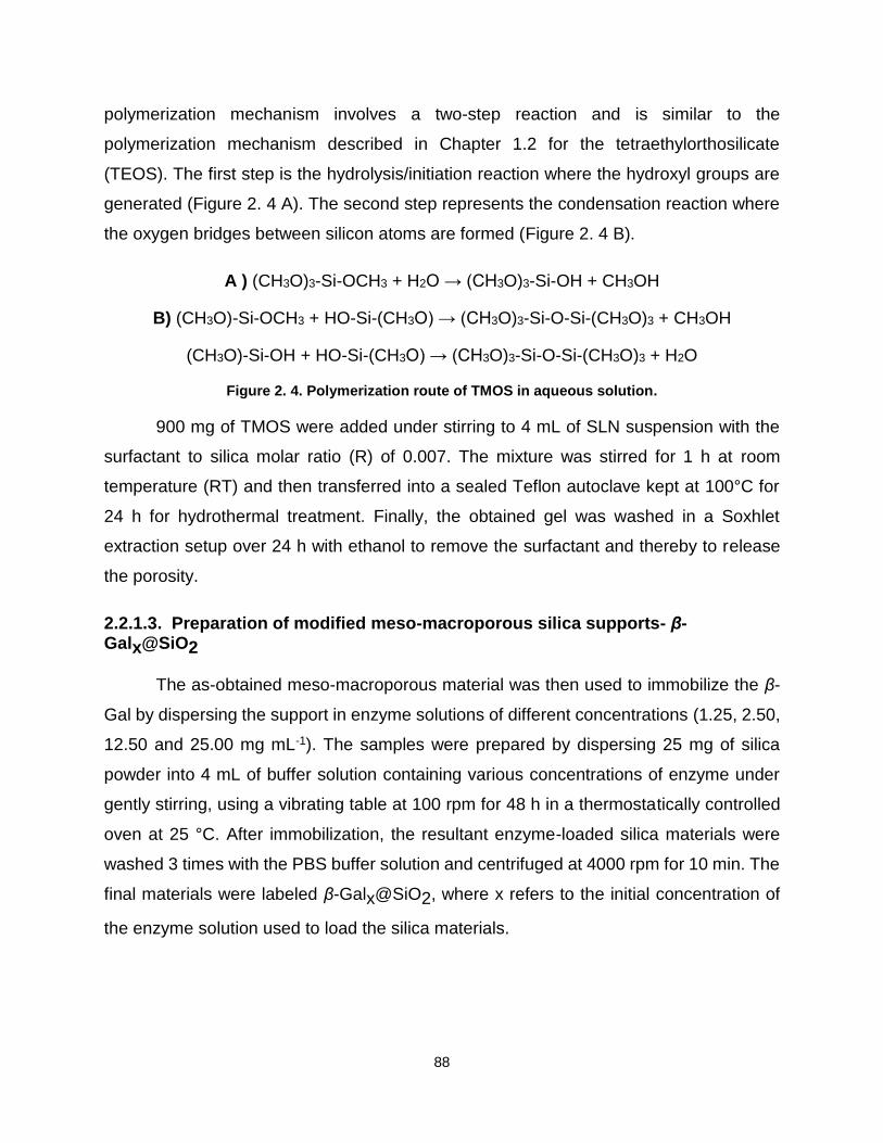
88
polymerization mechanism involves a two-step reaction and is similar to the
polymerization mechanism described in Chapter 1.2 for the tetraethylorthosilicate
(TEOS). The first step is the hydrolysis/initiation reaction where the hydroxyl groups are
generated (Figure 2. 4 A). The second step represents the condensation reaction where
the oxygen bridges between silicon atoms are formed (Figure 2. 4 B).
A ) (CH3O)3-Si-OCH3 + H2O → (CH3O)3-Si-OH + CH3OH
B) (CH3O)-Si-OCH3 + HO-Si-(CH3O) → (CH3O)3-Si-O-Si-(CH3O)3 + CH3OH
(CH3O)-Si-OH + HO-Si-(CH3O) → (CH3O)3-Si-O-Si-(CH3O)3 + H2O
Figure 2. 4. Polymerization route of TMOS in aqueous solution.
900 mg of TMOS were added under stirring to 4 mL of SLN suspension with the
surfactant to silica molar ratio (R) of 0.007. The mixture was stirred for 1 h at room
temperature (RT) and then transferred into a sealed Teflon autoclave kept at 100°C for
24 h for hydrothermal treatment. Finally, the obtained gel was washed in a Soxhlet
extraction setup over 24 h with ethanol to remove the surfactant and thereby to release
the porosity.
2.2.1.3. Preparation of modified meso-macroporous silica supports- β-Galx@SiO2
The as-obtained meso-macroporous material was then used to immobilize the β-
Gal by dispersing the support in enzyme solutions of different concentrations (1.25, 2.50,
12.50 and 25.00 mg mL-1). The samples were prepared by dispersing 25 mg of silica
powder into 4 mL of buffer solution containing various concentrations of enzyme under
gently stirring, using a vibrating table at 100 rpm for 48 h in a thermostatically controlled
oven at 25 °C. After immobilization, the resultant enzyme-loaded silica materials were
washed 3 times with the PBS buffer solution and centrifuged at 4000 rpm for 10 min. The
final materials were labeled β-Galx@SiO2, where x refers to the initial concentration of
the enzyme solution used to load the silica materials.

89
2.2.2. Liposomes coated silica particles (LCSP)
2.2.2.1. Preparation of liposomes
The phospholipid 1,2-dioleoyl-sn-glycero-3-phosphocholine (DOPC) (Figure 2. 5)
was used as pH responsive protective coating (see Chapter 3.2). This lipid has two oleic
acids attached on the phosphatidylcholine head-group. The quaternary ammonium group
has a positive charge and the phosphate group has a negative charge. This type of lipid
is able to form liposomes and is naturally present in the double layer of biological
membranes.
Figure 2. 5. Structure of DOPC.
Phospholipid liposomes were prepared by the lipid film hydration method. In the
experiment, 390 μl of DOPC (Avanti®, 850375C, ≥ 63 mM) were mixed in chloroform with
97μl Texas Red, a fluorescent dye. The chloroform was evaporated under a dry nitrogen
stream to form a thin lipid film. The lipid was then hydrated with 1 ml of TRIS buffer (20
mM TRIS, 150 mM NaCl, pH 7.4) and the solution was sonicated for 30 min using an
ultrasonic sonotrode, operating with a MS-72 titanium tip at 30% of the maximum power.
At the end of the liposomes preparation the solution was centrifuged in order to remove
the titanium particles released by the sonication tip.
2.2.2.2. Modification of porous silica particles (ESP)
A diluted solution of Ha-lactase at a concentration of 5.36 mg protein/mL was
prepared from the enzyme stock solution by adding in 50:50 wt% PBS buffer (pH 6.5) and
glycerol. Then, 4 mL of enzyme solution was added to 250 mg of a specific silica material
KROMASIL® 300-10-SIL (SP), provided from AksoNobel Germany, in order to proceed
the enzyme immobilization as schematized in Figure 2. 6. The mixture was left under
stirring (150 rpm) at room temperature (20°C) over 3 hours. After immobilization, the
resulted enzyme-loaded silica was recovered under centrifugation using a centrifuge filter
(size 0.2 μm) and washed with PBS buffer. Finally, the obtained supported enzyme
catalyst was dried overnight under a fume hood at room temperature.

90
Figure 2. 6. Modification of porous silica particles
2.2.2.3. Preparation of liposomes coated silica particles (LCSP)
The LCSP were prepared as schematized in Figure 2. 7. After the adsorption of
enzyme onto silica, approximately 80 mg of modified silica were dispersed in the
liposomes solution and incubated for 1 hour at room temperature under stirring (150 rpm).
Along incubation time, as described by Mornet e al.[12], the pre-prepared liposomes will
first adhere to mesoporous silica surface, undergo gradual deformation, break and
spread on the particle surface forming a continuous bilayer phospholipid film. A final
centrifugation step was performed to separate the coated silica particles (LCSP) from the
remaining liposomes. After being washed with TRIS buffer 6 times, the formed LCSP
were dispersed in TRIS solution and stored at 4°C.
Figure 2. 7 Preparation of liposomes coated silica particles (LCSP).
2.2.3. Double emulsion type Solid Lipid Nanoparticles (SLN) synthesis
SLNs loaded β-Gal were prepared by W1/O/W2 emulsification melted dispersion
method. The system was composed of shea butter as oil phase, β-galactosidase solution
as the water phase 1 (W1) and a solution of Tween 20 as water phase 2 (W2). A schematic

91
representation of this preparation method for solid lipid nanoparticles preparation is
shown in the Figure 2. 8. 0.600 ml of enzyme solution was added to a mixture of shea
butter/PGPR (1000mg/300mg) under a vortex. To prepare the double emulsion, the
inverse emulsion previously prepared was added into 10 g of aqueous solution of 20 %
Tween 20 (wt%) under vortex. The sample temperature was maintained at 40°C all along
the procedure. The obtained solution was cooled down slowly to 20°C under vortex in
which the crystallization process of shea butter occurred and the colloidal suspension
was stored at 4°C.
Figure 2. 8. Schematic representation of preparation method for solid lipid nanoparticles.
SLNs were prepared without organic solvents, by melt dispersion technique, using
the melted lipid instead of a lipid solution in an organic solvent. The enzyme is exposed
to 40 °C temperature (much lower than the unfolding temperature[13]) for a short period of
time, which reduces the possibility of enzyme degradation. Moreover, the enzyme
solution contains 50% glycerol that enhances the thermal stability of β-Gal [14].
In this formulation of the water-in-oil-in-water double emulsions (W/O/W), PGPR
was used to stabilize the first inverse water-in-oil emulsion (W1/O) and Tween® 20 was
used to stabilize the dispersion of W1/O emulsion in water leading to W1/O/W2 from which
the solid lipid particles suspension is generated. The shea butter was chosen as the oil
phase.

92
The polyglycerol polyricinoleate (PGPR) has a chemical backbone of polyglycerol
grafted with pendant chains of polymerized ricinoleic acid, labeled R in Figure 2. 9.
Ricinoleic acid is an unsaturated omega-9 fatty acid [15]. PGPR is already used in
chocolate and chocolate base products, salad dressing and backed products [16][17][18].
This emulsifier is very lipophilic and weakly soluble in water due to the few ether and
hydroxyl groups are present in the chemical backbone. Thus, PGPR is a water in oil
emulsifier (HLB <1).
Figure 2. 9. Structure of PGRP.
The main component of shea butter are triglycerides (Figure 2. 10). The
triglycerides contained in the shea butter used are derived mainly from stearic acid (36-
50%) and oleic acid (40-50%) extracted from the nuts of African shea tree (Vitellaria
paradoxa)(Appendix 2). It was provided by ieS LABO (France).
.
Figure 2. 10. General chemical structure of a triglyceride.
93
2.2.4. Preparation of hybrid materials
2.2.4.1. Preparation of hybrid alginate silica particles (ASP)
Sodium alginate was dissolved in deionized water with a final concentration of 1%
wt. A 2 mL aliquot of the alginate solution was mixed with 0.735 mL of TMOS under vortex
agitation. This mixture was then added drop wise into a mixture of 0.3 g PGPR and 1.5 g
hexane. After one minute, in which silica polymerization is initiated, by the presence of
alginate molecules[19], 3 mL solution of 0.2 M MgCl2 and 50% enzyme solution was added
drop wise and kept under vortex agitation for 1 minute. To prevent denaturation of the
enzyme in the presence of Ca2+, Mg2+ has been used. The beads, thus formed were left
to crosslink with the divalent cation for a half an hour. After washing with deionized water
and centrifuged on a 0.2 μm filter, the particles were lyophilized overnight.
2.2.4.2. Preparation of alginate core silica shell materials (SAM)
Sodium alginate was dissolved in deionized water with a final concentration of 1%
wt. 2 mL of the alginate solution was added drop wise into a mixture of 0.3 g PGPR and
1.5 g hexane. After one minute, 3mL solution of 0.2 M MgCl2 and 50% enzyme solution
was added dropwise and kept under vortex agitation for 1 minute. After the crosslink of
the alginate with the divalent cation for half an hour, 0.735 mL of TMOS was added and
left to polymerize for another half an hour. After washing with deionized water and
centrifuged on a 0.2 μm filter, the particles were lyophilized overnight.
2.2.4.3. Preparation of alginate particles (AP)
The procedure of alginate particles (AP) was quite similar with the preparation of
SAP, except that after the crosslinking of the alginate, the emulsion was broken using
isopropanol. After washing with deionized water and centrifuged on a 0.2 μm filter, the
particles were lyophilized overnight.

94
2.3. Characterization methods
2.3.1. Protein Quantification Assay
The amount of -galactosidase non-entrapped in the system presented in
Chapters 3 and 4 was quantified with Thermo Scientific™ Pierce™ BCA Protein Assay.
The Thermo Scientific™ Pierce™ BCA Protein Assay is a commercial preparation kit for
the colorimetric detection and quantitation of the amount of protein in a sample. The
method is based on biuret reaction, in which the reduction of copper II (Cu2+) to copper I
(Cu1+) occurs in presence of four specific amino acids contained in the protein backbones
(cysteine, cystine, tryptophan and tyrosine). Consequently, the chelation takes place
between two molecules of bicinchoninic acid (BCA) and the Cu1+ previously formed
leading to a purple-colored product absorbing visible light at λmax=562nm (Figure 2. 11)
[20]. The absorbance of the water-soluble complex is linear with increasing protein
concentrations in a wide range of concentrations from 0 to 2000 µg/mL. The color
obtained is not only resulting from the presence of functional groups is the solution [21].
Figure 2. 11. Scheme of the chemical reaction of Cu+ with two BCA molecules.
Usually, in the case of this method, the calibration curves are determined and
reported for bovine serum albumin (BSA) as a standard reference. Thus, a series of
dilutions of known concentration of BSA were prepared from the stock protein (2 mg/mL)
in different media (water, simulated gastric fluid (SGF) and simulated intestinal fluid
(SIF)). The slope and the intercept values of the calibration curves obtained in the three
media are rather close meaning that the simulated body fluids do not change the
complexation route of BCA with Cu+, as presented in the Figure 2. 12.

95
AH2O=0.00345[BSA]+0.14386
ASIF=0.003420[BSA]+0.14533
ASGF=0.00365[BSA]+0.14237
Figure 2. 12. The calibration curves obtained for chelation of BSA with Cu+ in water, SGF and SIF.
2.3.2. Detection of enzyme and material activity
Following a typical procedure, the ONPG solution was prepared in PBS buffer,
prepared two hours prior to use, at a concentration of 25 mg/L. The substrate was left to
react with the enzyme for 10 minutes. The reaction was then stopped by adding a solution
containing 50mM Na2CO3 and 7 mM EDTA. The pH shifts towards more basics values
(pH 8.3), at which the ortho-nitrophenol is entirely in its base conjugated form, the ortho-
nitrophenolate (Figure 2. 13 B, ε= 4.6 mM-1 cm-1 at max=420 nm).
0 50 100 150 200 250
0.2
0.4
0.6
0.8
1.0
1.2
H2O
SGF
SIF
Abso
rba
nce
[BSA] g/ml

96
Figure 2. 13. A) Hydrolysis reaction of ONPG into galactose and ortho-nitrophenol, B) chemical shift between ortonitrophenol and ortonitrophenolat with the change of pH.
For both free and immobilized/encapsulated enzyme on silica materials the activity
was defined in international units (IU), 1 IU corresponding to the amount of enzyme
catalyzing the conversion of 1 mol of substrate per minute at 25°C in Chapter 3.1 and at
30°C in Chapter 3.2 and 4.2.
In Chapter 3.1, in the case of the free enzyme, the reaction was carried out by
mixing 0.5 mL of 40 mM ONPG and 0.5 mL of enzyme solution at 25°C and run over 10
min. Adding 0.5 mL of 500 mM Na2CO3 solution stopped the reaction. The absorbance of
ONP was measured at 420 nm. For the activity of the immobilized β-Gal on meso-
macroporous silica materials, 1 mg of each powder of loaded silica materials was
transferred into a tube containing 0.5 mL of buffer solution, to which 0.5 mL of 40 mM of
ONPG solution was subsequently added. The reaction was stopped after 10 min by
adding 0.5 mL of 500 mM Na2CO3 solution. To remove the silica material, samples were
centrifuged (4000 rpm during 30 sec) and the absorbance value of the ONP-containing
supernatant was measured at 420 nm. For both free and immobilized/encapsulated
enzyme on silica materials the activity was defined in international units (IU), 1 IU
corresponding to the amount of enzyme catalyzing the conversion of 1 mol of substrate
per minute at 25°C.
pH<7.23
pH>7.23
A
B
ortho-nitrophenyl-ß-galactoside (ONPG) galactose ortho-nitrophenol
colorless
yellow
ortho-nitrophenol ortho-nitrophenolate
Lactase

97
In Chapter 3.2, the assay for the free enzyme activity was performed by mixing 2.5
ml of o-nitrophenyl-β- d-glactopyranoside (ONPG, 25 mg/L) and 0.5 ml of diluted enzyme
solution (diluted 12000 times from the initial stock). The mixture was incubated at 30°C
for 10 minutes before being stopped by adding 1 mL of a solution of Na2CO3 (50 mM). All
experiments were performed in triplicate. For the activity of the immobilized β-Gal on silica
particles (SP) and liposomes coated silica particles (LCSP), approximately 8 mg of SP
and 100 μl of LCSP suspension was transferred into a tube containing 1.6 mL of buffer
solution (PBS), from which 0.125ml was analyzed. To this solution, 0.5 mL of ONPG
solution was subsequently added. The reaction was stopped after 10 min by adding 0.25
mL Na2CO3 solution. To remove the silica material, samples were centrifuged (13500 rpm
for 120 sec). The activity of the SP was determined at 22.4 U/mg of silica material and
the activity of LCSP as 10.9 U/mg of silica. For both free and immobilized/encapsulated
enzyme on silica materials the activity was defined in units (U), 1 U corresponding to the
amount of enzyme catalyzing the conversion of 1 mol of substrate per minute at 30°C.
In Chapter 4.2, the same protocol was followed as in Chapter 3.1, with the
exception that approximately 10 mg of materials were dispersed in 2 mL of buffer solution.
The dilutions and the protocol remained the same.
2.3.3. Enzyme release in simulated gastro-intestinal fluid
A controlled release of the enzyme out of materials was done in vitro in simulated
gastric fluid (SGF) and in simulated intestinal fluid (SIF). The materials were analyzed two
by two, uncoated SP carrier was compared with the lipid covered silica particle (LCSP),
while hybrid alginate silica particles (ASP) was compared with alginate core silica shell
materials (SAM). In order to simulate the passage of the materials through the gastro
intestinal tract four samples of each material were tested as presented in Table 2. 2. Each
type of sample underwent four experiments under magnetic stirring at 150 rpm. In the first
one the sample (LCSP1 or SP1/ ASP1 or SAM1) was kept for 1H in SGF, the second
one, the carrier (LCSP2 or SP2/ ASP2 or SAM2) was kept for 2H in SGF. The third and
the four experiments are a successive immersion of the sample in SGF over 2H followed
by 1H or 2H in the SIF. In the case of LCSP and SP, the samples from the experiments

98
3 and 4 have been centrifuged before being added to the SIF (Table 2. 2). In the case of
ASP and SAM, all samples were separated from the solutions by centrifugation after each
hour and introduced into a new solution.
Table 2. 2. Incubation time in simulated gastro-intestinal fluids (SGF and SIF).
Experiment Samples Incubation treatment
(time and media)
1 LCSP1 and SP1/ ASP1
and SAM1 1H in SGF
2 LCSP2 and SP2/ ASP2
and SAM2 2H in SGF
3 LCSP3 and SP3/ ASP3
and SAM3 2H in SGF followed by 1H
in SIF
4 LCSP4 and SP4/ ASP4
and SAM4 2H in SGF followed by 2H
in SIF LCSP: liposomes coated enzyme-silica particles, SP: enzyme adsorbed on silica particles, ASP: hybrid alginate silica particles and SAM: alginate core silica shell materials (SAM).
All the solutions were analyzed to quantify the enzyme released and its activity.
2.3.4. Dynamic light scattering
Particle sizing distribution and their hydrodynamic radius (RH) were obtained by
dynamic light scattering (DLS) using a Malvern 3000HSA Zetasizer instrument equipped
with a He-Ne laser (633 nm, 5 mW). Prior to measurement, the dispersions of SLN (used
for the preparation of meso-macroporos materials in Chapeter 3.1), were diluted with
Millipore water until the count rate intensity was above 500 kcps. The samples were
placed in disposable polystyrene cells, and the experiments were performed at constant
temperature (25°C).
2.3.5. SAXS measurements
Small angle X-ray scattering (SAXS) measurements were carried out using a
SAXSess mc2 (Anton Paar) apparatus with slit collimation. It is coupled with an ID 3003
laboratory X-Ray generator (general electric), equipped with a sealed X-ray tube
(PANalytical, CuKα radiation λ= 0.1542 nm) operating at 40 kV and 50 mA. A multilayer
mirror and a block collimator provide a monochromatic primary beam. A translucent beam
stop allows the measurement of an attenuated primary beam at q=0. Meso-macroporous
materials (Chapter 3.1) were introduced into a powder cell (sandwiched in between two

99
Kapton foils), whereas aqueous dispersions and shea butter ( Chapter 4.1) are placed in
quartz capillaries of 1.5 mm of diameter. Samples are placed inside an evacuation
chamber at 309 mm from the sample holder. Acquisition times are typically in the range
of 30 minutes. The scattered X-ray beam is recorded by a CCD detector (Princeton
Instruments, 2084×2084 pixels array with 24×24 μm2 pixel size) in the q range from 0.09
to 5 nm-1, and treated the with SAXSquant software for smearing signal correction. All
data was corrected for background scattering from the respective empty cells. For the
lipid dispersions, the scattering data were corrected by the water filled capillary.
2.3.6. Nitrogen sorption analysis
The pore size and the texture parameters of the bare and enzyme-loaded silica
materials (Chapter 3) were determined by nitrogen sorption isotherms at 77 K using a
Micromeritics Tristar device. For that purpose, all materials were degassed under vacuum
over for 24 h at 20°C to remove water and CO2 physically adsorbed at the surface of the
samples. The specific surface area (SBET) was determined by applying the
Brunauer−Emmett−Teller (BET) theory whereas pore volume and average pore size were
obtained using the Barrett−Joyner−Halenda method (BJH) applied to the desorption
branch of the isotherms.
2.3.7. Microscopy
Morphology and porosity of the bare meso-macroporous silica material were
observed by transmission electronic microscopy (TEM). The powder was first ground and
then suspended in ethanol by sonication. A drop of the dispersion was spread out on the
TEM carbon lacey grid and dried at room temperature before observation (Chapter 3.1)
Particle size of simple and double emulsions was determined by optical
microscopy, using an Olympus BX51 equipped with a Toupca camera, with a TouView
software (Chapter 4.1). Droplet size, size polydispersity and morphology of prepared
Pickering emulsions (Chapter 5) were evaluated by optical microscopy (Olympus BX51
microscope). The observation of the samples was carried in bright field mode.
100
Morphology of the bare silica material (Kromasil®)(Chapter 3.2) and hybrid
alginate-silica (Chapter 4.2) were observed by scanning electronic microscopy (SEM) at
an accelerating voltage of 2.0 kV and working distance of about 10 mm. The powder was
spread out on the SEM carbon patch and dried at room temperature before observation.
The fluorescence microscopy measurements presented in Chapter 3.2 were
performed at the Institute for Physical Chemistry, Heidelberg, Germany, using an Axio
Observer inverted microscope (Carl Zeiss AG), equipped with a PlanNeofluoar
63x/1.25/PH3 antiflex oil-immersion objective with a built-in lambda-quarter plate, a filter
cube with the fitting filter set for Texas Red or FITC (ʎexcitation = 595 nm; emission 615
nm). The light source consisted of a high-pressure metal halide lamp HXP 120V. The
images were recorded with an Orca ER CCD camera (Hamamatsu Photonics) with an
adjusted exposure time of 50 to 300 milliseconds.
2.3.8. Fourier Transform Infrared spectroscopy – FTIR
Attenuated total reflectance Fourier transformed infrared (ATR- FTIR) spectra
were collected using a IRaffinity-1 spectrometer (Shimadzu) coupled with a PIKE
Technologies GladiATR accessory with a diamond crystal. The software for collecting and
viewing spectra was the LabSolutions IR (Shimadzu). The absorbance spectrum of each
sample has been obtained by accumulating 32 scans at 4.0 cm-1 of resolution. The
background correction was made with the spectrum signal of the clean ATR crystal
exposed to the ambient atmosphere. The bare and enzyme-loaded meso-macroporous
silica materials in Chapter 3.1 and the hybrid silica materials in Chapter 4.2 were analyzed
by FTIR.
2.3.9. Thermogravimetric analysis (TGA)
The enzyme adsorption efficiency was quantified by thermogravimetric analysis
(TGA) using a Netzsch STA 449F1 thermobalance. Approximately 10 to 15 mg of each
sample were heated up to 800 °C at 5 °C min-1 under air for the decomposition of
organics. The results were used in the work presented in Chapter 3.

101
2.3.10. Zeta potential measurements
Zeta potential was measured using a Malvern Zetasizer 3000 HS instrument,
based on 0.5 mg·mL-1 suspension of bare or enzyme-loaded meso-macroporous silica
dispersed by sonication in a water bath in aqueous solution at pH ranging from 2.5 to 9.
The bare and modified silica materials presented in Chapter 3 were analyzed by Zeta
potential.
2.3.11. DSC
The calorimetric measurements (DSC) (work done at Institute for Physical
Chemistry, Heidelberg, Germany) were performed using a VP-DSC calorimeter
(MicroCal, Inc., Northampton, MA, U.S.A.) with a scan speed of 90°C/hour, in the
temperature range 10-50°C.

102
Bibliography
[1] A. J. Engelen, P. H. Randsdorp, J. AOAC Int. 82, 112–8. [2] M. Minekus, M. Alminger, P. Alvito, S. Ballance, T. Bohn, C. Bourlieu, F. Carrière, R. Boutrou, M. Corredig, D.
Dupont, et al., Food Funct. 2014, 5, 1113–24. [3] S. Kim, M.-J. Stébé, J.-L. Blin, A. Pasc, J. Mater. Chem. B 2014, 2, 7910–7917. [4] I. Zafeiri, J. E. Norton, P. Smith, I. T. Norton, F. Spyropoulos, J. Colloid Interface Sci. 2017, 500, 228–240. [5] S. D. Singh-Joy, V. C. McLain, Int. J. Toxicol. 2008, 27 Suppl 2, 93–128. [6] L.-Y. Zhao, W.-M. Zhang, J. Drug Target. 2017, 25, 471–484. [7] J.-L. Blin, J. Jacoby, S. Kim, M.-J. Stébé, N. Canilho, A. Pasc, Chem. Commun. (Camb). 2014, 50, 11871–4. [8] S. Kim, R. Diab, O. Joubert, N. Canilho, A. Pasc, Colloids Surf. B. Biointerfaces 2015, 140, 161–168. [9] A. Pasc, J.-L. Blin, M.-J. Stébé, J. Ghanbaja, RSC Adv. 2011, 1, 1204. [10] A. E. . Palmqvist, Curr. Opin. Colloid Interface Sci. 2003, 8, 145–155. [11] J. Y. Ying, C. P. Mehnert, M. S. Wong, Angew. Chemie Int. Ed. 1999, 38, 56–77. [12] S. Mornet, O. Lambert, E. Duguet, A. Brisson, Nano Lett. 2005, 5, 281–285. [13] M. Sutter, S. Oliveira, N. N. Sanders, B. Lucas, A. Van Hoek, M. A. Hink, A. J. W. G. Visser, S. C. De Smedt,
W. E. Hennink, W. Jiskoot, J. Fluoresc. 2007, 17, 181–192. [14] V. Athès, D. Combes, Enzyme Microb. Technol. 1998, 22, 532–537. [15] J. Kiefer, K. Frank, F. M. Zehentbauer, H. P. Schuchmann, Biosensors 2016, 6, 1–11. [16] E. O. Afoakwa, Chocolate Science and Technology, 2010. [17] Nordisk Ministerrad, Food Additives in Europe 2000 : Status of Safety Assessments of Food Additives
Presently Permitted in the EU, Nordic Council Of Ministers, 2002. [18] N. T. Dunford, Food and Industrial Bioproducts and Bioprocessing, Wiley-Blackwell, 2012. [19] S. Xu, Z. Jiang, Y. Lu, H. Wu, W. Yuan, 2006, 511–517. [20] P. K. Smith, R. I. Krohn, G. T. Hermanson, A. K. Mallia, F. H. Gartner, M. D. Provenzano, E. K. Fujimoto, N.
M. Goeke, B. J. Olson, D. C. Klenk, Anal. Biochem. 1985, 150, 76–85. [21] K. J. Wiechelman, R. D. Braun, J. D. Fitzpatrick, Anal. Biochem. 1988, 175, 231–237.

103
Chapter 3. Physisorption on silica
The use of enzymes as biocatalysts in industrial applications, such as food[1],
energy-biodiesel [2] or pharmaceutical synthesis [3] is increasing due to their high catalytic
activity and selectivity. However, enzymes have a poor reusability and a low operational
stability because of their sensitivity to pH and temperature. The immobilization of
enzymes is one of the most promising methods to maintain enzyme performance and
stability with the possibility to recover and reuse the catalyst. Supported enzymatic
catalyst is also interesting to use in particular reaction conditions when enzyme is
operating in organic solvent, for instance when a transesterification are required [4–6].
Different immobilization strategies have been developed to prepare supported enzyme
catalysts: entrapment, microencapsulation and cross-linked enzyme crystals (CLEC) or
aggregates (CLEA) [7]. These methods are based on physical adsorption, covalent
attachment and affinity of the enzyme to the support.
Depending on the immobilization method, the chemical and physical properties of
the enzyme can be altered. For example, through a covalent bonding between the
enzyme and the support, the active conformation of an protein can be strongly modified
and induce a decrease in the enzymatic activity [8]. However, the physical adsorption of
the enzyme generally occurs through weak forces such as hydrophobic interaction,
hydrogen bonds, electrostatic, van der Waals forces or ionic interactions that do not
change dramatically the native conformation and the activity of the enzyme [9,10].
The materials employed for enzyme adsorption can be organic or inorganic but not
all the enzymes can be easily immobilized on them. Investigating suitable solid supports
for enzyme immobilization is still a current scientific challenge, depending on each
specific enzyme and for each industrial application. The criteria to choose a suitable
carrier for a given enzyme and its application include: stability (or reactivity), cost,
availability and the type of the reactor in which it will be used. The surface area, the
particle size, the pore size and the structure, the type of functional groups at the surface
of the pores are the physico-chemical parameters that should also be considered in the
choice of the support.

104
The most common organic carriers are synthetic or natural polymers. Natural
supports used in enzyme immobilization are either pure calcium alginate [11] or in mixture
with gelatin and transglutaminase [12]. Cellulose [4,13,14] and chitosan in hydrogel form [15–
18], in microcrystalline form [19], or agarose gel are commonly used as supports [20].
Synthetic polymers such as poly(vinyl alcohol) [21][22], cross-linked poly(vinyl alcohol) [23],
poly(N-methylolacrylamide) [24], polypropylene [25–27], poly(acrylic acid-co-
acrylamide)/hydrotalcite nanocomposite hydrogels [28], poly(hydroxybutyrate)
nanoparticles [29] and beads [30], poly(o-toluidine) [31] and poly(acrylonitrile) [32] are also
used for enzyme immobilization.
Concerning the inorganic supports, literature reports for the immobilization of
enzyme the use of metals and also many oxides such as alumina gel [11], aluminum [33] or
aluminosilicates [34] (class of compounds made of aluminum, silicon and oxygen), titania
sol-gel [35], gold [36,37], cordierite, mullite [38], halloysite [39], mica [40] and hydroxyapatite [41]
as powder or as ceramic [42], bentonite [43] or mesoporous activated carbon [44] of different
pore sizes.
Compared with the organic resin supports, the inorganic materials like amorphous
silica present convenient properties for protein immobilization such as: high surface area,
thermal stability, good mechanical properties, low swelling in organic solvents while
withstanding high flow rates in continuous reactors. The silica materials are nontoxic,
microbial resistant and they also exhibit a high biocompatibility, biodegradability. For this
reasons amorphous silicon dioxide have been intensively studied as carriers [45–47]. Many
amorphous porous silica materials with different morphological parameters have been
extensively synthesized for its adsorption properties. The mesoporous silica SBA-15
(Santa Barbara Amorphous) presenting a hexagonal array of pores (5 to 30 nm size
diameter), a large volume of meso- and microporosity (~ 1.0 cm3·g-1, ~ 0.8 cm3·g-1,
respectivly) and a high surface area from 500 to 1400 m2·g-1 is an excellent support used
for enzyme immobilization [48].
However, some other mesoporous silicas used for enzyme immobilization have
been documented in literature: the mesoporous MCM-41 (Mobil Composition of Matter, 2

105
to 3 nm pore size) [49], the large to ultra large pore size MSU-H (Michigan State University,
7.6-11.9 nm pore size) [50], the FDU-12 (Fudan University Material, 8.9 nm pore size) [51],
the large-pore mesoporous silica nanoparticles with a cubic Ia3d structure of pores (KIT-
6, 8 nm pore size) [52], different size of mesoporous silica particles [53], the folded sheet
mesoporous silica [54], silicas with small surface areas [55–57] or silica gels (particle size
0.040-0.063 nm) [58]. Finally, vesicular silica [59,60] and fumed silica [61], that have well-
developed surface areas, small particles and high mechanical strength have also been
investigated for the enzyme immobilization.
The affinity between the enzyme and the support is a key factor of getting an
efficient enzymatic catalyst. Thus, the surface of the support is commonly modified with
chemical groups that can physically interact with specific chemical groups bear by the
enzyme. Usually, the grafting groups are chosen with at least two reactive groups, one
that can chemically anchor on the support and the other that physically interact with the
enzyme. This is the case of glutaraldehyde (CH2(CH2CHO)2) that contains two reactive
aldehyde groups [36,38,62], one that can connect to the -OH groups of the support and the
other with a -NH2 groups of the enzyme. In fact, glutaraldehyde is the most common linker
used for its bifunctional carbonyl groups that have high affinity to bacteria, fungi and
protein.
According to the support and the enzyme immobilized, the linker can be adapted.
For instance, silanes are used to modify silica materials. The most frequently used silanes
are 3-amino-propyltrimethoxysilane [59,63], 3-aminopropyltriethoxy-silane [52,64,65],
mercaptopropyl-trimethoxysilane or mercaptopropyl-triethoxysilane [37], n-
octyltriethoxysilane [57], phenyltrimethoxy-silane, vinyltri-methoxysilane [51] and [3-
(trimethoxy-silyl)propyl] octadecyl dimethyl ammonium chloride [66]. They can also be
grafted on gold particles surface [37].
Polymers can be synthetized through specific monomer(s) and at different chain
lengths, and besides being a class of support, they can also be used as linkers. Polymers
as polyethyleneimine [67] polystyrene [19] or poly(styrene sulfonate) [68], acrylonitrile
copolymers [69] or second generation polycationic dendronized polymer (de-PG2) [56] can
106
be employed to improve the affinity of the enzyme for the chosen support. Moreover, in
some complex elaborated catalysts, enzyme–support affinity is increased by coating silica
by conducting polymers as polypyrrole [70,71] or polyamidoamine dendrimers [72].
The linkers with acid-base properties like amines are also interesting to modify the
support surface because hydrogen bonding is favored with enzyme. For example, diethyl-
amine [73,74], diethylaminoethyl (DEAE) [75] or mono-aminomethyl-N-amino-ethyl [20] are
the most commonly used amino linkers. Moreover, long chain carboxylic acid as erucic
acid [76] or short chain carboxylic acid as itaconic acid that contain two carboxyl groups
and an additional reactive carbonyl group [17] are also used as modifiers.
Oxygen plasma [4] and plasma polymerization of allyl-alcohol, allyl-amine and
acrylic acid were also proposed [14] as a new approach for support functionalization.
However, they present the disadvantage of having a high cost.

107
3.1. Preferential adsorption of β-galactosidase regarding Hierarchical Meso-Macro porosity of a Silica Material 2
Since their discovery in 1992 [77], silica mesoporous materials have been widely
used in enzyme immobilization due to their tunable pore size, volume and their large
specific surface area. These materials can entrap a large amount of enzymes, and the
immobilization can be done either by chemisorption or physisorption. The pores are a
favorable environment for enhancing the thermal and pH stability of the enzymes as well
as their resistance to high salt concentrations. [45,78,79] However, the confinement of the
enzyme in pores [45] and/or in a small pore size of the material, or on non-open-pore
structures [79] can lead to a decrease of the enzymatic activity and might exhibit significant
resistance to the substrate diffusion. By increasing the pore size (e.g. from meso- to
macropore), one can expect an increase of the diffusion rates of substrates to the active
sites of the enzyme and a larger enzyme mobility/flexibility within the cavities, resulting
thus in a better enzyme activity [80]. Indeed, macroporous silica materials have a high
mass transfer rate due to the interconnection of their broad pores, in addition to a good
mechanical and thermal stability. However, even if the enzyme can be easily immobilized
on this kind of materials, it can be also leached out easily, particularly when the pH of the
media varies. To overcome this problem, the enzyme can be retained inside the material
by crosslinking or by aggregation [81].
Hierarchical porous materials combine the properties of mesopores, such as high
surface area and controllable pore size/volume, with those of macropores, providing high
diffusion and throughput rates [82]. Although they are widely used in chemical catalysis [83],
only a few examples have been reported in the literature for enzyme encapsulation. For
instance, Cao et al. [84] used hierarchical silica spheres to encapsulate glucose oxidase
by physisorption. Meso-macroporous silica materials prepared by polycondensation of
sodium silicate were used to physically
[80] and chemically adsorb β-galactosidase [85,86]
and lipases [87–89]. Also, lipase was entrapped in solid lipid nanoparticles (SLN; W/O/W
2 This subchapter is based on the article: I.-A. Pavel, S. F. Prazeres, G. Montalvo, C. Garcıa Ruiz, V. Nicolas, A. Celzard, D. Francois, L. Canabady-Rochelle, N. Canilho, A. Pasc (2017). “Effect of Meso vs Macro Size of Hierarchical Porous Silica on the Adsorption and Activity of Immobilized β-Galactosidase” Langmuir, 33(13), 3333–3340. https://doi.org/10.1021/acs.langmuir.7b00134 [129]

108
type) covered by a meso-macroporous silica shell [90] or covalently attached to a silica
foam [91]. The fish-in-net technique was used to entrap various enzymes (umarase,
trypsin, lipase, and porcine liver esterase) inside the macroporous cages while the
mesopores provided a path for the diffusion of reactants [92]. Using this technique, β-
galactosidase and lysozyme were co-immobilized to prevent bacterial contamination of
the silica matrix in the industrial production of low lactose milk dairy [93]. Macro-
mesoporous silica spheres prepared with a micro-device were used as a support for
penicillin G acylase that was covalently attached through grafted aminopropyl and
glutaraldehyde chemical groups [94]. Recently, catalase was used to prove the efficiency
of hierarchical macro/mesoporous amino-grafted silica spheres as enzyme carriers [95].
In the present study, β-galactosidase (β-Gal) from Kluyveromices lactis was
immobilized into hierarchical meso-macroporous silica by physical adsorption. The
enzyme adsorption pathway was investigated as a function of pore size and related to the
specific activity measured for the loaded silica material.
3.1.2. Morphology and texture of bare and enzyme-loaded silica supports
The meso-macroporous silica supports were obtained through a dual templating
mechanism, as detailed in Chapter 2. But briefly, the material preparation combined Solid
Lipid Nanoparticles (SLN) and micelles formed by a Pluronic® block-copolymer surfactant
used as templates for macropores and mesopores, respectively. The solid lipid
nanoparticles were of approximately 200 nm in diameter as shown by the size distribution
graph determined by dynamic light scattering (DLS) and presented on Figure 3. 1. The
morphology of the meso-macroporous silica was analyzed by transmission electron
microscopy (TEM).

109
Figure 3. 1. Size distribution of SLN obtained from DLS.
TEM micrographs (Figure 3. 2) made on the silica support clearly show a dual
meso-macroporosity where the mesoporosity induced by the non-ionic surfactant is
imprinted in the walls of the SLN-templated macropores. This results are in agreement
with our lab previous publications [90,96–98].
Figure 3. 2. TEM pictures of bare silica material showing the mesopores network interconnecting with macroporous walls.
100 150 200 250 300 350
Inte
nsity (
a.u
)
Diameter (nm)

110
In agreement with TEM micrographs, the SAXS pattern of the bare SiO2 material
presented in Figure 3. 3 confirmed the worm-like arrangement of mesopores with an
average periodic Bragg distance (dBragg) of 12.4 nm. Similar pore structure where previous
obtained in our lab [90,97]. As for the enzyme-loaded silica, the intensity of the diffusion
peaks decreased with the increase of the concentration of the feed solution from 1.25 to
12.50 mg·mL-1. As a matter of fact, when the mesoporosity is filled with organic molecules
the scattering contrast consecutively decreases. This progressive extinction of the Bragg
peak stopped defining the sample prepared at an enzyme concentration of 25.00 mg mL-
1, meaning that the corresponding material contained less β-Gal.
Figure 3. 3. SAXS patterns of the bare and enzyme-loaded meso-macroporous silica support
materials.
Nitrogen sorption measurements were performed on bare and enzyme-loaded
silica materials as shown in Figure 3. 4. Bare silica exhibited a type IV isotherm,
characteristic of a mesoporous material (Appendix 1). However, at high relative pressure
(p/p0 around 0.9), a steep increase of the values of adsorbed volume was observed,
suggesting the presence of macropores and/or interparticular spaces. The pore size
distribution obtained by the BJH method applied to the adsorption branch of the isotherm
evidenced that the average mesopore size (Ø) was 9 nm for the bare silica. Moreover,
0.2 0.4 0.6 0.8 1.0 1.2 1.4
1E-4
1E-3
0.01
0.1
1
10
10.8 nm
11.2 nm
11.9 nm
I (q)
q, nm-1
SiO2
-Gal25.00
@SiO2
-Gal12.50
@SiO2
-Gal2.50
@SiO2
-Gal1,25
@SiO2
12.4 nm
11.6 nm

111
the specific surface area (SBET) and the pore volume (Vp) of the meso-macroporous bare
material (SiO2) were around 660 m2.g-1 and 1.23 cm3.g-1, respectively (see Table 3. 1).
Figure 3. 4. N2 adsorption-desorption isotherms and corresponding pore size distributions.
As expected, sorption data revealed that the values of SBET, Vp and pore diameter
of the β-Gal-loaded silica materials dramatically decreased with respect to bare silica.
Upon increasing the initial concentration of the feed solution from 1.25 to 12.50 mg.mL-1,
the specific surface area still decreased from 166 m2.g-1 (for -Gal1.25 @SiO2) to a
threshold value of 85 m2.g-1 on average (for -Gal2.50 @SiO2 and -Gal12.50 @SiO2).
Likewise, for the same range of concentration, the pore volume values dropped by half
from 0.26 cm3.g-1 to a minimum average of 0.14 cm3.g-1 while the pore diameter only
slightly decreased, from 6.4 to 5.6 nm. This evolution of the texture parameters indicates
that, in the dilute regime, the enzyme uptake of the mesopores increased with the
concentration of the feed solution. However, the material prepared with the most
concentrated solution of 25.00 mg.mL-1, β-Gal25.00@SiO2, presented significantly higher
pore texture parameters with SBET, Vp, and Ø of 239 cm2.g-1, 0.52 cm3.g-1 and 6.8 nm,
respectively. Those values remained lower than the ones of the bare silica, indicating that
the enzyme was still physisorbed in the mesopores, but less than in the materials
prepared with lower concentrations of enzyme.
0.2 0.4 0.6 0.8 1.00
500
1000
A
dso
rbe
d v
olu
me
(cm
3/g
.ST
P)
Relative pressure (p/p0)
SiO2
-Gal25.00
@SiO2
-Gal12.50
@SiO2
-Gal2.50
@SiO2
-Gal1.25
@SiO2
10 200.0
0.1
0.2
5.6 to 6.4 nm
6.8 nm
dV
/dp
(cm
3/g
.nm
-1)
Pore diameter (nm)
9 nm

112
A quantitative estimation of the variation of the silica wall thickness (ε) was made,
by subtracting the pore diameters of each sample from the dBragg distances (see Table 3.
1). A net thickening of the material wall (from 3.4 to 5.9 nm) was observed as soon as the
bare material was loaded with a diluted feed solution, 1.25 mg.mL-1 to 12.50 mg.mL-1.
Then, just like for the other texture parameters, ε also decreased to 4.4 nm in the case of
β-Gal25.00@SiO2 sample, indicating again a lower uptake of the enzyme in the mesopores
when using higher enzyme feed solutions.
Table 3. 1. Parameters of bare and β-Gal-loaded meso-macroporous silica supports obtained from SAXS, nitrogen adsorption and thermogravimetric analysis. aBragg distance determined by SAXS, bSBET: specific surface area calculated from BET theory, cVp : pore volume, dØ : pore diameter, εe
wall thickness (ε = dBragg - Ø), fmass ratio, g Infrared band area ratio, gAamide:ASiO2 : ratio of ATR peak areas.
SAXS N2 adsorption TGA ATR
dBragga
(nm)
SBETb
(m2 g-1)
Vpc
(cm3 g-1) Ød (nm)
εe
(nm)
mβ-Gal: mSiO2f
(loading, wt%)
Aamide:ASiO2g
SiO2 12.4 657 1.23 9 3.4 0 0
-Gal1.25
@SiO2 10.8 166 0.26 6.4 5.1 1.17 (54) 0.11
-Gal2.50
@SiO2 11.2 80 0.12 5.6 5.6 1.45 (59) 0.15
-Gal12.50
@SiO2 11.6 90 0.17 5.7 5.9 1.55 (61) 0.13
-Gal25.00
@SiO2 11.9 239 0.52 6.8 4.4 0.84 (45) 0.05
N2 adsorption measurements can only provide information on the enzyme
presence in the mesopores. To get more information on the total enzyme loading in both
mesopores and macropores, thermogravimetric analysis was performed (Figure 3. 5).
The respective enzyme loading for each sample is presented in Table 3. 1. It should be
noted that the values of the -Gal loading into those meso-macroporous silica materials
are rather high compared to the one in organic resins [99] or hybrid materials [100]. The
evolution of the loading values was in line with the trends previously observed by nitrogen
sorption analysis and SAXS. Indeed, the -Gal loading increased progressively in our
case from 54 to 61 wt.% when the concentration of the enzyme solution increased from
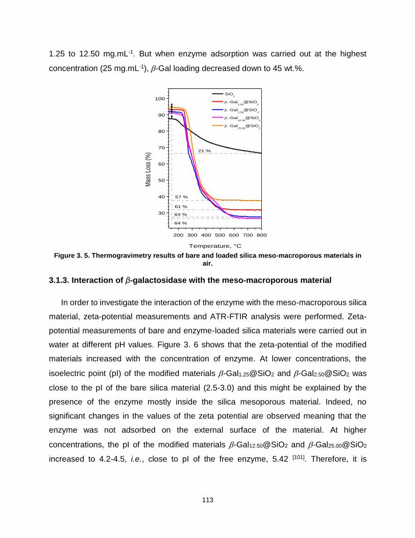
113
1.25 to 12.50 mg.mL-1. But when enzyme adsorption was carried out at the highest
concentration (25 mg.mL-1), -Gal loading decreased down to 45 wt.%.
Figure 3. 5. Thermogravimetry results of bare and loaded silica meso-macroporous materials in
air.
3.1.3. Interaction of β-galactosidase with the meso-macroporous material
In order to investigate the interaction of the enzyme with the meso-macroporous silica
material, zeta-potential measurements and ATR-FTIR analysis were performed. Zeta-
potential measurements of bare and enzyme-loaded silica materials were carried out in
water at different pH values. Figure 3. 6 shows that the zeta-potential of the modified
materials increased with the concentration of enzyme. At lower concentrations, the
isoelectric point (pI) of the modified materials -Gal1.25@SiO2 and -Gal2.50@SiO2 was
close to the pI of the bare silica material (2.5-3.0) and this might be explained by the
presence of the enzyme mostly inside the silica mesoporous material. Indeed, no
significant changes in the values of the zeta potential are observed meaning that the
enzyme was not adsorbed on the external surface of the material. At higher
concentrations, the pI of the modified materials -Gal12.50@SiO2 and -Gal25.00@SiO2
increased to 4.2-4.5, i.e., close to pI of the free enzyme, 5.42 [101]. Therefore, it is
200 300 400 500 600 700 800
30
40
50
60
70
80
90
100
64 %
61 %
64 %
57 %
Mas
s Lo
ss (%
)
Temperature, °C
SiO2
Gal1.25
@SiO2
Gal2.50
@SiO2
Gal12.50
@SiO2
Gal25.00
@SiO2
21 %

114
reasonable to assume that the enzyme progressively filled the mesopores and then the
macropores of the silica material.
Figure 3. 6. Zeta-potential measurements for the bare and the enzyme-loaded meso-macroporous
silica.
On another hand, infrared experiments were carried out and the spectra are
presented in
Figure 3. 7, without any correction. ATR-FTIR spectra of the bare material shows
the typical bands of silica at 1065, 960 and 800 cm-1, corresponding to Si-O-Si and Si-
OH stretching vibrations [102]. After enzyme immobilization, the spectra exhibited a slight
displacement of these bands (from 1065 to 1053 cm-1 and from 960 to 956 cm-1,
respectively), which can be attributed to the interactions between the enzyme and the
silica support. The band at 1651 cm-1 (C=O stretching vibration) is characteristic of the
amide I, whereas the band at 1535 cm-1 (N-H bending vibrations) is representative of the
amide II. These bands are the consequence of the immobilization of β-Gal on the meso-
macroporous silica by physical adsorption. In fact, other researchers have used these
bands to characterize the presence of the enzyme adsorbed on the support [103]. All the
spectra, including bare silica, showed some broad bands at around 2900 cm-1,
characteristic of C-H vibrations and related to the presence of the surfactant (Pluronic®
2 3 4 5 6 7 8 9 10-40
-35
-30
-25
-20
-15
-10
-5
0
5
10
15
20
25
Zeta
pote
ntial (m
V)
pH
SiO2
-Gal25,00
@SiO2
-Gal12.50
@SiO2
-Gal2.50
@SiO2
-Gal1.25
@SiO2

115
P123), which was not completely removed after the Soxhlet extraction process. Indeed,
thermogravimetric analysis of the bare silica evidenced a mass loss of almost 21 wt.%
that corresponds to the remaining surfactant (Figure 3. 5). In agreement with the results
obtained by TGA for the enzyme-loaded materials, one can also observe that the area
ratios of the characteristic amide/silica bands (1651 cm-1 and 1058 cm-1, respectively)
followed the same trend as a function of the enzyme concentration in the feed solution
(Table 3. 1). When using diluted solutions to immobilize the enzyme inside the meso-
macroporous silica material, the loading rate increased with the concentration from 1.25
to 2.50 mg mL-1, and remained constant when increasing further the initial concentration
of enzyme to 12.50 mg.mL-1. Interestingly, when directly immobilizing the enzyme from
the stock solution at 25 mg.mL-1, the amount of encapsulated enzyme was lower. Thus,
a selective adsorption occurred during the loading of the meso-macroporous silica
material: (1) for diluted feed solution, the enzyme is preferentially physisorbed into
mesopores and the loading rate is rather high (54-61 wt.%) and (2) for a concentrated
feed solution, more enzyme located into macropores but the loading rate is smaller (45
wt.%).
3500 3000 1500 1000
0.0
0.5
1.0
1.5
2.0
2.5
3.0
3.5
4.0
-Gal1.25
@SiO2
-Gal2.50
@SiO2
-Gal12.50
@SiO2
800
-Gal25.00
@SiO2
Lyophilized -Gal
1535
1651956
960
1053
Abs
orba
nce
(a.u
.)
Wavenumber (cm-1
)
1065
SiO2

116
Figure 3. 7. ATR-FTIR spectra of dried enzyme, bare silica, and meso-macroporous silica materials prepared at different feed solution concentrations.
In order to rationalize the physisorption mechanism of the enzyme into the meso-
macroporous silica materials, the structure and the morphology of the enzyme was further
considered. Using the molecular visualization program VMD[104], amino-acid distribution
and geometrical sizes of β-Gal oligomers from Kluyveromyces lactis have been analysed
based on the crystal structure reported previously by Pereira-Rodríguez et al. [105,106].
Briefly, β-Gal forms a homo-oligomer of four subunits (A–B–C–D) that can be described
as a dimer of dimers. Each chain consists of 1024 residues with a molecular mass of 119
kDa. Monomers A–C and B–D form two identical dimers. The assembly of these dimers
essentially occurs through interactions between monomers A and B, although there are
also some contacts between monomers A and D, and monomers B and C that help
stabilizing the tetramer. The dimer interfaces involve a significant proportion of
hydrophobic interactions, whereas the tetramer interface results mostly from interactions
between polar and/or charged residues (see Figure 3. 8). As a consequence, the energy
for dissociating the tetrameric assembly into two dimers is much lower (~ 6 kcal.mol-1)
than the energy required to dissociate the dimer into two monomers (~ 20 kcal.mol-1) [107].
In standard conditions, dimers and tetramers can definitely coexist and both exhibit an
equal enzymatic activity[108]. The presence of silica can however displace the equilibrium
between the two structural organizations.

117
Figure 3. 8. (left) Residues of β-Gal dimer involved in the tetramerization interface and (right) residues of β-Gal monomer involved in the dimerization interface. Hydrophobic, polar, acidic and
basic interfacial residues are colored in white, green, red and blue, respectively.
The planar surfaces of the tetramer expose an excess of positively charged
residues (~81 basic vs 73 acidic residues), as shown in Figure 3. 9. Upon dissociation of
the tetramer into two dimers, the solvent accessible surface area increases by 11% per
dimer, and the number of accessible positively charged residues also steps-up. Thus, in
the presence of negatively charged silanol groups, the equilibrium between the two
oligomeric organizations is prone to be displaced toward the dimer.
Figure 3. 9. Acidic (red) and basic (blue) surface residues of β-Gal tetramer. Only residues having a surface accessible solvent area (calculated with a probe sphere radius of 0.14 nm) larger than
0.5 nm2 are represented.
Spatial extension of β-Gal Kluyveromyces lactis was inferred from the crystal
structure of the tetramer (PDB 3OBA). Figure 3. 10, Figure 3. 11 and Figure 3. 12 show
that the tetramer, the dimer and the monomer can be contained in boxes of dimensions
15.1 nm 17.1 nm 10.7 nm, 11.9 nm 15.6 nm 7.2 nm, and 7.2 nm 11.7 nm 6.3
nm respectively. Size-wise, only the monomer and the dimer are susceptible to migrate
into the mesopores of the hybrid silica material (measured average diameter of 9 nm),
while the bulkier tetramer can only be physisorbed in macropores. Therefore, the
following mechanism of the physisorption of the enzyme in the meso-macroporous silica
material can be postulated: at low enzyme concentration, silica mesopores are
progressively filled when increasing the concentration of the feed solution (from 1.25 to
12.50 mg.mL-1) with active dimers.

118
Figure 3. 10. (Left) Front and (right) side views of X-ray crystal structure of β-Gal tetramer (PDB
3OBA).
Figure 3. 11. (Left) Front and (right) side views of X-ray crystal structure of β-Gal dimer (PDB
3OBA).
Figure 3. 12. (Left) Front and (right) side views of X-ray crystal structure of β-Gal monomer (PDB
3OBA).
However, when the feed solution reaches 25.00 mg.mL-1, protein interactions
leading to aggregation become important enough to limit or block the diffusion of the
enzyme dimers in mesopores consistently with the lower uploading rate observed at high
initial concentrations of the (Figure 3. 14)
171 Å
151 Å
171 Å
107 Å
156 Å
119 Å
156 Å
72 Å
117 Å
72 Å
117 Å
63 Å

119
3.1.4. Activity of free and immobilized enzyme into meso-macroporous silica
materials
The enzyme activity was determined spectrophotometrically as detailed in Chapter
2. In the investigated range of concentration, the activity of free -Gal from Kluyveromyces
lactis was independent of the enzyme concentration. Such an effect may be explained by
both the absence of association and dissociation processes and by the specific activities
of various oligomers at equilibrium being identical to each other. This behavior was
already observed with -Gal from Penicillium canescens fungi, which also showed an
equilibrium between monomers/dimers and tetramers, the active forms being dimers and
tetramers [109]. The calculated specific activity of β-Gal was 104 U.mg-1 of enzyme (see
Figure 3. 13).
Figure 3. 13. Specific activity of the enzyme (red) and corresponding absorbance of ONP at 420
nm (black).
Upon physisorption in the meso-macroporous material, the enzyme specific
activity depended on its location within the pores. When the enzyme was preferentially
adsorbed (as dimers) in the mesopores, the specific activity increased with the increase
of loading degree (Figure 3. 14). This behavior is often encountered for enzymes
adsorbed within mesopores [110]. More interestingly, the specific activity of the enzyme
physisorbed in the macropores was two times higher than that of the enzyme entrapped
0.0000 0.0002 0.0004 0.0006 0.0008 0.0010
0.0
0.2
0.4
0.6
0.8
1.0
1.2
1.4
1.6
1.8
2.0
80
85
90
95
100
105
110
115
Absorbance
Spe
cific
Act
ivity
(U
/mg
)
Abs
orba
nce
(42
0)
- Gal (mg/mL)
Specific activity

120
into mesopores. This might be due not only to the adsorption phenomenon but also to the
increased release of the substrate.
Figure 3. 14. Evolution of the specific activity and loading degree showing the preferential adsorption of the enzyme either in mesopores (meso) or in macropores (macro), depending on the
initial concentration.
3.1.5. Conclusion
β-Gal from Kluyveromices lactis was immobilized into hierarchical
macro/mesoporous silica by physical adsorption. The support was obtained by a
cooperative templating mechanism, using Pluronic® P123 micelles as porogen of
mesopores on one hand, and a transcription mechanism using solid lipid nanoparticles
templating macropores on the other hand. The enzyme was a tetramer, i.e., a dimer of
dimers with low dissociation energy in solution. The adsorption of enzyme at low
concentrations in water took place preferentially in the mesopores as dimers or
monomers, while the tetrameric form was adsorbed in the macropores. The enzyme
immobilized in the macropores showed a higher specific activity than the one immobilized
in the mesopores. Beyond food application, designed materials are of particular interest
to bioconversion, bioremediation or biosensing when coupling the designed support with
other enzymes.

121
3.2 Silica-coated liposomes for β-galactosidase delivery
The first time that mesoporous silica nanoparticles were studied as drug delivery
system was in 2001 [111]. Since then, mesoporous silica nanoparticles have been widely
involved in the elaboration of biomedical applications [112]. Drug delivery and bio-imaging
are topics in which the use of amorphous silica is becoming popular especially because
of its high drug uptake capacity. Furthermore, amorphous silica was recognized as safe
by FDA [113] and authorized as an additive in Europe [114]. Particle size, shape, surface
area and structure of the pores play apparently a role in biocompatibility and
biotranslocation [112].
However, biocompatibility is mainly influenced by the surface properties of the solid
carrier. In the case of silica materials, the silanol groups exposed on the surface can
interact, denature or destroy the structure of biological molecules (cellular membrane
lipids and proteins) [115]. Thus, to improve the biocompatibility and to increase the in vivo
circulation time, the surface of the silica can be functionalized with different chemical
groups, modified by polyethylene glycols (PEGs) or coated with a lipid layer [116] according
to the intended medical application.
Comparing bare and lipid-coated silica nanoparticles in mice, Van Schooneveld et
al. [117] observed a 10-fold improvement of biocompatibility and half-lives of blood
circulation of silica. For this, a variety of therapeutic agents were encapsulated in silica-
coated lipid. KLA pro-apoptotic peptide, a programmed cell death-inducing peptide used
for cancer treatment, was encapsulated in mesoporous silica nanoparticles modified with
dipalmitoyl phosphatidylcholine (DPPC), PEG-grafed distearoyl
phosphatidylethanolamine (DSPE-PEG2000) and cholesterol lipid bilayer [118]. In
oncology therapy, axitinib and celastrol [119], gemcitabine and paclitaxel [120] were co-
delivered from a lipid bilayer-supported mesoporous silica nanoparticles in multi-targeted
cancer therapy and pancreatic cancer, respectively. Stimuli responsive carriers have also
been investigated to treat multidrug resistance [121] and cancer [122]. For example, hybrid
lipid stimuli-responsive mesoporous silica nanoparticles were prepared to release
doxorubicin or to carry anti-EGFR antibodies to target individual leukemia cells [123].

122
In the present work, β-galactosidase from Kluyvermyces Lactis was immobilized
into low porosity silica particles by physical adsorption and coated with a pH responsive
1,2-dioleoyl-sn-glycero-3-phosphocholine (DOPC) phospholipid bilayer. As shown in the
previous chapter the presynthetized meso-macroporous silica presents a bulk morphlogy.
In order to be able to fulfill the mouth feel requirements (< 25 µm), we choose an industrial
available silica, KROMASIL® 300-10-SIL. The adsorption and release of the enzyme was
investigated in vitro under simulated gastrointestinal fluids.
3.2.2. Characterization of bare and enzyme-loaded silica
The morphology and structure of a commercial bare KROMASIL® 300-10-SIL
silica particles were assessed by Scanning electron microscopy (SEM).
Figure 3. 15. SEM micrographs of bare silica (SP), and Size distribution of the micro-particles determined from SEM pictures.
The particles have a spherical shape with a size of 9 ±1 μm as determined from
SEM micrographs measurements (Figure 3. 15).
Nitrogen sorption measurements were performed on bare particles (SP) and
enzyme-loaded silica materials (ESP) as shown in Figure 3. 16. Bare silica exhibited a
type II isotherm, with a hysteresis loop type H3, associated with the capillary
condensation of the N2 and is characteristic to the mesoporous materials with slit-shaped
pores [124]. The pore size distribution obtained by the Dubinin–Radushkevich calculation
6 8 10 12 140
10
20
30
40
50
60
Co
un
t
Diameter (m)

123
method evidenced that the bare silica material exhibits microporosity (Ø < 2 nm) and
mesoporosity (average mesopore size of 25 nm). Moreover, the specific surface area
(SBET) and the pore volume (Vp) of the bare material were small, around 66 m2.g-1 and
0.33 cm3.g-1, respectively (see Table 3. 2). The total pore volume, V0.97, was calculated
from nitrogen adsorption at a relative pressure of 0.97. These data reveal that the
micrometric silica particles have a rather low porosity. Compared with the bare silica, the
β-Gal-loaded silica (ESP) have lower textural parameters values (SBET, Vp and pore
diameter). In fact, as summarize in Table 3. 2, the specific surface area decreased from
66 m2·g-1 to 15 m2·g-1, the volume pore (Ø) decreased from 0.33 cm3.g-1 to 0.05 cm3.g-1
while the mesopore diameter decreased from 25.1 to 21.5 nm. This, and the fact that the
intensity of the pore size distribution significantly decrease, is an indication that the
enzyme adsorbed is uptake by the mesopores and is also onto the surface of silica
particles blocking the micropores.
Figure 3. 16. N2 adsorption-desorption isotherms and corresponding pore size distributions.
0.0 0.2 0.4 0.6 0.8 1.00
50
100
150
200
250
300
350
Adsorb
ed v
olu
me (
cm
3/gS
TP
)
Relative pressure (p/p0)
SP
ESP
0.0 0.5 1.0 1.5 15 20 25 300.00
0.05
0.10
dV
/dp(c
m3/gn
m-1)
Pore diameter (nm)
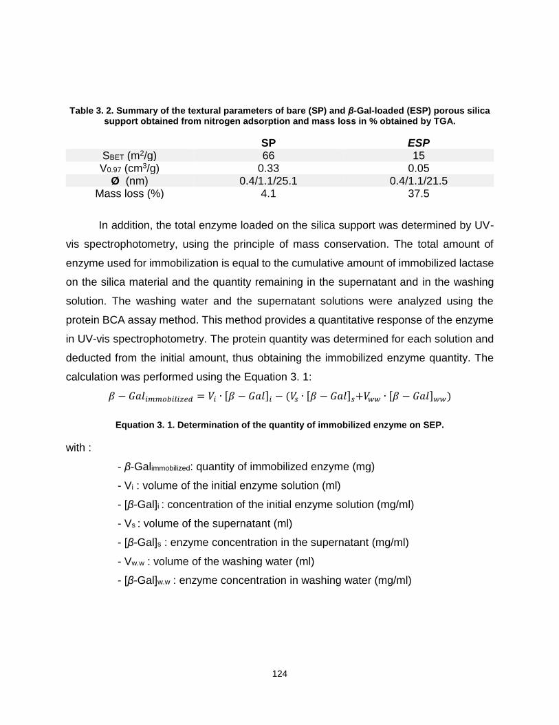
124
Table 3. 2. Summary of the textural parameters of bare (SP) and β-Gal-loaded (ESP) porous silica support obtained from nitrogen adsorption and mass loss in % obtained by TGA.
In addition, the total enzyme loaded on the silica support was determined by UV-
vis spectrophotometry, using the principle of mass conservation. The total amount of
enzyme used for immobilization is equal to the cumulative amount of immobilized lactase
on the silica material and the quantity remaining in the supernatant and in the washing
solution. The washing water and the supernatant solutions were analyzed using the
protein BCA assay method. This method provides a quantitative response of the enzyme
in UV-vis spectrophotometry. The protein quantity was determined for each solution and
deducted from the initial amount, thus obtaining the immobilized enzyme quantity. The
calculation was performed using the Equation 3. 1:
𝛽 − 𝐺𝑎𝑙𝑖𝑚𝑚𝑜𝑏𝑖𝑙𝑖𝑧𝑒𝑑 = 𝑉𝑖 ∙ [𝛽 − 𝐺𝑎𝑙]𝑖 − (𝑉𝑠 ∙ [𝛽 − 𝐺𝑎𝑙]𝑠+𝑉𝑤𝑤 ∙ [𝛽 − 𝐺𝑎𝑙]𝑤𝑤)
Equation 3. 1. Determination of the quantity of immobilized enzyme on SEP.
with :
- β-Galimmobilized: quantity of immobilized enzyme (mg)
- Vi : volume of the initial enzyme solution (ml)
- [β-Gal]i : concentration of the initial enzyme solution (mg/ml)
- Vs : volume of the supernatant (ml)
- [β-Gal]s : enzyme concentration in the supernatant (mg/ml)
- Vw.w : volume of the washing water (ml)
- [β-Gal]w.w : enzyme concentration in washing water (mg/ml)
SP ESP SBET (m2/g) 66 15 V0.97 (cm3/g) 0.33 0.05
Ø (nm) 0.4/1.1/25.1 0.4/1.1/21.5 Mass loss (%) 4.1 37.5

125
The amount of enzyme adsorbed on silica material was calculated at 7.5 mg
enzyme/g of silica support, and the value was determined by dividing the quantity of
immobilized enzyme by the quantity of silica used for the immobilization.
The immobilization yield is calculated using the following formula:
𝑌𝑖𝑒𝑙𝑑 (%) = 100𝐶𝑖 − (𝐶𝑠+𝐶𝑤𝑤)
𝐶𝑖
Equation 3. 2 Calculation of immobilization yield.
where Ci, is the concentration of enzyme in the immobilization solution, Cs and Cww
are the enzyme concentration in the supernatant and in the washing solution,
respectively. The enzyme adsorption yield was calculated at 34.8 ± 4.3%
Thermogravimetric analysis (TGA) was also used to quantify the amount of
enzyme adsorbed on the silica material (Figure 3. 17 and Table 3.2). Two different mass
loss steps were detected in the curve thermogravimetric curve obtained for the loaded
silica. The first step of mass lost occurring from room temperature (RT) to 100°C can be
associated with the evaporation of physically adsorbed water molecules on the loaded
silica surface [125]. This phenomenon is also present in the unmodified inorganic particles
(analyzed as received without further modification). A second mass loss step is detected
between 100 and 450°C for the sample ESP and it corresponds to around 33% of weight
loss of the total sample. This value indicates that the loaded silica bears 330 mg of organic
matter per g of silica and is much higher than the amount calculated previously by the
UV-vis spectrophotometric method (7.5 mg enzyme/g silica support). This can lead to the
conclusion that beside enzyme, other organic species were absorbed onto the silica,
probably glycerol. The source of the glycerol is the solution in which the enzyme is
formulated. Its presence in the materials has a positive aspect, since the glycerol is known
for protecting the enzyme against external factors that can inactivate the β-Galactosidase
enzyme [126]. This can also ensure that the enzyme does not lose its activity during the
absorption and the formation of the 1,2-dioleoyl-sn-glycero-3-phosphocholine (DOPC)
liposomes double layer.

126
Figure 3. 17. Mass loss determined by thermogravimetric analysis made on bare (SP) and loaded
silica materials (ESP) in air.
3.2.3. Characterization of liposomes coated silica particles (LCSP)
After enzyme loading, silica particles were incubated with the 1,2-dioleoyl-sn-
glycero-3-phosphocholine (DOPC) lipids, resulting in the liposome-coated silica particles
(LCSP), following the procedure described in Chapter 2. The presence of lipids on the
surface of resulting LCSP material was confirmed by fluorescence microscopy in
presence of a fluorescent dye (Texas Red).
The fluorescent microscopy experiments were made on the samples in simulated
gastric fluid (SGF), simulated intestinal fluid (SIF) and in SIF containing pancreatin
enzymes (and, more precisely, a mixture of amylase, lipase and protease). The
preparation of stock simulated digestion fluids solutions is presented in Chapter 2. The
samples were kept for 2 hours in SGF (Figure 3. 18 A) and 2 hours in SIF (Figure 3. 18
B) prior observation. As presented in Figure 3. 18 A and B, the micrometric silica particles
are fluorescent. This is undoubtedly due to the presence of phospholipid bilayer around
the particles since the Texas Red dye is only soluble in lipids. In addition, the silica
remains covered with the lipid bilayer in SGF as well as in SIF. However, the addition of
pancreatin in the SIF leads to the disappearance of fluorescence probably indicating the
100 200 300 400 500 600 700 800
60
70
80
90
100
37.5 %
Ma
ss L
oss (
%)
Temperature (C)
SP
ESP
4.1 %

127
removal of the lipid bilayers (Figure 3. 18 C). This experiment evidences the presence of
the liposomes around the silica nanoparticles surface.
A B C
Figure 3. 18. Fluorescence microscopy images obtained for LCSP in SGF (A), SIF (B) and in SIF containing pancreatin (C) (scale bar 50 μm).
3.2.4. Enzyme activity during the immobilization and incubation
The enzyme loaded silica microparticles (ESP) initially described were dried.
Approximately 85 mg of ESP were dispersed in 1 ml of TRIS buffer (pH 7.4) containing
the liposomes leading to the formation of LCSP material. The liposomes that did not
adhere to the surface were removed by washing the LCSP sample 7 times with the buffer.
The washing waters were used to determine the quantity of the enzyme that leaked from
silica during the incubation. The same principle of mass conservation was applied as in
the case of the determination of the amount of enzyme present in the ESP, and the
Equation 3. 3 was used.
𝛽 − 𝐺𝑎𝑙𝑟𝑒𝑐𝑜𝑣𝑒𝑟𝑒𝑑 = ∑ 𝑉𝑛
7
𝑛=1
[𝛽 − 𝐺𝑎𝑙]𝑛
Equation 3. 3. Determination of the quantity of enzyme leaked from LCSP during incubation.
where β-Galrecovered (mg) is the total amount of the enzyme that was recovered in
the washing waters, Vn (ml) is the volume of each washing water with the enzyme
concentration [β-Gal]n (mg/ml)
The total amount of enzyme that was recovered in the washing water (that leaked
from ESP) was determined at 0.66 mg of protein/g of silica. Thus, the amount of enzyme
adsorbed into LCSP was calculated at 6.8 mg enzyme/g silica, corresponding to the total
amount of enzyme immobilized on the ESP material (7.5 mg enzyme/g silica) minus the

128
amount of enzyme that leaked out of ESP material and minus the one that was recovered
in the washing solutions.
It should also be noted that the leaked enzyme has almost the same activity as the
enzyme in the initial solution used for immobilization: 5025 U/mg versus 5223 U/mg,
respectively. The lipid incubation step did not deactivate the enzyme.
The leakage during the incubation with lipids was minimal and the specific activities
of the materials were assessed by enzymatic reaction with ONPG. The specific activity of
the ESP was determined at 22.4 U/mg of silica material and the activity of LCSP was
measured at 10.9 U/mg of silica. The activity of LCSP material was smaller than the
activity of ESP material. This was expected, since the coating with the lipid double layer
decreases the diffusion of the substrate ONPG from the solution to the surface of silica
where the enzyme is absorbed.
3.2.5. Enzyme release
A controlled release of the enzyme out of the uncoated ESP carrier and the lipid
covered particle (LCSP), was done in vitro in simulated gastric fluid (SGF, pH3) and
simulated intestinal fluid (SIF, pH7) as presented in Chapter 2.
In order to simulate the passage of the materials through the gastro- intestinal tract
four samples of each material were tested as presented in Table 2. 2. (Chapter 2). Each
type of sample underwent four experiments under magnetic stirring at 150 rpm. In the first
one, the sample LCSP1 (or ESP1) was kept for 1H in SGF. In the second, the carrier
LCSP2 (or ESP2) was kept for 2H in SGF. The third and the four experiments were a
successive immersion of the samples LCSP3 (or ESP3) and LCSP4 (or ESP4) in SGF
over 2H followed by 1H or 2H in the SIF respectively. The samples LCSP3 (or ESP3) and
LCSP4 (or ESP4) from the experiments 3 and 4 have been centrifuged before being
added to the SIF. The Figure 3. 19 displays the cumulative quantity of enzyme released
e.g. after 3H in the simulated gastro-intestinal fluid, the total quantity of enzyme release
is the sum of the quantity of enzyme released after 2H in SGF and the quantity of enzyme
released after 1H in SIF. The values are given in Figure 3.19 are in weight percentage

129
(wt.%), which represents the quantity of enzyme release /initial enzyme loading per mg
of silica.
A clear difference in release profiles of coated (LCSP) and uncoated (ESP) silica
was observed. In SGF (pH 3), after the first hour, only a small quantity of enzyme was
released from LCSP 4.6 wt.% compared with 61.2 wt.% for ESP. After 2H in SGF, the
enzyme quantity released form LCSP remain 4.6 wt.% and in the case of ESP, the amount
remained close to 61.7 wt.%. After 1H in SIF (pH7), the ESP carrier released in total 83.8
wt.% of enzyme that was initially absorbed. In the consecutive 2H in SIF, 96.2 wt.% of the
lactase has been released. However, for LCSP, the total amount of enzyme released
reached only 6.3 wt.% in the first hour of immersion in SGF and approximately the same
amount as in the second hour (7.2 wt.%). According to the results obtained from the
consecutive immersion test (experiment 3 and 4), it can be clearly noticed that the amount
of released enzyme increased in SIF media. The silica SiO2-bilayer interactions are
controlled by van der Waals forces. At low pH, the 1,2-dioleoyl-sn-glycero-3-
phosphocholine (DOPC) bilayer is fused to the surface of silica due to the favorable van
der Waals forces [127]. As the pH value rises the electrostatic repulsion between the
negatively charged silica surface and liposomes increases, and a ~1 nm water layer
separates the DOPC bilayer and silica surface.[128] As it was expected the interactions
between the lipid layer and silica changed in SIF pH7 leading to the leak of the enzyme.

130
Figure 3. 19. The quantity of the enzyme release from LCSP and ESP in SGF (pH 3) and SIF (pH 7).
In order to verify if the enzyme released during the simulated digestion remains
activity, the recovered solutions from in vitro in simulated gastric fluid (SGF) and
simulated intestinal fluid (SIF) were analyzed. In terms of activity, the enzyme released
from ESP and LCSP displays very different behaviors as presented in Figure 3. 20. The
enzyme liberated by ESP along the experiments did not present any activity, compared
to enzyme recovered from LCSP sample that was still retaining a part of its reactivity
when put in SIF. When dispersed in the SIF (pH 7), the enzyme released from LCSP
increased its activity progressively: from 64 U/mg in SGF the enzyme activity increased
to 236 U/mg after 1H in SIF and to 351 U/mg after 2H in SIF. The conclusion that can be
drawn from these results is that the lipid bilayer indeed provides protection against the
acidic pH and that it works as a slow release system, which gradually liberates active
enzyme.
1 h in SGF 2 h in SGF 1 h in SIF 2 h in SIF
0
10
20
30
40
50
60
70
80
90
100
82.8 %
96.2 %
61.7 %61.2 %
7.2 %6.3 %4.6 %
ESP
LCSP %
En
zym
e r
ele
ase
d (
%)
Time(hours)
4.6 %

131
Figure 3. 20. The activity (B) of the enzyme release from LCSP and ESP in SGF (pH 3) and SIF (pH
7).
To confirm that the enzyme retains its activity during the gastric simulation, the
specific activities of the materials ESP and LCSP (Figure 3. 21.) were compared with the
initial ESP and LCSP. The ESP material retained only a fraction of its original activity
when added to the SGF, it decreased over the time, and an insignificant increase was
observed in the SIF. The final specific activity for ESP sample was calculated at 0.32
U/mg of silica. The specific activity of the LCSP sample also decreased when added in
SGF, but a significant part of specific activity was retained, with a final value of 0.875
U/mg of silica. Although the specific activity variation along the four experiments has the
same tendency for both LCSP and ESP catalyst, the presence of DOPC liposomes
protects the enzyme from the acidic pH inactivation (SGF at pH3).
1 h in SGF 2 h in SGF 1 h in SIF 2 h in SIF
0
50
100
150
200
250
300
350
64 U/mg
LCSP
ESP
Rele
ased e
nzym
e (
U/m
g)
Time (hours)
64 U/mg
236 U/mg
351 U/mg

132
Figure 3. 21. Activity of silica materials used in the simulated gastro-intestinal digestion fluids (SGF and SIF).
3.2.5. Conclusion
β-Gal from Kluyveromices lactis was immobilized into low porosity silica particles
by physical adsorption and then coated with DOPC bilayers. This system presented a
controlled release pattern over the pH ranges of simulated gastric and intestinal fluids.
The presence of liposomes not only provides a controlled release, but also acts as a
protective coating from pH inactivation in acidic pH (SGF) for the immobilized enzyme.
Compared to the ESP material, LCSP sample preserved much better the activity of the
enzyme (0.875 U/mg of silica vs. 0.32U/mg of silica) for a similar initial loading. Moreover,
the enzyme immobilized in LCSP had, in the end of the simulated gastro-intestinal
digestion experiment, the same behavior as the free enzyme, namely an increase of
activity at optimal pH values. The enzyme was not denatured by the long stay in acidic
pH. Although the loading in LCSP is slightly smaller than in the ESP material (6.8 vs. 7.5
mg), one can conclude that this type of immobilization could preserve the enzyme until
the intestine where the lactase should be released. Thus, LCSP is a promising candidate
for the delivery of enzymes, with the guarantee of safely immobilizing the enzyme intact.
The main improvement for the LCSP system that could be envisioned is an
increase of its loading capacity. Since the enzyme is quite a large molecule (14 nm), a
1 h in SGF 2 h in SGF 1 h in SIF 2 h in SIF
0.2
0.4
0.6
0.8
1.0
1.2
1.4
1.6
LCSP
ESP
Mate
rial activity (m
ol/m
in/m
g o
f sili
ca
)
Time (hours)

133
more porous type of silica would ensure a larger amount of encapsulated enzyme. This
could be achieved in the sol-gel phase, via the insertion of organic molecules. Another
direction for further research is the variation of the lipids (or lipid mixtures) used in the
production of the liposomes. By changing the composition of the lipids, different
permeability to chemical species, in and from the carrier system, can be obtained. This
would potentially lead to the tailoring of delivery systems perfectly adapted to specific
triggers.

134
Bibliography
[1] D. Dale, Microencapsulation in the Food Industry, Elsevier, 2014. [2] L. Fjerbaek, K. V. Christensen, B. Norddahl, Biotechnol. Bioeng. 2009, 102, 1298–1315. [3] D. J. Pollard, J. M. Woodley, Trends Biotechnol. 2007, 25, 66–73. [4] H. A. Gulec, Colloids and Surfaces B-Biointerfaces 2013, 104, 83–90. [5] A. M. Klibanov, Nature 2001, 409, 241–246. [6] P. V. Iyer, L. Ananthanarayan, Process Biochem. 2008, 43, 1019–1032. [7] C. Garcia-Galan, Á. Berenguer-Murcia, R. Fernandez-Lafuente, R. C. Rodrigues, Adv. Synth. Catal. 2011,
353, 2885–2904. [8] L. Cao, Curr. Opin. Chem. Biol. 2005, 9, 217–26. [9] K. Hernandez, R. Fernandez-Lafuente, Enzyme Microb. Technol. 2011, 48, 107–122. [10] E. T. Hwang, M. B. Gu, Eng. Life Sci. 2013, 13, 49–61. [11] A. Gupta, K. Prabhu, J. Gen. Appl. Microbiol. 1995, 41, 399–407. [12] H. Y. Kawaguti, P. H. Carvalho, J. A. Figueira, H. Sato, 2011, 2011, DOI 10.4061/2011/791269. [13] P. M. Kosaka, Y. Kawano, O. A. El Seoud, D. F. S. Petri, 2007, 12167–12173. [14] K. Labus, I. Gancarz, J. Bryjak, Mater. Sci. Eng. C 2012, 32, 228–235. [15] B. Krajewska, 2004, 35, 126–139. [16] M. Nasratun, A. . Hasrul, A. . Sureena, M. . Nurul Aini, A. . Ruwaida, M. . Shalyda, A. Ideris, A. . Rozaimi, J. .
Sharfuddin, N. I. . Ahmad Nordin, J. Appl. Sci. 2010, 21, 2701–2704. [17] G. Bayramoglu, I. Gursel, M. Yilmaz, M. Y. Arica, J. Chem. Technol. Biotechnol. 2012, 87, 530–539. [18] M. Alatorre-meda, P. Taboada, J. Sabín, B. Krajewska, L. M. Varela, J. R. Rodríguez, 2009, 339, 145–152. [19] L. B. R. Castro, F. F. Silva, A. M. Carmona-Ribeiro, M. Kappl, D. F. S. Petri, J. Phys. Chem. B 2007, 111,
8520–8526. [20] M. Pešić, C. López, G. Álvaro, J. López-Santín, J. Mol. Catal. B Enzym. 2012, 84, 144–151. [21] A. A. Mendes, P. C. Oliveira, A. M. Vélez, R. C. Giordano, R. D. L. C. Giordano, H. F. De Castro, Int. J. Biol.
Macromol. 2012, 50, 503–511. [22] D. Kubác, A. Čejková, J. Masák, V. Jirků, M. Lemaire, E. Gallienne, J. Bolte, R. Stloukal, L. Martínková,
2006, 39, 59–61. [23] T. K. Ozturk, A. Kilinc, "Journal Mol. Catal. B, Enzym. 2010, 67, 214–218. [24] J. C. Santos, G. F. M. Nunes, V. H. Perez, H. F. De Castro, 2007, 1255–1261. [25] P. Torres, D. Reyes-duarte, N. Lo, M. Ferrer, A. Ballesteros, F. J. Plou, 2008, 43, 145–153. [26] E. Hita, A. Robles, B. Camacho, A. Ramírez, L. Esteban, M. J. Jiménez, M. M. Muñío, P. A. González, E.
Molina, 2007, 42, 415–422. [27] H. A. Akdogan, N. K. Pazarlioglu, Process Biochem. 2011, 46, 840–846. [28] Y. Zhang, T. Zhi, L. Zhang, H. Huang, H. Chen, 2009, 50, 5693–5700. [29] V. Deepak, R. Kumar, K. Kalishwaralal, S. Gurunathan, Bioresour. Technol. 2009, 100, 6644–6646. [30] A. A. Mendes, L. Freitas, A. K. F. De Carvalho, P. C. De Oliveira, H. F. De Castro, 2011, 2011, DOI
10.4061/2011/967239. [31] P. C. Ashly, P. V Mohanan, Process Biochem. 2010, 45, 1422–1426. [32] S. Sakai, Y. Liu, T. Yamaguchi, R. Watanabe, M. Kawabe, K. Kawakami, Bioresour. Technol. 2010, 101,
7344–7349. [33] R. Reshmi, G. Sanjay, S. Sugunan, Catal. Commun. 2006, 7, 460–465. [34] S. Wanjari, C. Prabhu, T. Satyanarayana, A. Vinu, S. Rayalu, Microporous Mesoporous Mater. 2012, 160,
151–158. [35] J. Yu, H. Ju, 2002, 74, 3579–3583. [36] M. Delvaux, S. Demoustier-Champagne, Biosens. Bioelectron. 2003, 18, 943–951. [37] E. J. Cho, S. Jung, H. J. Kim, Y. G. Lee, K. C. Nam, H.-J. Lee, H.-J. Bae, Chem. Commun. 2012, 48, 886–
888. [38] K. M. de Lathouder, D. T. J. van Benthem, S. A. Wallin, C. Mateo, R. F. Lafuente, J. M. Guisan, F. Kapteijn,
J. A. Moulijn, J. Mol. Catal. B Enzym. 2008, 50, 20–27. [39] R. Zhai, B. Zhang, L. Liu, Y. Xie, H. Zhang, J. Liu, CATCOM 2010, 12, 259–263. [40] U. H. Zaidan, M. B. A. Rahmana, S. S. Othman, M. Basri, E. Abdulmalek, R. N. Z. R. A. Rahman, A. B.
Salleh, Food Chem. 2012, 131, 199–205. [41] C. Fargues, M. Bailly, G. Grevillot, L. Laboratoire, C. Ensic-cnrs, G. Bp, N. Cedex, 1998, 16, 5–16. [42] Q. Liu, X. Kong, C. Zhang, Y. Hua, 2012, DOI 10.1002/jsfa.5997. [43] F. A. Erhardt, H.-J. Jördening, 2007, 131, 440–447. [44] L. J. Kennedy, P. K. Selvi, A. Padmanabhan, K. N. Hema, G. Sekaran, 2007, 69, 262–270. [45] N. Carlsson, H. Gustafsson, C. Thörn, L. Olsson, K. Holmberg, B. Åkerman, Adv. Colloid Interface Sci. 2014,
205, 339–60. [46] E. Magner, Chem. Soc. Rev. 2013, 42, 6213–6222.

135
[47] M. Hartmann, X. Kostrov, Chem. Soc. Rev. 2013, 6277–6289. [48] Y. Yokogawa, R. Yamauchi, A. Saito, Y. Yamato, T. Toma, Biomed. Mater. Eng. 2017, 28, 37–46. [49] E. Weber, D. Sirim, T. Schreiber, B. Thomas, J. Pleiss, M. Hunger, R. Gläser, V. B. Urlacher, 2010, 64, 29–
37. [50] W. hua Yu, M. Fang, D. shen Tong, P. Shao, T. ning Xu, C. hui Zhou, Biochem. Eng. J. 2013, 70, 97–105. [51] S. B. Hartono, S. Z. Qiao, J. Liu, K. Jack, B. P. Ladewig, Z. Hao, G. Qing, M. Lu, J. Phys. Chem. C 2010,
114, 8353–8362. [52] M. Falahati, A. A. Saboury, L. Ma’mani, A. Shafiee, H. A. Rafieepour, Int. J. Biol. Macromol. 2012, 50, 1048–
1054. [53] R. H. Chang, J. Jang, K. C. Wu, 2011, 12, 2844–2850. [54] T. Y. Nara, H. Togashi, C. Sekikawa, K. Inoh, K. Hisamatsu, K. Sakaguchi, F. Mizukami, T. Tsunoda,
"Journal Mol. Catal. B, Enzym. 2010, 64, 107–112. [55] B. E. Grabicka, M. Jaroniec, 2010, 385–396. [56] S. Fornera, T. Bauer, a. D. Schlüter, P. Walde, J. Mater. Chem. 2012, 22, 502. [57] M. M. Zheng, Y. Lu, L. Dong, P. M. Guo, Q. C. Deng, W. L. Li, Y. Q. Feng, F. H. Huang, Bioresour. Technol.
2012, 115, 141–146. [58] M. S. Bhattacharyya, A. Singh, U. C. Banerjee, Bioresour. Technol. 2010, 101, 1581–1586. [59] G. Zhou, K. K. Fung, L. W. Wong, Y. Chen, R. Renneberg, S. Yang, Talanta 2011, 84, 659–665. [60] C. Wu, G. Zhou, X. Jiang, J. Ma, H. Zhang, H. Song, Process Biochem. 2012, 47, 953–959. [61] M. Kramer, J. C. Cruz, P. H. Pfromm, M. E. Rezac, P. Czermak, J. Biotechnol. 2010, 150, 80–86. [62] L. Thudi, L. S. Jasti, Y. Swarnalatha, N. W. Fadnavis, K. Mulani, S. Deokar, S. Ponrathnam, J. Mol. Catal. B
Enzym. 2012, 74, 54–62. [63] H. S. Mansur, R. L. Oréfice, Z. P. Lobato, W. L. Vasconcelos, E. S. Mansur, L. J. C. Machado, Adsorption
2001, 105–116. [64] M. Falahati, L. Ma'Mani, A. A. Saboury, A. Shafiee, A. Foroumadi, A. R. Badiei, Biochim. Biophys. Acta
- Proteins Proteomics 2011, 1814, 1195–1202. [65] M. Vinoba, M. Bhagiyalakshmi, S. K. Jeong, Y. I. I. Yoon, S. C. Nam, Colloids Surfaces B Biointerfaces 2012,
90, 91–96. [66] D. T. Tran, C. L. Chen, J. S. Chang, J. Biotechnol. 2012, 158, 112–119. [67] J. J. Karimpil, J. S. Melo, S. F. D’Souza, Int. J. Biol. Macromol. 2012, 50, 300–302. [68] F. Xu, W. H. Wang, Y. J. Tan, M. L. Bruening, Anal. Chem. 2010, 82, 10045–10051. [69] T. Godjevargova, R. Nenkova, V. Konsulov, J. Mol. Catal. B Enzym. 2006, 38, 59–64. [70] S. W. Kwon, B. O. Jeong, E. H. Lee, Y. S. Kim, Y. Jung, 2012, 33, 1593–1596. [71] P. Xiao, X. Lv, Y. Deng, 2012, 2719, DOI 10.1080/00032719.2012.673103. [72] S. Wang, P. Su, F. Ding, Y. Yang, "Journal Mol. Catal. B, Enzym. 2013, 89, 35–40. [73] H. Kawakita, K. Sugita, K. Saito, M. Tamada, T. Sugo, H. Kawamoto, J. Memb. Sci. 2002, 205, 175–182. [74] H. Kawakita, K. Sugita, K. Saito, M. Tamada, T. Sugo, H. Kawamoto, Biotechnol. Prog. 2002, 18, 465–469. [75] S. Karboune, R. Neufeld, S. Kermasha, J. Biotechnol. 2005, 120, 273–283. [76] Y. Z. Chen, C. T. Yang, C. B. Ching, R. Xu, Langmuir 2008, 24, 8877–8884. [77] J. S. Beck, J. C. Vartuli, W. J. Roth, M. E. Leonowicz, C. T. Kresge, K. D. Schmitt, C. T. W. Chu, D. H. Olson,
E. W. Sheppard, S. B. McCullen, et al., J. Am. Chem. Soc. 1992, 114, 10834–10843. [78] S. Hudson, J. Cooney, E. Magner, Angew. Chem. Int. Ed. Engl. 2008, 47, 8582–94. [79] C.-H. Lee, T.-S. Lin, C.-Y. Mou, Nano Today 2009, 4, 165–179. [80] C. Bernal, L. Sierra, M. Mesa, ChemCatChem 2011, 3, 1948–1954. [81] M. Wang, W. Qi, Q. Yu, R. Su, Z. He, Biochem. Eng. J. 2010, 52, 168–174. [82] P. Colombo, C. Vakifahmetoglu, S. Costacurta, J. Mater. Sci. 2010, 45, 5425–5455. [83] C. M. A. Parlett, K. Wilson, A. F. Lee, Chem. Soc. Rev. Chem. Soc. Rev 2013, 42, 3876–3893. [84] S. Cao, L. Fang, Z. Zhao, Y. Ge, S. Piletsky, A. P. F. Turner, Adv. Funct. Mater. 2013, 23, 2162–2167. [85] C. Bernal, L. Sierra, M. Mesa, J. Mol. Catal. B Enzym. 2012, 84, 166–172. [86] C. Bernal, P. Urrutia, A. Illanes, L. Wilson, N. Biotechnol. 2013, 30, 500–506. [87] C. Bernal, A. Illanes, L. Wilson, Langmuir 2014, 30, 3557–3566. [88] N. Guajardo, C. Bernal, L. Wilson, Z. Cabrera, Process Biochem. 2015, 50, 1870–1877. [89] N. Guajardo, C. Bernal, L. Wilson, Z. Cabrera, Catal. Today 2015, 255, 21–26. [90] J.-L. Blin, J. Jacoby, S. Kim, M.-J. Stébé, N. Canilho, A. Pasc, Chem. Commun. (Camb). 2014, 50, 11871–4. [91] N. Brun, A. Babeau-Garcia, M.-F. Achard, C. Sanchez, F. Durand, G. Laurent, M. Birot, H. Deleuze, R.
Backov, Energy Environ. Sci. 2011, 4, 2840. [92] X. Y. Yang, Z. Q. Li, B. Liu, A. Klein-Hofmann, G. Tian, Y. F. Feng, Y. Ding, D. Su, F. S. Xiao, Adv. Mater.
2006, 18, 410–414. [93] H. Li, S. Li, P. Tian, Z. Wu, Z. Li, Molecules 2017, 22, DOI 10.3390/molecules22030377. [94] J. Zhao, Y. Wang, G. Luo, S. Zhu, Bioresour. Technol. 2011, 102, 529–35. [95] J. Li, L.-S. Li, L. Xu, Microporous Mesoporous Mater. 2016, 231, 147–153. [96] S. Kim, M.-J. Stébé, J.-L. Blin, A. Pasc, J. Mater. Chem. B 2014, 2, 7910–7917.

136
[97] S. Kim, R. Diab, O. Joubert, N. Canilho, A. Pasc, Colloids Surf. B. Biointerfaces 2015, 140, 161–168. [98] A. Pasc, J.-L. Blin, M.-J. Stébé, J. Ghanbaja, RSC Adv. 2011, 1, 1204. [99] S. Gürdaş, H. A. Gülec, M. Mutlu, Food Bioprocess Technol. 2012, 5, 904–911. [100] T. Jesionowski, J. Zdarta, B. Krajewska, Adsorption 2014, 20, 801–821. [101] Z. Quinn Z.K, C. Xiao Dong, Biochem. Eng. J. 2001, 9, 33–40. [102] S. F. Prazeres, C. G. Ruiz, G. M. García, Appl. Spectrosc. Rev. 2015. [103] W. Humphrey, A. Dalke, K. Schulten, J. Mol. Graph. 1996, 14, 33–38. [104] R. F. Pereira-Rodríguez, Ángel Leiro, M. E. Cerdán, M. I. González Siso, M. B. Fernández, J. Mol. Catal. B
Enzym. 2008, 52–53, 178–182. [105] A. Pereira-Rodríguez, R. Fernández-Leiro, M. I. González-Siso, M. E. Cerdán, M. Becerra, J. Sanz-Aparicio,
J. Struct. Biol. 2012, 177, 392–401. [106] A. Pereira-Rodríguez, R. Fernández-Leiro, M. I. González Siso, M. E. Cerdán, M. Becerra, J. Sanz-Aparicio,
Acta Crystallogr. Sect. F. Struct. Biol. Cryst. Commun. 2010, 66, 297–300. [107] E. Krissinel, K. Henrick, J. Mol. Biol. 2007, 372, 774–97. [108] M. Becerra, E. Cerdán, M. I. G. Siso, Biotechnol. Tech. 1998, 12, 253–256. [109] O. S. Pilipenko, L. F. Atyaksheva, O. M. Poltorak, E. S. Chukhrai, Russ. J. Phys. Chem. A 2007, 81, 990–
994. [110] C. Lei, T. a Soares, Y. Shin, J. Liu, E. J. Ackerman, Nanotechnology 2008, 19, 125102. [111] M. Vallet-Regi, A. Rámila, R. P. Del Real, J. Pérez-Pariente, Chem. Mater. 2001, 13, 308–311. [112] F. Tang, L. Li, D. Chen, Adv. Mater. 2012, 24, 1504–1534. [113] FDA, 2009, Www.accessdata.fda.gov/scripts/fcn/gras_notices/GR. [114] C. Contado, L. Ravani, M. Passarella, Anal. Chim. Acta 2013, 788, 183–192. [115] I. I. Slowing, C. W. Wu, J. L. Vivero-Escoto, V. S. Y. Lin, Small 2009, 5, 57–62. [116] A. E. Nel, L. Mädler, D. Velegol, T. Xia, E. M. V. Hoek, P. Somasundaran, F. Klaessig, V. Castranova, M.
Thompson, Nat. Mater. 2009, 8, 543–557. [117] M. M. Van Schooneveld, E. Vucic, R. Koole, Y. Zhou, J. Stocks, D. P. Cormode, C. Y. Tang, R. E. Gordon, K.
Nicolay, A. Meijerink, et al., Nano Lett. 2008, 8, 2517–2525. [118] Y. Jin, N. Zhang, C. Li, K. Pu, C. Ding, Y. Zhu, Colloids Surfaces B Biointerfaces 2017, 151, 240–248. [119] J. Y. Choi, T. Ramasamy, S. Y. Kim, J. Kim, S. K. Ku, Y. S. Youn, J. R. Kim, J. H. Jeong, H. G. Choi, C. S.
Yong, et al., Acta Biomater. 2016, 39, 94–105. [120] H. Meng, M. Wang, H. Liu, X. Liu, A. Situ, B. Wu, Z. Ji, C. H. Chang, A. E. Nel, ACS Nano 2015, 9, 3540–
3557. [121] N. Han, Q. Zhao, L. Wan, Y. Wang, Y. Gao, P. Wang, Z. Wang, J. Zhang, T. Jiang, S. Wang, ACS Appl.
Mater. Interfaces 2015, 7, 3342–3351. [122] Z. Wang, Y. Tian, H. Zhang, Y. Qin, D. Li, L. Gan, F. Wu, Int. J. Nanomedicine 2016, 11, 6485–6497. [123] P. N. Durfee, Y. S. Lin, D. R. Dunphy, A. J. Muñiz, K. S. Butler, K. R. Humphrey, A. J. Lokke, J. O. Agola, S.
S. Chou, I. M. Chen, et al., ACS Nano 2016, 10, 8325–8345. [124] J. Sun, D. Ma, H. Zhang, F. Jiang, Y. Cui, R. Guo, X. Bao, Langmuir 2008, 24, 2372–2380. [125] S. H. Ahn, S. H. Kim, S. G. Lee, J. Appl. Polym. Sci. 2004, 94, 812–818. [126] V. Athès, D. Combes, Enzyme Microb. Technol. 1998, 22, 532–537. [127] P. S. Cremer, S. G. Boxer, J. Phys. Chem. B 1999, 103, 2554–2559. [128] F. Wang, J. Liu, Small 2014, 10, 3927–3931. [129] I.-A. Pavel, S. F. Prazeres, G. Montalvo, C. Garcıa Ruiz, V. Nicolas, A. Celzard, F. Dehez, L. Canabady-
Rochelle, N. Canilho, A. Pasc, Langmuir 2017, 33, 3333–3340.

137
Chapter 4. Encapsulation of β-galactosidase in responsive carriers allowing
release triggered by either temperature or pH
The encapsulation strategies involve the entrapment of active agents (cells,
enzymes, food ingredients) in/by the matrix of the carrier with the purpose of preventing
premature release or degradation of the active moiety particularly when it should be
delivered to a targeted site like intestine, as the case of this work. Encapsulation is
required to protect the agent from moisture[1], heat[2], oxygen or light[3] and sometimes to
improve the shelf-life[4]. However, encapsulation can also be a strategy for masking
undesirable odor, taste and color or, for preventing reactions and interactions between
the ingredients and the active agent. Another very important reason for choosing
encapsulation is to control the delivery. In addition, encapsulation can modify the physical
characteristics of the components (e.g. from liquid to solid) leading to easier handling,
separation and may also confer adequate concentration and uniform dispersion in the
mixture [5]. The use of the entrapment techniques has increased in the food industry.
The encapsulation of flavours and aromas improves the stability for volatile
molecules [6] against evaporation and chemical reactions (interaction with food or other
flavours or oxidation). Organosulfurs [7], herbal and plants extract [3] that present health
benefits, are encapsulated to mask an unpleased smell. Bioactive components, such as
lipids, peptides (fragments of proteins), vitamins, minerals, antioxidants, cells (probiotic)
display multiple health benefits. Their encapsulation protects them from external factors
during storage, and from acidic pH during their passage through gastro-intestinal track [8].
Probiotic bacteria are encapsulated to increase their bioavailability and functionality
against a pH variation, the digestive enzymes of stomach or, to resist to mechanical stress
or transport conditions [9].
Enzymes are used in various food industries (starch, baking, brewing, dairy,
vegetables, fruits, fats and oils, fish and meat industry) mainly as processing agents. The
enzymes can be added in different production steps: preparation, processing, treatment,
packaging, transportation or in the storage stage [10]. Enzymes easily denature, thus
encapsulation provides them a longer lifetime, and improves the resistance to harsh pH

138
and to temperature. The encapsulated enzymes present in some specific cases new
catalytic properties compared with the free enzyme [6].
Various techniques can be applied to entrap enzyme, including: extrusion, coating,
liposome entrapment [11], spray drying, agglomeration or multistage drying, spray chilling
or spray cooling, fluidized bed granulation or coating, coacervation, inclusion
complexation, centrifugal extrusion, high shear granulation, melt extrusion and melt
injections, rotational suspension separation, emulsification and sol-gel encapsulation [12].
All these methods present advantages and drawbacks, but the encapsulation approach
of the enzyme depends on its intended application in the food manufacture process. For
example Dusterhoft et al. [13] entrapped amylase enzyme (used in the baking industry to
maintain freshness of the final product during storage) via spray coating and chilling in a
core-shell particle. The encapsulated enzymes were protected against temperature and
the release was controlled during dough baking [2]. Using spray coating, Soloman et al.
[14] investigated functional coating of lactase with acacia gum, shellac and
hydroxypropylmethyl cellulose to protect the enzyme from low pH 2.
The shape of the capsule depends on the entrapment method used. Figure 4. 1
resents various forms of capsules:
• the well-defined core-shell morphology is the simplest form (mononuclear), which
can be a sphere or an irregular shape
• the polynuclear capsules present multiple cores inside a shell
• the multi-wall capsules present more than one wall around the core, each wall can
be made from the same material or may be different
• the matrix type, where the active agent is dispersed into a matrix (usually a
polymer)

139
.
Figure 4. 1 Various forms of capsules (reproduced from [15]).
The encapsulation ingredients used in food applications must be “generally
recognized as safe” (GRAS) and approved by the governmental agencies (EFSA-
European Food Safety Authority and FDA-Food and Drug Administration) [6]. Obviously,
the protective shell must be from a food grade material and be biodegradable.
The most used materials for encapsulation in food applications are
polysaccharides and derivatives: plants exudates and extracts of acacia, mesquite gums,
pectins, galactomannans, soluble soybean polysaccharides or marine extracts such as
alginate and carrageenan. Starch and their derivatives –cellulose, syrups, amylose and
amylopectin, dextrins and maltodextrins or polydextrose are also used for encapsulation.
Another category of polysaccharides used for encapsulation, like chitosan, dextran, and
xanthan, have microbial or animal origins. Other common encapsulating natural agents
include: milk proteins (casein), waxes (candellina wax, carnauba wax and beeswax), fatty
acids or alcohols, lipids, or phospholipids. Other organic materials that have been used
for encapsulation are polyvinylpyrrolidone (PVP), paraffin oil or shellac [5].
Among the inorganic materials used for enzyme encapsulation, mesoporous silica
is the most used. Silica materials exhibit a high degree of biocompatibility,
biodegradability and nontoxicity, and some resistance to microbial attack. The high
Mononuclearsphereshape
Mul -wall
Mononuclearirregularshape
Matrix
Polynuclear
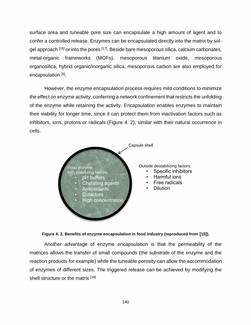
140
surface area and tuneable pore size can encapsulate a high amount of agent and to
confer a controlled release. Enzymes can be encapsulated directly into the matrix by sol-
gel approach [16] or into the pores [17]. Beside bare mesoporous silica, calcium carbonates,
metal-organic frameworks (MOFs), mesoporous titanium oxide, mesoporous
organosilica, hybrid organic/inorganic silica, mesoporous carbon are also employed for
encapsulation [6].
However, the enzyme encapsulation process requires mild conditions to minimize
the effect on enzyme activity, conferring a network confinement that restricts the unfolding
of the enzyme while retaining the activity. Encapsulation enables enzymes to maintain
their viability for longer time, since it can protect them from inactivation factors such as
inhibitors, ions, protons or radicals (Figure 4. 2), similar with their natural occurrence in
cells.
Figure 4. 2. Benefits of enzyme encapsulation in food industry (reproduced from [15]).
Another advantage of enzyme encapsulation is that the permeability of the
matrices allows the transfer of small compounds (the substrate of the enzyme and the
reaction products for example) while the tuneable porosity can allow the accommodation
of enzymes of different sizes. The triggered release can be achieved by modifying the
shell structure or the matrix [18]
Food enzyme with stabilizing factors,
• pH buffers • Chelating agents • Antioxidants • Cofactors • High concentration
Outside destabilizing factors:
• Specific inhibitors • Harmful ions • Free radicals • Dilution
Capsule shell

141
Finding a suitable solution for enzyme encapsulation in the food industry, in spite
of increasing demand, is still challenging. Since it requires mild reaction conditions
compatible with the manipulation of biological macromolecules good physical properties
of the resulting carriers in term of stability, biocompatibility and loading capacities,are
difficult to achieve..
The release of the active ingredient from the carrier can be stage-specific, side-
specific or triggered by external stimuli. Temperature (high or low), pH, enzyme trigger,
irradiation, shear or pressure release (mechanical, mastication), osmotic shock, moisture
or solvent release (via rehydration, dissolution) are used as external stimuli. Triggered
release seeks to deliver the active ingredient in a different manufacture stage or a specific
location within the body after food ingestion.
The design of the capsule is made according to the release mechanism. Dispersive
or water-soluble materials (e.g. proteins, carbohydrates) are used for water trigger
release. Fats, lipids and waxes are used in the capsules that have a temperature trigger
release. Specific parts in the gastro-intestinal track have specific pH values and specific
enzymes. A starch capsule can be used for the release of the active ingredient in the
mouth, since starch is hydrolysed by amylase (present in the mouth). The protease in the
stomach can disintegrate by hydrolysis a capsule made of protease. Resistant materials
to the acidic pH of the stomach, denatured proteins, food polymers (e.g. zein, shellac)
can be used for the delivery in the intestine, where the capsule becomes soluble at basic
pH values.

142
4.1. Thermo-responsive food grade delivery system for the treatment of lactose
intolerance
The solid lipid nanoparticles (SLNs) are alternative colloidal carriers to emulsions,
liposomes, polymer micro- or nanoparticles. SLNs can encapsulate both hydrophobic and
hydrophilic drugs, enabling thus the enhancement of drug absorption in the
gastrointestinal tract [19]. SLNs have gained increase attention from food, cosmetic and
pharmaceutics industry.
There are different formulation procedures to prepare SLNs. In High-Pressure
Homogenization (HPH) technique, a liquid is pushed with pressure through a micron size
gap. Although this is an energy intensive process, it is also an efficient dispersing
technique. The method starts from the dispersion of the drug in the melted lipid (5-10˚C
above the melting point). It can be performed either at high temperature (hot HPH) or low
temperature (< RT, cold HPH).
There are two other high energy dispersive techniques are high shear
homogenization and ultrasonication techniques. In both cases, the lipid particle solution
is obtained by dispersion of the melted lipid in a hot aqueous phase containing surfactant
as particles stabilizer. The solid lipid nanoparticles are obtained by cooling down the
previous dispersion.
SLNs can also be obtained from low energy techniques such as: microemulsion,
membrane contractor, phase inversion temperature, coacervation and double emulsion.
In the microemulsion, the melted lipids are mixed with a hot surfactant solution. Due to
the high ratio of lipid/surfactant, a microemulsion is spontaneous formed under gentle
stirring[20]. The hot microemulsion is dispersed in a high amount of cold water and the
lipids solidify forming the SLNs dispersion. The membrane contractor technique employs
a cylindrical membrane module. While working at the melting temperature of the lipid, in
the internal channel of the membrane, a surfactant solution is circulated while the melted
lipid is presses through pores [21]. By cooling the SLNs dispersion is formed.

143
Phase inversion temperature technique is another way to prepare SLNs. In fact,
by changing the temperature, the HLB index of the surfactant changes, and the O/W type
emulsion can change to a W/O emulsion. The SLNs formulation is formed at low
temperatures.
Emulsification-solvent evaporation technique, emulsification solvent diffusion
technique, solvent injection technique, supercritical fluid technique are methods that
require the use of an organic solvent. The emulsification-solvent evaporation technique
involves three steps. After the liposomes are dissolved in an organic solvent, they are
dispersed in an aqueous solution by high-speed homogenizer. This dispersion is then
passed through HPH and the SLNs nanoemulsion is obtained. The emulsification solvent
diffusion technique requires the use of an organic solvent miscible with water. By
dispersing the solvent solution that contains the lipids in water, under stirring, the SLNs
are formed due to the diffusion of the organic solvent. The solvent injection technique is
based on the same principle as emulsification solvent diffusion technique. The lipids
dissolved in a water-miscible solvent, are injected in a surfactant solution [21].
Hydrophobic compounds can be dispersed directly in the lipid phase, while
hydrophilic phase can be dispersed in the inner water phase of a double emulsion W/O/W.
Only a few examples of lipid formulation for peptides and proteins entrapment have been
reported in the literature [22].
The first example concerns the encapsulation of insulin, a model peptide, into
particles of tripalmitin (Dynasan®116) [23,24] or glyceryl monostearate and cetyl palmitate
[25,26], by the solvent evaporation method. Using the same procedure, catalase, an
enzyme that can prevent the accumulation of toxic levels of hydrogen peroxide, was
encapsulated in lecithin/triglyceride[27] and in soybean phosphatidylcholine [28]. However,
the use of organic solvents has several drawbacks. It can decrease enzyme activity [29]
and increase the toxicity of the final product [30].
The melted dispersion technique is a solvent free route allowing the preparation of
double emulsions of W/O/W from melted lipids. The emulsification is carried out at a
temperature above the melting point of the lipid. By cooling down to room temperature or

144
lower, the solid lipid particles are formed, leading to a colloidal suspension. Reithmeier
[23] compared the solvent evaporation technique with the melt dispersion technique for the
encapsulation of insulin. The best encapsulation efficiency was obtained using the W/O/W
melt dispersion technique.
Thermal responsive delivery drug [31] and protein [32] systems are widely
researched. They usually use polymers as the thermal responsive component. Although
there are couple of groups that are investigating lipids [33] or wax [34,35] formulations
thermo-responsive, up to our knowledge, this is the first time that enzyme encapsulation
in SLNs has been design as a thermal trigger response system.
Used in pharmaceutical, cosmetic and food industry, shea butter was chosen as
the oil phase. Being a triglyceride mainly formed with stearic and oleic acid, it presents a
low melting temperature (between 25°C and 45°C) [36]. The closeness to body
temperature makes shea butter a good candidate for the preparation of temperature
triggered SLNs. In addition, shea butter has antioxidant and anti-inflammatory properties
due to the presence of α-tocopherol and polyphenols that can enrich foods. These are
investigated for the prevention of oxidation in human cells and may reduce the chance of
degenerative diseases [36]. Although it is widely used in industry, only few examples have
been reported in literature for the preparation of shea butter based solid lipid
nanoparticles or nanostructured lipid carriers (NLC). Substances like anesthetics
(lidocaine [37]), polyphenols (curcumin [38]), anti-inflamatory (nimesulide [39], fragrances
((α)-amyl-cinnamal, cinnamal, cinnamyl alcohol, eugenol, geraniol,hydroxycitronellal, and
isoeugenol [40]), moisturizing agents (sodium scetylated hyaluronate and ceramides [41])
or peptides (heptapeptide-acetyl-DEETGEF-OH) [42] have been so far encapsulated in
shea butter carriers, but to the best of our knowledge there was no study concerning the
encapsulation of enzymes.
In the present study, the SLNs formulated as delivery systems were obtained by
the melted dispersion technique through a double W/O/W emulsion in which the β-
galactosidase from Kluyveromyces Lactis was incorporated in the inner water phase. The

145
release of the enzyme was trigger by thermal response and investigated by in situ UV-
Vis spectroscopy.
4.1.1. SLNs particle characterization
The SLNs preparation was described in Chapter 2. The particles shape and the
emulsion texture were visualized by optical microscopy for the simple emulsion (W1/O)
and for the double emulsion (W1/O/W2) (Figure 4. 3). The water phase W1 droplets in the
double emulsion (W1/O/W2) are separated by a thin film of continuous phase (the shea
butter phase), as the W1/O emulsion is highly concentrated. The size distribution of the
water droplets in the inverse emulsion W1/O and the particle size of the double emulsion
(W1/O/W2) was established using a droplet and counting from the images. The diameter
of aqueous droplets where the enzyme is solubilized was determined as 0.8 ±0.3 μm
whereas the size of the W1/O/W2 double emulsion was very polydisperse from 20 to 140
μm.
Figure 4. 3. Optical microscopy pictures and size distributions of inverse emulsion (W1/O) (left) and optical microscopy pictures of double emulsion (W1/O/W2) (right).
Furthermore, it was found that nanometric sizing of a hard fat has an influence on
its intrinsic properties such as the melting temperature (Tm), as it was demonstrated by
Bunjes et al. [43] in the case of triglycerides. In addition, Haji Ali et al. [38] showed, by
performing differential scanning calorimetry (DSC) experiments, that the melting
temperature of shea butter shifts from 37 °C in bulk state to 33°C once in SLN form.
0.0 0.5 1.0 1.50
5
10
15
20
25
30
35
Co
un
t
Size (m)
W1/O

146
The SLNs formulated in this work are slightly different since they entrap water and
enzymes molecules. In order to figure out if the Tm of this type of SLN changed, differential
scanning calorimetry has been also performed on bulk shea butter , on a solution of free
enzyme in the presence of the SLNs (β-Gal+SLN) and on a colloidal suspension of SLNs
encapsulating enzymes molecules (β-Gal@SLN) (Figure 4. 4). In the case of shea butter
two melting temperatures have been detected at 15 ºC and 30 ºC. The physical and
thermal properties of the natural lipids are given by the different compositions of
triglycerides. The presence of polyunsaturated triglycerides in their structure (< 20wt%)
lead to a polymorphic behavior [37] hence the presence of the two peaks in the bulk shea
butter thermogram. In the case of SLN only one peak appears in the DSC thermogram
(Figure 4. 4), corresponding to the melting of the shea butter and the rupture of the SLN.
The thermograms for free enzyme in the presence of the SLNs (β-Gal+SLN) and colloidal
suspension of SLNs encapsulating enzymes (β-Gal@SLN) are the same indicating that
the presence of the enzyme doesn’t change the structure of the SLN.
Figure 4. 4. DSC thermograms free enzyme in the presence of the SLNs (β-Gal+SLN) and colloidal suspension of SLNs encapsulating enzymes (β-Gal@SLN).
The same trend was observed from the small angle scattering data (Figure 4. 5 A)
recorded for bulk shear butter between 10°C to 50°C. The presence of the broad pick at
~45 Å at low temperature (10ºC) is correlated with a two-chain (2L) packed layers and the
10 20 30 40 50
0.000
0.001
0.002
0.003
0.004
0.005
0.006
0.007
0.008
Cp (
kcal/m
lC
)
Temperature (C)
-Gal +SLN
-Gal@SLN

147
small pick (~ 66 Å) is correlated with triple-chain length (3L) of the triglycerides. The
presence of a 3L is confirmed at 20°C and 30 °C. This type of crystallinity is formed by
saturated fatty acids (2L structure) and unsaturated triglycerides (3L structure)[38]. At
temperatures higher than 30°C, the peaks that correspond to 2L and 3L crystallinity
disappear at high contact angle that corresponds with the melting of the lipid (Figure 4. 5
A).
The SAXS patterns obtained for the samples β-Gal+SLN and β-Gal@SLN (Figure 4.
5 B and C) are rather similar with each other for all the temperatures, with one peak (4.6
Å) related to a crystalline beta phase which intensity decreases with the increase of
temperature.
Figure 4. 5. SAXS patterns obtained for the bulk shea butter (A), free enzyme in the presence of the SLNs (β-Gal+SLN) (B), and colloidal suspension of SLNs encapsulating enzymes (β-Gal@SLN)
(C).
4.1.2. In situ UV-visible spectroscopy of enzyme activity
The release of the enzyme out of the SLNs was performed by in situ UV-visible
spectroscopy through the hydrolyses reaction of o-nitrophenyl-β-D-galactopyranoside
(ONPG) catalysed by the β-Gal. Preliminary experiments had to be performed in order to
establish the effect of the temperature on the physical and the chemical parameters of o-
nitrophenol (ONP) product: the acidic constant (pKa) and the extinction coefficient ().
0.1 1 10
Inte
nsity (
a.u
)
q(nm-1)
10C
20C
30C
40C
50C
A
0.1 1 10
B
Inte
nsity (
a.u
)
q(nm-1)
10C
20C
30C
40C
50C
0.1 1 10
C
Inte
nsity (
a.u
)
q(nm-1)
10C
20C
30C
40C
50C

148
The absorbance coefficient ε was calculated from the Beer-Lambert law (Equation
4. 1) by measuring the adsorption of consecutive dilutions of a pre-synthetized ONP
solution (0.8 mM) at λ410 nm at different temperatures (10, 20, 30, 40 and 45° C). The
curves absorbance vs concentration (Figure 4. 6) were plotted and the slope of each
straight lines represents εl from the Beer-Lambert equation.
𝐴 = 𝜀 ∙ 𝑙 ∙ 𝑐
Equation 4. 1. Beer-Lambert equation.
where ε is the absorption coefficient (cm-1mol-1), l is the light pass length (cm) and c is the
concentration of the solution (M).
Figure 4. 6. The absorbance of different ONP concentrations at different temperatures.
The values determined for ε from Figure 4. 6 were taken into account to calculate
the ε for each temperature (Figure 4. 7).
0.0 2.0x10⁻⁴
4.0x10⁻⁴
6.0x10⁻⁴
0.0
0.2
0.4
0.6
0.8
1.0
10C
20C
30C
40C
45C
Ab
so
rba
nce
[ONP](M)

149
Figure 4. 7. The ε variation in a temperature range from 10 to 45ºC.
The values for the acidic constant pKa determined by Robinson et al. [44] were
taken into account to calculate the pKa for each temperature (Figure 4. 8).
Figure 4. 8.The pKa variation in a temperature range from 10 to 45ºC.
As can be observed from Figure 4. 7 and Figure 4. 8 the temperature plays an
important role. The values for pKa increase with 140 % from 10 ˚C to 45 ˚C and the
values for ε increase with 40 % in the range of temperature used. For this reason, UV-
10 15 20 25 30 35 40 45
0.12
0.14
0.16
0.18
0.20
0.22
0.24
0.26
0.28
pK
a
Temperature (C)
10 15 20 25 30 35 40 45
950
1000
1050
1100
1150
1200
1250
1300
(cm
-1m
ol-1
)
Temperature (C)

150
visible spectroscopy results were corrected by the appropriate pKa and ε for each
temperature.
The hydrolyses of ONPG by β-galactosidase was followed in situ by UV-visible
spectrophotometry over a thermal ramp ranging from 10°C to 45°C. A spectrum was
recorded every 2.5°C for 5 minutes. More precisely, the experiment was performed as
follow: 2.5 ml of ONPG solution (25 mg/L) was kept in a quartz cuvette under magnetic
stirring over 5 minutes at the desired temperature. Then 0.1 ml of diluted free enzyme
solution was added to tempered ONPG. The free enzyme solution was diluted 12000
times from the initial stock solution. The corrected rate formation of ONP ( 𝑉𝑖𝑐𝑜𝑟𝑒𝑐𝑡𝑒𝑑 ) was
calculated from the slope (Absorbance vs time) recorded at λ410 nm, and corrected with
the ε (Figure 4. 7) and pKa (Figure 4. 8) values of ONP for each temperature according
to the Equation 4. 2 [45].
𝑽𝒊𝒄𝒐𝒓𝒆𝒄𝒕𝒆𝒅 = 𝑽𝒊𝟒𝟏𝟎∙ (
𝟏
𝜺+
𝟏
𝜺 ∙ 𝒑𝑲𝒂)
Equation 4. 2. Vi correction equation.
Where 𝑉𝑖𝑐𝑜𝑟𝑒𝑐𝑡𝑒𝑑 (M/s) is the initial corrected rate for the ONP formation, 𝑉𝑖420 (M/s)is
the initial rate obtained from the UV taken at λ410 nm, ε (cm-1mol-1) is the absorbance
coefficient and pKa is the ionization constant of ONP.
Figure 4. 9 displays the profiles of the initial rates of the ONP transformation
catalyzed by the free enzyme (β-Gal) at different temperatures. Each experimental point
represents the average of 4 repeated measurements.
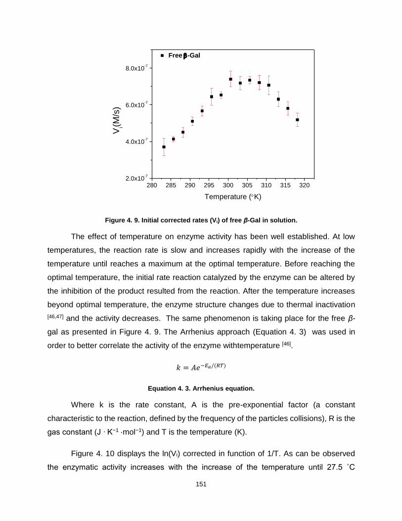
151
Figure 4. 9. Initial corrected rates (Vi) of free β-Gal in solution.
The effect of temperature on enzyme activity has been well established. At low
temperatures, the reaction rate is slow and increases rapidly with the increase of the
temperature until reaches a maximum at the optimal temperature. Before reaching the
optimal temperature, the initial rate reaction catalyzed by the enzyme can be altered by
the inhibition of the product resulted from the reaction. After the temperature increases
beyond optimal temperature, the enzyme structure changes due to thermal inactivation
[46,47] and the activity decreases. The same phenomenon is taking place for the free β-
gal as presented in Figure 4. 9. The Arrhenius approach (Equation 4. 3) was used in
order to better correlate the activity of the enzyme withtemperature [46].
𝑘 = 𝐴𝑒−𝐸𝑎/(𝑅𝑇)
Equation 4. 3. Arrhenius equation.
Where k is the rate constant, A is the pre-exponential factor (a constant
characteristic to the reaction, defined by the frequency of the particles collisions), R is the
gas constant (J · K−1 ·mol−1) and T is the temperature (K).
Figure 4. 10 displays the ln(Vi) corrected in function of 1/T. As can be observed
the enzymatic activity increases with the increase of the temperature until 27.5 ˚C
280 285 290 295 300 305 310 315 3202.0x10
-7
4.0x10⁻⁷
6.0x10⁻⁷
8.0x10⁻⁷
Free -Gal
Vi(M
/s)
Temperature (K)

152
followed by a region where it remains constant. After 37.5 C the activity slowly decreases.
This means that the optimal temperature for the enzyme is 37.5 C. The degradation trend
of the enzyme is a straight line and a first-order reaction in agreement with Arrhenius’
equation and with the literature [48].
Figure 4. 10. ln (Vi) vs 1/T for the free β-Gal in solution.
It has been reported in literature that many proteins retained or increased their
activity when absorbed on the hydrophobic surfaces [49] or on SLNs [22]. For this reason,
the activity of the free enzyme in the presence of SLNs was analyzed in function of
temperature. The SLNs were prepared as detailed in Chapter 2, by replacing the solution
of enzyme with a mixture containing PBS/Glycerol (50/50 wt%). After the formation of the
SLNs, an enzyme solution was added, having the same final concentration as the enzyme
solution used for the analysis of the free enzyme.
The Figure 4. 11 displays the profiles of the initial rates of the ONP transformation
catalyzed by β-Gal put in presence of SLNs (β-Gal+SLN) at different temperatures. Each
experimental point represents the average of 2 repetitions. The values for these
experiments were very similar and the calculated error is negligible. It can be observed
that the initial rate for β-Gal+SLN has a similar trend as the initial rate for free β-Gal with
an increase of more than 35% all over the temperature range.
0.0035 0.0034 0.0033 0.0032 0.0031
-14.8
-14.6
-14.4
-14.2
-14.0
Free -Gal
ln(V
i)
1/T
ln(Vi)=4001 (1/T)-27
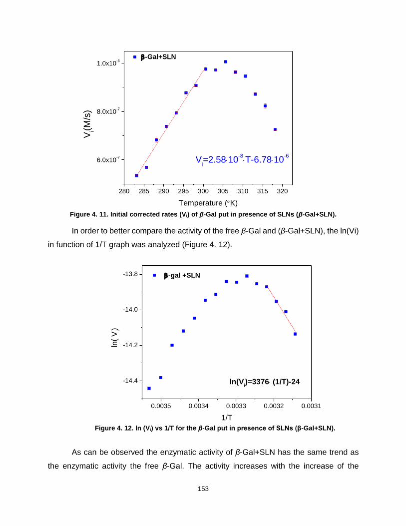
153
Figure 4. 11. Initial corrected rates (Vi) of β-Gal put in presence of SLNs (β-Gal+SLN).
In order to better compare the activity of the free β-Gal and (β-Gal+SLN), the ln(Vi)
in function of 1/T graph was analyzed (Figure 4. 12).
Figure 4. 12. ln (Vi) vs 1/T for the β-Gal put in presence of SLNs (β-Gal+SLN).
As can be observed the enzymatic activity of β-Gal+SLN has the same trend as
the enzymatic activity the free β-Gal. The activity increases with the increase of the
280 285 290 295 300 305 310 315 320
6.0x10⁻⁷
8.0x10⁻⁷
1.0x10⁻⁶
Vi=2.5810
-8T-6.7810
-6
-Gal+SLN
Vi(M
/s)
Temperature (K)
0.0035 0.0034 0.0033 0.0032 0.0031
-14.4
-14.2
-14.0
-13.8
ln(Vi)=3376 (1/T)-24
-gal +SLN
ln(
Vi)
1/T

154
temperature until 27.5 ˚C followed by a region where it remains constant. After 37.5 ˚C
(the optimal temperature) the activity slowly decreases. Compared with the Free β-Gal,
the β-Gal+SLN not only has a higher activity (by comparing the values of Vi and ln(Vi)
respectively) but also has lower rate of degradation (by comparing the slope of the regions
where the enzyme degrades). As it can be observed the slope for the degradation of the
free β-Gal is higher (4001 M·K·s-1) compared with the slope for the degradation of β-
Gal+SLN (3376 M·K·s-1).
In order to observe the effect of the encapsulation on the enzyme, the colloidal
suspension of SLNs encapsulating enzymes molecules (β-Gal@SLN) was analyzed in
the same way. The final concentration of the enzyme used for encapsulation was the
same as the one used to analyze the free β-Gal and β-Gal+SLN.
The Figure 4. 13 displays the profiles of the initial rates of the ONP transformation
catalyzed by β-Gal+SLN. Each experimental point represents an average of 3 repetitions.
Figure 4. 13. Initial corrected rates (Vi) of the colloidal suspension of SLNs encapsulating enzymes
molecules (β-Gal@SLN).
The concentration of β-Gal+SLN and the initial concentration of enzyme used to
form β-Gal@SLN formulation were the same, therefore, one can assume that the enzyme
280 285 290 295 300 305 310 315 3202.0x10
-7
4.0x10⁻⁷
6.0x10⁻⁷
8.0x10⁻⁷
Vi=2.5810
-8T-7.0710
-6
-Gal@SLN
Vi(M
/s)
Temperature (K)
155
that is released from the SLNs presents the same behavior as the enzyme in the presence
of SLN. It can be observed by comparing the Figure 4. 13 with Figure 4. 11, that at a
temperature below 28°C, the initial rate Vi has a linear increase with the increase of the
temperature for both samples. The slopes in this region, for β-Gal+SLN and for β-
Gal@SLN are the same (2.58·10-8 Ms-1K-1). This indicates that in case of β-Gal@SLN
no release is taking place in this region as the concentration of the enzyme is constant
as in the case of β-Gal+SLN. The difference in the slopes equation is the intercept value
that is smaller for β-Gal@SLN compare to β-Gal+SLN. This indicates that the enzyme
quantity present in the sample β-Gal@SLN is low. The substrate (hydrophilic) does not
have access to the enzyme that is entrapped in the lipids (hydrophobic) and only the
enzyme that is outside the SLN (the enzyme that was not entrapped) can hydrolyse the
substrate.
To quantify the amount of enzyme encapsulated in the SLNs the Vi of the enzyme
in the solution of the studied samples (β-Gal+SLN and β-Gal@SLN) were compared.
Starting from Michaelis-Menten equation (Equation 4. 4) and the fact that the both
samples have the same behavior at low temperatures (they have the same rate constant
k) the Equation 4. 5 was used to calculate the encapsulation efficiency.
𝑉𝑖 = 𝑘[𝐸]
Equation 4. 4. Michaelis-Menten equation.
Where Vi is the initial speed of the reaction (M/s), k is the rate constant (M/s·mg)
and [E] is the concentration of the enzyme (mg/ml).
The Vi value at 20°C was taken into account to minimize the errors that can appear
at 10°C due to air humidity that can influence the results. As it can be observed, the error
bar of the value Vi at 10°C for β-Gal@SLN is higher than the error bar for the Vi value at
20°C. Another reason that the Vi values at 20°C were taken into consideration is because
the SAXS patterns obtained for that sample (Figure 4. 5) at 10°C and 20°C are the same,
proving that the crystalline structure remains the same.

156
The encapsulation efficiency (EE), defined as the percentage of β-Gal
encapsulated in the SLN compared with the amount of enzyme used for the preparation
of the formulation ([𝛽 − 𝐺𝑎𝑙]+ (Equation 4. 5). This value was calculated to be
38.92±3.48%
%𝐸𝐸 =
𝑉@
𝑉+[𝛽 − 𝐺𝑎𝑙]+
[𝛽 − 𝐺𝑎𝑙]𝑡∗ 100
Equation 4. 5. Encapsulation efficiency (EE).
Where %EE is the encapsulation efficiency, V@ is the rate of the enzyme that is in
the solution of the formulation β-Gal@SLN, V+ and [β-Gal]+ is the rate and the
concentration of enzyme that is in the solution of the formulation β-Gal+SLN and [β-Gal]t
is the total enzyme concentration used.
In order to better understand the activity of β-Gal@SLN and to observe where and
if a release is taking place, the ln(Vi) in function of 1/T graph was analyzed (Figure 4. 14).
Figure 4. 14. ln (Vi) vs 1/T for the β-Gal@SLN.
0.0035 0.0034 0.0033 0.0032 0.0031
-15.2
-15.0
-14.8
-14.6
-14.4
-14.2
-14.0
ln(Vi)=1954 (1/T)-20
-Gal@SLN
ln(V
1)
1/T
157
As can be observed the enzymatic activity of β-Gal@SLN has the same trend as
the enzymatic activity of the free β-Gal and β-Gal+SLN for the region 10-27.5 ˚C, they
increases with the increase of the temperature. On a closer look, it can be observed that
in the region between 27.5 ˚C and 37.5 ˚C the enzymatic activity slowly increases for β-
Gal@SLN. Taking into account that β-Gal+SLN and β-Gal@SLN have the same
behavior, this indicates that the enzyme starts to be released from the SLNs. The slope
of the regions where the enzyme degrades,temperatures higher than 37.5 ˚C which
represents the optimal temperature for the enzyme were compared. The slope for the
degradation of β-Gal@SLN (1954 MKs-1), which is correlated with the enzyme
degradation, is 57% smaller than in the case of β-Gal+SLN (3376 MKs-1). The
interpretation of this result is that two distinct phenomena occur in the same time, the
release of the enzyme combined with the partial inactivation of the enzyme.
4.1.3. Conclusions and perspectives
In this study β-Gal from Kluyveromyces lactis was entrapped in thermo-
responsive shea butter solid lipid nanoparticles. The SLN formulation was prepared from
food grade ingredients, through a safe and low energy method (melt dispersion
technique) without organic solvent. The enzyme was encapsulated through a double
emulsion affording a loading efficiency of 40% in the SLNs (β-Gal@SLN). According to
the in situ experiments made under thermal variation, no release of the enzyme was
detected at a temperature below 27.5°C, while the carriers are still solid. The UV-visible
spectroscopy experiments made on the β-Gal+SLN sample also shown that in presence
of the lipid particles, the activity of the enzyme increased by 35% on an interval from 10°C
to 45°C, compared to the native activity of the free enzyme in the same range of
temperatures. The enhanced enzymatic activity effect was maintained for the sample β-
Gal@SLN. Concerning the enzyme release behavior observed for the formulation (β-
Gal@SLN), the presence of an increase in activity in the values of Vi starting from 27.5
˚C coincided with the melting temperature of the SNLs (28 ˚C). This means that the
encapsulated enzyme in the SLNs is released with the temperature trigger. These results
are encouraging for continued investigation towards the formulation of non-toxic carriers
for temperature triggered release of enzymes in the human body.

158
In perspective, the improvement of this delivery system is tied to the melting point
of the SLNs. The increase of the melting point from 28°C for the previously described
SLNs to a temperature closer to the human body, 37.5°C, could be attained by the
addition of a small percentage of food grade fatty acids such as palmitic or stearic acids.
These lipids have a higher melting point compare to shea butter, and they present a very
good compatibility with triglyceride fatty acids in general[50]. The resulting future SLNs
would present a melting point closer to the body temperature which improved the
structural integrity and homogeneity of the carrier until the intestine.

159
4.2. pH-responsive hybrid silica-alginate carrier for lactose intolerance treatment
Alginate is a polysaccharide biopolymer recognized as biocompatible, food grade
and abundant in nature. This linear polymer is an anionic block copolymer composed of
1-4-linked residues of β-D-mannuronic acid block covalently linked to the α-L-guluronic
acid block. A large density of the electrostatic interactions between the carboxylate
groups (-COO-) of the α-L-guluronic residues and multivalent cation, like Mg2+, Ca2+ or
Fe3+ the mostly used, lead to the formation of a hydrogel as shown in the Figure 4. 15.
The calcium alginate hydrogels are of interest to control and to target a release
phenomenon specifically in the gastrointestinal tract, as this polymer is pH sensitive. In
fact, at low pH (e.g. stomach), the alginate hydrogel shrinks as the carboxylate groups
are been totally protonated (-COOH, pKa~3.5), while above in basic pH (~7) the hydrogel
swells since most of the carboxyl groups are negatively charged. As a consequence, the
alginate chains start to repel each other and water infiltration occurs in the polymer
network [51].
Figure 4. 15. Arrangement of alginate chains in presence of a divalent cations
The enzyme encapsulation procedure is always carried out under very mild
conditions. Polysaccharides such as alginate were already utilized to entrap enzymes [53].
The only drawbacks of using a polymeric hydrogel are related to the large pores that can
appear in the network which can lead to enzyme leakage [54–56] or to the diffusion of an

160
acidic solution inside the hydrogel that may denature the enzyme, as it may happen in
the stomach.
In order to take advantage of the pH sensitive release mechanism presented by
the alginate while mitigating the mass transfer phenomena associated with the polymeric
matrix, we propose here the combination of mesoporous silica together with an alginate
matrix in the elaboration hybrid organic-inorganic particles. The hybrid organic-inorganic
composite materials could combine the advantages of both materials. Many researchers
have prepared hybrid silica-polysaccharide composites for enzyme immobilization to
improve the mechanical properties of alginate hydrogel. As an example, the alginate/silica
biocomposites can be synthesized by impregnation of mesoporous silica particles, such
as MCM-41, with alginic acid solution that contained β-galactosidase from K. fragilis,
followed by the ionic gelation of the biopolymer. The hybrid material exhibited a higher
stability upon ageing ( kept one week at 5˚C) compared with the alginate gel alone [57].
In this work, the β-galactosidase entrapment efficiency was studied and compared
for alginate/silica hybrid particles (ASP) and alginate/silica core-shell materials (SAM).
Tetramethyl orthosilicate (TMOS) was used as silica precursor, since the methanol that
is released during hydrolysis-condensation of the silicon alkoxides is less harmful for the
enzyme compared to ethanol, which is released when using tetraethyl orthosilicate source
(TEOS) [58].
4.2.1. Particle characterization
The detailed protocol preparation of the two types of hybrid inorganic-organic
samples ASP and SAM has been described in Chapter 2. Alginate beads (AP) were
prepared to compare physical characteristics of the hybrid material with the organic
alginate beads. Once obtained, the materials were lyophilized, as preparation for electron
microscopy. The morphology and the structure of alginate, and the core-shell and the
hybrid alginate silica particles were assessed by scanning electron microscopy. All the
samples kept their form after the immobilization. The AP (Figure 4. 16 A) and ASP (Figure
4. 17 A) materials have a sphere-like form while in the case of SAM ( Figure 4. 18 A) the
alginate beads seem to beentrapped in a silica network. From the SEM micrographs, the
surface of the particles can be closely observed. AP (Figure 4. 16 B) and ASP (Figure 4.

161
17 B) surface present a similar coarse surface indicating the presence of small particles,
while the SAM (Figure 4. 18 B) surface has the appearance of a continuous network.
Figure 4. 16. SEM micrographs of (A) alginate particle and (B) zoom on the surface of the material.
Figure 4. 17. SEM micrographs of (A) hybrid alginate-silica particle (ASP) and (B) zoom on the surface of the material.

162
Figure 4. 18. SEM micrographs of (A) the core-shell alginate/silica materials (SAM), (B) zoom on the surface of the material.
The apparent crystals-like structures on the surface of AS and ASP were analyzed
by the Energy Dispersive Spectroscopy (EDS) analysis. The imprint of magnesium is
present, as are oxygen and silicon as can be observed in Figure 4. 19. As such, the small
particles present on the surface of both AS and ASP are the crystalized magnesium salts.
Figure 4. 19. EDS analysis on the AP surface (A) on the ASP surface (B) and on the small particles on the coarse surface.
C
O
Mg
keV0
5
10
15
20
25
30
35
0 5 10 15 20
Energy (KeV)
A
O
Si
keV0
5
10
15
20
25
30
0 5 10 15 20
B
C O
Mg
Si
keV0
5
10
15
20
25
30
35
0 5 10 15 20
C

163
In order to compare the silica network for ASP and SAM both materials were
calcined according to the following ramp settings: 1h at 100°C, 2h at 350°C and 3h at
550°C. After the calcination, all organic compounds are removed. As it can be observed
in Figure 4. 20 A the silica that resulted from the ASP forms a continuous network, with
small inorganic particles (Figure 4. 20 B). As shown above, these small particles are a
mixture of magnesium salt and silica that formed during the synthesis (Figure 4. 19 C).
The porous sponge like structure of the silica confirms that the ASP was initially a mixture,
and that the alginate and the silica polymerize together.
Figure 4. 20. SEM micrographs of calcined ASP (A) and zoom on the surface of the silica material (B).
Compared with the calcinated ASP that is a continuous network, the silica structure
remained after calcination of SAM (Figure 4. 21) is a framework displaying sphere-shaped
holes. This is the result of the removal of the alginate beads in the calcination process.

164
Figure 4. 21. SEM micrographs of calcined SAM (A) and zoom on the silica material (B).
Infrared experiments were carried out on the three materials, AP, ASP and SAM.
The spectra are presented in Figure 4. 22 without supplementary processing.
Figure 4. 22. ATR-FTIR spectra of dried enzyme, AP, ASP and SAM.
ATR-FTIR spectra of ASP and SAM display the typical bands of silica the bands
at 1057, 960 and 790 cm-1, corresponding to common Si-O-Si and Si-OH stretching
described in literature as characteristic of silica materials. At the same time, the bands at
165
1057 and 790 cm-1 can be related to the presence of Si-O-C and C-O-C bonds[59]. The
characteristic bands of the alginate ( the ones that do not overlap with the characteristic
bands for the enzyme) at 1730 cm-1 (corresponding of C=O stretch of COOH) and at 2850
cm-1 and 2924 cm-1 (corresponding to the C-H stretch) [60] can be clearly seen in the SAM
spectra. In the ASP spectra, these bands are less well represented, probably indicating
strong interactions between silica and the alginate. The presence of 1651 cm-1 and 1535
cm-1 bands in all three materials indicate the presence of the encapsulated enzyme [61].
To confirm the presence of the enzyme, the three materials AP, ASP and SAM
were dispersed into a solution containing 1 M NaOH, the standard used to dissolve silica
networks, and 0.9 wt% of NaCl, to dissolve the alginate network. These solutions were
analyzed with the BCA assays in order to quantify the amount of enzyme (E) entrapped
in the different systems. The results are as following:
- in AP sample: 46.500±0.064 mg (E)/g (AP)
- in ASP sample: 106.000±0.001 mg (E)/g (ASP)
- in SAM sample: 619.000±0.006 mg (E)/g (SAM)
The simple alginate beads encapsulated the lowest quantity of enzyme.
Comparing the AP and ASP, the hybrid material encapsulated more enzyme. This result
is expected, according to the literature[54], as the alginate gel has a more porous structure
and the enzyme can easily leak during the washing step. The SAM material presented
the highest load of enzyme. This can be due to the fact that a silica network covers the
already formed alginate beads, preventing a leakage and assuring a better encapsulation.
Only the ASP and SAM materials were taken into consideration and analyzed in a
simulated gastro-intestinal digestion for two reasons. The high porosity of the alginate
promotes a rapid leakage and a fast inactivation of the enzyme in the acidic pH. The
second reason is that the isopropanol was used to release the alginate beads from the
emulsion and the enzyme most likely loses a part of its activity. That is why the AP
material was only used in the physical characterization of the materials.

166
4.2.2. Enzyme release in gastro intestinal simulated fluids
In order to simulate the passage of the hybrid alginate-silica materials through the
gastric system four samples for each ASP and SAM were prepared in separated batches,
as described in Chapter 2. In a standard experiment, the silica samples were suspended
consecutively in two simulated gastro-intestinal fluids for up to 2h in each: the simulated
gastric fluid first (SGF) and the simulated intestinal fluid second (SIF); the suspension
was kept under stirring (150 rpm). For each sample, the supernatant recovered after
centrifugation, was used to quantify and determine the activity of the enzyme released,
while the hybrid materials were used to determine the specific activity of the material.
Table 4. 1 displays the cumulative quantity of enzyme released e.g. after 3H in the
simulated gastro-intestinal fluid, the total quantity of enzyme released is the sum of the
quantity of enzyme released after 2H in SGF and the quantity of enzyme released after
1H in SIF. The values that are given in the Table 4. 1 are in weight percentage (quantity
of enzyme release /initial quantity of enzyme encapsulated per mg of material).
A clear difference in release profiles of ASP and SAM materials was observed.
The ASP materials have a very slow release. In SGF (pH 3) almost no release is
observed, 0.002 wt % of enzyme is released after the first hour and 0.025 wt% of enzyme
is released in the second hour. When added in SIF (pH 7) a steady and (still) slow release
is observed, 0.114 wt % in the first hour and 0.195 wt% in the second hour. This means
that only 0.205 mg of enzyme was released from 105 mg that was encapsulated per gram
of silica during the simulated gastro-intestinal fluid. Conversely more enzyme is released
from SAM. This is a clear difference observed from the data. During SAM incubation in
SGF (pH 3), 3.83 wt % of enzyme is released in the first hour and 17.61 wt % in the
second hour. The release is progressively increased in SIF (pH 7). Then 42.69 wt % is
released in the first hour and 82.92 wt % is released in the second hour. This means that
almost 520 mg of enzyme was released from 619 mg that was encapsulated per g of
silica during the simulated gastro-intestinal fluid.
Table 4. 1.Quantity of enzyme released from ASP and SAM in SGF (pH 3) and SIF (pH 7) in wt%.

167
1 h in SGF 2 h in SGF 2 h in SGF and
1h in SIF
2 h in SGF and 2h in SIF
ASP 0.002 0.025 0.114 0.195 SAM 3.83 17.61 42.69 82.92
In order to check if the enzyme released during the simulated digestion kept its
activity, the recovered solutions from in vitro simulated gastric fluid (SGF) and simulated
intestinal fluid (SIF) were analyzed. In terms of activity, the enzyme released from ASP
and SAM displays very a different behavior, that are opposite compared with the released
profiles. Table 4. 2 presents the enzymatic activity in U/mg of enzyme. The enzyme
liberated by SAM along the experiments lost almost all its activity, having 6.7 U/mg after
the first hour in SGF decreasing at 0.44 U/mg after the second hour in SGF. When the
enzyme is released in SIF fluid (pH 7) the enzyme continues to remain almost inactive.
Compared to the enzyme recovered from SAM, the enzyme released from ASP sample
still retained part of its reactivity. When added in the acidic pH, the enzyme lost a part of
its activity after the first hour continuing to decrease in the second hour when is released
in SGF. In SGF (pH=7) the activity of the enzyme remains almost constant.
Table 4. 2. Activity of the enzyme (U/mg) released from ASP and SAM in SGF (pH 3) and SIF (pH 7).
1 h in SGF 2 h in SGF 2 h in SGF
and 1h in SIF
2 h in SGF and
2h in SIF ASP 1739.131 108.74 37.56 35.75 SAM 6.70 0.44 0.33 0.45
To verify if the enzyme retains a part of its activity during the gastric simulation, the
specific activities of the materials used in the experiment were compared with the initial
ASP and SAM specific activities (Figure 4. 23). The same phenomenon occurred, as
observed in case of the enzyme released, concerning the activity measured for the
materials after their incubation in the simulate gastric system as presented in Figure 4.
23 . The graph displays the enzymatic activity of the SAM and ASP materials, compared
to the activity of the free enzyme. Both SAM and ASP materials start losing their activity
after the first hour in SGF (pH 3) and continue to decrease after the second hour in the
acidic pH. Compared with ASP activity, the SAM activity does not increase when the

168
materials are inserted in a basic pH. The activity of the free enzyme follows the same
trend as the activity of the SAM materials.
Figure 4. 23. Activity of silica materials after a stay in the simulated gastro-intestinal fluids (SGF
and SIF) (on the left Y axe) compared with the activity of the free enzyme (on the right Y axe).
This concludes that, although the SAM material can encapsulate a larger quantity
of enzyme and presents a better release compared with ASP materials, the SAM
materials are unable to protect the enzyme form the acidic pH. The ASP materials can
better protect the encapsulated enzyme, but the tight network hinder the release. The
typical use of this type of materials is in catalysis, where they constitute enzyme supports.
The supports provide a well protected environment for the enzyme, hindering its leakage
during the process and during the repeatability cycles [54,62]. The results of our
experiments are in good accord with this fact.
4.2.3. Conclusion and perspectives
Enzyme encapsulated core–shell alginate/silica (SAM) and hybrid alginate silica
materials (ASP) were synthetized by direct emulsion (water-in-oil type emulsion). The
encapsulated enzyme presented a higher release from SAM, but with a rapid decrease
of its activity in acidic pH. In the case of SAM, the trigger pH worked correctly, the material
presenting a low release in acidic pH and a high release at neutral pH, however the
natural inactivation of both the released and the encapsulated enzyme in the acidic pH

169
occurred. The ASP encapsulated enzyme presents a slow release during the incubation
in the simulated gastro-intestinal fluids, but an increase of specific activity of the material
was noticed when introduced into basic pH. The activity of the ASP liberated enzyme
along the 4h is much greater than in the case of SAM. The very slow liberation of the
enzyme, coupled with the protection from pH inactivation propose the ASP materials as
a viable biocatalyst option for the removal of lactose in the dairy industry. As a perspective
towards a better triggering at basic pH values the modification of the silica composition is
envisioned. For example the insertion of NH2 moieties in the silica structure via (3-
Aminopropyl)triethoxysilane (APTES) could act as basicpoles for the local increase of pH.
This design change might allow the protection from inactivation of the enzyme over the
acidic pH range.

170
Bibliography
[1] C. Bernal, L. Sierra, M. Mesa, ChemCatChem 2011, 3, 1948–1954. [2] M. C. Horn, Methods and Compositions for Retarding the Staling of Baked Goods, 2003, EP1533840 B1. [3] M. Vinceković, M. Viskić, S. Jurić, J. Giacometti, D. Bursać Kovačević, P. Putnik, F. Donsì, F. J. Barba, A.
Režek Jambrak, Trends Food Sci. Technol. 2017, 69, DOI 10.1016/j.tifs.2017.08.001. [4] F. Kayaci, T. Uyar, Food Chem. 2012, 133, 641–649. [5] V. Nedovic, A. Kalusevic, V. Manojlovic, S. Levic, B. Bugarski, Procedia Food Sci. 2011, 1, 1806–1815. [6] D. Dale, Microencapsulation in the Food Industry, Elsevier, 2014. [7] C. Acosta, E. Pérez-Esteve, C. A. Fuenmayor, S. Benedetti, M. S. Cosio, J. Soto, F. Sancenón, S. Mannino,
J. Barat, M. D. Marcos, et al., ACS Appl. Mater. Interfaces 2014, 6, 6453–6460. [8] P. de Vos, M. M. Faas, M. Spasojevic, J. Sikkemaa, Int. Dairy J. 2010, 292–302. [9] M. Chávarri, I. Marañón, M. C. Villarán, Probiotics 2012, 501–540. [10] A. K. Patel, R. R. Singhania, A. Pandey, Curr. Opin. Food Sci. 2016, 7, 64–72. [11] P. Walde, S. Ichikawa, Biomol. Eng. 2001, 18, 143–177. [12] A. C. Pierre, Biocatal. Biotransformation 2004, 22, 145–170. [13] E.-M. Dusterhoft, M. Minor, K. Nikolai, N. Hargreaves, Simon Huscroft, U. Scharf, Encapsulated Functional
Bakery Ingredients, 2006, US20060110494 A1. [14] N. A. Solomon, Lactase Formulations, 2013, US20130058913 A1. [15] B. Gibbs, S. Kermasha, I. Alli, C. Mulligan, Int J Food Sci Nutr. 1999, 50, 213–224. [16] R. A. Sheldon, Adv. Synth. Catal. 2007, 349, 1289–1307. [17] N. Carlsson, H. Gustafsson, C. Thörn, L. Olsson, K. Holmberg, B. Åkerman, Adv. Colloid Interface Sci. 2014,
205, 339–60. [18] B. Innovation, Bioencapsulation Innov. 2013, 1–24. [19] P. Severino, T. Andreani, A. S. Macedo, J. F. Fangueiro, M. H. A. Santana, A. M. Silva, E. B. Souto, J. Drug
Deliv. 2012, 2012, 750891. [20] S. Morel, E. Ugazio, R. Cavalli, M. R. Gasco, Int. J. Pharm. 1996, 132, 259–261. [21] P. Ganesan, D. Narayanasamy, Sustain. Chem. Pharm. 2017, 6, 37–56. [22] A. J. Almeida, E. Souto, Adv. Drug Deliv. Rev. 2007, 59, 478–90. [23] H. Reithmeier, J. Herrmann, A. Göpferich, J. Control. Release 2001, 73, 339–350. [24] M. García-Fuentes, D. Torres, M. J. Alonso, Colloids Surfaces B Biointerfaces 2003, 27, 159–168. [25] M. Trotta, R. Cavalli, M. E. Carlotti, L. Battaglia, F. Debernardi, Int. J. Pharm. 2005, 288, 281–288. [26] M. Gallarate, M. Trotta, L. Battaglia, D. Chirio, J. Microencapsul. 2009, 26, 394–402. [27] C. Qi, Y. Chen, Q.-Z. Jing, X.-G. Wang, Int. J. Mol. Sci. 2011, 12, 4282–93. [28] C. Qi, Y. Chen, J.-H. Huang, Q.-Z. Jin, X.-G. Wang, J. Sci. Food Agric. 2012, 92, 787–93. [29] I. K. Voets, W. a Cruz, C. Moitzi, P. Lindner, E. P. G. Arêas, P. Schurtenberger, J Phys Chem B 2010, 114,
11875–11883. [30] W. Mehnert, K. Mader, Adv. Drug Deliv. Rev. 2001, 47, 165–196. [31] M. Liu, H. Du, W. Zhang, G. Zhai, Mater. Sci. Eng. C 2017, 71, 1267–1280. [32] K. Yong, S. Hong, 2007, 32, 669–697. [33] B. J. Sun, H. C. Shum, C. Holtze, D. A. Weitz, ACS Appl. Mater. Interfaces 2010, 2, 3411–3416. [34] M. Depardieu, M. Nollet, M. Destribats, V. Schmitt, R. Backov, Part. Part. Syst. Charact. 2013, 30, 185–192. [35] M. Nollet, M. Depardieu, M. Destribats, R. Backov, V. Schmitt, Part. Part. Syst. Charact. 2013, 30, 62–66. [36] F. G. Honfo, N. Akissoe, A. R. Linnemann, M. Soumanou, M. A. J. S. Van Boekel, Crit. Rev. Food Sci. Nutr.
2014, 54, 673–86. [37] L. N. M. Ribeiro, M. C. Breitkreitz, V. A. Guilherme, G. H. R. da Silva, V. M. Couto, S. R. Castro, B. O. de
Paula, D. Machado, E. de Paula, Eur. J. Pharm. Sci. 2017, 106, 102–112. [38] H. A. Hassan, M. Florentin, B. N. I. Sandrine, D. Pierrick, J. Jordane, L. Michel, Eur. J. Lipid Sci. Technol.
2015, n/a-n/a. [39] R. P. Raffin, A. Lima, R. Lorenzoni, M. B. Antonow, C. Turra, M. P. Alves, S. B. Fagan, J. Biomed.
Nanotechnol. 2012, 8, 309–15. [40] A. Cortial, M. Vocanson, E. Loubry, S. Briançon, Flavour Fragr. J. 2015, 30, 467–477. [41] I. P. Kaur, R. Agrawal, Recent Pat. Drug Deliv. Formul. 2007, 1, 171–182. [42] F. Suter, D. Schmid, F. Wandrey, F. Zülli, Eur. J. Pharm. Biopharm. 2016, 108, 304–309. [43] H. Bunjes, M. H. J. Koch, K. Westesen, Langmuir 2000, 16, 5234–5241. [44] R. A. Robinson, A. Peiperl, 1981, 2572, 1–2. [45] K. Selmeczi, C. Michel, A. Milet, I. Gautier-luneau, C. Philouze, J. Pierre, D. Schnieders, A. Rompel, C. Belle,
Chem. - A Eur. J. 2007, 9093–9106. [46] H. Zhang, L. Wang, Q. Shen, B. Wu, P. Gao, Acta Biochim. Biophys. Hung. 2011, 43, 409–417. [47] M. E. Peterson, R. M. Daniel, M. J. Danson, R. Eisenthal, Biochem. J. 2007, 402, 331–337. [48] Z. Quinn Z.K, C. Xiao Dong, Biochem. Eng. J. 2001, 9, 33–40.
171
[49] T. Zoungrana, G. H. Findenegg, W. Norde, Prog. Biotechnol. 1998, 15, 495–504. [50] D. Valeri, A. J. A. Meirelles, J. Am. Oil Chem. Soc. 1997, 74, 1221–1226. [51] Z. Zhang, R. Zhang, L. Chen, Q. Tong, D. J. McClements, Eur. Polym. J. 2015, 72, 698–716. [52] K. Kashima, I. Masanao, in Adv. Desalin., 2012, pp. 3–36. [53] T. Haider, Q. Husain, Int. J. Biol. Macromol. 2007, 41, 72–80. [54] Y. Lu, Z. yi Jiang, S. wei Xu, H. Wu, Catal. Today 2006, 115, 263–268. [55] A. Blandino, M. Macı, D. Cantero, M. Macías, Mac, Enzyme Microb. Technol. 2000, 27, 319–324. [56] A. Blandino, M. Macias, D. Cantero, Proc. Biochem. 2001, 36, 601–606. [57] T. Coradin, J. Livage, Comptes Rendus Chim. 2003, 6, 147–152. [58] D. Avnir, T. Coradin, O. Lev, J. Livage, J. Mater. Chem. 2006, 16, 1013–1030. [59] G. E. Chernev, B. I. Samuneva, P. R. Djambaski, I. M. M. Salvado, H. V. Fernandes, Cent. Eur. J. Chem.
2006, 4, 81–91. [60] G. Lawrie, I. Keen, B. Drew, A. Chandler-Temple, L. Rintoul, P. Fredericks, L. Grøndahl, Biomacromolecules
2007, 8, 2533–2541. [61] I.-A. Pavel, S. F. Prazeres, G. Montalvo, C. Garcıa Ruiz, V. Nicolas, A. Celzard, F. Dehez, L. Canabady-
Rochelle, N. Canilho, A. Pasc, Langmuir 2017, 33, 3333–3340. [62] S. Xu, Z. Jiang, Y. Lu, H. Wu, W. Yuan, 2006, 511–517.

172
General Conclusions and Perspectives
Designing biocatalyst and smart enzyme delivery systems remains a challenge for
the food industry. Thus, the main goal of this PhD work was to investigate new formulation
methods, to design bio-responsive carriers of interest for catalysis in food production, and
for the elaboration of solid diary lactase supplements.
This work presented the investigation pathways and the interpretation results of
the chemical properties and responsive behaviors of four different carriers for the β-
galactosidase immobilization. The formulation routes chosen were mainly based on two
types of enzyme immobilization strategies: the physical adsorption and the entrapment.
All the carriers obtained apply to two types of physiological stimuli, either the pH found in
the small intestine or to body temperature.
Among the organic and inorganic matrix supports, the physical adsorption of
enzymes was widely done on amorphous silica supports since 2001. Porosity is a
parameter that can afford a high loading material. That is why, as discussed in Chapter
3, amorphous silica materials with hierarchical meso-macroporosity were used as β-
galactosidase supports building the design and fabrication of a bio-catalyst. A series of
enzyme-supported catalysts were prepared from different concentrations of free β-
galactosidase (e.g. enzyme feed solutions). The enzymatic activity of the bio-catalyst was
determined as a function of the loading degree. Surprisingly, it was observedthat a
selective adsorption of the enzyme in hierarchical meso or macropores occurred. Based
on enzyme dimensional simulations it was hypothesized that, at low enzyme feed solution
concentrations, the adsorption of the lactase took place preferentially in the mesopores
as dimers or monomers, while the tetrameric form was adsorbed in the macropores.
Consequently, a bio-catalyst prepared from high concentration enzyme feed solution
yielded the lowest loading degree. However, this catalyst presented the highest
enzymatic activity.
In Chapter 3, another silica based carrier was designed as a pH responsive
system, that would retain the lactase in the gastric fluid but to release it in the small
intestine fluid, based on the change in pH. β-galactosidase was physically adsorbed on

173
low-porosity micrometric silica beads. The beads were then covered by a biocompatible
coating of 1,2-dioleoyl-sn-glycero-3-phosphocholine (DOPC) liposomes. This formulation
strategy afforded liposome-coated silica particles with a controlled release pattern over
gastric to intestinal pH (pH3 to pH7). Interestingly, the presence of liposomes also proved
to be a protective coating against acidic pH, preventing the denaturation of the adsorbed
enzyme. The obtained results recommend employing this type of formulation as a
promising carrier for the release of lactase in the small intestine.
Chapter 4 covered the formulation of stimuli-responsive carriers prepared in the
context of enzyme entrapment strategies. The first approach was to entrap lactase in
food-grade solid lipid nanoparticles (SLN) through a water/oil/water double emulsion. The
advantage of using a low melting point solid lipid, shea butter, conferred a thermal
response to the carrier. The carrier would melt slowly around body temperature affording
the release of the enzyme. The enzyme was encapsulated into this type of nanoparticles
(up to 40% loading efficiency), via the inner water phase of a double emulsion. This
procedure limits its diffusion and ensures its protection. The enzyme release kinetic was
followed by in situ UV-vis spectrophotometry under a thermal range control (10°C to
45°C), an innovative way according to literature. The enzymatic activity of three types of
samples (free β-Gal, β-Gal+SLN, β-Gal@SLN) were measured in order to determine the
thermal threshold release and the activity of the enzyme in presence of the SLN. By this
analytical method, the starting temperature release was detected at 27.5°C. The activity
of the enzyme freed from the SLN, increased by 35% compared to free enzyme. Given
the precise temperature of release and the reproducibility of the results, such a carrier
could be adjusted for body temperature triggered release, and further, could serve as a
reliable delivery system for β-Galactosidase.
The entrapment of lactase in a hybrid carrier that combined alginate gels and silica
was investigated and the results were presented in the second part of Chapter 4. This
approach aimed to design a high loaded pH responsive carrier, that can protect the
enzyme from the gastric fluids, while allowing its delivery in the small intestine. The
protection and release properties of two types of carriers prepared via emulsions were
investigated and compared, core-shell alginate/silica and hybrid alginate-silica materials.
According to the results, the hydrid alginate-silica carrier presented a slower release of

174
the enzyme. It displayed a higher activity and a good prevention against acidic pH
inactivation.
The different types of carriers obtained by the defined formulation strategies are
promising candidates in overcoming some challenges of the food industry. There are
some general perspectives can still be explored in order to improve this work. Regarding
silica supported enzyme catalyst incased in the phospholipid liposome layer, the main
drawback of that carrier is its weak loading degree. This point could be improved by using
porous silica supports presenting higher pore sizes relative to the enzyme dimension.
Another strategy could be changing the lipid nature or type. This type of carrier may allow
a triggered release, based on body temperature.
In the case of the SLN based carrier, the improvement of this delivery system is
tied to the melting point of the lipid (mp). A very good correlation between this and enzyme
release was already achieved. The increase from 32.5°C melting point of shea butter to
37.5°C, human body temperature could be attained by the addition of a small percentage
of fatty acid. Palmitic or stearic acids have high melting points, they are safe for human
consumption and present a very good compatibility with other fats. The resulting SLNs
would present a higher overall melting point and would keep their structural integrity and
homogeneity.
Another strategy would be the formation of a shell of silica around the SLN. An
interesting opportunity is to replace the surfactant for the formation of the double emulsion
with silica nanoparticles, to form a Pickering emulsion. A Pickering emulsion is the
emulsion stabilized by solid particles, such assilica nanoparticles. The particles act by
being adsorbed irreversibly at the interface of two phases and reduce the interfacial
tensions.
A brief exploration of this principle has already been done, applied directly on this
subject by using silica nanoparticles to stabilize SLNs. Thus, in order to form an inverse
emulsion, the silica nanoparticles had to be partially hydrophobized. For this, stabilizing
HDK® T40 hydrophilic fumed-silica nanoparticles (NPs) purchased from Wacker
(Germany), were modified with stearic acid. Using the modified NPs and a hard oil at
room temperature, Witocan P (kindly provided by Palsgaard) a solid lipid nanoparticles
dispersion, stabilized by NPs was obtained, as shown the figure below. This type of SLN

175
Pickering-like stabilized suspension, presented a very good preservation of structural
integrity, which is the first criterium of enzyme entrapment. As a preliminary result, this
finding is very encouraging towards this strategy.
Figure 1. Witocan P solid lipid nanoparticles dispersion stabilized by Pickering
emulsion
This work aims to prove that enzymatic catalysis is a thing of the present, and that
it can be adapted to any specific challenges. During this project, we tried to showcase the
variety and richness of possibilities of encapsulation techniques. Four distinct types of
systems have been designed and characterized, with the goal to emphasize how each
certain method can accommodate different requirements of the material. My hope is that
this work will contribute to the further development of supported enzymatic catalysis and
to that of smart foods.

176
Conclusions générales et perspectives
La conception de biocatalyseurs et de systèmes intelligents pour la libération
d’enzymes, restent un défi pour l’industrie alimentaire. Ainsi, l’objectif principal de ce
travail de doctorat était d’élaborer de nouvelles méthodes de formulation pour la
conception de transporteurs bio-sensibles d’intérêt pour la catalyse de la réaction
d’hydrolyse du lactose et ainsi élaborer des suppléments solides de lactase pour
l’industrie agroalimentaire.
Ce travail a montré les voies de recherche et la caractérisation des propriétés
chimiques et les comportements bio-sensibles de quatre systèmes de transport conçu
pour l’immobilisation de β-galactosidase. Tous les systèmes de transport obtenus
présentent des réponses à deux types de stimuli physiologiques, soit la température soit
le pH. De plus, les voies de formulation choisis étant dictées par deux types de stratégies
d’immobilisation d’enzyme : l’adsorption physique et l’emprisonnement.
Parmi les matrice supports, organiques et inorganiques, l’adsorption physique des
enzymes était faite largement sur les supports de silice amorphe depuis 2001. Le degré
et la nature de la porosité sont des paramètres qui permettre d’atteindre un taux
d’adsorption important dans le matériau. C’est pourquoi, comme discuté au chapitre 3,
les matériaux de silice amorphe avec une méso-macro porosité hiérarchique, ont été
utilisés comme support pour l’élaboration d’un biocatalyseur. Une série de catalyseurs
supportés a été préparée à partir de différentes concentrations de β-galactosidase
(solutions « feed » d’enzyme). L’activité enzymatique du catalyseur a été déterminée par
rapport au dégrée d’adsorption en enzyme. Curieusement, une adsorption sélective
d’enzyme dans les méso-macro pores hiérarchiques a eu lieu. A partir de simulations
dimensionnelles de l’enzyme, les hypothèses suivantes ont été émises : à une
concentration faible de solution enzymatique l’adsorption de la lactase se passe
préférentiellement dans les mésopores sous la forme de dimers ou de monomères, alors
que la forme tétramérique est préférentiellement adsorbée dans les macropores à plus
haute concentration. En conséquence, un biocatalyseur préparé à une concentration
élevée en enzyme présentait le dégrée minimum de chargement, néanmoins ce
catalyseur avait une plus grande activité enzymatique étant donné que le tétramère est
la forme la plus active.

177
Dans le chapitre 3, un autre transporteur à base de silice a été conçu pour être
sensible au pH physiologique, et protéger la lactase dans le fluide gastrique mais
permettre sa libération dans l’intestin grêle. Ainsi, après l’adsorption physique sur des
billes micrométriques de silice avec une faible porosité, les billes ont été couvertes par
une couche biocompatible de liposomes à base de 1,2-dioleoyl-sn-glycero-3-
phosphocholine (DOPC). Cette stratégie de formulation a généré des particules de silice
enrobées de liposomes, montrant une libération contrôlée au pH intestinal (pH7). De
facon intéressante d’après les résultats, la présence des liposomes a joué le rôle d’une
couche protectrice à pH acide, empêchent ainsi la dénaturation de l’enzyme immobilisée.
A partir des résultats obtenus, ce type de formulation peut conduire à l’élaboration d’un
transporteur prometteur pour la libération de la lactase dans l’intestine.
Le chapitre 4 décrit la formulation de transporteurs dans lesquels la stratégie
d’emprisonnement de l’enzyme a été choisi. La première approche était tout d’abord
l’emprisonnement de la lactase dans les nanoparticules solides lipidiques (SLNs) par une
double émulsion eau/huile/eau. L’usage de SLN assurait la libération de l’enzyme par un
stimuli thermique, la température du corps humain. Le taux d’encapsulation dans les
nanoparticules a été déterminé allant jusqu’à 40% dans la phase intérieure de l’émulsion
double qui limite sa diffusion et assure sa protection. La cinétique de libération de
l’enzyme a été suivie in situ par spectrophotométrie UV-vis dans un intervalle de
température allant de 10°C to 45. Cette méthodologie a été très peu explorée d’après la
littérature. L’activité enzymatique pour trois types d’échantillons (β-Gal libre, β-Gal+SLN,
β-Gal@SLN), a été mesurée pour déterminer la limite thermale de libération et l’activité
de l’enzyme en présence des SLNs. Par cette méthode analytique, le début de libération
a été détectée à 27.5°C et l’activité de l’enzyme libre en présence des SLNs a été
quantifiée à 35% supérieur à l’enzyme seule en solution. D’après ces résultats, un tel
transporteur pourrait être reformulé pour obtenir une libération de la de β-Galactosidase
à la température du corps.
Finalement, l’emprisonnement de la lactase dans un transporteur hybride qui
combine l’alginate et la silice a aussi été étudié et présenté dans le Chapitre 4. Cette
approche a visé la conception un transporteur hautement chargé en enzyme qui
répondrait au pH pour ainsi protéger l’enzyme du fluide gastrique mais permet sa livraison

178
dans l’intestin. Les propriétés de protection et de libération pour deux types de
transporteurs également produits à partir d’émulsions ont été investigués et comparés.
Les deux types de transporteurs sont les suivants : core/shell alginate/silice et hybride
alginate-silice. D’après les résultats, le transporteur hybride alginate-silice a présenté une
libération plus lente de l’enzyme, une activité supérieure et une meilleure protection
contre le pH acide que le système core/shell alginate/silice.
Les deux types de transporteurs fabriqués par la voie d’émulsion sont prometteurs
pour remplir les défis de l’industrie alimentaire. Cependant, les perspectives suivantes
peuvent être explorées pour améliorer ce travail.
Concernant l’enzyme supportée par les billes de silice enrobées de la couche de
liposomes phospholipidiques, l’inconvénient majeure de ce transporteur est le faible
degré d’adsorption d’enzyme. Ce point pourrait être amélioré par l’usage de supports
sphériques avec un volume libre plus important et une dimension de pores supérieure à
la taille de l’enzyme.
En perspective, l’amélioration dans le cas des transporteurs à base de SLNs est
liée du point de fondre. Une très bonne corrélation entre la température de fusion et la
libération de l’enzyme a été déterminé. L’incrément de 4°C, du point de fusion du beurre
de karité par l’ajout d’acide gras tels que l’acide palmitique ou l’acide stéarique,
permettrait d’induire le relargage de l’enzyme à la température du corps humain. Les
SLNs résultants présenteraient un point de fusion global plus élevé tout en gardant leur
homogénéité et intégrité structurale.
Une autre stratégie pour favoriser l’adhésion des SLN à l’intestin serait la formation
d’une couche de silice autour des SLN. Une voie facile est le replacement du surfactant
pour la formation de la double émulsion par des nanoparticules de silice, pour former une
émulsion Pickering. Une émulsion Pickering est une émulsion stabilisée par des
particules solides, comme les nanoparticules de silice. Les particules sont absorbées
irréversiblement à l’interface entre les deux phases liquides, réduisant ainsi les tensions
interfaciales.
Une courte exploration a été faite en utilisant des nanoparticules de silice
hydrophiles de type HDK® T40. Cependant pour former une émulsion inverse les
particules de silice ont été hydrophobisées au préalable par des chaines d’acide
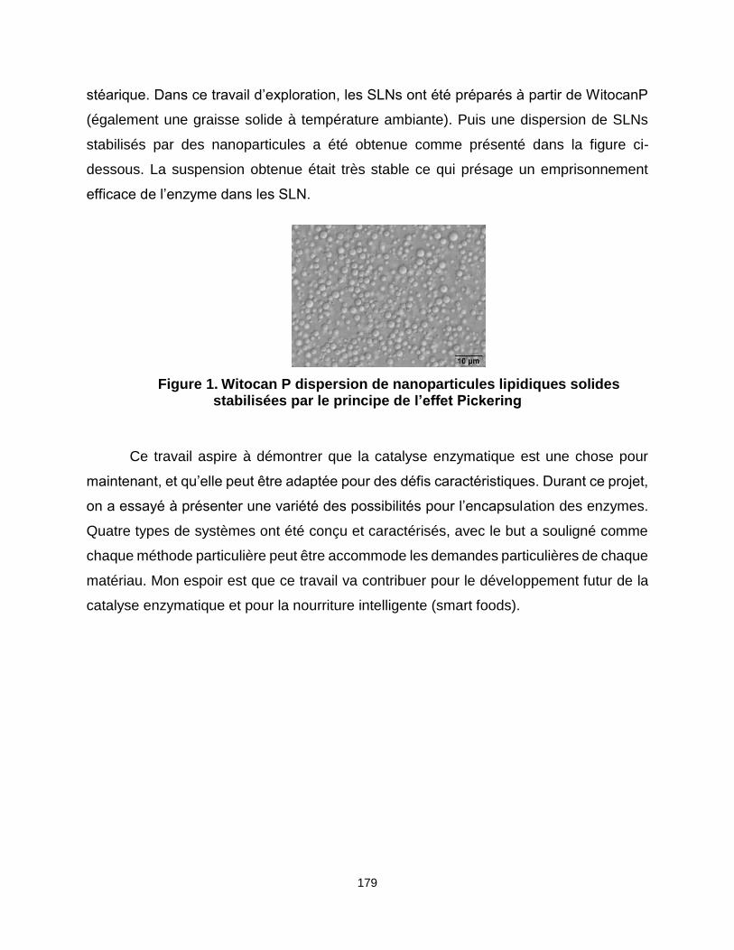
179
stéarique. Dans ce travail d’exploration, les SLNs ont été préparés à partir de WitocanP
(également une graisse solide à température ambiante). Puis une dispersion de SLNs
stabilisés par des nanoparticules a été obtenue comme présenté dans la figure ci-
dessous. La suspension obtenue était très stable ce qui présage un emprisonnement
efficace de l’enzyme dans les SLN.
Figure 1. Witocan P dispersion de nanoparticules lipidiques solides stabilisées par le principe de l’effet Pickering
Ce travail aspire à démontrer que la catalyse enzymatique est une chose pour
maintenant, et qu’elle peut être adaptée pour des défis caractéristiques. Durant ce projet,
on a essayé à présenter une variété des possibilités pour l’encapsulation des enzymes.
Quatre types de systèmes ont été conçu et caractérisés, avec le but a souligné comme
chaque méthode particulière peut être accommode les demandes particulières de chaque
matériau. Mon espoir est que ce travail va contribuer pour le développement futur de la
catalyse enzymatique et pour la nourriture intelligente (smart foods).

180
Appendix 1 Techniques of characterization
DLS
Dynamic light scattering (DLS) (also known as Quasi- Elastic Light Scattering or
Photon Correlation Spectroscopy) is a technique allowing the evaluation of particle size
and size distribution of emulsions, micelles, polymers, proteins, nanoparticles and
colloids. The sample is illuminated by a laser beam and the time-dependent fluctuations
of the scattered light are detected at a known scattering angle θ (90° in our case) by a
photon detector. The particles undergo a random motion due to thermal agitation (or
Brownian motion) leading to the scattering of light in all directions. This motion results in
fluctuations of the distances between the particles, and hence also in fluctuations of the
phase relations of the scattered light waves. Additionally, the number of particles within
the scattering volume may also vary in time. All the fluctuating scattered intensity is
recorded and mathematically treated to plot a so-called correlation function from which a
diffusion coefficient (D) of the scatters is determined. Then with the Stockes-Einstein
equation, the hydrodynamic radius (RH) of the particles is calculated:
with kB the Boltzmann constant, T the absolute temperature, η the viscosity of the
continuous medium and D the diffusion coefficient.
Small angle X-ray scattering (SAXS)
Small angle X-ray scattering (SAXS) is a technique able to indirectly define the
size, the shape and the internal structure of samples being powder, fibers, colloidal
dispersions of solid particles, peptides, polymers or nanostructured systems. The
dimensional detection size range extends from 1 to 100 nm. This technique is based on
the differences of scattering intensity induced by electron densities variation between the
scatters and the continuous medium. The diffusion intensity curves l(q) is usually
represented as a function of the scattering vector, q

181
where θ is the scattering angle and λ is the X-ray beam wavelength. The diffusion of the
X-ray waves gives a scattering pattern following Bragg’s law. When an intense scattering
is observed at small angles it means that the distance between the electrons is large. If
there is a periodic arrangement in the sample or a monodisperse size, a multiple
scattering pattern is recorded.
were dh,k,l is the repetition distance, θ is the diffraction semi-angle, n is the reflection order,
λ is the X-ray wavelength.
The dimensional and structural parameters of the studied system are given by the
relationship between the repeat distances determined mathematically from the Bragg
reflection patterns.
Nitrogen sorption analysis
Adsorption is a surface phenomenon consisting of the
physical adhesion of atoms, ions, or molecules from a gas, liquid, or dissolved solid
(adsorbate) to a surface (adsorbent) through Van der Waals forces.
The nitrogen adsorption-desorption technique provides information about the

182
texture characteristics of a solid e.g. surface area, pore size and pore size distribution.
Usually, the probing adsorbate used is N2 with a boiling temperature of 77K. Under
controlled pressure, the gaseous molecules adsorbed to the material in layers. According
to Brunauer, Emmett and Teller theory (BET) which is an extension of Langmuir theory,
adsorbate gas molecules are more realistically adsorbed in multilayers.
The quantity of adsorbed gas is plotted as a function of the equilibrium pressure
(P/P0). According to IUPAC the isotherms are classified according to six types:
Figure Appendix 1 -1 Isotherms physisorption (IUPAC classification)[335]
I- Type I isotherm indicates that the molecules of the adsorbate form a monolayer. It rises
sharply at low relative pressures and reaches a plateau: the amount of gas adsorbed by
the unit mass of solid reaches a limited value, as the relative pressure(P/P0) is almost 1.
This type of isotherm indicates the existence of micropores.
- Type II isotherm indicates an unrestricted monolayer-multilayer formation after the
formation of a monolayer at low relative pressure, and is found for non-porous or
macroporous adsorbents.

183
- Type III isotherm is not common and shows that no monolayer is formed at the surface
of the solid (unrestricted multilayer). This type of isotherm is characteristic of a weak
adsorbent-adsorbate interaction because the interactions between adsorbed molecules
are stronger compared with the interactions between the adsorbent surface and
adsorbate.
- Type IV isotherm indicates the existence of mesopores. At relatively small pressure the
fast increase of the adsorbed nitrogen quantity corresponds to the adsorption of a
monolayer. The presence of the mesopores (2-5 nm) can be observed at higher relative
pressures when the adsorbed volume is translated by a capillary condensation
phenomenon, followed by a plateau. Because of this phenomenon (irreversible nature of
the capillary condensation), a hysteresis loop appears when the nitrogen starts the
desorption process.
- Type V isotherm is attributed to monolayer-multilayer adsorption, it follows the same
path as Type II isotherms but, it also exhibits a hysteresis loop due to capillary
condensation, which is associated with the mechanism of pore filling and emptying.
- Type VI isotherm is associated with layer-by-layer adsorption on a highly uniform non-
porous surface. The step-height represents the monolayer capacity for each adsorbed
layer and, in the simplest case, remains nearly constant for two or three adsorbed layers.
The specific surface area, can be calculated from adsorption-desorption isotherms using
the following algorithm
The Brunauer-Emmett-Teller equation is used to determine surface area of the porous
material from the monolayer volume capacity (Vm):
With V the adsorbed volume at a pressure P; Vm is the monolayer volume; P0 is the
saturating vapor pressure of the adsorbate gas; C is constant.

184
where σ is the molecular cross-sectional area (16.2 Å2 for N2 at 77 K); Vm is the molar
gas volume; Na is Avogadro number.
The pore size distribution is calculated using the BJH method (Barrett, Jayne and
Halenda). This theory is based on the phenomenon of capillary condensation which
appears in the mesopores, and by applying the Kelvin law, which links the pressure P at
which the condensation happens to the curvature radius of the meniscus of the formed
liquid.
where γ is the surface tension at a temperature T; rP is the pore radius; t is the thickness
of the adsorbed layer.
This calculation is applied to the adsorption branch of the isotherm. The pore size
range that can be detected by this technique is between 1.8 and 50 nm.

185
Appendix 2 Shea butter technical sheet
INTEREXPORT SERVICES
Fiche technique / Technical specifications
Beurre de KARITE raffiné/ Refined SHEA butter
Date : 28/03/2012
Reference ies LABO HV014
Mode d'extraction / Extraction process : Extrait par pression puis raffiné
Obtained by pressure and refined
Origine / Origin : AFRIQUE / AFRICA
Aspect / Appearance : Solid couleur crème/Solid colour cream
Nom INCI / INCI name : BUTYROSPERMUM PARKII BUTTER
Nom CTFA /CTFA name : BUTYROSPERMUM PARKII (SHEA) BUTTER
N° CAS / CAS number : 194043-92-0
N° EINECS / EINECS number : -
Durée de vie recommandée / Shelf life : 2 ans / 2 years
CARACTERISTIQUES/CHARACTERISTICS Valeur/Value
Indice d’acide / Acid index (mgKOH /g)..........................................................max 2
Indice de péroxyde / Peroxide index (méq O2/kg) ............................................max 5
Indice d’iode / Iodine value (gI2 /100g)..........................................................51 - 72
Teneur en insaponifiable / Unsaponifiable matter ........................................% min 4
Point éclair / Flash point ...............................................................................°C >290
Point d’ébullition / Boiling point ..................................................................°C >200
Point de fusion / Melting point ..................................................................°C 32 - 44
COMPOSITION EN ACIDES GRAS/COMPOSITION IN FATTY ACIDS %
C12:0 Acide laurique / Lauric acid ...................................................................max 1
C14:0 Acide myristique / Myristic acid ............................................................max 1
C16:0 Acide palmitique / Palmitic acid ............................................................3 - 10
C18:0 Acide stéarique / Stearic acid ...............................................................36 - 50
C18:1 Acide oléique / Oleic acid ....................................................................40 - 50
C18:2 Acide linoléique / Linoleic acid ...............................................................3 - 8
C18:3 Acide linolénique / Linolenic acid .........................................................max 1
C20:0 Acide arachidique / Arachidic acid ........................................................max 3
C20:1 Acide gadoléique / Eicosenoic acid........................................................max 1
C22:0 Acide béhénique / Behenic acid .............................................................max 1
C24:0 Acide lignocérique / Tetracosenoic acid ................................................max 1
Huiles végétales – Producteur d’extraits végétaux et de macérats huileux – Producteur BIO depuis 1995
Zone artisanale – F 04 700 ORAISON Tél. 33 4 92 72 55 55 Fax. 33 4 92 72 55 56.
E-mail: [email protected] www.ieslabo.com
SAS au capital de 37 740€ - APE 2042 Z - SIRET 334 042 496 00060 - TVA FR 33 334 042 496

186
Appendix 3 Halactase technical sheet
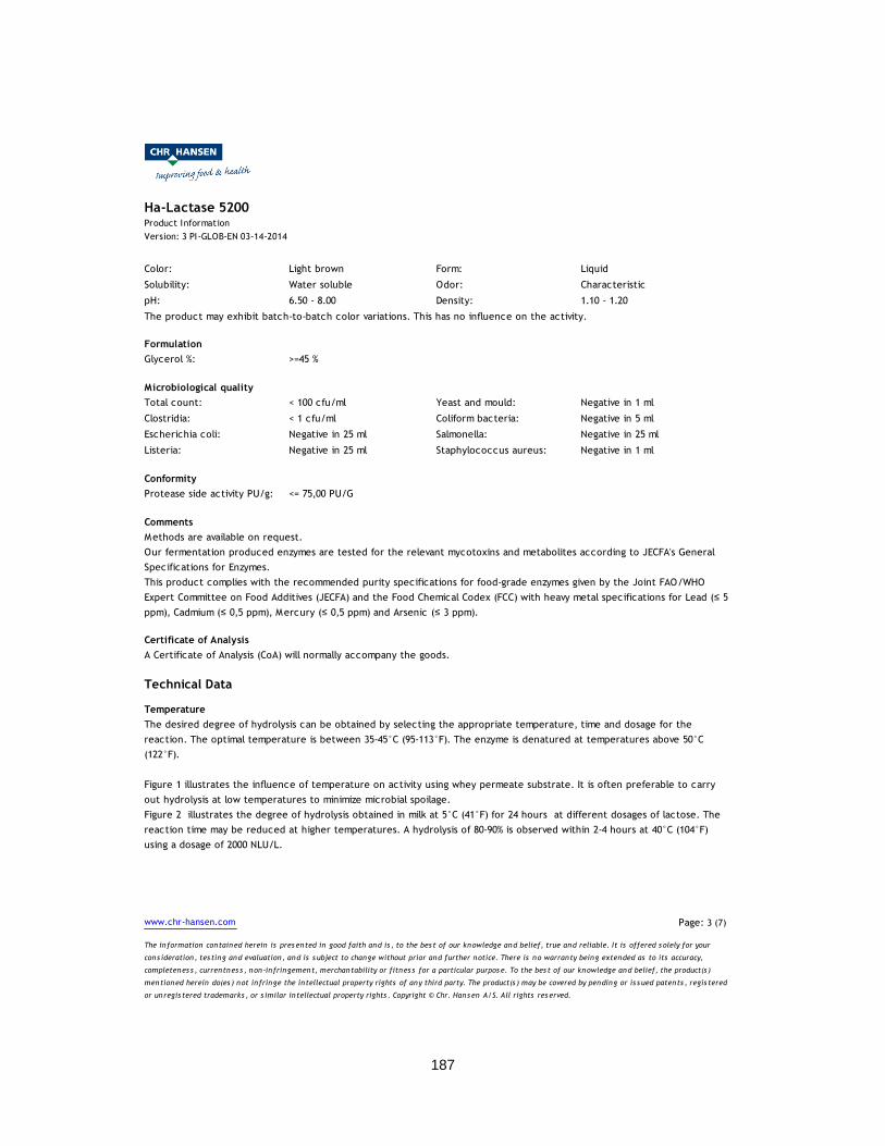
187

188

189

190

191
Copyright © 2022 FDOKUMEN