Emphysema is associated with increased inflammation in lungs of atherosclerosis-prone mice by...
10
RESEARCH Open Access Emphysema is associated with increased inflammation in lungs of atherosclerosis-prone mice by cigarette smoke: implications in comorbidities of COPD Gnanapragasam Arunachalam, Isaac K Sundar, Jae-woong Hwang, Hongwei Yao, Irfan Rahman * Abstract Background: Chronic obstructive pulmonary disease is associated with numerous vascular effects including endothelial dysfunction, arterial stiffness and atherogenesis. It is also known that a decline in lung function is associated with increased cardiovascular comorbidity in smokers. The mechanism of this cardiopulmonary dual risk by cigarette smoke (CS) is not known. We studied the molecular mechanisms involved in development of emphysema in atherosclerosis-prone apolipoprotein E-deficient (ApoE -/- ) mice in response to CS exposure. Methods: Adult male and female wild-type (WT) mice of genetic background C57BL/6J and ApoE -/- mice were exposed to CS, and lung inflammatory responses, oxidative stress (lipid peroxidation products), mechanical properties as well as airspace enlargement were assessed. Results and Discussion: The lungs of ApoE -/- mice showed augmented inflammatory response and increased oxidative stress with development of distal airspace enlargement which was accompanied with decline in lung function. Interestingly, the levels and activities of matrix metalloproteinases (MMP-9 and MMP-12) were increased, whereas the level of eNOS was decreased in lungs of CS-exposed ApoE -/- mice as compared to air-exposed ApoE -/- mice or CS-exposed WT mice. Conclusion: These findings suggest that CS causes premature emphysema and a decline of lung function in mice susceptible to cardiovascular abnormalities via abnormal lung inflammation, increased oxidative stress and alterations in levels of MMPs and eNOS. Background Chronic obstructive pulmonary disease (COPD) is char- acterized by chronic airflow limitation resulting from excessive airway inflammatory response mediated by cigarette smoke (CS). Comorbidities such as cardiovascu- lar disease, diabetes, lung cancer, and osteoporosis are more prevalent in smokers and patients with COPD [1-3]. Recent studies have shown that smokers with altered forced expiratory volume in one second (FEV 1 ) and airflow limitation are associated with arterial stiff- ness, exaggerated atherosclerosis and vice-versa [2,4,5]. Growing evidence also indicates that inflammation, endothelial dysfunction and oxidative modification of lipids play an important role in the pathogenesis of ather- osclerosis and COPD [3,6,7]. In addition to CS, alcohol consumption is also one among the important contribut- ing factors involved in the pathogenesis of COPD and atherosclerosis and their co-morbidities [8,9]. Apolipoprotein E-deficient (ApoE -/- ) mice develop atherosclerosis due to an accumulation of cholesterol ester-enriched particles in the blood resulting from a lack of triglyceride and cholesterol metabolism/lipid transport [10]. These mice have a shorter life-span and age faster than wild-type counterparts [11]. CS exposure to ApoE -/- mice promotes arterial thrombosis and mod- ulates the size and composition of neointimal lesions/ thickening [12], which is associated with increased * Correspondence: [email protected] Department of Environmental Medicine, Lung Biology and Disease Program, University of Rochester Medical Center, Rochester, NY, USA Arunachalam et al. Journal of Inflammation 2010, 7:34 http://www.journal-inflammation.com/content/7/1/34 © 2010 Arunachalam et al; licensee BioMed Central Ltd. This is an Open Access article distributed under the terms of the Creative Commons Attribution License (http://creativecommons.org/licenses/by/2.0), which permits unrestricted use, distribution, and reproduction in any medium, provided the original work is properly cited.
-
Upload
independent -
Category
Documents
-
view
1 -
download
0
Transcript of Emphysema is associated with increased inflammation in lungs of atherosclerosis-prone mice by...
RESEARCH Open Access
Emphysema is associated with increasedinflammation in lungs of atherosclerosis-pronemice by cigarette smoke: implications incomorbidities of COPDGnanapragasam Arunachalam, Isaac K Sundar, Jae-woong Hwang, Hongwei Yao, Irfan Rahman*
Abstract
Background: Chronic obstructive pulmonary disease is associated with numerous vascular effects includingendothelial dysfunction, arterial stiffness and atherogenesis. It is also known that a decline in lung function isassociated with increased cardiovascular comorbidity in smokers. The mechanism of this cardiopulmonary dual riskby cigarette smoke (CS) is not known. We studied the molecular mechanisms involved in development ofemphysema in atherosclerosis-prone apolipoprotein E-deficient (ApoE-/-) mice in response to CS exposure.
Methods: Adult male and female wild-type (WT) mice of genetic background C57BL/6J and ApoE-/- mice wereexposed to CS, and lung inflammatory responses, oxidative stress (lipid peroxidation products), mechanicalproperties as well as airspace enlargement were assessed.
Results and Discussion: The lungs of ApoE-/- mice showed augmented inflammatory response and increasedoxidative stress with development of distal airspace enlargement which was accompanied with decline in lungfunction. Interestingly, the levels and activities of matrix metalloproteinases (MMP-9 and MMP-12) were increased,whereas the level of eNOS was decreased in lungs of CS-exposed ApoE-/- mice as compared to air-exposed ApoE-/-
mice or CS-exposed WT mice.
Conclusion: These findings suggest that CS causes premature emphysema and a decline of lung function in micesusceptible to cardiovascular abnormalities via abnormal lung inflammation, increased oxidative stress andalterations in levels of MMPs and eNOS.
BackgroundChronic obstructive pulmonary disease (COPD) is char-acterized by chronic airflow limitation resulting fromexcessive airway inflammatory response mediated bycigarette smoke (CS). Comorbidities such as cardiovascu-lar disease, diabetes, lung cancer, and osteoporosis aremore prevalent in smokers and patients with COPD[1-3]. Recent studies have shown that smokers withaltered forced expiratory volume in one second (FEV1)and airflow limitation are associated with arterial stiff-ness, exaggerated atherosclerosis and vice-versa [2,4,5].Growing evidence also indicates that inflammation,
endothelial dysfunction and oxidative modification oflipids play an important role in the pathogenesis of ather-osclerosis and COPD [3,6,7]. In addition to CS, alcoholconsumption is also one among the important contribut-ing factors involved in the pathogenesis of COPD andatherosclerosis and their co-morbidities [8,9].Apolipoprotein E-deficient (ApoE-/-) mice develop
atherosclerosis due to an accumulation of cholesterolester-enriched particles in the blood resulting from alack of triglyceride and cholesterol metabolism/lipidtransport [10]. These mice have a shorter life-span andage faster than wild-type counterparts [11]. CS exposureto ApoE-/- mice promotes arterial thrombosis and mod-ulates the size and composition of neointimal lesions/thickening [12], which is associated with increased
* Correspondence: [email protected] of Environmental Medicine, Lung Biology and Disease Program,University of Rochester Medical Center, Rochester, NY, USA
Arunachalam et al. Journal of Inflammation 2010, 7:34http://www.journal-inflammation.com/content/7/1/34
© 2010 Arunachalam et al; licensee BioMed Central Ltd. This is an Open Access article distributed under the terms of the CreativeCommons Attribution License (http://creativecommons.org/licenses/by/2.0), which permits unrestricted use, distribution, andreproduction in any medium, provided the original work is properly cited.
oxidative stress, reduced glutathione levels and mito-chondrial damage leading to atherosclerotic lesion for-mation [6,13-17]. Massaro and Massaro have recentlyshown that these mice have an impaired pulmonarymorphology and functional phenotype with a rapiddecline in lung function as they age [18]. However, theunderlying molecular mechanism of the pulmonary phe-notype was not studied. We used the ApoE-/- mice,which are prone to develop atherosclerosis [19,20], tounderstand the molecular mechanism of pulmonaryphenotype in response to CS exposure, as well as tostudy the concept of accelerated decline in lung functionand aging in cardiopulmonary comorbid conditions. Wedetermined the inflammatory response, oxidative stress(lipid peroxidation products), levels/activities of matrixmetalloproteinases (MMP-9 and MMP-12) and NAD+-dependent deacetylase sirtuin 1 (SIRT1) which is shownto regulate endothelial nitric oxide synthase (eNOS)activity (endothelial function) in lungs of ApoE-/- miceexposed to CS.
MethodsReagentsUnless otherwise stated, all biochemical reagents used inthis study were purchased from Sigma Chemicals Co.,St. Louis, MO, USA. Antibodies used to detect proteinsinclude mouse specific SIRT1 and eNOS (Cell Signaling,Danvers, MA), MMP-9 and MMP-12 (Santa Cruz Bio-technology, Santa Cruz, CA) for western blotting andimmunoprecipitation.
AnimalsAdult male and female wild-type (WT) mice of geneticbackground C57BL/6J and ApoE-/- mice [19,20] (Strainnumber, B6.129P2-Apoetm1Unc/J; stock number, 002052,backcrossed to C57BL/6J for 10 generations, JacksonLaboratory, Bar Harbor, ME) were housed in the inhala-tion facility of the University of Rochester. These micewere fed with regular standard Chow diet during hous-ing and experimental procedures. ApoE-/- mice showedobvious signs of atherosclerotic lesions in the aorticsinus and ascending aorta after feeding with Chow dietat 24 weeks of age with an early onset of signs seenafter approximately 3-4 months of age (Jackson Lab).ApoE-/- mice develop atherosclerotic plaques at 2-3months after feeding with a high-fat Western-type diet[20]. All experimental protocols described in this studywere approved by the animal research committee of theUniversity of Rochester.
CS exposureAdult mice (12 weeks old, body weight ranging from 30-40 g, male and female) were exposed to CS for 3 daysusing Baumgartner-Jaeger CSM2082i automated
cigarette smoking machine (CH Technologies, West-wood, NJ) [21,22]. The smoke was generated from 3R4Fresearch cigarettes (University of Kentucky, Lexington,KY). Mainstream CS was diluted with filtered air, anddirected into the exposure chamber. Monitoring of theCS exposure (TPM per cubic meter of air, mg/m3) wasperformed in real-time using MicroDust Pro-aerosolmonitor (Casella CEL, Bedford, UK) and verified dailyby gravimetric sampling. The smoke concentration wasset at a nominal value of approximately 300 mg/m3
TPM by adjusting the flow rate of the dilution air[21,22]. The control mice were exposed to filtered air inan identical manner.
Bronchoalveolar lavage and tissue harvestThe mice were intraperitoneally injected with 100 mg/kgbody weight of pentobarbiturate (Abbott laboratories,Abbott Park, IL) and killed by exsanguination. Thelungs were lavaged three times with 0.6 ml of 0.9%sodium chloride and removed en bloc. The bronchoal-veolar lavage (BAL) fluid cell pellet was resuspended insaline, and the total cell number was counted with ahemocytometer. Differential cell count (500 cells/slide)was performed on cytospin-prepared slides (ThermoShandon, Pittsburgh, PA) stained with Diff-Quik (DadeBering, Newark, DE).
Cytokine analysisThe levels of proinflammatory mediators such as mono-cyte chemoattractant protein-1 (MCP-1) and chemokinekeratinocyte chemoattractant (KC) in lung homogenateswere measured by ELISA using respective duo-antibodykits (R&D Systems, Minneapolis, MN) according to themanufacturer’s instructions.
Immunohistochemical staining for tissue macrophagesImmunohistochemical staining for macrophages in lungsections was performed as described previously [21,22].The number of Mac-3-positive cells in each lung section(5 random microscopic fields per lung section in 3 dif-ferent sections) was counted manually at ×200 magnifi-cation and averaged.
Lipid peroxidation products assay in lung homogenateThe right lung lobe was homogenized with ice-cold20 mM Tris-HCl (pH 7.4) and centrifuged at 3,000 g at4°C for 10 min, and the supernatants were collected.Butylated hydroxytoluene (5 mM) was added to thesupernatant to prevent further peroxidation, and thesamples were immediately frozen in liquid nitrogen.Lipid peroxidation products [malondialdehyde (MDA)and 4-hydroxy-2-nonenal (4-HNE)] were measuredusing a lipid peroxidation kit (Enzo Life Sciences, PA)according to the manufacturer’s instructions [22].
Arunachalam et al. Journal of Inflammation 2010, 7:34http://www.journal-inflammation.com/content/7/1/34
Page 2 of 10
Measurement of lung mechanical propertiesLung mechanical properties were determined usingScireq Flexivent apparatus (Scireq, Monteral, Canada).The dynamic lung compliance and lung resistance weremeasured in mice anesthetized by sodium pentobarbital(50 mg/kg, intraperitoneally) and paralyzed with pancur-onium (0.5 mg/kg, intraperitoneally). A tracheotomywas performed and an 18-guage cannula was inserted 3mm into an anterior nick in the exposed trachea andconnected to a computer controlled rodent ventilator.Initially, the mice were ventilated with room air (150breaths/min) at a volume of 10 ml/kg body mass. After3 minutes of ventilation, measurement of lung mechani-cal properties were initiated by the computer generatedprogram to measure dynamic lung compliance andresistance. These measurements were repeated threetimes for each animal.
Hematoxylin and Eosin (H&E) staining and mean linearintercept analysisMouse lungs (which had not been lavaged) after CS expo-sure were inflated by 1% low-melting agarose at a pressureof 25 cm H2O, and then fixed with neutral buffered forma-lin. Tissues were embedded in paraffin, sectioned (4 μm),and stained with hematoxylin and eosin (H&E). The alveo-lar size was estimated from the mean linear intercept (Lm)of the airspace which is a measure of airspace enlarge-ment/emphysema. Lm was calculated for each samplebased on 10 random fields observed at a magnification of×200 using cross-lines as described previously [21,22].
ImmunoblottingProteins (20 μg) from lung tissue homogenates wereused for immunoblotting as described previously[21-24]. In brief, protein was electrophoresed on 7.5%SDS-PAGE gel and transblotted on nitrocellulose mem-brane (Amersham Biosciences, Piscataway, NJ). Mem-branes were blocked with 5% (w/v) non-fat milk in PBScontaining 0.1% (v/v) Tween 20 and then incubatedwith anti-SIRT1, anti-eNOS, anti-MMP-9 or anti-MMP-12 antibodies. After washing, bound antibody wasdetected using anti-rabbit/anti-mouse antibody linked tohorseradish peroxidase and bound complexes weredetected using enhanced chemiluminescence (PerkinElmer, Waltham, MA). Protein levels were measured byBCA kit as per the manufacturer’s instructions usingBSA as standards (Thermo Scientific, Rockford, IL).
SIRT1 deacetylase activity assaySIRT1 activity was assayed using a deacetylase colori-metric activity assay kit according to the manufacturer’sinstructions (Biomol International, Plymouth Meeting,PA). Briefly, SIRT1 was immunoprecipitated from wholelung homogenates (100 μg protein). After the final
washing, Color de Lys substrate reagent and NAD+ wereadded to the SIRT1 conjugated beads and incubated at37°C for 80 min. The substrate-SIRT1 mixture was thenplaced on a 96-well plate, and the Color de Lys developerreagent was added to the wells at 37°C for 20 min. Theplate was then read at 405 nm using a spectrophotometer(Model 680 microplate reader, Bio-Rad, Hercules, CA).
MMPs activity assay by zymographyThe zymography was performed to determine the activ-ity of MMPs in mouse lung as described previously [25].Briefly, lung tissues were homogenized in 400 μl lysisbuffer (50 mM Tris-HCl, pH 7.4, with protease inhibi-tors) on ice. One hundred micrograms of protein wasthen mixed with equal volume sample buffer (80 mMTris-HCl, pH 6.8, 4% SDS, 10% glycerol, 0.01% bromo-phenol blue) and then loaded on a 7.5% SDS-polyacryla-mide gel containing 1 mg/ml gelatin which was overlaidwith 5% stacking gel. After electrophoresis, gels wererinsed in distilled water, washed three times for 15 min-utes each in 150 ml 2.5% Triton X-100 solution. Gelswere then incubated in 100-150 ml of 50 mM Tris-HCl(pH 7.5), 10 mM CaCl2, 1 μM ZnCl2, 1% Triton X-100and 0.02% NaN3. After incubation, gels were stainedwith 100 ml Coomassie blue R-250 for 3 h and thendestained 1 h with destaining solution (50% methanol,10% acetic acid). Gels were washed in distilled water for20 minutes and then scanned. The intensity of bandswas quantified using image J software (Version 1.41,National Institutes of Health, Bethesda, MD, USA).
Statistical analysisData were presented as means ± SEM. Statistical analy-sis of significance was calculated using one-way analysisof variance followed by post hoc test for multigroupcomparisons using Stat View software. P < 0.05 wasconsidered as significant.
ResultsApoE-/- mice are susceptible to increased lunginflammatory cell influx in response to CSAugmented inflammatory response in the lung from envir-onmental stress or toxicants results in the activation ofinflammatory cascades in microvasculature and vesselwalls leading to a potentiation of atherogenesis[12,13,26,27]. Atherogenic prone ApoE
-/- were exposed toCS for 3 days, and the number of neutrophils and macro-phages in BAL fluid as well as in the lungs were deter-mined. CS exposure led to a higher number of neutrophilinflux in BAL fluid of ApoE-/- mice as compared to WTmice (Fig. 1A). However, CS exposure significantlydecreased the number of macrophages in BAL fluid ofApoE-/- mice, but not in WT mice (Fig. 1B). Interestingly,the macrophage infiltration in lung interstitium of
Arunachalam et al. Journal of Inflammation 2010, 7:34http://www.journal-inflammation.com/content/7/1/34
Page 3 of 10
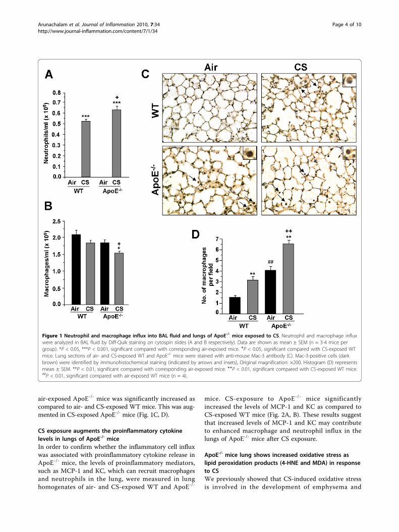
air-exposed ApoE-/- mice was significantly increased ascompared to air- and CS-exposed WT mice. This was aug-mented in CS-exposed ApoE-/- mice (Fig. 1C, D).
CS exposure augments the proinflammatory cytokinelevels in lungs of ApoE-/- miceIn order to confirm whether the inflammatory cell influxwas associated with proinflammatory cytokine release inApoE-/- mice, the levels of proinflammatory mediators,such as MCP-1 and KC, which can recruit macrophagesand neutrophils in the lung, were measured in lunghomogenates of air- and CS-exposed WT and ApoE-/-
mice. CS-exposure to ApoE-/- mice significantlyincreased the levels of MCP-1 and KC as compared toCS-exposed WT mice (Fig. 2A, B). These results suggestthat increased levels of MCP-1 and KC may contributeto enhanced macrophage and neutrophil influx in thelungs of ApoE-/- mice after CS exposure.
ApoE-/- mice lung shows increased oxidative stress aslipid peroxidation products (4-HNE and MDA) in responseto CSWe previously showed that CS-induced oxidative stressis involved in the development of emphysema and
Figure 1 Neutrophil and macrophage influx into BAL fluid and lungs of ApoE-/- mice exposed to CS. Neutrophil and macrophage influxwere analyzed in BAL fluid by Diff-Quik staining on cytospin slides (A and B respectively). Data are shown as mean ± SEM (n = 3-4 mice pergroup). *P < 0.05, ***P < 0.001, significant compared with corresponding air-exposed mice. +P < 0.05, significant compared with CS-exposed WTmice. Lung sections of air- and CS-exposed WT and ApoE-/- mice were stained with anti-mouse Mac-3 antibody (C). Mac-3-positive cells (darkbrown) were identified by immunohistochemical staining (indicated by arrows and insets), Original magnification: ×200. Histogram (D) representsmean ± SEM. **P < 0.01, significant compared with corresponding air-exposed mice. ++P < 0.01, significant compared with CS-exposed WT mice.##P < 0.01, significant compared with air-exposed WT mice (n = 4).
Arunachalam et al. Journal of Inflammation 2010, 7:34http://www.journal-inflammation.com/content/7/1/34
Page 4 of 10
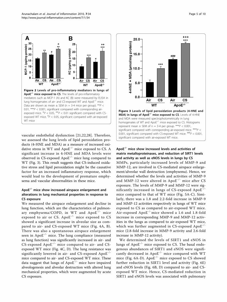
vascular endothelial dysfunction [21,22,28]. Therefore,we assessed the lung levels of lipid peroxidation pro-ducts (4-HNE and MDA) as a measure of increased oxi-dative stress in WT and ApoE-/- mice exposed to CS. Asignificant increase in 4-HNE and MDA levels wereobserved in CS-exposed ApoE-/- mice lung compared toWT (Fig. 3). This result suggests that CS-induced oxida-tive stress and lipid peroxidation might be the causativefactor for an increased inflammatory response, whichwould lead to the development of premature emphy-sema and vascular abnormalities in these mice.
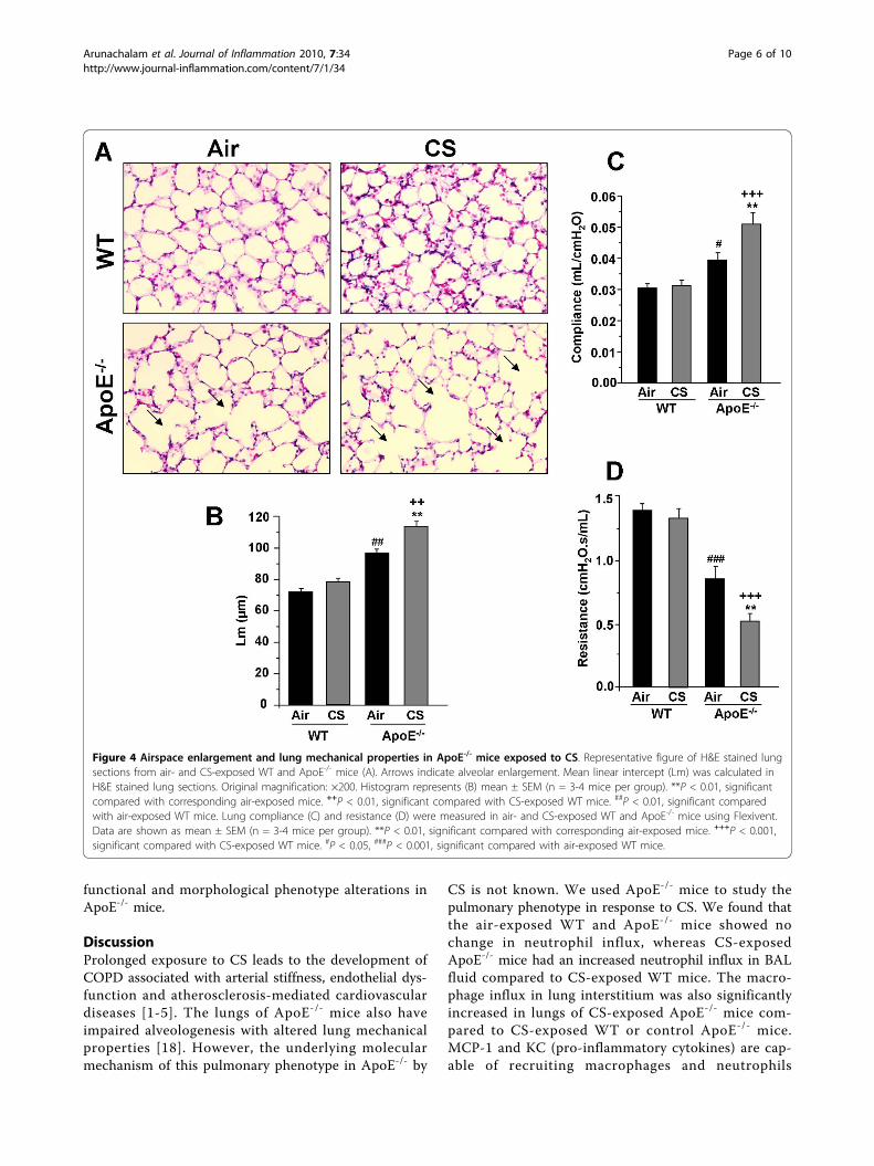
ApoE-/- mice show increased airspace enlargement andalterations in lung mechanical properties in response toCS exposureWe measured the airspace enlargement and decline inlung function, which are the characteristics of pulmon-ary emphysema/COPD, in WT and ApoE-/- miceexposed to air or CS. ApoE-/- mice exposed to CSshowed a significant increase in alveolar size as com-pared to air- and CS-exposed WT mice (Fig. 4A, B).There was also a spontaneous airspace enlargementseen in ApoE-/- mice. The lung compliance (measuredas lung function) was significantly increased in air- andCS-exposed ApoE-/- mice compared to air- and CS-exposed WT mice (Fig. 4C, D). The lung resistance wassignificantly lowered in air- and CS-exposed ApoE-/-
mice compared to air- and CS-exposed WT mice. Thesedata suggest that lungs of ApoE-/- mice have impairedalveologenesis and alveolar destruction with altered lungmechanical properties, which were augmented by acuteCS exposure.
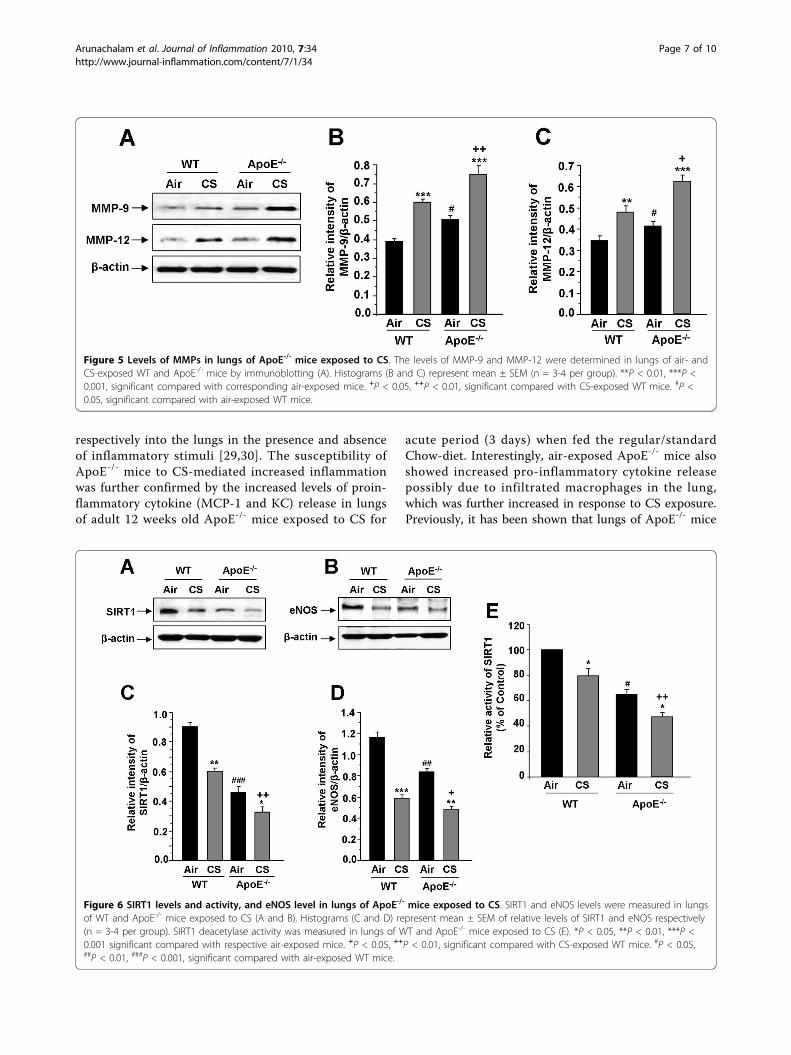
ApoE-/- mice show increased levels and activities ofmatrix metalloproteinases, and reduction of SIRT1 levelsand activity as well as eNOS levels in lungs by CSMMPs, particularly increased levels of MMP-9 andMMP-12, are involved in CS-mediated airspace enlarge-ment/alveolar wall destruction (emphysema). Hence, wedetermined whether the levels and activities of MMP-9and MMP-12 were altered in ApoE-/- mice after CSexposure. The levels of MMP-9 and MMP-12 were sig-nificantly increased in lungs of CS-exposed ApoE-/-
mice compared to that of WT mice (Fig. 5A-C). Simi-larly, there was a 1.8 and 2.2-fold increase in MMP-9and MMP-12 activities respectively in lungs of WT miceexposed to CS as compared to air-exposed WT mice.Air-exposed ApoE-/-mice showed a 1.6 and 1.8-foldincrease in corresponding MMP-9 and MMP-12 activ-ities in the lungs as compared to air-exposed WT mice,which was further augmented in CS-exposed ApoE-/-
mice (2.8-fold increase in MMP-9 activity and 2.6-foldincrease in MMP-12 activity).We determined the levels of SIRT1 and eNOS in
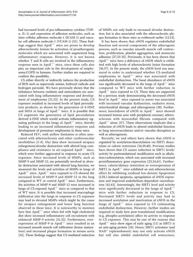
lungs of ApoE-/- mice exposed to CS. The basal endo-genous abundances of SIRT1 and eNOS were signifi-cantly decreased in ApoE-/- mice compared with WTmice (Fig. 6A-D). ApoE-/- mice exposed to CS showedfurther reduction in SIRT1 level and activity (Fig. 6E)and eNOS levels (Fig. 6B, D) compared to air- and CS-exposed WT mice. Hence, CS-mediated reduction inSIRT1 and eNOS levels was associated with pulmonary
Figure 2 Levels of pro-inflammatory mediators in lungs ofApoE-/- mice exposed to CS. The levels of pro-inflammatorymediators such as MCP-1 (A) and KC (B) were measured by ELISA inlung homogenates of air- and CS-exposed WT and ApoE-/- mice.Data are shown as mean ± SEM (n = 3-4 mice per group). **P <0.01, ***P < 0.001, significant compared with corresponding air-exposed mice. +P < 0.05, ++P < 0.01 significant compared with CS-exposed WT mice. #P < 0.05, significant compared with air-exposedWT mice
Figure 3 Levels of lipid peroxidation products (4-HNE andMDA) in lungs of ApoE-/- mice exposed to CS. Levels of 4-HNEand MDA were measured spectrophotometrically in lunghomogenates of WT and ApoE-/- mice exposed to CS. Histogramsrepresent mean ± SEM of n = 3-4 per group. ***P < 0.001,significant compared with corresponding air-exposed mice. +++P <0.001, significant compared with CS-exposed WT mice. ###P < 0.001,significant compared with air-exposed WT mice.
Arunachalam et al. Journal of Inflammation 2010, 7:34http://www.journal-inflammation.com/content/7/1/34
Page 5 of 10
functional and morphological phenotype alterations inApoE-/- mice.
DiscussionProlonged exposure to CS leads to the development ofCOPD associated with arterial stiffness, endothelial dys-function and atherosclerosis-mediated cardiovasculardiseases [1-5]. The lungs of ApoE-/- mice also haveimpaired alveologenesis with altered lung mechanicalproperties [18]. However, the underlying molecularmechanism of this pulmonary phenotype in ApoE-/- by
CS is not known. We used ApoE-/- mice to study thepulmonary phenotype in response to CS. We found thatthe air-exposed WT and ApoE-/- mice showed nochange in neutrophil influx, whereas CS-exposedApoE-/- mice had an increased neutrophil influx in BALfluid compared to CS-exposed WT mice. The macro-phage influx in lung interstitium was also significantlyincreased in lungs of CS-exposed ApoE-/- mice com-pared to CS-exposed WT or control ApoE-/- mice.MCP-1 and KC (pro-inflammatory cytokines) are cap-able of recruiting macrophages and neutrophils
Figure 4 Airspace enlargement and lung mechanical properties in ApoE-/- mice exposed to CS. Representative figure of H&E stained lungsections from air- and CS-exposed WT and ApoE-/- mice (A). Arrows indicate alveolar enlargement. Mean linear intercept (Lm) was calculated inH&E stained lung sections. Original magnification: ×200. Histogram represents (B) mean ± SEM (n = 3-4 mice per group). **P < 0.01, significantcompared with corresponding air-exposed mice. ++P < 0.01, significant compared with CS-exposed WT mice. ##P < 0.01, significant comparedwith air-exposed WT mice. Lung compliance (C) and resistance (D) were measured in air- and CS-exposed WT and ApoE-/- mice using Flexivent.Data are shown as mean ± SEM (n = 3-4 mice per group). **P < 0.01, significant compared with corresponding air-exposed mice. +++P < 0.001,significant compared with CS-exposed WT mice. #P < 0.05, ###P < 0.001, significant compared with air-exposed WT mice.
Arunachalam et al. Journal of Inflammation 2010, 7:34http://www.journal-inflammation.com/content/7/1/34
Page 6 of 10
respectively into the lungs in the presence and absenceof inflammatory stimuli [29,30]. The susceptibility ofApoE-/- mice to CS-mediated increased inflammationwas further confirmed by the increased levels of proin-flammatory cytokine (MCP-1 and KC) release in lungsof adult 12 weeks old ApoE-/- mice exposed to CS for
acute period (3 days) when fed the regular/standardChow-diet. Interestingly, air-exposed ApoE-/- mice alsoshowed increased pro-inflammatory cytokine releasepossibly due to infiltrated macrophages in the lung,which was further increased in response to CS exposure.Previously, it has been shown that lungs of ApoE-/- mice
Figure 5 Levels of MMPs in lungs of ApoE-/- mice exposed to CS. The levels of MMP-9 and MMP-12 were determined in lungs of air- andCS-exposed WT and ApoE-/- mice by immunoblotting (A). Histograms (B and C) represent mean ± SEM (n = 3-4 per group). **P < 0.01, ***P <0.001, significant compared with corresponding air-exposed mice. +P < 0.05, ++P < 0.01, significant compared with CS-exposed WT mice. #P <0.05, significant compared with air-exposed WT mice.
Figure 6 SIRT1 levels and activity, and eNOS level in lungs of ApoE-/- mice exposed to CS. SIRT1 and eNOS levels were measured in lungsof WT and ApoE-/- mice exposed to CS (A and B). Histograms (C and D) represent mean ± SEM of relative levels of SIRT1 and eNOS respectively(n = 3-4 per group). SIRT1 deacetylase activity was measured in lungs of WT and ApoE-/- mice exposed to CS (E). *P < 0.05, **P < 0.01, ***P <0.001 significant compared with respective air-exposed mice. +P < 0.05, ++P < 0.01, significant compared with CS-exposed WT mice. #P < 0.05,##P < 0.01, ###P < 0.001, significant compared with air-exposed WT mice.
Arunachalam et al. Journal of Inflammation 2010, 7:34http://www.journal-inflammation.com/content/7/1/34
Page 7 of 10
had increased levels of pro-inflammatory cytokine (TNF-a, IL-1) and expression of adhesion molecules, such asinter-cellular adhesion molecule-1 (ICAM-1) and vascu-lar cell adhesion molecule-1 (VCAM-1) [31]. These find-ings suggest that ApoE-/- mice are prone to developatherosclerotic lesions by activation of proatherogenicmolecules which are associated with augmented lunginflammatory response. However, it is not knownwhether T and B cells are involved in the inflammatoryresponse seen in ApoE-/- mice, since these cells alsoplay an important role in the development of emphy-sema/COPD in humans. Further studies are required toconfirm this possibility.CS either directly or indirectly induces the production
of ROS such as superoxide anions, hydroxyl radicals andhydrogen peroxide. We have previously shown that theimbalance between oxidants and antioxidants are asso-ciated with lung inflammatory response and develop-ment of emphysema [21,22]. In the present study, CSexposure resulted in increased levels of lipid peroxida-tion products, as shown by the generation of 4-HNEand MDA in lungs of ApoE-/- mice. It is possible thatCS augments the generation of lipid peroxidationderived 4-HNE which would activate inflammatory sig-naling pathways in the lungs of ApoE-/- mice, therebyleading to an increased inflammatory response anddevelopment of premature emphysema in these mice.Reduced FEV1 with airflow limitation is often asso-
ciated with atherosclerosis and other cardiovascularmorbidities [2-4]. Our data show increased airspaceenlargement/alveolar destruction with altered lung com-pliance and resistance in air-exposed ApoE-/- mice,which were further aggravated in response to acute CSexposure. Since increased levels of MMPs, such asMMP-9 and MMP-12, are potentially involved in alveo-lar destruction associated with altered lung function, wemeasured the levels and activities of MMPs in lungs ofApoE-/- mice. ApoE-/- mice exposed to CS showed theincreased levels of MMP-9 and MMP-12 in the lungcompared to WT or control ApoE-/- mice. Furthermore,the activities of MMP-9 and MMP-12 were increased inlungs of CS-exposed ApoE-/- mice as compared to thatof WT mice. It is possible that increased macrophageinfiltration into the lungs in response to CS exposuremay lead to elevated MMPs which might be the causefor airspace enlargement and lower lung functionobserved in these mice. It is noteworthy to mentionhere that ApoE-/- mice when fed with high cholesteroldiet show increased inflammatory cell recruitment withenhanced MMP-9 activity [31,32]. Furthermore, over-expression of MMP-9 in ApoE-/- mice resulted in anincreased smooth muscle cell infiltration (lesion matura-tion) and increased plaque formation in mouse aorta[32]. These findings suggest that CS-mediated induction
of MMPs not only leads to increased alveolar destruc-tion, but is also associated with the atherosclerotic pla-que formation in these mice as evidenced earlier [12,13].It has been shown that eNOS regulates endothelial
function and several components of the atherogenicprocess, such as vascular smooth muscle cell contrac-tion, proliferation, platelet aggregation, and monocyteadhesion [27,33-35]. Previously, it has been shown thatApoE-/- mice have a deficiency of eNOS which is exhib-ited with high levels of atherosclerotic lesion formation[36,37]. In the present study, the eNOS level was mea-sured in order to understand whether CS-mediatedemphysema in ApoE-/- mice was associated withendothelial dysfunction. The basal abundance of eNOSwas significantly decreased in the lungs of ApoE-/- micecompared to WT mice with further reduction inApoE-/- mice exposed to CS. These data are supportedby a previous study demonstrating the decreased eNOSlevel in ApoE-/- mice exposed to ozone was associatedwith increased vascular dysfunction, oxidative stress,mitochondrial damage, and atherogenesis [38]. Further-more, knockdown of eNOS in ApoE-/- mice showedincreased lesions area with peripheral coronary athero-sclerosis with myocardial fibrosis compared withApoE-/- alone [37]. These observations implicate that areduction of eNOS leads to altered endothelial functionin lung microvasculature and/or vascular disruption aswell as atherogenesis.Recently, we and others have shown that eNOS is
regulated by acetylation/deacetylation via SIRT1 deace-tylase or calorie restriction [34,39,40]. Previous studieshave shown that CS causes reduction in SIRT1 levels/activity by posttranslational modification such as alkyla-tion/carbonylation, which was associated with increasedproinflammatory gene expression [23,24,41]. Further-more, calorie/dietary restriction or overexpression ofSIRT1 in ApoE-/- mice exhibited an anti-atherosclerosiseffect by inhibiting oxidized low-density lipoprotein(LDL)-induced apoptosis, upregulation of eNOS expres-sion and improved endothelium-dependent vasorelaxa-tion [42,43]. Interestingly, the SIRT1 level and activitywere significantly decreased in the lungs of ApoE-/-
mice with further reduction in response to CS.Decreased SIRT1 levels and activity may lead toincreased acetylation and inactivation of eNOS in thelungs of ApoE-/- mice exposed to CS culminatingendothelial dysfunction. However, further studies arerequired to study how post-translational modifications(e.g. phospho-acetylation) affect its activity in responseto CS exposure. This may be one of the reasons thatApoE-/- mice show signs of early aging [11] as SIRT1 isan anti-aging protein [24]. Hence, SIRT1 activation (andNAD+ replenishment) may not only activate eNOSbut will also inhibit endothelial cell senescence,
Arunachalam et al. Journal of Inflammation 2010, 7:34http://www.journal-inflammation.com/content/7/1/34
Page 8 of 10
atherosclerosis and inflammatory response in the lung[23,44,45]. This is further validated by SIRT1 activationor calorie restriction in ApoE-/- mice leads to protectionagainst atherosclerosis progression by upregulatingeNOS [43-45]. Interestingly, our preliminary datashowed that overexpression of SIRT1 in ApoE-/- mice indouble transgenic mice protected, whereas knockdownof SIRT1 in ApoE-/- mice aggravated the lung phenotype(inflammation and emphysema).In summary, our study shows the augmented inflam-
matory response, increased oxidative stress, and airspaceenlargement with altered mechanical properties in lungsof ApoE-/- mice in response to CS, which was associatedwith increased MMPs, reduced SIRT1 activity andeNOS levels. These mice have an accumulation ofexcess lipids laden in blood and pulmonary arteries/lungmicrovasculature which can undergo rapid oxidation byCS-derived free radicals and oxidants leading to the gen-eration of secondary oxidized lipid mediators/peroxida-tion products/signaling molecules both systemically andlocally. This will trigger alterations in SIRT1, eNOS andabnormal inflammatory responses leading to pulmonaryfunctional and morphological phenotype. This may beone of the mechanisms linking CS-mediated accelerateddecline in lung function and aging in comorbidities ofcardiopulmonary diseases [46].
AbbreviationsApoE: apolipoprotein E; COPD: chronic obstructive pulmonary diseases; CS:cigarette smoke; eNOS: endothelial nitric oxide synthase; 4-HNE: 4-hydroxy-2-nonenal; MDA: malondialdehyde; MMPs: matrix metalloproteinases.
AcknowledgementsThis study was supported by the NIH 1R01HL085613, 1R01HL097751,1R01HL092842 and NIEHS ES-01247. We thank Dr. Donald Massaro(Georgetown University) for useful discussions.
Authors’ contributionsGA contributed in the study design and planning, and performed theexperiments. JH, IKS and HY participated and coordinated in completing thestudy. GA wrote the first draft of the manuscript. IR supervised the studyand contributed in data discussions and correcting the drafts. Furthermore,IR conceived the study, contributed in the study design, planning andrevised the manuscript. All authors read and approved the final manuscript.
Competing interestsThe authors declare that they have no competing interests.
Received: 12 May 2010 Accepted: 22 July 2010 Published: 22 July 2010
References1. Barnes PJ, Celli BR: Systemic manifestations and comorbidities of COPD.
Eur Respir J 2009, 33:1165-1185.2. Iwamoto H, Yokoyama A, Kitahara Y, Ishikawa N, Haruta Y, Yamane K,
Hattori N, Hara H, Kohno N: Airflow limitation in smokers is associatedwith subclinical atherosclerosis. Am J Respir Crit Care Med 2009, 179:35-40.
3. Maclay JD, McAllister DA, MacNee W: Cardiovascular risk in chronicobstructive pulmonary disease. Respirology 2007, 12:634-641.
4. Sin DD, Wu L, Man SF: The relationship between reduced lung functionand cardiovascular mortality: a population-based study and a systematicreview of the literature. Chest 2005, 127:1952-1959.
5. Sin DD, Wu L, Man SF: Systemic inflammation and mortality in chronicobstructive pulmonary disease. Can J Physiol Pharmacol 2007, 85:141-147.
6. Yang Z, Knight CA, Mamerow MM, Vickers K, Penn A, Postlethwait EM,Ballinger SW: Prenatal environmental tobacco smoke exposure promotesadult atherogenesis and mitochondrial damage in apolipoprotein E-/-mice fed a chow diet. Circulation 2004, 110:3715-3720.
7. Ota Y, Kugiyama K, Sugiyama S, Ohgushi M, Matsumura T, Doi H, Ogata N,Oka H, Yasue H: Impairment of endothelium-dependent relaxation ofrabbit aortas by cigarette smoke extract–role of free radicals andattenuation by captopril. Atherosclerosis 1997, 131:195-202.
8. Bertelli AA, Das DK: Grapes, wines, resveratrol, and heart health. JCardiovasc Pharmacol 2009, 54:468-76.
9. Kamholz KS: Wine, Spirits and the Lung: Good, Bad or Indifferent? TransAm Clin Climatol Assoc 2006, 117:129-145.
10. Mahley RW: Apolipoprotein E: cholesterol transport protein withexpanding role in cell biology. Science 1988, 240:622-630.
11. Ang LS, Cruz RP, Hendel A, Granville DJ: Apolipoprotein E, an importantplayer in longevity and age-related diseases. Exp Gerontol 2008,43:615-622.
12. Schroeter MR, Sawalich M, Humboldt T, Leifheit M, Meurrens K, Berges A,Xu H, Lebrun S, Wallerath T, Konstantinides S, Schleef R, Schaefer K:Cigarette smoke exposure promotes arterial thrombosis and vesselremodeling after vascular injury in apolipoprotein E-deficient mice.J Vasc Res 2008, 45:480-492.
13. Von Holt K, Lebrun S, Stinn W, Conroy L, Wallerath T, Schleef R: Progressionof therosclerosis in the Apo E-/- model: 12-month exposure to cigarettemainstream smoke combined with high-cholesterol/fat diet.Atherosclerosis 2009, 205:135-143.
14. Knight-Lozano CA, Young CG, Burow DL, Hu ZY, Uyeminami D,Pinkerton KE, Ischiropoulos H, Ballinger SW: Cigarette smoke exposure andhypercholesterolemia increase mitochondrial damage in cardiovasculartissues. Circulation 2002, 105:849-854.
15. Tani S, Dimayuga PC, Anazawa T, Chyu KY, Li H, Shah PK, Cercek B:Aberrant antibody responses to oxidized LDL and increased intimalthickening in apoE-/- mice exposed to cigarette smoke. Atherosclerosis2004, 175:7-14.
16. Gairola CG, Drawdy ML, Block AE, Daugherty A: Sidestream cigarettesmoke accelerates atherogenesis in apolipoprotein E-/- mice.Atherosclerosis 2001, 156:49-55.
17. Biswas SK, Newby DE, Rahman I, Megson IL: Depressed glutathionesynthesis precedes oxidative stress and atherogenesis in Apo-E(-/-) mice.Biochem Biophys Res Commun 2005, 338:1368-1373.
18. Massaro D, Massaro GD: Apoetm1Unc mice have impaired alveologenesis,low lung function, and rapid loss of lung function. Am J Physiol Lung CellMol Physiol 2008, 294:L991-L997.
19. Zhang SH, Reddick RL, Piedrahita JA, Maeda N: Spontaneoushypercholesterolemia and arterial lesions in mice lacking apolipoproteinE. Science 1992, 258:468-471.
20. Whitman SC: A practical approach to using mice in atherosclerosisresearch. Clin Biochem Rev 2004, 25:81-93.
21. Rajendrasozhan S, Chung S, Sundar IK, Yao H, Rahman I: Targeteddisruption of NF-�B1 (p50) augments cigarette smoke-induced lunginflammation and emphysema in mice: a critical role of p50 inchromatin remodeling. Am J Physiol Lung Cell Mol Physiol 2010, 298:L197-L209.
22. Yao H, Edirisinghe I, Rajendrasozhan S, Yang SR, Caito S, Adenuga D,Rahman I: Cigarette smoke-mediated inflammatory and oxidativeresponses are strain-dependent in mice. Am J Physiol Lung Cell Mol Physiol2008, 294:L1174-L1186.
23. Yang SR, Wright J, Bauter M, Seweryniak K, Kode A, Rahman I: Sirtuinregulates cigarette smoke-induced proinflammatory mediator release viaRelA/p65 NF-kappaB in macrophages in vitro and in rat lungs in vivo:implications for chronic inflammation and aging. Am J Physiol Lung CellMol Physiol 2007, 292:L567-L576.
24. Rajendrasozhan S, Yang SR, Kinnula VL, Rahman I: SIRT1, anantiinflammatory and antiaging protein, is decreased in lungs ofpatients with chronic obstructive pulmonary disease. Am J Respir CritCare Med 2008, 177:861-870.
25. Suga M, Iyonaga K, Okamoto T, Gushima Y, Miyakawa H, Akaike T, Ando M:Characteristic elevation of matrix metalloproteinase activity in idiopathicinterstitial pneumonias. Am J Respir Crit Care Med 2000, 162:1949-1956.
Arunachalam et al. Journal of Inflammation 2010, 7:34http://www.journal-inflammation.com/content/7/1/34
Page 9 of 10
26. Sun Q, Wang A, Jin X, Natanzon A, Duquaine D, Brook RD, Aguinaldo JG,Fayad ZA, Fuster V, Lippmann M, Chen LC, Rajagopalan S: Long-term airpollution exposure and acceleration of atherosclerosis and vascularinflammation in an animal model. JAMA 2005, 294:3003-3010.
27. Araujo JA, Barajas B, Kleinman M, Wang X, Bennett BJ, Gong KW, Navab M,Harkema J, Sioutas C, Lusis AJ, Nel AE: Ambient particulate pollutants inthe ultrafine range promote early atherosclerosis and systemic oxidativestress. Circ Res 2008, 102:589-596.
28. Edirisinghe I, Yang SR, Yao H, Rajendrasozhan S, Caito S, Adenuga D,Wong C, Rahman A, Phipps RP, Jin ZG, Rahman I: VEGFR-2 inhibitionaugments cigarette smoke-induced oxidative stress and inflammatoryresponses leading to endothelial dysfunction. FASEB J 2008, 22:2297-2310.
29. Fuentes ME, Durham SK, Swerdel MR, Lewin AC, Barton DS, Megill JR,Bravo R, Lira SA: Controlled recruitment of monocytes and macrophagesto specific organs through transgenic expression of monocytechemoattractant protein-1. J Immunol 1995, 155:5769-5776.
30. Lira SA, Zalamea P, Heinrich JN, Fuentes ME, Carrasco D, Lewin AC,Barton DS, Durham S, Bravo R: Expression of the chemokine N51/KC inthe thymus and epidermis of transgenic mice results in markedinfiltration of a single class of inflammatory cells. J Exp Med 1994,180:2039-2048.
31. Naura AS, Hans CP, Zerfaoui M, Errami Y, Ju J, Kim H, Matrougui K, Kim JG,Boulares AH: High-fat diet induces lung remodeling in ApoE-deficientmice: an association with an increase in circulatory and lunginflammatory factors. Lab Invest 2009, 89:1243-1251.
32. Lemaitre V, Kim HE, Forney-Prescott M, Okada Y, D’Armiento J: Transgenicexpression of matrix metalloproteinase-9 modulates collagen depositionin a mouse model of atherosclerosis. Atherosclerosis 2009, 205:107-112.
33. Edirisinghe I, Arunachalam G, Wong C, Yao H, Rahman A, Phipps RP, Jin ZG,Rahman I: Cigarette Smoke-induced Oxidative/Nitrosative Stress ImpairsVEGF- and Fluid Shear Stress-Mediated Signaling in Endothelial Cells.Antioxid Redox Signal 2010, 12:1355-1369.
34. Arunachalam G, Yao H, Sundar IK, Caito S, Rahman I: SIRT1 regulatesoxidant- and cigarette smoke-induced eNOS acetylation in endothelialcells: Role of resveratrol. Biochem Biophys Res Commun 2010, 393:66-72.
35. Takaya T, Hirata K, Yamashita T, Shinohara M, Sasaki N, Inoue N, Yada T,Goto M, Fukatsu A, Hayashi T, Alp NJ, Channon KM, Yokoyama M,Kawashima S: A specific role for eNOS-derived reactive oxygen species inatherosclerosis progression. Arterioscler Thromb Vasc Biol 2007,27:1632-1637.
36. Knowles JW, Reddick RL, Jennette JC, Shesely EG, Smithies O, Maeda N:Enhanced atherosclerosis and kidney dysfunction in eNOS(-/-)Apoe(-/-)mice are ameliorated by enalapril treatment. J Clin Invest 2000,105:451-458.
37. Kuhlencordt PJ, Gyurko R, Han F, Scherrer-Crosbie M, Aretz TH, Hajjar R,Picard MH, Huang PL: Accelerated atherosclerosis, aortic aneurysmformation, and ischemic heart disease in apolipoprotein E/endothelialnitric oxide synthase double-knockout mice. Circulation 2001,104:448-454.
38. Chuang GC, Yang Z, Westbrook DG, Pompilius M, Ballinger CA, White CR,Krzywanski DM, Postlethwait EM, Ballinger SW: Pulmonary ozone exposureinduces vascular dysfunction, mitochondrial damage, and atherogenesis.Am J Physiol Lung Cell Mol Physiol 2009, 297:L209-L216.
39. Nisoli E, Tonello C, Cardile A, Cozzi V, Bracale R, Tedesco L, Falcone S,Valerio A, Cantoni O, Clementi E, Moncada S, Carruba MO: Calorierestriction promotes mitochondrial biogenesis by inducing theexpression of eNOS. Science 2005, 310:314-317.
40. Mattagajasingh I, Kim CS, Naqvi A, Yamamori T, Hoffman TA, Jung SB,DeRicco J, Kasuno K, Irani K: SIRT1 promotes endothelium-dependentvascular relaxation by activating endothelial nitric oxide synthase. ProcNatl Acad Sci USA 2007, 104:14855-14860.
41. Caito S, Rajendrasozhan S, Cook S, Chung S, Yao H, Friedman AE,Brookes PS, Rahman I: SIRT1 is a redox-sensitive deacetylase that is post-translationally modified by oxidants and carbonyl stress. FASEB J PMID2010, 20385619, Apr 12 Epub ahead of print.
42. Zhang QJ, Wang Z, Chen HZ, Zhou S, Zheng W, Liu G, Wei YS, Cai H,Liu DP, Liang CC: Endothelium-specific overexpression of class IIIdeacetylase SIRT1 decreases atherosclerosis in apolipoprotein E-deficientmice. Cardiovasc Res 2008, 80:191-199.
43. Guo Z, Raymundo FM, Yang H, Ikeno Y, Nelson J, Diaz V, Richardson A,Reddick R: Dietary restriction reduces atherosclerosis and oxidative stress
in the aorta of apolipoprotein E-deficient mice. Mech Ageing Dev 2002,123:1121-1131.
44. Yu W, Fu YC, Chen CJ, Wang X, Wang W: SIRT1: a novel target to preventatherosclerosis. J Cell Biochem 2009, 108:10-13.
45. Ota H, Eto M, Ogawa S, Iijima K, Akishita M, Ouchi Y: SIRT1/eNOS axis as apotential target against vascular senescence, dysfunction andatherosclerosis. J Atheroscler Thromb 2010, 17:431-435.
46. Ukena C, Mahfoud F, Kindermann M, Kindermann I, Bals R, Voors AA, vanVeldhuisen DJ, Bohm M: The Cardiopulmonary continuum systemicinflammation as ‘common soil’ of heart and lung disease. Int J Cardiol2010, PMID 20570377, Jun 4.
doi:10.1186/1476-9255-7-34Cite this article as: Arunachalam et al.: Emphysema is associated withincreased inflammation in lungs of atherosclerosis-prone mice bycigarette smoke: implications in comorbidities of COPD. Journal ofInflammation 2010 7:34.
Submit your next manuscript to BioMed Centraland take full advantage of:
• Convenient online submission
• Thorough peer review
• No space constraints or color figure charges
• Immediate publication on acceptance
• Inclusion in PubMed, CAS, Scopus and Google Scholar
• Research which is freely available for redistribution
Submit your manuscript at www.biomedcentral.com/submit
Arunachalam et al. Journal of Inflammation 2010, 7:34http://www.journal-inflammation.com/content/7/1/34
Page 10 of 10