Morphological analysis of mouse lungs after treatment with magnetite-based magnetic fluid stabilized...
6
Journal of Magnetism and Magnetic Materials 293 (2005) 277–282 Morphological analysis of mouse lungs after treatment with magnetite-based magnetic fluid stabilized with DMSA Moˆnica Pereira Garcia a , Renata Miranda Parca a , Sacha Braun Chaves a , Luciano Paulino Silva a , Antonio Djalma Santos a , Zulmira Guerrero Marques Lacava a , Paulo Ce´sar Morais b , Ricardo Bentes Azevedo a, a Universidade de Brası´lia, Instituto de Biologia, Departamento de Gene´tica e Morfologia, 70910-900 Brası´lia-DF, Brazil b Universidade de Brası´lia, Instituto de Fı´sica, Nu´cleo de Fı´sica Aplicada, 70919-970 Brası´lia-DF, Brazil Available online 10 March 2005 Abstract Mouse lungs injected with magnetic fluids based on magnetite nanoparticles stabilized by 2,3-dimercaptosuccinic acid were studied. We observed clusters of magnetic nanoparticles inside blood vessels, within the organ parenchyma and cells, as well as increased numbers of leukocytes in the organ. Both the particle concentration and organ inflammation diminished in a time-dependent manner. r 2005 Elsevier B.V. All rights reserved. Keywords: Nanoparticles; Magnetic fluid; Dimercaptosuccinic acid (DMSA); Morphological analysis; Lung inflammation; Toxicity; Mice; Biocompatibility Magnetic nanoparticles (MNP) are traditionally ferrite-based materials with the general formula MFe 2 O 4 , where M is a divalent metal-ion [1].A solution of magnetic nanoparticles is considered a magnetic fluid (MF) if the solution is composed of monodomain MNP dispersed in an organic or inorganic liquid carrier forming an ultra-stable colloidal suspension [1]. Conventional MF are organic-based colloidal suspensions stabilized by steric repulsion after coating the magnetic nano- grains with surfactant agents. Ionic MF are water- based colloidal suspensions stabilized by coulom- bic repulsion after adding an electric surface charge density at the MNP [2]. Most biocompa- tible magnetic fluids instead are highly stable MF in physiological medium (neutral pH and 0.9% sodium chloride), which may hold a variety of biological effectors chemisorbed at the MNP surface [3]. From a general point of view the colloidal stability of the biocompatible MF upon clustering depends on both steric and coulombic repulsion [3]. Depending upon the particular application, nucleotides, oligonucleotides, peptides, ARTICLE IN PRESS www.elsevier.com/locate/jmmm 0304-8853/$ - see front matter r 2005 Elsevier B.V. All rights reserved. doi:10.1016/j.jmmm.2005.02.053 Corresponding author. Tel.:/fax: +55 61 349 6167. E-mail address: [email protected] (R.B. Azevedo).
Transcript of Morphological analysis of mouse lungs after treatment with magnetite-based magnetic fluid stabilized...
ARTICLE IN PRESS
Journal of Magnetism and Magnetic Materials 293 (2005) 277–282
0304-8853/$
doi:10.1016
�Corresp
E-mail a
www.elsevier.com/locate/jmmm
Morphological analysis of mouse lungs after treatment withmagnetite-based magnetic fluid stabilized with DMSA
Monica Pereira Garciaa, Renata Miranda Parcaa, Sacha Braun Chavesa,Luciano Paulino Silvaa, Antonio Djalma Santosa, Zulmira Guerrero Marques
Lacavaa, Paulo Cesar Moraisb, Ricardo Bentes Azevedoa,�
aUniversidade de Brasılia, Instituto de Biologia, Departamento de Genetica e Morfologia, 70910-900 Brasılia-DF, BrazilbUniversidade de Brasılia, Instituto de Fısica, Nucleo de Fısica Aplicada, 70919-970 Brasılia-DF, Brazil
Available online 10 March 2005
Abstract
Mouse lungs injected with magnetic fluids based on magnetite nanoparticles stabilized by 2,3-dimercaptosuccinic acid
were studied. We observed clusters of magnetic nanoparticles inside blood vessels, within the organ parenchyma and
cells, as well as increased numbers of leukocytes in the organ. Both the particle concentration and organ inflammation
diminished in a time-dependent manner.
r 2005 Elsevier B.V. All rights reserved.
Keywords: Nanoparticles; Magnetic fluid; Dimercaptosuccinic acid (DMSA); Morphological analysis; Lung inflammation; Toxicity;
Mice; Biocompatibility
Magnetic nanoparticles (MNP) are traditionallyferrite-based materials with the general formulaMFe2O4, where M is a divalent metal-ion [1]. Asolution of magnetic nanoparticles is considered amagnetic fluid (MF) if the solution is composed ofmonodomain MNP dispersed in an organic orinorganic liquid carrier forming an ultra-stablecolloidal suspension [1]. Conventional MF areorganic-based colloidal suspensions stabilized bysteric repulsion after coating the magnetic nano-
- see front matter r 2005 Elsevier B.V. All rights reserve
/j.jmmm.2005.02.053
onding author. Tel.:/fax: +55 61 349 6167.
ddress: [email protected] (R.B. Azevedo).
grains with surfactant agents. Ionic MF are water-based colloidal suspensions stabilized by coulom-bic repulsion after adding an electric surfacecharge density at the MNP [2]. Most biocompa-tible magnetic fluids instead are highly stable MFin physiological medium (neutral pH and 0.9%sodium chloride), which may hold a variety ofbiological effectors chemisorbed at the MNPsurface [3]. From a general point of view thecolloidal stability of the biocompatible MF uponclustering depends on both steric and coulombicrepulsion [3]. Depending upon the particularapplication, nucleotides, oligonucleotides, peptides,
d.
ARTICLE IN PRESS
M.P. Garcia et al. / Journal of Magnetism and Magnetic Materials 293 (2005) 277–282278
vitamins, and antibiotics can be bounded at theMNP surface. MF have been used for severalbiomedical applications, including separation andpurification of cells [4], contrast agents in magneticresonance imaging [5], and in the magnetichyperthermia of tumor cells [3]. The biologicaleffects of biocompatible MF must be extensivelyevaluated before medical and clinical applicationscan be put forward. The present study comple-ments and reinforces, in an extended time window,previous studies that had demonstrated a prefer-ential deposition of MNP stabilized with 2,3-dimercaptosuccinic acid (DMSA-MF) in lungfrom 5min up to 24 h after intravenous injectionof DMSA-MF in mice [6]. Most of MNP werefound within blood vessels, mainly capillaries, inspite of some MNP inside parenchyma cells.Though the amount of MNP in an animal’s lungdecreases as a function of time, we still found ahigh amount of magnetic materials 24 h afterinjection. Yet, some inflammatory cells groupswere observed in the organ, mostly associated withthe presence of MNP. As any foreign materialsMNP could induce an inflammatory process. Inaddition, lung cells may secrete inflammatorymediators and increase the local inflammation. Ina long-term scenario, if the inflammatory processis not controlled, it can lead to pulmonary fibrosis.The aim of this study was to investigate ifmorphological changes, such as chronic inflamma-tion and/or pulmonary fibrosis, occur in a long-term treatment of DMSA-MF in the lungs of mice.
DMSA-MF was synthesized by chemical co-precipitation of Fe(II) and Fe(III) ions in alkalinemedium. After precipitation, MNP were surface-coated with DMSA to obtain a stable MF sampleat physiological condition. In order to investigatethe effects of the DMSA-MF on the lungmorphology of adult female Swiss mice (nineanimals per group), 100 mL of 8.6� 1015 particles/cm3 of DMSA-MF dissolved in saline solution wasgiven as an intravenous bolus dose through theanimal’s tail vein. The average particle size was9.470.1 nm. The particle size polydispersity wasobtained from a transmission electron microscopy(TEM) micrograph. The particle size histogramobtained from the TEM data was curve-fittedusing the log-normal distribution function, as
described elsewhere [7]. The animals were killed24 h (EG1), 48 h (EG2), 7 days (EG7), 15 days(EG15), 30 days (EG30), and 90 days (EG90) afterinjection. Control animals received saline solutiononly (CG). Lungs were dissected en bloc and fixedfor light microscopy in buffered 4% paraformal-dehyde solution. After 8 h of immersion in fixative,the organs were dehydrated through ethanol(70%, 80%, 90%, and 100%), clarified in xylene,and embedded in paraplast. Serials sections werecut (5 mm), stained with hematoxylin and eosin(H&E) or Perl’s methods for histopathologicalanalysis. Perl’s method is a histochemical methodthat stains iron(III).
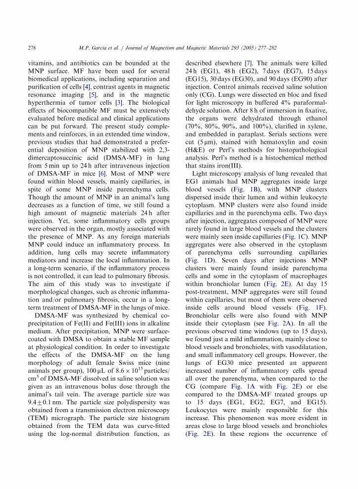
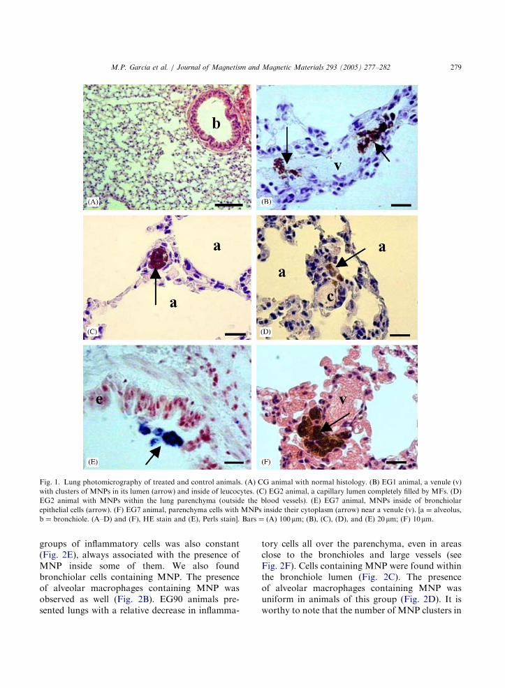
Light microscopy analysis of lung revealed thatEG1 animals had MNP aggregates inside largeblood vessels (Fig. 1B), with MNP clustersdispersed inside their lumen and within leukocytecytoplasm. MNP clusters were also found insidecapillaries and in the parenchyma cells. Two daysafter injection, aggregates composed of MNP wererarely found in large blood vessels and the clusterswere mainly seen inside capillaries (Fig. 1C). MNPaggregates were also observed in the cytoplasmof parenchyma cells surrounding capillaries(Fig. 1D). Seven days after injections MNPclusters were mainly found inside parenchymacells and some in the cytoplasm of macrophageswithin bronchiolar lumen (Fig. 2E). At day 15post-treatment, MNP aggregates were still foundwithin capillaries, but most of them were observedinside cells around blood vessels (Fig. 1F).Bronchiolar cells were also found with MNPinside their cytoplasm (see Fig. 2A). In all theprevious observed time windows (up to 15 days),we found just a mild inflammation, mainly close toblood vessels and bronchioles, with vasodilatation,and small inflammatory cell groups. However, thelungs of EG30 mice presented an apparentincreased number of inflammatory cells spreadall over the parenchyma, when compared to theCG (compare Fig. 1A with Fig. 2E) or elsecompared to the DMSA-MF treated groups upto 15 days (EG1, EG2, EG7, and EG15).Leukocytes were mainly responsible for thisincrease. This phenomenon was more evident inareas close to large blood vessels and bronchioles(Fig. 2E). In these regions the occurrence of
ARTICLE IN PRESS
Fig. 1. Lung photomicrography of treated and control animals. (A) CG animal with normal histology. (B) EG1 animal, a venule (v)
with clusters of MNPs in its lumen (arrow) and inside of leucocytes. (C) EG2 animal, a capillary lumen completely filled by MFs. (D)
EG2 animal with MNPs within the lung parenchyma (outside the blood vessels). (E) EG7 animal, MNPs inside of bronchiolar
epithelial cells (arrow). (F) EG7 animal, parenchyma cells with MNPs inside their cytoplasm (arrow) near a venule (v). [a ¼ alveolus,
b ¼ bronchiole. (A–D) and (F), HE stain and (E), Perls stain]. Bars ¼ (A) 100mm; (B), (C), (D), and (E) 20 mm; (F) 10 mm.
M.P. Garcia et al. / Journal of Magnetism and Magnetic Materials 293 (2005) 277–282 279
groups of inflammatory cells was also constant(Fig. 2E), always associated with the presence ofMNP inside some of them. We also foundbronchiolar cells containing MNP. The presenceof alveolar macrophages containing MNP wasobserved as well (Fig. 2B). EG90 animals pre-sented lungs with a relative decrease in inflamma-
tory cells all over the parenchyma, even in areasclose to the bronchioles and large vessels (seeFig. 2F). Cells containing MNP were found withinthe bronchiole lumen (Fig. 2C). The presenceof alveolar macrophages containing MNP wasuniform in animals of this group (Fig. 2D). It isworthy to note that the number of MNP clusters in
ARTICLE IN PRESS
Fig. 2. Lung photomicrography of intravenously injected DMSA-MF animals. (A) EG15 animal. Observe cells containing MNPs
attached to the epithelium of a bronchiole (arrow). (B) EG30 animal with macrophages inside a bronchiole lumen (arrow). Note the
presence of MNPs inside their cytoplasm. (C) and (D) EG90 animal. Observe macrophages containing MNPs inside bronchiole and
alveoli, respectively. (E) EG30 and (F) EG90 animals. Note the stronger inflammatory process close to the bronchiole (b) in (E) as
compared to (F). [a ¼ alveolus, b ¼ bronchiole. (B) and (E), HE stain and (A), (C), (D), (F), Perls stain]. Bars ¼ (A) and (B) 20mm; (C)
and (D) 10 mm; (E) and (F) 100mm.
M.P. Garcia et al. / Journal of Magnetism and Magnetic Materials 293 (2005) 277–282280
the lungs was observed to decrease as a function oftime. Control animals had normal histologicalappearance, as shown in Fig. 1A.
At this point, we note that normal lung presentsa morphological structure composed of conduct-
ing portions, which includes the internal bronchithat undergo extensive branching to give rise tobronchioles, the latter representing the terminalpart of the conducting passages. The respiratoryportion is that in which gas exchange occurs. It
ARTICLE IN PRESS
M.P. Garcia et al. / Journal of Magnetism and Magnetic Materials 293 (2005) 277–282 281
sequentially includes respiratory bronchioles (thinbronchioles that have sparse alveoli in their wall),alveolar ducts, and alveolar sacs whose wall iscomposed of only alveoli, and the alveoli itself.Alveoli are surrounded and separated from oneanother by a very thin connective tissue layer thatcontains numerous blood capillaries. The tissuebetween adjacent alveolar air space is calledalveolar septum. It is worthy to note that anincrease in inflammatory cells in the lung par-enchyma reveals that an inflammatory process hastaken place in that organ.
DMSA is a well-known molecule, often used asa chelating agent to remove heavy metals from anorganism, and therefore considered as a biocom-patible agent [8]. In part, this fact has been used tosupport its application as a biocompatible stabiliz-ing coating for MNP, once DMSA alone showslow toxicity in various biological systems alreadystudied [9–11]. Moreover, effector molecules canbe bound to the DMSA structure. Quantitativeiron determinations and microscopy studies withintravenous administration of ferrite particles,useful as contrast agents for magnetic resonanceimaging, demonstrated that particles are partiallydegraded and that iron is cleared from theorganism during a 3-month period without toxi-city response [12]. Since most of the iron-basedMNP and DMSA are not toxic, we would notexpect to have a strong inflammatory process afterDMSA-MF injection in the animals. However, thepresent study showed that in the time window of1–30 days there was an enhancement of inflam-matory cells in the lung parenchyma. Besides, suchan increase was always correlated to the areaswhere the MNP were found. The specific cause tothis inflammation was not determined in thisstudy. Nevertheless, some hypothesis could bemade. In spite of the fact that iron is the mostabundant metal in the animal’s body, when inexcess, it can lead to several pathologies. The basicmechanism related with the iron capacity to inducepathologies in the organism is the increase ofsuperoxide radicals. This increase involves thereduction of free oxygen induced by iron. Super-oxide radicals can react with cellular membranespromoting its peroxidation, as well as peroxida-tion of proteins and nucleic acids. All these cellular
disturbances could lead different cell types tosignal leukocytes and attract them to the locationof injury. Another possibility to explain theincrease of cell number in the lung parenchymais that leukocytes, specifically monocytes andneutrophils, were directly activated by MNPpresence. In this case these cells may incorporateMNP first and then produce pro-inflammatorymediators that attracted more leukocytes to thesite. This scheme would enhance the inflammatoryprocess. As a matter of fact, from 12 h after thetreatment Chaves et al. [6] observed neutrophilsand monocytes containing DMSA-MF nanopar-ticles within blood vessels, showing that the MNPdirectly activate leukocytes. This explanation doesnot exclude the possibility that peroxidation alsooccurs, since free oxygen is abundant in the lungand could react with iron. Indeed, further experi-ments are necessary to answer this question.
Morphological analysis showed that 90 daysafter DMSA-MF treatment, animals had theirlung parenchyma with similar morphology as theCG, except for a few cells that contained MNPwithin the parenchyma and bronchioles lumen,alveolar macrophages with MNP aggregates intheir cytoplasm, and few areas with small groupsof inflammatory cells. In general, all animals ofEG90 had a smaller amount of MNP in their lungthan all the other treated groups. The reduction ofMNP in the lung is more pronounced during thefirst two days, though it kept decreasing during thetime window of our experiment. Results obtainedby magnetic resonance showed that some MNPmigrate from the lung to other organs such as liverand spleen, where they are incorporated by thelocal macrophages [5]. This could explain theaccentuated decrease in MNP within the lungs.However, the presence of cells containing MNPinside bronchiole lumen could be a good pathwayto take the nanoparticles out, since cilia ofrespiratory cells in the bronchioles and bronchibeat mucus with dust toward the esophagus.Catabolism of iron by the macrophages, includingalveolar macrophages, is also a possibility toexplain the decrease of MNP in the lung. Thesetwo possibilities are most likely to be the only onesresponsible for the decrease of MNP in the latephase of this study. The decrease of inflammation
ARTICLE IN PRESS
M.P. Garcia et al. / Journal of Magnetism and Magnetic Materials 293 (2005) 277–282282
in the lung of EG30 animals along with thedecrease in MNP in this organ strongly suggestthat DMSA-MF nanoparticles are mainly respon-sible for this inflammatory process.
In conclusion, when DMSA-MF is injectedintravenously, the MNP are preferentially driventowards the lung, where inflammation is induced.However, the inflammatory process reduces as afunction of time, and it is not able to promotefurther pathologies such as pulmonary fibrosis.
References
[1] R.E. Rosenweig, Ferrohydrodynamics, Cambridge
University Press, New York, 1985.
[2] R. Massart, IEEE Trans. Magn. 17 (1981) 1247.
[3] D. Gunther, N. Buske, DE patents No. 4325386 (1993),
No. 4372826 (1993).
[4] P. Hermetin, R. Doenges, V. Franssen, et al., Bioconjugate
Chem. 1 (1990) 411.
[5] A.K. Fahlvek, J. Klaveness, D.D. Stark, J. Magn. Reson.
Imaging 3 (1993) 187.
[6] S.B. Chaves, L.M. Lacava, Z.G.M. Lacava, et al., IEEE
Trans. Magn. 38 (2002) 3231.
[7] B.M. Lacava, R.B. Azevedo, L.P. Silva, et al., Appl. Phys.
Lett. 77 (2000) 1876.
[8] A. Ferrer, An Sist. Sanit. Navar. 26 (2003) 141.
[9] R. Sivaprasad, M. Nagaraj, P. Varalakshmi, J. Nutr.
Biochem. 15 (2004) 18.
[10] M. Pande, S.J. Flora, Toxicology 177 (2002) 187.
[11] A.L. Miller, Altern. Med. Rev. 3 (1998) 199.
[12] B.R. Bacon, D.D. Stark, C.H. Park, et al., J. Lab. Clin.
Med. 110 (1987) 164.