Auscultation of the lungs - Repository of Kharkiv National ...
106
Auscultation of the lungs: the main and additional respiratory sounds, their diagnostic significance Professor T. ASHCHEULOVA Head of Propedeutics to Internal Medicine N1, Basis of Bioethics and Biosafety Kharkiv National Medical University
-
Upload
khangminh22 -
Category
Documents
-
view
1 -
download
0
Transcript of Auscultation of the lungs - Repository of Kharkiv National ...
Auscultation of the lungs: the main and additional respiratory sounds, their diagnostic significance Professor T. ASHCHEULOVA Head of Propedeutics to Internal Medicine N1, Basis of Bioethics and Biosafety
Kharkiv National Medical University
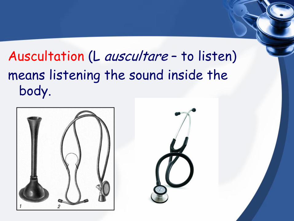
Auscultation (L auscultare – to listen)
means listening the sound inside the body.

Chapter 01 HISTORY
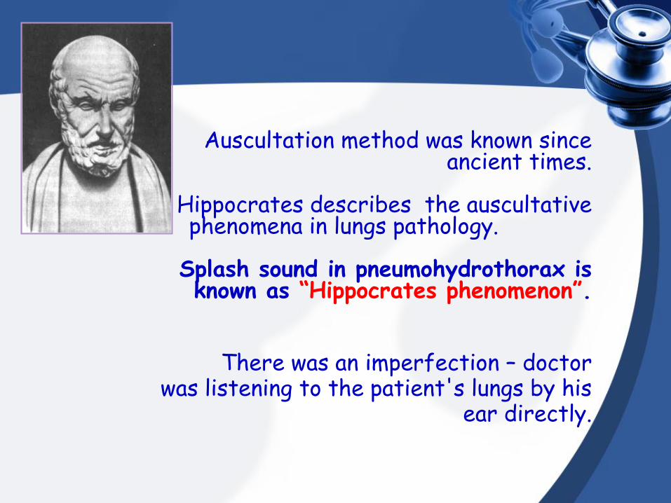
Auscultation method was known since ancient times.
Hippocrates describes the auscultative
phenomena in lungs pathology.
Splash sound in pneumohydrothorax is known as “Hippocrates phenomenon”.
There was an imperfection – doctor was listening to the patient's lungs by his
ear directly.
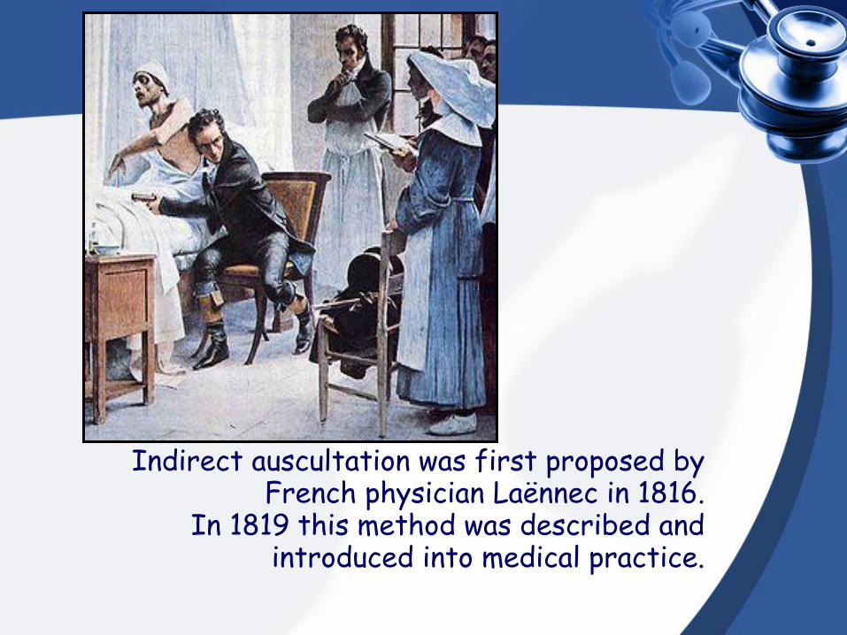
Indirect auscultation was first proposed by French physician Laënnec in 1816.
In 1819 this method was described and introduced into medical practice.
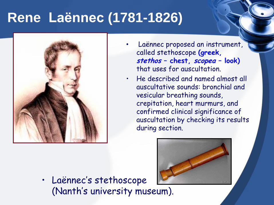
Rene Laënnec (1781-1826)
• Laënnec’s stethoscope (Nanth’s university museum).
• Laënnec proposed an instrument, called stethoscope (greek, stethos – chest, scopea – look) that uses for auscultation.
• He described and named almost all auscultative sounds: bronchial and vesicular breathing sounds, crepitation, heart murmurs, and confirmed clinical significance of auscultation by checking its results during section.
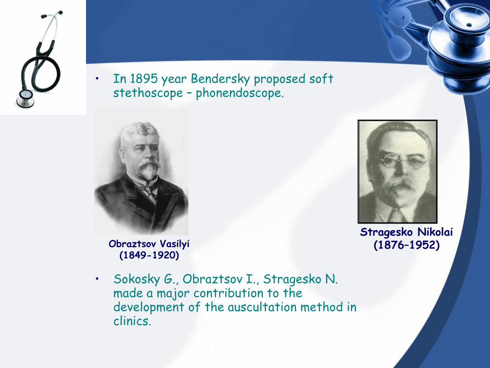
• In 1895 year Bendersky proposed soft stethoscope – phonendoscope.
• Sokosky G., Obraztsov I., Stragesko N. made a major contribution to the development of the auscultation method in clinics.
Obraztsov Vasilyi (1849-1920)
Stragesko Nikolai (1876–1952)
Chapter 02
PHYSICAL BASIS, TYPES
AND RULES OF AUSCULTATION
Physical basis and diagnostic value of the auscultation
PHYSICAL BASIS
Auscultation the same as percussion is based on the tissue’s ability to conduct a sound . On the sound’s variations basis a doctor can estimate changes in the tissues and organs.
Physical basis and diagnostic value of the auscultation
The difference is: in auscultation you evaluate a natural
sounds that appear in the result of organism’s activity and characterized a function;
in percussion – artificial sound that spreads and reflects from anatomical structures characterizes the structure of the organs and tissues.
Physical basis and diagnostic value of the auscultation
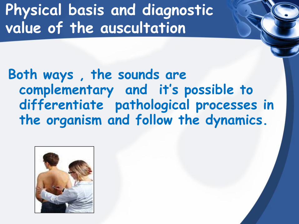
Both ways , the sounds are complementary and it’s possible to differentiate pathological processes in the organism and follow the dynamics.
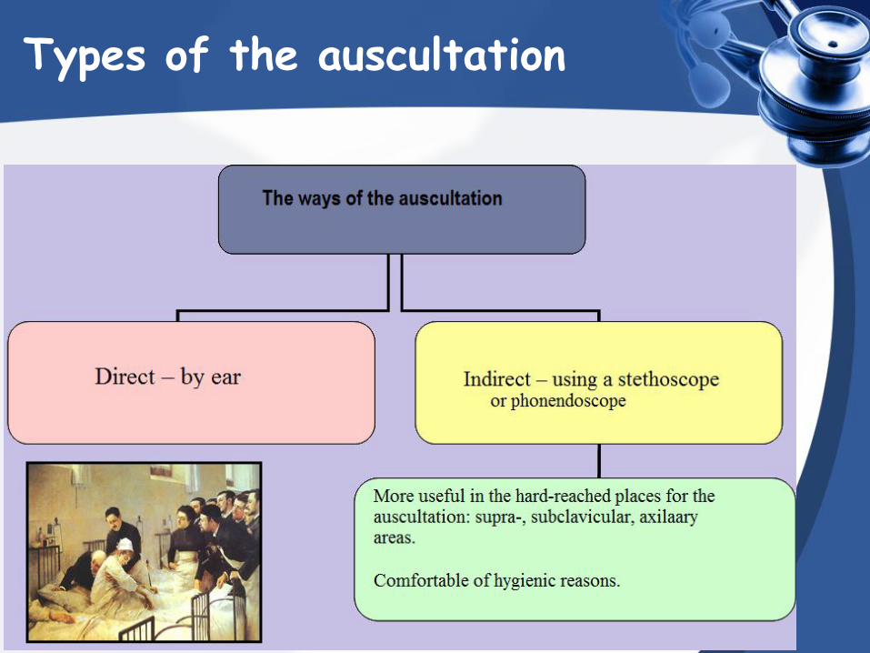
Types of the auscultation
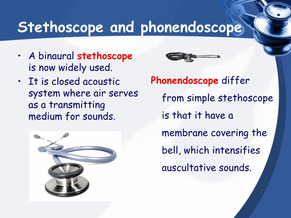
Stethoscope and phonendoscope
• A binaural stethoscope is now widely used.
• It is closed acoustic system where air serves as a transmitting medium for sounds.
Phonendoscope differ
from simple stethoscope
is that it have a
membrane covering the
bell, which intensifies
auscultative sounds.
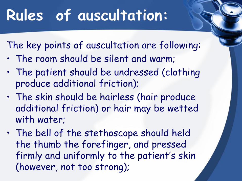
Rules of auscultation:
The key points of auscultation are following:
• The room should be silent and warm;
• The patient should be undressed (clothing produce additional friction);
• The skin should be hairless (hair produce additional friction) or hair may be wetted with water;
• The bell of the stethoscope should held the thumb the forefinger, and pressed firmly and uniformly to the patient’s skin (however, not too strong);
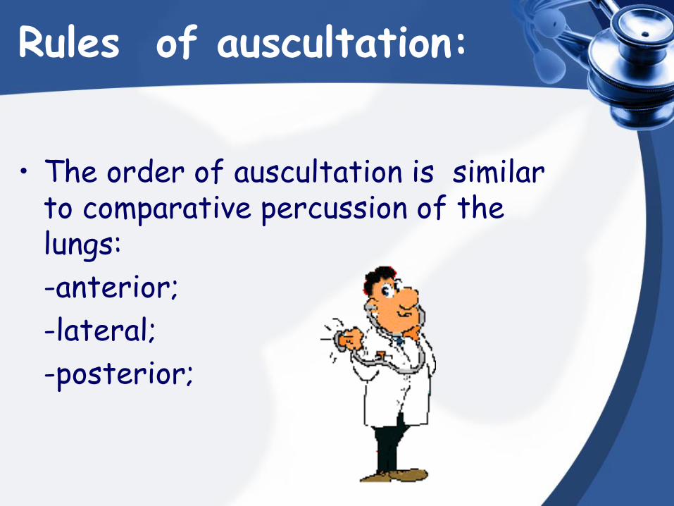
Rules of auscultation:
• The order of auscultation is similar to comparative percussion of the lungs:
-anterior;
-lateral;
-posterior;
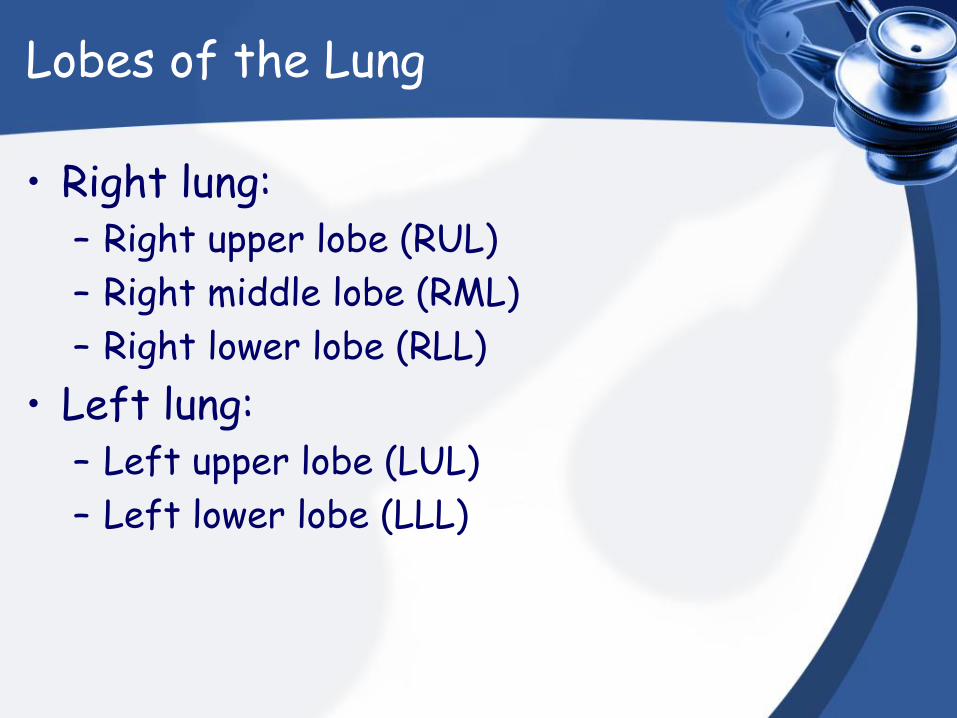
Lobes of the Lung
• Right lung: – Right upper lobe (RUL)
– Right middle lobe (RML)
– Right lower lobe (RLL)
• Left lung: – Left upper lobe (LUL)
– Left lower lobe (LLL)
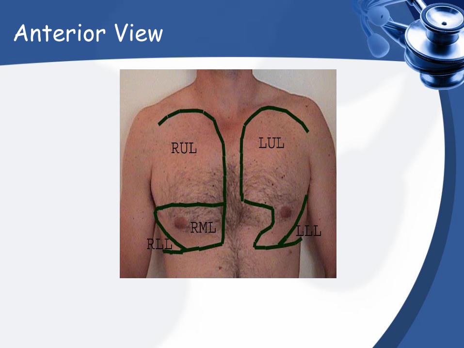
Anterior View
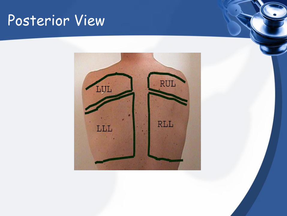
Posterior View
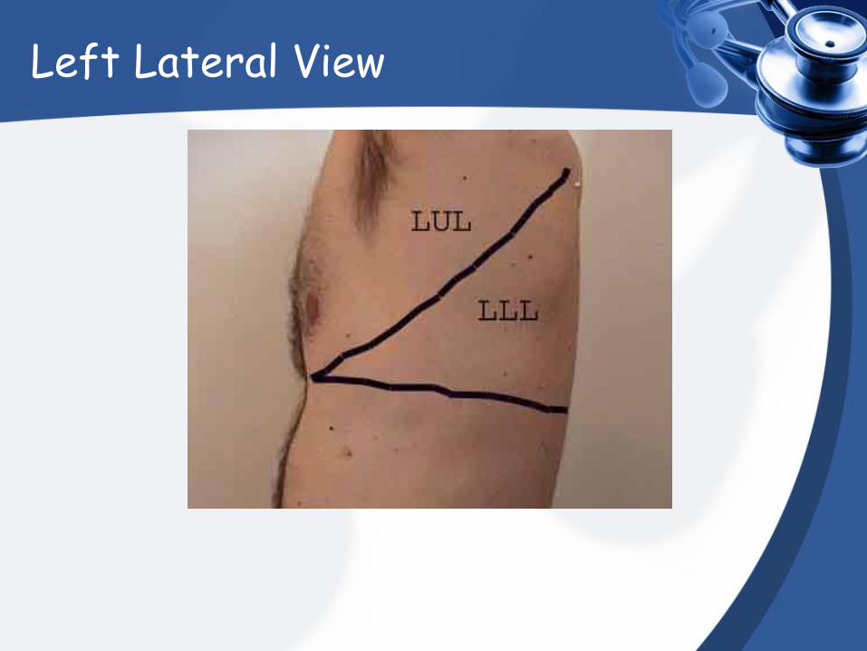
Left Lateral View
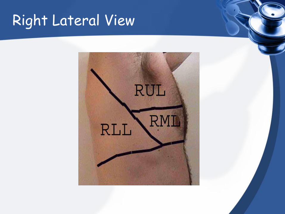
Right Lateral View
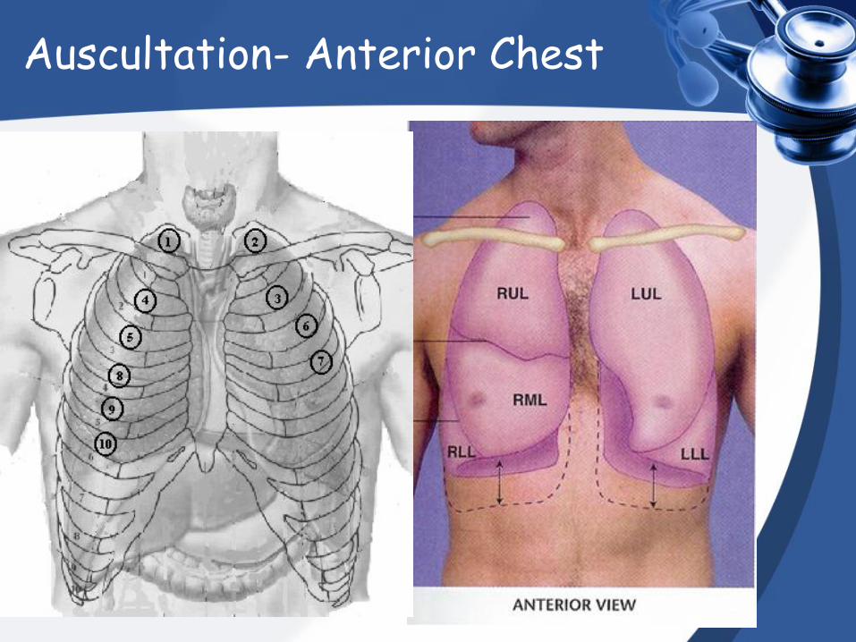
Auscultation- Anterior Chest
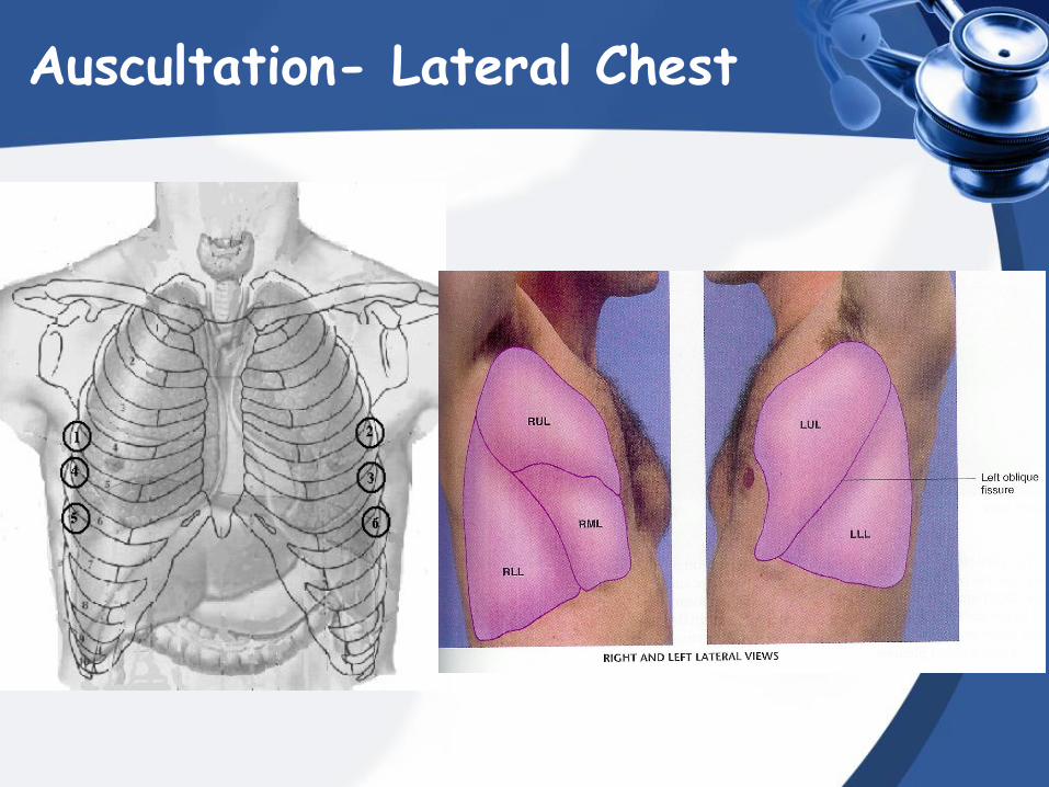
Auscultation- Lateral Chest
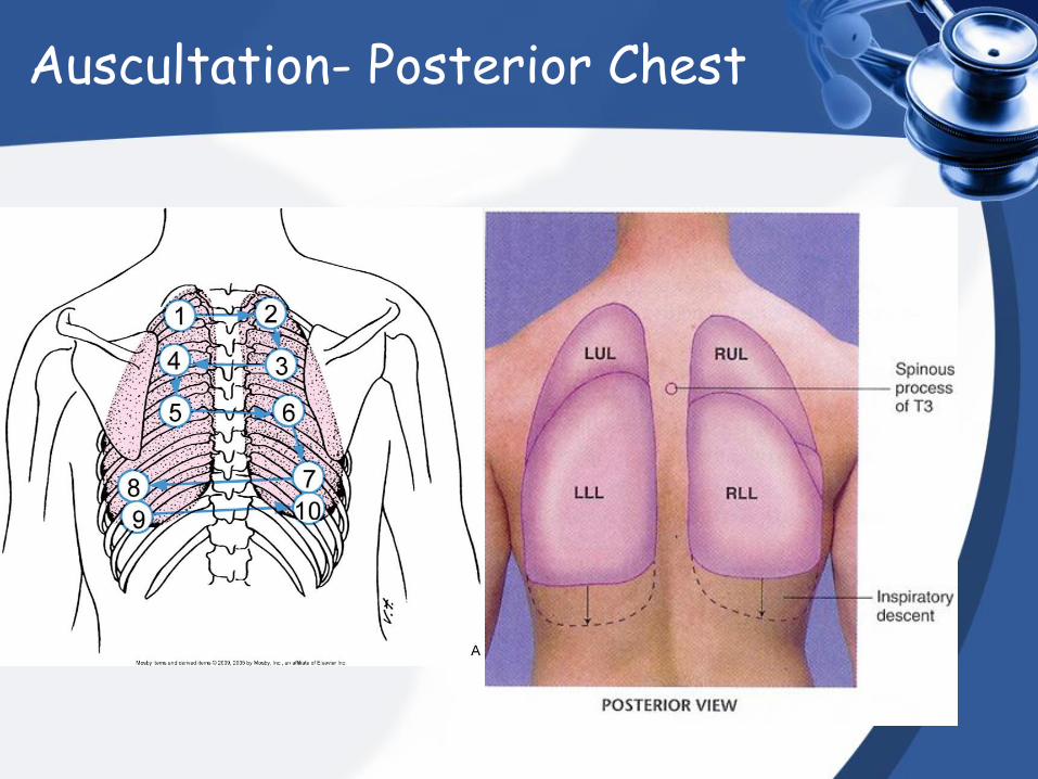
Auscultation- Posterior Chest

Chapter 03 Respiratory sounds
analysis
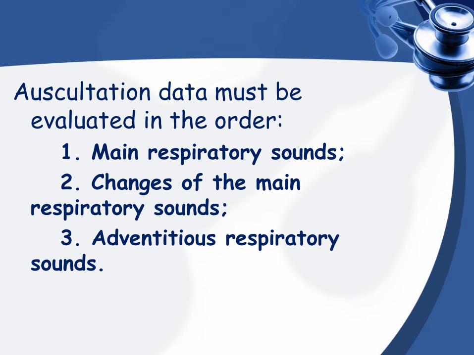
Auscultation data must be evaluated in the order:
1. Main respiratory sounds;
2. Changes of the main respiratory sounds;
3. Adventitious respiratory sounds.
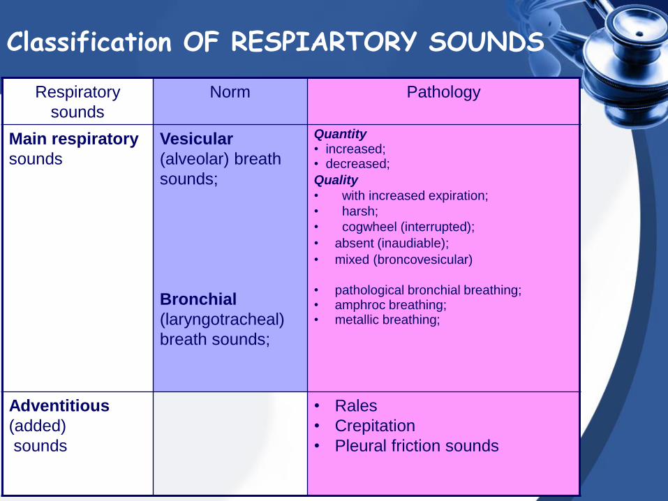
Classification OF RESPIARTORY SOUNDS
Respiratory
sounds
Norm Pathology
Main respiratory
sounds
Vesicular
(alveolar) breath
sounds;
Bronchial
(laryngotracheal)
breath sounds;
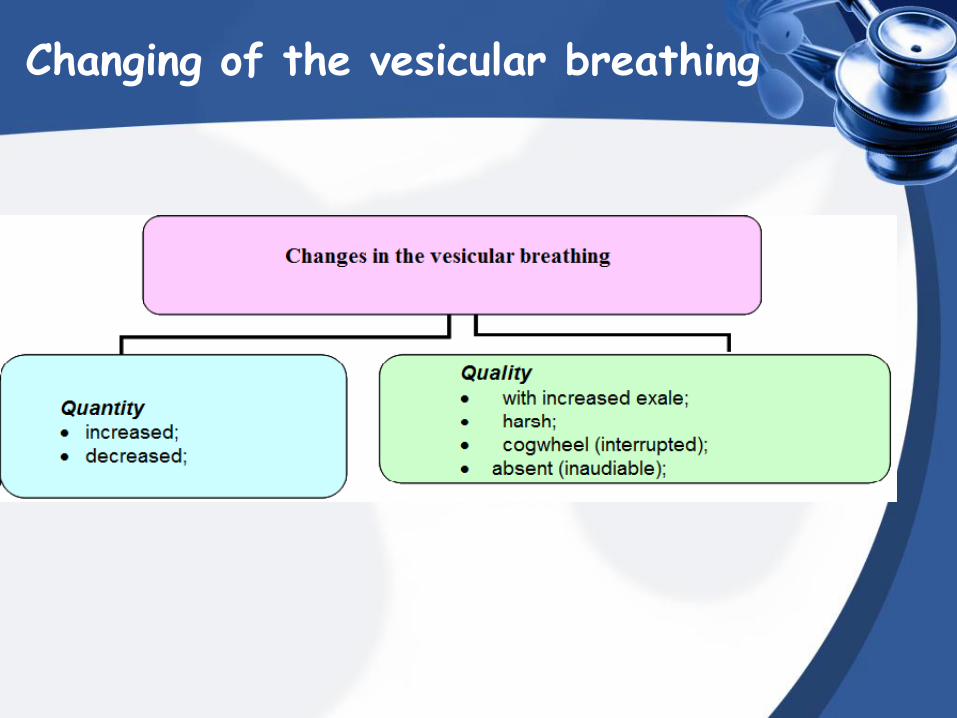
Quantity • increased; • decreased;
Quality
• with increased expiration;
• harsh;
• cogwheel (interrupted);
• absent (inaudiable);
• mixed (broncovesicular)
• pathological bronchial breathing; • amphroc breathing; • metallic breathing;
Adventitious
(added)
sounds
• Rales
• Crepitation
• Pleural friction sounds
Main respiratory
sounds
VESICULAR BREATHING
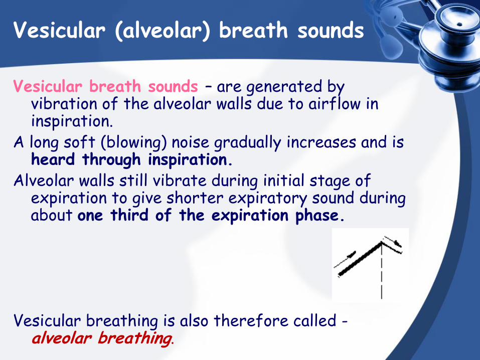
Vesicular (alveolar) breath sounds
Vesicular breath sounds – are generated by vibration of the alveolar walls due to airflow in inspiration.
A long soft (blowing) noise gradually increases and is heard through inspiration.
Alveolar walls still vibrate during initial stage of expiration to give shorter expiratory sound during about one third of the expiration phase.
Vesicular breathing is also therefore called -
alveolar breathing.
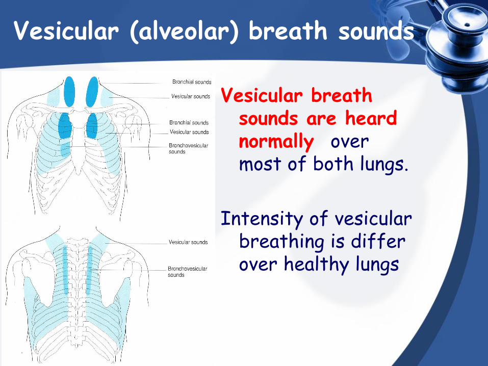
Vesicular (alveolar) breath sounds
Vesicular breath sounds are heard normally over most of both lungs.
Intensity of vesicular breathing is differ over healthy lungs
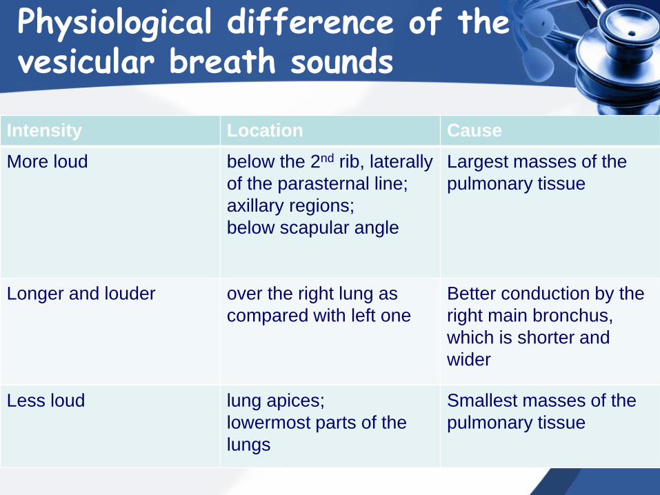
Physiological difference of the vesicular breath sounds
Intensity Location Cause
More loud below the 2nd rib, laterally
of the parasternal line;
axillary regions;
below scapular angle
Largest masses of the
pulmonary tissue
Longer and louder over the right lung as
compared with left one
Better conduction by the
right main bronchus,
which is shorter and
wider
Less loud lung apices;
lowermost parts of the
lungs
Smallest masses of the
pulmonary tissue
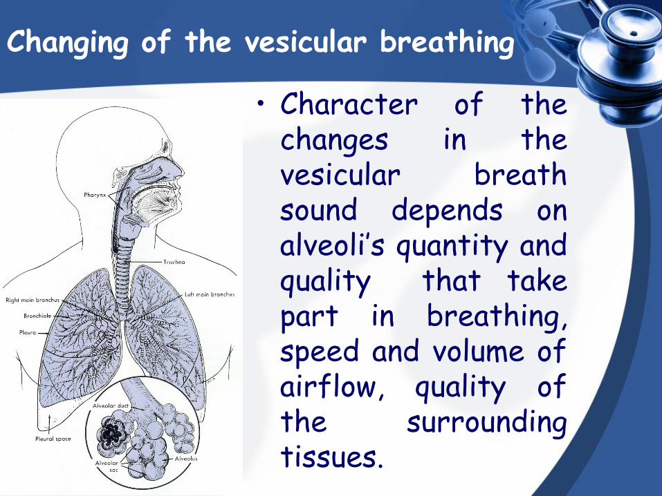
Changing of the vesicular breathing
• Character of the changes in the vesicular breath sound depends on alveoli’s quantity and quality that take part in breathing, speed and volume of airflow, quality of the surrounding tissues.
Changing of the vesicular breathing
Quantity changes of the vesicular breathing
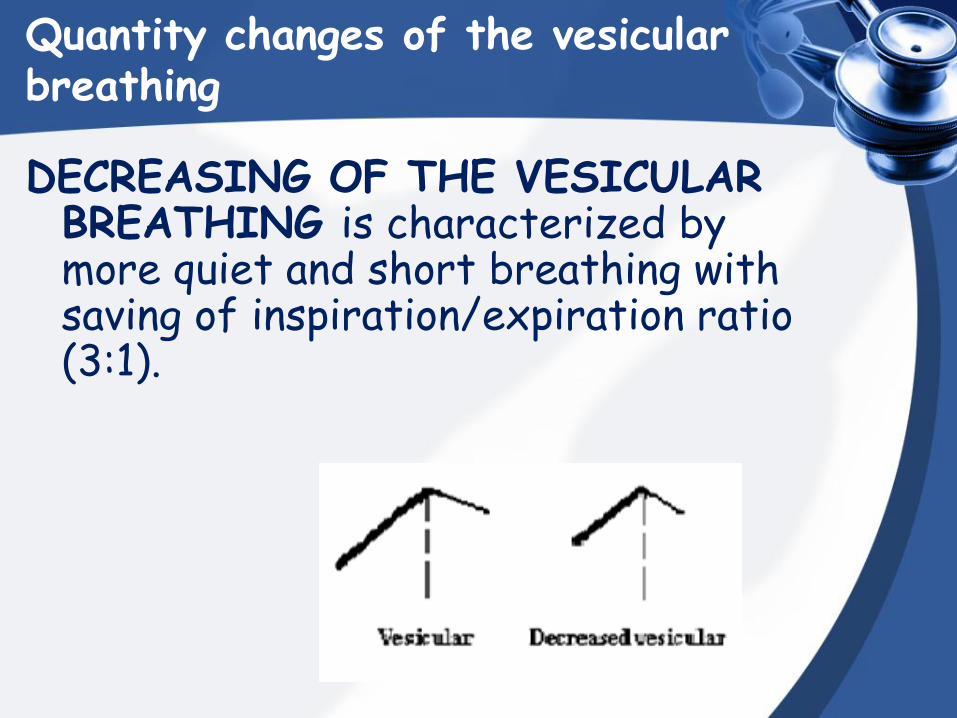
DECREASING OF THE VESICULAR BREATHING is characterized by more quiet and short breathing with saving of inspiration/expiration ratio (3:1).
Quantity changes of the vesicular breathing
DECREASING OF THE VESICULAR BREATHING can be physiological and pathological.
Physiological changes of the vesicular
breathing always involve both part of the chest, and breath sounds are equally changes at the symmetrical points of the chest
Quantity changes of the vesicular breathing
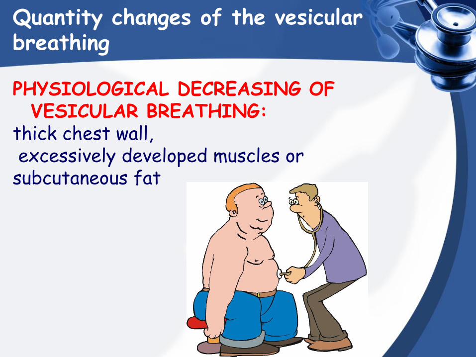
PHYSIOLOGICAL DECREASING OF VESICULAR BREATHING:
thick chest wall, excessively developed muscles or subcutaneous fat
Pathological decreasing of vesicular breathing
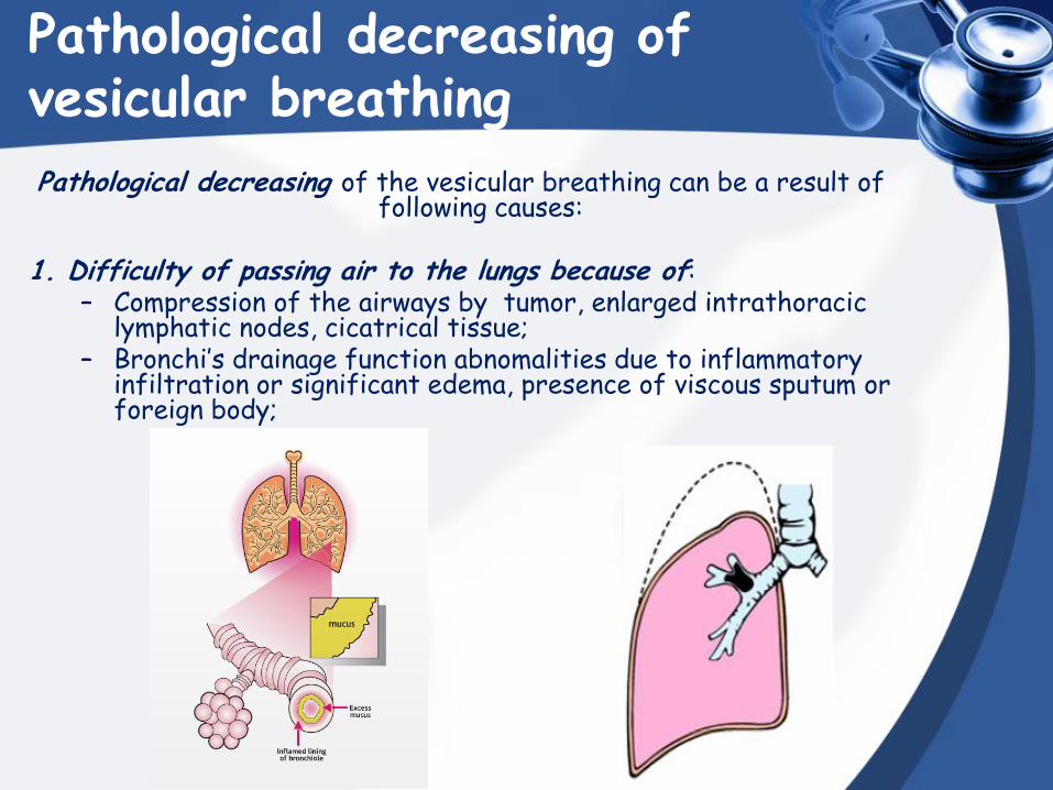
Pathological decreasing of the vesicular breathing can be a result of following causes:
1. Difficulty of passing air to the lungs because of:
– Compression of the airways by tumor, enlarged intrathoracic lymphatic nodes, cicatrical tissue;
– Bronchi’s drainage function abnomalities due to inflammatory infiltration or significant edema, presence of viscous sputum or foreign body;
Pathological decreasing of vesicular breathing
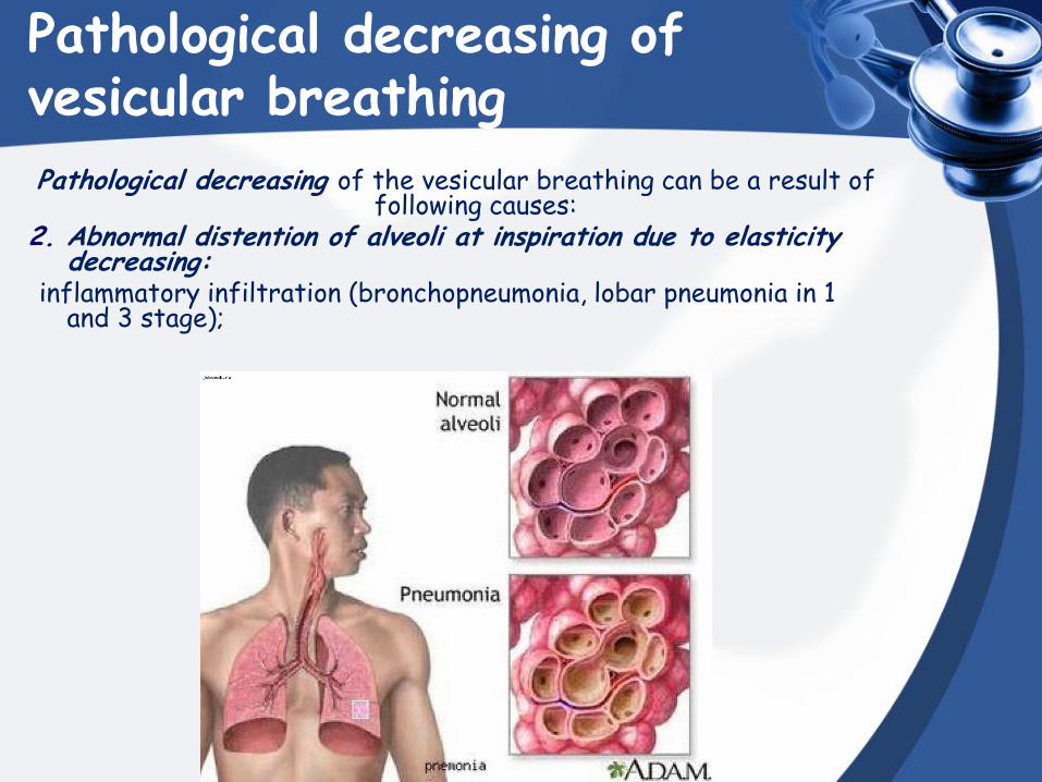
Pathological decreasing of the vesicular breathing can be a result of following causes:
2. Abnormal distention of alveoli at inspiration due to elasticity decreasing:
inflammatory infiltration (bronchopneumonia, lobar pneumonia in 1 and 3 stage);
Pathological decreasing of vesicular breathing
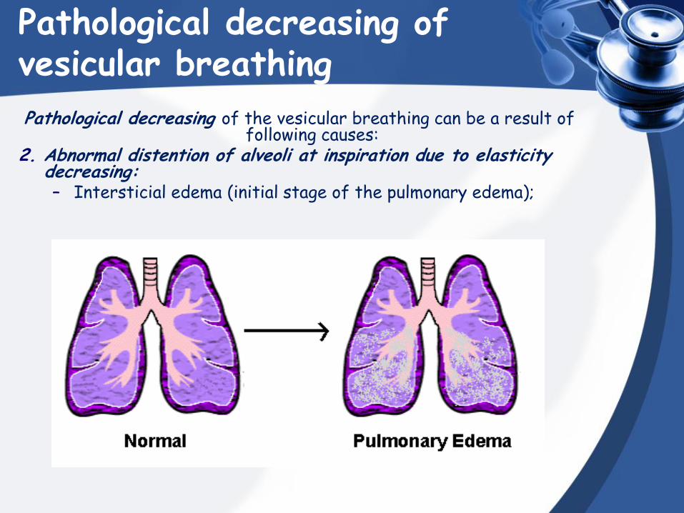
Pathological decreasing of the vesicular breathing can be a result of following causes:
2. Abnormal distention of alveoli at inspiration due to elasticity decreasing: – Intersticial edema (initial stage of the pulmonary edema);
Pathological decreasing of vesicular breathing
Pathological decreasing of the vesicular breathing can be a result of following causes:
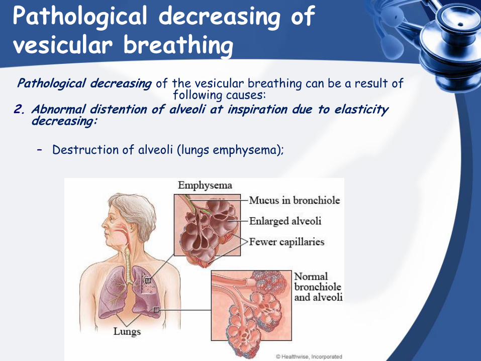
2. Abnormal distention of alveoli at inspiration due to elasticity decreasing:
– Destruction of alveoli (lungs emphysema);
Pathological decreasing of vesicular breathing
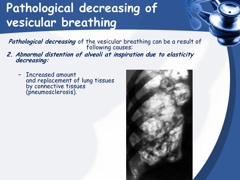
Pathological decreasing of the vesicular breathing can be a result of following causes:
2. Abnormal distention of alveoli at inspiration due to elasticity decreasing:
– Increased amount
and replacement of lung tissues by connective tissues (pneumosclerosis).
Pathological decreasing of vesicular breathing
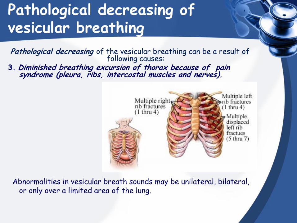
Pathological decreasing of the vesicular breathing can be a result of following causes:
3. Diminished breathing excursion of thorax because of pain syndrome (pleura, ribs, intercostal muscles and nerves).
Abnormalities in vesicular breath sounds may be unilateral, bilateral, or only over a limited area of the lung.
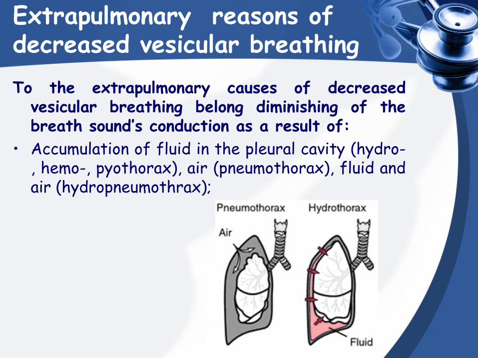
Extrapulmonary reasons of decreased vesicular breathing
To the extrapulmonary causes of decreased vesicular breathing belong diminishing of the breath sound’s conduction as a result of:
• Accumulation of fluid in the pleural cavity (hydro-, hemo-, pyothorax), air (pneumothorax), fluid and air (hydropneumothrax);
Extrapulmonary reasons of decreased vesicular breathing
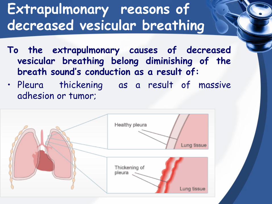
To the extrapulmonary causes of decreased vesicular breathing belong diminishing of the breath sound’s conduction as a result of:
• Pleura thickening as a result of massive adhesion or tumor;
Extrapulmonary reasons of decreased vesicular breathing
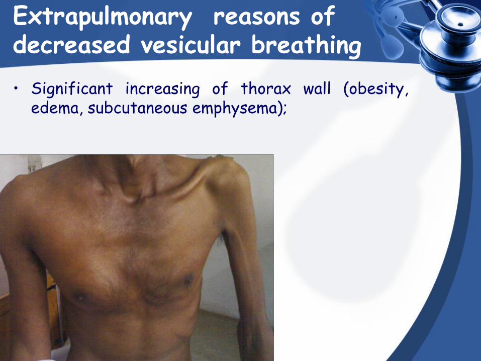
• Significant increasing of thorax wall (obesity, edema, subcutaneous emphysema);
Absence of the vesicular breathing
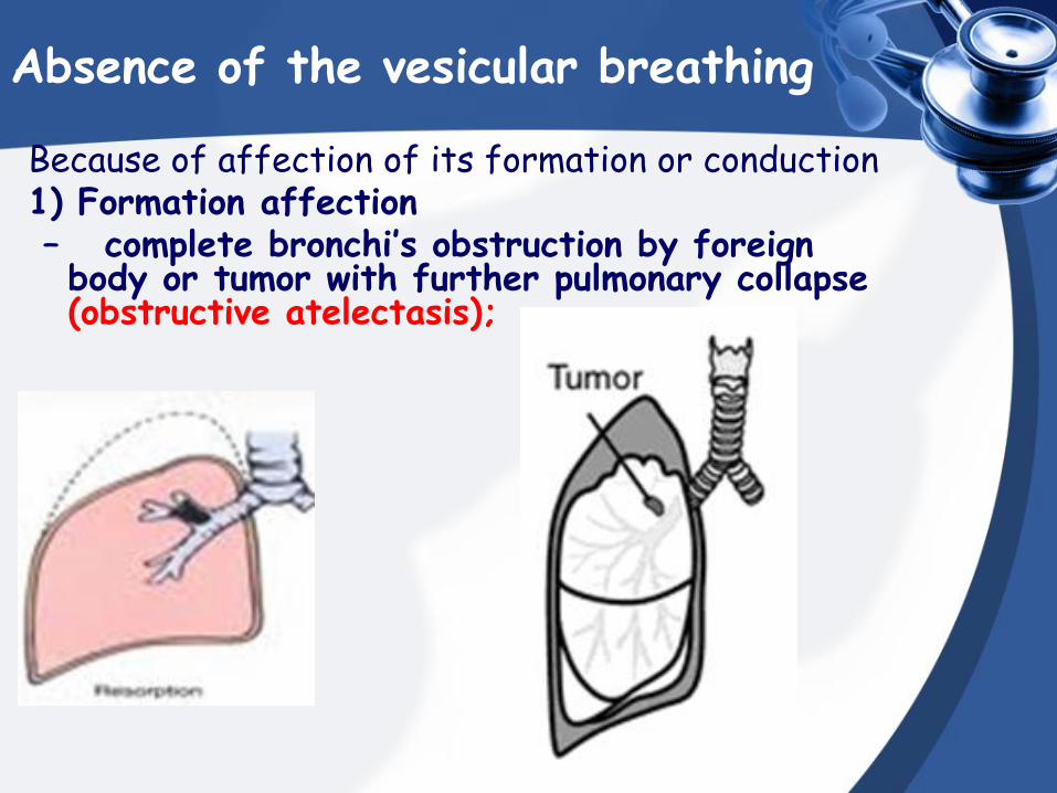
Because of affection of its formation or conduction 1) Formation affection – complete bronchi’s obstruction by foreign
body or tumor with further pulmonary collapse (obstructive atelectasis);
Absence of the vesicular breathing
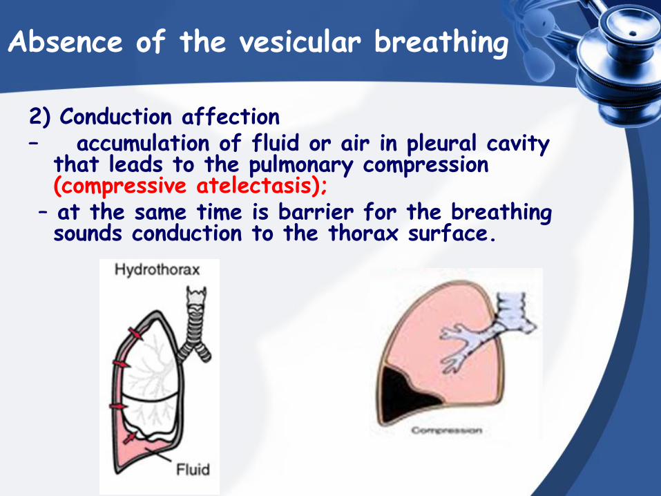
2) Conduction affection – accumulation of fluid or air in pleural cavity
that leads to the pulmonary compression (compressive atelectasis);
– at the same time is barrier for the breathing sounds conduction to the thorax surface.
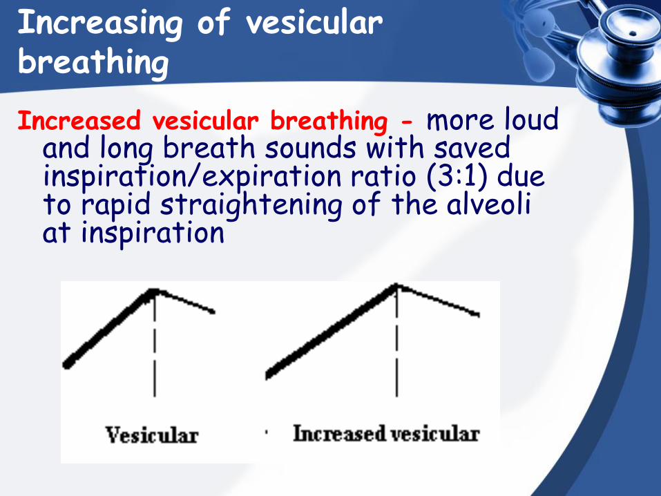
Increasing of vesicular breathing
Increased vesicular breathing - more loud and long breath sounds with saved inspiration/expiration ratio (3:1) due to rapid straightening of the alveoli at inspiration
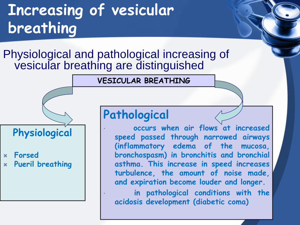
Increasing of vesicular breathing
Physiological and pathological increasing of vesicular breathing are distinguished
Physiological
Forsed Pueril breathing
VESICULAR BREATHING
Pathological • occurs when air flows at increased
speed passed through narrowed airways (inflammatory edema of the mucosa, bronchospasm) in bronchitis and bronchial asthma. This increase in speed increases turbulence, the amount of noise made, and expiration become louder and longer.
• in pathological conditions with the acidosis development (diabetic coma)
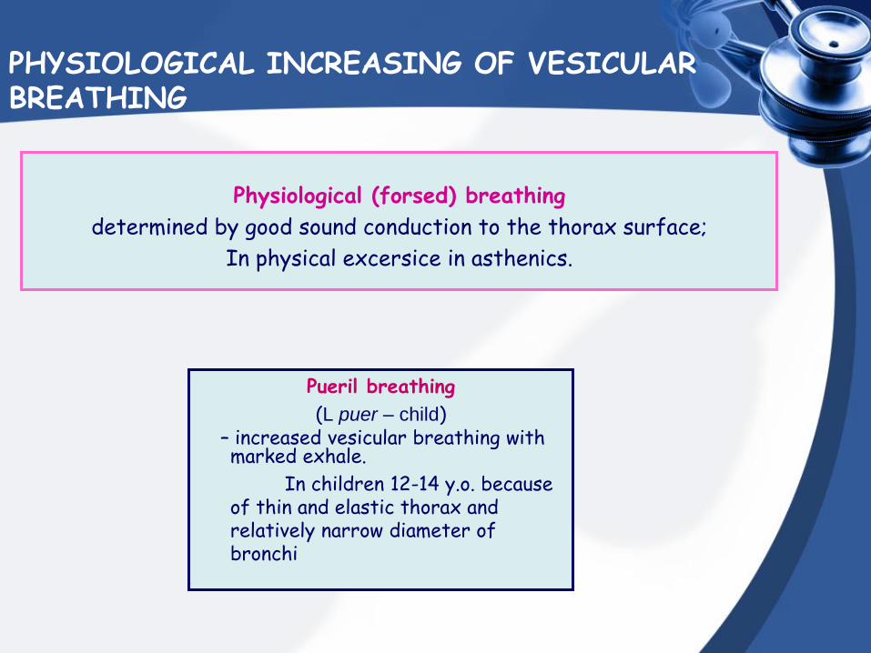
PHYSIOLOGICAL INCREASING OF VESICULAR BREATHING
Physiological (forsed) breathing
determined by good sound conduction to the thorax surface;
In physical excersice in asthenics.
Pueril breathing
(L puer – child) – increased vesicular breathing with
marked exhale.
In children 12-14 y.o. because of thin and elastic thorax and relatively narrow diameter of bronchi
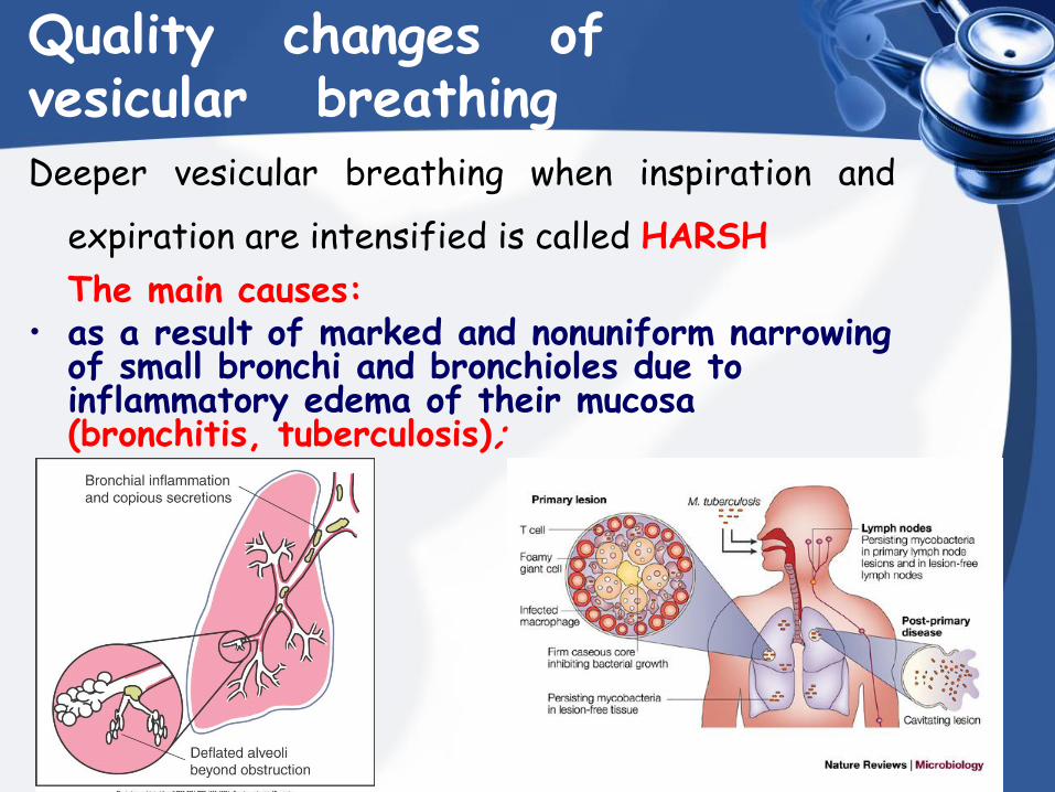
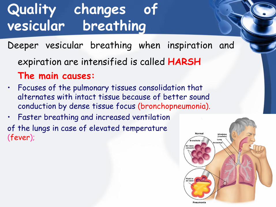
Quality changes of vesicular breathing Deeper vesicular breathing when inspiration and
expiration are intensified is called HARSH
The main causes: • as a result of marked and nonuniform narrowing
of small bronchi and bronchioles due to inflammatory edema of their mucosa (bronchitis, tuberculosis);
Quality changes of vesicular breathing Deeper vesicular breathing when inspiration and
expiration are intensified is called HARSH
The main causes: • Focuses of the pulmonary tissues consolidation that
alternates with intact tissue because of better sound conduction by dense tissue focus (bronchopneumonia).
• Faster breathing and increased ventilation
of the lungs in case of elevated temperature (fever);
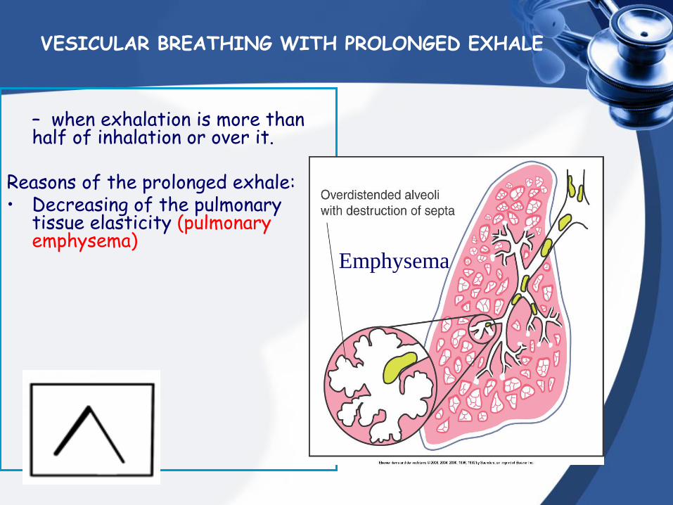
VESICULAR BREATHING WITH PROLONGED EXHALE
– when exhalation is more than half of inhalation or over it.
Reasons of the prolonged exhale: • Decreasing of the pulmonary
tissue elasticity (pulmonary emphysema)
Emphysema
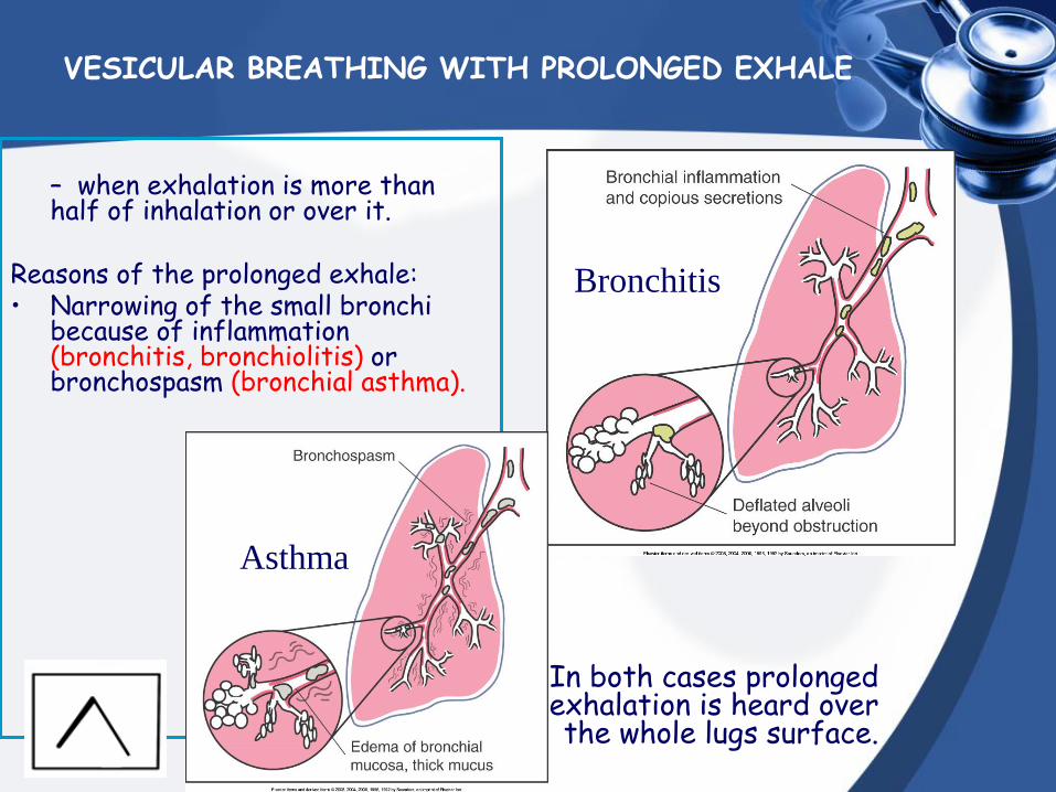
VESICULAR BREATHING WITH PROLONGED EXHALE
– when exhalation is more than half of inhalation or over it.
Reasons of the prolonged exhale: • Narrowing of the small bronchi
because of inflammation (bronchitis, bronchiolitis) or bronchospasm (bronchial asthma).
Bronchitis
Asthma
In both cases prolonged exhalation is heard over the whole lugs surface.
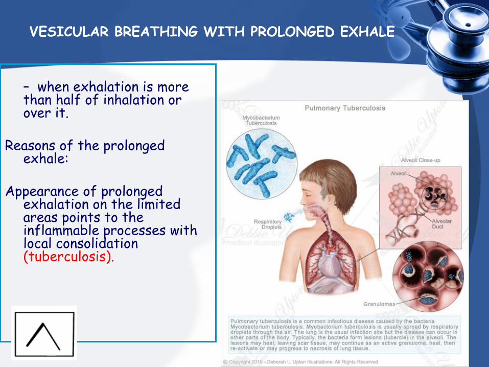
VESICULAR BREATHING WITH PROLONGED EXHALE
– when exhalation is more than half of inhalation or over it.
Reasons of the prolonged
exhale:
Appearance of prolonged exhalation on the limited areas points to the inflammable processes with local consolidation (tuberculosis).
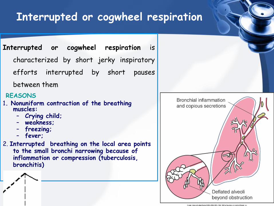
Interrupted or cogwheel respiration
Interrupted or cogwheel respiration is
characterized by short jerky inspiratory
efforts interrupted by short pauses
between them
REASONS 1. Nonuniform contraction of the breathing
muscles: – Crying child; – weakness; – freezing; – fever;
2. Interrupted breathing on the local area points to the small bronchi narrowing because of inflammation or compression (tuberculosis, bronchitis)
10 min.
Break 6
7
8
12 1
2
3
4
5
9
10
11

Main respiratory
sounds
BRONCHIAL
BREATHING
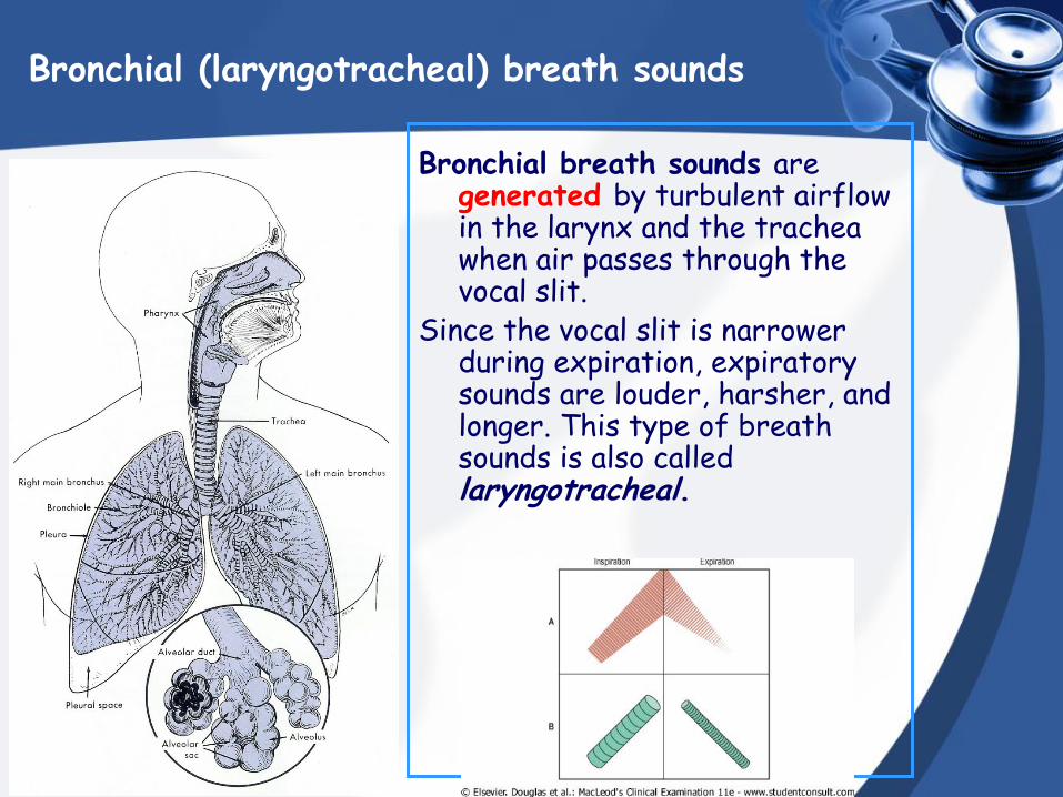
Bronchial (laryngotracheal) breath sounds
Bronchial breath sounds are generated by turbulent airflow in the larynx and the trachea when air passes through the vocal slit.
Since the vocal slit is narrower during expiration, expiratory sounds are louder, harsher, and longer. This type of breath sounds is also called laryngotracheal.
Bronchial (laryngotracheal) breath sounds
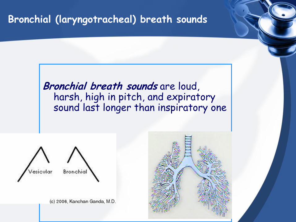
Bronchial breath sounds are loud, harsh, high in pitch, and expiratory sound last longer than inspiratory one
Bronchial (laryngotracheal) breath sounds
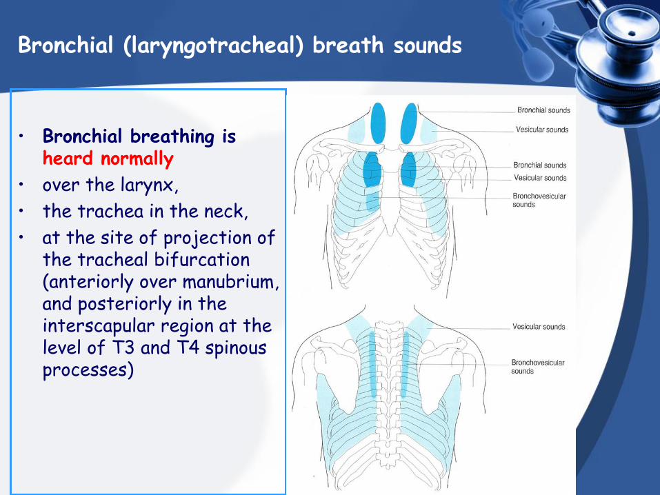
• Bronchial breathing is heard normally
• over the larynx,
• the trachea in the neck,
• at the site of projection of the tracheal bifurcation (anteriorly over manubrium, and posteriorly in the interscapular region at the level of T3 and T4 spinous processes)
Bronchial (laryngotracheal) breath sounds
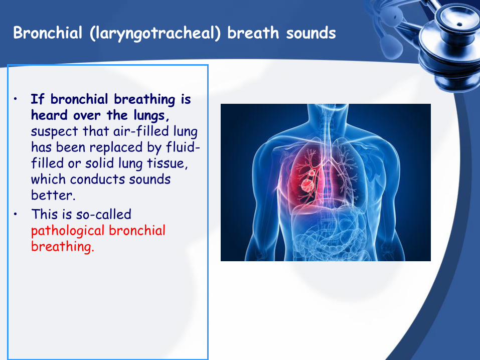
• If bronchial breathing is heard over the lungs, suspect that air-filled lung has been replaced by fluid-filled or solid lung tissue, which conducts sounds better.
• This is so-called pathological bronchial breathing.
Pathological bronchial breathing
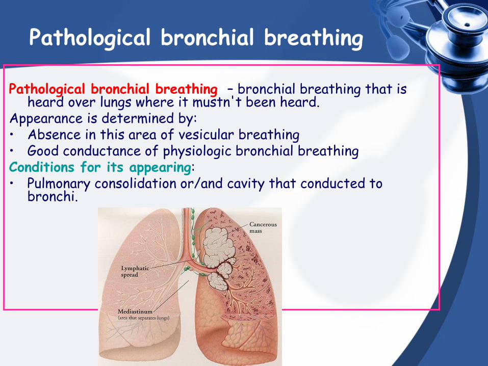
Pathological bronchial breathing – bronchial breathing that is heard over lungs where it mustn't been heard.
Appearance is determined by: • Absence in this area of vesicular breathing • Good conductance of physiologic bronchial breathing Conditions for its appearing: • Pulmonary consolidation or/and cavity that conducted to
bronchi.
Changes of the bronchial breathing
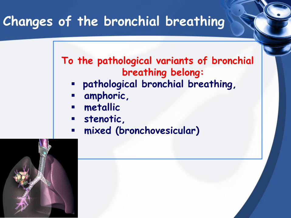
To the pathological variants of bronchial breathing belong:
pathological bronchial breathing, amphoric, metallic stenotic, mixed (bronchovesicular)
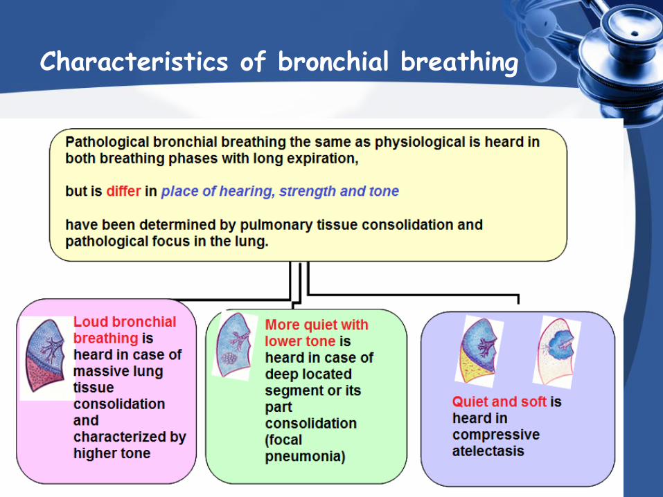
Characteristics of bronchial breathing
The reasons of pathological bronchial breathing
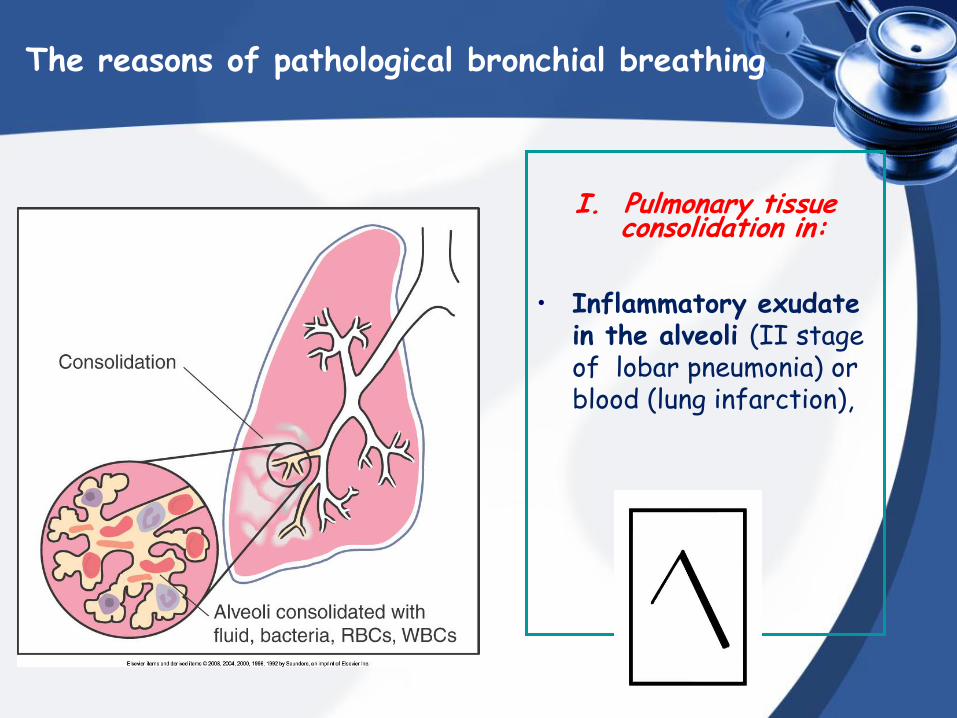
І. Pulmonary tissue
consolidation in:
• Inflammatory exudate in the alveoli (II stage of lobar pneumonia) or blood (lung infarction),
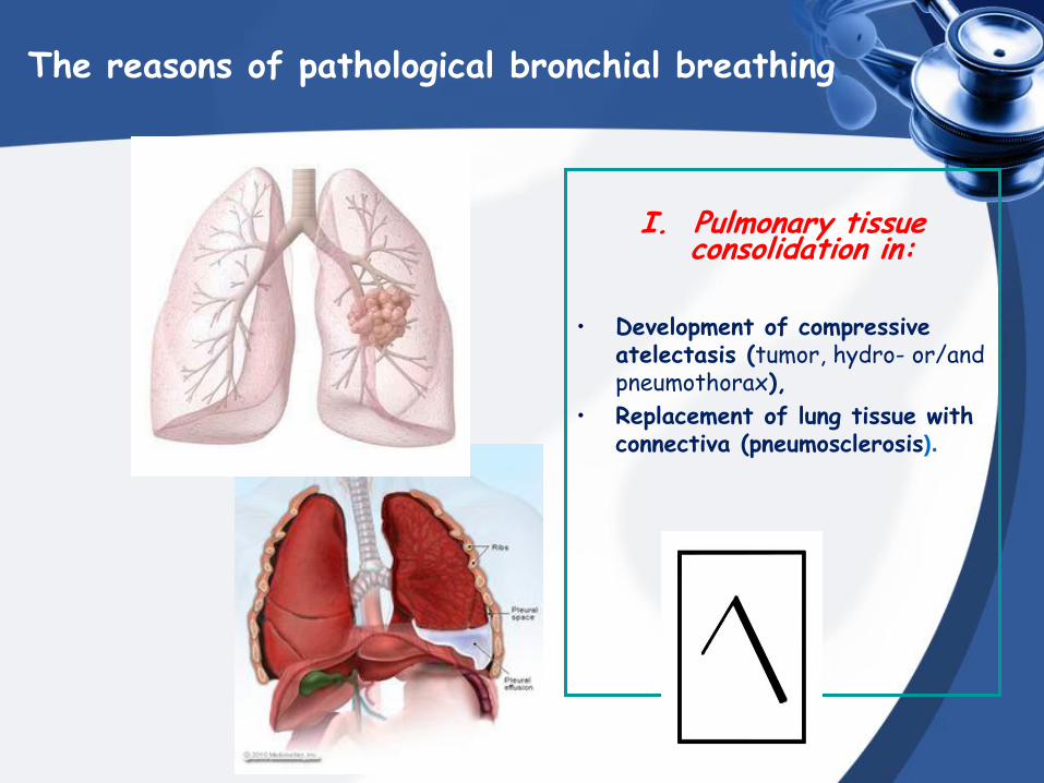
The reasons of pathological bronchial breathing
І. Pulmonary tissue consolidation in:
• Development of compressive
atelectasis (tumor, hydro- or/and pneumothorax),
• Replacement of lung tissue with connectiva (pneumosclerosis).
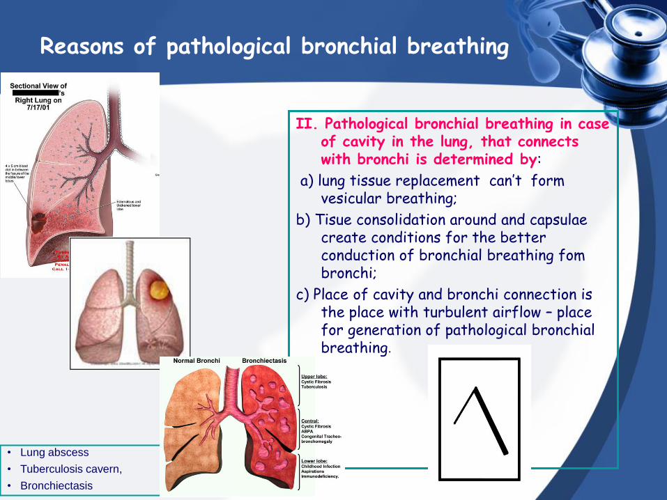
Reasons of pathological bronchial breathing
ІІ. Pathological bronchial breathing in case of cavity in the lung, that connects with bronchi is determined by:
а) lung tissue replacement can’t form vesicular breathing;
b) Tisue consolidation around and capsulae create conditions for the better conduction of bronchial breathing fom bronchi;
c) Place of cavity and bronchi connection is the place with turbulent airflow – place for generation of pathological bronchial breathing.
• Lung abscess
• Tuberculosis cavern,
• Bronchiectasis
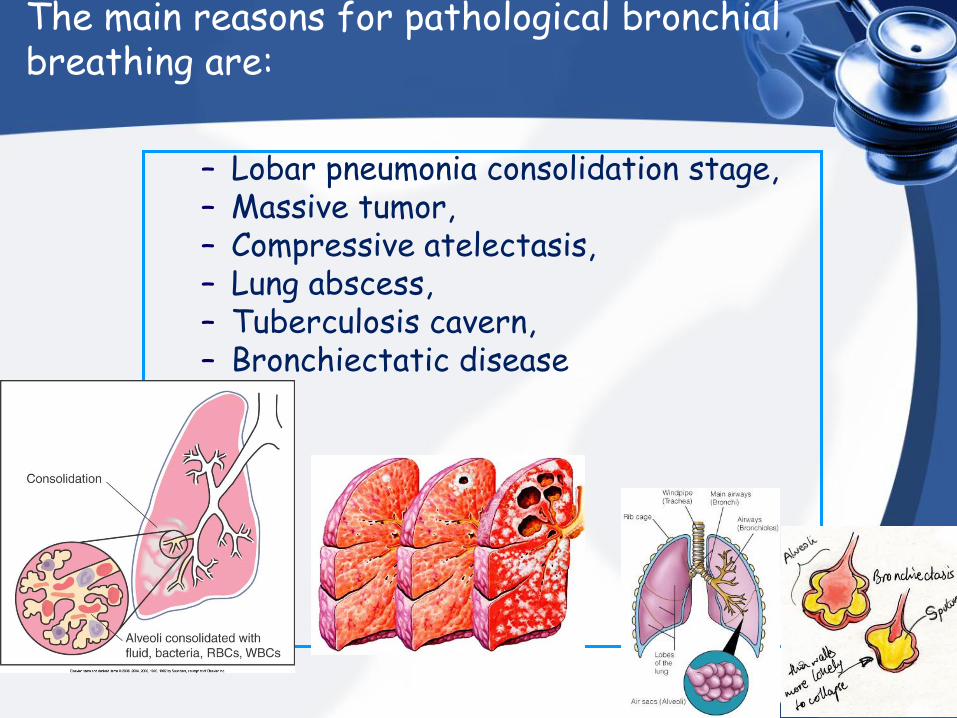
The main reasons for pathological bronchial breathing are:
– Lobar pneumonia consolidation stage, – Massive tumor, – Compressive atelectasis, – Lung abscess, – Tuberculosis cavern, – Bronchiectatic disease
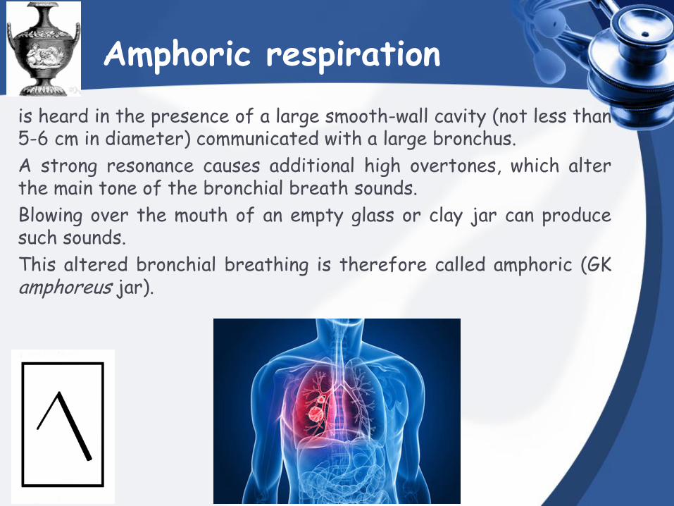
Amphoric respiration
is heard in the presence of a large smooth-wall cavity (not less than 5-6 cm in diameter) communicated with a large bronchus.
A strong resonance causes additional high overtones, which alter the main tone of the bronchial breath sounds.
Blowing over the mouth of an empty glass or clay jar can produce such sounds.
This altered bronchial breathing is therefore called amphoric (GK amphoreus jar).
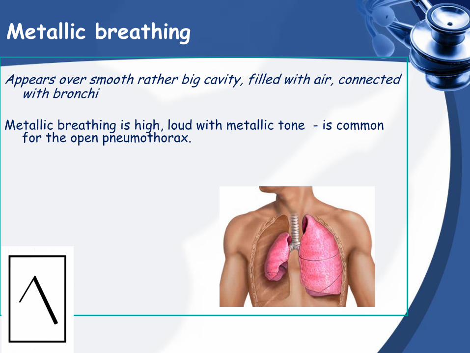
Metallic breathing
Appears over smooth rather big cavity, filled with air, connected with bronchi
Metallic breathing is high, loud with metallic tone - is common
for the open pneumothorax.
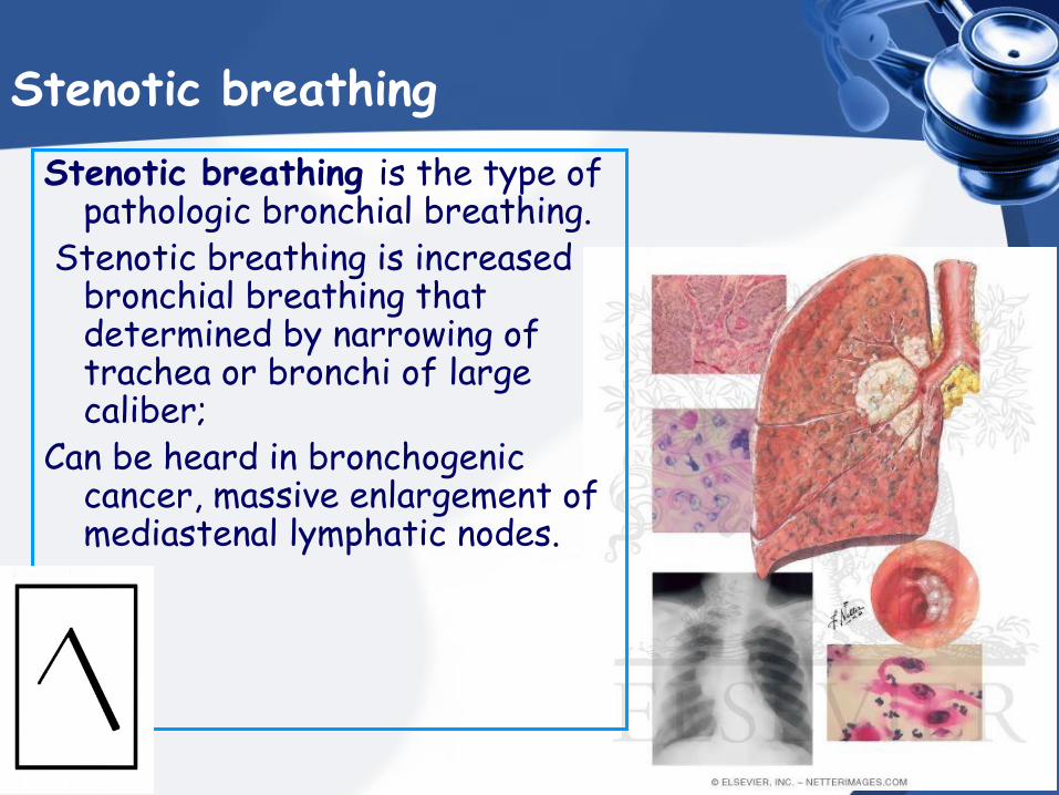
Stenotic breathing is the type of pathologic bronchial breathing.
Stenotic breathing is increased bronchial breathing that determined by narrowing of trachea or bronchi of large caliber;
Can be heard in bronchogenic cancer, massive enlargement of mediastenal lymphatic nodes.
Stenotic breathing
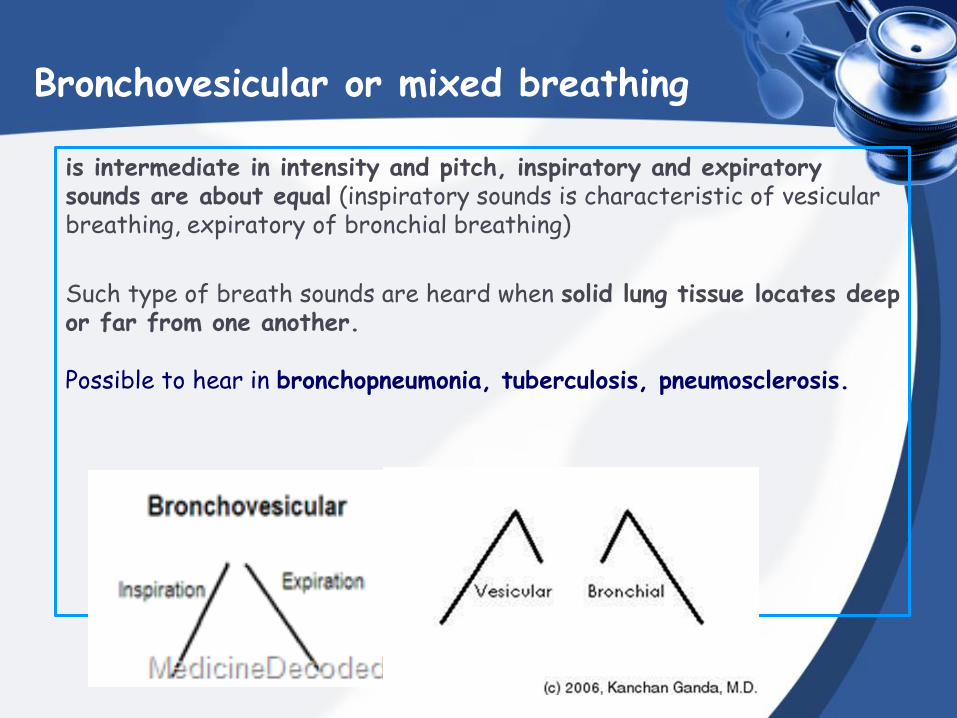
Bronchovesicular or mixed breathing
is intermediate in intensity and pitch, inspiratory and expiratory sounds are about equal (inspiratory sounds is characteristic of vesicular breathing, expiratory of bronchial breathing)
Such type of breath sounds are heard when solid lung tissue locates deep or far from one another. Possible to hear in bronchopneumonia, tuberculosis, pneumosclerosis.

Adventitious sounds
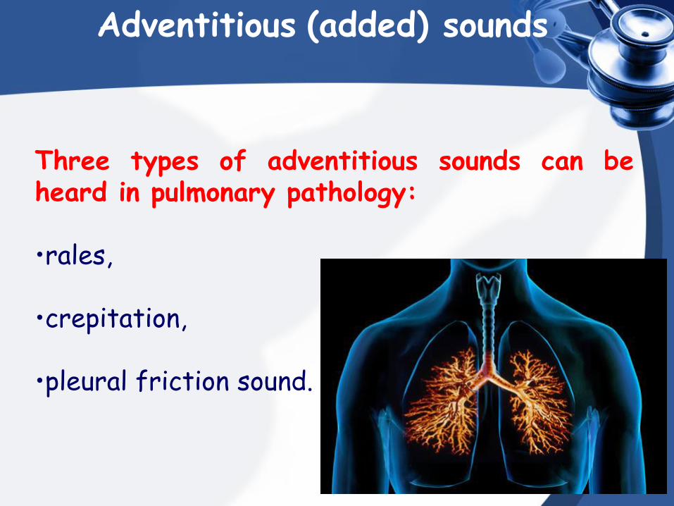
Adventitious (added) sounds
Three types of adventitious sounds can be heard in pulmonary pathology: •rales,
•crepitation,
•pleural friction sound.
Adventitious sounds
RALES
em
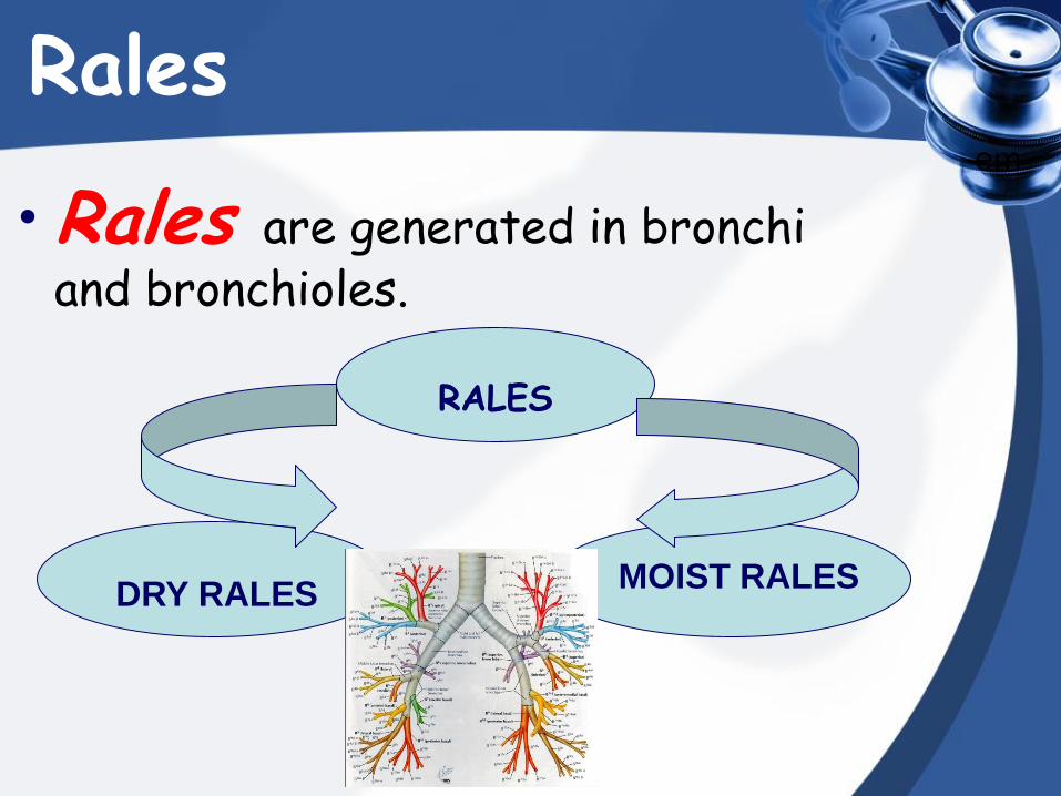
Rales
•Rales are generated in bronchi and bronchioles.
RALES
DRY RALES MOIST RALES
em
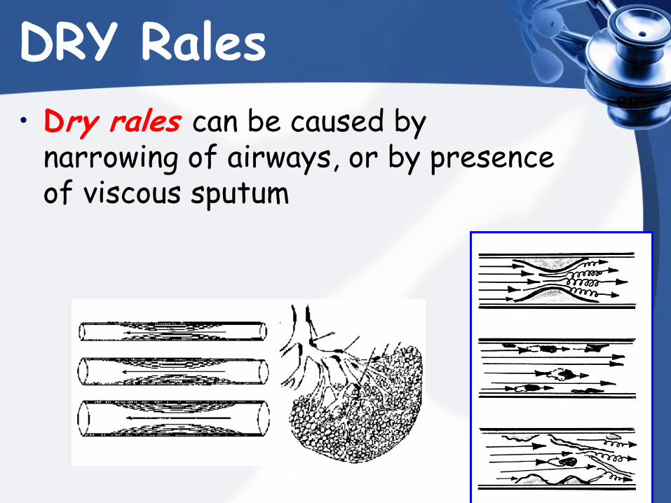
DRY Rales
• Dry rales can be caused by narrowing of airways, or by presence of viscous sputum
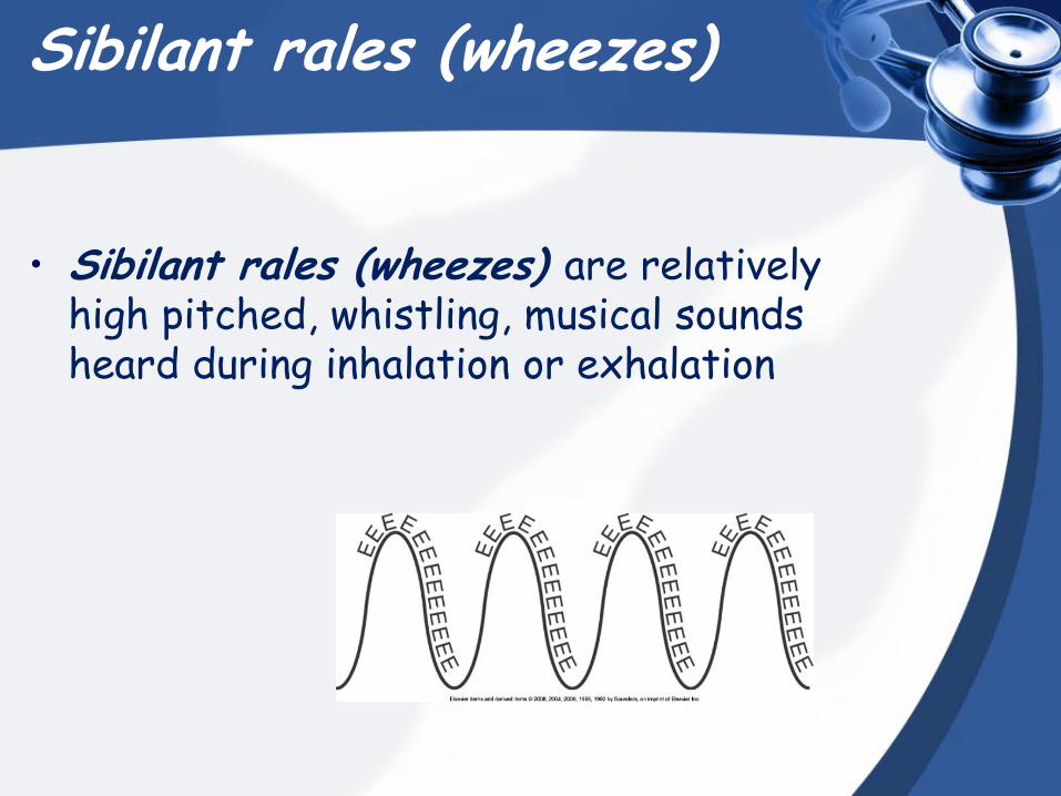
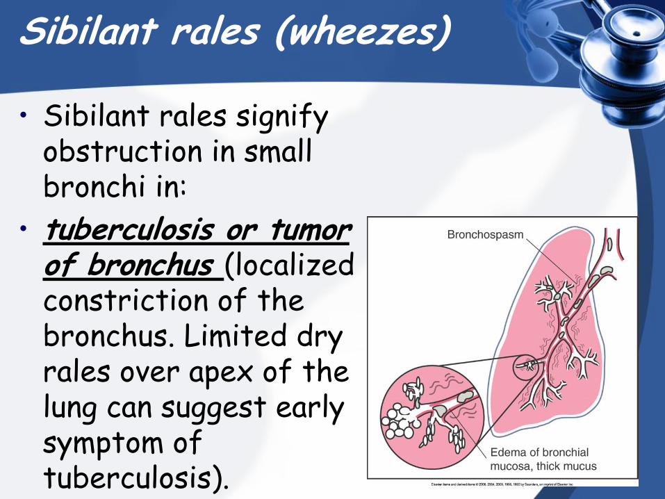
Sibilant rales (wheezes)
• Sibilant rales (wheezes) are relatively high pitched, whistling, musical sounds heard during inhalation or exhalation
Sibilant rales (wheezes)
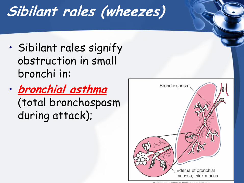
• Sibilant rales signify obstruction in small bronchi in:
• bronchial asthma (total bronchospasm during attack);
Sibilant rales (wheezes)
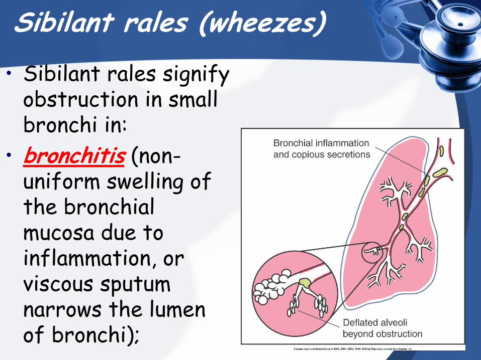
• Sibilant rales signify obstruction in small bronchi in:
• bronchitis (non-uniform swelling of the bronchial mucosa due to inflammation, or viscous sputum narrows the lumen of bronchi);
Sibilant rales (wheezes)
• Sibilant rales signify obstruction in small bronchi in:
• tuberculosis or tumor of bronchus (localized constriction of the bronchus. Limited dry rales over apex of the lung can suggest early symptom of tuberculosis).
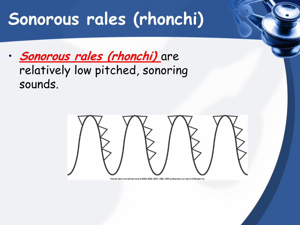
Sonorous rales (rhonchi)
• Sonorous rales (rhonchi) are relatively low pitched, sonoring sounds.
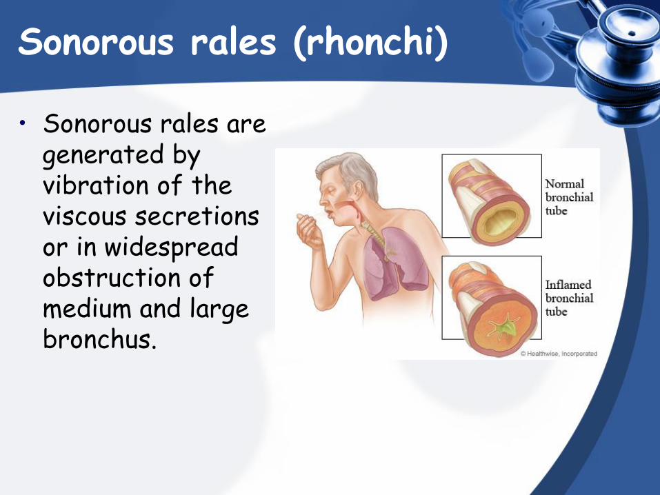
Sonorous rales (rhonchi)
• Sonorous rales are generated by vibration of the viscous secretions or in widespread obstruction of medium and large bronchus.
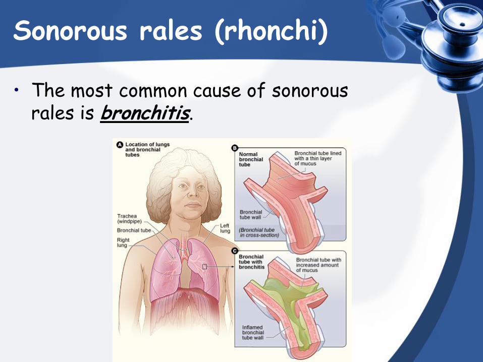
Sonorous rales (rhonchi)
• The most common cause of sonorous rales is bronchitis.
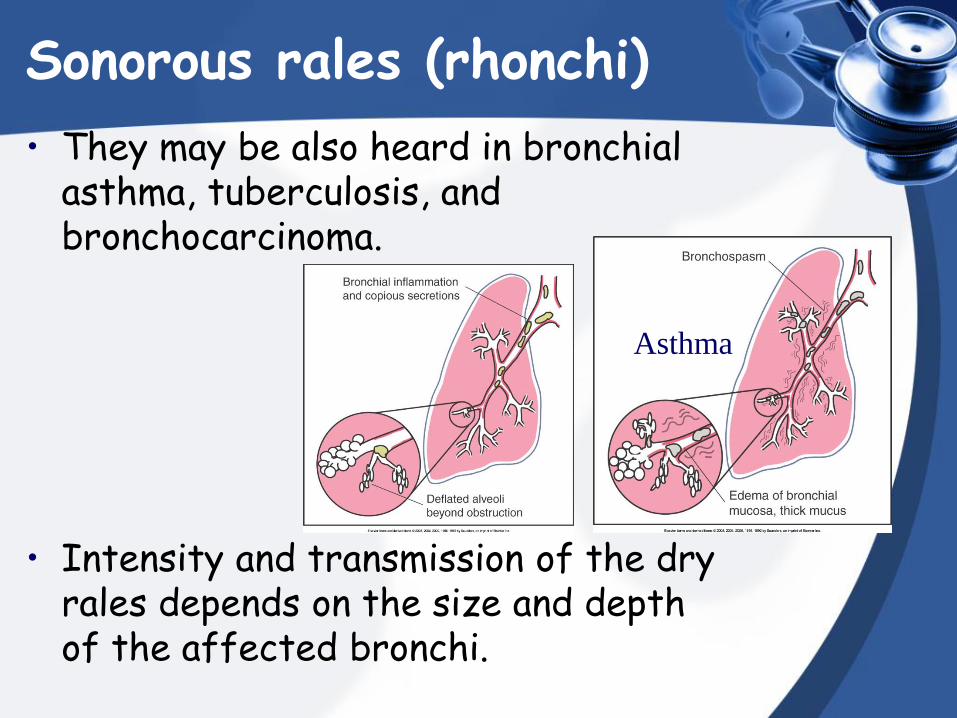
Sonorous rales (rhonchi)
• They may be also heard in bronchial asthma, tuberculosis, and bronchocarcinoma.
• Intensity and transmission of the dry rales depends on the size and depth of the affected bronchi.
Asthma
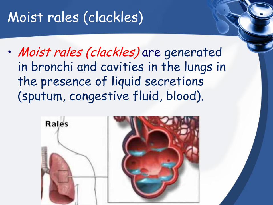
Moist rales (clackles)
• Moist rales (clackles) are generated in bronchi and cavities in the lungs in the presence of liquid secretions (sputum, congestive fluid, blood).
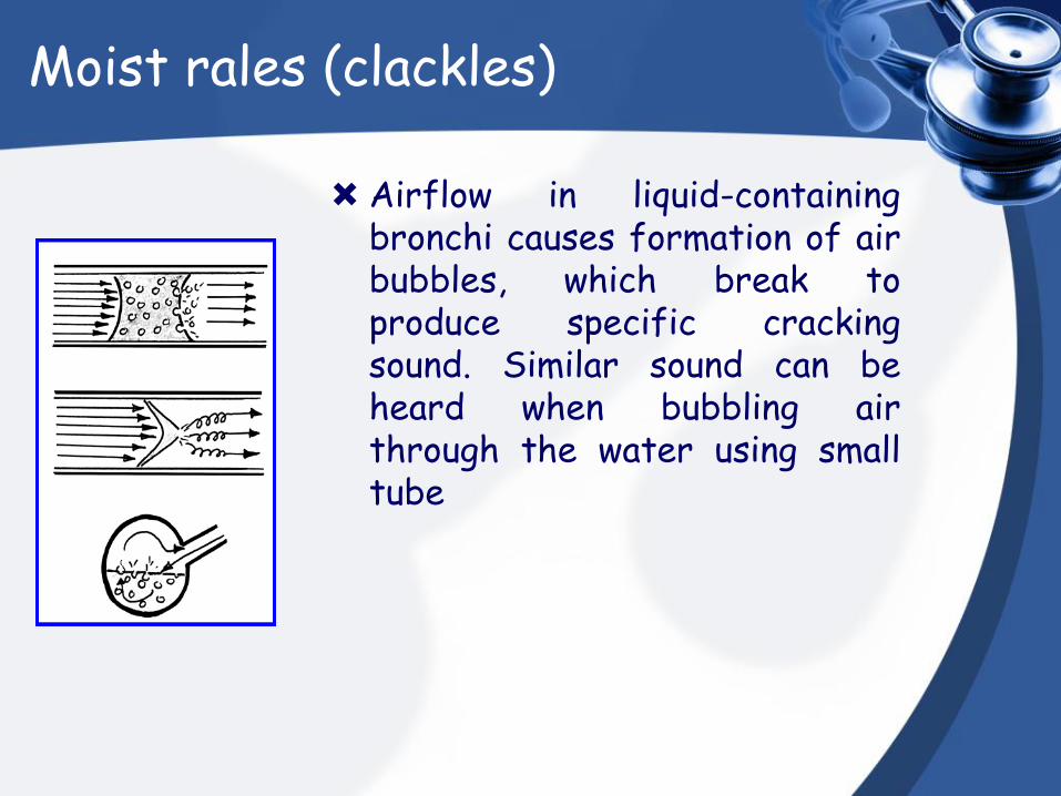
Moist rales (clackles)
Airflow in liquid-containing bronchi causes formation of air bubbles, which break to produce specific cracking sound. Similar sound can be heard when bubbling air through the water using small tube
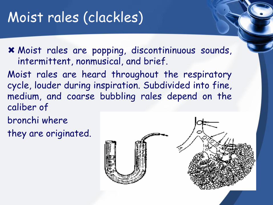
Moist rales (clackles)
Moist rales are popping, discontininuous sounds, intermittent, nonmusical, and brief.
Moist rales are heard throughout the respiratory cycle, louder during inspiration. Subdivided into fine, medium, and coarse bubbling rales depend on the caliber of
bronchi where
they are originated.
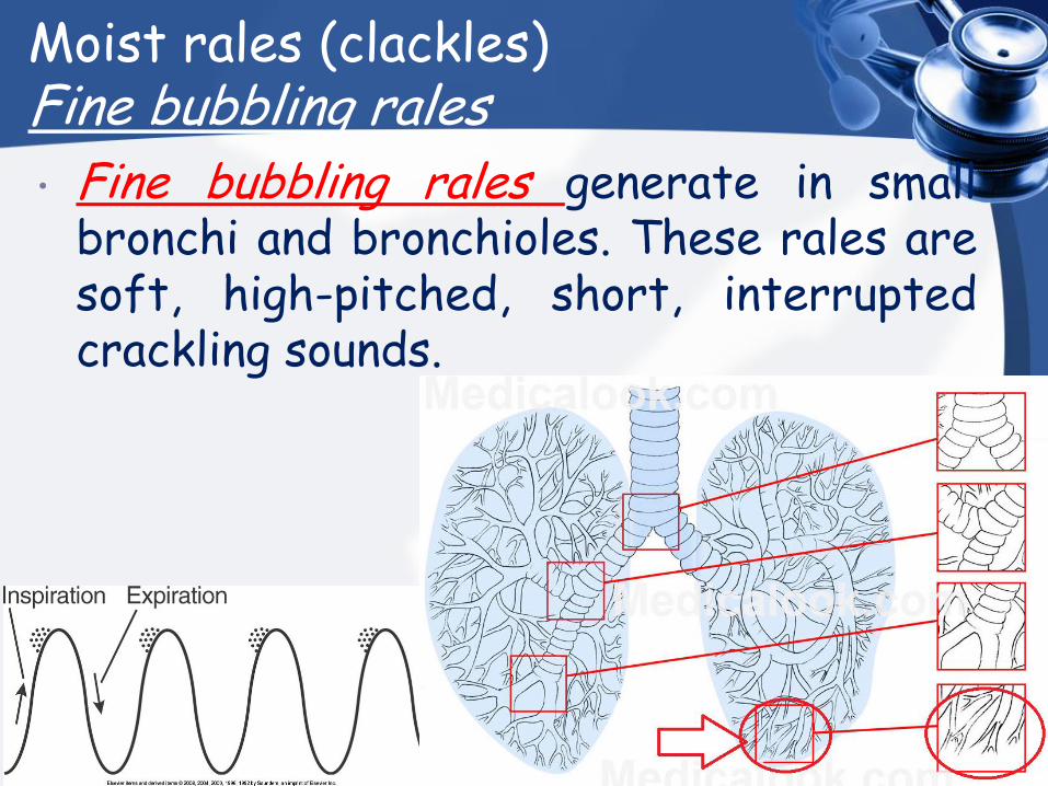
Moist rales (clackles) Fine bubbling rales
• Fine bubbling rales generate in small bronchi and bronchioles. These rales are soft, high-pitched, short, interrupted crackling sounds.
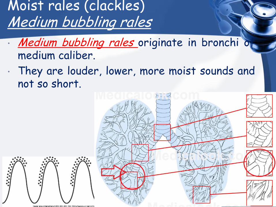
Moist rales (clackles) Medium bubbling rales • Medium bubbling rales originate in bronchi of
medium caliber.
• They are louder, lower, more moist sounds and not so short.
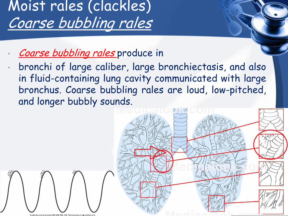
Moist rales (clackles) Coarse bubbling rales • Coarse bubbling rales produce in
• bronchi of large caliber, large bronchiectasis, and also in fluid-containing lung cavity communicated with large bronchus. Coarse bubbling rales are loud, low-pitched, and longer bubbly sounds.

Adventitious sounds
CREPITATION
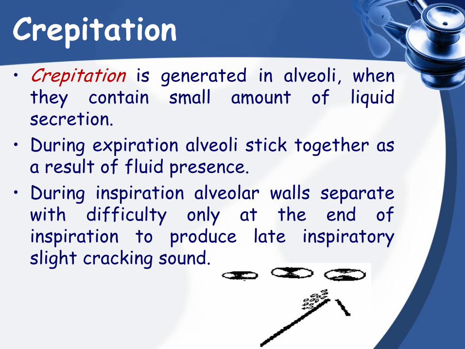
Crepitation • Crepitation is generated in alveoli, when
they contain small amount of liquid secretion.
• During expiration alveoli stick together as a result of fluid presence.
• During inspiration alveolar walls separate with difficulty only at the end of inspiration to produce late inspiratory slight cracking sound.
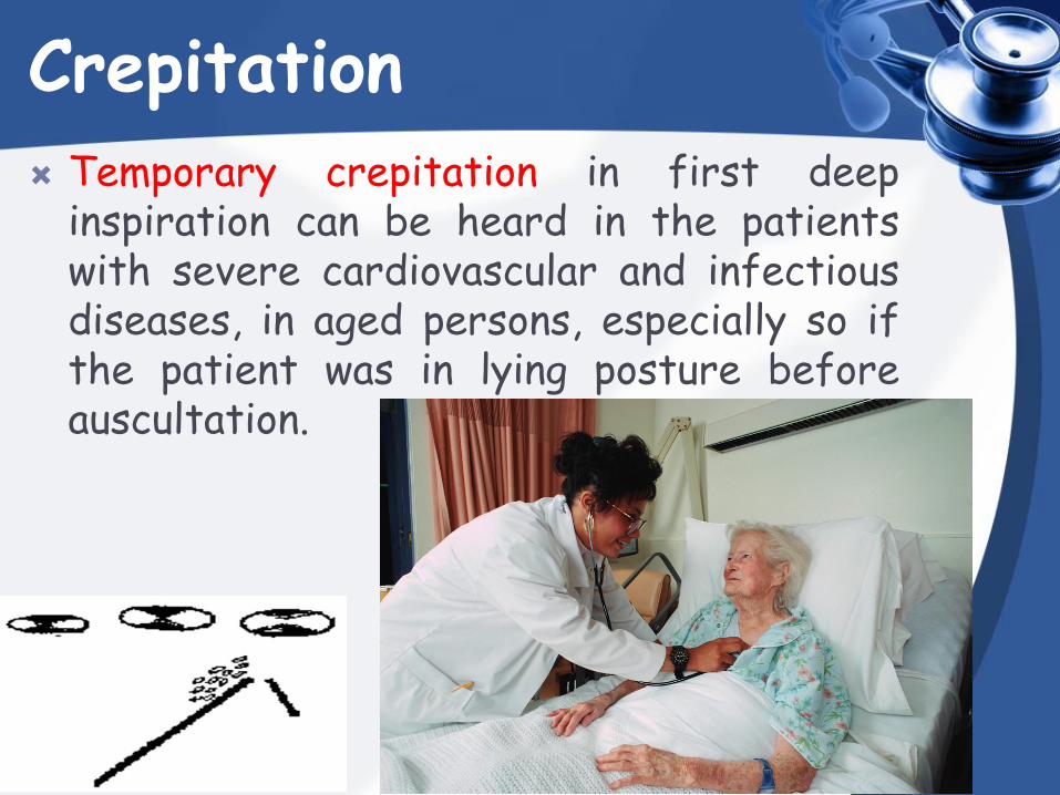
Crepitation Temporary crepitation in first deep
inspiration can be heard in the patients with severe cardiovascular and infectious diseases, in aged persons, especially so if the patient was in lying posture before auscultation.
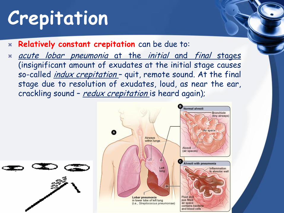
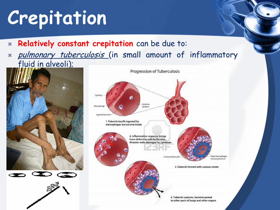
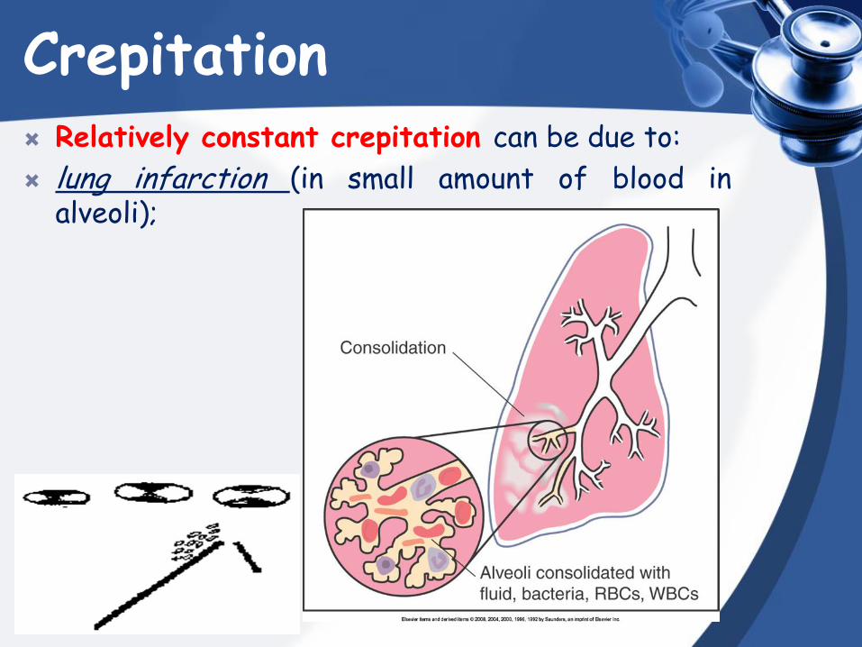
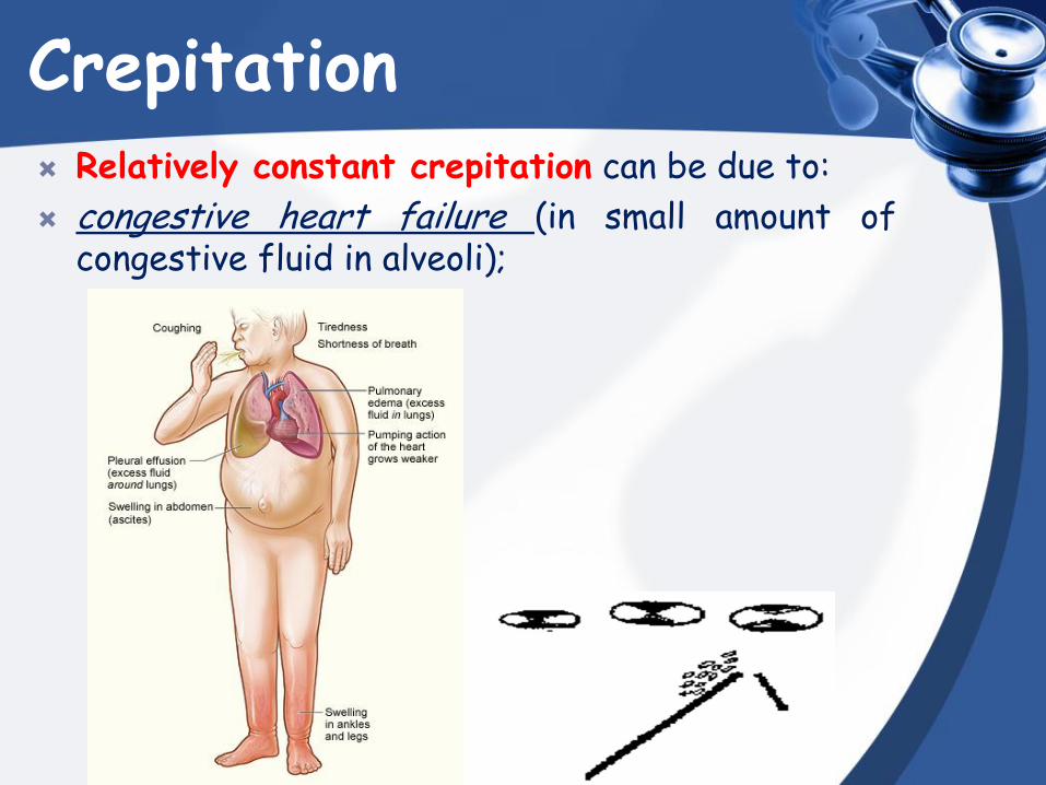
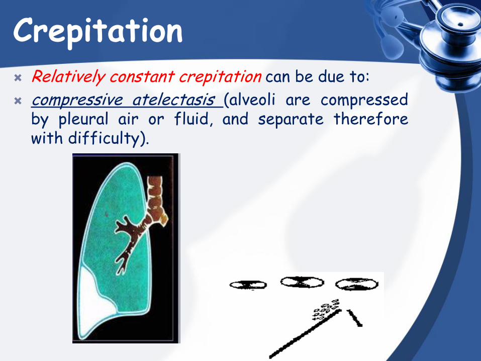
Crepitation Relatively constant crepitation can be due to:
acute lobar pneumonia at the initial and final stages (insignificant amount of exudates at the initial stage causes so-called indux crepitation – quit, remote sound. At the final stage due to resolution of exudates, loud, as near the ear, crackling sound – redux crepitation is heard again);
Crepitation Relatively constant crepitation can be due to:
pulmonary tuberculosis (in small amount of inflammatory fluid in alveoli);
Crepitation Relatively constant crepitation can be due to:
lung infarction (in small amount of blood in alveoli);
Crepitation Relatively constant crepitation can be due to:
congestive heart failure (in small amount of congestive fluid in alveoli);
Crepitation Relatively constant crepitation can be due to:
compressive atelectasis (alveoli are compressed by pleural air or fluid, and separate therefore with difficulty).
Adventitious sounds
PLEURAL FRICTION SOUND
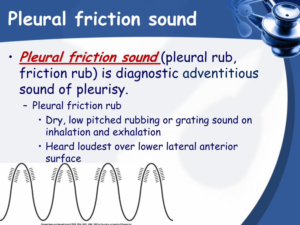
Pleural friction sound
• Pleural friction sound (pleural rub, friction rub) is diagnostic adventitious sound of pleurisy. – Pleural friction rub
• Dry, low pitched rubbing or grating sound on inhalation and exhalation
• Heard loudest over lower lateral anterior surface
Pleural friction sound
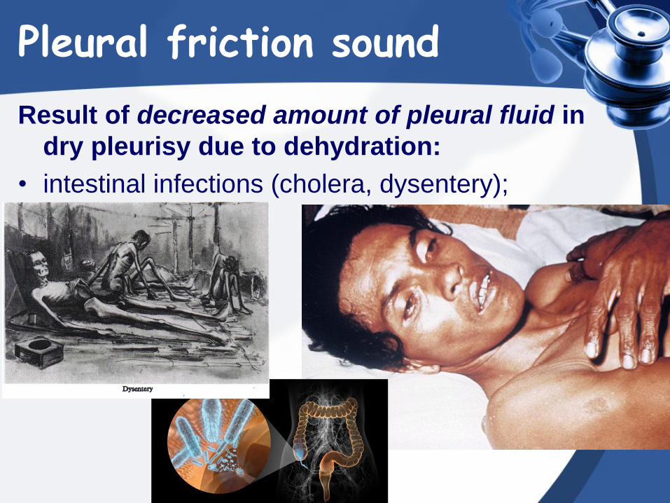
Result of decreased amount of pleural fluid in
dry pleurisy due to dehydration:
• intestinal infections (cholera, dysentery);
Pleural friction sound
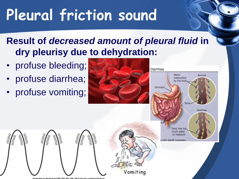
Result of decreased amount of pleural fluid in
dry pleurisy due to dehydration:
• profuse bleeding;
• profuse diarrhea;
• profuse vomiting;
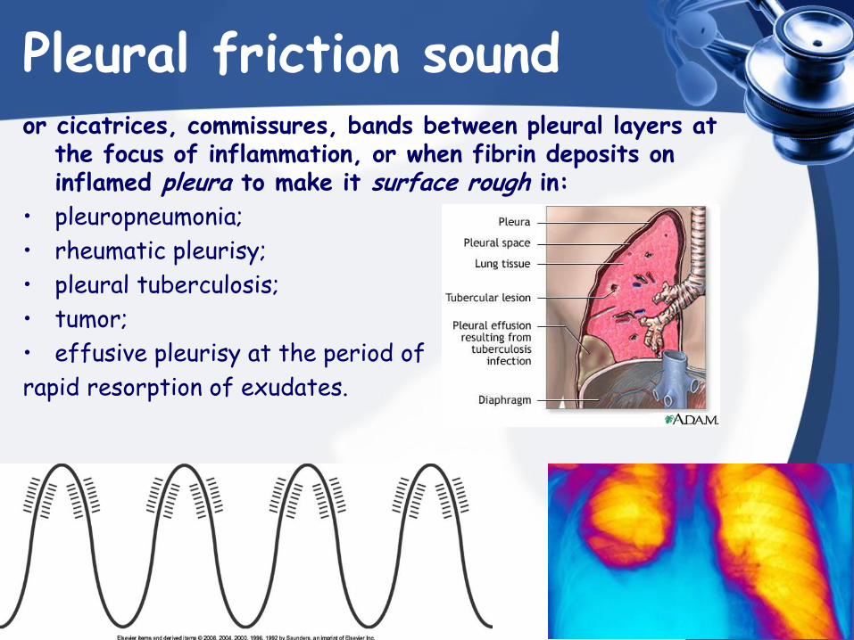
Pleural friction sound or cicatrices, commissures, bands between pleural layers at
the focus of inflammation, or when fibrin deposits on inflamed pleura to make it surface rough in:
• pleuropneumonia;
• rheumatic pleurisy;
• pleural tuberculosis;
• tumor;
• effusive pleurisy at the period of
rapid resorption of exudates.
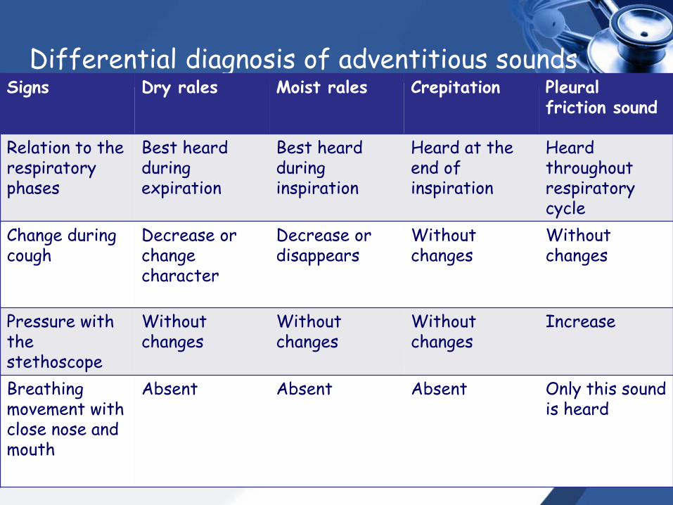
Differential diagnosis of adventitious sounds Signs Dry rales Moist rales Crepitation Pleural
friction sound
Relation to the respiratory phases
Best heard during expiration
Best heard during inspiration
Heard at the end of inspiration
Heard throughout respiratory cycle
Change during cough
Decrease or change character
Decrease or disappears
Without changes
Without changes
Pressure with the stethoscope
Without changes
Without changes
Without changes
Increase
Breathing movement with close nose and mouth
Absent Absent Absent Only this sound is heard

Thank you for your attention!
Any Questions?