Vascular changes in the lungs on the plain radiograph of the ...
8
Postgraduate Medical Journal (January 1970) 46, 3-10. Vascular changes in the lungs on the plain radiograph of the chest I. H. KERR M.B., M.R.C.P., F.F.R. Consultant Radiologist, Brompton Hospital, London, S. W.3 Summary The normal anatomy of the pulmonary vessels is considered and correlated with the angiographic appearances. Some pathological conditions causing an altera- tion in the vascular pattern as seen on the plain films are considered and the changes are described. It is concluded that an appreciation of these changes in vascular pattern may indicate to he clinician the presence of local or distant disease. Introduction Up to as recently as 1942 there was difference of opinion as to the nature of the shadows in the lung fields in normal radiographs, some authors still considering them to be due to the bronchi. However, Lodge in 1946 stated emphatically 'that the linear shadows seen transversing the lung fields in normal radiographs are produced by the blood-filled arteries and veins of the lung'. Further he concluded that many of the lobar and segmental arteries, and main pulmonary veins could be identified in the normal plain chest radiograph. Since that time the study of the pulmonary vascu- lature both in the normal and the abnormal has been greatly enhanced by pulmonary arteriography and there is a greater appreciation of the vascular changes in disease seen on the plain radiograph. Interpreta- tion of the chest radiograph is now considerably dependent on the identification of vascular changes, especially in the absence of abnormal pulmonary shadows. Normal anatomy of pulmonary vessels Arteries So that proficiency in the assessment of the vascu- lar pattern can be achieved, the observer should be familiar with the normal anatomy of both the pulmonary arterial tree, and the pulmonary veins. There are several normal variations described (Boyden, 1955; Jefferson, 1965). The intrapulmonary arteries are closely applied to the bronchi, though there are a greater number of variations in the origins of the lobar and segmental vessels than in those of the equivalent bronchi. Variations of arterial branching are greatest in the left upper lobe where there is no true typical pattern of branching. Beyond the origin of the segmental arteries, however, vessels remain intimately related to the bronchial axial pathways. On the right side, the right pulmonary artery reaches the lung by passing in front of the right descending bronchus, between the upper and middle lobe bronchi. It gives off its largest branch (the truncus anterior) to the upper lobe, and then descends on the lateral aspect of the bronchus as the pars interlobaris until the middle lobe and apical segment of lower lobe arteries take origin. From this point the artery continues to descend as the pars basalis, on the lateral side of the bronchus, to the basal segments of the lower lobe (see Fig. 1). The left pulmonary artery arches backwards over the left main bronchus, as the pars superior. It then descends, as on the right side, as the pars interlobaris, and then pars basalis, but on this side lying postero- lateral to the bronchus. There is no definite truncus anterior on the left side, the upper lobe being sup- plied by a variable number of smaller branches arising from the outer aspect of the left pulmonary artery as it curves posteriorly and downwards over the left bronchus. The arteries mainly give to the two hilar shadows on the plain frontal radiograph their characteristic shapes though that on the right is partly composed of the descending upper lobe veins (Lavender & Doppman, 1962). The right hilar shape is that of a wide angled 'V', with the apex pointing to the left. One arm of the 'V' is derived from the pars inter- lobaris and the pars basalis, and the other the truncus anterior and descending upper lobe vein. On the left side the hilum is shaped like a long fat comma, being produced by the shadows of the pars superior, pars interlobaris and pars basalis of the left pulmonary artery with the small upper lobe arteries arising from the outer curve of the comma. There is gradual diminution of the calibre of the artery shadows in proportion to the distance from the hila. The vessels are normally straight or gently curved, but not tortuous. The lower lobe vessels are fatter than the upper lobe vessels, as there is more ( copyright. on July 8, 2022 by guest. Protected by http://pmj.bmj.com/ Postgrad Med J: first published as 10.1136/pgmj.46.531.3 on 1 January 1970. Downloaded from
-
Upload
khangminh22 -
Category
Documents
-
view
2 -
download
0
Transcript of Vascular changes in the lungs on the plain radiograph of the ...

Postgraduate Medical Journal (January 1970) 46, 3-10.
Vascular changes in the lungs on the plain radiograph of the chest
I. H. KERRM.B., M.R.C.P., F.F.R.
Consultant Radiologist, Brompton Hospital, London, S. W.3
SummaryThe normal anatomy of the pulmonary vessels isconsidered and correlated with the angiographicappearances.Some pathological conditions causing an altera-
tion in the vascular pattern as seen on the plain filmsare considered and the changes are described.
It is concluded that an appreciation of thesechanges in vascular pattern may indicate to heclinician the presence of local or distant disease.
IntroductionUp to as recently as 1942 there was difference of
opinion as to the nature of the shadows in the lungfields in normal radiographs, some authors stillconsidering them to be due to the bronchi. However,Lodge in 1946 stated emphatically 'that the linearshadows seen transversing the lung fields in normalradiographs are produced by the blood-filled arteriesand veins of the lung'. Further he concluded thatmany of the lobar and segmental arteries, and mainpulmonary veins could be identified in the normalplain chest radiograph.
Since that time the study of the pulmonary vascu-lature both in the normal and the abnormal has beengreatly enhanced by pulmonary arteriography andthere is a greater appreciation of the vascular changesin disease seen on the plain radiograph. Interpreta-tion of the chest radiograph is now considerablydependent on the identification of vascular changes,especially in the absence of abnormal pulmonaryshadows.
Normal anatomy of pulmonary vesselsArteriesSo that proficiency in the assessment of the vascu-
lar pattern can be achieved, the observer should befamiliar with the normal anatomy of both thepulmonary arterial tree, and the pulmonary veins.There are several normal variations described(Boyden, 1955; Jefferson, 1965). The intrapulmonaryarteries are closely applied to the bronchi, thoughthere are a greater number of variations in theorigins of the lobar and segmental vessels than inthose of the equivalent bronchi. Variations of
arterial branching are greatest in the left upper lobewhere there is no true typical pattern of branching.Beyond the origin of the segmental arteries, however,vessels remain intimately related to the bronchialaxial pathways.On the right side, the right pulmonary artery
reaches the lung by passing in front of the rightdescending bronchus, between the upper and middlelobe bronchi. It gives off its largest branch (thetruncus anterior) to the upper lobe, and thendescends on the lateral aspect of the bronchus as thepars interlobaris until the middle lobe and apicalsegment of lower lobe arteries take origin. From thispoint the artery continues to descend as the parsbasalis, on the lateral side of the bronchus, to thebasal segments of the lower lobe (see Fig. 1).The left pulmonary artery arches backwards over
the left main bronchus, as the pars superior. It thendescends, as on the right side, as the pars interlobaris,and then pars basalis, but on this side lying postero-lateral to the bronchus. There is no definite truncusanterior on the left side, the upper lobe being sup-plied by a variable number of smaller branchesarising from the outer aspect of the left pulmonaryartery as it curves posteriorly and downwards overthe left bronchus.The arteries mainly give to the two hilar shadows
on the plain frontal radiograph their characteristicshapes though that on the right is partly composedof the descending upper lobe veins (Lavender &Doppman, 1962). The right hilar shape is that of awide angled 'V', with the apex pointing to the left.One arm of the 'V' is derived from the pars inter-lobaris and the pars basalis, and the other the truncusanterior and descending upper lobe vein. On the leftside the hilum is shaped like a long fat comma, beingproduced by the shadows of the pars superior, parsinterlobaris and pars basalis of the left pulmonaryartery with the small upper lobe arteries arising fromthe outer curve of the comma.There is gradual diminution of the calibre of the
artery shadows in proportion to the distance fromthe hila. The vessels are normally straight or gentlycurved, but not tortuous. The lower lobe vessels arefatter than the upper lobe vessels, as there is more
(
copyright. on July 8, 2022 by guest. P
rotected byhttp://pm
j.bmj.com
/P
ostgrad Med J: first published as 10.1136/pgm
j.46.531.3 on 1 January 1970. Dow
nloaded from
4 I. H. Kerr
I I;:: ··· *r·:i';':I'
···l:.L..····:'l:Ii
RPA .N:_i~i f. \j
. * ._ ,
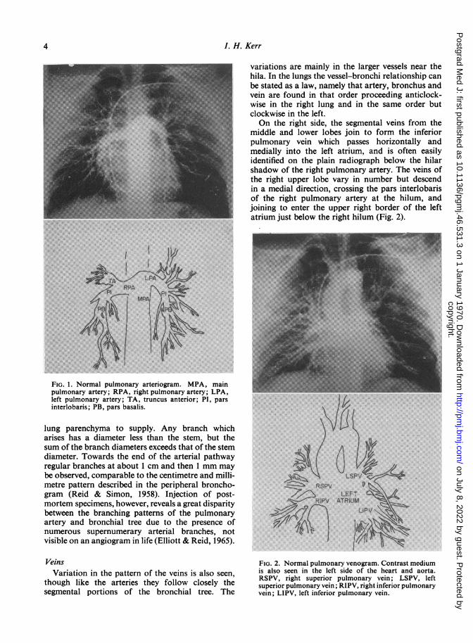
FIG. 1. Normal pulmonary arteriogram. MPA, mainpulmonary artery; RPA, right pulmonary artery; LPA,left pulmonary artery; TA, truncus anterior; PI, parsinterlobaris; PB, pars basalis.
lung parenchyma to supply. Any branch whicharises has a diameter less than the stem, but thesum of the branch diameters exceeds that of the stemdiameter. Towards the end of the arterial pathwayregular branches at about 1 cm and then 1 mm maybe observed, comparable to the centimetre and milli-metre pattern described in the peripheral broncho-gram (Reid & Simon, 1958). Injection of post-mortem specimens, however, reveals a great disparitybetween the branching patterns of the pulmonaryartery and bronchial tree due to the presence ofnumerous supernumerary arterial branches, notvisible on an angiogram in life (Elliott & Reid, 1965).
VeinsVariation in the pattern of the veins is also seen,
though like the arteries they follow closely thesegmental portions of the bronchial tree. The
variations are mainly in the larger vessels near thehila. In the lungs the vessel-bronchi relationship canbe stated as a law, namely that artery, bronchus andvein are found in that order proceeding anticlock-wise in the right lung and in the same order butclockwise in the left.On the right side, the segmental veins from the
middle and lower lobes join to form the inferiorpulmonary vein which passes horizontally andmedially into the left atrium, and is often easilyidentified on the plain radiograph below the hilarshadow of the right pulmonary artery. The veins ofthe right upper lobe vary in number but descendin a medial direction, crossing the pars interlobarisof the right pulmonary artery at the hilum, andjoining to enter the upper right border of the leftatrium just below the right hilum (Fig. 2).
........ .
...... :........: ..
R·PV ''..'.::'':.:T'::',: : :·i.i..:.:::~V...'.' - '"'/"' u v,,
FIG. 2. Normal pulmonary venogram. Contrast mediumis also seen in the left side of the heart and aorta.RSPV, right superior pulmonary vein; LSPV, leftsuperior pulmonary vein; RIPV, right inferior pulmonaryvein; LIPV, left inferior pulmonary vein.
copyright. on July 8, 2022 by guest. P
rotected byhttp://pm
j.bmj.com
/P
ostgrad Med J: first published as 10.1136/pgm
j.46.531.3 on 1 January 1970. Dow
nloaded from

Vascular changes in the plain radiograph of the chest
In the left lung the venous drainage is very similarto that on the right with the segmental vesselsjoining to form a superior pulmonary vein and aninferior pulmonary vein, which enter the left atrium.
It is the direction of the larger veins which makesthem identifiable on the plain radiograph. They passmedially to the left atrium 1 or 2 in. below thehilar shadows produced by the large trunks of theleft and right pulmonary arteries, and therefore,cross the branches of the arteries as these leave thehila.As with the arteries, in the erect position, the veins
of the upper lobes are smaller than those of the lowerlobes. In the supine position the upper lobe veins fillwith blood so that there is less difference in sizebetween them and the lower lobe veins, though theystill are smaller.
Changes in the normal vascular patternThere are few diseases of the lungs which do not
cause an alteration in some way, to the normal vesselpattern on the plain chest radiograph. The vesselsare visible because blood and vascular tissue aredenser than the air in the surrounding alveoli. Ifthere is no air in the alveoli, either because ofpulmonary collapse or consolidation, the vesselsbecome invisible and cannot be identified.Thus any condition which causes loss of air in the
alveoli, will cause a loss of the normal vascularpattern. Such diseases embrace a large portion ofpulmonary pathology and it is not the intention ofthis paper to provide an exhaustive list of pulmonarydiseases which cause alteration of the normal vascu-lar pattern, but rather to give some examples of theway in which observation of vascular change aids inthe interpretation of the plain chest film.
(1) Displacement of vesselsDisplacement of vessels in the lung indicates that
some portion of the lung is reduced in volume.Space occupying lesions such as distension cysts orbullae compress the adjacent lung with displacementof vessels.
Observing the displacement of the hila is of helpin identifying collapse of the lung. Thus in collapseof the left lower lobe (Fig. 3) the left hilum loses itsnormal round outer contour, as the pars inter-lobaris and pars basalis are displaced medially anddownwards behind the upper part of the shadow ofthe heart. The left hilar shadow is normally slightlyhigher than the right, and in left lower lobe collapseit is displaced inferiorly to lie about the same level asthat on the right or lower. The vessels in the upperlobe become splayed out as the upper lobe undergoescompensatory emphysema and the upper lobebecomes more transradiant. Similar changes areseen in right lower lobe collapse.
Fig. 3. Post-operative collapse of left lower lobe. Theleft pulmonary artery is displaced downwards so thatthe left hilum is small, and the normal round contourof the hilum is lost.
In upper lobe collapse or contraction the reverseoccurs, and there is elevation of the hilar shadows.In long-standing disease such as tuberculosis it is thedisplacement of the vessel shadows coupled withdisplacement of the mediastinal structures thatindicates fibrosis has taken place. An angularelevation of the hilar shadow with streaks of fibrosisleading to the apex of the lung is the typical appear-ance in long-standing upper lobe pulmonary tuber-culosis.
(2) Decrease in vascularity of lungDecrease in vacularity of the lung, either local or
general, may be very difficult to appreciate. Know-ledge of the normal appearances of the vessels isessential. Only after careful study of the vascularpattern in normal radiographs can the abnormal bedetected. Radiographic technique is critical. Anover-exposed film may suggest oligaemia, and anunder-exposed film plethora of the lung fields.Rotation of the patient may make one lung fieldappear oligaemic in comparison with the other. If theradiograph has not been taken on full inspiration thevessels appear more conspicuous, especially in thelower lung fields.
5
copyright. on July 8, 2022 by guest. P
rotected byhttp://pm
j.bmj.com
/P
ostgrad Med J: first published as 10.1136/pgm
j.46.531.3 on 1 January 1970. Dow
nloaded from

6 I. H. Kerr
(a) Generalized decrease ofpulmonary vascularity.There is generalized pulmonary oligaemia in cyanoticcongenital heart disease, usually the result of pul-monary stenosis or atresia, or tricuspid atresia inwhich a right to left shunt is present. The normalbronchial artery supply to the lung is usually sosmall as to be invisible on the plain radiograph, butunder these circumstances, the bronchial arteriesmay dilate to such an extent as to be visible (Fig. 4).They then produce a mottled pattern in the perihilarregions. This may be so marked as to obscure thefact that the pulmonary vessels are smaller thannormal.
Acquired generalized oligaemia is seen in pul-monary emphysema (Fig. 5). The radiologicalfeatures of emphysema have been described by Laws& Heard (1962) and Simon (1964). The mid-lungpulmonary arteries appear narrow, straight andsometimes of uniform calibre over several centi-metres instead of tapering towards the periphery.The finer vessels are reduced so that there is a ratherclear homogeneous transradiant background to thelung field. In several cases the main pulmonaryartery becomes enlarged and there is dilatation ofthe hilar vessels. In the normal person there isgradual reduction in size of the pulmonary vesselsfrom the hilum to the periphery, but in emphysemathere is a rather abrupt reduction in size between thehilar vessels and the mid-lung vessels. Other featuresof emphysema, the low flat diaphragm, large retro-sternal translucent zone, the narrow vertical heart,
;i....··:,-· ;·;··:;sli::i:PI"I'' ·:';:; ··'·;;' ·'':'':··:IZli.j.L ·: ··:ii·.i.ifii;E:::· ···.·,...:..".I:;'···::·iii.:::iii:.:::::.·.:·:· :.i:ijBIZLl;li::i:..l::l··:::·· ·-·vnw:inib:: .:·.··:·::::······Is: ....:i· I':l!:::;:::!i:.·:i·i·i:·: :i· :i' '-.1.·..
.iili·.··········:···· ·· i'',iJiilii4il;:iiiii: ii::::··I· ·i·iiie::· -::ci... iiiiili"""" iiiiii$9ii:i:l:i:,::*lis:··· ·:· .i3!2iiii :.·:·:·:··:;:·
":B:
:iiiiLii. i.2i.1:.·.·.... IdiPI1:Li"'
.iEii.i L·.··:::::n:w·' :' :···:·:·;::···:::::·: ·········:i··:···;: .:,::!: -::n::i::····;···;··':a:L.:Fl";i:"p -·:·.
···:r·:·· :··· WII'·:: Ig,:Ir i.ii!il'ril··:i,li' .:.."::'l:i8ili'iiiiiiC::.·.s·1:i·::i··.· :i;iiiiiids
:......·.:. t..l:i·ir·i·;i:i:·:::::::..:
"" :.·: ·il:li:'':i:iiiilii!iiiiiizilililiilnl :' '';:'·:·;i·:··ii·::;::··· ·I···· i·':: ·::·l.i il.li;iiiiiiiiiii.ii·iiiiiihil::'·'i· ':"':Y'':' :"·'e:··::···:i:·il:i:ii::::::.l. '...'.···i.·i :·.::'Til·::::::;:l: i::i:·:·:·:i··i·::·:: .. :adi.:l:;.::;:::'i':liiiiii.i:ii:l..l:.I .· ·.··.· :·:.::..i,,....li ·:r
nl.i,:w:···:·::a:;;:·: ·.:. i:·18iiai8ii9ii::iiP:'··.·..j 1. ''··Ei;.·:sipiFFSiCiiilix·.::::::l·il:.la:' -·:..:::i::.....:.·:·..·:
i··· ·:*:;·ilii t.i:··;·;· i··"i::·:%:8ia:ii:llI:"·:··'·
:I:.!::,:::::.
:·:·;:·i·::.... ..ifllif.:"i;l:s::.::s:
:.:···· :-.:·:·;%ti.sr·,:··:i.aP.:s.·-."3""""·C.-".L"IL:.aP.B.LB· Sss.SL·.:·'i:K:Si.'i::·:::'i:, b:·:I·I·;·······:·:%·:'·:I::ai.:i9:.:ia:n:
l.liFiil:lil
i·i;':'':
·:-I:l.s% ar .a:i·.
"·*· ::i
'?i%.;.lLi;lP:"*
:·····:·-::;.::.-
E:ii:''1PBl.%ars.l.Fi.Llili.lell:'·ir
z:·;:·.*.:·::··-·-;·:I:m: ;r;x; ·I::'·-··-··;·..i:i :''.'':"I""':"··'Li::::·:::;·i· i·: ifiii!i.l..i.88SI ..ii.iilii.lj
:W::l':.*n-rr.o·slll. C.pa::;i :.:C:::::W: Pi::
FIG. 4. Pulmonary atresia. No pulmonary arteries are seen. The vessel shadows are irregular, and areproduced by bronchial arteries and a few small pulmonary veins.
.:..;:.:..::::.::::: ::.s: :n:.......... ::··y:;::·:..
l <.~~;~;~;~~~;~~~~~~~ ~~:'I:
~"iiilli::i.:.i.ii I;i'i:i~iii·····:-~~~~~~~~~~~~~~~·· .:·... i:i· l:ci
-~~~~~~~~~~~~~~~~~~~~~~~~fliHi
Ciii-illi::!l;lj:l:::!·;: ·· ··.·: :·::: :· ·:: ·:lr Lfi!l
Ci,:liii l-ISel.:.:
- :l~~~~~~~~~~~~~~~~~·
.: ..: .. .....Wili
H.^ a,., .,::..
FIG. 5. Emphysema. There is marked reduction in thepulmonary vessel size in the mid lung field, particularlyin the right upper and mid zones.
copyright. on July 8, 2022 by guest. P
rotected byhttp://pm
j.bmj.com
/P
ostgrad Med J: first published as 10.1136/pgm
j.46.531.3 on 1 January 1970. Dow
nloaded from

Vascular changes in the plain radiograph of the chest
and bullae, may be present. Patients with asthmauncomplicated by emphysema may have radiologicalsigns of air trapping, low flat diaphragms, trans-radiant lungs, and narrow vertical heart, but theintrapulmonary vessels, unlike those in emphysema,are normal in size and not narrow. In primarypulmonary hypertension the main pulmonary arteryand hilar vessels are large, and there is reduction insize of the intrapulmonary vessels, but unlikeemphysema there is no evidence of air trapping;the diaphragm is normal.
(b) Localized oligaemia. Congenital absence of onepulmonary artery is rare but provides a characteristicradiographic appearance. The affected lung isreduced in volume but appears normally aerated.There is an absence of the normal vascular shadowat the hilum and in place of the branching system ofblood vessels normally seen, there is the network ofsmaller vessels, giving an irregular pattern, typical ofenlarged bronchial arteries. These are the onlyvessels supplying blood to the lung (Good, 1961).
Similar appearances are seen in hypoplasia of apulmonary artery (Belcher & Pattinson, 1957) thoughin most of these cases there are associated abnormali-ties of the bronchial tree.
In unilateral transradiant lung (Macleod's syn-drome), there is a uniform reduction in pulmonaryvasculature throughout the affected lung, or portionof lung. It is now considered that this condition isprobably due to arrested growth of the lung due tosevere infection in childhood (Reid et al., 1967).The observation in pulmonary embolism of
oligaemia of the lung goes back to Westermark in1938, and was much disputed for several years. Inboth acute and chronic pulmonary embolism theaffected area, whether segment, lobe or entire lungmay show oligaemia (Fleischner, 1962). Localizedoligaemia is easier to detect than widespreadoligaemia since a reduction in size of the vessels inone or more zones can be detected by reference tothe vessel size in other zones. The changes are mosteasily recognized in chronic pulmonary embolicdisease, though the radiograph may appear mis-leadingly normal in these patients (Chrispin, Good-win & Steiner, 1963). However, careful study ofplain radiographs taken in acute pulmonary embo-lism, particularly if massive, reveals detectableoligaemia in nearly all cases (Figs. 6 and 7). Shadowsdue to infarcts are present in only about two-thirdsof patients with pulmonary embolism (Kerr, Simon& Sutton, 1969). The quality of the radiograph isobviously of the utmost importance in assessingthese changes in patients who are frequently seriouslyill, and often unable to sit up. However, with goodtechnique and using modern high-powered portableunits a satisfactory film should be obtainable.
FIG. 6. Acute massive pulmonary embolism. Plainchest radiograph showing reduced vascularity of rightlung. Note particularly loss of small vessel pattern inthe periphery of the lung as compared with the left.
(3) Increase in pulmonary vascularity(a) Generalized. Astley (1959) reviewed some of the
radiological signs provided by the plain radiographand fluoroscopy, in congenital heart disease. Hedivided the patients into acyanotic and cyanoticgroups. The presence of pulmonary plethora in theacyanotic patients indicates a left to right shunt,either via a septal defect, or a patent ductus, oranomalous pulmonary veins. Pulmonary plethora incyanotic patients suggests the possibility of transpo-sition, truncus arteriosus, infantile co-arctation orthe Eisenmenger syndrome.
In patients with Eisenmenger syndrome wherethere is reversal of left to right shunt, the largesthearts and the biggest proximal pulmonary arteriesare seen in association with atrial septal defects(Rees & Jefferson, 1967). The plain radiograph isoften almost normal in ventricular septal defectwith reversed shunt particularly in older children.Pulmonary plethora may be acquired as the result
of a septal defect, either following infarction of theseptum or traumatic rupture. The onset of plethorais sudden and it may be difficult to distinguish frompulmonary congestion due to left heart failure inthese usually very ill patients, but if there has beenthe sudden development of a loud systolic murmurthe possibility of a septal defect should be considered.
In some cases of chronic bronchitis, Group 3 (syn.'blue bloater', cor pulmonale), described by Scarrow
7
....:c i
copyright. on July 8, 2022 by guest. P
rotected byhttp://pm
j.bmj.com
/P
ostgrad Med J: first published as 10.1136/pgm
j.46.531.3 on 1 January 1970. Dow
nloaded from

I. H. Kerr
FIG. 7. Acute massive pulmonary embolism. Pulmonary arteriogram of thesame patient as Fig. 6. showing embolism in the right main pulmonary artery.
(1966) where there is no evidence of emphysema,there may be enlargement of the intrapulmonaryarteries. Unlike the other groups, in which there isemphysema, a more normal type of terminalarborization of the pulmonary arteries is seen.
In polycythaemia, not secondary to heart orrespiratory disease, there is frequently an increase inthe size of the small pulmonary vessels in the peri-phery of the lung. There may be a mottling in themid and lower zones of the lungs present, due to alarge number of small peripheral vessels seen end-on(Pitman, Steiner & Szur, 1961).
Patients with cirrhosis of the liver may show smallill-defined nodular shadows in the lungs, pre-dominantly in both lower zones. Berthelot et al.(1966) inspected post-mortem lung specimens inthirteen patients with cirrhosis, six of whom hadnodular shadows. They showed small angiomata onthe pleural surfaces, looking like the spider naeviseen on the skin in such patients. There was fillingof an excessive number of peripheral small branchesof the pulmonary arteries within the respiratoryportion of the lung. The authors suggest that thenodular shadows might well arise from widespreaddilatation of these small vessels.
(b) Local. Where there is diminution of vascu-larity of the lungs of a patchy nature, localizedhyperaemia may occur in the normal areas of lung.This can be seen in pulmonary emphysema,'particu-larly of the bullous variety, and also in pulmonaryembolism.
There are two types of pulmonary arterio-venousfistula, both of which cause abnormally large vessels(Good, 1961). The first type, the more common, isbetween a pulmonary artery and a pulmonary vein.The second type is rare, and is between the systemiccirculation and the pulmonary vessels. The first, ifsevere enough, may produce cyanosis, as it causes aright to left shunt. The second is acyanotic as theblood flows from the oxygenated systemic circulationto the pulmonary circulation. There is usually around or oval shadow in the periphery of the lungconnected to the hilum by two large vessels. One ofthese is an artery and the other a vein, and usuallytheir connections can be identified, if not on theplain radiograph, by tomography (Fig. 8). Multiplefistulae are present in 30-40% of cases. A bruit maybe heard over the lesion. Some of the patients withpulmonary arterio-venous fistulae suffer fromhereditary haemorrhagic telangiectasia in whom rubycoloured lesions may be seen on the skin andmucous membranes.Pulmonary arterio-venous fistula should be dis-
tinguished from partial anomalous venous drainage,in which there is an anomalous course of thepulmonary veins. In both conditions a large veinmay be visible, but in arterio-venous fistula twovessels should be visible, the draining vein and thefeeding artery. In anomalous venous drainage onelarge vein is seen and it receives tributaries, giving ita branched appearance. Anomalous veins of theright lung may drain into the left atrium, the rightatrium, the hepatic vein or either vena cava. Veins
8
copyright. on July 8, 2022 by guest. P
rotected byhttp://pm
j.bmj.com
/P
ostgrad Med J: first published as 10.1136/pgm
j.46.531.3 on 1 January 1970. Dow
nloaded from

Vascular changes in the plain radiograph of the chest 9
···c. ·"·
·: 'i13 ·i·
':'.· :.. .·:···:ipl:ii (.·:··i· ··:·;:·. ···:·::iiilIrpili:lr "··::
''''' 'I··Ii:- --:-; .i:i:::: .'"":· ·:::.·'''''''
..:.:: : ii··i::·ii ·i'·:·::,··:··::··:· i·'···.i381 rCa.:···l'l
:··r·.··.:8:I· .liiiii"":': ":' ··· :·i:·::::: ":':: ': '' :·····:;:i::':'
I:·:··· Igil:····.· ·:·::·:I :··':::I··. :'.':"·'··:· '···:··:'ii::;:::.:::r:: .: ;·: ··.:i··:··.
I:....' ::·:;·.Ilassl.Hara.isll -- -P.EiliKI;:lil··I·
:: ':
.·..·.··.·..· ':":`";:'·.·:'·:···i:·:··::r::: ·';·· :-::: ·iI:i·l?i:i.:i:l:;i:·.·: ·.·:·.·:·i·
.i: ·:·::ii'iiiiii. :.::·:·
.:I····ii·iiiiiijiiiiiiiiil:;I::"'·..:.·. I:: ··i·;i:: :, :.: .:iii:il:ll'diiiii:iiili:.:i: ::.:I91::I:::j::":'.::.":::::....:'.:. i··.· .:..i· '· '' :"'·
:-··
···*ii· ·:··.:,iii:i:i,:i::ii:ii :i· ·:··::"'··:· '' ''':' ':....i:......:.i
··r···i·.: .:..:.·:·:r·:··:::: ..I ':·':,.,., .,..
FIG. 8. Arterio-venous fistula in the right lower lobe.Note the two vessels, the artery and the vein leadingto and from the lesion in the costo-phrenic sulcus.
in the left lung may drain into the left innominatevein. The commonest of these is seen as a largevessel running vertically in the right lung, gatheringtributaries, and passing through the diaphragm nearthe right cardiophrenic angle.
(4) Changes in pulmonary vasculature resulting frompulmonary venous hypertension
Simon (1958) demonstrated that in patients withmitral stenosis there is engorgement of the upperlobe veins, and narrowing of lower lobe veins, andthat the changes in vascularity provide some indi-cation of the severity of pulmonary venous hyper-tension. The upper lobe veins were graded by Simon.In the normal range (Grade 0) the veins are notconspicuous and are smaller than the arteries. InGrade 1 there is slight engorgement of the upper lobeveins which become easily identifiable by contrastto the arteries. They are one to one and a half timesthe normal width. In Grade 2 the main tributariesare obviously dilated two to three times normal. Thesmall tributaries are also prominent and traceableto the periphery. In Grade 3 the venous trunks andmajor tributaries are markedly dilated, three to fivetimes normal width.
Unfortunately in correlating physiological studies
with the degree of dilatation of the upper lobe veins,it is found that there is not a direct proportionalrelationship. The appearances are affected by thepulmonary artery pressure. As pulmonary hyper-tension progresses there is enlargement of the mainpulmonary artery and its proximal branches withdiminution of the peripheral vessels, which becomenarrower and more tortuous. These changes affectthe lower lobes first, then become widespread(Goodwin et al., 1955).The combined effects of pulmonary venous hyper-
tension and pulmonary arterial hypertension, aretherefore to reduce the peripheral pulmonary bloodflow so that the venous engorgement becomes less,thus explaining why some patients with severe mitralstenosis show only slight venous engorgement.Analysis of these changes on the plain radiograph,and correlation with physiological studies, shows thatassessment attains a clinically valuable degree cfaccuracy (Milne, 1963).Conclusion
Like observation of the hands, or of the mouth,in the physical examination of a patient, so the studyof the vascular changes on the plain chest radio-graph may provide the clinician with considerableinformation of both local and distant disease. Thecareful study of the vessels in the lungs may berewarding in supplying both anatomical and physio-logical information.
ReferencesASTLEY, R. (1959) Simple methods in the diagnosis of con-
genital heart disease. British Journal of Radiology, 32, 355.BELCHER, J.R. & PATTINSON, J.N. (1967) Hypoplasia of lobarpulmonary arteries: report of three cases. Journal ofThoracic Surgery, 34, 357.
BERTHELOT, P., WALKER, J.G., SHERLOCK, S. & REID, L.(1966) Arterial changes in the lungs in cirrhosis of theliver-lung spider naevi. New England Journal of Medicine,274, 291.
BOYDEN, E.A. (1955) Segmental Anatomy of the Lung.McGraw-Hill, New York.
CHRISPIN, A.R., GOODWIN, J.F. & STEINER, R.E. (1963) Theradiology of obliterative pulmonary hypertension andthrombo-embolism. British Journal of Radiology, 36, 705.
ELLIOTT, F.M. & REID, L. (1965) Some new facts about thepulmonary artery and its branching pattern. ClinicalRadiology, 16, 193.
FLEISCHNER, F.G. (1962) Pulmonary embolism. ClinicalRadiology, 13, 169.
GooD, C.A. (1961) Certain vascular abnormalities of thelungs. American Journal of Roentgenology, RadiumTherapy, and Nuclear Medicine, 85, 1009.
GOODWIN, J.F., HUNTER, J.D., CLELAND, W.P., DAVIES, L.G.& STEINER, R.E. (1955) Mitral valve disease and mitralvalvotomy. British Medical Journal, 2, 573.
JEFFERSON, K.E. (1965) The pulmonary vessels in the normalpulmonary angiogram. Proceedings of the Royal Society ofMedicine, 58, 677.
KERR, I.H., SIMON, G. & SUTTON, G.C. (1969) Pulmonaryembolic disease: the value of the plain radiograph. (Inpreparation.)
copyright. on July 8, 2022 by guest. P
rotected byhttp://pm
j.bmj.com
/P
ostgrad Med J: first published as 10.1136/pgm
j.46.531.3 on 1 January 1970. Dow
nloaded from

10I. H. Kerr
LAVENDER, P. & DOPPMAN, J. (1962) The hilum in pulmonaryvenous hypertension. British Journal of Radiology, 35, 303.
LAWS, J.W. & HEARD, B.E. (1962) Emphysema and the chestfilm: a retrospective radiological and pathological study.British Journal of Radiology, 35, 750.
LODGE, T. (1946) The anatomy of the blood vessels of thehuman lung as applied to chest radiology. British Journalof Radiology, 19, 1.
MACLEOD, W.M. (1954) Abnormal transradiency of one lung.Thorax, 9, 147.
MILNE, E.N.C. (1963) Physiological interpretation of theplain radiograph in mitral stenosis, including a review ofcriteria for the radiological estimation of pulmonary,arterial and venous pressures. British Journal of Radiology,36, 902.
PITMAN, R.G., STEINER, R.E. & SZUR, L. (1961) The radio-logical appearances of the chest in polycythaemia vera.Clinical Radiology, 12, 276.
REES, R.S.O. & JEFFERSON, K.E. (1967) The Eisenmengersyndrome. Clinical Radiology, 18, 366.
REID, L. & SIMON, G. (1958) The peripheral pattern in thenormal bronchogram and its relation to peripheral pul-monary anatomy. Thorax, 13, 103.
REID, L., SIMON, G., ZORAB, P.A. & SEIDELIN, R. (1967) Thedevelopment of unilateral hypertransradiancy of the lung.British Journal of the Diseases of the Chest, 61, 190.
SCARROW, G.D. (1966) The pulmonary angiogram in chronicbronchitis and emphysema. Clinical Radiology, 17, 54.
SIMON, G. (1964) Radiology and emphysema. ClinicalRadiology, 15, 293.
SIMON, M. (1958) The pulmonary veins in mitral stenosis.Journal of the Faculty of Radiologists, 9, 25.
WESTERMARK, N. (1938) On roentgen diagnosis of lungembolism. Acta radiologica, 19, 357.
copyright. on July 8, 2022 by guest. P
rotected byhttp://pm
j.bmj.com
/P
ostgrad Med J: first published as 10.1136/pgm
j.46.531.3 on 1 January 1970. Dow
nloaded from