Lysyl Oxidase Activity Is Dysregulated during Impaired Alveolarization of Mouse and Human Lungs
14
Lysyl Oxidase Activity Is Dysregulated during Impaired Alveolarization of Mouse and Human Lungs Arun Kumarasamy 1 , Isabelle Schmitt 1 , Alexander H. Nave 1 , Irwin Reiss 2 , Irene van der Horst 2 , Eva Dony 1 , Jesse D. Roberts, Jr. 3 , Ronald R. de Krijger 4 , Dick Tibboel 2 , Werner Seeger 1 , Ralph T. Schermuly 1,5 , Oliver Eickelberg 6 , and Rory E. Morty 1 1 Department of Internal Medicine, University of Giessen Lung Center, Justus Liebig University, Giessen, Germany; 2 Department of Pediatric Surgery, and 4 Department of Pathology, Josephine Nefkens Institute, Erasmus University Medical Center, Rotterdam, The Netherlands; 3 Departments of Anesthesia and Critical Care, Pediatrics and Medicine, and the Cardiovascular Research Center, Massachusetts General Hospital, Boston, and Harvard Medical School, Cambridge, Massachusetts; 5 Department of Lung Development and Remodelling, Max Planck Institute for Heart and Lung Research, Bad Nauheim, Germany; 6 Institute of Lung Biology and Disease, Helmholz Zentrum, Munich, Germany Rationale: Disordered extracellular matrix production is a feature of bronchopulmonary dysplasia (BPD). The basis of this phenomenon is not understood. Objectives: To assess lysyl oxidase expression and activity in the injured developing lungs of newborn mice and of prematurely born infants with BPD or at risk for BPD. Methods: Pulmonary lysyl oxidase and elastin gene and protein expression were assessed in newborn mice breathing 21 or 85% oxygen, in patients who died with BPD or were at risk for BPD, and in control patients. Signaling by transforming growth factor (TGF-b) was preemptively blocked in mice exposed to hyperoxia using TGF- b–neutralizing antibodies. Lysyl oxidase promoter activity was assessed using plasmids containing the lox or loxl1 promoters fused upstream of the firefly luciferase gene. Measurements and Main Results: mRNA and protein levels and activity of lysyl oxidases (Lox, LoxL1, LoxL2) were elevated in the oxygen- injured lungs of newborn mice and infants with BPD or at risk for BPD. In oxygen-injured mouse lungs, increased TGF-b signaling drove aberrant lox, but not loxl1 or loxl2, expression. Lox expression was also increased in oxygen-injured fibroblasts and pulmonary artery smooth muscle cells. Conclusions: Lysyl oxidase expression and activity are dysregulated in BPD in injured developing mouse lungs and in prematurely born infants. In developing mouse lungs, aberrant TGF-b signaling dysregulated lysyl oxidase expression. These data support the postulate that excessive stabilization of the extracellular matrix by excessive lysyl oxidase activity might impede the normal matrix remodeling that is required for pulmonary alveolarization and thereby contribute to the pathological pulmonary features of BPD. Keywords: lung development; septation; TGF-b; transforming growth factor Bronchopulmonary dysplasia (BPD) is a consequence of prolonged mechanical ventilation of premature infants with supplemental oxygen. The syndrome was first described by Northway and colleagues in 1967 (1) and remains a significant complication of premature birth, affecting nearly 10,000 newborns annually in the United States. BPD causes long- term respiratory consequences that reach beyond childhood (2). Today, BPD—which is also called ‘‘new BPD’’ or chronic lung disease of early infancy—is believed to result from an arrest of late lung development that is characterized by alveolar simplification (3, 4) and an arrest of microvascular development (5, 6) or microvascular dysangiogenesis (7, 8). Thus, BPD leads to a long-term reduction in the total number of alveoli, and hence, reduces the surface area available for gas exchange. The pathogenesis of new BPD is not fully understood; however, several lines of evidence indicate that a severely perturbed extracellular matrix (ECM) metabolism contributes to this disorder. For example, an accumulation of elastic fibers has been observed in patients with classic BPD (9, 10), and secondary collagen fibers in the saccular wall of patients who died with new BPD are ‘‘thickened, tortuous, and disorganized’’ (11). Excessive production and accumulation of elastin has also been reported in animal models of BPD. For example, in the injured lungs of premature lambs, increased deposition of elastic fibers has been associated with thickened walls of the terminal respiratory units (12). In addition, in the injured lungs of mouse pups with BPD, increased elastin synthesis has also been reported (13, 14). These observations are important, because the ECM plays a pivotal role in late lung development, and any perturbations to the formation and remodeling of matrix structures would affect the development of the immature lung (15). AT A GLANCE COMMENTARY Scientific Knowledge on the Subject The formation of extracellular matrix structures is per- turbed in bronchopulmonary dysplasia (BPD), which is believed to contribute to the arrested alveolarization seen in ‘‘new BPD.’’ What This Study Adds to the Field The expression and activity of lysyl oxidases, which regu- late collagen and elastin cross-linking, are dysregulated in BPD in mice and humans. These data could suggest that overstabilization of the extracellular matrix may impede normal matrix remodeling, thereby creating a less plastic lung structure. (Received in original form February 10, 2009; accepted in final form September 23, 2009) Supported in part by research grants 51-0031 from the von Behring-Ro ¨ntgen Foundation (R.E.M.), 62589035 from the University Medical Center Giessen and Marburg (R.E.M.); 521 from the Sophia Kinderziekenhuis Fonds (I.R., I.v.d.H., and D.T.); and by the Deutsche Forschungsgemeinschaft through: Collaborative Re- search Centre 547 ‘‘Cardio-Pulmonary Circulation’’ (W.S., R.T.S. and O.E.); Clinical Research Group 118 ‘‘Pulmonary Fibrosis’’ (I.R., W.S., R.T.S., and O.E.), and Excellence Cluster 147 ‘‘Cardio-Pulmonary System’’ (W.S., R.T.S., O.E., and R.E.M.), and MO 1789/1 (R.E.M.). Correspondence and requests for reprints should be addressed to Rory E. Morty, Ph.D., Department of Internal Medicine, University of Giessen Lung Center, Justus Liebig University, Aulweg 123 (Room 6-11), D-35392 Giessen, Germany. E-mail: [email protected] Am J Respir Crit Care Med Vol 180. pp 1239–1252, 2009 Originally Published in Press as DOI: 10.1164/rccm.200902-0215OC on September 24, 2009 Internet address: www.atsjournals.org
Transcript of Lysyl Oxidase Activity Is Dysregulated during Impaired Alveolarization of Mouse and Human Lungs

Lysyl Oxidase Activity Is Dysregulated during ImpairedAlveolarization of Mouse and Human Lungs
Arun Kumarasamy1, Isabelle Schmitt1, Alexander H. Nave1, Irwin Reiss2, Irene van der Horst2, Eva Dony1,Jesse D. Roberts, Jr.3, Ronald R. de Krijger4, Dick Tibboel2, Werner Seeger1, Ralph T. Schermuly1,5,Oliver Eickelberg6, and Rory E. Morty1
1Department of Internal Medicine, University of Giessen Lung Center, Justus Liebig University, Giessen, Germany; 2Department of Pediatric Surgery,
and 4Department of Pathology, Josephine Nefkens Institute, Erasmus University Medical Center, Rotterdam, The Netherlands; 3Departments of
Anesthesia and Critical Care, Pediatrics and Medicine, and the Cardiovascular Research Center, Massachusetts General Hospital, Boston, and
Harvard Medical School, Cambridge, Massachusetts; 5Department of Lung Development and Remodelling, Max Planck Institute for Heart and LungResearch, Bad Nauheim, Germany; 6Institute of Lung Biology and Disease, Helmholz Zentrum, Munich, Germany
Rationale: Disordered extracellular matrix production is a feature ofbronchopulmonarydysplasia (BPD).Thebasisof thisphenomenon isnot understood.Objectives: To assess lysyl oxidase expression and activity in theinjured developing lungs of newborn mice and of prematurely borninfants with BPD or at risk for BPD.Methods: Pulmonary lysyl oxidase and elastin gene and proteinexpression were assessed in newborn mice breathing 21 or 85%oxygen, in patients who died with BPD or were at risk for BPD, and incontrol patients. Signaling by transforming growth factor (TGF-b)was preemptively blocked in mice exposed to hyperoxia using TGF-b–neutralizing antibodies. Lysyl oxidase promoter activity wasassessed using plasmids containing the lox or loxl1 promoters fusedupstream of the firefly luciferase gene.Measurements and Main Results: mRNA and protein levels and activityof lysyl oxidases (Lox, LoxL1, LoxL2) were elevated in the oxygen-injured lungs of newborn mice and infants with BPD or at risk forBPD. In oxygen-injured mouse lungs, increased TGF-b signalingdrove aberrant lox, but not loxl1 or loxl2, expression. Lox expressionwas also increased in oxygen-injured fibroblasts and pulmonaryartery smooth muscle cells.Conclusions: Lysyl oxidase expression and activity are dysregulated inBPD in injured developing mouse lungs and in prematurely borninfants. In developing mouse lungs, aberrant TGF-b signalingdysregulated lysyl oxidase expression. These data support thepostulate that excessive stabilization of the extracellular matrix byexcessive lysyl oxidase activity might impede the normal matrixremodeling that is required for pulmonary alveolarization andthereby contribute to the pathological pulmonary features of BPD.
Keywords: lung development; septation; TGF-b; transforming growth
factor
Bronchopulmonary dysplasia (BPD) is a consequence ofprolonged mechanical ventilation of premature infants with
supplemental oxygen. The syndrome was first described byNorthway and colleagues in 1967 (1) and remains a significantcomplication of premature birth, affecting nearly 10,000newborns annually in the United States. BPD causes long-term respiratory consequences that reach beyond childhood(2). Today, BPD—which is also called ‘‘new BPD’’ or chroniclung disease of early infancy—is believed to result from anarrest of late lung development that is characterized byalveolar simplification (3, 4) and an arrest of microvasculardevelopment (5, 6) or microvascular dysangiogenesis (7, 8).Thus, BPD leads to a long-term reduction in the total numberof alveoli, and hence, reduces the surface area available for gasexchange.
The pathogenesis of new BPD is not fully understood;however, several lines of evidence indicate that a severelyperturbed extracellular matrix (ECM) metabolism contributesto this disorder. For example, an accumulation of elastic fibershas been observed in patients with classic BPD (9, 10), andsecondary collagen fibers in the saccular wall of patients whodied with new BPD are ‘‘thickened, tortuous, and disorganized’’(11). Excessive production and accumulation of elastin has alsobeen reported in animal models of BPD. For example, in theinjured lungs of premature lambs, increased deposition ofelastic fibers has been associated with thickened walls of theterminal respiratory units (12). In addition, in the injured lungsof mouse pups with BPD, increased elastin synthesis has alsobeen reported (13, 14). These observations are important,because the ECM plays a pivotal role in late lung development,and any perturbations to the formation and remodeling ofmatrix structures would affect the development of the immaturelung (15).
AT A GLANCE COMMENTARY
Scientific Knowledge on the Subject
The formation of extracellular matrix structures is per-turbed in bronchopulmonary dysplasia (BPD), which isbelieved to contribute to the arrested alveolarization seenin ‘‘new BPD.’’
What This Study Adds to the Field
The expression and activity of lysyl oxidases, which regu-late collagen and elastin cross-linking, are dysregulated inBPD in mice and humans. These data could suggest thatoverstabilization of the extracellular matrix may impedenormal matrix remodeling, thereby creating a less plasticlung structure.
(Received in original form February 10, 2009; accepted in final form September 23, 2009)
Supported in part by research grants 51-0031 from the von Behring-Rontgen
Foundation (R.E.M.), 62589035 from the University Medical Center Giessen and
Marburg (R.E.M.); 521 from the Sophia Kinderziekenhuis Fonds (I.R., I.v.d.H., and
D.T.); and by the Deutsche Forschungsgemeinschaft through: Collaborative Re-
search Centre 547 ‘‘Cardio-Pulmonary Circulation’’ (W.S., R.T.S. and O.E.);
Clinical Research Group 118 ‘‘Pulmonary Fibrosis’’ (I.R., W.S., R.T.S., and O.E.),
and Excellence Cluster 147 ‘‘Cardio-Pulmonary System’’ (W.S., R.T.S., O.E., and
R.E.M.), and MO 1789/1 (R.E.M.).
Correspondence and requests for reprints should be addressed to Rory E. Morty,
Ph.D., Department of Internal Medicine, University of Giessen Lung Center,
Justus Liebig University, Aulweg 123 (Room 6-11), D-35392 Giessen, Germany.
E-mail: [email protected]
Am J Respir Crit Care Med Vol 180. pp 1239–1252, 2009
Originally Published in Press as DOI: 10.1164/rccm.200902-0215OC on September 24, 2009
Internet address: www.atsjournals.org
Although many studies have examined the synthesis andproduction of ECM molecules in BPD, few studies to date haveaddressed the subsequent post-translational processing of thesemolecules, or the remodeling of the matrix itself (11, 12).Therefore, in this study the expression and activity of lysyloxidases, which play a key role in regulating the stability of theECM, have been examined in an animal model of BPD, as wellas in ventilated human neonates who have died either with BPDor at risk for BPD.
Five lysyl oxidases have been described: the archetypicalmember, Lox, and four closely related, or Lox-like (LoxL)members, named LoxL1–LoxL4 (16, 17). All lysyl oxidasescatalyze the oxidative deamination of lysine and hydroxylysineresidues to peptidyl a-aminoadipic-d-semialdehydes, generatingreactive semialdehydes that condense to form intramolecularand intermolecular covalent cross-links in elastin and collagenmolecules. This process drives fiber maturation and thus impartsstructural stability to the ECM (16, 17).
In this study it was hypothesized that lysyl oxidase expres-sion and activity are dysregulated in BPD. The data reportedhere demonstrate that lysyl oxidase expression and activity areindeed up-regulated in the injured lungs of newborn mice andhuman infants, and that this up-regulation is driven, in part, bythe excessive transforming growth factor (TGF)-b signalingassociated with oxygen injury to the lung. It is proposed that thepersistence and overabundance of elastin fibers and the forma-tion of thickened, tortuous, and irregularly distributed collagenfibers observed in BPD may result from ‘‘overstabilization’’ ofthese important matrix molecules through excessive lysyl oxi-dase activity, making them resistant to normal remodelingprocesses in the developing lung. This ‘‘locked’’ lung structuremay well underlie the arrested development of immature lungsand lead to the impaired septation and vascularization observedin BPD.
METHODS
Animal and Tissue Treatment
The animal ethics authority of the government of the State of Hessenand the Subcommittee for Research Animal Studies of the Massachu-setts General Hospital approved all animal procedures. The newbornmouse model of BPD used in this study, in which newborn mice areexposed to normoxic (21% O2) or hyperoxic (85% O2) gas mixtures forthe first 28 days after birth, has been defined and characterized byothers (14, 18, 19) and described previously by our own laboratory(20, 21). Mice were killed at days 1, 7, 14, 21, and 28 after birth foranalysis, which spans the period of late lung development in mice. Theprocessing of lung tissue from this model for RNA and protein analysis,as well as immunohistology, has been described previously (20, 21),as has the inhibition of TGF-b signaling in mouse pup lungs witha TGF-b1,2,3–neutralizing IgG1 antibody (1D11; Genzyme, Cam-bridge, MA) (22). In these studies, an isotype-matched nonimmuneIgG1 antibody (MOPC21; Sigma, St. Louis, MO) did not modulatepulmonary TGF-b signaling.
Human Tissue
Human material was used with the approval of the Human SubjectsReview Committees of the Erasmus University Medical Centre and theUniversity of Giessen Lung Center, which use human research pro-tection principles espoused in the Declaration of Helsinki. Neonatalhuman lung tissue was retrieved from archived autopsy material at theErasmus University Medical Centre and the University of GiessenLung Center. The clinical characteristics of the patients from which thespecimens were derived are presented in Table 1 (two groups of controlpatients) and Table 2 (patients with BPD or at risk for developingBPD, called the ‘‘BPD group’’). Two groups of control patients wereused: control group 1, which included patients with the gestational ageat birth and birth weight matched to the BPD group; and control group2, which included patients with the chronological age at death matchedto the chronological age at death of the BPD group. Tissue sampleswere collected at autopsy, within 24 hours of death. The average time
TABLE 1. CLINICAL CHARACTERISTICS OF CONTROL PATIENTS
Patient
(Material)*
Birth
Weight, g M/F
Gestational
Age, wk
Chron. Age
at Death, d
Combined
Lung Weight, g
Days
FIO2.0.50
Days
Mechanical
Ventilation Cause of Death/Autopsy Diagnosis and Medication
Control group 1, matched to BPD group (Table 2) for birth weight and gestational age at birth
1 (h, r) 954 M 26 ,1 14.6 0 0 Intracranial hemorrhage
2 (h, r) 914 M 27 ,1 15.5 0 0 DiGeorge syndrome (22q11.2 deletion);
intrauterine infection
3 (r) 1,210 M 28 ,1 ND 0 0 Hypoxic-ischemic encephalopathy
4 (h, r) 826 M 29 ,1 ND 0 0 Hydrocephalus, Arnold Chiari malformation
5 (r) 852 M 27 ,1 24.0 0 0 Hypoxic-ischemic encephalopathy
6 (h, r) 995 M 27 ,1 19.0 0 0 Placental abruption
7 (h, r) 758 M 26 ,1 17.0 0 0 Intrauterine infection
Median 914 27
Mean 6 SE 929 6 55 27 6 0.4
Control group 2, age matched to BPD group (Table 2) for chronological age at death
8 (h) 2,334 M 33 ,1 ND 0 0 Intrauterine death, no congenital abnormalities
evident
9 (h) 1,904 F 34 ,1 34.0 0 0 Intrauterine death (chorioamnionitis)
10 (r) 1,625 M 33 3 ND 0 0 Congenital heart malformation. Drugs: atropine,
prostaglandin A1
11 (h, r) 2,350 M 35 ,1 30.0 0 ,1 h Perinatal asphyxia. Drugs: atropine, adrenaline
12 (h) 2,040 M 33 ,1 ND 0 ,1 h Multiple congential anomalies (VACTERL).
Drugs: adrenaline
13 (r) 1,740 F 34 ,1 ND 0 0 Intrauterine death (ventriculomegaly)
14 (h, r) 1,800 M 32 5 ND 0 5 Meningoencephalitis
15 (h, r) 1,190 M 31 ,1 ND 0 0 Placental abruption
Median 1,852 33
Mean 6 SE 1,873 6 135 33 6 0.44
Definition of abbreviations: BPD 5 bronchopulmonary dysplasia; Chron. 5 chronological; F 5 female; M 5 male; ND 5 not determined; VACTERL 5 vertebra/anus/
cardiac/trachea/esophagus/radius/renal/limb anomalies.
* Material includes RNA (r) and histological sections (h).
1240 AMERICAN JOURNAL OF RESPIRATORY AND CRITICAL CARE MEDICINE VOL 180 2009

between death and tissue processing was similar in all three patientgroups.
Cell Culture
The NIH/3T3 fibroblast-like cell line and primary human fibroblasts andprimary human pulmonary artery smooth muscle cells (paSMC) werecultured as described previously (20, 23). The human fibroblast cell lineHFL1 was passaged as recommended by the American Type CultureCollection (ATCC). Gas tension in the culture media was monitoreddaily. When the headspace gas contained 21% oxygen, the cell culturemedia exhibited a PO2 of 142.4 6 4.9 mm Hg for NIH/3T3 cells and 137.46 8.3 mm Hg for paSMC. With the hyperoxic 85% oxygen-containingheadspace gas, the cell culture media exhibited a PO2 of 506.3 6 16.7 mmHg for NIH/3T3 cells and 466.9 6 29.4 mm Hg for paSMC over theexperimental time course (maximum 96 h). The pH and PCO2 values didnot appreciably change over the experimental time course.
Immunohistochemistry
Hematoxylin staining, Hart’s elastin staining, picrosirius red, andMasson’s trichrome staining for collagen, and staining for the expressionof lysyl oxidase isoforms was performed on 3-mm tissue sections asdescribed previously (20), with goat anti-Lox (SC-32409; blockingpeptide SC-32409P), goat anti-LoxL1 (SC-48720; blocking peptide SC-48720P), goat anti-LoxL2 (SC-48723; blocking peptide SC-487243), andgoat anti-LoxL3 (SC-48728; blocking peptide SC-48728P). All anti-
bodies were used at 1:50 dilution, and were from Santa Cruz (SanFrancisco, CA). In the antibody blocking studies, the antibodies werepreadsorbed with a 10-fold molar excess of the peptides describedabove, for 30 minutes at room temperature, before application to lungsections. Immune complexes were visualized with a Histostain Plus Kit(Zymed, San Francisco, CA).
Analysis of Gene Expression
Total RNA (collected from whole mouse lungs as described above orfrom peripheral regions of human lungs) was screened by semiquan-titative or quantitative real-time reverse transcription (RT) polymerasechain reaction (PCR) with the primers listed in Table 3, as describedpreviously (20). For quantification of relative mRNA expression bysemiquantitative RT-PCR, band intensities from specific samples werenormalized for loading using the constitutively expressed hspa8 orgapdh band amplified from the same sample. Densitometric analysis ofamplicon bands generated in the linear range of product amplificationwas performed using a GS-800 model densitometer with Quantity Onesoftware (both from Bio-Rad Laboratories, Munich, Germany). Forreal-time PCR, changes in mRNA expression were evident comparingDCt values, and using the hmbs gene as an internal control (23).
Protein Isolation and Immunoblotting
Protein was collected, transferred to charged membranes, and immu-noblots were probed as described previously (20) with the antibodies
TABLE 2. CLINICAL CHARACTERISTICS OF PATIENTS WITH BPD OR AT RISK FOR BPD
Patient
(Material)*
Birth
weight, g M/F
Gestational
age, wk
Chron. age
at death, d
Combined
lung
weight, g
Days
FIO2.0.5
Days
Mechanical
Ventilation†
Cause of Death/Microbiology/Autopsy
Diagnosis/Drugs‡
16 (h) 650 M 27 9 33.3 9 9 (c, hf) BPD, IRDS, sepsis. Drugs: surfactant, inotropes,
tobramycin, vancomycin, cortisone
17 (r)x 720 M 29 62 59.0 13 62 (c, hf) BPD, IRDS, Staphylococcus aureus sepsis. Drugs:
surfactant, inotropes, tobramycin, flucloxacillin,
cortisone
18 (h, r) 1055 M 32 18 ND 18 18 (c) BPD, ventricular septal defect, Edwards syndrome
(trisomy 18). Drugs: inotropes
19 (h, r) 825 F 27 6 35.0 6 6 (c, hf) BPD, IRDS, bronchopneumonia, intracranial
hemorrhage. Drugs: surfactant, inotropes,
tobramycin, penicillin
20 (h) 920 M 26 19 ND 17 19 (c, hf) BPD, retinopathy, patent ductus arteriosus;
Streptococcus, Staphylococcus and Ureaplasma
infection. Drugs: surfactant, indomethacin,
dexamethasone, inotropes, vancomycin,
penicillin, erythromycin
21 (h)x 650 M 27 32 44.0 16 32 (c) BPD, IRDS, Staphylococcus epidermidis infection.
Drugs: surfactant, dexamethasone, inotropes,
penicillin, vancomycin
22 (r)x 835 M 26 65 ND 27 65 (c, hf) BPD, cerebral bleeding, ductus arteriosus. Drugs:
surfactant, inotropes, dexamethasone, theophylline
23 (h, r)x 930 M 26 99 186.0 98 99 (c, hf) BPD, IRDS, pneumothorax, subependymal hemorrhage.
Drugs: surfactant, inotropes, dexamethasone,
tobramycin, penicillin, amphotericin
24 (h, r)x 1250 F 28 34 86.0 34 34 (c, hf) BPD, IRDS, Staphylococcus epidermidis sepsis. Drugs:
surfactant, furosemide, amoxicillin, erythromycin
25 (r)x 1220 M 31 35 95.0 35 35 (c, hf) BPD, IRDS, right ventricular hypertrophy, anemia,
rickets. Drugs: furosemide, amoxicillin, vancomycin
Median 878 27 33 18 33
Mean 6 SE 906 6 68 28 6 0.8 38 6 8 27 6 8 38 6 9
P value vs. CTRL1k 0.111 (NS) 0.403 (NS) 0.016
P value vs. CTRL2k ,0.001 ,0.001 0.949 (NS)
Definition of abbreviations: BPD 5 bronchopulmonary dysplasia; Chron. 5 chronological; CTRL 5 control; IRDS 5 infant respiratory distress syndrome; F 5 female;
M 5 male; ND 5 not determined; NS 5 not significant.
* Material includes RNA (r) and histological sections (h).† c, conventional ventilation; hf, high-frequency ventilation.‡ Inotropes include dopamine, dobutamine, and adrenaline.x Patients have clinically defined BPD.k By paired Student t-test, compared with the control 1 (CTRL1) and control 2 (CTRL2) groups in Table 1. In the case of chronological age at death, the postmenstrual
ages at death, rounded to the nearest full week, were compared.
Kumarasamy, Schmitt, Nave, et al.: Lox Activity is Dysregulated in BPD 1241

used for immunohistochemistry (1:750), as well as goat anti-LoxL4(SC-48732; Santa Cruz, 1:500) and mouse a-tubulin (SC-58667; SantaCruz, 1:2,000). Immune complexes were detected using peroxidase-conjugated secondary antibodies: rabbit anti-mouse (R&D Systems,Wiesbaden, Germany; 1:2,500) and donkey anti-goat (Santa Cruz,1:1,000). Densitometric analysis of bands was performed as describedabove.
Elastin and Desmosine Measurements
Soluble elastin levels were determined using the FASTIN elastin assay(BioColor, County Antrim, UK) as per the manufacturer’s recommen-dations. Desmosine was measured using the procedures of Starcher andcolleagues (24) as modified by Hornstra and colleagues (25). The levelsof these ECM components were related to the total protein levels inthe samples.
Lysyl Oxidase Activity Assay
Lysyl oxidase activity is reported as b-aminopropionitrile-sensitiveactivity assessed using Amplex Red (Invitrogen, Karlsruhe, Germany)detection and 1,5-diaminopentane (Sigma, Taufkirchen, Germany) assubstrate, as described previously (26).
Luciferase-based Promoter Reporter Assay
The construction of luciferase-linked promoter reporter plasmidscontaining the 22,073/11434 proximal region of the murine lox geneor the 2712/21 proximal region of the human loxl1 gene in pGL2(Promega, Madison, WI) has already been described (27, 28), as has theconstruction of a luciferase-linked promoter reporter plasmid contain-ing 1,547 bp of 59 flanking sequence from the human lox gene witha pGL3 backbone (29).
Statistical Treatment of Data
Unless otherwise indicated, data are presented as mean 6 SD.Differences between groups were analyzed by analysis of variancewith the Student-Newman-Keuls post hoc test for multiple compari-
sons, or by Student t-test, with P values less than 0.05 regarded assignificant.
RESULTS
Elastin and Desmosine Metabolism Is Perturbed in
Oxygen-injured Mouse Lungs
Neonatal mice breathing 21% oxygen from Postnatal Day (P)1exhibited typical elastin deposition in developing alveoli thatappeared to be condensed into punctate foci in the tips ofdeveloping septa at days P7 and P28 (Figure 1A, arrows). Incontrast, mice exposed to 85% oxygen from P1 for similardurations exhibited disorganized elastin fiber networks in thedeveloping septa. Changes in septal collagen distribution werenot evident by Masson’s trichrome staining (Figures 1Ba and1Bb), but septal collagen abundance appeared to be increased,or collagen fibers were thickened, in lungs from 85% oxygen-exposed mice examined by picrosirius red staining under polar-ized light (Figures 1Bc and 1Bd). Soluble elastin protein levelswere comparable in normoxia- and hyperoxia-exposed pups atP14, but were elevated at P28 in hyperoxia-exposed pups(Figure 1C). Furthermore, levels of desmosine, a marker ofelastin cross-linking by lysyl oxidases, were elevated in the lungsof hyperoxia-exposed pups both at P14 and P28 (Figure 1D).Although the expression of elastin (eln) mRNA normallypeaked by P21, it remained high at P28 in mice breathing85% oxygen (Figure 1E). In addition to elastin, levels of mRNAencoding fibulin-5 and emilin-1, two important elastic fibercomponents, also appeared to be elevated in oxygen-injuredlungs of mouse pups (Figure 1E), indicating that in addition toelastin, the expression of other components of the elastic fibersare affected in lungs exposed to 85% oxygen. Together, thesedata indicate that elastin metabolism is dysregulated in the 85%
TABLE 3. PRIMERS USED FOR REVERSE TRANSCRIPTASE–POLYMERASE CHAIN REACTION
Gene Forward Primer Reverse Primer
Amplicon
Size, bp
Number of
Cycles
Annealing
Temperature, 8C
Semiquantitative real-time RT-PCR
Mouse
eln 59-GGAGTTCCCGGTGGAGTCTATT-39 59-ACCAGGAATGCCACCAACACCTG-39 1,079 25 60.0
emilin1 59-AACCGTGTCTCCACTCATGA-39 59-GGTGTCCAGTCGTTCGCAGA-39 1,470 26 60.0
fbln5 59-CTAGATATTGATGAATGCCG-39 59-TGCCTCTGAAGTTGATGACA-39 1,180 26 60.0
gapdh 59-AACTTTGGCATTGTGGAAGG-39 59-ACACATTGGGGGTAGGAACA-39 222 25 60.0
lox 59-ATGCGTTTCGCCTGGGCTGTGC-39 59-CTAATACGGTGAAATTGTGCAGCC-39 630 31 60.0
loxl1 59-AGCTTGCTCAACTCGGGCTCTGAG-39 59-TCAGGACTGGACGATTTTGCAG-39 1,668 32 57.5
loxl2 59-GGTGCTGAAGAATGGAGAATGGG-39 59-TTACTGTACAGAGAGCTGGTTATTT-39 1,295 32 57.5
loxl3 59-ATGAGAGCTGTCAGTGTGTGG-39 59-GCACCGAACTTCACTCAAGTGG -39 1,132 32 57.0
loxl4 59-ATGATGTGGCCCCAACCACC-39 59-TCAGATCAAGTTGTTCCTGAGTCGCT-39 2,273 32 57.0
Human
hspa8 59-TGGGTGGAGAAGATTTTGAC-39 59-ACCACAGGGAGGAGCTCCAC-39 1,219 24 60.0
lox 59-CCTGCTGATCCGCGACAACC-39 59-CCTGAGGCATACGCATGATGTCC-39 937 31 60.0
loxl1 59-CCGGACCTCAGCGCTCCGAGAGTAGC-39 59-CCACGTTGTTGGTGAAGTCAGACTCC-39 1,031 31 60.0
loxl2 59-GGTCTGCAGAGAGCTGGGCTTTGG-39 59-GGCAGTCGATGTCATGGCGGTACATG-39 1,000 31 60.0
loxl3 59-GGCTTCCTGACGGCTGGTCGCAAG-39 59-GTGCTGAGTGCAGCAACAGATCTGATG-39 1,034 31 60.0
loxl4 59-GCACAGAGAGCTCCTTGGACCAG-39 59-GCACAGAGAGCTCCTTGGACCAG-39 1,284 31 60.0
Quantitative real-time RT-PCR
Mouse
hmbs 59-GGTCCAGAAGATGACCCACA-39 59-AAGCTGCCGTGCAACATCCA-39 120 — —
Lox 59-GCACTGCACACACACAGGGA-39 59-TTAGTGTAGTCTGATTCAGG-39 120 — —
Human
hmbs 59-TTGCTGCTGTCCAGTGCCTA-39 59-AGATGAAGCCCCCACATACT-39 130 — —
lox 59-TGCCAGTGGATTGATATTAC-39 59-TACGGTGAAATTGTGCAGCC-39 130 — —
loxl1 59-CTGCTATGACACCTACAATG-39 59-TGTGTAGTGAATGTTGCATC-39 120 — —
loxl2 59-TCGGCCTCAGCCGCGCAGAC-39 59-CCTCCATGCTGTGGTAGTGC-39 120 — —
loxl3 59-ATCACGGATGTGAAGCCAGG-39 59-TGAAGGCATCACCAATGTGG-39 120 — —
loxl4 59-AAGTGGCAGAGTCAGATTTC-39 59-TGTTCCTGAGACGCTGTTCC-39 110 — —
Definition of abbreviation: RT-PCR 5 reverse transcriptase–polymerase chain reaction.
1242 AMERICAN JOURNAL OF RESPIRATORY AND CRITICAL CARE MEDICINE VOL 180 2009

oxygen-injured developing mouse lung, and that this dysregu-lation is associated with altered elastin turnover or cross-link-ing, as revealed by elevated desmosine levels.
Lysyl Oxidase Expression and Activity Are Elevated in
Oxygen-injured Mouse Lungs
The elevated desmosine levels in the 85% oxygen-exposedmouse pup lungs (Figure 1D) suggested that hyperoxia disruptsthe mechanisms responsible for ECM maturation. Becauseelastin cross-linking is primarily performed by lysyl oxidases,the expression and function of these enzymes were examined inthe injured developing lung. Lox was weakly expressed at P1 inthe air-breathing mouse pup lung, and was not detected at allbetween P7 and P28 (Figure 2). In contrast, pronounced loxgene expression (Figure 2A; quantified in Figure 2B) and Loxprotein expression (Figure 2C; quantified in Figure 2D) wasobserved in the lungs of hyperoxia-exposed mouse pups be-tween P7 and P28. The expression of loxl1 and loxl2 mRNA wasprogressively increased between P7 and P28 in normoxia-exposed pups, and although a similar trend was observed inhyperoxia-exposed pups, loxl1 and loxl2 mRNA expression was
higher in hyperoxia-exposed pups at P21 and P28 comparedwith age-matched normoxia-exposed pups (Figures 1A and 1B).This trend in expression was confirmed at the protein level forLoxL2 (Figures 2C and 2D). Strong LoxL1 protein expressionwas observed in hyperoxia-exposed pups throughout the periodP7 to P28 compared with relatively weak expression in nor-moxia-treated pups over the same time-frame. No appreciablechanges in the trends in loxl3 and loxl4 gene expression (Figure2A) or LoxL3 or LoxL4 protein expression (Figure 2C) wereevident in normoxia- versus hyperoxia-exposed pups over theperiod P7 to P28. Consistent with the trend of increasedexpression of some lysyl oxidases in oxygen-injured mousepup lungs, lysyl oxidase activity was increased in the lungs ofP14 and P28 hyperoxia-exposed pups compared with age-matched litter mates (Figure 2E).
The most pronounced changes in lysyl oxidase expression inthe lungs of hyperoxia-exposed pups were seen on P21 and P28.Therefore, P21 mouse lungs were stained for Lox, LoxL1,LoxL2, and LoxL3 to assess expression and localization of lysyloxidases in the developing septa (Figure 3). No immunoreac-tivity was observed for LoxL4 (indeed, LoxL4 appears to have
Figure 1. Elastin production and cross-linking are dysregulated in the injured developing mouse lung. (A) Hart’s stain for elastin in air-exposed
mouse pup lungs, indicating punctate elastin foci (arrows) in the developing septa at Postnatal Day (P)7 and P28 in the lungs of pups exposed to
21% O2 that are absent in the lungs of pups exposed to 85% O2. (B) Assessment of collagen morphology by Masson’s trichrome stain (a and b) inthe developing septa of P7 mouse pups exposed to 21% O2 or 85% O2. Collagen was also assessed by picrosirius red staining observed under
polarized light in mouse pups exposed to (c) 21% O2 or (d) 85% O2. Both (C ) soluble elastin and (D) desmosine were elevated in 85% O2-exposed
pups (open bars) compared with 21% O2-exposed pups (solid bars) at P28 (n 5 5). (E) Expression of elastin (the eln gene), fibulin-5 (the fbln5 gene),and emilin-1 (the emilin1 gene) mRNA monitored by semiquantitative reverse transcriptase–polymerase chain reaction in the first month of
postnatal life of pups exposed to 21% O2 or 85% O2. The constitutively expressed hspa8 and gapdh genes served as controls for loading
equivalence. *P , 0.01.
Kumarasamy, Schmitt, Nave, et al.: Lox Activity is Dysregulated in BPD 1243

the lowest expression of all lysyl oxidases in the lung; Figures2A and 2B), and these images have therefore been omitted.Staining specificity was confirmed by preadsorption of anti-bodies with a competing peptide (Figure 3; right column).Consistent with the mRNA and protein expression data (Figure2), pronounced immunoreactivity for Lox, LoxL1, and LoxL2was observed in the developing alveolar septa of hyperoxia-exposed pups, whereas immunoreactivity was less pronouncedfor the normoxia-treated group (Figure 3; central vs. leftcolumn), and no differences in staining intensity or localizationfor LoxL3 were observed between the normoxia- and hyper-oxia-treated groups.
Lysyl Oxidases Are Induced by Transforming Growth Factor-b
and Hyperoxia
Although these studies indicate that lysyl oxidase expressionand activity are increased in the hyperoxic mouse pup lung, themechanisms regulating these enzymes are unknown. TGF-b hasbeen broadly implicated in regulating the expression andactivity of lysyl oxidases (30, 31), and TGF-b signaling isdynamically regulated during mouse and human lung alveola-
rization (32). Furthermore, TGF-b signaling is abnormallyincreased in oxygen-injured mouse lungs (20, 22). Therefore,the effects of TGF-b, hyperoxia, and combinations thereof, onthe expression of lysyl oxidases in fibroblasts (both mousefibroblast-like NIH/3T3 cells and primary human lung fibro-blasts) and paSMC were assessed, because fibroblasts andsmooth muscle cells are the primary lysyl oxidase–producingcells (33). In mouse fibroblast-like NIH/3T3 cells, the expressionof the lox gene was not detected under baseline conditions ina 21% oxygen environment. However, exposure of these cells to85% oxygen increased lox mRNA expression, as did stimulationof NIH/3T3 cells with 2 ng/ml TGF-b (20, 23). A combination of85% oxygen and TGF-b had a synergistic effect, causing pro-nounced lox mRNA expression (Figure 4A). In contrast, hyper-oxia alone was unable to drive loxl1 gene expression in NIH/3T3 cells; however, stimulation with 2 ng/ml TGF-b increasedloxl1 gene expression in NIH/3T3 cells, and this induction wasincreased when cells were maintained in an 85% oxygenenvironment (Figure 4A). No loxl2 gene mRNA was detectedin NIH/3T3 cells, and neither 85% oxygen nor TGF-b had anyimpact on loxl3 and loxl4 gene expression by NIH/3T3 cells
Figure 2. Lysyl oxidase mRNA and protein expression is dysregulated in the injured developing mouse lung. (A) Expression of lysyl oxidase mRNAwas monitored by semiquantitative reverse transcriptase–polymerase chain reaction where the constitutively expressed hspa8 gene served as
a loading control. These data were quantified by densitometric analysis in B. (C ) Expression of lysyl oxidase protein was monitored by immunoblot,
using constitutively expressed a-tubulin as a loading control. These data were quantified by densitometric analysis in D. (E ) Lysyl oxidase activity,
assessed by an Amplex Red–based assay, was elevated in whole lung extracts from 85% O2-exposed pups (open bars) compared with 21% O2-exposed pups (solid bars) at P14 and P28. * P , 0.01 (n 5 4 for A and C, and n 5 5 for E ). AFU 5 arbitrary fluorescence units.
1244 AMERICAN JOURNAL OF RESPIRATORY AND CRITICAL CARE MEDICINE VOL 180 2009

(Figure 4A). Identical trends were observed in primary humanlung fibroblasts (data not shown).
In human paSMC, 2 ng/ml TGF-b increased mRNA expres-sion of the lox gene, both in 21% oxygen and 85% oxygenenvironments (Figure 4B). However, no synergistic effect of85% oxygen and TGF-b was observed, as had been observed inNIH/3T3 cells and human fibroblasts. Neither 85% oxygen norTGF-b, or combinations thereof, impacted the expression ofloxl1, loxl2, loxl3, or loxl4 (Figure 4B). Taken together, thesedata indicate that lox and loxl1 can be induced in fibroblasts byboth hyperoxia and TGF-b, and that these stimuli can actsynergistically. In contrast, TGF-b, but not hyperoxia alone,could drive lox expression in paSMC.
To further explore the mechanism of hyperoxia-induced loxand loxl1 gene expression, the NIH/3T3 mouse fibroblast cellline and the HFL1 human fibroblast cell line were transfectedwith luciferase-linked promoter reporter plasmids, in which thelox and loxl1 promoter regions were located upstream of a fireflyluciferase gene, and activity could be assessed in the cells bymeasuring the expression of firefly luciferase. Consistent withthe NIH/3T3 cell data presented in Figure 4A, TGF-b stimu-lated lox promoter activity, and this effect was enhanced in an85% oxygen environment (Figure 5A, solid bars). Similarly,TGF-b also stimulated loxl1 promoter activity in NIH/3T3 cells(Figure 5A, open bars); however, stimulation with TGF-b in an
85% oxygen environment did not have an additive effect onloxl1 promoter activity (Figure 5A, open bars). This contrastswith data presented in Figure 4A, however, might be explainedby the use of a human loxl1 promoter reporter construct ina mouse fibroblast-like cell line. Therefore, studies were alsoperformed in the transfected human HFL1 fibroblast cell line,where TGF-b (2 ng/ml) could stimulate loxl1 promoter activity,and this effect was enhanced in an 85% oxygen environment(Figure 5A; left panel, open bar).
Incubation of NIH/3T3 cells harboring a murine lox lucifer-ase-linked promoter reporter plasmid in an 85% oxygenenvironment caused a fivefold increase in lox promoter activitywithout addition of exogenous TGF-b (Figure 5B). This effectwas abrogated when a TGF-b–neutralizing antibody was addedto the cell-culture medium, suggesting that hyperoxia droveautocrine stimulation of the lox promoter through TGF-bproduction by NIH/3T3 cells. No autocrine activation of thehuman loxl1 promoter was observed in NIH/3T3 cells. In-terestingly, although a hyperoxia environment was able toaugment loxl1 promoter activity in response to exogenouslyapplied TGF-b (Figure 5A, right panel), no autocrine activationof the human loxl1 promoter was observed in HFL1 cells either(Figure 5B, right panel). This probably indicates that althoughthe loxl1 promoter can be activated by exogenously appliedTGF-b, the levels of TGF-b or the increased activity of the
Figure 3. Lysyl oxidase pro-tein expression is dysregulated
in the septa of oxygen-injured
mouse pup lungs. Increasedstaining intensity is observed
for Lox, LoxL1, and LoxL2 in
the septa of mice exposed to
85% O2 compared with 21%O2-exposed mouse pups at
P14. Antibody specificity was
confirmed by preadsorption of
antibodies with a competingpeptide, which had been used
as the immunogen for anti-
body generation. Results forLoxL4 are omitted as no im-
munoreactivity was observed
with the anti-LoxL4 antibody
on mouse lung sections.
Kumarasamy, Schmitt, Nave, et al.: Lox Activity is Dysregulated in BPD 1245

TGF-b pathways caused by exposure to hyperoxia are notsufficient to activate the loxl1 promoter. Taken together, thesedata suggest that increased oxygen tension augments Lox, andperhaps LoxL1 production by lung fibroblasts, through induc-tion of the lox and loxl1 promoter activity by TGF-b.
Attenuating TGF-b Signaling In Vivo Prevents Induction of
lox Expression by Hyperoxia
Baseline TGF-b signaling is increased in the lungs of mouse pupsexposed to 85% oxygen (20). To explore whether hyperoxiainduced abnormal lysyl oxidase expression in the lungs of mousepups through TGF-b–dependent signaling mechanisms, pups weretreated with TGF-b1,2,3–neutralizing antibodies before exposureto 85% oxygen. This has previously been demonstrated to dampenTGF-b signaling in the lungs of 85% oxygen-exposed pups, toimprove elastin biogenesis and assembly, and to limit the delete-rious effects of hyperoxia on alveologenesis and vasculogenesis(22). Treatment of pups with TGF-b1,2,3–neutralizing antibodiesbefore exposure to 85% oxygen reduced pulmonary lox mRNAexpression in the lung, as assessed by semiquantitative RT-PCR(Figure 6A), as well as quantitative real-time RT-PCR (Figure6B). No effects on the mRNA levels of loxl1, loxl2, loxl3, or loxl4were observed (data not shown). Moreover, Lox protein immu-noreactivity in the lungs of pups exposed to 85% oxygenappeared to be less intense in lung tissue from pups treatedwith TGF-b1,2,3–neutralizing antibodies, compared with tissue
from pups treated with an isotype-matched nonimmune controlantibody (Figure 6C). Additionally, improved elastin deposi-tion was evident in the lungs of mouse pups exposed tohyperoxia that were treated with TGF-b1,2,3–neutralizingantibodies (Figure 6D).
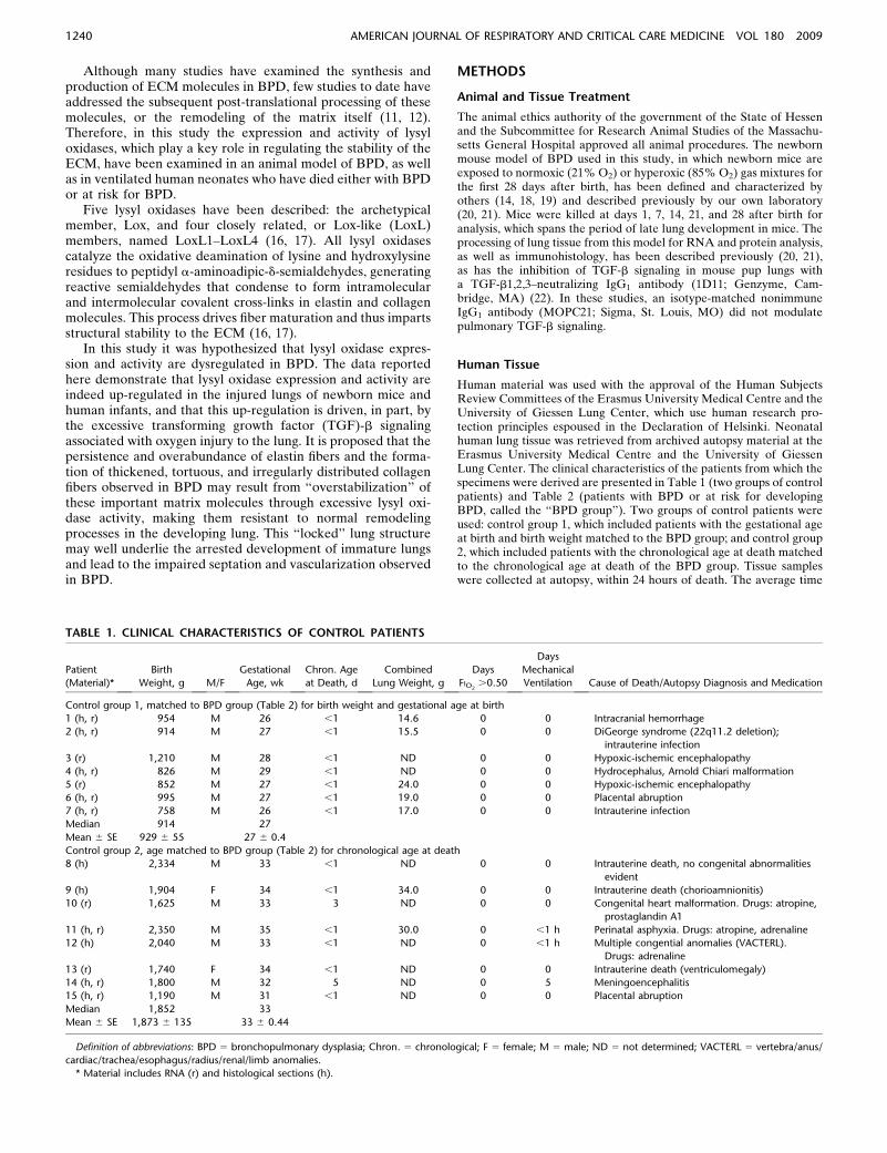
Lox and LoxL1 Expression Is Increased in Human Infants with
BPD or at Risk for BPD
The data presented above suggest that lysyl oxidase expressionis increased in the injured developing mouse pup lung. To assesswhether the expression of lysyl oxidases is similarly modulatedin the injured developing lung of humans, the immunoreactivityand mRNA levels of lysyl oxidases were examined in 10prematurely born infants who died either with BPD or hadbeen at risk for the development of BPD (Table 2; the ‘‘BPDgroup’’). The BPD group probably represents a mixture of newBPD and classic BPD. The use of surfactant in 8 out of 10patients places these patients in the post-surfactant era, and theextremely low birth weight (median, 878 g) and gestational age atbirth (27 wk) argue in favor of new BPD, although some degreeof fibrosis can be observed in some patients, which is morecharacteristic of classic BPD. Two groups of control patientswere used, which included either seven patients with thegestational age at birth and birth weight matched to the BPDgroup (control group 1; Table 1), or eight patients with the
Figure 4. Lysyl oxidase mRNA expression can be modulated in vitro by oxygen and transforming growth factor (TGF)-b. (A) Murine NIH/3T3
fibroblast-like cells and (B) human pulmonary artery smooth muscle cells were maintained under hyperoxic (85% O2) or normoxic (21% O2)
conditions for 24 hours, before addition of TGF-b (2 ng/ml) for an additional 24 hours, after which mRNA was isolated and assessed for lysyl oxidase
gene expression. Whole-lung mRNA from mouse or human lungs, respectively, served as a positive control for the polymerase chain reactions (PCR)(Pos.), whereas expression of the hspa8 gene was used as a loading control. The PCR amplicons derived from two separate cell cultures are
represented for each condition. For quantification, densitometric data for amplicons derived from six different cell cultures per condition were
averaged; { P , 0.01 comparing 85% O2 versus 21% O2 exposures in the presence of vehicle; x P , 0.01 comparing TGF-b–stimulated versus
unstimulated cells with 21% oxygen or 85% oxygen exposure; * P , 0.01 comparing 85% O2 versus 21% O2 exposures 24 hours after TGF-bstimulation.
1246 AMERICAN JOURNAL OF RESPIRATORY AND CRITICAL CARE MEDICINE VOL 180 2009

chronological age at death matched to the chronological age atdeath of the BPD group (control group 2; Table 1).
Lung sections from control patients (Figures 7A, 7B, 7E, and7F) exhibit normal lung expansion with loose extracellularmatrix in the interalveolar septa without inflammatory cells.The epithelial lining of the alveoli contains a single cell layerwithout epithelial defects, and hyaline membranes are notevident. In contrast, unequal lung expansion and broadenedintraalveolar septa are evident in lung sections from patientswith BPD (Figures 7C, 7D, 7G, and 7H), with desquamatedepithelial lining and casts evident in the larger airways, alongwith eosinophilic infiltrates and an increased abundance ofneutrophilic cells in the intraalveolar septa.
Pronounced immunoreactivity was observed for LoxL1 inthe smooth muscle layer of blood vessels in control infants (age-matched for chronological age at death with the BPD group)who died without apparent lung disease (Figures 7A and 7B)and in patients with BPD (Figures 7C and 7D). LoxL1immunoreactivity was also observed in the alveolar tissue ofpatients with BPD (Figures 7C and 7D) but not those of controlpatients (Figures 7A and 7B). A similar pattern of staining forLox was observed. Interestingly, although LoxL1 appeared tostain the entire vascular smooth muscle layer, Lox expressionappeared to be confined to the inner smooth muscle layer(Figure 7B vs. Figures 7F and 7H). Lox staining was evidentexclusively in the pulmonary vascular smooth muscle of controlpatients (Figures 7E and 7F), whereas lung sections from the
group of patients with BPD revealed additional staining in cellswithin the alveolar lumen and in cells lining the alveolar walls.The source of Lox and LoxL1 in the alveolar lumen and cellslining the alveolar walls has not been clarified. Both Lox andLoxL1, which are secreted enzymes, may be derived frominterstitial fibroblasts, or from the lung epithelium itself,because Lox and LoxL1 have now been detected in prostate,mammary, and retinal epithelial cells (34–36).
The antibody specificity was confirmed by preadsorption ofanti-Lox and anti-LoxL1 antibodies with a competing peptide(Figures 7I and 7J). No changes in the immunoreactivity ofLoxL1, LoxL2, LoxL3, or LoxL4 were seen in lung sectionsfrom any patients with BPD compared with control patients(data not shown).
A more quantitative assessment of lysyl oxidase expressionwas obtained by screening whole-lung RNA from patients withBPD and control patients by quantitative real-time RT-PCR.Consistent with the immunohistochemical data, an increase inlung lox mRNA expression levels was seen in patients withBPD versus both the control group with the gestational age atbirth and birth weight matched to the BPD group (controlgroup 1), and the control group with the chronological age atdeath matched to the chronological age at death of the BPDgroup (control group 2) (Figure 7K). In contrast, lung loxl1mRNA expression levels were increased in patients with BPDcompared with the control group 2, but not when comparedwith control group 1 (Figure 7K). The mRNA expression levels
Figure 5. Hyperoxia induced autocrine
activation of lysyl oxidase promoters in
NIH/3T3 through transforming growthfactor (TGF)-b. (A) Murine NIH/3T3 fi-
broblast-like cells and human HFL1 fibro-
blast-like cells were transfected withplasmid constructs in which the mouse
lox or human loxl1 promoters had been
inserted, upstream of a firefly luciferase
gene. The cells were maintained underhyperoxic (85% O2) or normoxic (21%
O2) conditions for 24 hours, before ad-
dition of TGF-b (2 ng/ml) for an addi-
tional 12 hours, after which cell extractswere assessed for firefly luciferase activ-
ity. To control for the effects of ligand
stimulation and hyperoxia on the base-
line transcriptional activity of cells, valueswere normalized for the transcriptional
activity of the pGL3-control vector (20).
(B) Murine NIH/3T3 fibroblast-like cellsand human HFL1 fibroblast-like cells
were treated as described in A, except
that medium was supplemented with
nonimmune IgG or anti–TGF-b1,2,3–neutralizing antibodies over the entire
time course of the experiment (10 mg/ml).
*P , 0.01 (n 5 5). ALU 5 arbitrary
luminescence units; n.i. 5 nonimmune.
Kumarasamy, Schmitt, Nave, et al.: Lox Activity is Dysregulated in BPD 1247

for loxl2, loxl3, and loxl4 were unchanged between the BPDand both control groups. As 6 out of 10 patients with BPD or atrisk for developing BPD received corticosteroids, these twosubgroups of patients (patients 17, 22, and 23 vs. patients 18, 19,24, and 25; Table 2) were assessed for differences in lox mRNAexpression. This comparison is relevant as corticosteroids havepowerful effect on gene expression. No difference in theexpression level of any lysyl oxidase mRNA was observedbetween the corticoid-treated and untreated groups of patients(results not shown).
DISCUSSION
The production and remodeling of the ECM are critical pro-cesses required for the secondary septation of the developinglung and the production and maturation of functional pulmo-nary gas exchange units (37, 38). The pathogenic mechanismsthat result in the perturbed deposition and remodeling of theECM observed in important developmental lung diseases suchas BPD (12, 15) are poorly understood. Although several
studies performed to date have examined the production ofECM molecules in BPD, few have addressed the subsequentprocessing of these molecules, which can facilitate the remodel-ing of the matrix itself. Therefore, in this study, the expressionand activity of lysyl oxidases, a family of enzymes that playa crucial role in regulating the stability of the ECM, wereexamined in an animal model of BPD, and in prematurely borninfants who have died with BPD or at risk for BPD.
The data presented here demonstrate the up-regulation ofthree elastin/collagen cross-linking enzymes (Lox, LoxL1, andLoxL2) in newborn animals with hyperoxic lung injury anddysregulated ECM development, and up-regulation of at leasttwo elastin/collagen cross-linking enzymes (Lox and LoxL1)in patients with BPD. Interestingly, two of these enzymes,Lox and LoxL1, have already been identified as beingtemporally regulated during normal mouse lung development(39). Additionally, both lox2/2 (25, 40) and loxl12/2 (41)mouse pups exhibit disturbed respiratory tract development,and although lox2/2 embryos develop to term, they die withinhours of birth with evidence of pronounced cardiopulmonary
Figure 6. Treatment of neonatal mice with transforming growth factor (TGF)-b1,2,3–neutralizing antibodies suppressed the induction of lox geneexpression by hyperoxia. Neonatal mice were treated either with control IgG or anti–TGF-b1,2,3–neutralizing antibodies before exposure to
hyperoxic (85% O2) or normoxic (21% O2) conditions for 10 days, and then killed. The expression of lysyl oxidases was assessed in mRNA from
mouse lungs by (A) semiquantitative reverse transcriptase–polymerase chain reaction (RT-PCR), where expression of the hspa8 gene was used as
a loading control; or (B) real-time quantitative RT-PCR (n 5 5); or (C ) by immunohistochemistry. Antibody specificity was confirmed bypreadsorption of antibodies with a competing peptide, which had been used as the immunogen for antibody generation (insets). Additionally, (D)
lung sections from mice were screened for assessment of elastin deposition in the developing septa by Hart’s elastin stain. *P , 0.01.
1248 AMERICAN JOURNAL OF RESPIRATORY AND CRITICAL CARE MEDICINE VOL 180 2009
failure (25, 40). These data suggest a key role for this family ofenzymes in the development of the respiratory tract. Addi-tional studies in these knockout mice have revealed key rolesfor lysyl oxidases in imparting tensile strength to connectivetissue, because they exhibit pathologies consistent with a lossof structural stability of the ECM. For example, soon afterparturition, lox2/2 pups exhibit ruptured arterial aneurysmsand ruptured diaphragms (25), whereas adult loxl12/2 micedevelop pelvic organ and rectal prolapse and excessive skinlaxity (41). In addition to the induction of lysyl oxidase geneexpression by hyperoxia reported in this study, mechanicalventilation of mice with room air or with 40% oxygen gasmixtures for 8 hours was also able to increase lox, but notloxl1 mRNA expression (13); however, a similar modulationof lysyl oxidase gene expression has not been observed inpremature lambs mechanically ventilated with high (FIO2 �0.26) levels of oxygen (42). Variable responses of elastingene expression to oxygen levels has also been reported.Although a mild increase in eln mRNA levels was observed inresponse to 85% oxygen exposure of mice in this study, Bruce
and colleagues (43) reported a decrease in elastin geneexpression in the lungs of rats exposed to 95% oxygenbetween P4 and P14. This paradox might be explained bythe higher oxygen concentrations used in the rat model, or bythe very different sensitivities to high oxygen concentrationsdisplayed by mammals, even between different mouse strains(44).
The transcriptional regulation of lysyl oxidases is poorlydescribed; however, several growth factors are known to drivelysyl oxidase expression (16, 17). Notable among these aremembers of the TGF-b family of peptide growth factors, whichare also key regulators of late lung development (45). Recently,excessive TGF-b activity has been identified as a key patho-genic factor in animal models of BPD (20, 22, 46), whichsuggested that dysregulated TGF-b signaling might underliethe aberrant expression of lysyl oxidases in BPD. Indeed,dampening of TGF-b signaling in the newborn lung injurymodel used in this study reduced the expression of the loxgene, although expression of other members of the lysyl oxidasefamily was unaffected. Thus, in the case of oxygen injury to the
Figure 7. The expression of Lox and LoxL1 was elevated in the lungs of neonatal patients who died with BPD or were at risk for BPD. The expressionof lysyl oxidases was assessed in the lungs of patients with BPD or at risk for BPD, as well as in control lungs. A low-power (200-mm scale) view of
representative sections from (A) patients in control group 2 (the histopathology of six patients [Table 1] was examined; in this case, sections from
patient 11 are illustrated) and from (C ) patients in the BPD group (the histopathology of seven patients [Table 2] was examined; in this case, sectionsfrom patient 24 are illustrated), stained for LoxL1. (B, D) High-power views (50-mm scale) are also illustrated for LoxL1 staining in the same sections
(the magnified area is demarcated in the low-power view by a black box). Representative medium-power views (100-mm scale) are illustrated for the
same two patients, stained for Lox (E, F ), with the corresponding high-power views (50-mm scale) illustrated in F and H (the magnified area is
demarcated in the low-power view by a black box). (I, J ) Antibody specificity was confirmed by preadsorption of antibodies with a competingpeptide, which had been used as the immunogen for antibody generation, before staining a section of lung tissue from the same patient with BPD.
Staining was consistently more intense in patients with BPD, and the sections illustrated are representative of the trends observed in a total of five
patients assessed per group (as indicated in Tables 1 and 2). The expression of lysyl oxidase mRNA was also assessed in mRNA isolated from the
lungs of seven patients in control group 1 (red), seven patients with BPD or at risk for BPD (green) and five patients in control group 2 (yellow) byquantitative real-time reverse transcriptase–polymerase chain reaction (K ). The bars represent the data range and the boxes represent lower and
upper quartiles. The line within the quartile box indicates the median; *P , 0.01.
Kumarasamy, Schmitt, Nave, et al.: Lox Activity is Dysregulated in BPD 1249

lungs of mouse pups, elevated Lox levels are attributable toincreased TGF-b signaling in the lungs of affected animals,which most likely drives Lox production by lung fibroblasts.Along these lines, elevated levels of TGF-b1 have also beendetected in bronchoalveolar lavage fluids of ventilated neonateswho went on to develop new BPD, versus ventilated neonateswho did not (47). This observation might suggest that elevatedlox mRNA levels seen in infants who died with BPD or were atrisk for BPD may be attributable to elevated TGF-b levels inthese neonates.
The increased expression of lysyl oxidase family membersreported here is consistent with the increased levels of tissuedesmosine, an enzymatic product of lysyl oxidase, that are ob-served in animal models of BPD (this study and Reference 12).Patients who develop BPD exhibit increased urinary desmosinelevels (48), which have been attributed to increased elastindegradation due to proteolysis in association with infection andprolonged exposure of infants to hyperoxia (49). However, datapresented here suggest that the increased desmosine levels may bedue, at least in part, to an overactive cross-linking system inaffected lungs, which could promote excessive cross-linking ofelastin and collagen fibers, perhaps resulting in the accumulation ofelastic fibers in peripheral lung tissue (9, 10). Such mechanismscould give rise to the ‘‘thickened, tortuous and disorganized’’collagen fibers that are often observed in patients who have diedwith BPD (11). Such disorganized collagen fibrils have beenobserved in the skin of patients with amyotrophic lateral sclerosiswho have excessive lysyl oxidase activity. In such patients, thethickness of collagen fibers directly correlated with the number ofstable collagen cross-links (50), further supporting the notion thatelevated lysyl oxidase activity might generate the thickenedcollagen fibers observed in the lungs of patients with BPD. Thisidea is also supported by the observation that elastic fibers in thelungs of lox2/2 pups are thinner than in wild-type age-matchedpups, and that collagen fibers in lox2/2 pups were dispersed andloose (51), contrasting with the thickened, tightly bundled fibersobserved in patients with BPD (11). Clearly, other mechanisms ofelastin fiber assembly may also be affected in BPD. Mechanicalventilation of mice with 40% oxygen gas mixtures for 8 hoursdecreased the expression of fibulin-5 and emilin-1 (13), which arecritical players in elastin fiber dynamics. In this study, both fibulin-5and emilin-1 expression appeared to be dysregulated at the mRNAlevel, with increased mRNA abundance evident in oxygen-injuredlungs. Dysregulated expression levels of both molecules wouldmost likely affect elastin fiber assembly, and suggests an avenueindependent of lysyl oxidases, which may impact elastin fiberformation and maintenance in oxygen-injured lungs.
Changes in lysyl oxidase activity and expression have notbeen causally implicated in the development of BPD in thisstudy. The early perinatal death of the lox2/2 knockout animalsprevents their use in studies of postnatal lung injury. However,with the data presented in mind, the authors would like topropose a novel hypothesis of how disordered elastin andcollagen cross-linking and deposition might inhibit alveolariza-tion in the injured developing lung. It is generally believed thatas the lung develops, a continuous cycle of ECM deposition andremodeling occurs: matrix scaffolding must be erected tosupport the developing structures of the lung and in somelocations the matrix must be removed and/or otherwise remod-eled to permit extension or reshaping of existing structures (15).The authors propose that excessive elastin and collagen cross-linking by overabundant lysyl oxidases might overstabilizeelastin and collagen and make them chemically resistant tothe normal proteolytic and other remodeling events that arerequired for successful ECM remodeling and alveolar andmicrovascular development.
This hypothesis is supported by studies in which it has beendemonstrated that the degree of elastin and collagen cross-linking determines the susceptibility of ECM fibers to pro-teolytic degradation, and hence, remodeling. Disruption ofelastin cross-linking through dietary copper restriction (whichdown-regulates the activity of Cu21-dependent lysyl oxidases)reduced the accumulation of elastin in the aorta of experi-mental animals (52), which was directly attributed to increasedproteolytic degradation of elastin molecules in copper-restrictedanimals, because elastin had become more susceptible to pro-teolysis as a result of reduced cross-linking (52). Indeed, theinability to cross-link tropoelastin renders this molecule sensi-tive to proteolysis by trypsin-like proteases (53). Conversely,hyper-crosslinking of collagen fibers in tissue-engineered ar-teries through stimulation of lysyl oxidase activity increased thestability of collagen fibers to proteolysis (54). Thus, elevatedlysyl oxidase activity observed in animal models of BPD—andin patients with BPD—may generate excessively cross-linkedelastin and collagen fibers that are resistant to proteolysis anddegradation-dependent remodeling.
Clearly, the impact of an overstabilized matrix would not belimited to the division of the alveolar septa, but could alsoextend to pulmonary vascular development. In addition todecreased alveolar septation, patients with BPD exhibit pro-nounced disruption of pulmonary vascular development (5–8).An inability to remodel the vascular matrix, as might occur inan overstabilized vascular matrix, would also have implicationsfor vascular development. Remodeling of the ECM by pro-teases promotes cell migration, which is critical for vasculardevelopment, and matrix-bound growth factors that are re-leased during vessel matrix remodeling, promoting properangiogenesis by regulating endothelial migration and growth(reviewed in Reference 55). These processes would be impededby an overstabilized ECM. Furthermore, Lox has already beenimplicated in several vascular pathologies (56) in which, whenoverexpressed, Lox can promote local neointimal thickeningand vessel muscularization. These observations are interesting,because in addition to dysregulated angiogenesis, the pulmo-nary vasculature in patients with BPD undergoes remodelingthat includes medial hypertrophy (thus, vessel thickening) andmuscularization of small arteries.
Thus, it is proposed here that as a consequence of up-regulated expression of ECM cross-linking enzymes such aslysyl oxidases, the ECM is less plastic and resists remodelingthat facilitates lung growth. This may, at least in part, underliethe impaired septation and vascular development seen inpatients with BPD and animal models of BPD.
Conflict of Interest Statement: None of the authors has a financial relationshipwith a commercial entity that has an interest in the subject of this manuscript.
Acknowledgment: The authors thank Istvan Vadasz and Zbigniew Zas1ona(University of Giessen Lung Center) for critical advice; Veronika Grau (Depart-ment of Experimental Surgery, University Hospital Giessen) for access to excellentmicroscopy facilities; Ewa Bieniek (University of Giessen Lung Center) forexcellent technical assistance with collagen staining; Pascal Sommer and RomainDebret (Institute de Biologie et Chemie des Proteins, Universite de Lyon, Lyon,France) for providing the promoter reporter constructs for the lox and loxl1genes; Cristina Rodrıguez (Cardiovascular Research Center, Institut Catala deCiencies Cardiovasculars, Barcelona, Spain) for providing promoter reporterconstructs for the human lox gene; and Herbert M. Kagan (Department ofBiochemistry, Boston University School of Medicine; Boston, MA) for providingcustom-made rabbit anti-Lox antibodies.
References
1. Northway WH Jr, Rosan RC, Porter DY. Pulmonary disease followingrespirator therapy of hyaline-membrane disease. Bronchopulmonarydysplasia. N Engl J Med 1967;276:357–368.
2. Thebaud B, Ladha F, Michelakis ED, Sawicka M, Thurston G, Eaton F,Hashimoto K, Harry G, Haromy A, Korbutt G, et al. Vascular
1250 AMERICAN JOURNAL OF RESPIRATORY AND CRITICAL CARE MEDICINE VOL 180 2009

endothelial growth factor gene therapy increases survival, promoteslung angiogenesis, and prevents alveolar damage in hyperoxia-induced lung injury: evidence that angiogenesis participates inalveolarization. Circulation 2005;112:2477–2486.
3. Bland RD. Neonatal chronic lung disease in the post-surfactant era. Biol
Neonate 2005;88:181–191.4. Jobe AH, Bancalari E. Bronchopulmonary dysplasia. Am J Respir Crit
Care Med 2001;163:1723–1729.5. Stenmark KR, Abman SH. Lung vascular development: implications for
the pathogenesis of bronchopulmonary dysplasia. Annu Rev Physiol2005;67:623–661.
6. Thebaud B, Abman SH. Bronchopulmonary dysplasia: where have all
the vessels gone? Roles of angiogenic growth factors in chronic lungdisease. Am J Respir Crit Care Med 2007;175:978–985.
7. De Paepe ME, Mao Q, Powell J, Rubin SE, DeKoninck P, Appel N, Dixon
M, Gundogan F. Growth of pulmonary microvasculature in ventilatedpreterm infants. Am J Respir Crit Care Med 2006;173:204–211.
8. De Paepe ME, Patel C, Tsai A, Gundavarapu S, Mao Q. Endoglin
(CD105) up-regulation in pulmonary microvasculature of ventilatedpreterm infants. Am J Respir Crit Care Med 2008;178:180–187.
9. Chambers HM, van Velzen D. Ventilator-related pathology in the
extremely immature lung. Pathology 1989;21:79–83.10. Hislop AA, Wigglesworth JS, Desai R, Aber V. The effects of preterm
delivery and mechanical ventilation on human lung growth. EarlyHum Dev 1987;15:147–164.
11. Thibeault DW, Mabry SM, Ekekezie II, Zhang X, Truog WE. Collagen
scaffolding during development and its deformation with chronic lungdisease. Pediatrics 2003;111:766–776.
12. Pierce RA, Albertine KH, Starcher BC, Bohnsack JF, Carlton DP,
Bland RD. Chronic lung injury in preterm lambs: disordered pulmo-nary elastin deposition. Am J Physiol 1997;272:L452–L460.
13. Bland RD, Ertsey R, Mokres LM, Xu L, Jacobson BE, Jiang S, Alvira
CM, Rabinovitch M, Shinwell ES, Dixit A. Mechanical ventilationuncouples synthesis and assembly of elastin and increases apoptosis inlungs of newborn mice. Prelude to defective alveolar septation during lungdevelopment? Am J Physiol Lung Cell Mol Physiol 2008;294:L3–L14.
14. Warner BB, Stuart LA, Papes RA, Wispe JR. Functional and patholog-
ical effects of prolonged hyperoxia in neonatal mice. Am J Physiol1998;275:L110–L117.
15. Bourbon J, Boucherat O, Chailley-Heu B, Delacourt C. Control
mechanisms of lung alveolar development and their disorders inbronchopulmonary dysplasia. Pediatr Res 2005;57:38R–46R.
16. Csiszar K. Lysyl oxidases: a novel multifunctional amine oxidase family.
Prog Nucleic Acid Res Mol Biol 2001;70:1–32.17. Kagan HM, Li W. Lysyl oxidase: properties, specificity, and biological
roles inside and outside of the cell. J Cell Biochem 2003;88:660–672.18. Bonikos DS, Bensch KG, Ludwin SK, Northway WH Jr. Oxygen toxicity
in the newborn. The effect of prolonged 100 per cent O2 exposure onthe lungs of newborn mice. Lab Invest 1975;32:619–635.
19. Obara H, Pappas CT, Northway WH Jr, Bensch KG. Comparison of the
effect of two and six week exposure to 80% and 100% oxygen on thelung of the newborn mouse: a quantitative SEM and TEM correlativestudy. Int J Radiat Oncol Biol Phys 1985;11:285–298.
20. Alejandre-Alcazar MA, Kwapiszewska G, Reiss I, Amarie OV, Marsh
LM, Sevilla-Perez J, Wygrecka M, Eul B, Kobrich S, Hesse M, et al.Hyperoxia modulates TGF-beta/BMP signaling in a mouse model ofbronchopulmonary dysplasia. Am J Physiol Lung Cell Mol Physiol2007;292:L537–L549.
21. Woyda K, Koebrich S, Reiss I, Rudloff S, Pullamsetti SS, Ruhlmann A,
Weissmann N, Ghofrani HA, Gunther A, Seeger S, et al. Inhibition ofPDE4 enhances lung alveolarisation in neonatal mice exposed tohyperoxia. Eur Respir J 2009;33:861–870.
22. Nakanishi H, Sugiura T, Streisand JB, Lonning SM, Roberts JD Jr.
TGF-beta-neutralizing antibodies improve pulmonary alveologenesisand vasculogenesis in the injured newborn lung. Am J Physiol LungCell Mol Physiol 2007;293:L151–L161.
23. Morty RE, Nejman B, Kwapiszewska G, Hecker M, Zakrzewicz A,
Kouri FM, Peters DM, Dumitrascu R, Seeger W, Knaus P, et al.Dysregulated bone morphogenetic protein signaling in monocrotaline-induced pulmonary arterial hypertension. Arterioscler Thromb VascBiol 2007;27:1072–1078.
24. Starcher B, Green M, Scott M. Measurement of urinary desmosine as an
indicator of acute pulmonary disease. Respiration 1995;62:252–257.25. Hornstra IK, Birge S, Starcher B, Bailey AJ, Mecham RP, Shapiro SD.
Lysyl oxidase is required for vascular and diaphragmatic developmentin mice. J Biol Chem 2003;278:14387–14393.
26. Palamakumbura AH, Trackman PC. A fluorometric assay for detection
of lysyl oxidase enzyme activity in biological samples. Anal Biochem2002;300:245–251.
27. Jourdan-Le Saux C, Gleyzal C, Raccurt M, Sommer P. Functional
analysis of the lysyl oxidase promoter in myofibroblast-like clonesof 3T6 fibroblast. J Cell Biochem 1997;64:328–341.
28. Cenizo V, Debret R, Sommer P, Andre V. Lysyl oxidase-like gene
transcription is down-regulated in aged skin fibroblasts througha Sp-1-rich promoter region. J Invest Dermatol 2008;128:S49.
29. Raposo B, Rodrıguez C, Martanez-Gonzalez J, Badimon L. High levels
of homocysteine inhibit lysyl oxidase (LOX) and downregulate LOXexpression in vascular endothelial cells. Atherosclerosis 2004;177:1–8.
30. Boak AM, Roy R, Berk J, Taylor L, Polgar P, Goldstein RH, Kagan
HM. Regulation of lysyl oxidase expression in lung fibroblasts bytransforming growth factor-beta 1 and prostaglandin E2. Am J RespirCell Mol Biol 1994;11:751–755.
31. Feres-Filho EJ, Choi YJ, Han X, Takala TE, Trackman PC. Pre- and
post-translational regulation of lysyl oxidase by transforming growthfactor-beta 1 in osteoblastic MC3T3–E1 cells. J Biol Chem 1995;270:30797–30803.
32. Alejandre-Alcazar MA, Michiels-Corsten M, Vicencio AG, Reiss I, Ryu
J, de Krijger RR, Haddad GG, Tibboel D, Seeger W, Eickelberg O,et al. TGF-beta signaling is dynamically regulated during the alveola-rization of rodent and human lungs. Dev Dyn 2008;237:259–269.
33. Reynaud C, Gleyzal C, Jourdan-Le Saux C, Sommer P. Comparative
functional study of the lysyl oxidase promoter in fibroblasts, Ras-transformed fibroblasts, myofibroblasts and smooth muscle cells. CellMol Biol (Noisy-le-grand) 1999;45:1237–1247.
34. Dairkee SH, Ji Y, Ben Y, Moore DH, Meng Z, Jeffrey SS. A molecular
‘signature’ of primary breast cancer cultures; patterns resemblingtumor tissue. BMC Genomics 2004;5:47.
35. Omori K, Fujiseki Y, Omori K, Suzukawa J, Inagaki C. Regulation of
the expression of lysyl oxidase mRNA in cultured rabbit retinalpigment epithelium cells. Matrix Biol 2002;21:337–348.
36. Ren C, Yang G, Timme TL, Wheeler TM, Thompson TC. Reduced lysyl
oxidase messenger RNA levels in experimental and human prostatecancer. Cancer Res 1998;58:1285–1290.
37. Mariani TJ, Sandefur S, Pierce RA. Elastin in lung development. Exp
Lung Res 1997;23:131–145.38. Roth-Kleiner M, Post M. Similarities and dissimilarities of branching
and septation during lung development. Pediatr Pulmonol 2005;40:113–134.
39. Kho AT, Bhattacharya S, Mecham BH, Hong J, Kohane IS, Mariani TJ.
Expression profiles of the mouse lung identify a molecular signatureof time-to-birth. Am J Respir Cell Mol Biol 2009;40:47–57.
40. Maki JM, Rasanen J, Tikkanen H, Sormunen R, Makikallio K, Kivirikko
KI, Soininen R. Inactivation of the lysyl oxidase gene Lox leads toaortic aneurysms, cardiovascular dysfunction, and perinatal death inmice. Circulation 2002;106:2503–2509.
41. Liu X, Zhao Y, Gao J, Pawlyk B, Starcher B, Spencer JA, Yanagisawa
H, Zuo J, Li T. Elastic fiber homeostasis requires lysyl oxidase-like 1protein. Nat Genet 2004;36:178–182.
42. Bland RD, Xu L, Ertsey R, Rabinovitch M, Albertine KH, Wynn KA,
Kumar VH, Ryan RM, Swartz DD, Csiszar K, et al. Dysregulation ofpulmonary elastin synthesis and assembly in preterm lambs withchronic lung disease. Am J Physiol Lung Cell Mol Physiol 2007;292:L1370–L1384.
43. Bruce MC, Bruce EN, Janiga K, Chetty A. Hyperoxic exposure of
developing rat lung decreases tropoelastin mRNA levels that reboundpostexposure. Am J Physiol 1993;265:L293–L300.
44. Hudak BB, Zhang LY, Kleeberger SR. Inter-strain variation in suscep-
tibility to hyperoxic injury of murine airways. Pharmacogenetics 1993;3:135–143.
45. Jankov RP, Tanswell KA. Growth factors, postnatal lung growth and
bronchopulmonary dysplasia. Paediatr Respir Rev 2004;5(suppl A):S265–275.
46. Groenman F, Unger S, Post M. The molecular basis for abnormal human
lung development. Biol Neonate 2005;87:164–177.47. Kotecha S, Wangoo A, Silverman M, Shaw RJ. Increase in the
concentration of transforming growth factor beta-1 in bronchoalveo-lar lavage fluid before development of chronic lung disease ofprematurity. J Pediatr 1996;128:464–469.
48. Bruce MC, Wedig KE, Jentoft N, Martin RJ, Cheng PW, Boat TF,
Fanaroff AA. Altered urinary excretion of elastin cross-links inpremature infants who develop bronchopulmonary dysplasia. AmRev Respir Dis 1985;131:568–572.
Kumarasamy, Schmitt, Nave, et al.: Lox Activity is Dysregulated in BPD 1251

49. Bruce MC, Schuyler M, Martin RJ, Starcher BC, Tomashefski JF Jr,Wedig KE. Risk factors for the degradation of lung elastic fibers inthe ventilated neonate. Implications for impaired lung developmentin bronchopulmonary dysplasia. Am Rev Respir Dis 1992;146:204–212.
50. Ono S, Yamauchi M. Collagen fibril diameter and its relation to cross-linking of collagen in the skin of patients with amyotrophic lateralsclerosis. J Neurol Sci 1993;119:74–78.
51. Maki JM, Sormunen R, Lippo S, Kaarteenaho-Wiik R, Soininen R,Myllyharju J. Lysyl oxidase is essential for normal development andfunction of the respiratory system and for the integrity of elastic andcollagen fibers in various tissues. Am J Pathol 2005;167:927–936.
52. Tinker D, Geller J, Romero N, Cross CE, Rucker RB. Tropoelastinproduction and tropoelastin messenger RNA activity. Relationship to
copper and elastin cross-linking in chick aorta. Biochem J 1986;237:17–23.
53. Brown-Augsburger P, Tisdale C, Broekelmann T, Sloan C, Mecham RP.Identification of an elastin cross-linking domain that joins threepeptide chains. Possible role in nucleated assembly. J Biol Chem1995;270:17778–17783.
54. Dahl SL, Rucker RB, Niklason LE. Effects of copper and cross-linkingon the extracellular matrix of tissue-engineered arteries. Cell Trans-plant 2005;14:861–868.
55. Sottile J. Regulation of angiogenesis by extracellular matrix. BiochimBiophys Acta 2004;1654:13–22.
56. Rodrıguez C, Martınez-Gonzalez J, Raposo B, Alcudia JF, Guadall A,Badimon L. Regulation of lysyl oxidase in vascular cells: lysyl oxidaseas a new player in cardiovascular diseases. Cardiovasc Res 2008;79:7–13.
1252 AMERICAN JOURNAL OF RESPIRATORY AND CRITICAL CARE MEDICINE VOL 180 2009