Electrochemical Testing of Laser Treated Bronze Surface
19
Accepted Manuscript Electrochemical Testing of Laser Treated Bronze Surface B.S. Yilbas, Ihsan-ul-Haq Toor, Jahanzaib Malik, F. Patel, C. Karatas PII: S0925-8388(13)00402-7 DOI: http://dx.doi.org/10.1016/j.jallcom.2013.02.094 Reference: JALCOM 27905 To appear in: Received Date: 11 November 2012 Revised Date: 9 February 2013 Accepted Date: 14 February 2013 Please cite this article as: B.S. Yilbas, I-u. Toor, J. Malik, F. Patel, C. Karatas, Electrochemical Testing of Laser Treated Bronze Surface, (2013), doi: http://dx.doi.org/10.1016/j.jallcom.2013.02.094 This is a PDF file of an unedited manuscript that has been accepted for publication. As a service to our customers we are providing this early version of the manuscript. The manuscript will undergo copyediting, typesetting, and review of the resulting proof before it is published in its final form. Please note that during the production process errors may be discovered which could affect the content, and all legal disclaimers that apply to the journal pertain.
Transcript of Electrochemical Testing of Laser Treated Bronze Surface
Accepted Manuscript
Electrochemical Testing of Laser Treated Bronze Surface
B.S. Yilbas, Ihsan-ul-Haq Toor, Jahanzaib Malik, F. Patel, C. Karatas
PII: S0925-8388(13)00402-7
DOI: http://dx.doi.org/10.1016/j.jallcom.2013.02.094
Reference: JALCOM 27905
To appear in:
Received Date: 11 November 2012
Revised Date: 9 February 2013
Accepted Date: 14 February 2013
Please cite this article as: B.S. Yilbas, I-u. Toor, J. Malik, F. Patel, C. Karatas, Electrochemical Testing of Laser
Treated Bronze Surface, (2013), doi: http://dx.doi.org/10.1016/j.jallcom.2013.02.094
This is a PDF file of an unedited manuscript that has been accepted for publication. As a service to our customers
we are providing this early version of the manuscript. The manuscript will undergo copyediting, typesetting, and
review of the resulting proof before it is published in its final form. Please note that during the production process
errors may be discovered which could affect the content, and all legal disclaimers that apply to the journal pertain.
Electrochemical Testing of Laser Treated Bronze Surface
B.S.Yilbas1*, Ihsan-ul-Haq Toor1, Jahanzaib Malik1, F. Patel1, C. Karatas2
1Dept. of Mechanical Engineering, King Fahd University of Petroleum and Minerals (KFUPM), Dhahran 31261, Kingdom of Saudi Arabia. Email: [email protected] 2Engineering Faculty, Hacettepe University, Ankara, Turkey.
Abstract
Electrochemical testing of laser treated bronze surface is carried out and corrosion resistance of
the surface is assessed. Morphological and metallurgical changes in the laser treated layer are
examined using scanning electron microscope, energy dispersive spectroscopy, and X-ray
diffraction. The pit sites formed at the surface are analyzed using scanning electron microscope.
It is found that laser treatment improves the corrosion resistance of the treated surface. Fine
grains are formed in the surface region of the laser treated layer, which are attributed to the large
cooling rates from the surface.
Keywords: Laser melting, corrosion resistance, bronze.
Introduction
Bronze is a copper alloy and it is widely used in marine environment due to its combination of
toughness and resistance to the aqueous corrosion. However, corrosion and erosion take place at
the fluid-metal interface and this adverse effect accelerates for the surfaces where the non-
uniform micro structures are present. In general grain refining, with uniform microstructures at
the surface vicinity of the alloy, improves the corrosion and erosion resistance at the surface.
One of the methods to improve the alloy microstructure in the surface vicinity is to introduce
control melting incorporating the high power laser beam. In addition, laser treatment of the
surfaces is involved with precision of operation, short treatment duration, shallow heat affected
zone, and low cost. However, the proper selection of the laser parameters is vital in surface
treatment process to avoid the surface asperities because of the excessive heating. The surface

asperities such as cavities and micro cracks act like a defect sites for accelerated corrosion at the
surface. Consequently, investigation into laser treatment of bronze surface in relation to
corrosion prevention in aqueous solution becomes essential.
Considerable research studies were carried out to examine laser treatment of bronze surfaces.
Laser treatment of aluminum bronze was studies by Xu et. al. [1]. They demonstrated that the
microstructure of the treated surface was cellulated crystals and it became dendritic crystals in
the midsection of the treated layer. Laser treating of sintered bronze was investigated by Gisario
et. al. [2]. They performed thermographic analysis on the treated samples to evaluate the thermal
dispersion at the surface. Laser treatment of bronze surface and corrosion resistance was
examined by Tang. et. al. [3]. They showed that the galvanic effect between the laser treated and
as-received samples were small, which justified the use of laser surface alloying as a feasible
method in the local surface treatment of bronze. The cavitation erosion resistance of laser treated
bronze was studies by Kwok et. al. [4]. Their findings revealed that the cavitation erosion
resistance of the laser treated surface was improved by at most 7.5 times that of as-received
bronze surface. In addition, laser treatment enhanced the corrosion resistance of the surface
considerably. Electrochemical response of the laser treated bronze surface was examined by
Klassen et. al. [5]. They illustrated that the oxide formed at the surface during laser treatment
behaved as the passivation film improving the corrosion resistance of the surface. The corrosion
resistance of laser treated bronze surface was studied by Mazurkevich et. al. [6]. They indicated
that the laser treatment of the surface improved the corrosion resistance notably. The influence of
laser treatment on the surface properties of copper alloys was investigated by Garbacz et. al. [7].
They incorporated Raman Spectroscopy to examine the phase composition of the corroded layers
at the laser treated surface. The corrosion characteristics of laser treated Ni-Al-bronze surface
were examined by Kawazoe et. al. [8]. Their findings revealed that Ni-Al-Bronze had quenching
characteristics closely related to that of steel and the corrosion resistance of the surface improved
after the laser treatment process. Laser treatment of bronze and titanium alloy was investigated
by Kac et al. [9]. They showed that the high chemical homogeneity and fine structure of the
melted zone were attributed to high cooling rates due to the short interaction time with Nd:YAG
pulsed laser radiation and relatively small volume of the melted material. Yilbas et al. [10]
studied laser treatment of bronze surfaces with the presence of B4C particles. They demonstrated
that the laser treated surface produced was relatively free from defects and asperities with a
microhardness that was notably higher than that of the as-received bronze substrate.
Laser surface treatment of bronze was investigated previously [11 - 12]. The main emphasis in
the previous studies was on the improvement of microhardness at the surface and corrosion
resistance of the treated surface was left obscure. Consequently, in the present study, laser
treatment of aluminum bronze surface is carried out and corrosion resistance of the treated
surface is examined incorporating the electrochemical tests in the electrolytic solution. The
morphological and microstructural changes in the laser treated layer are examined using
scanning electron microscope, energy dispersive spectroscopy, and X-ray diffraction. The pits
sites at the surface are also analyzed.
Experimental Work
The CO2 laser (LC-ALPHAIII) delivering a nominal output power of 2 kW was used to irradiate
the workpiece surface. The focusing lens has focal length of 127 mm and the focused laser beam
diameter was about 0.3 mm at the workpiece surface. Nitrogen emerging from the conical nozzle
was used as an assisting gas. The various surface treatment tests were carried out incorporating
different laser parameters and the laser parameters resulting in crack free surfaces with low
surface roughness were selected. The laser treatment conditions are given in Table 1. The laser
treatment experiments were repeated three times to ensure the same topology and similar
microstructures forming in the treated layer.
The aluminum bronze with elemental composition (wt%) of 9% Al, 3% Fe, and balance of Cu
was used in the experiments. The bronze sheet was 3 mm in thickness and the size of the samples
used in the experiments was 20•20 (length•width) mm.
Corrosion tests (Potentiodynamic polarization, Tafel behavior and electrochemical impedance
spectroscopy) were carried out in a three electrode cell, which composed of a specimen as a
working electrode, a Pt wire as a counter electrode, and a saturated calomel reference electrode
(SCE). The specimens were degreased in benzene, cleaned ultrasonically, and subsequently

washed with distilled water prior to electrochemical tests. The investigations were carried out
with an exposed working electrode area of 0.07 cm2 in 0.1M NaCl solution at room temperature
in PCI4/750 Gamry potentiostat and repeated several times to ensure the reproducibility of the
data. DC105 corrosion software was used to analyze the Tafel region, while Potentiodynamic
polarization experiments were performed at a scan rate of 0.5mV/s.
Electrochemical impedance spectroscopy (EIS) measurements were carried out at OCP, by
applying a sinusoidal potential perturbation of 10 mV with frequency sweep from 100 kHz to
0.01 Hz. The impedance data were analysed and fitted to appropriate equivalent electrical circuit
using the GAMRY framework software.
A Jeol 6460 electron microscopy was used for the SEM examinations and a Bruker D8
Advanced with CuK• radiation was used for XRD analysis. A typical setting for the XRD was
40 kV and 30 mA, the scanning angle (2•) was ranged over 20o-80o. The parabolically-shaped
Göbel Mirror was used in the Bruker D8 Advanced, which provided highly-parallel X-ray
beams.
Results and Discussion
Potentiodynamic response of laser treated bronze surface is investigated. Microstructural and
morphological changes in the laser treated layer are examined prior and after the electrochemical
tests.
Figures (1) show SEM micrographs of laser treated surface prior to the electrochemical tests.
The surface comprises of regular laser scan tracks, which are equally spaced at the surface. Since
the laser power intensity was adjusted to avoid evaporation at the surface, no laser induced cavity
is formed at the surface. In addition, no micro-cracks or some other surface asperities are visible.
It should be noted that the thermal conductivity of bronze is high; consequently, conduction heat
transfer from surface region to the solid bulk is high. This, in turn, suppresses the development
of high temperature gradients and high stress levels in the surface region. Hence, thermally
induced micro-cracks do not form at the surface. The close examination of the surface reveals the
overlapping of irradiated laser spots along the scanning tracks. The overlapping ratio is of the
order of 70%, which enables the continuous melting of the substrate along the scanning
directions at the surface. Although presence of overlapping of irradiated spots increases the
surface roughness, the roughness of the treated surface is in the order of 2 �m as evident from
figure (2), in which the surface roughness chart is shown. Since nitrogen is used as an assisting
gas during the laser treatment process, the surface color changes from bright brown to
mahogany, which indicates the formation of Cu3N compound at the surface during the treatment
process. The presence of small grains at the surface suggests that the dissolution of Cu3N takes
place in the surface region due to the high temperature, which is also observed in the previous
study [11]. It should be noted that high cooling rates at the surface contributes significantly to the
formation of fine grains at the surface while enhancing the microhardness as consistent with the
previous findings [11].
Figure (3) shows SEM micrographs of the cross-section of the laser treated layer. The treated
layer extends uniformly below the surface. The thickness of the treated layer is on the order of 40
�m. It is evident that no large scale defects including major cracks are observed across the cross-
section of the treated layer. Laser treated layer consists of mainly three regions. Fine grains
forming dense layer are present in the first region. The formation of the fine grains is attributed
to the high cooling rates at the surface of the treated layer. It should be noted that the convective
cooling of the assisting gas contributes considerably to the high cooling rates at the surface. In
addition, formation of Cu3N compound at the surface results in the volume shrinkage in the
surface vicinity, which in turn contributes to the formation of the dense layer in the surface
region. Although high cooling rates results in high temperature gradients and thermal stresses in
the surface region, no micro-cracks or crack-network is observed. This is attributed to the self-
annealing effect of the initially formed laser scanning tracks. It should be noted that laser scans
the surface along the tracks during the treatment process. Initially formed tracks cools slowly
because of the heat transfer from the recently formed scan tracks. Table 2 gives the EDS data at
the surface after the laser treatment process. Elemental composition remains almost uniform after
the laser treatment process. Although the quantification of light elements is involved with error
in the EDS data, the presence of nitrogen is evident from Table 2 indicating the formation of the
nitride compound at the surface. This can also be observed from the peaks of the X-ray
diffractogram as seen from figure (4), in which X-Ray diffraction of the laser treated surface is
shown. In the second layer, dendritic structure is formed because of relatively lower cooling rates
than that of the surface. As the depth below the surface increases large grains are formed in the
third region. The demarcation line is not observed at the interface of the treated layer and the
base material. This is because of the high thermal conductivity of bronze, which causes high rate
of heat conduction from the treated layer to the solid bulk while modifying the microstructure in
the interface region.
Figure (5) shows the results of Potentiodynamic polarisation response of laser treated and
untreated surfaces in 0.1M NaCl solution at room temperature. It is clear from figure (5) that
laser treated sample exhibits better corrosion resistance than the untreated specimen in terms of
pitting potential (Epit) and passive current density (ip). Corrosion potential (Ecorr) is found to be -
400 mV > -120 mV for laser treated and untreated specimens, respectively. Laser treated
specimen shows a pitting potential of 300mVSCE as compared to 70mVSCE for untreated
specimen. Passive current density (iP) as well as corrosion current (icorr) density of laser treated
specimen are much less than that of untreated specimen. All these results suggest that a stable
and more protective film is formed on the laser treated specimen surface; therefore, laser
treatment has a positive effect on the corrosion properties of bronze surface. Table 3 summarized
the results of potentiodynamic polarization.
To confirm the potentiodynamic polarisation data, the values of corrosion rate for two specimens
are calculated using Tafel analysis. It is found that corrosion rate of laser treated specimen
(0.00037mpy) is much lower than that of untreated specimen (0.0083mpy), which is in
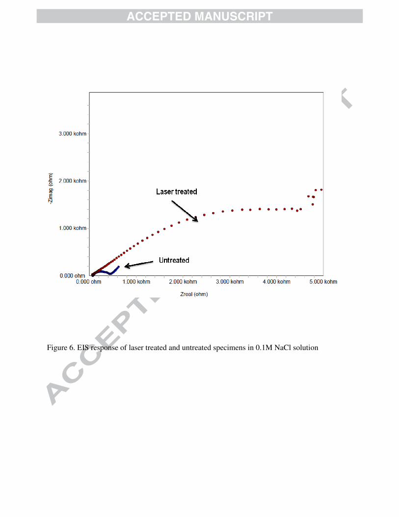
agreement the results shown in figure (5). Furthermore, electrochemical impedance spectroscopy
(EIS) measurements are carried out at OCP, by applying a sinusoidal potential perturbation of 10
mV with frequency sweep from 100 kHz to 0.01 Hz. The findings are shown in figure (6); in
which case, it can be observed that polarization resistance value (Rp) of laser treated specimen is
much higher than that of untreated specimen, suggesting that it has higher corrosion resistance.
The large semicircle of laser treated specimen corresponds to higher corrosion resistance
behaviour than untreated specimen. These results show the positive effect of laser treatment on
the corrosion properties of bronze.
Figure (7) shows SEM micrographs of pit sites at the laser treated and as-received workpiece
surfaces. It is evident from the SEM micrographs that pit site is smaller and shallow for laser
treated surface as compared to corresponding to as-received surface. Consequently, laser treated
layer acts as a passive layer lowering the surface pitting during the electrochemical testing. The
pits formed at the surface do not have a regular pattern and the secondary pitting in the pit sites is
not observed for both laser treated and as received surfaces.
Conclusion
Electrochemical response of laser treated bronze surface is investigated in aqueous solution.
Morphological and metallurgical changes in the laser treated layer are examined using the
analytical tools. Pit sites at the surfaces are analyzed incorporating scanning electron microscope.
It is found that laser treated surface is free from asperities such as cavities, voids, and micro-
cracks. The overlapping of the laser irradiated spots along the laser scanning tracks do not
contribute significantly to the surface roughness. Laser treated layer extends uniformly below the
surface with a thickness in the order of 40 �m. The close examination of the cross-sections of the
treated layer reveals that fine grains in a dense layer are formed in the surface region. This is
attributed to the high cooling rates at the surface. The presence of Cu3N nitrides is evident from
the X-Ray diffractogram, which contributes to the formation of the dense layer at the surface due
to the volume shrinkage. Dendritic structure is formed below the surface due to relatively slower
cooling rates as compared to that at the surface. Large grains are observed in the heat affected
zone region. The corrosion current density for the laser treated surface is much less than that of
the as-received surface indicating the laser treatment provides protective layer at the surface.
This finding is also supported by the electrochemical impedance spectroscopy results. The pits
sites on the laser treated surface appear to be shallower and smaller in size than that
corresponding to the as-received surface. The close examination of the pit sites revealed that no
secondary pitting takes place at the laser treated and untreated surfaces.
References

1. J-L. Xu, B. Yang, W. Gao, Z.-P. Wang, D.-W. Long, C.-Y. Ju, T. Yu, Microstructure and
performance of laser cladding on surface of aluminum bronze, J.l of Aeronautical Materials, Vol.
29, n 1, pp. 63-67, 2009.
2. A.�Gisario, D. Bellisario, F. Veniali, Thermal-morphological analysis of diode laser polishing
on sintered bronze, Int. J. of Material Forming, Vol. 3, n. 1, pp. 1067-1070, 2010.
3. C.H.�Tang, F.T. Cheng, H.C. Man, Laser surface alloying of a marine propeller bronze using
aluminium powder. Part II: Corrosion and erosion-corrosion synergism, Surface and Coatings
Technology, Vol. 200, n 8, pp. 2594-2601, 2006.
4. C.T. Kwok, P.K. Wong, W.K. Chan, F.T. Cheng, H.C. Man, Laser surface alloying of Mn-Ni-
Al bronze for cavitation erosion resistance, 29th International Congress on Applications of
Lasers and Electro-Optics, September 26 – 30, 2010, ICALEO 2010 - Congress Proceedings,
Vol. 103, pp. 1518-1524, 2010,
5. R.D. Klassen, C.V. Hyatt, P.R. Roberge, Passivation of laser-treated nickel aluminum bronze
as measured by electrochemical impedance spectroscopy, Canadian Metallurgical Quarterly,
Vol. 39, n 2, pp. 235-246, 2000.
6. A.N. Mazurkevich, I.N. Shiganov, R.S. Tretyakov, O.Y. Elagina, A.K. Prygaev, A.V.
Buryakin, Y.V. Baklanova, Study of corrosion resistance of coatings of different composition
prepared by laser surfacing, Chemical and Petroleum Engineering, Vol. 48, n 5-6, pp. 380-383,
2012.
7. H. Garbacz, E. Fortuna-Zalesna, J. Marczak, A. Koss, A. Zatorska, G. Z. Zukowska, T.
Onyszczuk, K. Kurzydlowski, Effect of laser treatment on the surface of copper alloys, Applied
Surface Science, Vol. 257, n 17, pp. 7369-7374, 2011.
8. T. Kawazoe, A. Ura, M. Saito, S. Nishikido, Erosion characteristics of surface hardened Ni-Al
bronze, Surface Engineering, Vol. 13, n 1, pp. 37-40, 1997.

9. S. Kac, A. Radziszewska, J. Kusinski, Structure and properties of the bronze laser alloyed with
titanium, Applied Surface Science, Vol. 253(19), pp. 7895-7898, 2007.
10. B.S. Yilbas, A. Matthews, A. Leyland, C. Karatas, S.S. Akhtar, B.J. Abdul Aleem, Laser
surface modification treatment of aluminum bronze with B4C, Applied Surface Science, Vol.
263, pp. 804-809, 2012.
11. B.S. Yilbas, S.S. Akhtar, C. Karatas, Laser nitriding of the surface of phosphor bronze, Int. J.
of Advanced Manufacturing Technology , pp. 1-13, 2012.
12. B.S. Yilbas, S.S. Akhtar, C. Karatas, C. Chatwin, Laser embedding of TiC particles into the
surface of phosphor bronze-bearing material, Surface and Interface Analysis, Vol. 44, pp. 831-
836, 2012.
Acknowledgements The authors acknowledge the support of Deanship of Scientific Research, King Fahd University
of Petroleum and Minerals, Dhahran, Saudi for supporting the funded project # SB111003.
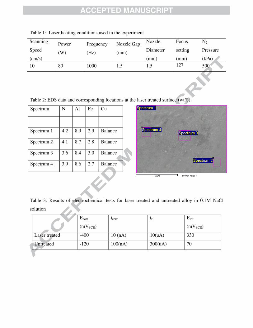
Table 1: Laser heating conditions used in the experiment
Scanning
Speed
(cm/s)
Power
(W)
Frequency
(Hz)
Nozzle Gap
(mm)
Nozzle
Diameter
(mm)
Focus
setting
(mm)
N2
Pressure
(kPa) 10 80 1000 1.5 1.5 127 500
�
�
Table 2: EDS data and corresponding locations at the laser treated surface (wt%).
Spectrum N Al Fe Cu
Spectrum 1 4.2 8.9 2.9 Balance
Spectrum 2 4.1 8.7 2.8 Balance
Spectrum 3 3.6 8.4 3.0 Balance
Spectrum 4 3.9 8.6 2.7 Balance
�
Table 3: Results of electrochemical tests for laser treated and untreated alloy in 0.1M NaCl
solution
Ecorr
(mVSCE)
icorr
iP
EPit
(mVSCE)
Laser treated -400 10 (nA) 10(uA) 330
Untreated -120 100(nA) 300(uA) 70
�

Figure 1. SEM micrographs of laser treated surface: a) Laser scanning tracks and
overlapping of irradiated spots, and b) Fine grains at the surface.
Laser Scan Tracks
Overlapping of Laser Spots
Fine Grains
a)
b)
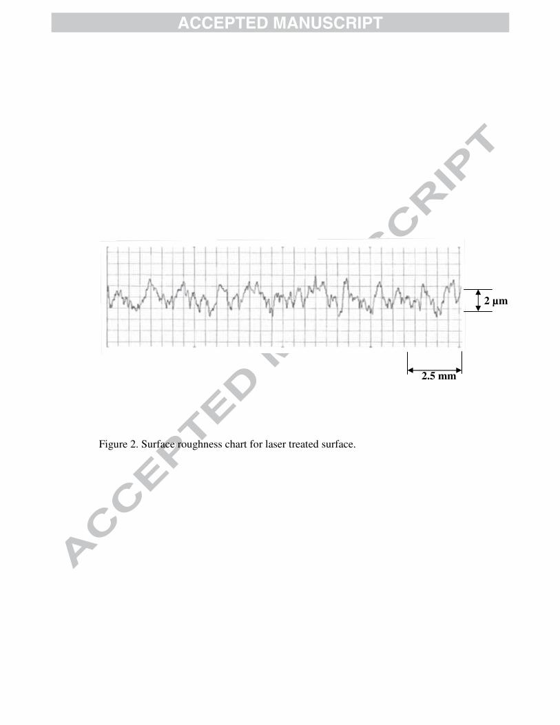
Figure 2. Surface roughness chart for laser treated surface.
2.5 mm
2 µm
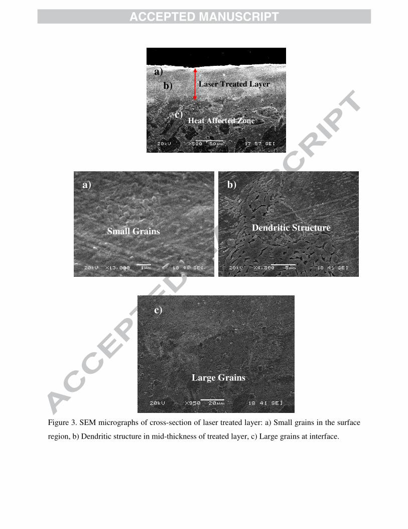
Figure 3. SEM micrographs of cross-section of laser treated layer: a) Small grains in the surface
region, b) Dendritic structure in mid-thickness of treated layer, c) Large grains at interface.
Laser Treated Layer
Heat Affected Zone
a)
b)
c)
a)
b)
c)
Small Grains Dendritic Structure
Large Grains
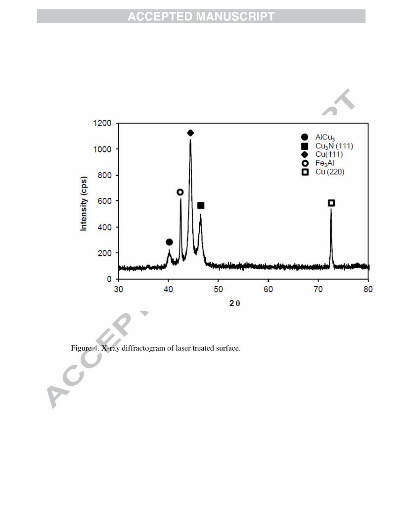
Figure 4. X-ray diffractogram of laser treated surface.
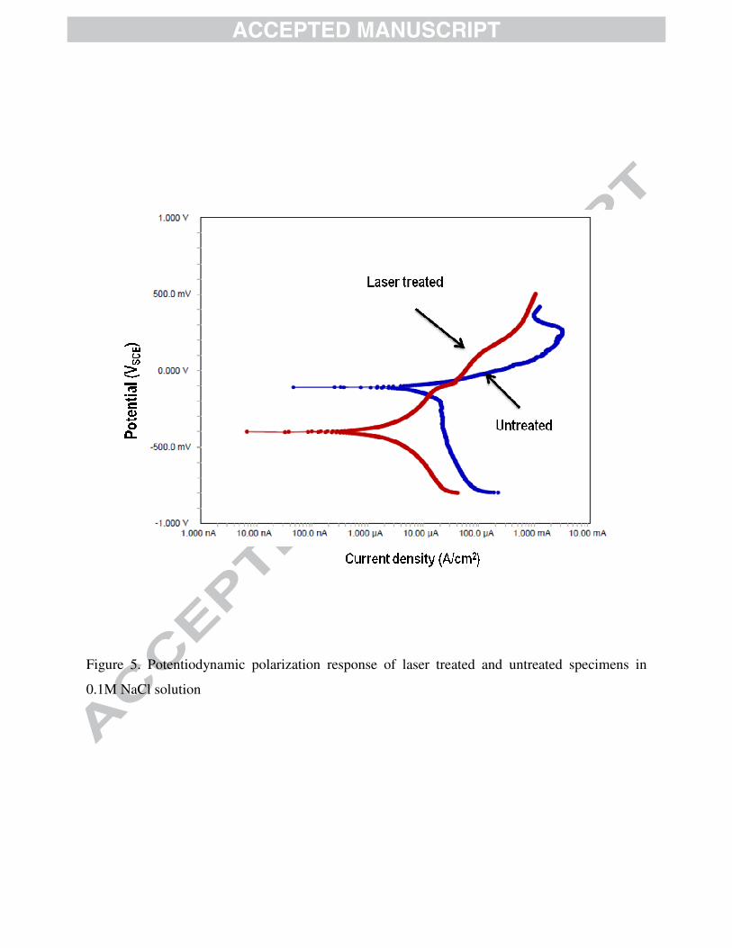
Figure 5. Potentiodynamic polarization response of laser treated and untreated specimens in
0.1M NaCl solution
�
�
��
�
�
�
�
�
Figure 6. EIS response of laser treated and untreated specimens in 0.1M NaCl solution
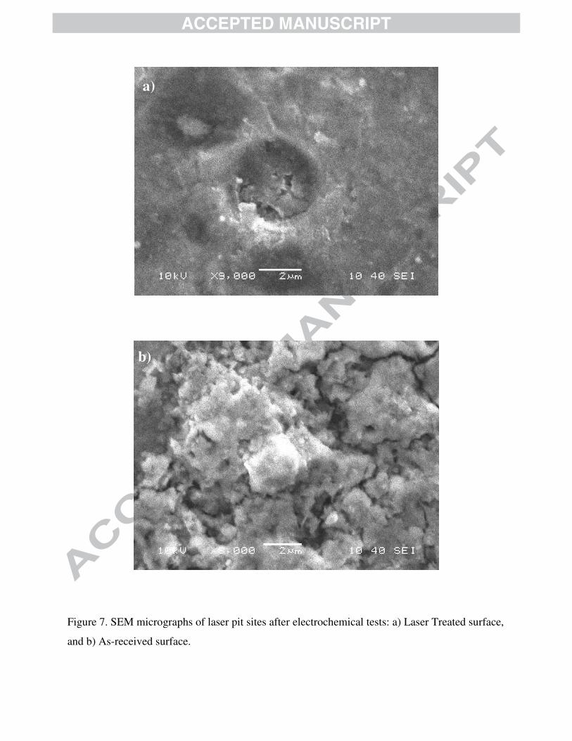
Figure 7. SEM micrographs of laser pit sites after electrochemical tests: a) Laser Treated surface,
and b) As-received surface.
a)
b)
�������������� ��
�
�
�
- ���������������������������� � ��������������
�
- ������������������������������ �� ������ � ������������ ����������������������� ����� ������ ���
�
- ��������� ���� �������������� ������� � �!"#��������� $��� ���
�
- %��������� �������� ��� �� ��� ��� � � ���� ������ ��� � � ������ ���� �� � ��� � ���$� ������ ���
� � ������� ���������������������
�
- &��� � �� �� �� ������� �������� �� ���� ������ �������� ������ ��� � ��� ����� ����� ����� � ���� ��"
����� �����������