Ageing effect of plasma‐treated wool
19
Ageing Effect of Plasma Treated Wool ABSTRACT Atmospheric pressure plasma treatment of wool fabric, with a relatively short exposure time, effectively removed the covalently-bonded lipid layer from the wool surface. The plasma- treated fabric showed increased wettability and the fibres showed greater roughness. XPS analysis showed a much more hydrophilic surface with significant increases in oxygen and nitrogen concentrations and a decrease in carbon concentration. Adhesion, as measured by scanning probe microscopy (SPM) force volume analysis, also increased, consistent with the more hydrophilic surface leading to a greater meniscus force on the SPM probe. The ageing of fibres from the plasma-treated fabric was assessed over a period of 28 days. Whilst no physical changes were observed, the chemical nature of the surface changed significantly. XPS showed a decrease in the hydrophilic nature of the surface with time, consistent with the measured decrease in wettability. This change is proposed to be due to the reorientation of proteolipid chains. SPM adhesion studies also showed the surface to be changing with time. After ageing for 28 days, the plasma-treated surface was relatively stable and still dramatically different from the untreated fibre, suggesting that the oxidation of the surface and modification or removal of the lipid layer were permanent. Keywords: Plasma, surface treatment, ageing, adhesion, hydrophilic/hydrophobic, wool 1
Transcript of Ageing effect of plasma‐treated wool

Ageing Effect of Plasma Treated Wool
ABSTRACT
Atmospheric pressure plasma treatment of wool fabric, with a relatively short exposure time,
effectively removed the covalently-bonded lipid layer from the wool surface. The plasma-
treated fabric showed increased wettability and the fibres showed greater roughness. XPS
analysis showed a much more hydrophilic surface with significant increases in oxygen and
nitrogen concentrations and a decrease in carbon concentration. Adhesion, as measured by
scanning probe microscopy (SPM) force volume analysis, also increased, consistent with the
more hydrophilic surface leading to a greater meniscus force on the SPM probe.
The ageing of fibres from the plasma-treated fabric was assessed over a period of 28 days.
Whilst no physical changes were observed, the chemical nature of the surface changed
significantly. XPS showed a decrease in the hydrophilic nature of the surface with time,
consistent with the measured decrease in wettability. This change is proposed to be due to the
reorientation of proteolipid chains. SPM adhesion studies also showed the surface to be
changing with time.
After ageing for 28 days, the plasma-treated surface was relatively stable and still
dramatically different from the untreated fibre, suggesting that the oxidation of the surface
and modification or removal of the lipid layer were permanent.
Keywords: Plasma, surface treatment, ageing, adhesion, hydrophilic/hydrophobic, wool
1

INTRODUCTION
The outermost layer of the wool fibre surface is covered by an external thin membrane called
the epicuticle, which is a thin band of proteinaceous material containing a high concentration
of the amino acid, cystine (Bradbury, 1973; Leeder & Bradbury, 1968), and a significant
proportion of bound lipid layer (Evans et al., 1985). A number of workers have studied the
surface of wool fibre and a wool surface model has been developed (Breakspear et al., 2005;
Evans et al., 2002; Huson et al., 2008; Leeder & Bradbury, 1968; Leeder & Bradbury, 1971;
McLaughlin & Pope, 1982; Negri et al., 1993a; Negri et al., 1996).
The model proposed by Evans et al (Evans et al., 1985) describes the epicuticle as an
outermost fatty acid monolayer (F- Layer) and a protein matrix. The major lipid component
of the fatty layer was identified as a methyl-branched, 21-carbon fatty acid (Evans et al.,
1985). Negri et al. (1993a) proposed a model in which the entire covalently-bound lipid
originated from the epicuticle. It was suggested that the intact lipid membrane is attached to
the protein through thioester bonds (Negri et al., 1993a; Negri et al., 1993b); the thickness
was estimated to be about 2.8-3.0 nm (Negri et al., 1993a). Scanning probe microscopy
results supported Negri’s estimation (Crossley et al., 2000).
Time of flight secondary ion mass spectrometry (ToF-SIMS) analysis of untreated wool has
identified a fragment at 325 amu as 18-methyleicosanoic acid (18- MEA) (C21H41O2) (Shao
et al., 1997; Volooj et al., 2000; Ward et al., 1993). Negri et al (Negri et al., 1993a)
described this lipid as oriented away from the fibre and thus forming a hydrophobic barrier at
the surface of each cuticle cell. Negri’s model also explains the presence of a high proportion
of carbon in the outer 3-5 nm of the cuticle as shown by XPS (Carr et al., 1986; Ward et al.,
1993).
Recently, Huson et al. (2008) and Maxwell & Huson (2005) argued that the lipid is an
integral part of the surface, rather than a discrete surface layer, and that the surface is capable
2

of altering its structure in response to different environments. It was postulated that under dry
conditions, the surface of the keratin fibres is rich in lipid and is hydrophobic, while in high
relative humidity (RH) or water, it is rich in protein and hydrophilic (Maxwell & Huson,
2005).
It has been shown that the chemical treatment using alkali, oxidants and reductants can
change the surface of wool from hydrophobic to hydrophilic (Leeder et al., 1985; Negri et al.,
1993b; Shao et al., 1997). Treatments with alcoholic KOH remove the 18-MEA from the
surface (Negri et al., 1996), while hydroxylamine treatment selectively removes 18-MEA
without producing any other chemical alteration to the fibre (Breakspear et al., 2005). In
addition, it has been shown that bleaching and permanent wave treatments with alkaline
peroxide remove some 18-MEA from the surface of hair fibre and oxidise cystine residue
(Robbins, 1988).
Surface analysis of plasma-treated wool has shown that plasma treatments modify the surface
of the wool fibre (Dai et al., 2001; Kan et al., 2004; Molina et al., 2005; Molina et al., 2002;
Naebe et al., 2010a; Naebe et al., 2010b; Ryu et al., 1991; Wakida et al., 1993). Regardless
of the treatment conditions used, the impact of plasma treatment is attributed to several
changes on the wool surface such as partial or total removal of the fatty acid layer, formation
of hydrophilic groups and an etching effect (Erra et al., 2002; Rakowski, 1997; Ryu et al.,
1987).
Although a number of workers studied the surface morphology of wool fibres using SPM,
their focus has been on estimating the thickness of the surface lipid layer and measuring the
mechanical properties of the fibre surface (Crossley et al., 2000; Gibson et al., 2001;
Maxwell & Huson, 2005). Few obtained the local physical information such as roughness and
adhesion. In addition, plasma treatment has been widely studied for its effect on the surface
of the wool fibre, but the ageing effect (changes on the plasma-treated wool fibre surface over
3

time) has received little attention. The objective of this study is to investigate the effect of
plasma treatment on the wool fibre surface, and especially the effects of ageing for a period
of 28 days after the treatment.
EXPERIMENTAL
1. Materials, plasma treatment, XPS and water uptake
The base fabric was a plain-weave pure wool fabric of 222 g/m2 manufactured from 20 µm
fibre, and cleaned by soxhlet extraction as described previously (Naebe et al., 2010a). A
plasma treatment with an exposure time of 30 sec was carried out using an Atmospheric
Plasma Treatment System from Sigma Technologies International (Arizona, USA) (APC
2000), operated at ambient temperature with helium as the plasma gas, as described previously
(Naebe et al., 2010a).
XPS analysis was conducted using an Axis Ultra spectrometer (Kratos Analytical Ltd., UK
equipped with a monochromatic X-ray source (Al Kα, hν = 1486.6 eV), operating at 150 W.
Water uptake by the fabric was conducted by weighing a vertically-clamped fabric sample
immersed in water as described previously (Naebe et al., 2010a) by weighing a vertically-
clamped fabric sample immersed in water. The equilibrium water uptake was defined as the
point where sample weight reached a constant value for 15 minutes. Samples were aged under
standard conditions and tested for wettability after 3 hours, and after 1, 2, 3, 7, 14, 21 and 28
days. A fresh sample was used for each measurement.
2. Scanning Probe Microscopy
Yarns were withdrawn from the fabric, cleaned by Soxhlet extraction and then treated with
plasma. Fibres were removed from the plasma-treated yarn and mounted on a microscope
slide using double-sided adhesive tape. The experiments were carried out in air under
4

ambient conditions. A Digital Instrument Dimension 3000 SPM was operated in contact
mode using a silicon nitride probe with a nominal spring constant of 0.58 N/m. Contaminants
were removed from the tip by treatment in a PSD-UV Ultra-Violet/Ozone Probe and Surface
Decontamination unit manufactured by Novascan Technologies (USA). During the
measurements, humidity was monitored by a TESTO Model 610 humidity and temperature
meter.
Height and deflection images were captured simultaneously using a scan rate of 1Hz.
Adhesion measurements were undertaken, using the force-volume mode, to collect a 16×16
array of force-distance curves over a 5×5 µm2 area of the wool fibre surface. The adhesion
data was collected from 5 to 7 areas on each of two fibres over a period of 28 days. Care was
taken to return the probe to exactly the same area on the fibre each time, as described
previously (Maxwell & Huson, 2005). Adhesion values are reported in nm of deflection;
however, an indication of the force can be obtained by multiplying the value by the cantilever
spring constant. Prior to examination of the samples, the position-sensitive detector was
calibrated by conducting a force measurement on a hard material (glass). The root-mean-
squared (rms) roughness values (Rq) were determined from height images that had previously
been flattened (second order). To isolate the analysis to specific areas and avoid unwanted
peripheral features, roughness analyses were carried out using a box curser to analyse only
selected portions of the image.
3. Scanning electron microscopy
Surface morphology of the samples was evaluated using a Field Emission Scanning Electron
Microscope (FE-SEM, Hitachi S4300 SE/N). Fabric samples were sputter-coated with gold
using a Bal-tec SCD50 sputter coater. The images were taken at a working distance of 15 mm
and an accelerating voltage of 1.2 kV.
5

RESULTS AND DISCUSSION
1. Surface Change after Treatment
1.1. Surface morphology
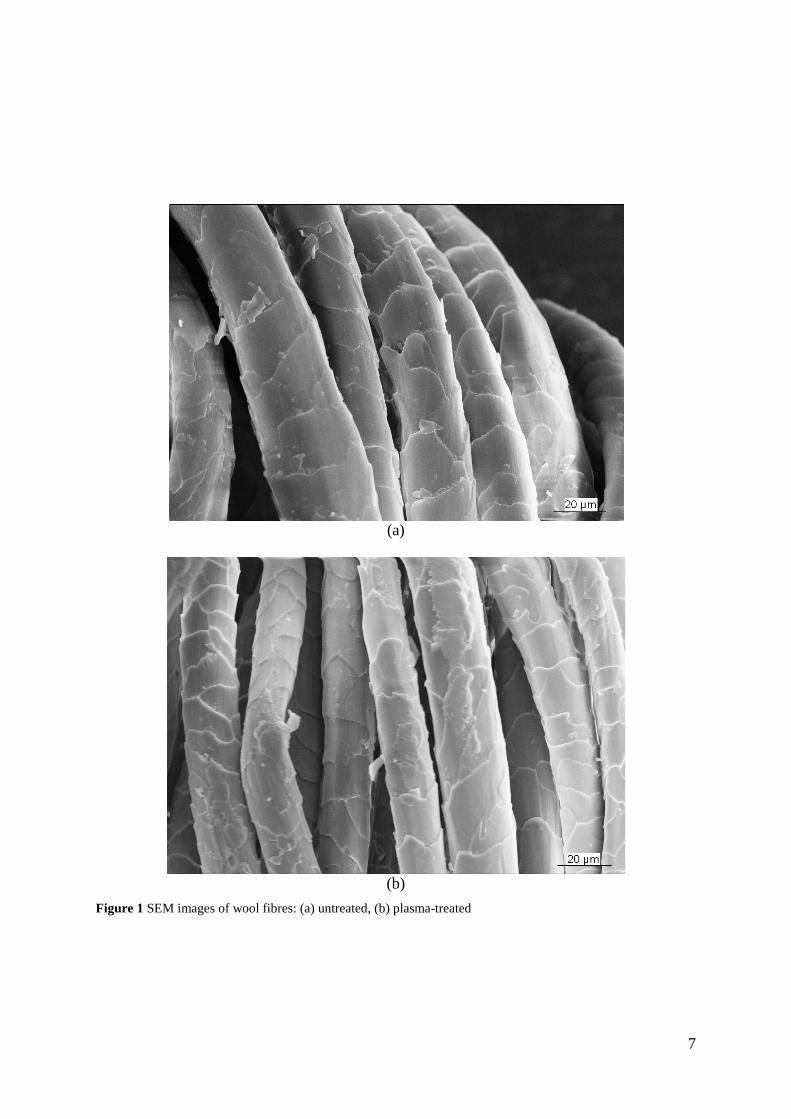
The SEM images for untreated and plasma-treated fibres are shown in Figure 1. There was no
apparent morphological change to the surface or cuticle edge of the treated wool fibre. In
contrast, SPM images showed significant microscopic changes on the fibre surface,
consistent with an etching effect of the plasma treatment, as previously reported (Cai & Qiu,
2008; Canal et al., 2007). This can be due to the difference between the two techniques in
interpreting subtle differences in height. SEM image were collected in vacuum on surfaces
coated with gold. In addition, changes in slope can result in an increase in electron reflection
from the sample surface, producing a higher intensity in the image. However, it can
sometimes be difficult to determine whether the feature is sloping up or down. On the other
hand, AFM images were collected in air on uncoated samples and yield direct height
information, determining whether a feature is a bump or pit is straightforward (Nessler,1999).
SPM images of the untreated fibres show more longitudinal surface striations compared to
the plasma-treated fibre (Figure 2). The three-dimensional nature of the SPM image was used
to calculate changes in roughness and surface area variations.
An increase in roughness (Rq), which leads to an increase in the surface area, of the plasma-
treated fibre relative to the untreated fibre can be seen in Figure 2. The Rq of the surface of
the untreated fibre varied between 25 and 43 nm (with an average of 33.4 nm and a standard
deviation of 6.3 nm) whereas that of the plasma-treated fibre varied from 70 to 93 nm (with
an average of 85.2 nm and a standard deviation of 6.1 nm). An increase in the average
roughness value of the treated wool surface under different plasma conditions has been
reported previously (Klausen et al., 1995; Phillips, 1995).
6
(a)
(b)
Figure 1 SEM images of wool fibres: (a) untreated, (b) plasma-treated
7

Figure 2 SPM analysis of wool fibres (3D surface plot, 5×5µm2): (upper image) untreated fibre, (lower image) plasma-treated fibre
8

1.2 Surface hydrophilicity immediately after treatment
Water uptake is defined here as the total movement of a liquid through the fabric driven by a
difference in surface energy between the fibre and the liquid and absorption of the liquid by
the fibre. The water uptake of untreated fabric and the fabric that had been treated with
plasma for 30 seconds was evaluated as soon as possible after treatment, usually within 4
hours. With the treated fabric, equilibrium uptake was achieved in less than 1 minute and it
showed very high (>300 %) initial water uptake at equilibrium. The untreated fabric showed
very little water uptake (0.03 %) after 15 minutes equilibration (Figure 3). A 30-second
plasma exposure time was judged to be sufficient to produce a significant change in surface
energy.
1.3. Surface analysis by XPS
Table 1 shows the surface composition of untreated and plasma-treated wool analysed by
XPS. A significant increase in oxygen and nitrogen concentration and a decrease in carbon
concentration were observed for the plasma-treated fabric compared to the untreated fabric.
The C/N ratio (an indicator of the level of covalently bound surface lipid) for plasma-treated
wool was consistent with values determined by chemical analysis for low-sulphur protein
fractions (4.4) and whole extract of wool (3.9) (Carr et al., 1986; Ward et al., 1993). A C/N
ratio of 3.4 has also been reported for the epicuticle (King & Bradbury, 1968). High
resolution XPS from a previous study (Naebe et al., 2010a) has shown that the plasma
treatment conditions used cause significant oxidation of the underlying protein matrix, in
particular the disulfide bonds, with the formation of cysteic acid residues.
A previous study (Naebe et al., 2010a) confirmed that plasma treatment under the conditions
used in the present study removed the 18-MEA, the major component of the lipid layer,
which is covalently bound through thioester linkages to the surface of the epicuticle (Negri et
9
al., 1993a). This resulted in exposure of the underlying, hydrophilic protein of the epicuticle,
thereby increasing wettability of the wool.
Table 1 Relative atomic concentration of untreated and plasma treated fabric
Elements Carbon (C1s) Oxygen (O1s) Nitrogen (N1s) Sulphur (S2P) C1s/ N1s
Binding Energy (eV) 284.0 530.0 399.0 167.0
Untreated 73.8 13.8 9.9 2.4 7.5
Plasma Treated 54.2 26.9 15.3 2.6 3.5
1.4. Surface analysis by SPM
SPM adhesion studies showed significant variability on both plasma-treated fibres and
untreated control fibres, as assessed from measurements taken at different parts of each fibre.
Nevertheless, the average adhesion on the plasma-treated fibre was significantly higher than
that of the untreated fibre. Table 2 shows the variation of average adhesion values for six
different areas on one untreated fibre and one plasma-treated fibre after 24 hrs conditioning.
Table 2 The average adhesion values of untreated and plasma-treated fibres from six different parts of the surface of each fibre
Sample Area
Mean STDEV 1 2 3 4 5 6
Untreated 83.3 37.1 81.2 37.8 84.2 76.3 66.7 22.8
Plasma Treated 127.3 146.7 135.8 146.1 156.2 121.8 133.8 13.0 The higher adhesion on the plasma-treated sample was consistent with the results on a micro-
patterned model surface used by Huson et al (Huson et al., 2008), which showed that
adhesion on a hydrophilic carboxylic acid surface was stronger than that on a hydrophobic
methyl surface.
The surface of the plasma-treated fibre was hydrophilic due to removal of the lipid layer. The
resultant higher water affinity increases the probability of a water bridge forming between the
probe tip and fibre specimen, the increased meniscus force in turn causing greater adhesion
between the tip and the sample. In addition, the higher water affinity is likely to affect the
10

real area of contact between the tip and sample (Bhushan & Jung, 2008), further increasing
the adhesion.
Unlike the results presented above, previous studies on hair (Bhushan & Chen, 2006;
Breakspear et al., 2005; LaTorre & Bhushan, 2005; 2006) and wool (Huson et al., 2008)
have shown that chemical treatment known to remove lipids from the surface generally
reduce adhesion. Huson et al acknowledged that this behaviour was unusual and speculated
that it could be due to the surface being able to absorb moisture, thereby affecting the
thickness of the adsorbed layer, and the possibility of ions or organic molecules leaching
from the bulk of the fibre and contaminating the adsorbed layer.
It is also possible that in the plasma-treated fibres used in the present study, oxidation of the
fibre surface might play a role in the increased adhesion. High resolution XPS analysis
showed significant oxidation of sulphur to sulphonic acid as a consequence of the plasma
treatment.
2 Surface Change with Ageing
2.1 Surface morphology
Changes in the surface morphology of the untreated and plasma-treated fibres were examined
by SPM. While the variation in roughness was observed between different areas of a sample,
no significant changes in surface roughness were observed on untreated and plasma-treated
fibres over a period of 28 days.
2.2 Surface hydrophiliciy changes with time
Untreated fabric and the fabric that had been treated with plasma for 30 seconds were stored
at 20+2 0C and 65+2 % RH and tested over a period of 28 days. The rate of water uptake was
used to determine the changes in the wetting properties of wool fabrics with ageing time.
11

Untreated
Figure 3 shows the percentage of water uptake at equilibrium versus ageing time. The
untreated fabric took up very little water and showed no change over the 28 days. In contrast,
the plasma-treated sample showed significantly higher initial water uptake, the equilibrium
value decreasing significantly over the first 3 days, and subsequently more slowly. After 28
days, although the plasma-treated fabric had lost some of its initial wettability, it was still
considerably more hydrophilic than the untreated fabric.
Figure 3 Equilibrium water uptake as a function of time; plasma-treated (■), untreated (▲). Note two different y-axis scales.
2.3 Surface analysis by XPS
Table 3 shows the surface composition of plasma-treated wool analysed by XPS for different
ageing times from one day to 28 days. An increase in carbon concentration and a decrease in
oxygen and nitrogen concentrations were observed. The sulphur content remained almost
constant over the entire period. The relative atomic concentration of carbon in the surface of
the plasma-treated sample increased rapidly and then reached a constant value (after
approximately 14 days) that was significantly less than that of the untreated surface (73.8 %).
12
Nitrogen concentrations decreased to an almost constant level over the same period. The C 1s
/N 1s ratio remained unchanged (~4.1) after 14 days of ageing.
Table 3 Surface analysis of plasma treated fabric over a period of one month Elements Binding Energy
(eV) Relative Atomic Concentration (%)
Storage time after plasma treatment (days) 1 2 3 7 14 21 28
Carbon (C1s) 284.0 54.2 54.5 54.8 55.9 57.0 57.3 57.3
Oxygen (O1s) 530.0 26.9 26.8 26.6 26.1 25.2 25.0 24.6
Nitrogen (N1s) 399.0 15.3 15.1 15.1 14.6 13.8 13.9 13.9
Sulphur (S2P) 167.0 2.6 2.6 2.5 2.5 2.6 2.6 2.6
C1s/ N1s 3.5 3.6 3.6 3.8 4.1 4.1 4.1
High resolution XPS sulphur spectra from plasma-treated wool samples after two, three and
seven days ageing are shown in Figure 4. The S 2P spectra consisted of two peaks at 164 eV
and 168 eV. The Peak at 164.0 eV corresponds to the presence of sulphur as a disulphide
linkage. The peak at higher bonding energy (168.0 eV) is attributed to the presence of
oxidised sulphur species (S6+) in the protein matrix after plasma treatment (Brack et al., 1996;
Molina et al., 2002). These oxidised species have been shown to be sulphonic acid (-SO3-)
(Bradley et al., 1994; Shao et al., 1997) and (S-SO3H) (Shao et al., 1997)
13

Figure 4 High resolution XPS sulphur spectra from plasma-treated wool after 2, 3 and 7 days ageing
The ratio of disulphide linkages to oxidised sulphur species (C-S/S-Oxidised) recorded from
S2p spectra of the plasma-treated fabrics after different ageing times has been summarised in
Table 4. The ratio increased significantly over the first 7 days before remaining constant,
suggesting that whilst the total sulphur had not changed (Table 3), the more oxidised sulphur
species had migrated away from the surface of the plasma-treated sample.
Table 4 Ratio of sulphur species of plasma treated fabric over a period of one month
Storage time after plasma treatment (days) 1 2 3 7 14 21 28
C-S/S-Oxidised 1.01 1.11 1.18 1.33 1.30 1.32 1.48
The results suggest that the surface of plasma-treated wool fibre is mobile and changes as the
fibre ages. The increase in carbon content can be attributed to the migration of the carbon
from the bulk of the fibre onto the surface. This migration would explain the decreased
wettability (Figure 3). The results also show a decrease in the polarity of the surface and a
14

decrease in oxygen (Table 1) and S6+ (Figure 4). This can be attributed to the orientation of
polar groups from the surface to the bulk phase, resulting in rearrangement of the surface of
the wool fibre.
2.4 Surface analysis by SPM
The effect of ageing plasma-treated fibres was studied by SPM adhesion measurements and
compared to an untreated control fibre. Figure 5 shows the average adhesion ratio of plasma-
treated to untreated samples over a period of 28 days. This ratio increased up to the ageing
time of 14 days and then the value remained relatively constant. After ageing for 28 days, the
surface showed three times more adhesion than that of the untreated fibre. For polymer
surfaces, Everaert et al. (1997) postulated that ageing or “hydrophobic recovery” changes the
hydrophilic nature of the surface created by a plasma treatment to a relatively or fully
hydrophobic surface over time. In the present study, ageing did not change the hydrophilic
surface of the plasma-treated wool to a completely hydrophobic surface. This would suggest
that the oxidation of the surface and modification or removal of the lipid layer were relatively
permanent and rearrangement of the surface would not return the fibre to its original
hydrophobic state.
Rearrangement on the surface was relatively rapid immediately after the plasma treatment,
and slowed with ageing time. The results agree with the model proposed by Huson et al.
(2008), suggesting that the disordered lipid layer is capable of rearranging in response to
changes in the environment, but the changes in adhesion measured by SPM are difficult to
explain. As the hydrophilic nature of the surface created by the plasma treatment changes to a
more hydrophobic surface over time, one would expect a reduction in surface adhesion. More
work is needed to expand the current understanding of the wool fibre surface to explain this
anomaly.
15

Figure 5 Average adhesion ratio of plasma-treated to untreated fibre over a period of 28 days for all fibres and areas measured
CONCLUSION
Atmospheric plasma treatment of wool fabric resulted in increased wettability of the fabric
and fibres, with greater roughness and adhesion as measured by SPM analysis. XPS
suggested a much more hydrophilic surface with a significant increase in oxygen and
nitrogen concentrations and a decrease in carbon concentration. These changes are consistent
with the removal of the covalently-bound lipid from the surface of the fibre. The significant
oxidation of the underlying protein matrix, in particular the disulfide bonds, with the
formation of cysteic acid residue was shown.
When the plasma-treated wool fibre was allowed to age, no changes were seen in the
morphology of the surface. However, wettability studies, as well as surface analyses using
SPM and XPS, showed that the chemical nature of the surface was changing with time. An
increase in carbon concentration and a decrease in oxygen and nitrogen concentrations were
observed over 14 days. After that period, the concentrations remained relatively constant.
16

Although the sulphur content was fairly stable over 28 days of ageing, the intensity ratio of
disulphide linkages to oxidised sulphur species (C-S/S-Oxidised) changed, showing that
(with time) the more oxidised sulphur species migrated away from the surface of the plasma-
treated sample. This increase in the hydrophobic nature of the surface was consistent with the
decrease in wettability with time, but at odds with the increased adhesion as measured by
SPM. After ageing for 28 days, the plasma-treated surface was relatively stable and still
dramatically different from the untreated fibre, suggesting that the oxidation of the surface
and modification or removal of the lipid layer were permanent. In spite of rearrangement of
the surface, the treated fibre remained considerably more hydrophilic than the untreated fibre.
References
Bhushan, B. & Chen, N. (2006). AFM studies of environmental effects on nanomechanical properties and cellular structure of human hair. Ultramicroscopy, 106, 755-764. Bhushan, B. & Jung, Y. C. (2008). Wetting, adhesion and friction of superhydrophobic and hydrophilic leaves and fabricated micro/nanopatterned surfaces. Journal of Physics: Condensed Matter, 20, 1-24. Brack, N., Lamb, R., Pham, D. & Turner, P. (1996). XPS and SIMS investigation of covalently bound lipid on the wool fibre surface. Surface and Interface Analysis, 24, 704-710. Bradbury, J. H. (1973). The structure and chemistry of keratin fibers, In Anfinsen, C. B.& Edsall, J. T. (Ed.), Advances in Protein Chemistry (pp.111-211). London: Academic Press. Bradley, R. H., Clackson, I. L. & Sykes, D. E. (1994). XPS of oxidized wool fibre surfaces. Surface and Interface Analysis, 22, 497-501. Breakspear, S., Smith, J. R. & Luengo, G. (2005). Effect of the covalently linked fatty acid 18-MEA on the nanotribology of hair's outermost surface. Journal of Structural Biology, 149, 235-242. Cai, Z. & Qiu, Y. (2008). Effect on the anti-felt properties of atmospheric pressure plasma treated wool. Journal of Applied Polymer Science, 107, 1142-1146. Canal, C., Molina, R., Bertran, E. & Erra, P. (2007). Polysiloxane sftener coatings on plasma-treated wool: Study of the surface interactions. Macromolecular Materials and Engineering, 292, 817-824. Carr, C. M., Leaver, I. H. & Hughes, A. E. (1986). X-ray photoelectron spectroscopic study of the wool fiber surface. Textile Research Journal, 56, 457-461.
17

Crossley, J. A. A., Gibson, C. T., Mapledoram, L. D., Huson, M. G., Myhra, S., Pham, D. K., Sofield, C. J., Turner, P. S. & Watson, G. S. (2000). Atomic force microscopy analysis of wool fibre surfaces in air and under water. Micron, 31, 659-667. Dai, X. J., Elms, F. M. & George, G. A. (2001). Mechanism for the plasma oxidation of wool fibre surfaces from XPS studies of self-assembled monolayers. Journal of Applied Polymer Science, 80, 1461-1469. Erra, P., Jovanèiæ, P., Molina, R., Jociæ, D. & Julia, M. R. (2002). Study of surface modification of wool fibres by means of SEM, AFM, and light microscopy. In Science, Technology and Education of Microscopy: an Overview (pp. 549-556). Badajoz: Formatex. Evans, D. J., Denning, R. J. & Church, J. S. (2002). Interface of keratin fibres with their environment. In Encyclopedia of Surface and Colloid Science (pp. 2628-2642). Marcel Dekker Inc. Evans, D. J., Leeder, J. D., Rippon, J. A. & Rivett, D. E. (1985). Separation and analysis of surface lipids of the wool fibre. In Proceedings of 7th International wool Research Conference (Tokyo, Japan), I, 135-142. Everaert, E. P., Chatelier, R. C., Mei, H. C. V. & Busscher, H. J. (1997). A quantitative model for the surface restructuring of repeatedly plasma treated silicone rubber. Plasmas and Polymers, 2, 41-51. Gibson, C. T., Myhra, S., Watson, G. S., Huson, M. G., Pham, D. K. & Turner, P. S. (2001). Effects of aqueous exposure on the mechanical properties of wool fibre analysis by atomic force microscopy. Textile Research Journal, 71, 573–581. Huson, M., Evans, D., Church, J., Hutchinson, S., Maxwell, J. & Corino, G. (2008). New insights into the nature of the wool fibre surface. Journal of Structural Biology, 163, 127-136. Kan, C. W., Chan, K. & Yuen, C. W. M. (2004). Surface characterisation of low temperature plasma treated wool fibre – The effect of the nature of gas. Fibers and Polymers, 5, 52-58. King, L. R. & Bradbury, J. H. (1968). The chemical composition of wool. Part 5. The epicuticle. Australian Journal of Biological Sciences, 21, 375-384. Klausen, T., Thomas, H. & Hocker, H. (1995). Influence of oxygen plasma treatment on the chemical and morphological changes of the wool fibre surface. In Proceedings of 9th International wool Research Conference (Biella, Italy), II, 241-248. LaTorre, C. & Bhushan, B. (2005). Nanotribological characterization of human hair and skin using atomic force microscopy. Ultramicroscopy, 105, 155-175. LaTorre, C. & Bhushan, B. (2006). Investigation of scale effects and directionality dependence on friction and adhesion of human hair using AFM and macroscale friction test apparatus. Ultramicroscopy, 106, 720-734. Leeder, J. D. & Bradbury, J. H. (1968). Conformation of epicuticle on keratin fibres. Nature, 218, 694-695. Leeder, J. D. & Bradbury, J. H. (1971). The discontinuous nature of the epicuticle on the surface of keratin fibres. Textile Research Journal, 41, 563-568. Leeder, J. D., Rippon, J. A. & Rivett, D. E. (1985). Modification of the Surface Properties of Wool by Treatment with Anhydrous Alkali. In Proceedings of 7th International wool Research Conference. (Tokyo, Japan), IV, 312-321. Maxwell, J. M. & Huson, M. G. (2005). Scanning probe microscopy examination of the surface properties of keratin fibres. Micron, 36, 127-136. McLaughlin, J. R. & Pope, C. G. (1982). Inverse gas chromatography of wool. Journal of Applied Polymer Science, 27, 1643-1654. Molina, R., Espinos, J. P., Yubero, F., Erra, P. & Gonzalez-Elipe, A. R. (2005). XPS Analysis of down stream plasma treated wool: Influance of the nature of the gas on the surface modification of wool. Applied Surface Science, 252, 1417-1429.
18

Molina, R., Jovančić, P., Comelles, F., Bertran, E. & Erra, P. (2002). Shrink-resistance and wetting properties of keratin fibres treated by glow discharge. Journal of Adhesion Science and Technology, 16, 1469-1485. Naebe, M., Cookson, P. G., Rippon, J. A., Brady, P. R., Brack, N., Riessen, G. V. & Wang, X. G. (2010a). Effects of plasma treatment of wool on the uptake of sulfonated dyes with different hydrophobic properties. Textile Research Journal, 80, 312-324. Naebe, M., Cookson, P. G., Rippon, J. A. & Wang, X. G. (2010b). Effects of levelling agent on the uptake of reactive dyes by untreated and plasma-treated wool. Textile Research Journal, 80, Negri, A. P., Cornell, H. J. & Rivett, D. E. (1993a). A model for the surface of keratin fibers. Textile Research Journal, 63, 109-115. Negri, A. P., Cornell, H. J. & Rivett, D. E. (1993b). The modification of the surface diffusion barrier of wool. Journal Society of Dyers and Colourists, 109, 296-301. Negri, A. P., Rankin, D. A., Nelson, W. G. & Rivett, R. E. (1996). Transmission electron microscope study of covalently-bound fatty acids in the cell membranes of wool fibres. Textile Research Journal, 66, 491–495. Nessler, R., (1999). Scanning Microscopy Technologies: Scanning Electron Microscopy and Scanning Probe Microscopy. Scanning, 21, 137. Phillips, T. L. (1995). Scanning force microscopy of the surface of wool fibres. Applied Chemistry. Royal Melbourne Institute of Technology. Master thesis. Rakowski, W. (1997). Plasma treatment of wool today. Part I- Fibre properties, spinning and shrinkproofing. Jounal of Socaiety of Dyers and Colourists, 113, 250-255. Robbins, C. R. (1988). Chemical and physical behaviour of human hair. New York, Spriner-Verlag. Ryu, J., Wakida, T., Kawamura, H., Goto, T. & Takagishi, T. (1987). Frictional coefficient of wool treated with low temperature plasma. Sen-i Gakkaishi, 43, 257-262. Ryu, J., Wakida, T. & Takagishi, T. (1991). Effect of corona discharge on the surface of wool and its application to printing. Textile Research Journal, 61, 595-601. Shao, J., Jones, D. C., Mitchell, R., Vickerman, J. C. & Carr, C. M. (1997). Time of flight secondary- ion mass spectrometric (ToF-SIMS) and X-ray photoelectron spectroscopic (XPS) analyses of the surface lipids of wool. Journal of Textile Institute, 88, 317-324. Volooj, S., Carr, C. M., Mitchell, R. & Vickerman, J. C. (2000). ToF-SIMS Analysis of bleaching of keratin fibres. Surface and Interface Analysis, 29, 422-430. Wakida, T., Tokino, S., Niu, S., Kawamura, H., Sato, Y., Lee, M., Uchiyama, H. & Inagaki, H. (1993). Surface characteristics of wool and poly (ethylene terphthalate) fabrics and film treated with low temperature plasma under atmospheric pressure. Textile Research Journal, 63, 433-438. Ward, R. J., Willis, H. A., George, G. A., Guise, G. B., Denning, R. J., Evans, D. J. & Short, R. D. (1993). Surface analysis of wool by X-ray photoelectron spectroscopy and static secondary ion mass spectrometry. Textile Research Journal, 63, 362-368.
19