Electrochemical and spectroscopic investigations of isoniazide and its analogs with ds.DNA at...
11
This article appeared in a journal published by Elsevier. The attached copy is furnished to the author for internal non-commercial research and education use, including for instruction at the authors institution and sharing with colleagues. Other uses, including reproduction and distribution, or selling or licensing copies, or posting to personal, institutional or third party websites are prohibited. In most cases authors are permitted to post their version of the article (e.g. in Word or Tex form) to their personal website or institutional repository. Authors requiring further information regarding Elsevier’s archiving and manuscript policies are encouraged to visit: http://www.elsevier.com/copyright
Transcript of Electrochemical and spectroscopic investigations of isoniazide and its analogs with ds.DNA at...
This article appeared in a journal published by Elsevier. The attachedcopy is furnished to the author for internal non-commercial researchand education use, including for instruction at the authors institution
and sharing with colleagues.
Other uses, including reproduction and distribution, or selling orlicensing copies, or posting to personal, institutional or third party
websites are prohibited.
In most cases authors are permitted to post their version of thearticle (e.g. in Word or Tex form) to their personal website orinstitutional repository. Authors requiring further information
regarding Elsevier’s archiving and manuscript policies areencouraged to visit:
http://www.elsevier.com/copyright
Author's personal copy
Original article
Electrochemical and spectroscopic investigations of isoniazide and its analogswith ds.DNA at physiological pH: Evaluation of biological activities
Nasima Arshad a,*, Uzma Yunus a, Shumaila Razzque a, Maliha Khan a, Samreen Saleemb, Bushra Mirza b,Naghmana Rashid a
aDepartment of Chemistry, Allama Iqbal Open University, Islamabad, PakistanbDepartment of Biochemistry, Quaid-i-Azam University, Islamabad, Pakistan
a r t i c l e i n f o
Article history:Received 2 March 2011Received in revised form11 October 2011Accepted 7 November 2011Available online 18 November 2011
Keywords:HydrazidsCyclic voltammetryUVeVis spectroscopyDNA bindingBinding site sizeBiological applications
a b s t r a c t
Interaction and binding of isonicotinic acid hydrazide (INH) and its two analogs; pyrazine carboxylic acidhydrazide (PCH) and 2,4-dihydroxy benzoic acid hydrazide (2,4-DHBAH) with DNA has been investigatedby UV-spectroscopy and cyclic voltammetry (CV) at physiological conditions of pH and temperature.Experimental results from both techniques were in good agreement and indicated stronger binding andformation of hydrazideseDNA complexes via intercalation. Among three hydrazides, 2,4-DHBAH showedgreater interaction toward DNA at stomach pH (4.7) as evident from its comparatively greater bindingconstant, {Kb; 2.02 � 104 M�1 (UV), 3.13 � 104 M�1 (CV)}. The greater binding site size (n ¼ 3) for 2,4-DHBAH at stomach pH inferred 3:1 binding stoichiometry and possibility of electrostatic interactionsor hydrogen bonding along with intercalative mode of interaction between 2,4-DHBAH and DNA. Thefree energies of hydrazideseDNA complexes indicated the spontaneity of their binding. 2,4-DHBAH hasshown promising anti-bacterial activities while anti-oxidant and cytotoxic potentials were exhibited byall three hydrazides.
� 2011 Elsevier Masson SAS. All rights reserved.
1. Introduction
Interaction between biomolecules is fundamental to numerousbiological processes [1]. Based on these interactions livingorganismmaintain complex regulatory and metabolic interactionalnetworks that together constitutes the process of life. A thoroughunderstanding of bimolecular interactions and pathways play keyrole in solving mystery of life [2].
Many small molecules that bind to DNA are extremely useful asbiochemical tools for the visualization of DNA both in vivo andin vitro and have been clinically proven beneficial although theirexact mode of action is unknown [3]. There has been a significantprogress made over a past few years in studies of DNA interactions.
Nucleic acids are common targets for antiviral, anticancer andantibiotic drugs [4e6]. A concrete knowledge of drugs nucleic acidinteraction, including sequence recognition, structural details,kinetics and thermodynamics of binding is prerequisite for theoptimization of drug effectiveness as well as to discover new drugs.The binding interaction of small molecules and their complexes
with DNA is of interest for both therapeutic and scientific reasons[7]. These interactions may also be used for conformationalrecognition to find new structures of DNA and sequence specificdifferences along the helix of DNA molecule [8e10]. In addition,drug-DNA interaction can be anticipated through electrochemicalchanges in the redox active molecule of drug [11e14].
Drug can bind to DNA both covalently as well as non-covalently(via intercalation mode or minor groove binding). The minorgroove of DNA (the negatively charged phosphates outside the DNAdouble helix) is the locus of action of large numbers of drugs, whilein an intercalation mode base pair of DNA unwind to accommodatethe drug [15,16]. Both intercalating agents and minor groovebinders are typified as anticancer, antiviral and antitumor, anti-bacterial and anti-fungal agents [17e22].
Hydrazides are the effective organic compound having thera-peutic properties [23] and known to possess biological effects suchas anti-fungal, anti-bacterial and anti-inflammatory activities [24].A series of substituted hydrazide derivatives have been synthesizedand screened for their in vitro antimicrobial activities againstmicroorganisms. The results of antimicrobial study indicated thatthe presence of electron withdrawing groups on the benzoic acidmoiety improved antimicrobial activity [25,26]. Isoniazid (INH),rifampin (RIF), ethambutol (EMB) and pyrazinamide (PZA) are well
* Corresponding author. Tel.: þ92 51 9057756; fax: þ92 51 9250081.E-mail address: [email protected] (N. Arshad).
Contents lists available at SciVerse ScienceDirect
European Journal of Medicinal Chemistry
journal homepage: http: / /www.elsevier .com/locate/ejmech
0223-5234/$ e see front matter � 2011 Elsevier Masson SAS. All rights reserved.doi:10.1016/j.ejmech.2011.11.014
European Journal of Medicinal Chemistry 47 (2012) 452e461
Author's personal copy
known antituberculosis drugs and direct DNA sequencing analysisof their genes i.e., katG, rpoB, embB and pncA, respectively, hasbeen reported as a rapid and useful molecular genetic method forthe detection and treatment of drug-resistant Mycobacteriumtuberculosis [27].
In present studies, an attempt is made to investigate the inter-action of isonicotinic acid hydrazide (INH) and its two analogs;pyrazine carboxylic acid hydrazide (PCH) and 2,4-dihydroxy ben-zoic acid hydrazide (2,4-DHBAH) (Scheme 1) with chicken blooddouble stranded (ds)-DNA (ck.DNA) using UVeVis spectroscopic(UV) and cyclic voltammetric (CV) techniques at stomach (4.7) andblood (7.4) pH under body temperature (37 �C). Also thesecompounds were investigated for anti-bacterial, anti-oxidant andcytotoxic activities.
2. Results and discussion
2.1. DNA binding study by UV-spectroscopy
The binding of drug to DNA has been characterized classicallythrough absorption titrations [28]. Generally, hypochromic (orhyperchromic) effect and red shift (or blue) shift are observed inthe absorption spectra of small molecules if they intercalate withDNA [29].
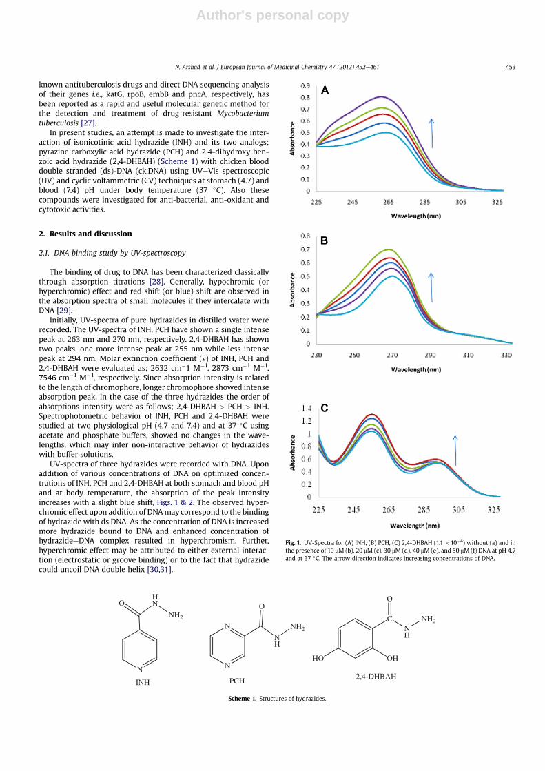
Initially, UV-spectra of pure hydrazides in distilled water wererecorded. The UV-spectra of INH, PCH have shown a single intensepeak at 263 nm and 270 nm, respectively. 2,4-DHBAH has showntwo peaks, one more intense peak at 255 nm while less intensepeak at 294 nm. Molar extinction coefficient (ε) of INH, PCH and2,4-DHBAH were evaluated as; 2632 cm�1 M�1, 2873 cm�1 M�1,7546 cm�1 M�1, respectively. Since absorption intensity is relatedto the length of chromophore, longer chromophore showed intenseabsorption peak. In the case of the three hydrazides the order ofabsorptions intensity were as follows; 2,4-DHBAH > PCH > INH.Spectrophotometric behavior of INH, PCH and 2,4-DHBAH werestudied at two physiological pH (4.7 and 7.4) and at 37 �C usingacetate and phosphate buffers, showed no changes in the wave-lengths, which may infer non-interactive behavior of hydrazideswith buffer solutions.
UV-spectra of three hydrazides were recorded with DNA. Uponaddition of various concentrations of DNA on optimized concen-trations of INH, PCH and 2,4-DHBAH at both stomach and blood pHand at body temperature, the absorption of the peak intensityincreases with a slight blue shift, Figs. 1 & 2. The observed hyper-chromic effect upon addition of DNAmay correspond to the bindingof hydrazide with ds.DNA. As the concentration of DNA is increasedmore hydrazide bound to DNA and enhanced concentration ofhydrazideeDNA complex resulted in hyperchromism. Further,hyperchromic effect may be attributed to either external interac-tion (electrostatic or groove binding) or to the fact that hydrazidecould uncoil DNA double helix [30,31].
N
HN
NH2
O
INH
N
N
NH
NH2
O
HO OH
CNH
NH2
O
2,4-DHBAHPCH
Scheme 1. Structures of hydrazides.
Fig. 1. UV-Spectra for (A) INH, (B) PCH, (C) 2,4-DHBAH (1.1 � 10�4) without (a) and inthe presence of 10 mM (b), 20 mM (c), 30 mM (d), 40 mM (e), and 50 mM (f) DNA at pH 4.7and at 37 �C. The arrow direction indicates increasing concentrations of DNA.
N. Arshad et al. / European Journal of Medicinal Chemistry 47 (2012) 452e461 453

Author's personal copy
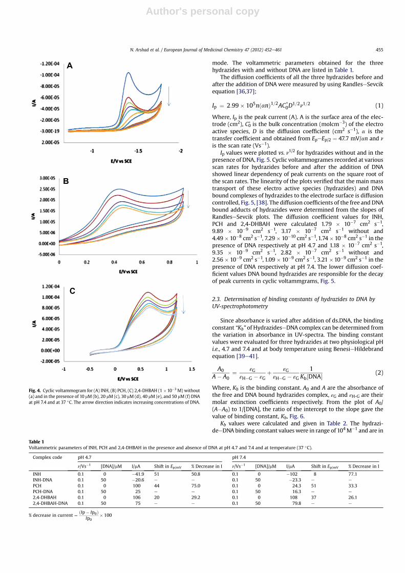
2.2. DNA binding study by cyclic voltammetry
Due to redox activity of most of the small molecules, cyclic vol-tammetry has proved as one of the important electrochemical tech-nique to elucidate mechanism of drug action in vivo [32e34]. Thecyclic voltammetric behavior of 1mMof INH, PCH and 2,4-DHBAH inthe absence and presence of 10 mMe50 mM DNA at GCE and underphysiological condition of temperature and pH are shown in Figs. 3 &4. INH showed a single irreversible and stable cathodic (reduction)peak at peak potential of �1.311 V at pH 4.7 and �1.446 V at pH 7.4.PCH and 2,4-DHBAH showed single irreversible anodic peak (oxida-tion) peaks at þ0.9082 V and þ0.7393 V, respectively at pH 4.7,and þ0.3931 V and þ0.5913 V, respectively at pH 7.4, versus SCE.
Upon the addition of various concentrations of DNA on fixedconcentration of INH, decrease in the reduction peak current valuesand a slight positive shift was observed. Cyclic voltammetricbehavior of 1 mM of PCH and 2,4-DHBAH in the presence andabsence of various concentrations of DNA was also observed asdecrease in oxidation peak current and a slight positive shifts in theanodic peak potentials. The substantial diminution in peak currentis attributed to the formation of slowly diffusing HydrazideeDNAsupramolecular complex due towhich the concentration of the freedrug (mainly responsible for the transfer of current) is lowered. Adecrease in the peak current along with the positive shifting in thepeak potential is attributed to characteristic behavior of the inter-calation of drug into DNA double helix [15,35]. Hence the observedbehavior of hydrazides in present CV studies for their interactionwith ds.DNA could be attributed to the possibility of intercalation
Fig. 2. UV-Spectra for (A) INH, (B) PCH, (C) 2,4-DHBAH (1.1 � 10�4) without (a) and inthe presence of 10 mM (b), 20 mM (c), 30 mM (d), 40 mM (e), and 50 mM (f) DNA at pH 7.4and at 37 �C. The arrow direction indicates increasing concentrations of DNA.
Fig. 3. Cyclic voltammogram for (A) INH, (B) PCH, (C) 2,4-DHBAH (1 �10�3 M) without(a) and in the presence of 10 mM (b), 20 mM (c), 30 mM (d), 40 mM (e), and 50 mM (f) DNAat pH 4.7 and at 37 �C. The arrow direction indicates increasing concentrations of DNA.
N. Arshad et al. / European Journal of Medicinal Chemistry 47 (2012) 452e461454
Author's personal copy
mode. The voltammetric parameters obtained for the threehydrazides with and without DNA are listed in Table 1.
The diffusion coefficients of all the three hydrazides before andafter the addition of DNA were measured by using RandleseSevcikequation [36,37];
Ip ¼ 2:99� 105nðanÞ1=2AC*0D
1=2n1=2 (1)
Where, Ip is the peak current (A). A is the surface area of the elec-trode (cm2), C0* is the bulk concentration (molcm�3) of the electroactive species, D is the diffusion coefficient (cm2 s�1), a is thetransfer coefficient and obtained from EpeEp/2 ¼ 47.7 mV/an and n
is the scan rate (Vs�1).Ip values were plotted vs. n1/2 for hydrazides without and in the
presence of DNA, Fig. 5. Cyclic voltammogrames recorded at variousscan rates for hydrazides before and after the addition of DNAshowed linear dependency of peak currents on the square root ofthe scan rates. The linearity of the plots verified that the main masstransport of these electro active species (hydrazides) and DNAbound complexes of hydrazides to the electrode surface is diffusioncontrolled, Fig. 5, [38]. The diffusion coefficients of the free and DNAbound adducts of hydrazides were determined from the slopes ofRandleseSevcik plots. The diffusion coefficient values for INH,PCH and 2,4-DHBAH were calculated 1.79 � 10�7 cm2 s�1,9.89 � 10�9 cm2 s�1, 3.17 � 10�7 cm2 s�1 without and4.49� 10�8 cm2 s�1, 7.29� 10�10 cm2 s�1, 1.74�10�8 cm2 s�1 in thepresence of DNA respectively at pH 4.7 and 1.18 � 10�7 cm2 s�1,9.35 � 10�9 cm2 s�1, 2.82 � 10�7 cm2 s�1 without and2.56 � 10�9 cm2 s�1, 1.09 � 10�9 cm2 s�1, 3.21 �10�9 cm2 s�1 in thepresence of DNA respectively at pH 7.4. The lower diffusion coef-ficient values DNA bound hydrazides are responsible for the decayof peak currents in cyclic voltammgrams, Fig. 5.
2.3. Determination of binding constants of hydrazides to DNA byUV-spectrophotometry
Since absorbance is varied after addition of ds.DNA, the bindingconstant “Kb” of HydrazideseDNA complex can be determined fromthe variation in absorbance in UV-spectra. The binding constantvalues were evaluated for three hydrazides at two physiological pHi.e., 4.7 and 7.4 and at body temperature using BenesieHildebrandequation [39e41].
A0
A� A0¼ εG
εH�G � εGþ εG
εH�G � εG
1Kb½DNA�
(2)
Where, Kb is the binding constant. A0 and A are the absorbance ofthe free and DNA bound hydrazides complex, εG and εH-G are theirmolar extinction coefficients respectively. From the plot of A0/(A�A0) to 1/[DNA], the ratio of the intercept to the slope gave thevalue of binding constant, Kb, Fig. 6.
Kb values were calculated and given in Table 2. The hydrazi-deeDNA binding constant values were in range of 104M�1 and are in
Fig. 4. Cyclic voltammogram for (A) INH, (B) PCH, (C) 2,4-DHBAH (1 �10�3 M) without(a) and in the presence of 10 mM (b), 20 mM (c), 30 mM (d), 40 mM (e), and 50 mM (f) DNAat pH 7.4 and at 37 �C. The arrow direction indicates increasing concentrations of DNA.
Table 1Voltammetric parameters of INH, PCH and 2,4-DHBAH in the presence and absence of DNA at pH 4.7 and 7.4 and at temperature (37 �C).
Complex code pH 4.7 pH 7.4
n/Vs�1 [DNA]/mM I/mA Shift in Ep/mV % Decrease in I n/Vs�1 [DNA]/mM I/mA Shift in Ep/mV % Decrease in I
INH 0.1 0 �41.9 51 50.8 0.1 0 �102 8 77.1INH-DNA 0.1 50 �20.6 e e 0.1 50 �23.3 e e
PCH 0.1 0 100 44 75.0 0.1 0 24.3 51 33.3PCH-DNA 0.1 50 25 e e 0.1 50 16.3 e e
2,4-DHBAH 0.1 0 106 20 29.2 0.1 0 108 37 26.12,4-DHBAH-DNA 0.1 50 75 e e 0.1 50 79.8 e e
% decrease in current ¼ ðIp� Ip0ÞIp0
� 100
N. Arshad et al. / European Journal of Medicinal Chemistry 47 (2012) 452e461 455

Author's personal copy
consistent with that reported for the interaction of anthracyclinemoleculeswithDNA (Kz 104�105M�1).Kbvalues depicted strongerinteractions of the three hydrazides with DNA at both pH values[19,40,42]. UV-spectral changes observed during hydrazideeDNAcomplex formation i.e., hyperchromic effect and hypsochromic shiftin present study inferred the possibility of intercalative mode ofinteraction. The greater value of binding constant may further beattributed to the small structureofhydrazidemoleculeswhoseplanerparts may intercalate between the adjacent DNA base pairs.
Binding constant is a measure of the complex stability. Inpresent studies, 2,4-DHBAH showed comparatively greater value ofbinding constant at stomach pH. The overall data also demon-strated greater binding constant values of the three hydrazides atstomach pH (4.7) as compared to blood pH (7.4), Table 2. The valuesof binding constants for all the three hydrazides at both pH valuesare comparable with the binding constant (1.7 � 104 M�1) ofa typical intercalator Lumazine. The order of binding constants ofthree hydrazids was as follows;
Kb(2,4-DHBAH) > Kb(INH) ¼ Kb(PCH) (at stomach pH)Kb(INH) > Kb(2,4-DHBAH) > Kb(PCH) (at blood pH)
From the values of binding constant “Kb”, Gibbs free energies“ΔG” of hydrazideseDNA complexes were calculated, using thefollowing equation;
DG ¼ �RTlnKb (3)
Free energies of all the three hydrazides at both pH values wereevaluated as negative values showing the spontaneity of hydrazi-deseDNA interaction, Table 1 [15,16].
2.4. Evaluation of binding constants and binding sites by cyclicvoltammetry
Based upon the decrease in peak current of hydrazides by theaddition of different concentration of ds.DNA, the binding constants,Kb for the three hydrazideseDNA complexes were calculatedaccording to the following equation [43].
I2p ¼ 1Kb½DNA�
�I2p0
� I2p�þ I2p0
� ½DNA� (4)
Where Kb is the binding constant, I and I0 are the peak currents withand without DNA. A plot of Ip2 vs. (Ip02 � Ip
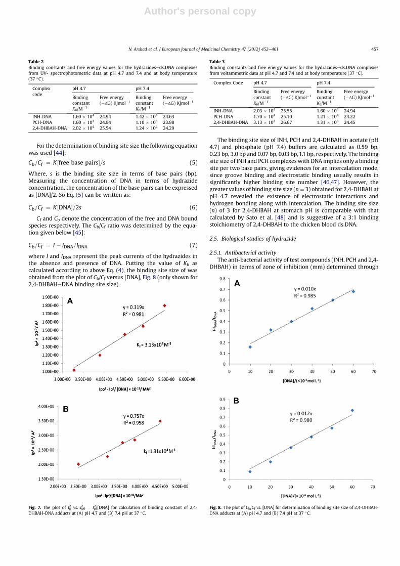
2)/[DNA] gave a straight linewith a slope equal to the reciprocal of binding constant, Kb, Fig. 7(only shown for 2,4-DHBAHeDNA binding). The binding constantvalues of three hydrazides with DNA were calculated at pH 4.7 and7.4 and at body temperature (37 �C) and the data are given inTable 3.
The order of binding constants for the three hydrazides obtainedby cyclic voltammetric data were as follow;
Kb(2,4-DHBAH) > Kb(INH) > Kb(PCH) (at stomach pH)Kb(INH) > Kb(2,4-DHBAH) > Kb(PCH) (at blood pH)
The data obtained for Kb from CV were in good agreement asobtained from UV-results and further confirmed the formation ofmost stable 2,4-DHBAHeDNA complex at stomach pH. From thebinding constant data, the standard Gibbs free energy changes forthree hydrazides were calculated, Table 2. The negative values of ΔGthrough voltammetric results also supported the UV-results of freeenergy changes and indicated the spontaneity of hydrazideseDNAbinding.
Fig. 5. Ip vs. n1/2 plots for 1.1 � 10�4 M 2,4-DHBAH at (A) pH 4.7 and (B) 7.4 pH in theabsence (1) and in the presence of 50 mL DNA (2) at scan rate of 20 mVs�1, 50 mVs�1,100 mVs�1, 150 mVs�1, 200 mVs�1, 250 mVs�1, and 300 mVs�1 in acetate buffer (pH;4.7) at 37 �C.
Fig. 6. Plot of A0/A-A0 vs.1/[DNA] for the application of BenesieHildebrand equation forcalculation of hydrazideeDNA binding constant at (A) pH 4.7 and (B) 7.4 pH at 37 �C.
N. Arshad et al. / European Journal of Medicinal Chemistry 47 (2012) 452e461456
Author's personal copy
For the determination of binding site size the following equationwas used [44]:
Cb=Cf ¼ K½free base pairs�=s (5)
Where, s is the binding site size in terms of base pairs (bp).Measuring the concentration of DNA in terms of hydrazideconcentration, the concentration of the base pairs can be expressedas [DNA]/2. So Eq. (5) can be written as:
Cb=Cf ¼ K½DNA�=2s (6)
Cf and Cb denote the concentration of the free and DNA boundspecies respectively. The Cb/Cf ratio was determined by the equa-tion given below [45]:
Cb=Cf ¼ I � IDNA=IDNA (7)
where I and IDNA represent the peak currents of the hydrazides inthe absence and presence of DNA. Putting the value of Kb ascalculated according to above Eq. (4), the binding site size of wasobtained from the plot of Cb/Cf versus [DNA], Fig. 8 (only shown for2,4-DHBAHeDNA binding site size).
The binding site size of INH, PCH and 2,4-DHBAH in acetate (pH4.7) and phosphate (pH 7.4) buffers are calculated as 0.59 bp,0.23 bp, 3.0 bp and 0.07 bp, 0.03 bp,1.1 bp, respectively. The bindingsite size of INH and PCH complexeswith DNA implies only a bindingsite per two base pairs, giving evidences for an intercalation mode,since groove binding and electrostatic binding usually results insignificantly higher binding site number [46,47]. However, thegreater values of binding site size (n¼ 3) obtained for 2,4-DHBAH atpH 4.7 revealed the existence of electrostatic interactions andhydrogen bonding along with intercalation. The binding site size(n) of 3 for 2,4-DHBAH at stomach pH is comparable with thatcalculated by Sato et al. [48] and is suggestive of a 3:1 bindingstoichiometry of 2,4-DHBAH to the chicken blood ds.DNA.
2.5. Biological studies of hydrazide
2.5.1. Antibacterial activityThe anti-bacterial activity of test compounds (INH, PCH and 2,4-
DHBAH) in terms of zone of inhibition (mm) determined through
Table 2Binding constants and free energy values for the hydrazideseds.DNA complexesfrom UV- spectrophotometric data at pH 4.7 and 7.4 and at body temperature(37 �C).
Complexcode
pH 4.7 pH 7.4
BindingconstantKb/M�1
Free energy(�ΔG) KJmol�1
BindingconstantKb/M�1
Free energy(�ΔG) KJmol�1
INH-DNA 1.60 � 104 24.94 1.42 � 104 24.63PCH-DNA 1.60 � 104 24.94 1.10 � 104 23.982,4-DHBAH-DNA 2.02 � 104 25.54 1.24 � 104 24.29
Fig. 7. The plot of Ip2 vs. Ip02 � Ip
2/[DNA] for calculation of binding constant of 2,4-DHBAH-DNA adducts at (A) pH 4.7 and (B) 7.4 pH at 37 �C.
Table 3Binding constants and free energy values for the hydrazideseds.DNA complexesfrom voltammetric data at pH 4.7 and 7.4 and at body temperature (37 �C).
Complex Code pH 4.7 pH 7.4
BindingconstantKb/M�1
Free energy(�ΔG) KJmol�1
BindingconstantKb/M�1
Free energy(�ΔG) KJmol�1
INH-DNA 2.03 � 104 25.55 1.60 � 104 24.94PCH-DNA 1.70 � 104 25.10 1.21 � 104 24.222,4-DHBAH-DNA 3.13 � 104 26.67 1.31 � 104 24.45
Fig. 8. The plot of Cb/Cf vs. [DNA] for determination of binding site size of 2,4-DHBAH-DNA adducts at (A) pH 4.7 and (B) 7.4 pH at 37 �C.
N. Arshad et al. / European Journal of Medicinal Chemistry 47 (2012) 452e461 457
Author's personal copy
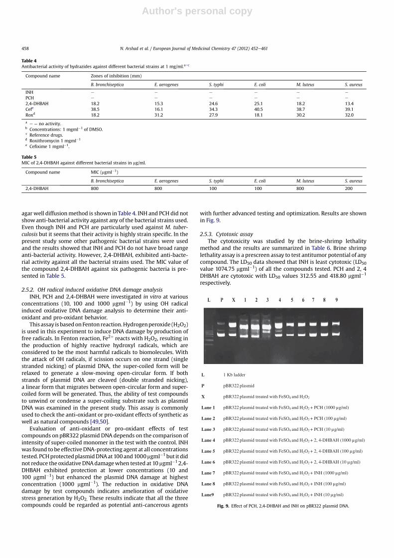
agar well diffusionmethod is shown in Table 4. INH and PCH did notshowanti-bacterial activity against any of the bacterial strains used.Even though INH and PCH are particularly used against M. tuber-culosis but it seems that their activity is highly strain specific. In thepresent study some other pathogenic bacterial strains were usedand the results showed that INH and PCH do not have broad rangeanti-bacterial activity. However, 2,4-DHBAH, exhibited anti-bacte-rial activity against all the bacterial strains used. The MIC value ofthe compound 2,4-DHBAH against six pathogenic bacteria is pre-sented in Table 5.
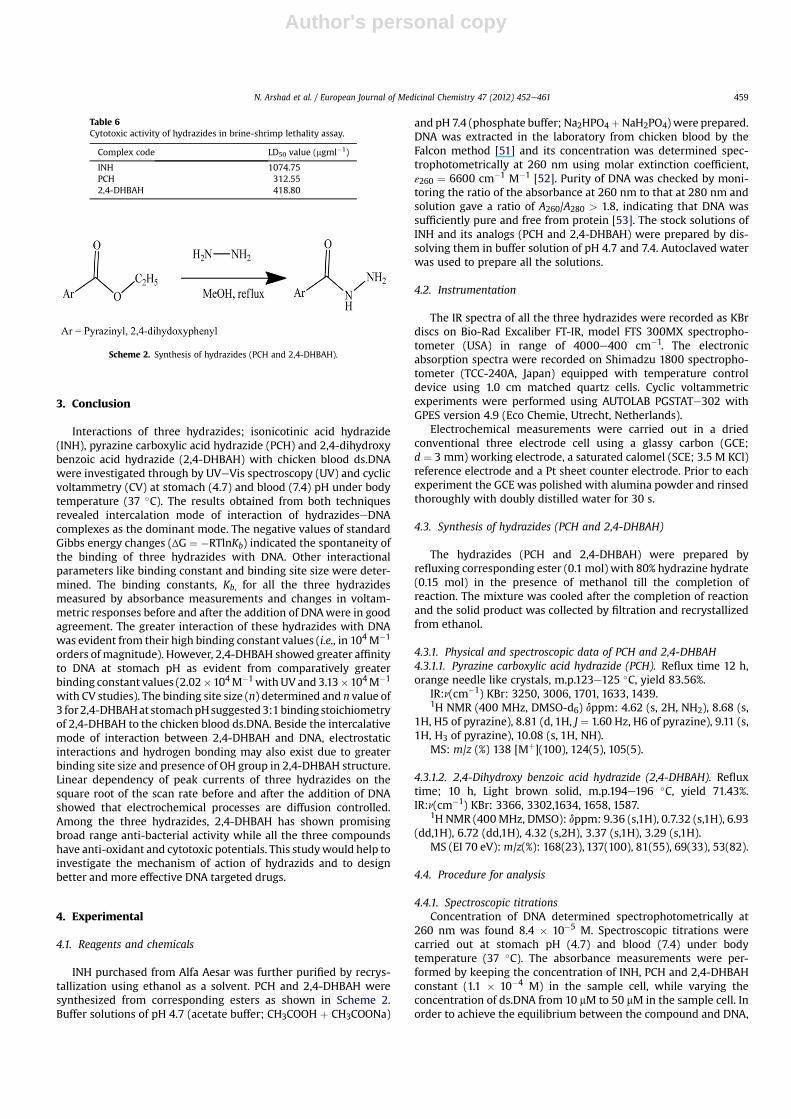
2.5.2. OH radical induced oxidative DNA damage analysisINH, PCH and 2,4-DHBAH were investigated in vitro at various
concentrations (10, 100 and 1000 mgml�1) by using OH radicalinduced oxidative DNA damage analysis to determine their anti-oxidant and pro-oxidant behavior.
This assay is basedonFenton reaction.Hydrogenperoxide (H2O2)is used in this experiment to induce DNA damage by production offree radicals. In Fenton reaction, Fe2þ reacts with H2O2, resulting inthe production of highly reactive hydroxyl radicals, which areconsidered to be the most harmful radicals to biomolecules. Withthe attack of OH radicals, if scission occurs on one strand (singlestranded nicking) of plasmid DNA, the super-coiled form will berelaxed to generate a slow-moving open-circular form. If bothstrands of plasmid DNA are cleaved (double stranded nicking),a linear form that migrates between open-circular form and super-coiled form will be generated. Thus, the ability of test compoundsto unwind or condense a super-coiling substrate such as plasmidDNA was examined in the present study. This assay is commonlyused to check the anti-oxidant or pro-oxidant effects of synthetic aswell as natural compounds [49,50].
Evaluation of anti-oxidant or pro-oxidant effects of testcompounds on pBR322 plasmid DNA depends on the comparison ofintensity of super-coiled monomer in the test with the control. INHwas found to be effectiveDNA-protecting agent at all concentrationstested. PCHprotectedplasmidDNAat100and1000mgml�1 but it didnot reduce the oxidative DNAdamagewhen tested at 10 mgml�12,4-DHBAH exhibited protection at lower concentrations (10 and100 mgml�1) but enhanced the plasmid DNA damage at highestconcentration (1000 mgml�1). The reduction in oxidative DNAdamage by test compounds indicates amelioration of oxidativestress generation by H2O2. These results indicate that all the threecompounds could be regarded as potential anti-cancerous agents
with further advanced testing and optimization. Results are shownin Fig. 9.
2.5.3. Cytotoxic assayThe cytotoxicity was studied by the brine-shrimp lethality
method and the results are summarized in Table 6. Brine shrimplethality assay is a prescreen assay to test antitumor potential of anycompound. The LD50 data showed that INH is least cytotoxic (LD50value 1074.75 mgml�1) of all the compounds tested. PCH and 2, 4DHBAH are cytotoxic with LD50 values 312.55 and 418.80 mgml�1
respectively.
Table 4Antibacterial activity of hydrazides against different bacterial strains at 1 mg/ml.aec
Compound name Zones of inhibition (mm)
B. bronchiseptica E. aerogenes S. typhi E. coli M. luteus S. aureus
INH e e e e e e
PCH e e e e e e
2,4-DHBAH 18.2 15.3 24.6 25.1 18.2 13.4Cefe 38.5 16.1 34.3 40.5 38.7 39.1Roxd 18.2 31.2 27.9 18.1 30.2 32.0
a e ¼ no activity.b Concentrations: 1 mgml�1 of DMSO.c Reference drugs.d Roxithromycin 1 mgml�1
e Cefixime 1 mgml�1.
Table 5MIC of 2,4-DHBAH against different bacterial strains in mg/ml.
Compound name MIC (mgml�1)
B. bronchiseptica E. aerogenes S. typhi E. coli M. luteus S. aureus
2,4-DHBAH 800 800 100 100 800 200
L 1 Kb ladder
P pBR322 plasmid
X pBR322 plasmid treated with FeSO4 and H2O2
Lane 1 pBR322 plasmid treated with FeSO4 and H2O2 + PCH (1000 µg/ml)
Lane 2 pBR322 plasmid treated with FeSO4 and H2O2 + PCH (100 µg/ml)
Lane 3 pBR322 plasmid treated with FeSO4 and H2O2 + PCH (10 µg/ml)
Lane 4 pBR322 plasmid treated with FeSO4 and H2O2 + 2, 4-DHBAH (1000 µg/ml)
Lane 5 pBR322 plasmid treated with FeSO4 and H2O2 + 2, 4-DHBAH (100 µg/ml)
Lane 6 pBR322 plasmid treated with FeSO4 and H2O2 + 2, 4-DHBAH (10 µg/ml)
Lane 7 pBR322 plasmid treated with FeSO4 and H2O2 + INH (1000 µg/ml)
Lane 8 pBR322 plasmid treated with FeSO4 and H2O2 + INH (100 µg/ml)
Lane9 pBR322 plasmid treated with FeSO4 and H2O2 + INH (10 µg/ml)
Fig. 9. Effect of PCH, 2,4-DHBAH and INH on pBR322 plasmid DNA.
N. Arshad et al. / European Journal of Medicinal Chemistry 47 (2012) 452e461458
Author's personal copy
3. Conclusion
Interactions of three hydrazides; isonicotinic acid hydrazide(INH), pyrazine carboxylic acid hydrazide (PCH) and 2,4-dihydroxybenzoic acid hydrazide (2,4-DHBAH) with chicken blood ds.DNAwere investigated through by UVeVis spectroscopy (UV) and cyclicvoltammetry (CV) at stomach (4.7) and blood (7.4) pH under bodytemperature (37 �C). The results obtained from both techniquesrevealed intercalation mode of interaction of hydrazideseDNAcomplexes as the dominant mode. The negative values of standardGibbs energy changes (ΔG ¼ �RTlnKb) indicated the spontaneity ofthe binding of three hydrazides with DNA. Other interactionalparameters like binding constant and binding site size were deter-mined. The binding constants, Kb, for all the three hydrazidesmeasured by absorbance measurements and changes in voltam-metric responses before and after the addition of DNAwere in goodagreement. The greater interaction of these hydrazides with DNAwas evident from their high binding constant values (i.e., in 104M�1
orders of magnitude). However, 2,4-DHBAH showed greater affinityto DNA at stomach pH as evident from comparatively greaterbinding constant values (2.02�104M�1withUV and 3.13�104M�1
with CV studies). The binding site size (n) determined and n value of3 for2,4-DHBAHat stomachpHsuggested3:1binding stoichiometryof 2,4-DHBAH to the chicken blood ds.DNA. Beside the intercalativemode of interaction between 2,4-DHBAH and DNA, electrostaticinteractions and hydrogen bonding may also exist due to greaterbinding site size and presence of OH group in 2,4-DHBAH structure.Linear dependency of peak currents of three hydrazides on thesquare root of the scan rate before and after the addition of DNAshowed that electrochemical processes are diffusion controlled.Among the three hydrazides, 2,4-DHBAH has shown promisingbroad range anti-bacterial activity while all the three compoundshave anti-oxidant and cytotoxic potentials. This studywould help toinvestigate the mechanism of action of hydrazids and to designbetter and more effective DNA targeted drugs.
4. Experimental
4.1. Reagents and chemicals
INH purchased from Alfa Aesar was further purified by recrys-tallization using ethanol as a solvent. PCH and 2,4-DHBAH weresynthesized from corresponding esters as shown in Scheme 2.Buffer solutions of pH 4.7 (acetate buffer; CH3COOH þ CH3COONa)
and pH 7.4 (phosphate buffer; Na2HPO4þNaH2PO4) were prepared.DNA was extracted in the laboratory from chicken blood by theFalcon method [51] and its concentration was determined spec-trophotometrically at 260 nm using molar extinction coefficient,ε260 ¼ 6600 cm�1 M�1 [52]. Purity of DNA was checked by moni-toring the ratio of the absorbance at 260 nm to that at 280 nm andsolution gave a ratio of A260/A280 > 1.8, indicating that DNA wassufficiently pure and free from protein [53]. The stock solutions ofINH and its analogs (PCH and 2,4-DHBAH) were prepared by dis-solving them in buffer solution of pH 4.7 and 7.4. Autoclaved waterwas used to prepare all the solutions.
4.2. Instrumentation
The IR spectra of all the three hydrazides were recorded as KBrdiscs on Bio-Rad Excaliber FT-IR, model FTS 300MX spectropho-tometer (USA) in range of 4000e400 cm�1. The electronicabsorption spectra were recorded on Shimadzu 1800 spectropho-tometer (TCC-240A, Japan) equipped with temperature controldevice using 1.0 cm matched quartz cells. Cyclic voltammetricexperiments were performed using AUTOLAB PGSTATe302 withGPES version 4.9 (Eco Chemie, Utrecht, Netherlands).
Electrochemical measurements were carried out in a driedconventional three electrode cell using a glassy carbon (GCE;d ¼ 3 mm) working electrode, a saturated calomel (SCE; 3.5 M KCl)reference electrode and a Pt sheet counter electrode. Prior to eachexperiment the GCE was polished with alumina powder and rinsedthoroughly with doubly distilled water for 30 s.
4.3. Synthesis of hydrazides (PCH and 2,4-DHBAH)
The hydrazides (PCH and 2,4-DHBAH) were prepared byrefluxing corresponding ester (0.1 mol) with 80% hydrazine hydrate(0.15 mol) in the presence of methanol till the completion ofreaction. The mixture was cooled after the completion of reactionand the solid product was collected by filtration and recrystallizedfrom ethanol.
4.3.1. Physical and spectroscopic data of PCH and 2,4-DHBAH4.3.1.1. Pyrazine carboxylic acid hydrazide (PCH). Reflux time 12 h,orange needle like crystals, m.p.123e125 �C, yield 83.56%.
IR:n(cm�1) KBr: 3250, 3006, 1701, 1633, 1439.1H NMR (400 MHz, DMSO-d6) dppm: 4.62 (s, 2H, NH2), 8.68 (s,
1H, H5 of pyrazine), 8.81 (d, 1H, J ¼ 1.60 Hz, H6 of pyrazine), 9.11 (s,1H, H3 of pyrazine), 10.08 (s, 1H, NH).
MS: m/z (%) 138 [Mþ](100), 124(5), 105(5).
4.3.1.2. 2,4-Dihydroxy benzoic acid hydrazide (2,4-DHBAH). Refluxtime; 10 h, Light brown solid, m.p.194e196 �C, yield 71.43%.IR:n(cm�1) KBr: 3366, 3302,1634, 1658, 1587.
1H NMR (400MHz, DMSO): dppm: 9.36 (s,1H), 0.7.32 (s,1H), 6.93(dd,1H), 6.72 (dd,1H), 4.32 (s,2H), 3.37 (s,1H), 3.29 (s,1H).
MS (EI 70 eV):m/z(%): 168(23), 137(100), 81(55), 69(33), 53(82).
4.4. Procedure for analysis
4.4.1. Spectroscopic titrationsConcentration of DNA determined spectrophotometrically at
260 nm was found 8.4 � 10�5 M. Spectroscopic titrations werecarried out at stomach pH (4.7) and blood (7.4) under bodytemperature (37 �C). The absorbance measurements were per-formed by keeping the concentration of INH, PCH and 2,4-DHBAHconstant (1.1 � 10�4 M) in the sample cell, while varying theconcentration of ds.DNA from 10 mM to 50 mM in the sample cell. Inorder to achieve the equilibrium between the compound and DNA,
Table 6Cytotoxic activity of hydrazides in brine-shrimp lethality assay.
Complex code LD50 value (mgml�1)
INH 1074.75PCH 312.552,4-DHBAH 418.80
Scheme 2. Synthesis of hydrazides (PCH and 2,4-DHBAH).
N. Arshad et al. / European Journal of Medicinal Chemistry 47 (2012) 452e461 459
Author's personal copy
solutions were allowed to stay for at least 5 min before eachmeasurements were made. After placing the sample solutionswithin the cell cavity and before running the spectra, wait for a fewseconds till required temperature was attained and shown ontemperature controlled device.
4.4.2. Voltammetric assayFirst a blank CVwas runwith the buffer solutions (4.7 and 7.4) at
37 �C, which showed no electroactivity in the potential range of ourinterest (�2 V toþ2 V). Cyclic voltammograms of INH, PCH and 2,4-DHBAH (1 mM) were recorded from �1 V to þ1.5 V vs. SCE beforeand after the addition of different volumes (ml) of the stock DNAsolution corresponding to the final concentration of DNA rangingfrom 10 mM to 50 mM within the cell. Scan rate of 100 mVs�1 wasused throughout the experiments. All measurements were made at37 �C after purging the solution in the cell with argon gas (99.999%)for at least 10e15 min for flushing out oxygen before every elec-trochemical assay.
4.4.3. Antibacterial assayAntimicrobial activity of INH, PCH and 2,4-DHBAH was inves-
tigated by using agar well diffusion method [54]. Six bacterialstrains; 2 g positive Staphylococcus aureus (ATCC 6538), Micro-coccus luteus (ATCC 10240) and 4 g negative Enterobacter aerogenes(ATCC 13048), Escherichia coli (ATCC 15224), Salmonella typhi(ATCC 6539), Bordetella bronchiseptica (ATCC 10580) cultured in LBat 37 �C for 24 h were used. Briefly, the broth culture of each teststrain (0.75 ml) was mixed in 75 ml of nutrient agar mediumseparately and poured into a 14 cm sterile petriplate. After solid-ification of medium, sterile metallic borer was used to dig 8 mmwells. One hundred micro liters of each test sample (1 mgml�1 inDMSO) was poured in its respective well. In each plate DMSO andstandard anti-bacterial drugs Roxyithromycin and Cefixime(1 mgml�1) served as negative and positive controls respectively.The plates were incubated at 37 �C for 24 h. The anti-bacterialactivity was calculated by measuring the diameter of zones ofinhibition around each well. The minimum inhibitory concentra-tion (MIC) of active compound was also determined by testingactivity of the serial dilutions.
4.4.4. OH radical induced oxidative DNA damage assayTo determine the anti-oxidant (protective) or pro-oxidant
(damaging) activity of test compounds (INH, PCH and 2,4-DHBAH), OH radical induced oxidative DNA damage assay wasperformed [55]. Plasmid pBR322 DNA was diluted using 50 mMphosphate buffer to get a concentration of 0.5 mg/3 ml. Each reactionmixture (15 ml) contained 3 ml of diluted pBR322, 5 ml of testcompounds at various concentrations (1000, 100 and 10 mgml�1),4 ml of 2 mM FeSO4 and 3 ml of 30% H2O2. For each experiment,pBR322 DNA treated with 30% H2O2 and 2 mM FeSO4 was used aspositive control (X). Hydrogen peroxide (H2O2) is used because itcan induce DNA damage by producing free radicals and FeSO4 actsas a catalyst in this experiment. Untreated pBR322 DNAwas used asnegative control (P) and a 1 kb DNA ladder (L) was used as marker.Reaction mixtures were incubated at 37 �C for 1 h in dark. Allreaction mixtures were subjected to 1% agarose gel electrophoresisin 1X TBE buffer. Ethidium bromide was used to stain DNA bands(super-coiled, linear and open-circular). Gels were visualized underUV, documented (Gel Doc, Bio Rad) and intensity of the bands wasdetermined.
4.4.5. Cytotoxic assayThe cytotoxity was studied by the brine-shrimp lethality assay
method [54]. Brine shrimps (Artemia salina) were hatched usingbrine shrimp eggs in sterile artificial sea water (prepared using sea
salt 38 gl�1) under constant aeration for 48 h at room temperature.After hatching, active shrimps free from eggs were collected frombrighter portion of the hatching chamber and used for the assay.Ten shrimps were drawn through a glass capillary and placed ineach vial containing 5 ml of artificial sea water with 10, 100 and1000 mgml�1
final concentration of each test compound from theirstock solutions. After 24 h, the number of surviving shrimp wascounted (13e15). Experiment was performed in triplicate. Datawasanalyzed with Finney computer program to determine LD50 (LethalDose that killed 50% of shrimps) values.
Acknowledgments
The authors are highly grateful to Ms. Asima Siddiqa (NationalCenter for Physics) for technical help in DNA extraction process andHigher Education Commission (HEC) Islamabad Pakistan forfinancial support.
References
[1] N. Brooijmans, K.A. Sharp, I.D. Kuntz, Proteins 48 (2002) 645e653.[2] T.L. Blundell, Nature 384 (1996) 23e36.[3] J.B. Chaires, Curr. Opin. Struct. Biol. 8 (1998) 314e320.[4] M. Maiti, G.S. Kumar, J. Nucleic Acids 2010 (2010) 23. doi:10.4061/2010/
593408 Article ID 593408.[5] M.J. Waring, Annu. Rev. Biochem. 50 (1981) 159e192.[6] L.H. Hurley, Biochem. Soc. Trans. 29 (2001) 692e696.[7] L. Jia, Inorg. Chim. Acta 363 (2010) 855e865.[8] J.H. Griffin, P.B. Dervan, J. Am.Chem. Soc. 109 (1987) 6840e6842.[9] J.K. Barton, Science 233 (1986) 727e734.
[10] E. Palecek, M. Fojta, Anal.Chem. 73 (2001) 74Ae83A.[11] S. Takenaka, Y. Uto, H. Kondo, T. Ihara, M. Takagi, Anal. Biochem. 218 (1994)
436e443.[12] X. Chu, G.L. Shen, J.H. Jiang, T.F. Kang, B. Xiong, R.Q. Yu, Anal. Chem. Acta 373
(1998) 29e38.[13] M. Aslanoglu, Acta Chem. Slov. 51 (2004) 107e116.[14] C. Zhoa, J.J. Zhu, J.J. Zhang, H.Y. Chen, Anal. Chem. Acta 394 (1999) 337e344.[15] M. Aslanoglu, Anal. Sci. 22 (2006) 439e443.[16] M. Aslanoglu, Turk. J.Chem. 29 (2005) 477e485.[17] P.G. Baraldi, Med. Res. Rev. 24 (2004) 475e528.[18] R.R. Monaco, Article ID 702317, J. Nucleic Acids 2010 4, doi:10.4061/2010/
702317.[19] N. Li, L. Ma, C. Yang, L. Guo, X. Yang, Biophys. Chem. 116 (2005) 199e205.[20] M.S. Shahabuddin, M. Gopal, S.C. Raghavan, J. Cancer Mol. 3 (2007) 139e146.[21] L.M. Wilhelmsson, N. Kingi, J. Bergman, J. Med. Chem. 51 (2008) 7744e7750.[22] S. Usha, I.M. Johnson, R. Malathi, Mol. Cell Biochem. 284 (2006) 57e64.[23] A. Ozdemir, G.T. Zitouni, Z.A. Kaplancikli, Y. Tunali, J. Enzym. Inhib. Med.
Chem. 24 (2009) 825e831.[24] H.M.A. Rahman, M.A. Hussein, Arch. Pharm. (Weinheim) 339 (2006) 378e387.[25] R.A. Mekheimer, A.M.A. Hameed, K.U. Sadek, ARKIVOC xvi (2008) 144e153.[26] S. Toliwal, K. Jadav, K. Patel, PMCID, Indian J. Pharm. Sci. 71 (2009) 144e148.[27] J.H. Choi, Chest 137 (2010) 393e400.[28] A.M. Pyle, J.P. Rehmann, R. Meshoyrer, C.V. Kumar, N.J. Turro, J.K. Barton, J. Am.
Chem. Soc. 111 (1989) 3051e3058.[29] Y. Sun, S. Bi, D. Song, C. Qiao, D. Mu, H. Zhang, Sensors and Actuators B 129
(2008) 799e810.[30] A. Tarushi, C.P. Raptopoulou, V. Psycharis, A. Terzis, G. Psomas,
D.P. Kessissoglou, Bioorg. Med. Chem. 18 (2010) 2678e2685.[31] G. Pratviel, J. Bernadou, B. Meunier, Adv. Inorg. Chem. 45 (1998) 251e312.[32] A. Shah, R. Qureshi, N.K. Janjua, S. Haque, S. Ahmad, Anal. Sci. 24 (2008)
1437e1441.[33] S. Bollo, L.J.N. Vergara, J.A. Squella, J. Electroanal. Chem. 562 (2004) 9e14.[34] P.C. Mandal, J. Electroanal. Chem. 570 (2004) 55e61.[35] X. Lu, M. Zhang, J. Kang, X. Wang, L. Zhuo, H. Liu, J. Inorg. Biochem. 98 (2004)
582e588.[36] J.E.B. Randles, Trans. Faraday Soc. 44 (1948) 322e327.[37] A. Sevcik, Collec. Czech. Chem. Commun. 13 (1948) 349e377.[38] S. Wang, T. Penz, C.F. Yang, Biophys. Chem. 104 (2003) 239e248.[39] G.J. Yang, J.J. Xu, H.Y. Chen, Z.H. Leng, Chin. J. Chem. 22 (2004) 1325e1329.[40] M.S. Ibrahim, Anal. Chem. Acta 443 (2001) 63e72.[41] F. Wang, Y. Xu, J. Zhao, H. Shengshui, J. Bioelectrochem. 70 (2007) 356e362.[42] H. Berg, G. Horn, U. Luthardt, Bioelectrochem. Bioenergy 8 (1981) 537e553.[43] J. Niu, G. Cheng, S. Dong, Electrochim. Acta 39 (1994) 2455e2460.[44] M.T. Carter, M. Rodriguez, A.J. Bard, J. Am. Chem. Soc. 111 (1989) 8901e8911.[45] M. Aslanoglu, C.J. Isaac, A. Houlton, B.R. Horrocks, Analyst 125 (2000)
1791e1798.[46] A. Shah, R. Qureshi, A.M. Khan, R.A. Khera, F.L. Ansari, J. Braz. Chem. Soc. 21(
(2010) 447e451.[47] Z. Xu, G. Bai, C. Dong, Bioorg. Med. Chem. 13 (2005) 5694e5699.
N. Arshad et al. / European Journal of Medicinal Chemistry 47 (2012) 452e461460

Author's personal copy
[48] S. Sato, H. Kondo, T. Nojima, S. Takenaka, Anal.Chem. 77 (2005) 7304e7309.[49] M. Zaheer, A. Shah, Z. Akhter, R. Qureshi, B. Mirza, M. Tauseef, M. Bolte, Appl.
Organomet. Chem. 25 (2011) 61e69.[50] G. Bibi, I. Haq, N. Ullah, A. Mannan, B. Mirza, Afr. J. Pharm. Pharmacol. 5 (2011)
252e258.[51] J. Sambrook, E.F. Fritsch, T. Maniatis, Molecular Cloning: A Laboratory Manual
Cold Spring Harbor, New York (1989).
[52] M.E. Reichmann, S.A. Rice, C.A. Thomas, P. Doty, J. Am. Chem. Soc. 76 (1954)3047e3053.
[53] S.S. Babkina, N.A. Ulakhovich, Anal. Chem. 77 (2005) 5678e5685.[54] M.S. Ahmad, M. Hussain, M. Hanif, S. Ali, M. Qayyum, B. Mirza, Chem. Bio. Drug
Des. 71 (2008) 568e576.[55] H. Nawaz, Z. Akhter, S. Yameen, H.M. Siddiqi, B. Mirza, A. Rifat, J. Organomet.
Chem. 694 (2009) 2198e2203.
N. Arshad et al. / European Journal of Medicinal Chemistry 47 (2012) 452e461 461