Egg beater as centrifuge: isolating human blood plasma from whole blood in resource-poor settings
6
Egg beater as centrifuge: isolating human blood plasma from whole blood in resource-poor settings† Amy P. Wong, Malancha Gupta, Sergey S. Shevkoplyas and George M. Whitesides * Received 10th June 2008, Accepted 8th August 2008 First published as an Advance Article on the web 14th October 2008 DOI: 10.1039/b809830c This paper demonstrates that a hand-powered egg beater can be modified to serve as a centrifuge for separating plasma from human whole blood. Immunoassays used to diagnose infectious diseases often require plasma from whole blood, and obtaining plasma typically requires electrically-powered centrifuges, which are not widely available in resource-limited settings. Human whole blood was loaded into polyethylene (PE) tubing, and the tubing was attached to the paddle of an egg beater. Spinning the paddle pelleted the blood cells to the distal end of the PE tubing; the plasma remained as the supernatant. A cholesterol assay (run on patterned paper) demonstrated the suitability of this plasma for use in diagnostic assays. The physics of the system was also analyzed as a guide for the selection of other rotating systems for use in centrifugation. Egg beaters, polyethylene tubing, and paper are readily available devices and supplies that can facilitate the use of point-of-care diagnostics at sites far from centralized laboratory facilities. Introduction This paper describes a method for separating blood plasma from human whole blood in polyethylene (PE) tubing using a hand- powered centrifuge (an egg beater). We isolated human plasma using an egg beater by attaching PE tubing containing blood to the paddle of an egg beater, and spinning its handle manually to pellet the blood cells. The plasma remained as the supernatant. The use of the plasma in a diagnostic assay for total cholesterol run on patterned paper demonstrated the practicality of this method. The use of the egg beater as a centrifuge requires little training, and the egg beater is widely available, inexpensive, robust, and completely hand-powered. Diagnostic tools used to detect concentrations of analytes in plasma isolated from whole blood often require expensive, electrically-powered centrifuges. This hand-powered centrifuge offers an alternative that may sometimes be more practical in the resource-poor settings of developing countries. In developing countries, infectious diseases are responsible for more than 50% of deaths. 1,2 Prompt and accurate diagnosis of infectious diseases would lead to more effective treatment, and lower rates of mortality, and would also ultimately reduce the expense of treating patients and the economic burden of the illness on society. 3 In the innovative developing countries (IDCs), 1 it is often difficult to reach populations that are distributed sparsely over rural areas. Health workers dispatched from centralized hospitals or laboratory facilities to serve these areas face chal- lenges such as difficult terrain, intermittent (or lack of) electricity, poorly-equipped facilities, an unskilled workforce, 4 and limited financial resources. The consequences of these limitations are that diagnostic medical technologies common in industrialized regions are either not usable or not affordable, and that the time required to reach the patient, transport samples to centralized laboratory facilities, and report a diagnosis to the patient can take days to months. Diagnosis of patients in the field enables prompt treat- ment (Fig. 1); point-of-care diagnosis relies on simple, inexpensive equipment that is available locally. Medical diagnostics—especially colorimetric assays and many immunological assays—rely upon obtaining clear bodily fluids such as blood plasma to diagnose diseases. Plasma is the liquid component of whole blood; it is usually obtained from whole blood by centrifugation with electrically-powered, bench-top centrifuges at speeds that generate approximately 1000 g for 15 minutes 5,6 This technique sediments blood cells, which interfere with assays because they scatter light, 7 aggregate, and lyse; lysis of cells releases intracellular components that can contaminate the sample of plasma. Standard centrifuges are impractical for use in severely resource-limited environments: 8 they require a source of electrical power, and are bulky, difficult to repair, and expensive (>$400). Locally available resources such as blenders and record players have been modified for scientific and biomedical use in laboratories, but still require electricity. 9,10 We believe that the hand-powered centrifuge—in combination with paper-based devices for running diagnostic assays 11 —provides a useful capa- bility for diagnostic analyses in regions with limited resources. Experimental Equipment and materials Polyethylene (PE) tubing (inner diameter: 1.57 mm, outer diameter: 2.08 mm) was purchased from VWR (Bridgeport, NJ, USA). Whole human blood (with ethylene diamine tetraacetic acid; EDTA) was obtained from Rockland Immunochemicals (Gilbertsville, PA, USA). Photoresist (SU-8 2010) to pattern the chromatography paper (Whatman No.1; VWR) was purchased Department of Chemistry and Chemical Biology, Harvard University, Cambridge, MA, 02138. E-mail: [email protected]; Fax: +617-495-9857; Tel: +617-495-9430 † Part of a special issue on Point-of-care Microfluidic Diagnostics; Guest Editors—Professor Kricka and Professor Sia. 2032 | Lab Chip, 2008, 8, 2032–2037 This journal is ª The Royal Society of Chemistry 2008 PAPER www.rsc.org/loc | Lab on a Chip
-
Upload
independent -
Category
Documents
-
view
0 -
download
0
Transcript of Egg beater as centrifuge: isolating human blood plasma from whole blood in resource-poor settings
PAPER www.rsc.org/loc | Lab on a Chip
Egg beater as centrifuge: isolating human blood plasma from whole bloodin resource-poor settings†
Amy P. Wong, Malancha Gupta, Sergey S. Shevkoplyas and George M. Whitesides*
Received 10th June 2008, Accepted 8th August 2008
First published as an Advance Article on the web 14th October 2008
DOI: 10.1039/b809830c
This paper demonstrates that a hand-powered egg beater can be modified to serve as a centrifuge for
separating plasma from human whole blood. Immunoassays used to diagnose infectious diseases often
require plasma from whole blood, and obtaining plasma typically requires electrically-powered
centrifuges, which are not widely available in resource-limited settings. Human whole blood was loaded
into polyethylene (PE) tubing, and the tubing was attached to the paddle of an egg beater. Spinning the
paddle pelleted the blood cells to the distal end of the PE tubing; the plasma remained as the
supernatant. A cholesterol assay (run on patterned paper) demonstrated the suitability of this plasma
for use in diagnostic assays. The physics of the system was also analyzed as a guide for the selection of
other rotating systems for use in centrifugation. Egg beaters, polyethylene tubing, and paper are readily
available devices and supplies that can facilitate the use of point-of-care diagnostics at sites far from
centralized laboratory facilities.
Introduction
This paper describes a method for separating blood plasma from
human whole blood in polyethylene (PE) tubing using a hand-
powered centrifuge (an egg beater). We isolated human plasma
using an egg beater by attaching PE tubing containing blood to
the paddle of an egg beater, and spinning its handle manually to
pellet the blood cells. The plasma remained as the supernatant.
The use of the plasma in a diagnostic assay for total cholesterol
run on patterned paper demonstrated the practicality of this
method. The use of the egg beater as a centrifuge requires little
training, and the egg beater is widely available, inexpensive,
robust, and completely hand-powered. Diagnostic tools used to
detect concentrations of analytes in plasma isolated from whole
blood often require expensive, electrically-powered centrifuges.
This hand-powered centrifuge offers an alternative that may
sometimes be more practical in the resource-poor settings of
developing countries.
In developing countries, infectious diseases are responsible for
more than 50% of deaths.1,2 Prompt and accurate diagnosis of
infectious diseases would lead to more effective treatment, and
lower rates of mortality, and would also ultimately reduce the
expense of treating patients and the economic burden of the illness
on society.3 In the innovative developing countries (IDCs),1 it is
often difficult to reach populations that are distributed sparsely
over rural areas. Health workers dispatched from centralized
hospitals or laboratory facilities to serve these areas face chal-
lenges such as difficult terrain, intermittent (or lack of) electricity,
poorly-equipped facilities, an unskilled workforce,4 and limited
financial resources. The consequences of these limitations are that
Department of Chemistry and Chemical Biology, Harvard University,Cambridge, MA, 02138. E-mail: [email protected];Fax: +617-495-9857; Tel: +617-495-9430
† Part of a special issue on Point-of-care Microfluidic Diagnostics; GuestEditors—Professor Kricka and Professor Sia.
2032 | Lab Chip, 2008, 8, 2032–2037
diagnostic medical technologies common in industrialized regions
are either not usable or not affordable, and that the time required
to reach the patient, transport samples to centralized laboratory
facilities, and report a diagnosis to the patient can take days to
months. Diagnosis of patients in the field enables prompt treat-
ment (Fig. 1); point-of-care diagnosis relies on simple, inexpensive
equipment that is available locally.
Medical diagnostics—especially colorimetric assays and many
immunological assays—rely upon obtaining clear bodily fluids
such as blood plasma to diagnose diseases. Plasma is the liquid
component of whole blood; it is usually obtained from whole
blood by centrifugation with electrically-powered, bench-top
centrifuges at speeds that generate approximately 1000 � g for 15
minutes5,6 This technique sediments blood cells, which interfere
with assays because they scatter light,7 aggregate, and lyse; lysis of
cells releases intracellular components that can contaminate the
sample of plasma. Standard centrifuges are impractical for use in
severely resource-limited environments:8 they require a source of
electrical power, and are bulky, difficult to repair, and expensive
(>$400). Locally available resources such as blenders and record
players have been modified for scientific and biomedical use in
laboratories, but still require electricity.9,10 We believe that the
hand-powered centrifuge—in combination with paper-based
devices for running diagnostic assays11—provides a useful capa-
bility for diagnostic analyses in regions with limited resources.
Experimental
Equipment and materials
Polyethylene (PE) tubing (inner diameter: 1.57 mm, outer
diameter: 2.08 mm) was purchased from VWR (Bridgeport, NJ,
USA). Whole human blood (with ethylene diamine tetraacetic
acid; EDTA) was obtained from Rockland Immunochemicals
(Gilbertsville, PA, USA). Photoresist (SU-8 2010) to pattern the
chromatography paper (Whatman No.1; VWR) was purchased
This journal is ª The Royal Society of Chemistry 2008
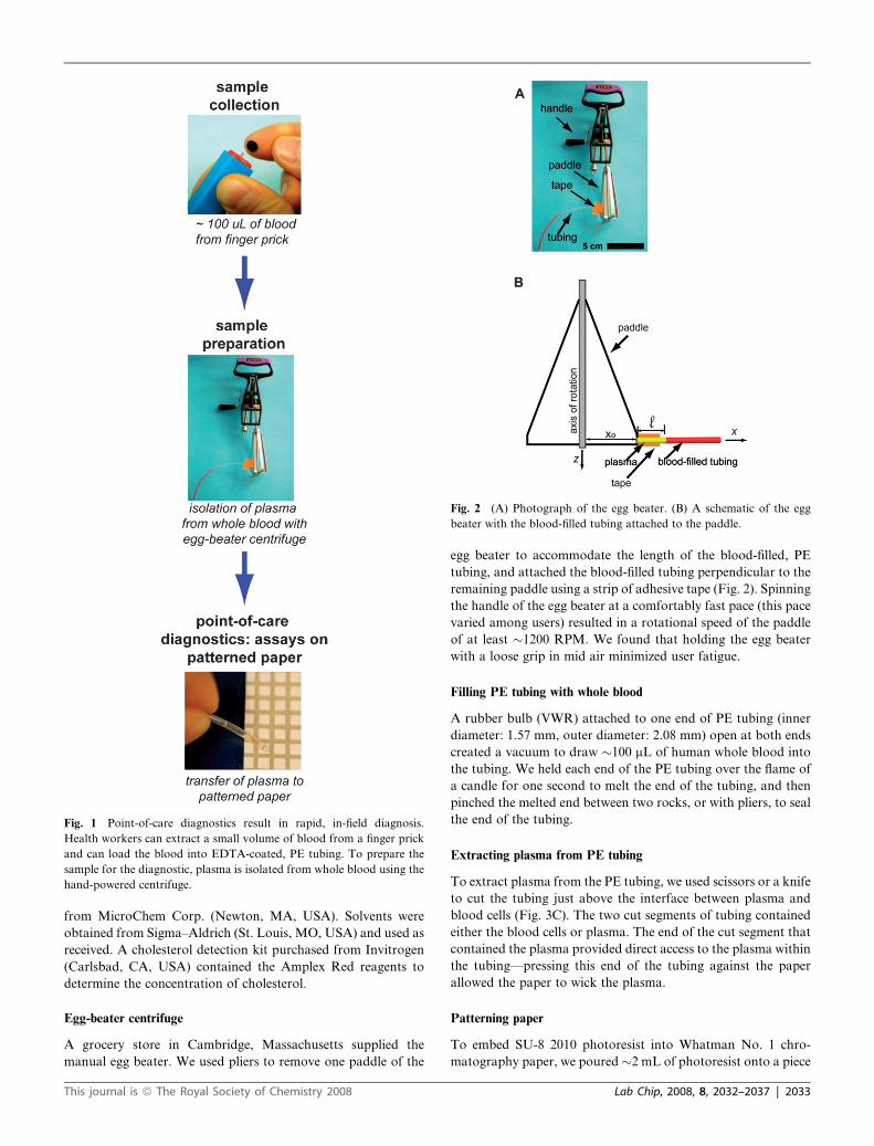
Fig. 1 Point-of-care diagnostics result in rapid, in-field diagnosis.
Health workers can extract a small volume of blood from a finger prick
and can load the blood into EDTA-coated, PE tubing. To prepare the
sample for the diagnostic, plasma is isolated from whole blood using the
hand-powered centrifuge.
Fig. 2 (A) Photograph of the egg beater. (B) A schematic of the egg
beater with the blood-filled tubing attached to the paddle.
from MicroChem Corp. (Newton, MA, USA). Solvents were
obtained from Sigma–Aldrich (St. Louis, MO, USA) and used as
received. A cholesterol detection kit purchased from Invitrogen
(Carlsbad, CA, USA) contained the Amplex Red reagents to
determine the concentration of cholesterol.
Egg-beater centrifuge
A grocery store in Cambridge, Massachusetts supplied the
manual egg beater. We used pliers to remove one paddle of the
This journal is ª The Royal Society of Chemistry 2008
egg beater to accommodate the length of the blood-filled, PE
tubing, and attached the blood-filled tubing perpendicular to the
remaining paddle using a strip of adhesive tape (Fig. 2). Spinning
the handle of the egg beater at a comfortably fast pace (this pace
varied among users) resulted in a rotational speed of the paddle
of at least �1200 RPM. We found that holding the egg beater
with a loose grip in mid air minimized user fatigue.
Filling PE tubing with whole blood
A rubber bulb (VWR) attached to one end of PE tubing (inner
diameter: 1.57 mm, outer diameter: 2.08 mm) open at both ends
created a vacuum to draw �100 mL of human whole blood into
the tubing. We held each end of the PE tubing over the flame of
a candle for one second to melt the end of the tubing, and then
pinched the melted end between two rocks, or with pliers, to seal
the end of the tubing.
Extracting plasma from PE tubing
To extract plasma from the PE tubing, we used scissors or a knife
to cut the tubing just above the interface between plasma and
blood cells (Fig. 3C). The two cut segments of tubing contained
either the blood cells or plasma. The end of the cut segment that
contained the plasma provided direct access to the plasma within
the tubing—pressing this end of the tubing against the paper
allowed the paper to wick the plasma.
Patterning paper
To embed SU-8 2010 photoresist into Whatman No. 1 chro-
matography paper, we poured �2 mL of photoresist onto a piece
Lab Chip, 2008, 8, 2032–2037 | 2033
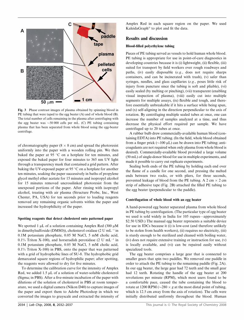
Fig. 3 Phase contrast images of plasma obtained by spinning blood in
PE tubing that were taped to the egg beater (A) and of whole blood (B).
The total number of cells remaining in the plasma after centrifuging with
the egg beater was �50 000 cells per mL. (C) PE tubing containing
plasma that has been separated from whole blood using the egg-beater
centrifuge.
of chromatography paper (8 � 8 cm) and spread the photoresist
uniformly into the paper with a wooden rolling pin. We then
baked the paper at 95 �C on a hotplate for ten minutes, and
exposed the baked paper for four minutes to 365 nm UV light
through a transparency mask that contained a grid pattern. After
baking the UV-exposed paper at 95 �C on a hotplate for another
ten minutes, soaking the paper successively in baths of propylene
glycol methyl ether acetate for 15 minutes and isopropyl alcohol
for 15 minutes removed uncrosslinked photoresist from the
unexposed portions of the paper. After rinsing with isopropyl
alcohol, treating with air plasma (Structure Probe, Inc., West
Chester, PA, USA) for ten seconds prior to loading reagents
removed any remaining organic solvents within the paper and
increased the hydrophilicity of the paper.
Spotting reagents that detect cholesterol onto patterned paper
We spotted 1 mL of a solution containing Amplex Red (300 mM
in dimethylsulfoxide (DMSO)), cholesterol oxidase (2 U mL�1 in
0.1M potassium phosphate, 0.05 M NaCl, 5 mM cholic acid,
0.1% Triton X-100), and horseradish peroxidase (2 U mL�1 in
0.1M potassium phosphate, 0.05 M NaCl, 5 mM cholic acid,
0.1% Triton X-100) in PBS, onto the paper that was patterned
with a grid of hydrophobic lines of SU-8. The hydrophobic grid
demarcated square regions of hydrophilic paper; after spotting,
the reagents were allowed to dry for five minutes.
To determine the calibration curve for the intensity of Amplex
Red, we added 1.5 mL of a solution of water-soluble cholesterol
(Sigma; in PBS). After a five-minute incubation of the paper with
dilutions of the solution of cholesterol in PBS at room temper-
ature, we used a digital camera (Nikon D40) to capture images of
the paper and export them to Adobe Photoshop in which we
converted the images to grayscale and extracted the intensity of
2034 | Lab Chip, 2008, 8, 2032–2037
Amplex Red in each square region on the paper. We used
KaleidaGraphª to plot and fit the data.
Results and discussion
Blood-filled polyethylene tubing
Pieces of PE tubing served as vessels to hold human whole blood.
PE tubing is appropriate for use in point-of-care diagnostics in
developing countries because it is (i) lightweight, (ii) flexible, (iii)
suited for transport by field workers over rough roadways and
paths, (iv) easily disposable (e.g., does not require sharps
containers, and can be incinerated with trash), (v) safer than
syringes, needles, and glass capillaries (e.g., poses little risk of
injury from puncture since the tubing is soft and pliable), (vi)
easily sealed (by melting or pinching), (vii) transparent (enabling
visual inspection of plasma), (viii) easily cut into multiple
segments for multiple assays, (ix) flexible and tough, and there-
fore essentially unbreakable if it hits a surface while being spun,
and (x) self-aligning in the direction perpendicular to the axis of
rotation. By centrifuging multiple sealed tubes at once, one can
increase the number of samples analyzed at a time, and thus
decrease the physical effort required per sample. We have
centrifuged up to 20 tubes at once.
A rubber bulb drew commercially-available human blood (con-
taining EDTA) into PE tubing. (In the field, whole blood obtained
from a finger prick (�100 mL) can be drawn into PE tubing; anti-
coagulants are not required when only plasma from whole blood is
desired). Commercially-available blood provided a large volume
(50 mL) of single-donor blood for use in multiple experiments, and
made it possible to carry out replicate experiments.
Sealing both ends of the PE tubing by holding each end over
the flame of a candle for one second, and pressing the melted
ends between two rocks, or with pliers, for three seconds,
prevented leakage of blood from the PE tubing. A 2 cm � 2 cm
strip of adhesive tape (Fig. 2B) attached the filled PE tubing to
the egg beater (perpendicular to the paddle).
Centrifugation of whole blood with an egg beater
A hand-powered egg beater separated plasma from whole blood
in PE tubing by centrifugation. (The particular type of egg beater
we used is sold widely in India for 105 rupees—approximately
$2.50 USD.) The manual egg beater represents a suitable device
for use in IDCs because it (i) is low-cost (and therefore unlikely
to be stolen from health workers), (ii) requires no electricity, (iii)
is sturdy enough to be sterilized and cleaned with boiling water,
(iv) does not require extensive training or instruction for use, (v)
is locally available, and (vi) can be repaired easily without
specialized tools.
The egg beater comprises a large gear that is connected to
smaller gears that spin two paddles. We removed one paddle in
order to attach the PE tubing to the remaining paddle (Fig. 2A).
In our egg beater, the large gear had 72 teeth and the small gear
had 12 teeth. Rotating the handle of the egg beater at 200
revolutions per minute (RPM), which most users found to be
a comfortable pace, caused the tube containing the blood to
rotate at 1200 RPM (�280 � g at the most distal point of tubing,
which is 12.5 cm away from the axis of rotation). The cells were
initially distributed uniformly throughout the blood. Human
This journal is ª The Royal Society of Chemistry 2008
blood cells are more dense (1.097 g mL�1)12 than the human
blood plasma (1.023 g mL�1),13 so, after ten minutes of centri-
fugation, a pellet of cells formed at the distal end of the tubing,
and the plasma (Fig. 3C) remained as the supernatant.
Mathematical solution for the time required to sediment cells by
centrifugation
We modeled the sedimentation of human blood cells due to
centrifugation as the motion of a spherical particle suspended in
a rotating vessel. In a rotating vessel, two forces act on
a spherical particle of radius R (m) and density rp (kg m�3), that
is suspended in a medium of density rm (kg m�3), and dynamic
viscosity h (kg m�1 s�1) (Fig. 2B). These forces are gravity, ~Fg,
and the Stokes force, ~F s, which is due to the viscous drag
exerted by the suspending medium on the particle. The balance
of forces is given by eqn (1),14 where m (kg) is the mass of the
particle and ~a (m s�2) is its acceleration in an inertial frame of
reference.
m~a ¼ ~Fg + ~F s (1)
The balance of forces (eqn (1)) in the frame of reference
rotating with the vessel at an angular velocity of ~u (rad s�1) is
given by eqn 2, where~v* is the velocity, and~a* is the acceleration
of the particle in the rotating frame of reference, and ~g is the
acceleration of gravity.14,15
m~a * ¼ 4p
3
�rp � rm
�R3~g � 6phR~v * � 4p
3
�rp � rm
�R3
�2~u �~v * þ~u � ð~u �~r Þ þ d~u
dt�~r
� (2)
In this equation, the first term on the right-hand side of the
equation is the force of gravity corrected for buoyancy, and the
second term is the Stokes force. The third term in eqn (2) is a sum
of three fictitious forces (centrifugal, Coriolis, and Euler forces)
arising from our use of a non-inertial, accelerating reference
frame (the frame of reference rotating with the vessel) to describe
the motion of the suspended particle.15 In this equation, we also
accounted for the effect of these inertial forces on the suspending
medium, as we did with the buoyant force and gravity. The
component form of eqn (2) is shown by eqn (3), where we assume
that the particle is traveling with its terminal velocity (~a* ¼ 0),
and that the angular velocity of the rotating vessel, ~u, does not
change with time.
0BB@
�6phRv*x � 4p3
�rp � rm
�R3
�2uv*y � u2x
��6phRv*y � 4p
3
�rp � rm
�R3
�� 2uv*x � u2y
�4p3
�rp � rm
�R3g � 6phRv*z
1CCA ¼~0 (3)
Eqn (4) shows the solution of the X and Y components of eqn
(3) for v*x—the velocity of the particle along the X axis, coaxial
with the long axis of the rotating vessel:
v*x ¼au
2ð1 þ a2Þx� a2u
2ð1 þ a2Þy; where a ¼ 4
9h
�rp � rm
�R2u (4)
This journal is ª The Royal Society of Chemistry 2008
We can estimate the dimensionless parameter a (a ¼ 2
9
ffiffiffiffiffiffiTa
p,
where Ta is the Taylor number characterizing the importance of
inertial forces relative to viscous forces) using the actual
parameters of the system: h ¼ 4.02 � 10�3 (kg m�1 s�1),13 rp ¼1097 (kg m�3), rm ¼ 1023 (kg m�3), R¼ 3.5 � 10�6 (m),16,17 and u
¼ 126 (rad s�1) (or about 1200 RPM), as shown in eqn (5).
a ¼ 4
9h
�rp � rm
�R2uz6:4 � 10�6 (5)
Because parameter a for our system is so small (eqn (5)), we
can safely neglect the second term of eqn (4) that is proportional
to a2; eqn (4) then becomes eqn (6).
v*x ¼au
2x; where a ¼ 4
9h
�rp � rm
�R2u (6)
We can use eqn (6) to predict the amount of plasma we can
obtain by centrifuging a piece of tubing (inner diameter, d ¼ 1.57
� 10�3 (m), total length, L ¼ 0.05 (m)) filled with �100 mL of
whole blood at 1200 RPM (angular velocity, u ¼ 126 (rad s�1))
for a set period of time, t0 (s). To perform this calculation, we
integrate eqn (6) in time to find the distance, l (m), a suspended
particle—a red blood cell—would be able to travel from its initial
position, x0 (m), at the top of the tubing (closest to the axis of
rotation) towards the distal end of the tubing (eqn (7)).
l ¼ x0
�exp
�2
9h
�rp � rm
�R2u2t0
�� 1
�(7)
The volume of cleared plasma, Vplasma (m3) is then given by
eqn (8) and the fraction of cleared plasma, fcleared, is given by eqn
(9).
Vplasma ¼p
4d2x0
�exp
�2
9h
�rp � rm
�R2u2t0
�� 1
�(8)
fcleared ¼ x0
L
�exp
�2
9h
�rp � rm
�R2u2t0
�� 1
�(9)
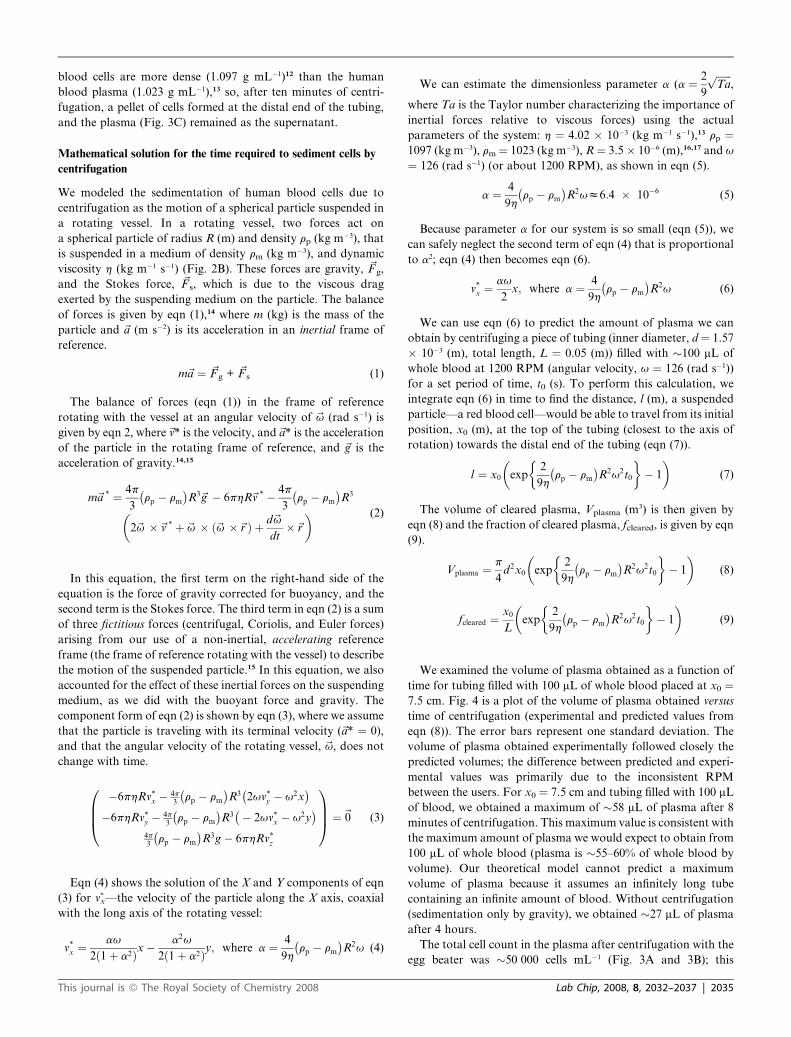
We examined the volume of plasma obtained as a function of
time for tubing filled with 100 mL of whole blood placed at x0 ¼7.5 cm. Fig. 4 is a plot of the volume of plasma obtained versus
time of centrifugation (experimental and predicted values from
eqn (8)). The error bars represent one standard deviation. The
volume of plasma obtained experimentally followed closely the
predicted volumes; the difference between predicted and experi-
mental values was primarily due to the inconsistent RPM
between the users. For x0 ¼ 7.5 cm and tubing filled with 100 mL
of blood, we obtained a maximum of �58 mL of plasma after 8
minutes of centrifugation. This maximum value is consistent with
the maximum amount of plasma we would expect to obtain from
100 mL of whole blood (plasma is �55–60% of whole blood by
volume). Our theoretical model cannot predict a maximum
volume of plasma because it assumes an infinitely long tube
containing an infinite amount of blood. Without centrifugation
(sedimentation only by gravity), we obtained �27 mL of plasma
after 4 hours.
The total cell count in the plasma after centrifugation with the
egg beater was �50 000 cells mL�1 (Fig. 3A and 3B); this
Lab Chip, 2008, 8, 2032–2037 | 2035
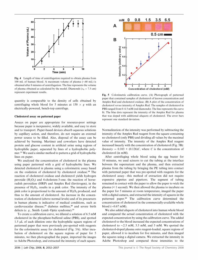
Fig. 4 Length of time of centrifugation required to obtain plasma from
100 mL of human blood. A maximum volume of plasma (�60 mL) is
obtained after 8 minutes of centrifugation. The line represents the volume
of plasma obtained as calculated by the model. Diamonds (x0 ¼ 7.5 cm)
represent experiment results.Fig. 5 Colorimetric calibration curve. (A) Photograph of patterned
paper that contained samples of cholesterol of known concentration and
Amplex Red and cholesterol oxidase. (B) A plot of the concentration of
cholesterol versus intensity of Amplex Red. The samples of cholesterol in
PBS ranged from 0–6.5 mM (red diamonds). The line represents the curve
fit. The blue dots represent the intensity of the Amplex Red for plasma
that was doped with additional aliquots of cholesterol. The error bars
represent one standard deviation.
quantity is comparable to the density of cells obtained by
centrifuging whole blood for 5 minutes at 150 � g with an
electrically-powered, bench-top centrifuge.
Cholesterol assay on patterned paper
Assays on paper are appropriate for resource-poor settings
because paper is inexpensive, widely available, and easy to store
and to transport. Paper-based devices absorb aqueous solutions
by capillary action, and therefore, do not require an external
power source to be filled. Also, disposal of the assay can be
achieved by burning. Martinez and coworkers have detected
protein and glucose content in artificial urine using regions of
hydrophilic paper, separated by lines of a hydrophobic poly-
mer.11 We used a similar method to pattern a grid of hydrophobic
lines on paper.
We analyzed the concentration of cholesterol in the plasma
using paper patterned with a grid of hydrophobic lines. We
detected cholesterol in plasma using a colorimetric assay based
on the oxidation of cholesterol by cholesterol oxidase.18 The
reaction of cholesterol oxidase and cholesterol yields hydrogen
peroxide (H2O2) and 4-cholesten-3-one; the reaction of horse-
radish peroxidase (HRP) and Amplex Red (Invitrogen), in the
presence of H2O2, results in a pink color. The intensity of the
pink color is proportional to the amount of H2O2 produced, and
thus to the amount of cholesterol. An increase in the concen-
tration of cholesterol (above normal levels) and of its precursors
in human plasma is indicative of medical conditions, such as
cardiovascular disease,19 diabetes mellitus,20 and severe birth
defects (e. g., Smith–Lemli–Optiz syndrome).21
To create a calibration curve, we diluted a solution of 6.5 mM
cholesterol in the phosphate-buffered saline (PBS), and spotted
1.5 mL of each dilution onto the hydrophilic square regions of
patterned paper that were previously pre-spotted with reagents
for the colorimetric assay for cholesterol (Fig. 5A). After incu-
bation of cholesterol on the square regions of paper for 5
minutes, we then photographed the paper, imported the images
to Adobe Photoshop, and extracted the intensity of each square.
2036 | Lab Chip, 2008, 8, 2032–2037
Normalization of the intensity was performed by subtracting the
intensity of the Amplex Red reagent from the square containing
no cholesterol (only PBS) and dividing all values by the maximal
value of intensity. The intensity of the Amplex Red reagent
increased linearly with the concentration of cholesterol (Fig. 5B):
Intensity ¼ 0.103 + (0.126)C, where C is the concentration of
cholesterol (in mM).
After centrifuging whole blood using the egg beater for
10 minutes, we used scissors to cut the tubing at the interface
between the supernatant and the plasma, and then extracted
plasma from the tubing by bringing the PE tubing into contact
with patterned paper that was pre-spotted with reagents for the
cholesterol assay—this method of extraction did not require
expensive pipettes and pipettors. The segment of tubing
remained in contact with the paper to allow the paper to wick the
plasma (< 1 second). We then allowed the plasma to incubate on
the paper for 5 minutes at room temperature, imaged the paper
with a digital camera, and extracted the intensity of squares in the
patterned paper.22 The calibration curve determined the
concentration of cholesterol in the commercially-available whole
blood (�0.67 mM).
We also added aliquots of cholesterol into human whole blood
and compared the actual concentration of cholesterol with the
expected concentration by using the calibration curve. The added
cholesterol to the blood increased the expected concentrations of
cholesterol to �2.5 mM, 3.5 mM, and 5 mM. We spotted the
cholesterol-doped plasma onto reagent-loaded, square regions of
paper, allowed it to incubate for five minutes, and then imaged
the squares using a digital camera. We extracted the intensity in
Adobe Photoshop and compared those intensities to the
This journal is ª The Royal Society of Chemistry 2008
theoretical concentration of cholesterol for a given intensity of
Amplex Red. The measured intensity of Amplex Red of the
cholesterol-doped plasma increased with the concentration of
cholesterol and corresponded to the expected concentration of
cholesterol for a given intensity (Fig. 5B).
Conclusions
This paper demonstrates the separation of plasma from whole
blood using a hand-powered centrifuge. Plasma isolated with
centrifuges that do not require electrical power enables the
detection of analytes in plasma in a field setting (away from
laboratory facilities). We used the plasma in a cholesterol assay
run on paper. The plasma can also, in principal, be used in assays
to diagnose other infectious diseases, such as Hepatitis B23 and
cysticercosis,24 which depend on plasma for rapid diagnostic tests.
Isolation of plasma typically requires the transportation of
samples of blood to central health facilities and the use of an
electrically-powered centrifuge to remove blood cells. This hand-
powered centrifuge enables health workers to isolate plasma in
the field, and to use existing rapid, diagnostic tests that depend
on plasma. This rapid diagnosis at the point of care decreases the
amount of time required to return a diagnosis to a patient and
decreases the delay between diagnosis and treatment. This hand-
powered centrifuge also reduces the burden upon health workers,
who are often limited by an inadequate workforce and insuffi-
cient medical supplies. The isolation of plasma at the point-of-
care enables health workers to travel to rural communities
without spending additional time traveling back to centralized
laboratory facilities to obtain a diagnosis. The egg beater is
a non-traditional medical supply that is widely available. Point-
of-care diagnosis also increases access to villagers who would be
otherwise unable to travel to central health facilities to receive
diagnosis and treatment. Although more point-of-care diagnos-
tics are needed in the field, we believe a tool to isolate plasma
from whole blood enables further development of diagnostics
that rely upon plasma.
Acknowledgements
This material is based on work supported by DARPA under
award No. HR0011-04-1-0032 and The California Institute of
Technology, and by NSF support of the Harvard MRSEC (NSF
#DMR-0213805).
This journal is ª The Royal Society of Chemistry 2008
References
1 C. M. Morel, T. Acharya, D. Broun, A. J. Dang, C. Elias,N. K. Ganguly, C. A. Gardner, R. K. Gupta, J. Haycock,A. D. Heher, P. J. Hotez, H. E. Kettler, G. T. Keusch,A. F. Krattiger, F. T. Kreutz, S. Lall, K. Lee, R. Mahoney,A. Martinez-Palomo, R. A. Mashelkar, S. A. Matlin, M. Mzimba,J. Oehler, R. G. Ridley, S. Pramilla, P. Singer and M. Y. Yun,Science, 2005, 309, 401–404.
2 World Health Organization, The World Health Report 2004http://www.who.int/whr/2004/en/overview_en.pdf.
3 World Health Organization, Global Plan to Combat NeglectedTropical Diseases 2008–2015, http://whqlibdoc.who.int/hq/2007/WHO_CDS_NTD_2007.3_eng.pdf.
4 United Nations Children’s Fund, The State of the World’s Children2008, http://www.unicef.org/sowc08/docs/sowc08.pdf.
5 J. Fraser Mustard, R. L. Kinlough-Rathbone and M. A. Packham, inMethods in Enzymology, Academic Press, 1989, vol. 169, pp. 3–11.
6 C. P. Y. Chan, K. W. Sum, K. Y. Cheung, J. F. C. Glatz,J. E. Sanderson, A. Hempel, M. Lehmann, I. Renneber andR. Renneberg, J. Immunol. Methods, 2003, 279, 91–100.
7 J. P. He, A. Karlsson, J. Swartling and S. Andersson-Engels, J. Opt.Soc. Am. A., 2004, 21, 1953–1961.
8 A. J. Herring, R. C. Ballard, V. Pope, R. A. Adegbola,J. Changalucha, D. W. Fitzgerald, E. W. Hook III, A. Kubanova,S. Mananwatte, J. W. Pape, A. W. Sturm, B. West, Y. P. Yin andR. W. Peeling, Sex. Transm. Infect., 2006, 82, 7–12.
9 E. Harris, EMBO Rep., 2004, 5, 7–11.10 J. M. Coloma and E. Harris, Br. Med. J., 2004, 329, 1160–1162.11 A. W. Martinez, S. T. Phillips, M. J. Butte and G. M. Whitesides,
Angew. Chem., Int. Ed., 2007, 46, 1318–1320.12 C. Y. Wang and J. B. Bassingthwaighte, J. Biomech. Eng., 2003, 125,
910–913.13 H. H. F. I. Van Breugel, P. G. De Groot, R. M. Heethaar and
J. J. Sixma, Blood, 1992, 80, 953–959.14 K. Batchelor, An Introduction to Fluid Dynamics, Cambridge
University Press, Cambridge, UK, 2000, ch. 4, pp. 229–240.15 K. Symon, Mechanics, Addison-Wesley Publishing Company,
Reading, Massachusetts, USA, 3rd edn, 1971, ch. 7, pp. 271–280.16 W. D. Corry and H. J. Meiselman, Biophys. J., 1978, 21, 19–34.17 W. D. Corry and H. J. Meiselman, Blood, 1978, 51, 693–701.18 D. M. Amundson and M. J. Zhou, J. Biochem. Biophys. Methods,
1999, 38, 43–52.19 K. K. Birtcher and C. M. Ballantyne, Circulation, 2004, 110, e296–
297.20 S. M. Grundy, I. J. Benjamin, G. L. Burke, A. Chait, R. H. Eckel,
B. V. Howard, W. Mitch, S. C. Smith, Jr. and J. R. Sowers,Circulation, 1999, 100, 1134–1146.
21 G. S. Tint, M. Irons, E. R. Elias, A. K. Batta, R. Frieden, T. S. Chenand G. Salen, New Engl. J. Med., 1994, 330, 107–113.
22 A. W. Martinez, S. T. Phillips, E. Carrilho, S. W. Thomas, H. Sindiand G. M. Whitesides, Anal. Chem., 2008, 80, 3699–3701.
23 E. B. Keeffe, D. T. Dieterich, J. M. Pawlotsky and Y. Benhamou,Clin. Gastroenterol. Hepatol., 2008, 6, 268–274.
24 A. Flisser and T. W. Gyorkos, Parasite Immunol., 2007, 29, 637–649.
Lab Chip, 2008, 8, 2032–2037 | 2037