Effects of the sol–gel route on the structural characteristics and antibacterial activity of...
8
Colloids and Surfaces B: Biointerfaces 116 (2014) 510–517 Contents lists available at ScienceDirect Colloids and Surfaces B: Biointerfaces jo ur nal ho me p ag e: www.elsevier.com/locate/colsurfb Effects of the sol–gel route on the structural characteristics and antibacterial activity of silica-encapsulated gentamicin G.G. Corrêa a , E.C. Morais a , R. Brambilla a , A.A. Bernardes a , C. Radtke a , D. Dezen b , A.V. Júnior b , N. Fronza b , J.H.Z. Dos Santos a,∗ a Universidade Federal do Rio Grande do Sul, Instituto de Química, Av. Bento Gonc ¸ alves, 9500, Porto Alegre 91501-970, RS, Brazil b Instituto Federal de Educac ¸ ão, Ciência e Tecnologia Catarinense, Campus Concórdia, SC, Brazil a r t i c l e i n f o Article history: Received 13 August 2013 Received in revised form 23 December 2013 Accepted 21 January 2014 Available online 6 February 2014 Keywords: Sol–gel Silica Gentamicin Antibacterial activity Encapsulation a b s t r a c t The effects of sol–gel processes, i.e., acid-catalyzed gelation, base-catalyzed gelation and base-catalyzed precipitation routes, on the encapsulation of gentamicin were investigated. The resulting xerogels were characterized using a series of complementary instrumental techniques, i.e., the adsorption/desorption of nitrogen, small-angle X-ray scattering, Fourier transform infrared spectroscopy, diffuse reflectance spec- troscopy, X-ray photoelectron spectroscopy, atomic force microscopy and scanning electron microscopy. The encapsulated gentamicin samples were tested against a series of Gram-positive and Gram-negative bacterial strains. The best antimicrobial activity was observed with the encapsulated gentamicin that was prepared via the precipitation route, even in comparison with the neat antibiotic, especially in the case of the Gram-positive strain Staphylococcus aureus. The gentamicin concentration on the outermost surface and the zeta potential were identified as factors that affected the highest efficiency, as observed in the case of encapsulation via the base-catalyzed process. © 2014 Elsevier B.V. All rights reserved. 1. Introduction Encapsulation represents a technological approach that consists of enveloping a given entity, such as drugs [1,2], catalysts [3], pesti- cides [4] and cells [5], with, for instance, a coating or a shell whose role depends on the final application. For instance, this shell may protect a given drug from deteriorating effects (e.g., vitamins from the effects of oxygen), may make manipulation easier and provide physical stability for sensors or may make transport easier and prevent the deactivation of a catalyst by a poison. In a medical context, the main aim of drug encapsulation is the control of the rate at which a drug leaves the encapsulating medium, as in the case of the controlled delivery of drugs. Such an approach is very effective for controlling the concentration of ther- apeutic agents in blood and for improving their bioavailability [6]. Other applications of encapsulation involve the use of encapsulated molecules for imaging and diagnostic techniques [7,8]. One class of drugs that has been investigated with a variety of encapsulation methods is antibiotics. In this context, biodegrad- able microspheres are useful for prolonged drug release and for targeting drugs to specific infection sites. In some cases, the encap- sulation of antibiotics in polymeric nanoparticles overcomes the ∗ Corresponding author. Tel.: +55 51 3316 7238; fax: +55 51 3316 7304. E-mail address: [email protected] (J.H.Z.D. Santos). problem of antibiotic deactivation because this encapsulation pre- vents interactions between the antibiotic and, for instance, the sputum contents in the case of inhalation. Examples of antibiotic encapsulation include levofloxacin in poly(lactic-co-glycolic acid) nanoparticles [9], ciprofloxacin in alginate/pectin microspheres [10], ofloxacin in chitosan microspheres [11] and violacein in poly- D,L-(lactide-co-glycolide) [12]. The encapsulation of gentamicin, which is an aminoglycoside antibiotic, for use in the treatment of several types of bacterial infections, especially those that are provoked by Gram-negative organisms, has been investigated in the literature. Gentamicin has been encapsulated in organic matrices, such as liposomes [13], Phospholipon ® 90G and Softisan ® 154, using a solid-reversed- micellar solution for intramuscular administration [14] and in biodegradable polymers (polylactic acid and cellulose acetate), which are a shell material, using the coaxial electrospinning tech- nique [15]. Inorganic carriers, either with or without organic counter- parts, have also been used in the encapsulation of drugs [16–18]. Silicon-based materials are usually preferred for drug delivery sys- tems because of their relative bio-inertness and their degradation into nontoxic silicic acid [6]. Furthermore, silica-based materi- als can easily be chemically modified, thus producing a broad range of hybrid materials [19]. In the case of gentamicin, several studies were conducted that combined mesoporous-based silica and layer-by-layer films, such as poly(allylamine hydrochloride) 0927-7765/$ – see front matter © 2014 Elsevier B.V. All rights reserved. http://dx.doi.org/10.1016/j.colsurfb.2014.01.043
-
Upload
independent -
Category
Documents
-
view
1 -
download
0
Transcript of Effects of the sol–gel route on the structural characteristics and antibacterial activity of...

Ea
GAa
b
a
ARR2AA
KSSGAE
1
ocrptpp
tmaaOm
eats
0h
Colloids and Surfaces B: Biointerfaces 116 (2014) 510–517
Contents lists available at ScienceDirect
Colloids and Surfaces B: Biointerfaces
jo ur nal ho me p ag e: www.elsev ier .com/ locate /co lsur fb
ffects of the sol–gel route on the structural characteristics andntibacterial activity of silica-encapsulated gentamicin
.G. Corrêaa, E.C. Moraisa, R. Brambillaa, A.A. Bernardesa, C. Radtkea, D. Dezenb,
.V. Júniorb, N. Fronzab, J.H.Z. Dos Santosa,∗
Universidade Federal do Rio Grande do Sul, Instituto de Química, Av. Bento Gonc alves, 9500, Porto Alegre 91501-970, RS, BrazilInstituto Federal de Educac ão, Ciência e Tecnologia Catarinense, Campus Concórdia, SC, Brazil
r t i c l e i n f o
rticle history:eceived 13 August 2013eceived in revised form3 December 2013ccepted 21 January 2014vailable online 6 February 2014
a b s t r a c t
The effects of sol–gel processes, i.e., acid-catalyzed gelation, base-catalyzed gelation and base-catalyzedprecipitation routes, on the encapsulation of gentamicin were investigated. The resulting xerogels werecharacterized using a series of complementary instrumental techniques, i.e., the adsorption/desorption ofnitrogen, small-angle X-ray scattering, Fourier transform infrared spectroscopy, diffuse reflectance spec-troscopy, X-ray photoelectron spectroscopy, atomic force microscopy and scanning electron microscopy.The encapsulated gentamicin samples were tested against a series of Gram-positive and Gram-negative
eywords:ol–gelilicaentamicinntibacterial activity
bacterial strains. The best antimicrobial activity was observed with the encapsulated gentamicin thatwas prepared via the precipitation route, even in comparison with the neat antibiotic, especially in thecase of the Gram-positive strain Staphylococcus aureus. The gentamicin concentration on the outermostsurface and the zeta potential were identified as factors that affected the highest efficiency, as observedin the case of encapsulation via the base-catalyzed process.
ncapsulation
. Introduction
Encapsulation represents a technological approach that consistsf enveloping a given entity, such as drugs [1,2], catalysts [3], pesti-ides [4] and cells [5], with, for instance, a coating or a shell whoseole depends on the final application. For instance, this shell mayrotect a given drug from deteriorating effects (e.g., vitamins fromhe effects of oxygen), may make manipulation easier and providehysical stability for sensors or may make transport easier andrevent the deactivation of a catalyst by a poison.
In a medical context, the main aim of drug encapsulation ishe control of the rate at which a drug leaves the encapsulating
edium, as in the case of the controlled delivery of drugs. Such anpproach is very effective for controlling the concentration of ther-peutic agents in blood and for improving their bioavailability [6].ther applications of encapsulation involve the use of encapsulatedolecules for imaging and diagnostic techniques [7,8].One class of drugs that has been investigated with a variety of
ncapsulation methods is antibiotics. In this context, biodegrad-
ble microspheres are useful for prolonged drug release and forargeting drugs to specific infection sites. In some cases, the encap-ulation of antibiotics in polymeric nanoparticles overcomes the∗ Corresponding author. Tel.: +55 51 3316 7238; fax: +55 51 3316 7304.E-mail address: [email protected] (J.H.Z.D. Santos).
927-7765/$ – see front matter © 2014 Elsevier B.V. All rights reserved.ttp://dx.doi.org/10.1016/j.colsurfb.2014.01.043
© 2014 Elsevier B.V. All rights reserved.
problem of antibiotic deactivation because this encapsulation pre-vents interactions between the antibiotic and, for instance, thesputum contents in the case of inhalation. Examples of antibioticencapsulation include levofloxacin in poly(lactic-co-glycolic acid)nanoparticles [9], ciprofloxacin in alginate/pectin microspheres[10], ofloxacin in chitosan microspheres [11] and violacein in poly-D,L-(lactide-co-glycolide) [12].
The encapsulation of gentamicin, which is an aminoglycosideantibiotic, for use in the treatment of several types of bacterialinfections, especially those that are provoked by Gram-negativeorganisms, has been investigated in the literature. Gentamicinhas been encapsulated in organic matrices, such as liposomes[13], Phospholipon®90G and Softisan® 154, using a solid-reversed-micellar solution for intramuscular administration [14] and inbiodegradable polymers (polylactic acid and cellulose acetate),which are a shell material, using the coaxial electrospinning tech-nique [15].
Inorganic carriers, either with or without organic counter-parts, have also been used in the encapsulation of drugs [16–18].Silicon-based materials are usually preferred for drug delivery sys-tems because of their relative bio-inertness and their degradationinto nontoxic silicic acid [6]. Furthermore, silica-based materi-
als can easily be chemically modified, thus producing a broadrange of hybrid materials [19]. In the case of gentamicin, severalstudies were conducted that combined mesoporous-based silicaand layer-by-layer films, such as poly(allylamine hydrochloride)
aces B
araawhtcisr
ptxloept
csmi(mat
2
2
lDBaw
2
oc(asgttchcuaBswmiSS
G.G. Corrêa et al. / Colloids and Surf
nd poly(styrene sulfonate) [20]. Other examples have beeneported, including the preparation of poly(lactic-co-glycoliccid)/mesoporous silica [21,22], a SiO2-CaO-P2O5 sol–gel glass [23]nd silica [24,25]. For the latter, the encapsulation of the drugas achieved using the sol–gel process of tetraethoxysilane withydrochloric acid, i.e., an acid-catalyzed process. The mild condi-ions of the sol–gel encapsulation route are beneficial as the processan be conducted at room temperature [26]. In previous stud-es, we used the sol–gel process to develop molecular imprintedilica-based materials with pharmaceuticals as templates for envi-onmental matrix pre-concentrations [25,27,28].
In the sol–gel process, there are several routes that enable theroduction of silica-based materials, which in turn affect the struc-ural, textural and morphological characteristics of the resultingerogels. To the best of our knowledge, the effect of the encapsu-ation method on the biological efficacy of an antibiotic as a resultf the choice of the sol–gel route has not been reported in the lit-rature. In the present paper, we report the effect of three sol–gelrocesses on the encapsulation of gentamicin: acid-catalyzed gela-ion, base-catalyzed gelation and the base precipitation route.
The resulting materials were characterized using a series ofomplementary instrumental techniques, i.e., elemental analy-is, porosimetry from the adsorption/desorption of nitrogen (BETethod), small-angle X-ray scattering (SAXS), Fourier transform
nfrared spectroscopy (FT-IR), diffuse reflectance spectroscopyDRS), X-ray photoelectron spectroscopy (XPS), atomic force
icroscopy (AFM) and scanning electron microscopy (SEM). Thentimicrobial effects of encapsulated gentamicin samples wereested against fifteen bacterial strains of pharmaceutical interest.
. Experimental
.1. Reagents and chemicals
Gentamicin (IQ Soluc ões Químicas SA, Santos, Brazil), tetraethy-orthosilicate (TEOS, Shinetsu, Tokyo, Japan), chloridric acid (Synth,iadema, Brazil), ammonium hydroxide (Quimex, São Paulo,razil), agar MH (Oxoid, Wade Road Basingstoke, Hampshire, UK)nd the gentamicin positive control disk (DME, Arac atuba, Brazil)ere used as received.
.2. Synthesis of xerogels
Xerogels were synthesized via the sol–gel method using onef three processes: (i) an acid-catalyzed route (A), (ii) a base-atalyzed route (B1) and (iii) a base-catalyzed route by precipitationB2). In route A, 8.6 mL of chloridric acid (0.2 M) (a catalyst) wasdded to a 250, 500, 750 or 1000 mg solution of gentamicin dis-olved in 10 mL of TEOS. The mixture was stirred for 24 h untilelation occurred. The resulting precipitate was dried at roomemperature and ground, thus producing the xerogel SILAG. Inhe base-catalyzed route, ammonium hydroxide was used as theatalyst in varying amounts. In route B1, 5 mL of ammoniumydroxide (2.8%) was added to a 500 mg solution of gentami-in dissolved in 10 mL of TEOS. The mixture was stirred for 24 hntil gelation occurred, followed by drying at room temperaturend grinding, thus producing xerogel SILBG. In the case of route2, 20 mL of ammonium hydroxide (28%) was added to a 500 mgolution of gentamicin dissolved in 10 mL of TEOS. The mixtureas stirred for 20 min until precipitation occurred. The resulting
aterial was dried at room temperature and ground, thus produc-ng SILBP. The three corresponding materials were labeled SILAG,ILBG and SILBP, and their respective blanks were SILA, SILB andILP.
: Biointerfaces 116 (2014) 510–517 511
2.3. Characterization of the xerogels
The carbon and nitrogen content of the xerogels was determinedusing a PerkinElmer (Wellesley, MA, USA) M-CHNSO/2400 ana-lyzer. SEM experiments were conducted on a JEOL (Tokyo, Japan)JSM/6060 microscope. The samples were fixed on carbon tape andthen coated with gold using conventional sputtering techniques.
AFM images were obtained using a Nanoscope IIIa atomic forcemicroscope (Digital Instruments Co.) in contact mode with siliconnitride probes. The WSMX 4.0 software from Nanotec Electronic S.L.was used for image treatment. The surface roughness was quan-tified using the root-mean-squared roughness (Rrms), which wasobtained from the standard deviation (S.D.) of the data from theAFM images, as determined using software and the standard defi-nition shown in equation (1):
Rrms =√∑N
n=1(zn − z)2
N − 1(1)
where zn represents the height of the nth data point z, is the meanheight of zn in the AFM topography and N is the number of datapoints [29].
The specific surface area was determined from the Brunauer-Emmett-Teller (BET) equation (P/Po = 0.05–0.35), and a nitrogenadsorption isotherm was measured at -196 ◦C in a Gemini 2375(Micromeritics, Norcross, GA, USA). The samples were previouslydegassed (10−2 mbar) for 8 h at 150 ◦C.
SAXS experiments were conducted on the D2A and D11Abeamlines at the Brazilian Synchrotron Light Laboratory (LNLS,Campinas, Brazil) at a wavelength of 1.488 nm. The incidentbeam was detected at two different sample-to-detector distances(1549.8 mm and 2245.7 mm) to increase the range of the scatter-ing vector q (q = (4�/�) sin�, where 2� is the scattering angle). Thedried samples were placed between two Kapton® foils, and thecollimated X-ray beam was passed through the chamber that con-tained the stainless steel sample holder. All measurements wereperformed at room temperature. Silver behenate powder was usedas a standard to calibrate the sample-to-detector distance, detec-tor tilt and direct beam position. Transmission, dark current andKapton® foil corrections were performed on the 2D images beforefurther data processing. The isotropic scattering patterns were radi-ally averaged. SAXS data analysis was performed using the Irenaevaluation routine [30], which was implemented in the IgorProSoftware (WaveMetrics, Portland, USA) [31]. A multilevel unifiedfit was used to describe the two levels of structural organizationevident in the scattering data. In this method, the scattering pro-vided by each structural level is the sum of a Guinier exponentialform and a structurally limited power-law tail. A generalized equa-tion that represents any number of levels can be written as [32,33]:
I(q) =n∑
i=1
Gi exp
(−q2R2
gi
3
)+ Bi exp
(−q2R2
g(i+1)
3
)
×[
(erf(qRgi/√
6))3
q
]Pi
(2)
where n is the number of structural levels observed, G is the Guinierprefactor, Rg is the radius of gyration and B is a prefactor specificto the power-law scattering, which is specified as the decay of theexponent P.
For the Fourier transform infrared spectroscopy (FT-IR) mea-
surements, spectra were recorded at room temperature on aBomem MB-102 Spectrometer; 36 scans with a resolution of 4 cm−1were combined to generate the spectra. The samples were preparedby sample dilution in KBr to generate pellets and were analyzed in
5 aces B: Biointerfaces 116 (2014) 510–517
assct3swtStt
Avlfit1(mmt
�
wtpp
2
2
fiS41asSP8Aps
2
tmmtscisiscsbgpt
Table 1Elemental analysis of the produced xerogels.
Xerogels Element contenta ± Db (%) Gentamicinencapsulated (mg g−1)
C N
SILAG 0.56 ± 0.03 0.17 ± 0.02 11.2SILBG 0.81 ± 0.01 0.42 ± 0.01 27.8SILBP 0.77 ± 0.02 0.84 ± 0.04 55.6
a Element content = average calculated from triplicate.
12 G.G. Corrêa et al. / Colloids and Surf
bsorbance mode. For the analysis of the xerogels using UV–Visiblepectroscopy, the solids were analyzed using a diffuse reflectancepectroscopy (DRS) accessory equipped with a round sampling cupovered by a quartz window. The spectra were recorded at roomemperature on a Varian Cary 100 UV–Visible spectrophotometer;2 scans in the 200–800 nm range were combined to generate thepectra. X-ray photoelectron spectroscopy (XPS) measurementsere performed on an Omicron-SPHERA station using Al K� radia-
ion (1486.6 eV). The anode was operated at 225 W (15 kV, 15 mA).urvey spectra were recorded with a 50 eV pass energy. The detec-ion angle of the photoelectrons (�) with respect to the normal ofhe sample was fixed at 53◦ for all measurements.
Zeta potential measurements were conducted using a Zeta PALSnalyzer (Brookhaven Instruments). The Zeta Potential Analyzerersion 3.18 (Brookhaven Instruments) software was used to col-ect the data. In a typical experiment, 50 mg of the sample wasrst diluted in 20 mL of MilliQ water and then filtered through Ver-iPure NYLON syringe filters (13 mm, 0.45 �m, 100/pk filter). Next,.5 mL of the filtrate was introduced into a polystyrene cuvettesquare, 10 mm, 4.5 mL, four-sided clear), and the instrument auto-
atically calculated the zeta potential from the electrophoreticobility (which is related to the � potential at the interface) using
he Smoluchowski equation [34]:
E = 4�ε0εr�
6�(1 + r) (3)
here ε0 and εr are the dielectric constant and electrical permit-ivity, respectively, of vacuum, is the solution viscosity, r is thearticle radius and = (2n0z2e2/εrε0kBT)1/2 is the Debye–Hückelarameter.
.4. Antimicrobial activity
.4.1. Bacterial strainsThe encapsulated gentamicin samples were tested against
fteen bacterial strains, namely, seven gram-positive strains,taphylococcus aureus ATCC 25923, Streptococcus pneumoniae ATCC9619, Streptococcus pyogenes CCCD S012, Bacillus cereus ATCC1778, Bacillus subtilis ATCC 6633, Enterococcus faecalis ATCC 19433nd Listeria monocytogenes ATCC 7644, and eight gram-negativetrains, Escherichia coli ATCC 25922, Proteus vulgaris ATCC 13315,almonella tythimurium ATCC 14028, Shigella flexneri ATCC 12022,seudomonas aeruginosa CCCD P003, Serratia marcescens ATCC100, Enterobacter aerogenes ATCC 13048 and Enterobacter cloacaeTCC 13047. All of the strains were stored at −20 ◦C in the appro-riate medium with 10% glycerol and were sub-cultured in trypticoy agar (TSA) slants maintained at 4 ◦C.
.4.2. Disc-diffusion assayThe detection of the inhibitory effects of the samples on
he bacteria tested was conducted using the agar disc-diffusionethod (NCCLS/CLSI) with the following modification: approxi-ately 106 CFU mL−1 suspensions of the tested bacteria were used
o inoculate the agar plates. Briefly, bacterial cultures from TSAlants were grown overnight at 35 ◦C in tryptic soy broth (TSB), andulture suspensions were adjusted to approximately 108 CFU mL−1
n a 0.9% saline solution by visual comparison with a 0.5 McFarlandtandard. The adjusted suspensions were then diluted to approx-mately 106 CFU mL−1 in a saline solution and inoculated using aterile swab in Mueller-Hinton agar plates (in the case of Strepto-occus pneumoniae and Streptococcus pyogenes, tests have requiredupplementation with blood). Solutions of each tested substance,
oth neat gentamicin (the reference antibiotic) and encapsulatedentamicin, were diluted to 5 mg L−1 in distilled water. Sterileaper discs (9 mm in diameter) were impregnated with 25 �L ofhe diluted samples and placed on the center of the inoculatedb D = t.sd/(n)1/2; where Student’s t, 90% of confidence where sd = standard devia-tion and n = degrees of freedom.
plates, which were incubated at 36 ◦C for 18–24 h. The diametersof the inhibition zones were measured in millimeters. Tests wereperformed in triplicate, and the results are presented as a mean(±standard deviation).
3. Results and discussion
In silica prepared by the sol–gel method based on silicon alkox-ides, two fundamental concurrent reactions occur: hydrolysis andcondensation [35]. In the first step, hydrolysis leads to the for-mation of Si–OH bonds with alcohol release. In the second step,condensation reactions produce water or alcohol, leading to thegeneration of siloxane (Si-O-Si) bonds, which form the bulk silicaskeleton. The rate between these two reactions is affected by thechoice of the catalyst: an acid catalyst promotes the hydrolysis reac-tion, while a base catalyst favors the condensation reaction. Thus,the nature of the sol–gel route may affect the final textural proper-ties of the materials and, in the case of the encapsulation process,the content of the incorporated target molecule. Table 1 shows theelemental analysis data for the resulting xerogels.
As shown in Table 1, the acid-catalyzed route produces thelowest amount of encapsulated gentamicin. Due to the acidroute favoring hydrolysis, it engenders the formation of weaklycrosslinked gels, while the base-catalyzed route produces a muchmore complex network of hierarchical structures. SILBP showedthe highest gentamicin encapsulated content. These results are inagreement with the nitrogen adsorption measurements: SILBG andSILBP presented very low surface areas of 8 m2 g−1 and 3 m2 g−1,respectively. A xerogel with a high surface area of 326 m2 g−1 wasachieved via the acid-catalyzed route. The highest surface areameasured was with SILAG, perhaps as a result of the texture gen-erated from the choice of the encapsulation route, i.e., a ramifiedstructure. Therefore, the structure exhibited for the acid-catalyzedsol–gel route may favor the leaching of gentamicin from the form-ing silica network during the encapsulation process, which in turnmay lead to the low gentamicin content.
FT-IR analyses were performed on the resulting hybrid materi-als to further investigate the structural effect of encapsulating thegentamicin in the silica network.
According to the literature, silica-based materials have a long-range amorphous structure due to a random network of elementarySiO4 units that are locally arranged into cyclosiloxanes, whichmostly contain four and six Si atoms [36]. The relative proportionsof these cyclic units can be obtained from the deconvolution of the�as(Si–O)Si-O-Si infrared band. The four components obtained werepreviously assigned to the longitudinal and transversal optical dou-blets (LO/TO) in four-fold, (SiO)4, and six-fold, (SiO)6, siloxane rings[37]. The deconvoluted FT-IR profiles of three typical samples areshown in Fig. 1.
These proportions may be estimated for each sample using thefollowing ratio: (areas of the LO6 + TO6 components)/(total areaof the �as(Si–O)Si-O-Si band). The presence of gentamicin in thesilica-based hybrid materials obtained from the sol–gel syntheses

G.G. Corrêa et al. / Colloids and Surfaces B: Biointerfaces 116 (2014) 510–517 513
(a) (b)
(c)
140 0 1300 12 00 1100 100 0 90 0
TO6
TO4
LO4
LO6
Wavenumber (cm-1)
Abs
orba
nce
(a.u
.)
1400 13 00 120 0 1100 100 0 90 0
TO6
TO4
LO4
LO6
Abs
orba
nce
(a.u
.)
Wavenumber (cm-1)
1400 13 00 12 00 11 00 10 00 90 0
TO6
TO4
LO4
LO6Abso
rban
ce (a
.u.)
umbe -1
ectra
rsastgntwotaa
s
bsoiictta
cXsytato
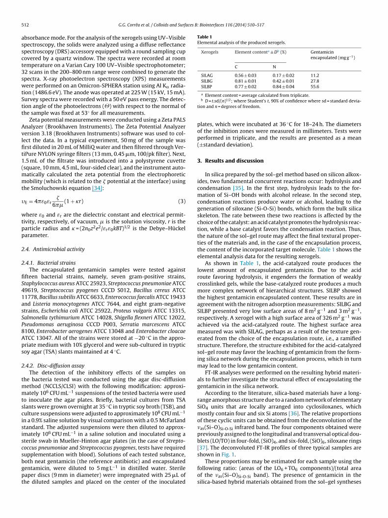
determined. Level 2, which is located in the q region below 0.01 A–1,provides information on the organization of these particles, i.e., onthe structure of the fractal clusters (secondary particles) that result
0
200
400
600
800
213
206 254
264
Ref
lect
ance
(a.u
.)
(a)
(b)
Waven
Fig. 1. Deconvolution of the Si-O region in the FT-IR sp
esulted in different network structures depending on the synthe-is protocol. For the SILBP system, the presence of gentamicin led ton approximate 30% decrease in the proportion of six-fold ((SiO)6)iloxane rings in comparison to the SILBP system. In the case ofhe systems that were prepared by gelation routes, the addition ofentamicin in the sol–gel syntheses resulted in an increase in theumber of (SiO)6 siloxane rings in comparison with the bare sys-ems. For SILBG, this increase was 9%. Finally, in the case of SILAG,hich was prepared under acid conditions, an increase of 24% was
bserved compared to the bare silica SILAG. These results suggesthat the addition of gentamicin in the synthesis of SILBP produced
more compact and denser network in comparison to the SILBGnd SILAG systems (see Supplementary Table 1).
The systems were further characterized by diffuse reflectancepectroscopy (DRS) in the UV-Vis region (Fig. 2).
As shown in Fig. 2, spectrum (a) shows the maximum absorptionands of neat gentamicin at 206 and 254 nm. These bands corre-pond to the n → �* transitions [38] of the isolated electron pairsf oxygen and nitrogen atoms. After encapsulation within the sil-ca matrix (spectrum b), these peaks shifted to 213 nm and 264 nm,ndicating a bathochromic effect, which is most likely due to thehange in medium that is caused by the silica framework. Thus,his process caused a reduction in the energy levels of the n → �*
ransitions of the isolated electrons from the oxygen and nitrogentoms [38].
An attempt to estimate the amount of encapsulated gentami-in on the outermost external surface of the grain was made using-ray photoelectron spectroscopy (XPS). In the case of silica, theampled measured depth was in the range of 5 nm. The surface anal-sis expressed as N/Si, in which the amount of nitrogen is assigned
o gentamicin while that of Si is assigned to silica, was 0.08, 0.04nd 0.49 for SILAG, SILBG and SILBP, respectively. Thus, the precipi-ation route produces the highest encapsulated gentamicin contentn the outermost external surface.r (cm )
of hybrid materials: (a) SILBP, (b) SILBG and (c) SILAG.
The SAXS technique was also used for the characterization ofsilica to elucidate the structures of the materials. The SAXS curvesof these materials have a structure that is formed by organizationlevels consisting of a Guinier region (level 1) and a Potency Law(level 2). A typical SAXS curve for the SILAG system is shown inFig. 3.
According to Fig. 3, the unified set of SAXS data reveals that thematerials are arranged in a structure consisting of two organizationlevels. By adjusting level 1, which is located in the q region above0.03 A–1, the radius of gyration (Rg) of the primary particles can be
200 300 400 500
Wavelength (nm)
Fig. 2. Diffuse reflectance spectra of (a) gentamicin and (b) SILAG.
514 G.G. Corrêa et al. / Colloids and Surfaces B: Biointerfaces 116 (2014) 510–517
0.01 0.11E-4
1E-3
0.01
0.1
1 Scatt ering in ten sity Unified fit Level 1 fit Level 2 fit
Inte
nsity
(a.u
.)
q (Å-1)
q-4,0
Fig. 3. Typical SAXS curve for the SILAG supports, and the curve fit from the unifiedm
fpaesefpsdsi
pRsc
e
Table 2SAXS data for silica-based mixed oxide determined from the curve fits.
System Level 1 Level 2
Rga (nm) Pb Rg
a (nm) P
SILAG 3.9 ± 0.02 4.0 – 3.8SILBG 2.9 ± 0.11 4.0 – 3.9SILBP 4.4 ± 0.12 4.0 – 4.0
D = t.sd/(n)1/2; where Student’s t, 90% of confidence where sd = standard deviationand n = degrees of freedom.
results for the preliminary assessment of the antibacterial activity
TA
odel.
rom the aggregation of the primary particles. The structure of therimary particle clusters that constitute level 2 can be obtained bynalyzing the power law exponent of the scattering curve. If thexponent of the power law (I � q−p) is between 1.0 and 3.0, theecondary particles have a mass fractal structure [39]. When thexponent is between 3.0 and 4.0, the secondary particles have aractal surface. In the case of an exponent of 4.0, the secondaryarticles have a dense core and a uniform surface. In the presenttudy, all of the SAXS curve systems revealed the presence of twoistinct organizational levels. The results obtained from the unifiedet of SAXS curves for the silica-based mixed oxides are presentedn Table 2.
According to Table 2, the radius of gyration (Rg) of the primaryarticles for the investigated systems was between 2.9 and 4.4 nm.egarding the organization of the primary particles, the valuesuggest the formation of secondary particles with fractal surfaceharacteristics.
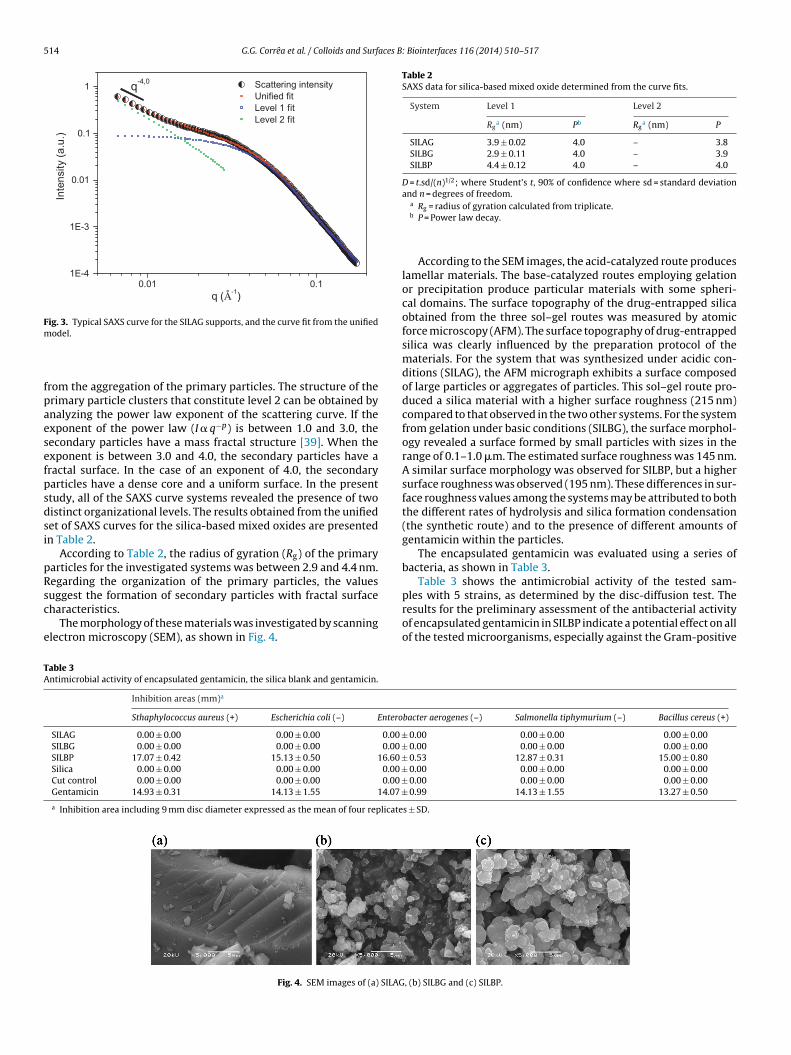
The morphology of these materials was investigated by scanninglectron microscopy (SEM), as shown in Fig. 4.
able 3ntimicrobial activity of encapsulated gentamicin, the silica blank and gentamicin.
Inhibition areas (mm)a
Sthaphylococcus aureus (+) Escherichia coli (–) Entero
SILAG 0.00 ± 0.00 0.00 ± 0.00 0.00 ±SILBG 0.00 ± 0.00 0.00 ± 0.00 0.00 ±SILBP 17.07 ± 0.42 15.13 ± 0.50 16.60 ±Silica 0.00 ± 0.00 0.00 ± 0.00 0.00 ±Cut control 0.00 ± 0.00 0.00 ± 0.00 0.00 ±Gentamicin 14.93 ± 0.31 14.13 ± 1.55 14.07 ±a Inhibition area including 9 mm disc diameter expressed as the mean of four replicate
Fig. 4. SEM images of (a) SILAG
a Rg = radius of gyration calculated from triplicate.b P = Power law decay.
According to the SEM images, the acid-catalyzed route produceslamellar materials. The base-catalyzed routes employing gelationor precipitation produce particular materials with some spheri-cal domains. The surface topography of the drug-entrapped silicaobtained from the three sol–gel routes was measured by atomicforce microscopy (AFM). The surface topography of drug-entrappedsilica was clearly influenced by the preparation protocol of thematerials. For the system that was synthesized under acidic con-ditions (SILAG), the AFM micrograph exhibits a surface composedof large particles or aggregates of particles. This sol–gel route pro-duced a silica material with a higher surface roughness (215 nm)compared to that observed in the two other systems. For the systemfrom gelation under basic conditions (SILBG), the surface morphol-ogy revealed a surface formed by small particles with sizes in therange of 0.1–1.0 �m. The estimated surface roughness was 145 nm.A similar surface morphology was observed for SILBP, but a highersurface roughness was observed (195 nm). These differences in sur-face roughness values among the systems may be attributed to boththe different rates of hydrolysis and silica formation condensation(the synthetic route) and to the presence of different amounts ofgentamicin within the particles.
The encapsulated gentamicin was evaluated using a series ofbacteria, as shown in Table 3.
Table 3 shows the antimicrobial activity of the tested sam-ples with 5 strains, as determined by the disc-diffusion test. The
of encapsulated gentamicin in SILBP indicate a potential effect on allof the tested microorganisms, especially against the Gram-positive
bacter aerogenes (–) Salmonella tiphymurium (–) Bacillus cereus (+)
0.00 0.00 ± 0.00 0.00 ± 0.00 0.00 0.00 ± 0.00 0.00 ± 0.00
0.53 12.87 ± 0.31 15.00 ± 0.80 0.00 0.00 ± 0.00 0.00 ± 0.00 0.00 0.00 ± 0.00 0.00 ± 0.00
0.99 14.13 ± 1.55 13.27 ± 0.50
s ± SD.
, (b) SILBG and (c) SILBP.

G.G. Corrêa et al. / Colloids and Surfaces B: Biointerfaces 116 (2014) 510–517 515
Table 4Zeta potential of SILAG, SILBG, SILPG and their respective blanks.
Zeta potentiala ± Db (mV)
System Blank Encapsulated
SILAG 5.41 ± 0.320 8.80 ± 1.07SILBG −27.7 ± 2.26 6.67 ± 1.17SILBP −35.5 ± 0.36 27.8 ± 1.06
a Zeta potential = average calculated from quintuplicate.
t
stttaGbc
oopTwtndTtt
t
csioafgltapt
atehtgrwtet
mtcata
bial
acti
vity
of
the
SILB
P
seri
es, s
ilic
a
blan
k
and
gen
tam
icin
.
Inh
ibit
ion
dia
met
er
(mm
)a
S.
aure
us
(+)
S.
pneu
mon
iae
(+)
S.
pyog
enes
(+)
B.
cere
us
(+)
B.
subt
ilis
(+)
E.
faec
ales
(+)
L.
mon
ocyt
o-ge
nes
(+)
E.
coli
(−)
P.
vulg
aris
(−)
S.
tiph
ymur
ium
(−)
S.
flexn
eri (
−)
P.
aeru
gino
sa(−
)S.
mar
cesc
ens
(−)
E.
aero
gene
s(−
)E.
cloa
cae
(−)
021
.80
±
1.25
17.7
3
±
1.01
17.6
7
±
1.47
17.0
7
±
1.10
18.6
7
±
1.40
13.3
7
±
1.33
18.0
7
±
1.70
15.8
7
±
1.21
17.2
7
±
0.95
13.6
0
±
0.87
–
14.7
3
±
0.50
15.2
0
±
0.20
16.6
0
±
0.72
16.0
7
±
0.50
19.8
7
±
1.85
15.9
3
±
1.33
15.8
0
±
0.87
16.9
3
±
1.67
16.1
3
±
1.17
11.9
3
±1.3
716
.47
±
1.10
15.3
3
±
0.42
17.0
0
±
0.87
13.3
3
±
0.70
–
14.6
7
±
0.31
15.0
7
±
0.90
16.6
7
±
1.92
14.5
3
±
0.99
17.0
7
±
0.42
15.2
0
±
1.25
14.8
7
±
1.80
15.0
0
±
0.80
14.2
7
±
1.10
10.8
7
±
0.76
16.2
0
±
1.56
15.1
3
±
0.50
17.2
7
±
1.03
12.8
7
±
0.31
–
12.7
3
±
0.50
15.0
0
±
1.40
16.6
0
±
0.53
13.2
0
±
0.20
16.3
3
±
1.36
13.6
7
±
0.99
13.8
7
±
0.99
12.1
3
±
0.70
14.1
3
±
1.30
10.8
0
± 0.
53
16.1
3
±
1.36
15.0
7
±
1.80
15.7
3
±
1.21
10.9
3
±
0.42
–
12.6
7
±
0.95
10.2
7
±
0.31
13.6
0
±
1.03
13.1
3
±
1.67
–
–
–
–
–
–
–
–
–
–
–
–
–
–
–ol
–
–
–
–
–
– –
–
–
–
–
–
–
–
–14
.93
±
0.31
15.6
0
±
0.62
16.0
7
±
1.14
13.2
7
±
0.50
17.2
0
±
0.72
13.5
3
±
1.60
17.3
3
±
0.51
14.1
3
±
1.55
18.3
3
±
1.50
14.1
3
±1.5
5
10.8
0
±
1.03
16.2
7
±
1.21
15.9
3
±
0.64
14.0
7
±
0.99
16.8
0
±
0.40
tion
incl
ud
ing
9
mm
dis
c
dia
met
er
exp
ress
ed
as
the
mea
n
of
thre
e
rep
lica
tes
±
SD, (
–)
no
anti
mic
robi
al
acti
vity
.
b D = t.sd/(n)1/2; where Student’s t, 90% of confidence where sd = standard devia-ion and n = degrees of freedom.
train Staphylococcus aureus. The higher efficacy of gentamicin onhis microorganism is most likely due to the low intrinsic resis-ance of Gram-positive bacteria to antimicrobial agents becausehese bacteria do not have specific membrane receptors or perme-ses to control the entrance of a substance into the cell, whereasram-negative bacteria lipopolysaccharides have an outer mem-rane that limits access, particularly for lipophilic substances, toell membrane phospholipids [40].
Another factor that may have influenced the improved efficiencyf the encapsulated antibiotic from the SILBP route is the amountf gentamicin contained on the outermost particle surface, thusroviding a controlled release of the molecule during the reaction.he amount of gentamicin contained on the SILBP capsule surfacesas determined from XPS analysis (N/Si = 0.49) and was higher than
hat in SILAG (0.08) and SILBG (0.04), which did not exhibit anyoticeable inhibitory effect against the tested bacteria, most likelyue to the low gentamicin concentration present on their surfaces.hus, SILAG and SILBG do not exhibit any inhibition potential duehe low amount of molecules on the surface, as characterized byhe XPS technique.
The zeta potential results of SILAG, SILBG, SILBP and their respec-ive blanks are shown in Table 4.
SILBP has the highest zeta potential, which is positive. Theell wall of Gram-positive bacteria (peptidoglycan) has a negativeuperficial charge due to the phosphoryl groups that are local-zed as teichuronic acid substituents. Because the peptidoglycanf Gram-negative bacteria is sequestered in the periplasmic spacend not exposed to the extracellular environment, the negative sur-ace charge in these microorganisms is conferred by the phosphorylroups and carboxyl-2-ceto-3-deoxyoctonate lipopolysaccharidesocated on the outside membrane [9]. Therefore, the material withhe highest positive zeta potential (SILBP) could have higher inter-ctions with “tight junctions” and with the cell membrane, so thearacellular permeation of hydrophilic compounds [9], such as gen-amicin, feasibly increases their antimicrobial activity.
A factor that appears to influence the gentamicin antimicrobialctivity is the N/Si ratio, which was measured by XPS. By analyzinghe data obtained from XPS, it can be seen that the SILBP materialxhibits the highest N/Si ratio, indicating that this material has aigher gentamicin concentration on the surface. This fact explainshe zeta potential result, considering that the NH2 groups fromentamicin confer a positive charge to the material surface. A cor-elation (R2 = 0.9998) between the N/Si ratio and the zeta potentialas observed, indicating that there is a strong correlation between
he data. Therefore, pharmaceutical diffusion into the medium isasier than for other materials with lower N/Si ratio values, in whichhe drug was less available.
Based on these exploratory results, different amounts of genta-icin were encapsulated by the three different routes to evaluate
he effect of the antibiotic content on some textural and structural
haracteristics of the resulting encapsulated systems and on thentimicrobial activity. Furthermore, a broad range of bacteria wasested. Better results continued to be shown by the SILBP systems,s shown in Table 5. Data involving the other systems (SILAG and Table
5A
nti
mic
ro
SILP
G
100
SILP
G
750
SILP
G
500
SILP
G
250
Sili
ca
Cu
t
con
trG
ENTA
aIn
hib
i

5 aces B
ST
rtrfa
aa1T
roitnwad(s
oottadrprc1
bembicoreiacEbvtfmt
4
asiOc
[
[[[
[
[
[
[[[[[[
[
[[
[
[
[
[[[[[[
[
16 G.G. Corrêa et al. / Colloids and Surf
ILBG) are presented as supplementary material (Supplementaryables 2 and 3).
For almost all the systems, higher encapsulation amountsesulted in more intense antimicrobial activity. A linear correla-ion (R2 > 0.9) between the encapsulated amount (in fact, the N/Siatio determined by XPS) and the inhibition diameter were foundor S. aureus, S. pneumoniae, S. pyogenes, B. subtilis, E. faecalis, E. colind E. cloacae.
From the textural point of view, increasing the encapsulatedmount from 250 to 1000 mg induces a decrease in the surfacerea: 382, 326, 71 and 1 m2 g−1, respectively, for 250, 500, 750 and000 mg of gentamicin in the case of SILAG (see Supplementaryable 3).
According to the literature, the presence of encapsulated mate-ials in a reaction medium does not generally affect the spreadf its molecules; therefore, the effect on the microorganism [41],.e., the structure-activity relation, is not altered, thus preventinghe effectiveness of bacterial defense and/or resistance mecha-isms through enzymatic action. However, in the present case,e observed that the encapsulation method appeared to affect the
ctivity of the drug (as in the case of S. aureus, in which the inhibitioniameter increases from 14.93 (not-encapsulated) to 21.80 mm1000 mg encapsulated system), most likely due to the textural andurface characteristics of the final xerogel).
Another factor that may have influenced the improved efficiencyf the encapsulated antibiotic from the SILBP route is the amountf gentamicin contained on the outermost surface of the particles,hus making more drug available. The amount of gentamicin con-ained on the SILBP capsule surfaces was determined from XPSnalysis (N/Si) and was higher than that in SILAG and SILBG, whichid not exhibit any noticeable inhibitory effect against the bacte-ia tested, most likely due to the low gentamicin concentrationresent on the surfaces. Furthermore, in the case of SILBP, the N/Siatio increased with increasing amounts of encapsulated gentami-in: 0.28, 0.49, 0.74 and 1.13, respectively, for 250, 500, 750 and000 mg of encapsulated gentamicin.
The fact that the surface phenomenon between the antimicro-ial agent and the microorganism cell is crucial for the inhibitoryffect cannot be neglected. All aminoglycosides, including genta-icin, act via the same inhibition mechanism; they exert their
actericidal effect by binding to bacterial ribosome. Therefore, its necessary that the antibiotic molecule remains attached to theell wall to affect the cell permeability and enable the transportf the active fraction of the antibiotic [42–44]. In this sense, theoughness of SILBP (195 nm, as detected by AFM) may have influ-nced the antimicrobial effect, as verified by the increase in thenhibition diameter found for the microorganisms Staphylococcusureus, Streptococcus pneumoniae, Streptococcus pyogenes, Bacillusereus, Bacillus subtilis, Listeria monocytogenes, Escherichia coli andnterobacter aerogenes. Conversely, a small decrease in the inhi-ition diameter was observed for Enterococcus faecalis, Proteusulgaris, Salmonella typhimurium, Pseudomonas aeruginosa, Serra-ia marcescens and Enterobacter cloacae. The surface roughnessrom this treatment may facilitate contact between the antibiotic
olecule and the cell membrane, thus favoring the cellular inhibi-ion mechanism of aminoglycosides.
. Conclusions
For the encapsulation of gentamicin within silica-based materi-ls, it has been shown that the sol–gel route affects the elemental,
tructural, textural and morphological characteristics of the result-ng xerogel, which in turn determine the antimicrobial activity.f the evaluated routes, the route involving base-catalyzed pre-ipitation produced the most active system, even higher than[[[
: Biointerfaces 116 (2014) 510–517
that observed for the bare antibiotic. The surface concentration ofgentamicin and the zero potential of the xerogel appear to varydepending on the encapsulation route, which is therefore a relevantfactor in the antimicrobial effect.
Acknowledgments
This work was financed by CNPq. The authors are thankful tothe LNLS for the measurements that were performed in the SAXSbeamline (Project #SAXS1 12495).
Appendix A. Supplementary data
Supplementary material related to this article can be found,in the online version, at http://dx.doi.org/10.1016/j.colsurfb.2014.01.043.
References
[1] Q. Guo, P. Luo, Y. Luo, F. Du, W. Lu, S. Liu, J. Huang, J. Yu, Colloids Surf. B:Biointerfaces 100 (2012) 138.
[2] X. Zhang, F. Du, J. Huang, W. Lu, S. Liu, J. Yu, Colloids Surf. B: Biointerfaces 100(2012) 155.
[3] A.G. Fisch, N.S.M. Cardozo, A.R. Secchi, F.C. Stedile, N.P.D. Silveira, J.H.Z. Santos,J. Non Cryst. Solids 354 (2008) 3973.
[4] J. Li, J. Yao, Y. Li, Y. Shao, J. Environ. Sci. Health B 47 (2012) 795.[5] S. Sakai, H. Inagaki, K. Inamoto, M. Taya, Biomaterials 33 (2012) 6721.[6] R. Ciriminna, M. Sciortino, G. Alonzo, A.D. Schrijver, M. Pagliaro, Chem. Rev. 111
(2011) 765.[7] K.Y. Choi, E.J. Jeon, H.Y. Yoon, B.S. Lee, J.H. Na, K.H. Min, S.Y. Kim, S.J. Myung,
S. Lee, X. Chen, I.C. Kwon, K. Choi, S.Y. Jeong, K. Kim, J.H. Park, Biomaterials 33(2012) 6186.
[8] D.J. Lee, G.Y. Park, K.T. Oh, N.M. Oh, D.S. Kwag, Y.S. Youn, Y.T. Oh, J.W. Park, E.S.Lee, Int. J. Pharm. 434 (2012) 257.
[9] W.S. Cheow, K. Hadinoto, Colloids Surf. A: Physicochem. Eng. Aspects 370(2010) 79.
10] G.A. Islan, I.P. de Verti, S.G. Marchetti, G.R. Castro, Appl. Biochem. Biotechnol.167 (2012) 1408.
11] A.D. Sezer, J. Akbua, Pharm. Dev. Technol. 17 (2010) 118.12] D. Martins, F.T.M. Costa, M. Brocchi, N. Durán, J. Nanopart. Res. 13 (2011) 355.13] P. Lutwyche, C. Cordeiro, D.J. Wiseman, M. St-Louis, M. Uh, M.J. Hope, M.S.
Webb, B.B. Finlay, Antimicrob. Agents Chemother. 42 (1998) 2511.14] E.C. Umeyor, F.C. Kenechukwu, J.D. Ogbonna, S.A. Chime, A. Attama, J. Microen-
capsul. 29 (2012) 296.15] K. Vichitchote, P. Threepopnatkul, S. Suttiruengwong, C. Kulsetthanchalee,
Mater. Sci. Forum 714 (2012) 263.16] J. Pang, Y. Luan, X. Yang, Y. Jiang, L. Zhao, Y. Zong, Z. Li, Mini Rev. Med. Chem.
12 (2012) 775.17] P.P. Nampi, V.S. Mohan, A.K. Sinha, H. Varma, Mater. Res. Bull. 47 (2012) 1379.18] K.K. Qian, R.H. Bogner, J. Pharm. Sci. 101 (2012) 444.19] S.J. McInner, N.H. Voelcker, Future Med. Chem. 1 (2009) 1051.20] Y. Zhu, J. Shi, Micropor. Mesopor. Mater. 103 (2007) 243.21] J.M. Xue, M. Xi, J. Control. Release 98 (2004) 209.22] J.M. Xue, C.H. Tan, D. Lukito, J. Biomed. Mater. Res. B, Appl. Biomater. 78 (2006)
417.23] L. Meseguer-Olmo, M. Ros-Nicolás, V. Vicente-Ortega, M. Alcaraz-Banos,
M. Clavel-Sainz, D. Arcos, C.V. Ragel, M. Vallet-Regí, C. Meseguer-Ortiz, J.Orthopaedic Res. 24 (2006) 454.
24] J-X. Wang, Z-H. Wang, J-F. Chen, J. Yun, Mater. Res. Bull. 43 (2008) 3374.25] E.C. Morais, G.G. Correa, R. Brambilla, C. Radtke, I. Baibich, J.H.Z. Santos, Colloids
Surf. B, Biointerfaces 103 (2012) 422.26] U. Schubert, N. Hüsing, second ed., Synthesis of Inorganic Materials, Wiley,
Weinheim, 2005.27] E.C. Morais, G.G. Correa, R. Brambilla, P.R. Livotto, M.B. Cardoso, J.H.Z. Santos, J.
Sol-Gel Sci. Technol. 1 (2012) 1.28] E.C. Morais, G.G. Correa, R. Brambilla, J.H.Z. Santos, A.G. Fisch, J. Sep. Sci. 36
(2012) 636.29] Y. Leprince-Wang, K. Yu-Zhang, Surf. Coat Technol. 140 (2001) 155.30] J. Ilavsky, P.R. Jemian, J. Appl. Crystallogr. 42 (2009) 347.31] S.R. Kline, J. Appl. Crystallogr. 39 (2006) 895.32] G. Beaucage, J. Appl. Crystallogr. 28 (1995) 717.33] G. Beaucage, J. Appl. Crystallogr. 29 (1996) 134.34] R.J. Hunter, Zeta Potential in Colloid Science, first ed., Academic Press, New
York, 1981.35] C.J. Brinker, G.W. Scherer, Sol–gel Science: The Physics and Chemistry of Sol–Gel
Processing, first ed., Academic Press, New York, 1990.36] A. Fidalgo, L.M. Ilharco, Chem. Eur. J. 10 (2004) 392.37] A. Fidalgo, R. Ciriminna, L.M. Ilharco, M. Pagliaro, Chem. Mater. 17 (2005) 6686.38] D.H. Williams, I. Fleming, Spectroscopic Methods in Organic Chemistry, first
ed., McGraw-Hill, New York, 1966.
aces B
[[[[
G.G. Corrêa et al. / Colloids and Surf
39] P.W. Schmidt, J. Appl. Crystallogr. 24 (1991) 414.40] A.D. Russel, J. Appl. Bacteriol. 71 (1991) 191.41] T.J. Nicholls, A.R. Goldsmith, A. Dawson, Phys. Rev. 68 (1988) 133.42] A.P. Taylor, K.S. Finnie, J.R. Bartlett, P.J. Holden, J. Sol-Gel Sci. Technol. 32 (2004)
223.
[
[
: Biointerfaces 116 (2014) 510–517 517
43] F.O. Donnell, T.J.P. Smyth, V.N. Ramachandran, W.F. Smyth, Int. J. Antimicrob.Agents 35 (2010) 30.
44] M.J. Blanco-Príeto, C. Lecaroz, M.J. Renedo, J. Kunkova, C. Gamazo, Int. J. Pharm.242 (2002) 203.