Effects of food processing on masticatory strain and craniofacial growth in a retrognathic face
23
Effects of food processing on masticatory strain and craniofacial growth in a retrognathic face Daniel E. Lieberman a *, Gail E. Krovitz b , Franklin W. Yates a , Maureen Devlin a , Marisa St. Claire c a Department of Anthropology, Peabody Museum, Harvard University, 11 Divinity Avenue, Cambridge, MA 02138, USA b Department of Anthropology, 409 Carpenter Building, Penn State University, College Park, PA 16802, USA c Bioqual Corporation, 9600 Medical Center Drive, Rockville, MD 20850-3336, USA Received 17 November 2003; accepted 19 March 2004 Abstract Changes in the technology of food preparation over the last few thousand years (especially cooking, softening, and grinding) are hypothesized to have contributed to smaller facial size in humans because of less growth in response to strains generated by chewing softer, more processed food. While there is considerable comparative evidence to support this idea, most experimental tests of this hypothesis have been on non-human primates or other very prognathic mammals (rodents, swine) raised on hard versus very soft (nearly liquid) diets. Here, we examine facial growth and in vivo strains generated in response to raw/dried foods versus cooked foods in a retrognathic mammal, the rock hyrax (Procavia capensis). The results indicate that the hyrax cranium resembles the non-human primate cranium in having a steep gradient of strains from the occlusal to orbital regions, but differs from most non-anthropoids in being primarily twisted; the hyrax mandible is bent both vertically and laterally. In general, higher strains, as much as two-fold at some sites, are generated by masticating raw versus cooked food. Hyraxes raised on cooked food had significantly less growth (approximately 10%) in the ventral (inferior) and posterior portions of the face, where strains are highest, resembling many of the differences evident between humans raised on highly processed versus less processed diets. The results support the hypothesis that food processing techniques have led to decreased facial growth in the mandibular and maxillary arches in recent human populations. 2004 Elsevier Ltd. All rights reserved. Keywords: Hyrax; Skull growth; Mandible; Maxilla; Cooking; Mastication; Strain Introduction Understanding how the face resists and responds to masticatory forces is important for testing hypotheses about the effects of chewing on facial growth. While multiple genetic and * Corresponding author. Tel.: +1-617-495-5470; fax: +1-617-496-8041 E-mail address: [email protected] (D.E. Lieberman). Journal of Human Evolution 46 (2004) 655–677 0047-2484/04/$ - see front matter 2004 Elsevier Ltd. All rights reserved. doi:10.1016/j.jhevol.2004.03.005
Transcript of Effects of food processing on masticatory strain and craniofacial growth in a retrognathic face

Effects of food processing on masticatory strain andcraniofacial growth in a retrognathic face
Daniel E. Liebermana*, Gail E. Krovitzb, Franklin W. Yatesa, Maureen Devlina,Marisa St. Clairec
aDepartment of Anthropology, Peabody Museum, Harvard University, 11 Divinity Avenue, Cambridge, MA 02138, USAbDepartment of Anthropology, 409 Carpenter Building, Penn State University, College Park, PA 16802, USA
cBioqual Corporation, 9600 Medical Center Drive, Rockville, MD 20850-3336, USA
Received 17 November 2003; accepted 19 March 2004
Abstract
Changes in the technology of food preparation over the last few thousand years (especially cooking, softening, andgrinding) are hypothesized to have contributed to smaller facial size in humans because of less growth in response tostrains generated by chewing softer, more processed food. While there is considerable comparative evidence to supportthis idea, most experimental tests of this hypothesis have been on non-human primates or other very prognathicmammals (rodents, swine) raised on hard versus very soft (nearly liquid) diets. Here, we examine facial growth and invivo strains generated in response to raw/dried foods versus cooked foods in a retrognathic mammal, the rock hyrax(Procavia capensis). The results indicate that the hyrax cranium resembles the non-human primate cranium in havinga steep gradient of strains from the occlusal to orbital regions, but differs from most non-anthropoids in being primarilytwisted; the hyrax mandible is bent both vertically and laterally. In general, higher strains, as much as two-fold at somesites, are generated by masticating raw versus cooked food. Hyraxes raised on cooked food had significantly less growth(approximately 10%) in the ventral (inferior) and posterior portions of the face, where strains are highest, resemblingmany of the differences evident between humans raised on highly processed versus less processed diets. The resultssupport the hypothesis that food processing techniques have led to decreased facial growth in the mandibular andmaxillary arches in recent human populations.� 2004 Elsevier Ltd. All rights reserved.
Keywords: Hyrax; Skull growth; Mandible; Maxilla; Cooking; Mastication; Strain
Introduction
Understanding how the face resists andresponds to masticatory forces is important fortesting hypotheses about the effects of chewingon facial growth. While multiple genetic and
* Corresponding author. Tel.: +1-617-495-5470; fax:+1-617-496-8041
E-mail address: [email protected](D.E. Lieberman).
Journal of Human Evolution 46 (2004) 655–677
0047-2484/04/$ - see front matter � 2004 Elsevier Ltd. All rights reserved.doi:10.1016/j.jhevol.2004.03.005

environmental factors influence facial growth,several lines of evidence suggest that changes indiet and food processing technology contribute tosome proportion of variations in facial size andshape. Human diets since the Middle Paleolithichave changed substantially in content (Stiner et al.,1999; Wrangham et al., 1999; Richards et al., 2001;Stiner, 2001), and in how they are processedthrough cooking, soaking, leaching, and grinding(Rangel et al., 1985; Brace et al., 1987, 1991; Shiauet al., 1999; Wrangham and Conklin-Brittain,2003). Food processing improves digestibility, butalso makes food softer and smaller in particle size,requiring less occlusal force per chew and fewerchewing cycles per unit of food (Lucas and Luke,1984; Lukacs, 1989; Lieberman, 1993; Agrawalet al., 1997; Strait, 1997). In turn, softer and moreprocessed foods are widely hypothesized to lead toless facial growth, especially in the lower face andthe alveolar crests, because of the potential effectsof force-generated strain (see Carlson, 1976;Carlson and Van Gerven, 1977; Corruccini andBeecher, 1982). Strain can stimulate periostealgrowth and/or inhibit resorption in skeletallyimmature animals, perhaps to adapt bone shapeand structure so that applied forces elicit strainenergies below threshold ranges (Rubin andLanyon, 1984; Biewener et al., 1986; Martin et al.,1998; Carter and Beaupre, 2001; Currey, 2002;Lieberman et al., 2003). In addition, low magni-tudes and frequencies of loading can lead to localbone resorption. Loss of dental function in themandible can decrease alveolar crest height andramus length by up to 50% (Carlsson and Persson,1967; Israel, 1973; Sugimura et al., 1984).
While one might expect nutritional improve-ments since the Middle Paleolithic to contribute toincreases in overall cranial size (see Kiliaridis et al.,1992), the evidence points to a trend towardssmaller facial size, with the most dramaticdecreases occurring after the Neolithic. Compari-sons of Nubian populations prior to and after theintroduction of agriculture show significant reduc-tions in many mid- and lower facial dimensionsincluding infraorbital height (7.8%), masseter ori-gin length (26.3%), mandibular corpus length(22%), and mandibular symphysis thickness(15.3%)—despite concurrent increases in brain size
(Carlson, 1976; Carlson and Van Gerven, 1977).Similar decreases in mandibular and maxillaryarch size, especially in the alveolar crests, occurredover the last century among Australian aboriginesand other populations who have transitioned tomodern, processed diets (Corruccini, 1984, 1990;Lukacs, 1989). Significant, but less extreme,decreases in facial size are typical in populationsfollowing the industrial revolution. For example, acomparison of late medieval and recent Finns(with presumably no major genotypic differences)reveals a 6% decrease in mandible length despiteoverall skull size increases (Varrela, 1992).
While softer, more processed foods may con-tribute to facial diminution, few studies haveexperimentally tested these effects in mammals,only one (Bouvier and Hylander, 1996) quantifiedgrowth in response to in vivo strains, and only afew studies have specifically looked at humans.Most human studies have shown that young adultswith larger muscle cross-sectional areas and/orhigher bite forces have larger, less variably-sizedfaces than those who produce less bite force(e.g., Ingervall and Helkimo, 1978; Dechow andCarlson, 1983; Kiliaridis et al., 1989; Kiliaridis,1995; English et al., 2002). Such correlations aredifficult to interpret, however, because smaller jawsmay result from smaller muscles, but both vari-ables may covary as a result of other factors. Onlyone study (Ingervall and Bitsanis, 1987) directlyquantified human facial growth responses to load-ing by examining the effects of chewing a hardresinous gum for two hours/day for one year in 13Greek children between ages 7 and 12. Treatmentgroup individuals were able to produce signifi-cantly more force than controls, and had signifi-cantly longer mandibular and maxillary arches.No human studies have examined the effects ofmasticatory loading during early facial growth,when such effects are likely to be greatest, andnone have quantified strains or site-specific growthrates.
Most experimental data on facial growthresponses to masticatory loading come from studieson non-human anthropoids and other mammalianmodels. One primate study (Corruccini andBeecher, 1982; Beecher et al., 1983) compared 19adult squirrel monkeys (Saimiri sciureus) raised on
D.E. Lieberman et al. / Journal of Human Evolution 46 (2004) 655–677656

soft food diets with 24 controls raised on hardfood diets; another study (Corruccini and Beecher,1984) compared 16 adult baboons (Papio cyno-cephalus) who were raised on a hard diet for twoyears with 24 baboons raised on a soft diet. Inboth studies, animals who chewed harder foodhad significantly wider and taller faces, thickermandibular corpora, and taller palates. However,primates raised on softer food often had seriousmalocclusions from narrowed maxillary arches,rotated and displaced teeth, crowded premolars,and palatal arching, suggesting abnormal growthpatterns. A related study on macaques obtainedsimilar results, but also showed that Haversianremodeling rates were higher in monkeys fed hardfood (Bouvier and Hylander, 1981, 1996).
Experiments on non-primates reveal a similarpicture. Rats raised on soft food have smallerjaw adductor muscles, generate lower mandibularstrains, and have significantly decreased anteriorfacial height, shorter mandibles, and smallermuscle attachment areas (Kiliaridis et al., 1986;Engstrom et al., 1986; Yamada and Kimmel,1991). Several studies (Moore, 1965; Kiliaridis,1989; Kiliaridis et al., 1992) examined interactionsbetween nutrition and strain by comparing man-dibular growth in rats whose diets varied in hard-ness and calcium content. Rats fed low calciumdiets had significantly smaller mandibles in alldimensions, whereas the rats fed soft food (regard-less of calcium content) had mandibles that wereonly shorter in vertical height and condyle size.Thus strain and nutrition both influence jaw size,but only strain affects jaw shape. Few controlledstudies have been done on larger species. Oneexception is Ciochon et al. (1997), who comparedeight minipigs raised for eight months on nutri-tionally identical soft and hard diets; the four pigsraised on soft food had serious malocclusions, anddiffered significantly in facial shape with shortermandibular rami and narrower midfaces than pigsraised on hard food.
Problems addressed in this study
Two major problems need to be addressedregarding the effects of softer, more processed diets
on facial growth in humans. First, most exper-iments on non-humans have been long-termstudies that compared facial growth in subjects fedhard versus extremely soft food, typically chowthat was softened to an almost liquid state. Asnoted above, animals raised on soft food in thesestudies not only had smaller faces but also devel-oped serious malocclusions from abnormal facialgrowth. It is not known to what extent thesedifferences resulted from low strains versus a nearabsence of loading (see Bertram and Swartz, 1991).Obviously, humans raised on cooked food do nottypically develop severe facial dysplasia, highlight-ing the need to examine variations in facial growthin response to treatments that better reflect theeffects of food processing technology.
A second problem is that additional experimen-tal data are needed on the relationship between thesite specific strain and growth in differently-shapedfaces. Many studies, primarily on anthropoids,show that the non-human face is characterized bya steep strain gradient, with high strains near theocclusal plane, moderate strains in the middle face,and low strains in the upper face (Hylander et al.,1991a; Hylander and Johnson, 1992; Ross andHylander, 1996; Ross, 2001). During unilateralmastication, the zygomatic arch and postorbitalseptum are subject to bending (Hylander, 1986;Hylander et al., 1991a; Herring et al., 1996; Ross,2001), and the mandible is subject to a combina-tion lateral transverse bending (wishboning),sagittal bending, and twisting about the longitudi-nal axis (Hylander, 1979, 1985, 1988; Daegling andHylander, 1998). There is no consistent evidencefor a predominant pattern of deformation in themiddle and upper face of primates, in spite ofmodels that the primate face is bent or sheared inthe sagittal plane during incision or mastication(see Ross, 2001). Evidence for twisting is mixed.Strains measured in the dorsal interorbital regionand the lateral surface of the postorbital in galagos(Ravosa et al., 2000a,b) and the medial orbital wallin owl monkeys and galagos (Ross, 2001) indicatetwisting, but circumorbital strains in several non-human anthropoids (Aotus, Macaca, Papio) donot, despite predictions that anthropoids shouldexperience more twisting than strepsirrhinesbecause of more recruitment of balancing side
D.E. Lieberman et al. / Journal of Human Evolution 46 (2004) 655–677 657

adductor force (Hylander et al., 1991a; Ross andHylander, 1996).
These results raise the question of how varia-tions in facial shape, including the unique architec-ture of the human face, influence patterns of strainand growth. Compared to other primates, thehuman face (see Fig. 1) is not only tall, wide, flatand oriented primarily in the coronal plane, but itis also both retrognathic (defined here as having
postcanine teeth beneath the orbits rather thanunder a rostrum), and retracted (defined here ashaving the orbits beneath the anterior cranialfossa) (Lieberman and Crompton, 2000). Notethat retrognathy as defined here, does not describethe position of the lower face as a whole relativeto the upper face (prognathy). Here, we focus onthe issue of retrognathy, which is also present in afew non-human anthropoids (e.g., Saimiri, Aotus).Retrognathy is interesting, in part because the roleof the rostrum in dissipating forces generated bymastication or incision in anthropoids is poorlyknown, and partly because previous studies ofin vivo strain in primate faces have not includedstrain gauges on the rostrum. A number of studies(e.g., Demes, 1982; Greaves, 1985; Preuschoft et al.,1986) have modeled the rostrum as a combinationtube and beam that resists twisting around ananteroposterior axis and bending or shearing in thesagittal plane. In addition, Rafferty et al. (2003)have shown that the rostrum in swine is normallysubject to shearing forces, but also to torsionduring unilateral mastication. It is therefore poss-ible that postcanine retrognathy, in which theocclusion does not occur below a rostrum, mayresult in a less steep strain gradient because ofproportionately more stress transmission from thepoint of occlusion into the middle and upperportions of the face. In other words, a rostrummay function to dissipate occlusal forces awayfrom the orbital region of the face. If so, then onemight also expect unilateral mastication in a retro-gnathic animal to cause more twisting in theinfraorbital, orbital and supraorbital regions.
Theoretically, retrognathy could also affect theabsolute forces generated and the relative amountof twisting caused by differential recruitment ofbalancing versus working side jaw adductors.Depending on muscle position, retracting the post-canine tooth row relative to the TMJ can augmentthe mechanical advantage of the adductor musclesby reducing load arm length relative to lever armlength. If the adductor muscles generate similarcontractile forces (see, however, Demes and Creel,1988) then mastication could generate higherocclusal forces, hence higher strains in mammalswith retrognathic postcanine teeth. Retracting thebite point relative to the TMJ, however, may
Fig. 1. Lateral views of human (top), rock hyrax (middle), andbaboon (bottom) adult skulls scaled to same length. Note thatthe entire molar row lies beneath or posterior to the plane of theorbits (dashed line) in humans and hyraxes, but not in baboons(which are a particularly prognathic primate). See text forfurther discussion of craniofacial differences.
D.E. Lieberman et al. / Journal of Human Evolution 46 (2004) 655–677658

permit less force to be generated, and also has thepotential to move the midline resultant of muscleforce outside the triangle of support defined by thetwo TMJs and the bite point (Greaves, 1978),often leading to a reduction in balancing sidemuscle force when chewing on posterior teeth(Spencer, 1998, 1999). Such reductions might re-sult in less torsion, depending on their relativeposition along the anteroposterior axis of the face,since working side adductor and occlusal forcestend to cancel each other out (see Fig. 2).
In order to examine effects of cooking andretrognathic faces on facial strain and growth, wepresent here preliminary data from a non-primatemodel, the rock hyrax (Procavia capensis), whichwe compare to published data from non-humanprimates. The hyrax is one of several mam-mals with substantially retrognathic postcanine
dentitions including a few primate species (notablycallitrichids and owl monkeys), Proboscidia, someRodentia (e.g., beavers), and several breeds ofdogs (most notably bulldogs and King Charlesspaniels). Of these, the hyrax—an herbivorous,sub-ungulate order that includes three genera,Procavia, Dendrohyrax, and Heterohyrax—is auseful experimental model for several reasons.Hyraxes have a generalized, masticatory system(Janis, 1983; Franks et al., 1985), with severalmorphological similarities to non-human anthro-poids. Hyraxes have thin-enameled lophodontmolars and premolars, the latter of which aremolarized with buccal and lingual wear facets(Janis, 1983). The hyrax mandible has a fusedsymphysis, a relatively deep and wide man-dibular corpus, and an enlarged ascending ramusthat provides ample insertion area for a large
Fig. 2. A, anterior view of hyrax skull showing: Fws, working side masseter force; Fbs, balancing side masseter force; Fb, bite force;condylar reaction forces are not shown. B, model of strain orientations during twisting in rostrum; C, model of strain orientationsduring shearing in rostrum. Divergent arrows indicate tension; convergent arrows indicate compression.
D.E. Lieberman et al. / Journal of Human Evolution 46 (2004) 655–677 659

masseter-medial pterygoid complex (Janis, 1983).Proportions of the jaw adductor muscles in pri-mates and hyraxes are similar: the masseter andmedial pterygoid are the dominant jaw adductors,the temporalis is relatively small, and the hasstrong divisions between a dorsoventrally orientedsuperficial portion and a highly transversely ori-ented deep portion (Janis, 1979, 1983). Electro-myogram (EMG) and kinematic data onmastication in the rock hyrax indicate that thepower stroke is predominantly transverse, withdistinct buccal and lingual phases of occlusion(Wen, 1984; German and Franks, 1991). Althoughhyraxes have a rostrum (Fig. 1), it is short, andbears only some diminutive premolars, as well ascaniniform incisors (no canines) that are not usedfor unilateral mastication, are rarely used for inci-sion, and instead are mostly used for display,grooming and fighting (Janis, 1979, 1983).
We stress that we do not consider hyraxes ananalogy or straightforward model for humans interms of facial biomechanics. Although hyraxeshave retrognathic postcanine teeth and a generallyprimate-like pattern of chewing, their faces are notretracted below the anterior cranial fossa; theyhave short infraorbital regions; narrow faces withdivergent, non-frontated orbits; and a partiallycomplete postorbital bar, often connected by asubstantial postorbital ligament. In addition, themetopic suture remains unfused in hyraxes. Rockhyraxes, however, are otherwise useful as exper-imental models because they mature rapidly, theyare docile if raised in captivity, and able to chew awide range of diets (Griner, 1968; Rubsamen et al.,1982). The tree hyrax (Dendrohyrax) might be abetter model because it has complete lateralorbital rims, but this species is not available forexperimental studies.
Hypotheses to be tested
The general hypothesis tested here is thatmastication of tough, hard foods compared tocooked, soft foods generates higher magnitudes ofstrains that stimulate more bone growth in theface. This general hypothesis is divided into twosets of specific hypotheses about (1) the patternof strains generated by masticating different food
types, and (2) the effect of strain patterns onregional growth.
In terms of strain, we test two specific hypoth-eses. First, cooked foods are predicted to generatesimilar patterns but lower magnitudes of strainthan uncooked, raw foods. Second, the pattern ofstrain in the hyrax face is predicted to follow agradient characteristic of the primate face in whichstrains are higher near the site of occlusion anddissipate dorsally away from the tooth row (seeHylander and Johnson, 1992). In terms of osteo-genic growth in response to strain, we test threespecific hypotheses. First, animals raised onharder, uncooked foods are predicted to havemore facial growth than animals raised on softer,more processed foods. Second, variations in theamounts of regional growth in the face are pre-dicted to correlate with strain magnitudes in thatregion. And third, growth is predicted to occurin the planes of deformation, thereby potentiallylowering strain magnitudes.
An additional goal of this study is to relate invivo strain data from the hyrax to existing modelsof facial biomechanics, summarized in Figs. 2 and3, for the cranium and mandible, respectively. Ifthe face and rostrum act like a cylinder that istwisted by a combination of dorsally-directedforces on the working side point of occlusion andventrally-directed force at both zygomatic arches(Fig. 2a), then principal strains will have similarmagnitudes on the balancing and working sides at45( relative to the long axis in the direction oftorsion, with 90( shifts from working to balancingside (Fig. 2b) (see Hylander et al., 1991a). In con-trast (or in addition), if the face resists forces as ashort beam subjected to bending and/or shearing inthe sagittal plane (see Ross, 2001), then the ventraland dorsal aspects will be tensed or compressed,respectively, along their long axes (Fig. 2c).
Models for orientations of strain in the man-dibular corpus and symphysis have been outlinedin Hylander (1984) and Crompton (1995) and areillustrated in Fig. 3. Bending in the parasagittalplane will cause the ventral margin of the man-dibular corpus to experience compression alongthe long axis (hence tension perpendicular tothe long axis) on the balancing side; in contrast,bending will cause the ventral margin of the
D.E. Lieberman et al. / Journal of Human Evolution 46 (2004) 655–677660

working side corpus to experience tension alongthe long axis anterior to the bite point, and com-pression along the long axis posterior to the bitepoint (Fig. 3a, b). Twisting of the mandibularcorpus around its long axis will cause the orienta-tion of tension to be 45( relative to the long axis inthe direction of twisting (Fig. 3c). In the anterioraspect of the symphysis, dorsoventral shear willcause the orientation of tension to be 45( relativeto the sagittal plane; lateral transverse bending(wishboning) will mediolaterally compress thesymphysis; and twisting of the two corpora willmediolaterally compress the superior margin andmediolaterally tense the inferior margin (whatmight be called coronal bending) (Fig. 3d–f; fordetails, see Hylander, 1984).
Materials and methods
Treatment groups
Two groups of rock hyraxes were studied.Craniofacial growth was examined in a group
of eight juvenile hyraxes. These animals wereapproximately 5–6 months old at the start of thetreatment period with unerupted first permanentmolars. Treatment period was 98 days. Animalswere divided randomly into hard and soft foodtreatment groups. Both groups were fed the samediet: 1 slice sweet potato (approximately 2$ thick),half of an apple, half a carrot, 1 kale stem (approxi-mately 2$ thick), supplemented with 125 g rabbitchow. In the soft food group, the vegetables werecut into pieces and microwaved for 3–5 minutes
Fig. 3. Simplified biomechanical model of mastication in the hyrax mandible. A, lateral view of hyrax mandible showing: Fc, condylarreaction force (bilateral); Fm, masseter force (bilateral); Fb, bite force. Plus and minus signs denote regions of the ventral margin of thecorpus that are compressed or tensed relative to the bite point on the working side (WS) and balancing side (BS). B, Schematic modelof strain orientations in the mandibular corpus during bending; C, Schematic model of strain orientations in the mandibular corpusduring twisting. D–F, schematic models of strain orientations in the mandibular symphysis during dorso-ventral shear, wishboning(lateral transverse bending), and coronal bending (from twisting of each corpus). Divergent arrows indicate tension, convergent arrowsindicate compression. See text for further details.
D.E. Lieberman et al. / Journal of Human Evolution 46 (2004) 655–677 661

until they were considerably softened, and therabbit chow was softened through soaking inwater. In the hard food group, the vegetables weredehydrated in a Nesco Gardenmaster (NescoCorp., Twin Rivers, WI) food dessicator forseveral hours, increasing food toughness and hard-ness. Both groups were given water ad libitum.
A second group of three adult hyraxes was usedsolely for strain gauge studies to provide data onmagnitude and patterns of strain generated bymasticating hard and soft food. These animalswere habituated to chew most of the same foodsfed to the hard versus soft treatment groupsdescribed above (dried and cooked apple, carrot,kale, sweet potato, and lettuce) in a clear plasticbox (0.5 m3). On several occasions, 3 strain gaugeswere applied surgically to these animals (seebelow). After recording strains immediately fol-lowing surgery and on the subsequent day, thestrain gauges were removed and the animalsallowed to recover for a minimum of six months.Strain gauges were never applied twice to the samelocation in each animal.
Gauge and electrode application and recording
Small (5 mm2) rosette (45() insulated FRA-1-11rosette strain gauges (Sokki Kenkyujo, Tokyo,Japan) with 120�0.5 Ohm resistance weresoldered to 36-gage insulated silver wire (MicroMeasurements, Raleigh, NC), sealed with twolayers of Micro Measurements A and D coat, andsoldered to a 6-pin connector. The orientation ofthe A-element was painted on the surface of eachgauge using metallic ink, and the gauges weresterilized in Betadyne and rinsed in alcohol. Inaddition, EMG electrodes were made using coated0.004 mm silver wire (California Fine Wire Co.,Laguna Beach, CA) using procedures outlined inLieberman and Crompton (2000). Animals werefasted for 24 hours prior to surgery; anesthesiawas induced with Ketamine (0.2 mg/kg), Xylazine(1.0 mg/kg) and Atropine (0.04 mg/kg), and main-tained by Isofluorane (to effect). A maximum ofthree gauges were applied to each animal at differ-ent sites (see below) using a sterile surgical pro-cedure. Each incision site was sterilized, and thenperfused with Bupivacaine to provide long-term
local anesthesia and minimize swelling. Afterreflecting any overlying tissue, the periosteum atthe gauge site was perfused with Bupivacaine,a small window was cut, and the periosteumremoved with a periosteal elevator, and any vesselscauterized. Exposed bone was cleaned with 100%chloroform, and the gauge affixed with methyl-2-cyano-acrylate glue using two minutes of appliedpressure. The position of each gauge and theorientation of the A-element was measured relativeto standard anatomical planes. The incision wasthen sutured closed; gauge wires were passedextracutaneously beneath flexible bandages to theback of the neck.
In most animals, EMG electrodes were insertedvia a hypodermic needle into the left and rightposterior m. temporalis to help determine side ofchew. Both the EMG and strain gauge connectorswere stitched to a surface bandage at the back ofthe neck (with loops to provide strain relief) andcovered with a protective, removable layer ofpadding.
Six different gauge sites were used (see Fig. 4):(1) the ventral margin of the mandibular corpusbelow the premolars (thus anterior to most bitepoints); (2) the ventral margin of the mandibularsymphysis; (3) the middle of the zygomatic arch;(4) the dorsal surface of the rostrum lateral andanterior to the premaxillary suture in the plane ofthe diastema; (5) the interorbital region, just left ofthe metopic suture; and (6) the interorbital region,on top of the metopic suture.
Following surgery, animals were given a generalanalgesic (Flunixamine, 1 cc/kg, im), and allowedto recover in a clear plastic feeding box. Straingauges were connected to a Vishay 2120Wheatstone bridge amplifier; bridge circuits werebalanced and calibrated when the animal was stillasleep. EMG wires were connected to a MotionLab MA 300 EMG amplifier (Motion Analysis,Baton Rouge, LA) with a 60 Hz high-pass filter.Most animals were hungry within a few hoursof surgery, and were fed as soon as they werealert. During recording sessions strains and EMGwere recorded on a TEAC RD-145T 16 channeldigital tape recorder (TEAC Corp, Tokyo, Japan)while the animals were chewing various fooditems. EMG amplifications were adjusted at the
D.E. Lieberman et al. / Journal of Human Evolution 46 (2004) 655–677662

beginning of the recording session, and straingauges were periodically re-balanced when theanimal was not eating. While chewing, the animalswere videoed in frontal view with a SONY
DCRVX-1000 digital camera (SONY, New York,NY) at 60 Hz with a small diode light in the cornerof the field of view synchronized to a 1 V signalrecorded on the tape recorder. Animals werere-recorded between 3 and 24 hours followingsurgery.
Following the second recording, each animalwas anaesthetized as described above; sutures wereremoved, the gauges exposed and the orientationsof the A-elements re-measured. Gauges were thenremoved, the incision site cleaned and re-sutured,and the animal allowed to recover.
Waveform analysis
Strain data from long chewing sequences ofdifferent foods were sampled from the taperecorder on a Macintosh G4 computer using anIonete A-D board (GW Instruments, SomervilleMA) at 250 Hz. A Superscope 3.0e (GW Instru-ments, Somerville MA) virtual instrument (writtenby DEL) was used to determine the zero offset,calculate strains in microstrain units (µε) of prin-cipal tension (ε1) and compression (ε2) from rawvoltage data using shunt calibration signalsrecorded during the experiments, and calculate theorientation of principal tension in degrees relativeto the A-element of each gauge (ε1() using formu-lae from Biewener (1992). All waveform data wereoutput to Igor Pro v. 3.01 (Wavemetrics Inc., LakeOswego, NY) for visualization and analysis. AnIGOR macro was used to measure peak ε1, ε2, ε1(and shear (�, calculated as ε1�ε2) for a minimumof 20 strokes on the active and balancing side foreach food type for each animal (in a few cases,fewer than 20 chews were available). Side of chewwas determined by using selected sequences inwhich synchronized video recordings of the animalwere related to patterns of strain as well as therelative timing of contractions of the posteriorm. temporalis, which continues to contract on thebalancing side after cessation of activity onthe working side. For these sequences, data wereanalyzed at 5,000 Hz.
Morphometric measurements and analysis
Computed tomography (CT) scans were takenof the hard and soft treatment group animals at
Fig. 4. Locations of strain gauges used in experiment, and meanorientations of principal strain orientation (ε1() during masti-cation of hard food at each location during working (w) andbalancing (b) side chews. Arrow lengths are only approximatelyproportional to shear (�) magnitudes. See tables for variation inorientations of strain at each sites.
D.E. Lieberman et al. / Journal of Human Evolution 46 (2004) 655–677 663

the beginning and end of the treatment periodusing a GE Medical Scanner. Animals weresedated prior to scanning with Ketamine(0.2 mg/kg), Xylazine (1.0 mg/kg) and Atropine(0.04 mg/kg). Slice thickness was 1.0 mm. Twenty-four cranial and ten mandibular landmarks (listedin Table 1) were collected from each scan usingETDIPS (www.cc.nih.gov/cip/software/etdips/).Cranial landmarks were selected with an emphasison primary sites of growth in the lower face,especially sutures. Measurement error in land-marking was analyzed by comparing calipermeasurements of the linear distances between asubset of 13 landmarks on a single cranium withthe mean of the inter-landmark distances obtainedfrom a CT scan of the same cranium; this pro-cedure was repeated five times. The error measure-ment for the inter-landmark distances, comparedto the mean of the caliper measurements, rangedfrom 0.2–9.0% (0.3–1.7 mm), mean=4.31%. Unfor-tunately, it is difficult to define landmarks toquantify accurately the height and thickness of themandibular corpus and symphysis. Consequently,linear caliper measurements of maximum corpuswidth and height at M1, and maximum height andwidth of the symphysis were taken post-mortem ineach animal.
The three dimensional landmark coordinatedata were analyzed using Euclidean DistanceMatrix Analysis (EDMA) to assess the influenceof experimental treatment on facial shape andgrowth. EDMA analyzes all possible linearinter-landmark distances without reference to acoordinate system to quantify differences in bothform and growth between samples (Lele andRichtsmeier, 1991; Lele, 1993). Form matrices foreach individual were first calculated as theEuclidean distance matrix of all inter-landmarkdistances, and then scaled by the geometric meanof all inter-landmark distances to compare facialshape. WinEDMA software (http://oshima.la.psu.edu/jrlab/adm/edma.html) was used in for allanalyses. Form difference matrices (FDM) werethen calculated as the ratio of like linear distancesbetween samples. To compare longitudinal 3Dfacial growth between hard and soft foodtreatment groups, growth difference matrices(Richtsmeier and Lele, 1993) were calculated as the
Table 1Three-dimensional landmarks used
CRANIAL1 Nasion2 Nasospinale3 Rhinion4 Most lateral point on coronal suture at intersection with
orbital rim, right5 Most lateral point on coronal suture at intersection with
orbital rim, left6 Most dorsal point on zygomaticomaxillary suture
(zygomaxillare superior), right7 Most dorsal point on zygomaticomaxillary suture
(zygomaxillare superior), left8 Right maxillary tuberosity9 Left maxillary tuberosity
10 Right midrostral point (intersection ofnasal/maxilla/premaxilla sutures)
11 Left midrostral point (intersection ofnasal/maxilla/premaxilla sutures)
12 Midpalatal suture (intersection of palatomaxillary andmidpalatal sutures)
13 Right inferior maxilla (lowest lateral point on rightmaxilla between M1 and M2)
14 Left inferior maxilla ( lowest lateral point on left maxillabetween M1 and M2)
15 Basion16 External occipital protuberance (EOP)17 Opisthion18 Bregma19 Most ventral point on zygomaticomaxillary suture
(zygomaxillare inferior), right20 Most ventral point on zygomaticomaxillary suture
(zygomaxillare inferior), left21 Right maxilla at P3 (suture anterior to right first
premolar)22 Left maxilla at P3 (suture anterior to left first premolar)23 Right zygomatico-temporal suture, most dorsal point24 Left zygomatico-temporal suture, most dorsal point
MANDIBULAR1 Infradentale superior2 Right condylion laterale3 Left condylion laterale4 Right gonion5 Left gonion6 Gnathion7 Right superior mandible (right highest lateral point on
mandible between M1 and M2)8 Left superior mandible (left highest lateral point on
mandible between M1 and M2)9 Right inferior mandible (right lowest lateral point on
mandible between M1 and M2)10 Left inferior mandible (left lowest lateral point on
mandible between M1 and M2)
D.E. Lieberman et al. / Journal of Human Evolution 46 (2004) 655–677664

mean ratio of like linear distances (scaled by eachindividual’s overall size) of the mean FDMs for thehard versus soft food treatment groups at the endversus beginning of the treatment period. Ratiosdifferent from 1.0 indicate inter-landmark dis-tances in which more or less growth has occurredin one of the samples. Significant differencesbetween hard and soft food treatment groups foreach linear distance were tested using 1000 boot-straps with replacement to compute 90% confi-dence intervals (�=0.10; p=0.10) (Lele andRichtsmeier, 1991, 1995). Note that confidenceintervals were calculated using 1,000 bootstrapanalyses for each interlandmark ratio separately.Two-tailed test of significance were used toavoid making a priori assumptions of which inter-landmark distances would be relatively larger orsmaller. We report only results for which the 90%confidence interval is <1.0 or >1.0, and include theconfidence intervals in Tables 8 and 9.
Results
Strain gauge analysis
Tables 2–7 summarize means and standarddeviations for in vivo peak strains calculated frommultiple chews (peak ε1, ε2, �, ε1(, and ε1/ε2) bygauge location and food type for each subject, withtwo individuals for most gauge locations. Rangesare not included to save space, but may beobtained from the authors. Figure 4 illustrates theorientations of principal strains for each location.In one experiment, one individual (Hyrax 2,10/’00), chewed only on the right side (hence onlyleft side data are presented). In addition, strainvalues for soft food were not recorded in allexperiments, preventing comparison of strain mag-nitudes for soft and raw/dried (hereafter referredto as hard) food for the interorbital region and thedorsal rostrum.
Table 2In vivo strains from the zygomatic arch (left side)
Food Side Animal N Tension, ε1 Compression, ε2 Shear, �max Angle* ε1/|ε2|
soft carrot W (left) H1 (6/’00) 23 193 (310) �137 (18) 329 (48) �80.96 (2.87) 1.41 (0.12)soft carrot B (right) H1 (6/’00) 15 109 (20) �58 (20)B 167 (37)B �77.49 (7.36) 2.05 (0.67)dry apple W (left) H1 (6/’00) 123 891 (156) �838 (178) 1729 (300) �79.41 (3.30) 1.09 (0.20)dry apple B (right) H1 (6/’00) 18 570 (83)B �499 (87)B 1069 (93)B �79.16 (3.59) 1.18 (0.29)soft apple W (left) H1 (6/’00) 14 352 (121)S �283 (94)S 635 (215)S �79.57 (2.49) 1.24 (0.06)soft apple B (right) H1 (6/’00) 22 163 (17)S,B �147 (21)S,B 310 (37)S,B �87.17 (4.92) 1.12 (0.09)
kale W (left) H3 (10/’00) 77 421 (85) �275 (76) 696 (160) �76.52 (2.77) 1.56 (0.14)kale B (right) H3 (10/’00) 35 199 (42)B �126 (28)B 325 (69)B �80.44 (4.05) 1.60 (0.11)dry carrot W (left) H3 (10/’00) 27 460 (77) �305 (62) 765 (137) �79.10 (2.40) 1.52 (0.12)dry carrot B (right) H3 (10/’00) 27 176 (42)B �136 (26)B 312 (67)B �82.36 (3.40) 1.29 (0.11)soft apple W (left) H3 (10/’00) 8 281 (78) �206(60)S 487 (138)S �79.93 (2.08) 1.37 (0.07)soft apple B (right) H3 (10/’00) 32 155 (31)S,B �114 (25)S 270 (56)S,B �81.28 (1.50) 1.36 (0.07)dry apple W (left) H3 (10/’00) 33 510 (93) �356 (86) 866 (175) �77.31 (1.72) 1.45 (0.09)dry apple B (right) H3 (10/’00) 3 295 (130)B �201 (107) 495 (237)B �76.70 (1.14) 1.43 (0.002)
Mean hard food W 4 570.50 �443.50 1014.00 �78.09 1.41Mean hard food B 4 310.00 �240.50 550.25 �79.67 1.38Mean soft food W 3 275.33 �208.67 483.67 �80.15 1.34Mean soft food B 3 142.33 �106.33 249.00 �81.98 1.51
Values in parentheses are standard deviations.*Angle relative to the coronal plane.BStrain values (within individuals) for balancing side significantly different (p<0.05) from working side (ANOVA).SStrain values (within individuals) for soft food significantly different (p<0.05) from hard food (ANOVA).
D.E. Lieberman et al. / Journal of Human Evolution 46 (2004) 655–677 665

Strain differences between hard and cookedfoods do not vary in terms of strain orientationor mode (ε1/ε2) at any site, but they do vary inmagnitude at several sites. In the zygomaticarch (Table 2), which is pulled ventrally by the
masseter, strains are approximately twice as highfor hard versus cooked foods, but with no signifi-cant difference in angle or mode. Working sidestrains are also approximately twice the magnitudeof balancing side strains for all food types.
Table 3In vivo strains in the mandibular corpus
Food Side Animal N Tension, ε1 Compression, ε2 Shear, �max Angle* ε1/|ε2|
dry apple W (left) H3 (7/’00) 29 186 (44) �495 (120) 681 (162) 81.92 (5.25) 0.38 (0.03)dry apple B (left) H3 (7/’00) 33 271 (63)B �651 (132) 922 (182)B 4.05 (4.13)B 0.42 (0.07)soft apple W (left) H3 (7/’00) 23 194 (37) �508 (92) 702 (128) 78.04 (6.42) 0.38 (0.02)soft apple B (left) H3 (7/’00) 5 163 (26)S �424 (33)B 587 (58)S 6.05 (1.46)B 0.38 (0.03)soft carrot W (left) H3 (7/’00) 28 137 (50) �427 (138) 564 (185) 82.93 (6.86) 0.32 (0.04)soft carrot B (left) H3 (7/’00) 20 206 (122) �564 (292) 770 (413) 5.56 (3.56)B 0.36 (0.04)kale W (left) H3 (7/’00) 18 274 (19) �727 (44) 1001 (61) 85.85 (5.43) 0.38 (0.02)kale B (left) H3 (7/’00) 7 337 (51)B �757 (78) 1093 (119) 5.54 (3.81)B 0.44 (0.05)
sweet potato W (right) H2 (10/’00) 31 122 (31) �253 (66) 374 (96) �82.06 (2.04) 0.46 (0.05)dry apple W (right) H2 (10/’00) 36 111 (35) �222 (74) 333 (110) �82.81 (1.51) 0.51 (0.04)dry carrot W (right) H2 (10/’00) 39 125 (26) �283 (61) 408 (86) �82.56 (1.18) 0.45 (0.03)
Mean hard food B 2 304 �704 1008 4.8 0.43Mean hard food W 5 218 �396 559 83.6 0.44Mean soft food B 2 184 �494 679 5.8 0.37Mean soft food W 2 166 �468 633 80.5 0.35
Values in parentheses are standard deviations.*Angle relative to the coronal plane.BStrain values (within individuals) for balancing side significantly different (p<0.05) from working side (ANOVA).SStrain values (within individuals) for soft food significantly different (p<0.05) from hard food (ANOVA).
Table 4In vivo strains in the mandibular symphysis
Food Side Animal N Tension, ε1 Compression, ε2 Shear, �max Angle* ε1/|ε2|
dry apple B (left) H3 (7/’02) 62 211 (51) �457 (101) 667 (151) 39.03 (3.0) 0.46 (0.04)soft apple B (left) H3 (7/’02) 28 178 (34) �407 (94) 595 (137) 38.71 (1.14) 0.44 (0.01)soft carrot B (left) H3 (7/’02) 48 175 (77) �487 (208) 691 (279) 37.91 (7.2) 0.43 (0.07)kale B (left) H3 (7/’02) 25 284 (36) �596 (81) 879 (117) 39.25 (2.35) 0.48 (0.01)
sweet potato W (left) H2 (10/’00) 31 239 (160) �470 (120) 709 (239) �43.63 (20.21) 0.49 (0.25)dry apple W (left) H2 (10/’00) 36 246 (159) �385 (130) 632 (261) �34.52 (21.98) 0.64 (0.32)dry carrot W (left) H2 (10/’00) 39 181 (84) �492 (150) 673 (218) �46.39 (11.44) 0.37 (0.12)
Mean hard food B 2 245 �464 688 36.7 0.55Mean hard food W 5 222 �449 671 �41.5 0.50Mean soft food B 2 177 �447 643 38.3 0.44Mean soft food W 0 na na na na na
Values in parentheses are standard deviations.*Relative to sagittal plane; NB H3 is a right side symphysis; H2 is a left side symphysis (hence opposite signs for angles).
D.E. Lieberman et al. / Journal of Human Evolution 46 (2004) 655–677666

Differences in strains generated by masticatinghard versus cooked food are less extreme in themandible. In the mandibular corpus (Table 3),mastication of hard foods generate approximately48% higher shear strains on the balancing side, butapproximately equal shear strains on the workingside; in the symphysis (Table 4), hard foods gener-ate only slightly but not significantly higher bal-ancing side shear strains. Strain orientations in the
mandible do not differ with food hardness, butε1/ε2 ratios are slightly lower for cooked foods,reflecting relatively more compression. Unfortu-nately, no cooked food strain data were recordedfor the dorsal rostrum (which experiencedextremely low strains) or the interorbital region(which experienced moderately low strains). Fur-ther experiments are needed, but two results sug-gest a likelihood of some difference in strain
Table 5In vivo strains from on the anterior dorsal rostrum (left of midline)
Food Side Animal N Tension, ε1 Compression, ε2 Shear, �max Angle* ε1/|ε2|
raw carrot W (left) H3 (3/’02) 13 43 (18) �113 (55) 156 (74) �49.85 (1.18) 0.39 (0.05)raw carrot B (right) H3 (3/’02) 50 34 (13) �18 (8)B 52 (21)B 54.17 (3.79)B 2.00 (0.34)B
raw lettuce W (left) H3 (3/’02) 29 32 (9) �104 (28) 136 (36) �50.16 (2.07) 0.31 (0.03)raw lettuce B (right) H3 (3/’02) 20 19 (7)B �16 (4)B 35 (11)B 48.44 (20.21)B 1.11 (0.17)B
raw apple W (left) H3 (3/’02) 4 35 (24) �102 (79) 137 (103) �48.00 (10.60) 0.38 (0.06)raw apple B (right) H3 (3/’02) 6 24 (3) �14 (1)B 38 (3)B 32.38 (5.34)B 1.69 (0.28)B
Mean hard food W 6 37 �106 143 �49.3 0.36Mean hard food B 6 27 �16 43 44.9 1.60
Values in parentheses are standard deviations.*Angle relative to the sagittal plane.BStrain values (within individuals) for balancing side significantly different (p<0.05) from working side (ANOVA).
Table 6In vivo strains in interorbital region (left of midline)
Food Side Animal N Tension, ε1 Compression, ε2 Shear, �max Angle* ε1/|ε2|
raw carrot B (right) H2 (2/’02) 34 126 (45)B �74 (31) 200 (74) 14.81 (2.50)B 1.76 (0.30)B
raw carrot W (left) H2 (2/’02) 12 53 (17) �102 (42) 155 (58) �79.90 (1.40) 0.54 (0.12)raw potato B (right) H2 (2/’02) 30 273 (64)B �159 (44) 432 (107)B 13.03 (0.36)B 1.74 (0.11)B
raw potato W (left) H2 (2/’02) 35 57 (11) �151 (34) 208 (42) �76.17 (1.99) 0.39 (0.07)raw apple B (right) H2 (2/’02) 32 254 (113)B �140 (68)B 394 (180)B 12.87 (0.65)B 1.86 (0.19)B
raw apple W (left) H2 (2/’02) 13 40 (9) �99 (21) 139 (16) �76.28 (3.18) 0.42 (0.19)
raw carrot W (left) H3 (3/’02) 67 150 (86) �35 (11) 186 (92) �45.85 (2.57) 4.52 (2.28)raw carrot B (right) H3 (3/’02) 13 16 (5)B �231 (62)B 247 (67) 48.60 (0.98)B 0.07 (0.01)B
raw lettuce W (left) H3 (3/’02) 46 166 (58) �28 (8) 194 (62) �44.57 (1.60) 5.82 (1.89)raw lettuce B (right) H3 (3/’02) 29 21 (3)B �193 (23)B 214 (26) 49.19 (0.53)B 0.11 (0.007)B
raw apple W (left) H3 (3/’02) 10 107 (42) �34 (6) 140 (46) �46.36 (2.24) 3.17 (1.18)raw apple B (right) H3 (3/’02) 4 10 (2)B �138 (33)B 147 (34) 45.89 (2.47)B 0.07 (0.02)B
Mean hard food W 6 96 �75 170 �45.0 2.48Mean hard food B 6 117 �156 272 47.2 0.94
Values in parentheses are standard deviations.*Angle relative to the sagittal plane.BStrain values (within individuals) for balancing side significantly different (p<0.05) from working side (ANOVA).
D.E. Lieberman et al. / Journal of Human Evolution 46 (2004) 655–677 667
magnitude between hard versus cooked foods inthe interorbital region. First, interorbital strainsgenerated by chewing different foods vary substan-tially (e.g., strains from chewing raw potato areapproximately twice that for carrot). Second,strains in the metopic suture (Table 7) generatedby masticating hard food are approximately twicethat of cooked food.
Tables 2–7 indicate that strain magnitudes differconsiderably between locations, with evidence fora gradient of strains relative to the occlusal planeor sites of muscle origin/insertion. Consideringonly hard foods (for which data exist for alllocations), mean shear values (�) in the mandibleare approximately 600–1000 µε in the corpus, and600–800 µε in the symphysis; mean shear values inthe working side zygomatic are similarly high(approximately 1000 µε). In contrast, working andbalancing side strains during mastication ofhard foods are moderate in the interorbital region(170 µε), and very low (50 µε) in the dorsalrostrum.
Analyses of principal strain orientation(summarized in Fig. 4) indicate a complex picture.The zygomatic arch is always bent approxi-mately perpendicular relative to the coronal plane(Table 2) regardless of side or food type as a resultof the ventrally-directed force of the masseter.Orientation of principal strain in the mandibular
corpus (Table 3) is approximately vertical (in thecoronal plane) during balancing side mastication,and shifts almost 90( to horizontal (aligned withthe sagittal plane) during working side mastica-tion, consistent with a pattern of strain dominatedby bending in the sagittal plane. Orientation ofprincipal strain in the symphysis (Table 4) isapproximately 45( relative to sagittal planeduring both working and balancing side chewswith an approximately 90( change across side-shifts, consistent with a strain regime dominatedby dorsoventral shear (Hylander, 1984). The 45(orientation of ε1(, however, could also result fromtwisting of the balancing side corpus around alongitudinal axis and/or from lateral transversebending (see below).
The patterns of strain orientation in the rostrum(Table 5) and interorbital (Table 6) regions differfrom those documented in the mandible and zygo-matic arch. Principal strain orientations in bothregions are approximately 45( relative to the sag-ittal plane in the expected orientation of twisting(towards the balancing side), with 90( shifts fromworking to balancing side. However in one animal(H2, 2/02) working and balancing side angles inthe interorbital region consistently differ in pre-dicted orientation by approximately 30(. Inaddition, strain mode shifts considerably betweenbalancing and working side chews, with high ratios
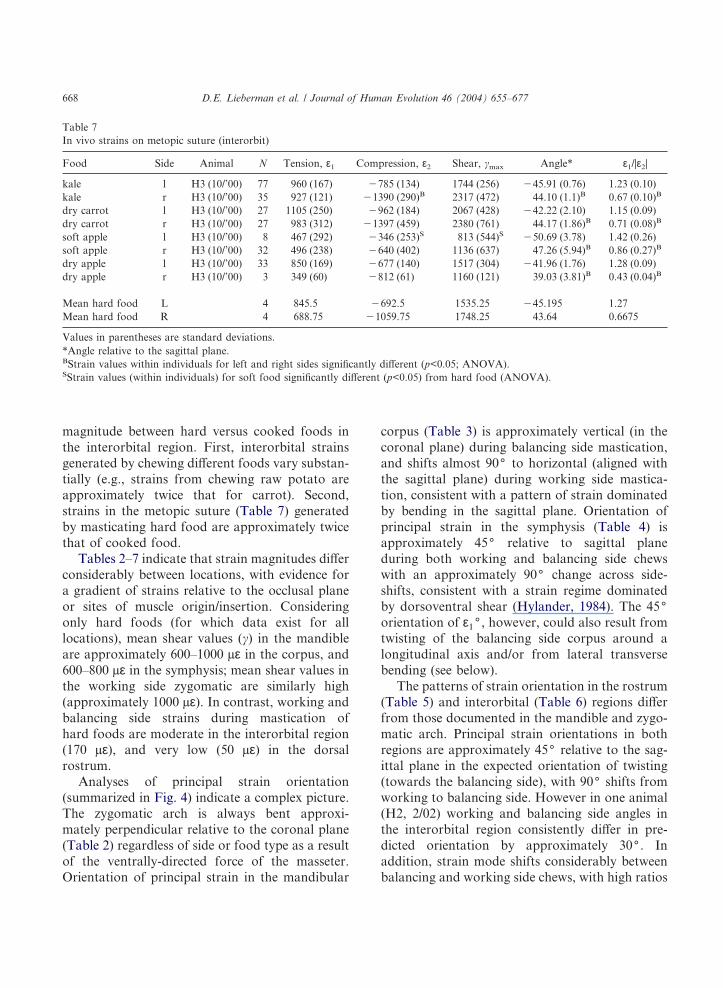
Table 7In vivo strains on metopic suture (interorbit)
Food Side Animal N Tension, ε1 Compression, ε2 Shear, �max Angle* ε1/|ε2|
kale l H3 (10/’00) 77 960 (167) �785 (134) 1744 (256) �45.91 (0.76) 1.23 (0.10)kale r H3 (10/’00) 35 927 (121) �1390 (290)B 2317 (472) 44.10 (1.1)B 0.67 (0.10)B
dry carrot l H3 (10/’00) 27 1105 (250) �962 (184) 2067 (428) �42.22 (2.10) 1.15 (0.09)dry carrot r H3 (10/’00) 27 983 (312) �1397 (459) 2380 (761) 44.17 (1.86)B 0.71 (0.08)B
soft apple l H3 (10/’00) 8 467 (292) �346 (253)S 813 (544)S �50.69 (3.78) 1.42 (0.26)soft apple r H3 (10/’00) 32 496 (238) �640 (402) 1136 (637) 47.26 (5.94)B 0.86 (0.27)B
dry apple l H3 (10/’00) 33 850 (169) �677 (140) 1517 (304) �41.96 (1.76) 1.28 (0.09)dry apple r H3 (10/’00) 3 349 (60) �812 (61) 1160 (121) 39.03 (3.81)B 0.43 (0.04)B
Mean hard food L 4 845.5 �692.5 1535.25 �45.195 1.27Mean hard food R 4 688.75 �1059.75 1748.25 43.64 0.6675
Values in parentheses are standard deviations.*Angle relative to the sagittal plane.BStrain values within individuals for left and right sides significantly different (p<0.05; ANOVA).SStrain values (within individuals) for soft food significantly different (p<0.05) from hard food (ANOVA).
D.E. Lieberman et al. / Journal of Human Evolution 46 (2004) 655–677668

(>1.0) of tensile to compressive strain on thebalancing side, and low ratios (<1.0) on the work-ing side. Comparison of Tables 6 and 7 indicatethat strains recorded on the metopic suture differfrom those measured just lateral to the sutureprimarily in terms of magnitude (mean shear val-ues on the suture are 1,641 µε, approximately sixtimes as high) and the ratio of tension/compression(which range from 0.7–1.4), but not in terms ofangle, which also indicates twisting.
Form and growth difference analyses
Table 8 summarizes and Fig. 5 illustrates resultsof the form difference analysis comparing hardversus soft diet treatment groups at the end ofthe treatment period. Only interlandmark differ-ences significant at the �=0.10 level are listed. Asone might expect for an experiment of only100 days, there are no major differences in overallcranial shape between the groups, but several
Table 8Form difference matrix analysis results (numbers match distances in Fig. 5)
Ratio
No. % (HD/SD) (HD/SD) Confidence interval
Dimensions of posterior face/neurocranium significantly larger in hard diet groupPosterior zygomatic—EOP 1 14.1 1.141 1.015–1.265Posterior zygomatic—basion 2 13.3 1.133 1.068–1.202Posterior zygomatic—opisthion 3 12.8 1.128 1.034–1.226Basion—EOP 4 7.5 1.075 1.043–1.105Frontal/zygomatic junction—basion 5 5.5 1.055 1.013–1.099Maxillary tuberosity—EOP 6 5.4 1.054 1.013–1.091Maxillary tuberosity—EOP 6 4.5 1.045 1.001–1.083Maxillary tuberosity—contralateral posterior zygomatic 7 3.2 1.032 1.006–1.064Zygomaxillare superior—basion 8 2.7 1.027 1.001–1.056
Dimensions of posterior face/neurocranium significantly smaller in hard diet groupNasion—posterior diastema 9 �7.4 0.926 0.888–0.959Maxilla at P3—ipsilateral posterior zygomatic 10 �7.1 0.929 0.883–0.978Midrostral point—ipsilateral posterior zygomatic 11 �6.3 0.937 0.880–0.991Midpalatal suture—inferior zygomatic 12 �5.7 0.943 0.893–0.996Maxilla at P3—contralateral posterior zygomatic 13 �4.9 0.951 0.909–0.991Midrostral point—contralateral posterior zygomatic 14 �4.7 0.953 0.915–0.990Nasion—posterior zygomatic 15 �4.1 0.953 0.911–0.993Nasion—inferior zygomatic 16 �3.6 0.959 0.924–0.992
Dimensions of anterior face significantly larger in hard diet groupLeft midrostral point—right maxilla at P3 17 9.8 1.098 1.011–1.188Nasale—midpalatal suture 18 7.1 1.071 1.013–1.126Right inferior zygomatic—left midrostral point 19 5.3 1.053 1.028–1.080Right inferior zygomatic—nasale 20 5.2 1.052 1.001–1.104Right inferior zygomatic—left inferior maxilla 21 3.9 1.039 1.009–1.070Right inferior zygomatic—left zygomaxillare superior 22 3.0 1.030 1.005–1.056
Dimensions of anterior face significantly smaller in hard diet groupNasion—bregma 23 �11.3 0.887 0.798–0.972Left zygomaxillare superior—bregma 24 �6.1 0.939 0.883–0.996Right frontal/zygomatic junction—right maxilla at P3 25 �4.7 0.953 0.917–0.995Opisthion—right maxilla at P3 26 �3.0 0.970 0.941–0.999
D.E. Lieberman et al. / Journal of Human Evolution 46 (2004) 655–677 669

interlandmark dimensions are significantly differ-ent in relative length between the hard- and soft-diet groups, especially in the ventral and posteriorportions of the face, and the neurocranium. In
particular, size-adjusted distances between the pos-terior zygomatic arch and landmarks on the pos-terior and inferior aspects of the cranium (externaloccipital protuberance, basion and opisthion) are12–14% greater in the hard diet group; in addition,in the hard diet group the distance from themaxillary tuberosity is approximately 5% longer tothe external occipital protuberance and 3.2%greater to the zygomatic arch. Several differencesin shape are also evident in the anterior, rostralportion of the face. Notably, interlandmark dis-tances between the lower maxilla (e.g., the mid-rostral point, nasale, the inferior maxilla) and thecontralateral zygomatic arch are significantlylonger, and the rostrum is both wider (midrostralpoint—right maxilla at P3) and taller (nasale—midpalatal suture).
In the analysis of cranofacial shape, some inter-landmark distances were smaller in the hard dietgroup than in the soft diet group, particularly inthe dorsal and rostral portions of the cranium.Such a pattern is evident from Table 8 and Fig. 4.For example, the distance from nasion to bregmais 11.3% smaller, and the distance from nasion tothe landmarks on the zygomatic arches is 3.6–7.4%smaller in the hard diet group. In addition, thedistance between the back of the face and severalpoints on the anterior rostrum tend to be relativelysmaller in the hard diet group.
Table 9 summarizes and Fig. 6 illustrates theresults of the scaled growth difference comparisonsbetween the hard and soft diet treatment groupsthat occurred during the treatment period. Onlyinterlandmark differences significant at the �=0.10level are listed. In general, differences in growthbetween the treatment groups reflect the differ-ences in shape noted above, except that there areno landmarks for which significantly less growthoccurred in the hard diet group. In particular, thehard diet group had more mediolateral and antero-posterior growth (16–17%) between the zygomaticarches and points on the posterior and inferioraspects of the cranial vault (e.g., basion, opisthion,external occipital protuberance), and 7.5–8.5%more growth between the maxillary tuberositiesand points on the neurocranium. The hard dietgroup also had more anteroposterior growth(7–8%) between the zygomatic arch and landmarks
Fig. 5. Results of EDMA form difference matrix analysis ofhyraxes fed hard versus soft food. Details of analysis aresummarized in the text. Solid lines are inter-landmark distances(scaled by the geometric mean of all interlandmark distances)significantly longer in the hard food group; dashed lines areinter-landmark distances (scaled by the geometric mean of allinterlandmark distances) significantly longer in the soft foodgroup. Numbers indicate dimensions in Table 8.
D.E. Lieberman et al. / Journal of Human Evolution 46 (2004) 655–677670

on the maxilla and rostrum. Many of the signifi-cant differences in growth are between postero-lateral and anteromedial landmarks of the midfaceand maxilla (see below).
The growth difference analysis revealed no sig-nificant differences in the growth of mandibulardimensions between landmarks identified on thehard and soft diet groups, possibly due to fewerbiologically relevant landmarks on the mandible.However, linear caliper measurements takendirectly on the mandibles and standardized bymandibular length indicate 15% greater corpusheight and 14% greater corpus thickness in thehard diet group at M1 (p<0.03, Mann–Whitney Utest). Relative height and thickness of the sym-physis, however, was not significantly differentbetween the two groups.
Discussion
The above results support the general hypoth-esis that human faces may have become relativelysmaller despite increases in body size because ofreduced levels of strain generated by chewingsofter, more processed food. This hypothesis wasdivided into several more specific hypotheses aboutstrain and growth. The first strain hypothesis, thatsoft, cooked foods generate less strain than hard,uncooked foods, was partially supported, although
the picture is somewhat complex. In particular, inthe zygomatic arch, strain generated by hard foodis approximately twice that of cooked food, par-ticularly on the balancing side; balancing but notworking side mandibular corpus strains are alsoabout twice as high when chewing hard food.Interestingly, symphysis strains did not differ sig-nificantly between hard versus soft foods, andstrain differences are less marked in regions withlower strain magnitudes such as the interorbitaland the rostrum. These results need to be corrob-orated with further experiments. Differencesbetween balancing and the working side strainspresumably result from less recruitment of balanc-ing side adductor force when chewing softer foods(a hypothesis that requires further testing withEMG data). One important point to note is thatthe above data compare strains generated by mas-ticating desiccated versus microwaved food ratherthan hard food with completely softened, nearlyliquid food. In such cases, differences in strainare even greater, approximately by an order of amagnitude (Lieberman et al., unpublished data).
The above data also support the second strainhypothesis, that the pattern of strains in the hyraxface follows a gradient in which strains are highernear the sites of occlusion and muscle insertionand dissipate dorsally away from the tooth row. Inthe hyrax, strain magnitudes are several timeshigher in the mandible and in the zygomatic arch
Table 9Growth difference matrix analysis results (scaled). (Numbers match distances in Fig. 6)
Ratio
No. % (HD/SD) (HD/SD) Confidence interval
Anterior facial dimensions with significantly more growth in hard diet groupMidrostral point—contralateral inferior zygomatic 1 8.1 1.081 1.003–1.166Inferior maxilla—contralateral inferior zygomatic 2 6.8 1.068 1.009–1.133
Posterior face/neurocranium dimensions with significantly more growth in hard diet groupPosterior zygomatic—EOP 3 17.1 1.171 1.003–1.369Posterior zygomatic—basion 4 16.5 1.165 1.046–1.304Posterior zygomatic—opisthion 5 15.9 1.159 1.027–1.310Posterior zygomatic—contralateral maxillary tuberosity 6 6.1 1.061 1.005–1.124Maxillary tuberosity—EOP 7 7.5 1.075 1.016–1.129Maxillary tuberosity—EOP 8 8.5 1.085 1.026–1.139Basion—Bregma 9 7.6 1.076 1.010–1.144Basion—EOP 10 10.5 1.105 1.022–1.183
D.E. Lieberman et al. / Journal of Human Evolution 46 (2004) 655–677 671

than in the top of the skull, resembling the gradientdocumented in several species of non-humanprimates (Hylander et al., 1991a; Hylander andJohnson, 1992; Hylander and Ravosa, 1992; Ross
and Hylander, 1996; Ravosa et al., 2000b). It ispossible that the facial gradient is a little steeper inhyraxes than in non-human primates. In macaquesand baboons, strains in the infraorbital and zygo-matic regions are about 2.8 times higher than inthe interorbital region, and about 5.8 times higherthan in the dorsal interorbital region (Hylanderet al., 1991a; Hylander and Johnson, 1992).Although gauge locations in the hyrax are slightlydifferent, working side strains in the zygomaticarch during hard food mastication are about sixtimes higher than in the interorbital region, andabout seven times higher than in the dorsal ros-trum. Although the morphological and bio-mechanical bases for strain gradients are not wellunderstood, it is interesting to note that the hyraxpattern is more similar to non-human primatesthan to miniswine (Herring et al., 2001), which areeven more prognathic than primates and have anapparently much steeper strain gradient. Forexample, in miniswine, shear strain in the zygo-matic arch is about twice that of strain in thedorsal rostrum (the maxillary bone) but approxi-mately ten times higher than in the interorbitalfrontal (Herring and Mucci, 1991; Rafferty andHerring, 1999; Herring et al., 2001). More precisecomparisons of strain gradients in hyraxes andother species, however, will require more dataon more functionally comparable locations frommultiple species, and from additional subjects.
There are also some interesting similaritiesand differences in terms of the pattern of strainorientation in the hyrax compared to the patterndocumented in various non-human primates andother mammals. As noted above, the generalpattern of strain in the hyrax mandibular corpusappears to be somewhat similar to non-humanprimates (Hylander, 1979; Hylander et al., 1987) aswell as miniswine (Herring et al., 2001), withpredominantly sagittal bending; in addition, thepattern of strain in the hyrax symphysis corre-sponds best either to dorso-ventral shearing or toa combination of wishboning and twisting—notunlike the pattern documented in non-humananthropoids (Hylander, 1984; Hylander andJohnson, 1992). However, additional data frommore subjects as well as other gauge positions arenecessary to distinguish between these different
Fig. 6. Results of EDMA growth difference matrix analysis ofhyraxes fed hard versus soft food. Details of analysis aresummarized in the text. Lines are inter-landmark dimensions inwhich significantly more growth occurred in the hard versussoft food hyrax treatment group. Numbers indicate dimensionsin Table 8. There were no inter-landmark distances in whichsignificantly more growth occurred in the soft food hyraxtreatment group.
D.E. Lieberman et al. / Journal of Human Evolution 46 (2004) 655–677672

possibilities. For example, the highly transversenature of the hyrax power stroke in combinationwith the lateral orientation of the deep massetersuggest that wishboning is likely to occur, butfurther studies are necessary with larger samplesizes, EMG data, and gauges on the dorsal andventral margins of the symphysis.
Perhaps the most interesting but puzzling differ-ence between the hyrax and some non-humanprimates is the predominance of twisting evident inthe rostrum and interorbital region in the hyrax. Ingauges mounted on the metopic suture, just off themidline in the interorbital region, and on therostrum, the angle of principal strain in the hyraxis generally close to 45( to the long axis of thecranium in the direction of occlusion, and changes90( with side shifts. This pattern has been docu-mented in swine (Herring and Teng, 2000), andnon-anthropoids such as Otolemur by Ravosaet al. (2000a,b), but not consistently in non-humananthropoids which appear to be somewhat vari-able in this regard (Hylander et al., 1991a,b; Rossand Hylander, 1996; Ross, 2001). There are severalpossibilities for this similarity to strepsirrhines, aswell as to some anthropoids (see Ross, 2001). Onepossibility may be related to retrognathy. As notedabove, the rostrum probably withstands much ofthe twisting stress generated by unilateral occlu-sion in animals whose teeth lie beneath a rostrumin front of the orbital plane. In contrast, stressesmay be greater in the orbital region in retrognathicmammals in which strains cannot be dissipatedsubstantially in the rostrum. This hypothesis seemsunlikely, however, given the substantial retro-gnathy in Aotus, which do no show strong evi-dence for twisting (Ross and Hylander, 1996).Alternatively, twisting in the hyrax skull may be afunction of its generally narrow, tubular shape inwhich little mass is distributed far from the skull’slong axis. Finally, twisting of the orbital region isalso influenced by the relative proportion of work-ing side to balancing side (W/B) adductor muscleforce, which has been estimated to be substantiallyhigher in strepsirrhines than anthropoids based onEMG activity ratios (Hylander et al., 2000). How-ever, judging from the high ratio of W/B strains inthe zygomatic arch, the hyrax probably has highW/B adductor ratios that will generate less torque,
leading to less twisting. In order to test thishypothesis, data on W/B adductor force ratios areneeded for the hyrax.
In short, the pattern of deformation docu-mented in the hyrax face is mostly consistent withtwisting in the face, bending in the zygomatic arch,and a combination of sagittal and lateral trans-verse bending in the mandibular corpus in whichstrains decline steeply from the region of occlusionto the upper face (note that the high strains in themetopic suture do not disprove the existence ofthe strain gradient described here for facial bones;strains are always higher in sutures than in bones[Herring and Teng, 2000]). In addition, strainsgenerated by masticating hard food are typicallyhigher than those generated by masticating cookedfood. Regardless of what aspects of facial shapeand muscle recruitment cause this pattern, thesestrains are predicted to influence facial growth inanimals raised on different diets. In particular, wetested three growth hypotheses: animals raised onharder, uncooked foods were predicted to havemore facial growth than animals raised on softer,more processed foods; variations in regionalgrowth were predicted to correlate with regionalstrain; and growth is predicted to occur in theplane of deformation. The experiment reportedhere examined only a short period of growth (98days) in a tiny sample. Nevertheless, the results aresignificant with respect to all three hypotheses. Thehyraxes raised on cooked food diets had signifi-cantly less facial growth than animals raised onharder foods. As noted above, the growth differ-ence matrix revealed no interlandmark distances inwhich the soft food group experienced moregrowth than the hard diet group. Instead, the hardfood animals grew more, particularly in transversedimensions between the lower face and the zygo-matic arch, and between the zygomatic arch andthe posterior portions of the cranium. These effectsare reflected in the shape differences detectedbetween the two groups. Animals raised on hardfood had transversely wider and longer faces pri-marily along the ventral aspect of the cranium,with correspondingly smaller dimensions of thedorsal portion of the rostrum and between theanterior rostrum and the posterior portions ofthe face. In addition, the mandibular corpus in the
D.E. Lieberman et al. / Journal of Human Evolution 46 (2004) 655–677 673

hard food group was significantly thicker andtaller than in the soft food group.
If we consider the strain results along the withdata on facial growth rates, then it appears poss-ible that, as predicted, variations in the amountsof regional growth correlate to some extent withregional strain magnitudes. For the most part,dimensions in the more ventral part of the facewith the highest strains (particularly the zygo-matic), experienced the most growth, and arerelatively larger in the hard versus soft diet group.Moreover, as predicted, the major vectors ofgrowth correspond approximately to the observedplanes of deformation. As noted above thecranium in the hyrax appears to be predominantlytwisted around an anteroposterior axis. The mosteffective way to counteract twisting is to add massin the coronal plane at the margins of the faceaway from the midline axis. While growth in thehyrax skull is evidently complex, the above results(Tables 8 and 9; Figs. 5 and 6) indicate that thehyraxes fed hard food had relatively more growthin posteromedial and anterolateral directions inthe more posterior portions of the face. Inaddition, the mandibular corpus is both dorso-ventrally taller and mediolaterally thicker in thehard food group, which would counteract sagittaland lateral transverse bending, respectively. How-ever, no significant differences were evident in thesymphysis where lateral transverse bending forceswere concentrated (but where no significant differ-ence in hard versus soft foods were measured),highlighting the lack of any simple correspondencebetween strain magnitudes and growth.
More detailed experiments on larger samplesizes are necessary to confirm the above results.Nonetheless, these data lend support to thehypothesis that masticating softer, more processed(cooked) foods while the animals are growing canlead to reduced facial growth in mammals withretracted molar rows. These data augment theresults of previous studies, both experimental(Bouvier and Hylander, 1981; Corruccini andBeecher, 1982, 1984; Yamada and Kimmel, 1991;Ciochon et al., 1997) and comparative (Carlson,1976; Carlson and Van Gerven, 1977; Ingervalland Helkimo, 1978; Corruccini, 1990; Varrela,1992) that indicate that a soft food diet stimulates
less growth in the cranium, particularly in themandibular and maxillary arches. However, incontrast to previous experimental studies citedabove, this study compared cooked versus driedfoods rather than hard versus nearly liquid diets.The results are therefore more comparable in somerespects to the biomechanical effects of techno-logical changes in food processing that occurredduring the last few thousand years.
One difficult problem that requires furtherresolution is the extent to which the differencesbetween hyraxes and non-human anthropoids arerelevant to human facial biomechanics. We do notwish in any way to suggest that the hyrax is astraightforward analog for human facial bio-mechanics and growth. As noted above, hyraxesdiffer from humans and other primates in anumber of important features, most crucially inthe size and position of much of the face, which inhumans is taller, wider, and oriented in the coronalplane. Biomechanically, the greatest differencebetween hyraxes very prognathic mammals such asswine and most non-human anthropoids that havebeen studied appears to be the prevalence oftwisting in the upper face. The extent to whichtwisting is a function of postcanine retrognathyor other factors needs to be examined usingadditional animal models, and will benefit fromtesting with finite element modeling (e.g., Koriothand Versluis, 1997). However, it is interestingthat the size and shape differences documented inthis study between hyraxes fed hard and cookedfood are both qualitatively and quantitativelycomparable to the differences (summarized above)in facial size that have been measured in severalhuman studies (e.g., Corruccini et al., 1985;Ingervall and Bitsanis, 1987; Varrela, 1992;Lieberman, 1998). In particular, after only threemonths, the mean difference in growth amonginterlandmark distances in which there weresignificant contrasts between treatment groups isapproximately 9%. These differences mostly werein mandibular corpus size, the size of the maxillaryarch, and the position of the zygomatic archrelative to the rest of the face.
In short, these results support the hypothesisthat human faces have become relatively smallerdespite increases in body size as a result of changes
D.E. Lieberman et al. / Journal of Human Evolution 46 (2004) 655–677674

in mechanical properties of food. Additionalresearch is needed to refine this hypothesis. Inparticular, larger sample sizes and longer treat-ment periods should reveal additional and poten-tially significant results in both the cranium andmandible, and provide more data on the natureand pattern of strains generated by mastication indifferent regions of the skull and mandible. Inaddition, further research is needed to determinethe relationship between specific parameters ofmechanical loading that cooking affects (mostnotably strain magnitude, number of strain cycles)and rates of site specific growth in the face atsutures and growth fields that generate the differ-ences in facial size and shape documented here andin other studies.
Acknowledgements
We thank Phil Amsterdam, A. Biewener, AWCrompton, Leanne DeNenno, John Landon,Naresh Nayaranaj, Masrour Makaremi, JohnPolk, Pedro Ramirez, Campbell Rolian, and LeeTuanquin for their assistance in various aspects ofthis project. Callum Ross and four anonymousreferees provided helpful comments. Financialsupport was provided by NIH SBIR DE13875-01,the Bioqual Corporation, Harvard University, andthe George Washington University.
References
Agrawal, K.R., Lucas, P.W., Prinz, J.F., Bruce, I.C., 1997.Mechanical properties of foods responsible for resistingfood breakdown in the human mouth. Arch. Oral Biol. 42,1–9.
Beecher, R.M., Corruccini, R.S., Freeman, M., 1983. Cranio-facial correlates of dietary consistency in a nonhumanprimate. J. Craniofac. Gen. Dev. Biol. 3, 193–202.
Bertram, J., Swartz, S.M., 1991. The ‘Law of Bone Transfor-mation’: A case of crying Wolff? Biol. Rev. 66, 245–273.
Biewener, A.A., 1992. In vivo measurement of bone strainand tendon force. In: Biewener, A.A. (Ed.), Biomechanics—Structures and Systems: A Practical Approach. OxfordUniversity Press, Oxford, pp. 123–147.
Biewener, A.A., Swartz, S.M., Bertram, J., 1986. Bonemodeling during growth: Dynamic strain equilibrium in thechick tibiotarsus. Calc. Tiss. Int. 39, 390–395.
Bouvier, M., Hylander, W., 1981. Effect of bone strain oncortical bone structure in macaques (M. mulatta). J.Morphol. 167, 1–12.
Bouvier, M., Hylander, W., 1996. The function of secondaryosteonal bone. Mechanical or metabolic? Arch. Oral Biol.41, 941–950.
Brace, C.L., Rosenberg, K., Hunt, K.D., 1987. Gradual changein human tooth size in the late Pleistocene andpost-Pleistocene. Evolution 41, 705–720.
Brace, C.L., Smith, S.L., Hunt, K.D., 1991. What big teeth youhad grandma! Human tooth size, past and present. In:Kelley, M.A., Larsen, C.S. (Eds.), Advances in DentalAnthropology. Wiley-Liss, New York, pp. 33–57.
Carlson, D.S., 1976. Temporal variation in prehistoric Nubiancrania. Am. J. Phys. Anthrop. 45, 467–484.
Carlson, D.S., Van Gerven, D.P., 1977. Masticatory functionand Post-Pleistocene evolution in Nubia. Am. J. Phys.Anthrop. 46, 495–506.
Carlsson, G.E., Persson, G., 1967. Morphological changesin the mandible after extraction and wearing of dentures:A longitudinal, clinical and x-ray cephalometric studycovering 5 years. Odontol. Rev. 18, 27–54.
Carter, D.R., Beaupre, G.S., 2001. Skeletal Form andFunction: Mechanobiology of Skeletal Development, Agingand Regeneration. Cambridge University Press, Cambridge.
Ciochon, R.L., Nisbett, R.A., Corruccini, R.S., 1997. Dietaryconsistency and craniofacial development related to masti-catory function in minipigs. J. Craniofac. Genet. Dev. Biol.17, 96–102.
Corruccini, R.S., 1984. An epidemiologic transition in dentalocclusion in world populations. Am. J. Orthod. 86,419–426.
Corruccini, R.S., 1990. Australian aboriginal tooth succession,interproximal attrition, and Begg’s theory. Am. J. Orthod.Dentofac. Orthop. 97, 349–357.
Corruccini, R.S., Beecher, R., 1982. Occlusal variation relatedto soft diet in a nonhuman primate. Science 218, 74–76.
Corruccini, R.S., Beecher, R., 1984. Occlusofacial morphologi-cal integration lowered in baboons raised on soft diet. J.Craniofac. Genet. Dev. Biol. 4, 135–142.
Corruccini, R.S., Henderson, A.M., Kaul, S.S., 1985. Bite forcevariation related to occlusal variation in rural and urbanPunjabi. Arch. Oral Biol. 30, 65–69.
Crompton, A.W., 1995. Masticatory function in non-mammalian cynodonts and early mammals. In: Thomason,J.J. (Ed.), Functional Morphology in VertebratePaleontology. Cambridge University Press, Cambridge, pp.55–75.
Currey, J.D., 2002. Bones: Structure and Mechanics. PrincetonUniversity Press, Princeton.
Daegling, D.J., Hylander, W.L., 1998. Biomechanics of torsionin the human mandible. Am. J. Phys. Anthrop. 105, 73–87.
Dechow, P., Carlson, D., 1983. Occlusal force and craniofacialbiomechanics during growth. Am. J. Phys. Anthrop. 83,219–237.
Demes, B., 1982. The resistance of primate skulls againstmechanical stresses. J. Hum. Evol. 11, 687–691.
D.E. Lieberman et al. / Journal of Human Evolution 46 (2004) 655–677 675

Demes, B., Creel, N., 1988. Bite force, diet and cranial mor-phology of fossil hominids. J. Hum. Evol. 17, 657–670.
English, J.D., Buschang, P.H., Throckmorton, G.S., 2002.Does malocclusion affect masticatory performance? AngleOrthod. 72, 21–27.
Engstrom, C., Kiliaridis, S., Thilander, B., 1986. The relation-ship between masticatory muscle function and cranial mor-phology. II.A histological study in the growing rat fed a softdiet. Eur. J. Orthod. 8, 271–279.
Franks, H.A., German, R.Z., Crompton, A.W., Hiiemae,K.M., 1985. Mechanism of intra-oral transport in a herbi-vore, the hyrax (Procavia syriacus). Arch. Oral Biol. 30,539–544.
German, R.Z., Franks, H.A., 1991. Timing in the movementof jaws, tongue, and hyoid during feeding in the hyrax,Procavia syriacus. J. Exp. Zool. 257, 34–42.
Greaves, W.S., 1978. The jaw lever system in ungulates: a newmodel. J. Zool. Lond. 184, 271–285.
Greaves, W.S., 1985. The mammalian postorbital bar as atorsion-resisting helical strut. J. Zool. Lond. 207, 125–136.
Griner, L.A., 1968. The rock hyrax (Procavia capensis): apotential laboratory animal. Lab. Anim. Care 18, 144–150.
Herring, S.W., Rafferty, K.L., Liu, Z.J., Marshall, C.D., 2001.Jaw muscles and the skull in mammals: the biomechanics ofmastication. Comp. Biochem. Physiol. A Mol. Integr.Physiol. 131, 207–219.
Herring, S.W., Mucci, R.J., 1991. In vivo strain in cranialsutures: the zygomatic arch. J. Morphol. 207, 225–239.
Herring, S.W., Teng, S., 2000. Strain in the braincase and itssutures during function. Am. J. Phys. Anthrop. 112,575–593.
Herring, S.W., Teng, S., Huang, X., Mucci, R.J., Freeman, J.,1996. Patterns of bone strain in the zygomatic arch. Anat.Rec. 246, 446–457.
Hylander, W.L., 1979. The functional significance of primatemandibular form. J. Morphol. 160, 223–239.
Hylander, W.L., 1984. Stress and strain in the mandibularsymphysis of primates: A test of competing hypotheses. Am.J. Phys. Anthrop. 64, 1–46.
Hylander, W.L., 1985. Mandibular function and biomechanicalstress and scaling. Am. Zool. 25, 315–330.
Hylander, W.L., 1986. In vivo bone strain as an indicator ofmasticatory force in Macaca fascilularis. Arch. Oral Biol.31, 149–157.
Hylander, W.L., 1988. Implications of in vivo experiments forinterpreting the functional significance of “robust” austra-lopithecine jaws. In: Grine, F. (Ed.), Evolutionary Historyof the “Robust” Australopithecines. Aldine De Gruyter,New York, pp. 55–83.
Hylander, W.L., Johnson, K.R., 1992. Strain gradients in thecraniofacial region of primates. In: Davidovitch, Z. (Ed.),The Biological Mechanisms of Tooth Movement andCraniofacial Adaptation. Ohio State University College ofDentistry, Columbus, pp. 559–569.
Hylander, W.L., Ravosa, M.J., 1992. An analysis of thesupraorbital region of primates: a morphometric and exper-
imental approach. In: Smith, P., Tchernov, E. (Eds.), Struc-ture, Function, and Evolution of Teeth. Freund, Tel Aviv,pp. 233–255.
Hylander, W.L., Johnson, K.R., Crompton, A.W., 1987. Load-ing patterns and jaw movement during mastication inMacaca fascicularis: A bone strain, electromyographic andcineradiographic analysis. Am. J. Phys. Anthrop. 72,287–314.
Hylander, W.L., Picq, P.G., Johnson, K.R., 1991a.Masticatory-stress hypotheses and the supraorbital regionof primates. Am. J. Phys. Anthrop. 86, 1–36.
Hylander, W.L., Picq, P.G., Johnson, K.R., 1991b. Function ofthe supraorbital region of primates. Arch. Oral Biol. 36,273–281.
Hylander, W.L., Ravosa, M.J., Ross, C.F., Wall, C.E.,Johnson, K.R., 2000. Symphyseal fusion and jaw-adductormuscle force. Am. J. Phys. Anthrop. 107, 257–271.
Ingervall, B., Bitsanis, E., 1987. A pilot study on the effect ofmasticatory muscle training on facial growth in long-facechildren. Eur. J. Orthodont. 9, 15–23.
Ingervall, B., Helkimo, E., 1978. Masticatory muscle force andfacial morphology in man. Arch. Oral Biol. 23, 203–206.
Israel, H., 1973. Age factor and the pattern of change incraniofacial structures. Am. J. Phys. Anthrop. 39, 111–128.
Janis, C.M., 1979. Mastication in the hyrax and its relevance toungulate dental evolution. Paleobiology 5, 50–59.
Janis, C.M., 1983. Muscle of the masticatory apparatus in twogenera of Hyrax (Procavia and Heterohyrax). J. Morphol.176, 61–87.
Kiliaridis, S., 1989. Muscle function as a determinant ofmandibular growth in normal and hypocalcaemic rat. Eur.J. Orthod. 11, 298–308.
Kiliaridis, S., 1995. Masticatory muscle influence on cranio-facial growth. Acta Odontol. Scand. 53, 196–202.
Kiliaridis, S., Engstrom, C., Chavez, L.M.E., 1992. Influenceof masticatory muscle function on craniofacial growth inhypocalcemic rats. Scand. J. Dent. Res. 100, 330–336.
Kiliaridis, S., Engstrom, C., Thilander, B., 1986. The relation-ship between masticatory muscle function and cranialmorphology. I. A cephalometric longitudinal analysis inthe growing rat fed a soft diet. Eur. J. Orthodont. 7,273–283.
Kiliaridis, S., Mejersjo, C., Thilander, B., 1989. Muscle func-tion and craniofacial morphology: a clinical study inpatients with myotonic dystrophy. Eur. J. Orthodont. 11,131–138.
Korioth, T.W.P., Versluis, A., 1997. Modeling the mechanicalbehavior of the jaws and their related structures by finiteelement (FE) analysis. Crit. Rev. Oral Biol. Med. 8, 90–104.
Lele, S., 1993. Euclidean Distance Matrix Analysis (EDMA):Estimation of mean form and mean form difference.Mathematical Geology 25, 573–602.
Lele, S., Richtsmeier, J.T., 1991. Euclidean Distance MatrixAnalysis: A coordinate-free approach for comparing bio-logical shapes using landmark data. Am. J. Phys. Anthrop.86, 415–427.
D.E. Lieberman et al. / Journal of Human Evolution 46 (2004) 655–677676

Lele, S., Richtsmeier, J.T., 1995. Euclidean distance matrixanalysis: Confidence intervals for form and growthdifferences. Am. J. Phys. Anthrop. 98, 73–86.
Lieberman, D.E., 1993. Life history variables preserved indental cementum microstructure. Science 261, 1162–1164.
Lieberman, D.E., 1998. Sphenoid shortening and the evolutionof modern human cranial shape. Nature 393, 158–162.
Lieberman, D.E., Crompton, A.W., 2000. Why fuse the man-dibular symphysis: A comparative analysis. Am. J. Phys.Anthrop. 112, 517–540.
Lieberman, D.E., Pearson, O.M., Polk, J.D., Demes, B.,Crompton, A.W., 2003. Optimization of bone growth andremodeling in response to loading in tapered mammalianlimbs. J. Exp. Biol. 206, 3125–3138.
Lucas, P.W., Luke, D.A., 1984. Chewing it over—basic princi-ples of food breakdown. In: Chivers, D.J., Wood, B.A.,Bilsborough, A. (Eds.), Food Acquisition and Processing inPrimates. Plenum, New York, pp. 283–302.
Lukacs, J.R., 1989. Dental Paleopathology: methods for recon-structing dietary patterns. In: Iscan, M.R., Kennedy,K.A.R. (Eds.), Reconstruction of Life from the Skeleton.Alan R Liss, New York, pp. 261–286.
Martin, R.B., Burr, D.B., Sharkey, N., 1998. Skeletal TissueMechanics. Springer, New York.
Moore, W.J., 1965. Masticatory function and skull growth. J.Zool. Lond. 146, 123–131.
Preuschoft, H., Demes, B., Meyer, M., Bar, H.F., 1986. Thebiomechanical principles realized in the upper jaw of long-snouted primates. In: Else, J.G., Lee, P.C. (Eds.), PrimateEvolution. Cambridge University Press, Cambridge, pp.249–264.
Rafferty, K.L., Herring, S.W., 1999. Craniofacial sutures: mor-phology, growth, and in vivo masticatory strains. J.Morphol. 242, 167–179.
Rafferty, K.L., Herring, S.W., Marshall, C.D., 2003. Bio-mechanics of the rostrum and the role of facial sutures. J.Morphol. 257, 233–244.
Rangel, R.D., Oyen, O.J., Russell, M.D., 1985. Changes inmasticatory biomechanics and stress magnitude that affectgrowth and development of the facial skeleton. Prog. Clin.Biol. Res. 187, 281–293.
Ravosa, M.J., Noble, V.E., Hylander, W.L., Johnson, K.R.,Kowalski, E.M., 2000a. Masticatory stress, orbital orienta-tion and the evolution of the primate postorbital bar. Jhum. Evol. 38, 667–693.
Ravosa, M.J., Johnson, K.R., Hylander, W.L., 2000b. Strain inthe galago facial skull. J. Morphol. 245, 51–66.
Richards, M.P., Pettitt, P.B., Stiner, M.C., Trinkaus, E., 2001.Stable isotope evidence for increasing dietary breadth in theEuropean mid-Upper Paleolithic. Proc. Natl. Acad. Sci. 98,6528–6532.
Richtsmeier, J.T., Lele, S., 1993. A coordinate-free approach tothe analysis of growth patterns: Models and theoreticalconsiderations. Biol. Rev. 68, 381–411.
Ross, C.F., 2001. In vivo function of the craniofacial haft: theinterorbital “pillar”. Am. J. Phys. Anthrop. 116, 108–139.
Ross, C.F., Hylander, W.L., 1996. In vivo and in vitro bonestrain in the owl monkey circumorbital region and thefunction of the postorbital septum. Am. J. Phys. Anthrop.101, 183–215.
Rubin, C.T., Lanyon, L.E., 1984. Dynamic Strain Similarity invertebrates: an alternative to allometric limb bone scaling. J.Theoret. Biol. 107, 321–327.
Rubsamen, K., Hume, I.D., Englehard, W.V., 1982. Physiologyof the rock hyrax. Comp. Biochem. Physiol. 72A, 271–277.
Shiau, Y.Y., Peng, C.C., Hsu, C.W., 1999. Evaluation of bitingperformance with standardized test-foods. J. Oral Rehabil.26, 447–452.
Spencer, M.A., 1999. Constraints on masticatory system evolu-tion in anthropoid primates. Am. J. Phys. Anthrop. 108,450–483.
Spencer, M.A., 1998. Force production in the primate mastica-tory system: electromyographic tests of biomechanicalhypotheses. J. Hum. Evol. 34, 25–54.
Stiner, M.C., 2001. Thirty years on the “broad spectrumrevolution” and paleolithic demography. Proc. Natl. Acad.Sci. 98, 6993–6996.
Stiner, M.C., Munro, N.D., Surovell, T.A., Tchernov, E.,Bar-Yosef, O., 1999. Paleolithic population growth pulsesevidenced by small animal exploitation. Science 283,190–194.
Strait, S., 1997. Tooth use and the physical properties of food.Evol. Anthrop. 5, 199–211.
Sugimura, T., Inada, J., Sawa, S., Kakudo, Y., 1984. Dynamicresponses of the skull caused by loss of occlusal force. J.Osaka Dental Univ. 18, 29–42.
Varrela, J., 1992. Dimensional variation of craniofacial struc-tures in relation to changing masticatory-functionaldemands. Eur. J. Orthodont. 14, 31–36.
Wen, P.T., 1984. Electromyographic and CineradiographicStudy of Translation in the Hyrax. Unpub. Thesis: HarvardUniversity.
Wrangham, R.W., Conklin-Brittain, N., 2003. Cooking as abiological trait. Comp. Biochem. Physiol. A 136, 35–46.
Wrangham, R.W., Jones, J.H., Laden, G., Pilbeam, D.,Conklin-Brittain, N., 1999. The raw and the stolen. Cook-ing and the ecology of human origins. Curr. Anthrop. 40,567–594.
Yamada, K., Kimmel, D.B., 1991. The effect of dietary consist-ency on bone mass and turnover in the growing ratmandible. Arch. Oral Biol. 36, 129–138.
D.E. Lieberman et al. / Journal of Human Evolution 46 (2004) 655–677 677