European Guideline Craniofacial Microsomia
100
European Guideline Craniofacial Microsomia Ruben W. Renkema, MD and the ERN CRANIO Working Group on Craniofacial Microsomia Index Summary 2387 Chapter 1. Introduction 2393 1.1 Incentive for making the guideline 2393 1.2 Purpose of the guideline 2393 1.3 Scope of the guideline 2393 1.4 Relationship to other congenital facial malformations 2394 1.5 Intended users of the guideline 2394 1.6 About craniofacial microsomia 2394 1.7 European Reference Networks 2394 Chapter 2. Methodology 2394 2.1 Validity of the guideline 2394 2.2 General information 2394 2.3 Aim and target audience guideline 2394 2.3.1 Aim of the Guideline 2394 2.3.2 Target audience 2395 2.3.3 Patient population 2395 2.4 Steering group 2395 2.5 Conflicts of interest 2395 2.6 Patient perspectives 2396 2.7 Implementation 2397 2.8 Methods 2397 2.8.1 Bottleneck analysis 2397 2.8.2 Questions and outcomes 2397 2.8.3 Literature search and selection of literature 2397 2.8.4 Quality assessment of individual studies 2398 2.8.5 Summary of literature 2398 2.8.6 Quality of evidence 2398 2.8.7 Formulating conclusions 2398 2.8.8 Considerations 2398 2.8.9 Formulating recommendations 2400 2.8.10 Conditions (organisation of care) 2400 2.8.11 Knowledge gap 2400 2.8.12 Evaluation and authorisation phase 2400 Chapter 3. Diagnostic criteria for craniofacial microsomia 2400 3.1 Based on which criteria is a child or adult with craniofacial microsomia diagnosed? 2401 Chapter 4. Screening, monitoring and indication for treatment 2403 4.1 Breathing difficulties in craniofacial microsomia 2403 From the Department of Plastic and Reconstructive Surgery and Hand Surgery and Department of Maxillofacial Surgery, Dutch Craniofacial Center, Erasmus Medical Center, Rotterdam, The Netherlands. Received March 29, 2020. Accepted for publication April 21, 2020. Address correspondence and reprint requests to Ruben W. Renkema, Dutch Craniofacial Center, Erasmus Medical Center, Rotterdam, The Netherlands; E-mail: [email protected] Supplemental digital contents are available for this article. Direct URL citations appear in the printed text and are provided in the HTML and PDF versions of this article on the journal’s Web site (www.jcraniofacialsurgery.com). This is an open access article distributed under the terms of the Creative Commons Attribution-Non Commercial-No Derivatives License 4.0 (CCBY-NC-ND), where it is permissible to download and share the work provided it is properly cited. The work cannot be changed in any way or used commercially without permission from the journal. Copyright # 2020 The Author(s). Published by Wolters Kluwer Health, Inc. on behalf of Mutaz B. Habal, MD. ISSN: 1049-2275 DOI: 10.1097/SCS.0000000000006691 EDUCATIONAL SUPPLEMENT The Journal of Craniofacial Surgery Volume 31, Number 8S, November/December 2020 2385
-
Upload
khangminh22 -
Category
Documents
-
view
1 -
download
0
Transcript of European Guideline Craniofacial Microsomia

Index
Summary
Chapter 1. I
1.1 Incen
1.2 Purpo
1.3 Scope
1.4 Relat
1.5 Intend
1.6 Abou
1.7 Europ
Chapter 2. M
2.1 Valid
2.2 Gener
2.3 Aim a
2.3.1 A
2.3.2 T
2.3.3 P
2.4 Steeri
2.5 Confl
2.6 Patien
2.7 Imple
2.8 Metho
2.8.1 B
2.8.2 Q
2.8.3 L
2.8.4 Q
2.8.5 S
2.8.6 Q
2.8.7 F
2.8.8 C
2.8.9 F
2.8.10
2.8.11
2.8.12
Chapter 3. D
3.1 Based
From the DErasmus
Received MAccepted foAddress cor
E-mail:Supplement
this articThis is an op
where itpermissi
Copyright #
ISSN: 1049DOI: 10.109
EDUCATIONAL SUPPLEMENT
The Journa
European Guideline Craniofacial MicrosomiaRuben W. Renkema, MD and the ERN CRANIO Working Group on Craniofacial Microsomia
2387
ntroduction 2393
tive for making the guideline 2393
se of the guideline 2393
of the guideline 2393
ionship to other congenital facial malformations 2394
ed users of the guideline 2394
t craniofacial microsomia 2394
ean Reference Networks 2394
ethodology 2394
ity of the guideline 2394
al information 2394
nd target audience guideline 2394
im of the Guideline 2394
arget audience 2395
atient population 2395
ng group 2395
icts of interest 2395
t perspectives 2396
mentation 2397
ds 2397
ottleneck analysis 2397
uestions and outcomes 2397
iterature search and selection of literature 2397
uality assessment of individual studies 2398
ummary of literature 2398
uality of evidence 2398
ormulating conclusions 2398
onsiderations 2398
ormulating recommendations 2400
Conditions (organisation of care) 2400
Knowledge gap 2400
Evaluation and authorisation phase 2400
iagnostic criteria for craniofacial microsomia 2400
on which criteria is a child or adult with craniofacial microsomia diagnosed? 2401
2403
2403
epartment ofMedical Cenarch 29, 2020r publicationrespondencer.renkema@eal digital contle on the jouen access artiis permissibleon from the j
2020 The A-22757/SCS.00000
l of Cranio
es in craniofacial micros
Chapter 4. Screening, monitoring and indication for treatment
4.1 Breathing difficulti omia
Plastic and Reconstructive Surgery and Hand Surgery and Department of Maxillofacial Surgery, Dutch Craniofacial Center,ter, Rotterdam, The Netherlands..April 21, 2020.and reprint requests to Ruben W. Renkema, Dutch Craniofacial Center, Erasmus Medical Center, Rotterdam, The Netherlands;rasmusmc.nlents are available for this article. Direct URL citations appear in the printed text and are provided in the HTML and PDF versions ofrnal’s Web site (www.jcraniofacialsurgery.com).cle distributed under the terms of the Creative Commons Attribution-Non Commercial-No Derivatives License 4.0 (CCBY-NC-ND),to download and share the work provided it is properly cited. The work cannot be changed in any way or used commercially without
ournal.uthor(s). Published by Wolters Kluwer Health, Inc. on behalf of Mutaz B. Habal, MD.
00000006691
facial Surgery � Volume 31, Number 8S, November/December 2020 2385

4.1.1 What is the type, prevalence and severity of breathing difficulties in craniofacial microsomia? 2404
4.1.2 What is the policy for screening and monitoring of breathing difficulties (OSA) in patients with craniofacial microsomia? 2405
4.1.3 What are the indications and policy for treatment of breathing difficulties (OSA) in patients with craniofacial microsomia? 2406
4.2 Feeding difficulties in craniofacial microsomia 2409
4.2.1 What is the type, prevalence and severity of feeding difficulties in craniofacial microsomia? 2409
4.2.2 What is the policy for screening and monitoring of feeding difficulties in patients with craniofacial microsomia? 2411
4.2.3 What are the indications and policy for treatment of feeding difficulties in patients with craniofacial microsomia? 2412
4.3 Speech and Language difficulties in craniofacial microsomia 2413
4.3.1 What is the type, prevalence and severity of speech difficulties in craniofacial microsomia? 2414
4.3.2 What is the policy for screening and monitoring of speech and language difficulties in patients with craniofacial microsomia? 2416
4.3.3 What are the indications and policy for treatment of speech difficulties in patients with craniofacial microsomia? 2418
4.4 Hearing difficulties in craniofacial microsomia 2420
4.4.1 What is the type, prevalence and severity of hearing difficulties in craniofacial microsomia? 2420
4.4.2 What is the policy for screening and monitoring of hearing difficulties in patients with craniofacial microsomia? 2424
4.4.3 What are the indications and policy for treatment of hearing difficulties in patients with craniofacial microsomia? 2425
4.5 Eye anomalies in craniofacial microsomia 2426
4.5.1 What is the type, prevalence and severity of eye anomalies in craniofacial microsomia? 2427
4.5.2 What is the policy for screening and monitoring of eye anomalies in patients with craniofacial microsomia? 2428
4.5.3 What are the indications and policy for treatment of eye anomalies in patients with craniofacial microsomia? 2429
4.6 Dental deformities in craniofacial microsomia 2432
4.6.1 What is the type, prevalence and severity of dental deformities in craniofacial microsomia? 2432
4.6.2 What is the policy for screening and monitoring of dental deformities in patients with craniofacial microsomia? 2433
4.6.3 What are the indications and policy for treatment of dental deformities in patients with craniofacial microsomia? 2434
4.7 Vertebral anomalies in craniofacial microsomia 2436
4.7.1 What is the type, prevalence and severity of vertebral anomalies in craniofacial microsomia? 2437
4.7.2 What is the policy for screening and monitoring of vertebral anomalies in patients with craniofacial microsomia? 2438
4.7.3 What are the indications and policy for treatment of vertebral anomalies in patients with craniofacial microsomia? 2439
4.8 Psychosocial difficulties in craniofacial microsomia 2442
4.8.1 What are the type, prevalence and severity of psychosocial difficulties in craniofacial microsomia? 2443
4.8.2 What is the policy for the screening and monitoring of psychosocial difficulties in patients with craniofacial microsomia? 2445
4.8.3 What are the indications and policies for the treatment of psychosocial difficulties in patients with craniofacial microsomia? 2446
Chapter 5. Surgical treatment 2449
5.1 Mandible & Maxilla 2449
5.1.1 What is the indication for surgical treatment of mandibular and maxillary deformity in patients with craniofacial microsomia? 2450
5.1.2 What is the most optimal treatment modality and its timing for mandibular/maxillary deformity in patients with craniofacialmicrosomia regarding severity, breathing problems, occlusal problems and aesthetics?
2452
5.2 Facial nerve 2460
5.2.1 What is the indication for surgical treatment of facial nerve anomaly in patients with craniofacial microsomia? 2461
5.2.2 What is the most optimal treatment modality for facial nerve anomaly in patients with craniofacial microsomiarelated to functional deficits and aesthetics?
2463
5.3 Soft tissues 2468
5.3.1 What is the indication for surgical treatment of soft tissue deficiency in patients with craniofacial microsomia? 2469
5.3.2 What is the most optimal treatment modality for soft tissue deficiency in patients with craniofacial microsomiarelated to severity and its timing?
2470
5.4 Microtia 2473
5.4.1 What is the indication for surgical treatment of ear deformity in patients with craniofacial microsomia? 2474
5.4.2 What is the most optimal treatment modality for ear deformity in patients with craniofacial microsomia related to its timing? 2475
Chapter 6. Organisation of care 2480
Minimal care standards and monitoring outcomes 2480
6.1 What are the minimal care standards to treat patients with craniofacial microsomia and how should outcomes of care be monitored? 2481
Appendix 1. Additional questions
Appendix 2. Literature searches
Appendix 3. Bottlenecks from patient’s perspective
Appendix 4. Definitions
Appendix 5. Reporting speech outcomes
Appendix 6. Evidence table
Renkema et al The Journal of Craniofacial Surgery � Volume 31, Number 8S, November/December 2020
2386 # 2020 The Author(s). Published by Wolters Kluwer Health, Inc. on behalf of Mutaz B. Habal, MD
The Journal of Craniofacial Surgery � Volume 31, Number 8S, November/December 2020 European Guideline Craniofacial Microsomia
SUMMARY
T his guideline provides the following chapters:
Chapter 3 Diagnostic criteria for craniofacial microsomia
Chapter 4 Screening, monitoring and indication for treatment
4.1 Breathing difficulties in craniofacial microsomia
4.2 Feeding difficulties in craniofacial microsomia
4.3 Speech difficulties in craniofacial microsomia
4.4 Hearing difficulties in craniofacial microsomia
4.5 Eye anomalies in craniofacial microsomia
4.6 Dental deformities in craniofacial microsomia
4.7 Vertebral anomalies in craniofacial microsomia
4.8 Psychosocial difficulties in craniofacial microsomia
Chapter 5 Surgical treatment of craniofacial microsomia
5.1 Mandible & Maxilla
5.2 Facial nerve
5.3 Soft tissues
5.4 Microtia
Chapter 6 Organisation of care
The following recommendations were agreed on:Chapter 3. Diagnostic criteria for craniofacial microsomia3.1 On which criteria is a child or adult with craniofacial
microsomia diagnosed?
#
Terminology
� It is advised to exclusively use the term craniofacialmicrosomia. Discard the use of other terms such asGoldenhar syndrome, hemifacial microsomia or aur-
2020 T
iculo-oculo-vertebral spectrum.
Diagnostic criteria� It is advised to use the diagnostic criteria for craniofacial
microsomia developed by the ICHOM Craniofacial Microsomiagroup.
CFM is defined by:
he Author(s). Published
2 major criteria, or1 major þ 1 minor criteria, or3þ minor criteria
Major criteria
Mandibular hypoplasiaMicrotiaOrbital / facial bone hypoplasiaAsymmetric facial movementMinor criteria
Facial soft tissue deficiencyPre-auricular tagsMacrostomiaCleftingEpibulbar dermoidsHemivertebraeChapter 4.1 Breathing difficulties in craniofacial microsomia4.1.1 What is the type, prevalence and severity of breathing
difficulties in craniofacial microsomia?Since this question does not relate to interventions or diagnos-
tics, only conclusions without any considerations, rationale orrecommendations are provided.
by Wolters Kluwer Health, Inc. on b
4.1.2 What is the policy for screening and monitoring ofbreathing difficulties (OSA) in patients with craniofacial micro-somia?
eh
� All patients with craniofacial microsomia should bescreened with a questionnaire biannually, at least up tothe age of six, in the outpatient department for a clinicalhistory of obstructive sleep apnoea.
� If there is a suspicion of obstructive sleep apnoea basedon a questionnaire, a polysomnography (sleep study)
alf of
has to be performed.
� All patients who have Pruzansky-Kaban IIb or IIImandibles and/or are bilaterally affected have toundergo a polysomnography (sleep study) to screen
for obstructive sleep apnoea in the first year of life.4.1.3 What are the indications and policy for treatment ofbreathing difficulties (OSA) in patients with craniofacial micro-somia?
� Treatment of children with craniofacial microsomia andobstructive sleep apnoea has to be discussed in a
multidisciplinary team.� Treatment of children with craniofacial microsomia andobstructive sleep apnoea depends on the age of the child,
the severity of symptoms and the level of obstruction.� In older children with mild to severe obstructive sleepapnoea, adenotonsillectomy (ATE) may be the treat-
ment of first choice.� In young infants and children with craniofacialmicrosomia and obstructive sleep apnoea non-surgicalrespiratory support has to be considered to treat
obstructive sleep apnoea.� In children with craniofacial microsomia and severeobstructive sleep apnoea a tracheostomy has to be
considered at all ages.� Mandibular distraction osteogenesis (MDO) should beconsidered to treat patients with severe obstructive sleepapnoea who have a tracheostomy or to reduce the
necessity for a tracheostomy or respiratory support.Mutaz B. Habal, MD 2387
Renkema et al The Journal of Craniofacial Surgery � Volume 31, Number 8S, November/December 2020
Chapter 4.2 Feeding difficulties in craniofacial microsomia
4.2.1 What is the type, prevalence and severity of feeding
difficulties in craniofacial microsomia?Since this question does not relate to interventions or diagnos-tics, only conclusions without any considerations, rationale orrecommendations are provided.
4.2.2 What is the policy for screening and monitoring of feedingdifficulties in patients with craniofacial microsomia?
23
� Children with craniofacial microsomia should bescreened with a questionnaire biannually, at least upto the age of six, and monitored regularly for feeding
88
difficulties by a paediatrician or multidisciplinary team.
� The WHO or national Growth Charts can be used to
monitor growth and screen for feeding difficulties.� A speech and language therapist should be involved in
patients who require tube feeding.4.2.3 What are the indications and policy for treatment offeeding difficulties in patients with craniofacial microsomia?
� Children with craniofacial microsomia with feedingdifficulties should be treated by a multidisciplinary
team.� Feeding strategies are guided by the severity of feeding
difficulties.Chapter 4.3 Speech difficulties in craniofacial microsomia4.3.1 What is the type, prevalence and severity of speech and
language difficulties in craniofacial microsomia?Since this question does not relate to interventions or diagnos-
tics, only conclusions without any considerations, rationale orrecommendations are provided.
4.3.2 What is the policy for screening and monitoring of speechand language difficulties in patients with craniofacial microsomia?
� Screen preverbal communication and babbling skills atthe age of nine months to decide if interventionis warranted.
� Evaluate receptive and expressive language skills at theage of two years and biannually until the age of eightyears in all patients with craniofacial microsomia.Those identified with difficulties should be referred totheir community speech and language therapist servicefor ongoing intervention.
� Oral-facial evaluation of structure and function isrecommended at each screening consultation to
# 2020 The Author(s). Published by
examine any impact of asymmetry on speech produc-tion. This should include examination of facialsymmetry, lips, dental occlusion and intra-oral exami-nation of tongue movement, dentition, hard palate andsoft palate movement on sustained ‘ah’ vowel.
� Screen patients with tracheostomy for speaking valvesuitability or an augmentative and alternative commu-nication system.
� Social communication skills should be monitored intangent with all of the afore-mentioned communicationskills.
� Children with craniofacial microsomia and associatedcleft palate should be screened annually from 2–5 yearsby the Cleft-Craniofacial speech and language therapistand should follow the local Cleft Palate Protocol.Velopharyngeal dysfunction should be assessed fromthe age of two years or when verbal output has emerged.
� Children with craniofacial microsomia without a cleftpalate should also be screened at the age of two years toexamine for potential risk of velopharyngeal dysfunc-tion related to their asymmetrical structure. If velo-pharyngeal dysfunction is identified, these childrenshould follow the same pathway as children with a
Wolte
cleft palate.
4.3.3 What are the indications and policy for treatment of speechand language difficulties in patients with craniofacial microsomia?
� Recommend early language stimulation for delayedbabble onset from nine months.
� Facilitate receptive and expressive language develop-ment using a range of behavioural techniques such asmodelling, imitation, repetition and extension.
� Patients with cleft speech characteristics should havearticulation therapy when identified. Direct therapyusing an articulation approach is recommended fromage three onwards
� Monitor patients with tracheostomy and speaking valveuse on a regular base.
� Introduce low or high tech augmentative and alternativecommunication systems to children who are non-verbalor whose speech is unintelligible. These includegestures, signing, symbols, word boards, communica-tion boards and books, as well as Voice OutputCommunication Aids (VOCAs). A low-tech systemsuch as signing can be introduced from one.
� Intervention for social communication difficulties isrecommended; e.g. development of non-verbal com-munication skills (e.g. eye contact, turn-taking);conversational skills, recognitions of emotions and
emotional regulation.Chapter 4.4 Hearing difficulties in craniofacial microsomia4.4.1 What is the type, prevalence and severity of hearing
difficulties in craniofacial microsomia?Since this question does not relate to interventions or diagnos-
tics, only conclusions without any considerations, rationale orrecommendations are provided.
4.4.2 What is the policy for screening and monitoring of hearingdifficulties in patients with craniofacial microsomia?
rs Kluwer Health, Inc. on behalf of Mutaz B. Habal, MD
The Journal of Craniofacial Surgery � Volume 31, Number 8S, November/December 2020 European Guideline Craniofacial Microsomia
#
� Perform neonatal hearing test in all new-borns withcraniofacial microsomia. If indicated, complete audio-logical evaluation in an experienced audiology centreshould be performed before the age of three months toensure timely treatment.
� Re-evaluate hearing tests in patients with craniofacialmicrosomia by the age of 24–30 months.
� Regularly perform otoscopy and audiometry in patientswith craniofacial microsomia including microtia and/orcleft palate by the ENT doctor/otolaryngologist.
� Audiologic intervention should be initiated before theage of six months in patients with congenital hearing
2020 T
loss.
4.4.3 What are the indications and policy for treatment ofhearing difficulties in patients with craniofacial microsomia?
� Treat moderate to severe hearing loss, either with non-surgical or surgical options.
� Coordinate surgical approach and timing in a multidis-ciplinary team regarding hearing augmentation andother surgical procedures including ear reconstruction
and mandibular surgeries.Chapter 4.5 Eye anomalies in craniofacial microsomia4.5.1 What is the type, prevalence and severity of eye anomalies
in craniofacial microsomia?Since this question does not relate to interventions or diagnos-
tics, only conclusions without any considerations, rationale orrecommendations are provided.
4.5.2 What is the policy for screening and monitoring of eyeanomalies in patients with craniofacial microsomia?
� All patients with craniofacial microsomia should bescreened at least once during the visual development(before the age of five) by an orthoptist andophthalmologist. Depending on the results, follow-up
visits need to be scheduled on a regular basis.4.5.3 What are the indications and policy for treatment of eyeanomalies in patients with craniofacial microsomia?
� Children with ocular disturbances need to be evaluatedby a specialised orthoptist and ophthalmologist duringthe visual development (before the age of five).
� Optimal spectacle correction should be provided in caseof a refractive error.
� Amblyopia should be treated before the age of six.� When surgery is considered this has to be discussed in a
multidisciplinary team, carefully evaluating the harmsand the benefits, especially in the case of young childrenin whom vision is still developing.
� Ultrasound imaging of the ocular dermoid needs to beconducted if extension posteriorly and into the orbit
is suspected.he Author(s). Published by Wolters Kluwer Health, Inc. on b
Chapter 4.6 Dental deformities in craniofacial microsomia4.6.1 What is the type, prevalence and severity of dental
deformities in craniofacial microsomia?Since this question does not relate to interventions or diagnos-
tics, only conclusions without any considerations, rationale orrecommendations are provided.
4.6.2 What is the policy for screening and monitoring of dentaldeformities in patients with craniofacial microsomia?
eh
� Patients with craniofacial microsomia should haveroutine dental care.
� Patients with craniofacial microsomia should be seenfrom age five by an orthodontist within a multidisci-plinary team to diagnose dental deformities.
� Perform screening for dental deformities by intra-oralinspection and standard dental records.
� Take orthodontic records in a structured schedule, at 6,
alf of
9, 12, 15 and 18 years of age.
4.6.3 What are the indications and policy for treatment of dentaldeformities in patients with craniofacial microsomia?
� Dentofacial orthopaedic treatment can be consideredappropriate in very mild craniofacial microsomiacases. In severe craniofacial microsomia patients,current evidence does not promote activatortreatment.
� Orthodontic treatment should be discussed and coordi-nated in a multidisciplinary team depending on the
decision to conduct orthognathic surgery or not.Chapter 4.7 Vertebral anomalies in craniofacialmicrosomia
4.7.1 What is the type, prevalence and severity of vertebralanomalies in craniofacial microsomia?
Since this question does not relate to interventions or diagnos-tics, only conclusions without any considerations, rationale orrecommendations are provided.
4.7.2 What is the policy for screening and monitoring ofvertebral anomalies in patients with craniofacial microsomia?
� Screening questions and clinical examinations relatedto neck/back symptoms should be undertaken atinitial consultation and as part of pre-operativeworkup.
� All patients with craniofacial microsomia who haveneurologic symptoms (e.g., paraesthesia, numbness, orweakness) or neck pain suggestive of neuronal injuryshould be evaluated as soon as possible by a(paediatric) neurologist.
� Patients should be referred appropriately and attentionto the cervical spine should be payed when patients are
undergoing general anaesthesia.4.7.3 What are the indications and policy for treatment ofvertebral anomalies in patients with craniofacial microsomia?
Mutaz B. Habal, MD 2389
Renkema et al The Journal of Craniofacial Sur
23
� Surgical fusion and/or bracing in patients with vertebralanomalies may be necessary to obtain spinal stabilityand to prevent secondary injury of the spinal structures.
� A multidisciplinary approach in treatment and timing is
90
warranted to optimise outcomes for these patients.
Chapter 4.8 Psychosocial difficulties in craniofacial microsomia4.8.1 What is the type, prevalence and severity of psychosocial
difficulties in craniofacial microsomia?Since this question does not relate to interventions or diagnos-
tics, only conclusions without any considerations, rationale orrecommendations are provided.
4.8.2 What is the policy for screening and monitoring ofpsychosocial difficulties in patients with craniofacial microsomia?
� All craniofacial microsomia patients should have access toa clinical psychology servicewith appropriate professionalexpertise and knowledge of craniofacial microsomia.
� Time points for reviews and screening should observekey life transitions such as birth, starting school,
transition to secondary school, etc.� To measure psychosocial wellbeing and family stress,validated self-reported psychological outcome measuresshould be obtained from to all craniofacial microsomiapatients as a matter of routine to screen for the presence ofbehavioural, emotional, social and/or learning difficul-ties. This includes the CleftQ, CFEQ, YP-CORE, HADSand Distress Thermometer for Parents and should beperformed at age 2, 5, 8 and 22. Elevated scores shouldalert clinicians to the potential need for furtherassessment or support. Standardised measures shouldassess levels of emotional distress as well as evaluate
difficulties related to visible differences.4.8.3 What are the indications and policy for treatment ofpsychosocial difficulties in patients with craniofacial microsomia?
Parental adjustment and support
� Parents of newly diagnosed children with craniofacialmicrosomia should have access to a specialist clinicalpsychology service with expertise and knowledge ofthe condition.
� Information on support groups and organisations should
be provided, both at initial contact and at regular review.Behavioural and/or learning difficulties
� When appropriate, clinicians should liaise with localservices and schools to discuss the child’s support needs.
� Cognitive assessment may be offered if warranted
# 2020 The Author(s). Published
gery � Volume 31, Number 8S, November/December 2020
by
Coping with visible difference
� Patients with craniofacial microsomia should haveaccess to specialist psychological support, particularlythose who are presenting with low self-esteem,depression/low mood, anxiety, appearance- or treat-ment-related concerns, including adjustment difficul-ties or trauma as a result of surgical/medicalinterventions.
� Clinicians with appropriate professional expertise incraniofacial microsomia should consider liaising withlocal schools to offer advice on how to support childrenwho have visible differences.
� Information about support groups and organisationsshould be provided.
� Psychological input is required pre- and post- facialsurgery to monitor expectation and acceptance.
� The psychologist is part of the coordinated care in themultidisciplinary team. See recommendations in
Wolte
Chapter 6.
Chapter 5.1 Mandible & Maxilla5.1.1 What is the indication for surgical treatment of mandibular
and maxillary deformity in patients with craniofacial microsomia?
� Consider surgical management (tracheostomy, adeno-tonsillectomy, mandibular and/or maxillary surgery) inpatients with craniofacial microsomia for the treatmentof breathing problems if non-surgical therapy fails or toend non-surgical therapy.
� Inform patients and parents about of the uncertainty ofrespiratory outcomes following mandibular and/ormaxillary surgery for OSA in patients with CFM.
� If surgical treatment of the mandibular/maxillarydeformity in patients with craniofacial microsomia isindicated to prevent or treat psychosocial problems, it isimportant to inform the patient about the potentialbenefits and harms and to ensure that the patients/parents have a realistic view of what can be expected.
� It is advised to integrate the (surgical) treatment of themandibular/maxillary deformity in patients with cra-niofacial microsomia in the planning of other surgeries,especially for those that affect facial symmetry, palsy,soft tissue augmentation and treatment of atresia
or microtia.5.1.2 What is the most optimal treatment modality and its timingfor mandibular/maxillary deformity in craniofacial microsomiaregarding severity, breathing problems, occlusal problems andaesthetics?
Obstructive sleep apnoea
� Start with non-surgical treatment for the management(e.g. oxygen, CPAP) of mild-moderate OSA in infantswith craniofacial microsomia. See Chapter 4.1 –Breathing difficulties for recommendations.
� Perform a tracheostomy or mandibular distractionosteogenesis in infants with mandibular hypoplasia
rs Kluwer Health, Inc. on behalf of Mutaz B. Habal, MD
The Journal of Craniofacial Surgery � Volume 31, Number 8S, November/December 2020 European Guideline Craniofacial Microsomia
#
and severe OSA who do not respond to non-surgical treatment.
� If the aim of surgical treatment is to end non-surgicaltreatment (e.g. CPAP), perform elective mandibulardistraction osteogenesis.
� Mandibular reconstruction with costochondral bone
2020 T
grafts should be performed after the age of six.
Occlusal problems
� For patients with craniofacial microsomia and severeocclusal problems, perform mandibular distractionosteogenesis in mixed dentition phase.
� A combined orthodontic and orthognathic surgery planis mandatory to achieve and optimise stable long-term outcomes.
� Perform secondary orthognathic surgery to correct
occlusion at skeletal maturity.Aesthetic problems
� Postpone surgical correction of the mandibular/maxil-lary deformity for aesthetic reasons in patients withcraniofacial microsomia until skeletal maturity.
� The implications of early surgery (i.e. repeat surgery)for psychosocial reasons should be discussed within themultidisciplinary team and with patient and caregivers.
� Psychological input is required pre- and post-opera-
tively to monitor expectation and acceptance.3D planning
� Use 3D planning to optimise surgical outcome ofmandibular and maxillary surgery in patients with
CFM.Chapter 5.2 Facial nerve5.2.1 What is the indication for surgical treatment of facial nerve
anomaly in patients with craniofacial microsomia?
Indications for treatment
� Provide all patients with craniofacial microsomia withpsychosocial support.
� Refer all craniofacial microsomia patients withlagophthalmos to an ophthalmologist.
� Surgical treatment of the upper or lower eyelids shouldbe considered in patients with craniofacial microsomiaand loss of function of the upper facial nerve branches.
� Coordinate the timing of facial reanimation surgery inpatients with craniofacial microsomia in the planning ofother major surgeries.
� Facial movement should by assessed with the CleftQ
Appearance at age 8, 12, and 22.he Author(s). Published by Wolters Kluwer Health, Inc. on b
5.2.2 What is the most optimal treatment modality for facialnerve anomaly in patients with craniofacial microsomia related to
functional deficits and aesthetics?eh
Eye
� Correct lagophthalmos due to facial palsy in patientswith craniofacial microsomia with placement of a goldweight or platinum chain, muscle transfers and/ortendon slings, or cross-facial nerve grafting.
� Tarsorrhaphy as a treatment for lagophthalmos in
alf of
patient with craniofacial microsomia is discouraged.
Oral
� Start with the injection of botulinum toxin in the non-affected depressor labii inferioris muscle if therapy isindicated in patients with craniofacial microsomia andasymmetrical lip depression due to facial palsy.
� Consider myomectomy of the non-affected depressorlabii inferioris muscle if the outcome of treatment withbotulinum toxin injections are satisfactory.
� Consider the use of dynamic techniques such asdigastric muscle transfers if the outcome of treatmentwith botulinum toxin injections are not satisfactory.
� Perform imaging of the digastric muscle prior tosurgical muscle transfer due to the high prevalence of
agenesis of the anterior belly of the digastric muscle.Aesthetics
� Psychological input is required pre- and post-opera-tively to monitor expectation and acceptance.
� Strive for spontaneous facial animations by using across-facial nerve graft with a free flap.
� Consider functional muscle transfer from the age offour onwards.
� Reserve the use of the masseteric nerve to innervate thefree muscle transfer for patients in whom cross-facialnerve grafting is not favourable, in bilateral cases, or asa babysit procedure.
� Consider the use of regional muscle transfers to achievefacial animation in patients with craniofacial micro-somia and facial palsy if cross-facial nerve grafting withfree muscle transfers is not preferred.
� A facial physical therapist is part of the multidisciplin-ary team.
� Collect clinician- and patient-reported outcome mea-
sures pre- and posttreatment.Chapter 5.3 Soft tissues5.3.1 What is the indication for surgical treatment of soft tissue
deficiency in patients with craniofacial microsomia?
� The indication for surgical treatment of soft tissuedeficiency in patients with craniofacial microsomia ismainly aesthetic. Inform the patient about the potential
Mutaz B. Habal, MD 2391
Renkema et al The Journal of Craniofacial Surgery � Volume 31, Number 8S, November/December 2020
23
benefits and harms to ensure that the patient has arealistic view of what can be expected.
� Patients’ difficulties with facial form/asymmetry shouldbe assessed with the CleftQ Appearance at age 8, 12,
92
and 22.
5.3.2 What is the most optimal treatment modality for soft tissuedeficiency in patients with craniofacial microsomia related toseverity and its timing?
� Psychological input is required pre- and post-opera-tively to monitor expectations and acceptance.
� Reconstruct soft tissue deficiencies in patients withcraniofacial microsomia with fat grafting from childhood.
� Free tissue transfer is only considered in patients with avery severe soft tissue deficiency.
� Alloplastic implants to correct soft tissue deficiency inpatients with craniofacial microsomia are ideallyperformed at skeletal maturity.
� The use of pedicled flaps for correction of soft tissuedeficiency in patients with craniofacial microsomia isstrongly discouraged.
� Coordinate the timing of surgical treatment of softtissue deficiency in patients with craniofacial micro-somia with the planning of other surgeries, especiallyfor surgeries that affect facial symmetry such as
mandibular surgeries or placement of facial implants.Chapter 5.4 Microtia5.4.1 What is the indication for surgical treatment of ear
deformity in patients with craniofacial microsomia?
� The indication for auricular reconstruction in patientswith craniofacial microsomia is aesthetic and psycho-social. Inform the patient about the potential benefitsand harms to ensure that the patient has a realistic viewof what can be expected.
� Provide all patients with craniofacial microsomia withpsychosocial support.
� Use the PROM Ear-Q pre- and postoperatively to assess
benefit of treatment.5.4.2 What is the most optimal treatment modality for eardeformity in patients with craniofacial microsomia related to itstiming?
� Patients should be treated within a multidisciplinaryteam setting.
� Discuss the advantages and disadvantages of the varioustreatment modalities with the patient and base thechoice for treatment on patients’ preferences.
� Psychological input is required pre- and postoperativelyto monitor expectation and acceptance.
� Ear reconstruction with rib grafts is the first choiceof treatment.
� Perform ear reconstruction with rib from the age ofeight onwards.
# 2020 The Author(s). Published by
� Treatment before the age of eight is not recommended,but if chosen, use external silicone prosthesis attachedwith adhesives.
� If chosen, place polyethylene implants (Medpore) fromthe age of six onwards.
� Osseointegrated implants are an option forsalvage procedures.
� Outcome measures should be obtained pre- and
Wolte
postoperatively with all techniques and interventions.
Chapter 6 What are the minimal care standards to treatpatients with craniofacial microsomia and how should outcomesof care be monitored?
Information
� The multidisciplinary team should provide informationregarding the condition and treatment options based onthe present craniofacial microsomia guideline in their
own language.Referral
� Patients should be referred to the multidisciplinary
craniofacial microsomia team in a timely manner.Collaboration
� Care for patients with craniofacial microsomia shouldbe delivered by the multidisciplinary team.
� The clinical pathway based on this guideline should
be followed.Communication
� Communication between and within teams (also indifferent hospitals) should be initiated to facilitate thebest possible treatment. A contact person in each centre- a care coordinator - clarify and facilitate communica-tion between different institutions and within her/his
own institution.Conditions
� A craniofacial centre has the following care providers:
� Maxillofacial surgeon
� Plastic surgeon
� ENT/audiology
� Psychology
� Orthodontics
� Ophthalmologist
� Paediatric anaesthesiologists
rs K
luwer Health, Inc. on behalf of Mutaz B. Habal, MD
The Journal of Craniofacial Surgery � Volume 31, Number 8S, November/December 2020 European Guideline Craniofacial Microsomia
#
� Team coordinator
� Paediatrician
� Clinical geneticist
� Paediatric intensivist
� Neurosurgeon and/or orthopaedic surgeon forspinal anomalies
� Paediatric radiologist
� Social worker
� Speech therapists
� Pedagogical worker
� (Facial) physical therapist
� Prosthetist
� Respiratory team
�A craniofacial centre has access to the following carefacilities:
� (3D)photography, roentgen, CT, MRI, 3D-planningfacility
� Paediatric ICU
� Sleep study facility
� Audiological evaluation
� Dental lab
2020
TheTransitional care
� Continuity of care should be ensured for patients with
craniofacial microsomia who reach adulthood.Centralisation
� Patients with craniofacial microsomia are only treatedfor craniofacial microsomia-related difficulties in acentres that meets the criteria (including volume of
care) defined by the ERN-CRANIO.Monitoring
� Patient measure should be performed as stated ineach chapter.
� Adhere to the ERN-CRANIO registry.
CHAPTER 1. INTRODUCTION
1.1 Incentive for making the guidelineCraniofacial microsomia (CFM) is estimated to occur in 1:3000
to 1:5000 live births and is the second most common congenitaldisorder of the face after cleft lip and palate (1). Diagnosis,treatment and outcome assessment is challenging due to a widephenotypic spectrum (1). The diagnosis is based on clinical
Author(s). Published by Wolters Kluwer Health, Inc. on b
assessment, and no clear diagnosis criteria exist. As a result,treatment options vary within and among different European coun-tries and are often based on expert opinion. So far, no internationalguideline has been developed. Since practice and expert opinionsvary, it is relevant to discuss the available literature, current practiceand current experiences with different healthcare professionals inEurope. An international guideline will result in a more aligned anduniform organisation of care for patients with CFM Europeancountries.
1.2 Purpose of the guidelineThere is a need to establish an international guideline regarding
patients with CFM in collaboration with a number of Europeancountries due to the wide phenotypic spectrum and variety ofdiagnostic criteria and treatment options for CFM. The guidelineshould fit the current practice in the countries involved and will givehealthcare professionals tools to align and standardise healthcare intheir own country and in other European countries.
The guideline can support healthcare professionals in discuss-ing the use of certain techniques or instruments with other health-care professionals or their national council. In addition, thisguideline will provide CFM patients (and their parents) andhealthcare professionals with an overview of the optimal careconcerning the various and multidisciplinary aspects of craniofa-cial microsomia.
1.3 Scope of the guidelineThe guideline focusses on all patients with CFM. This includes
patients with Goldenhar syndrome, hemifacial microsomia, oculo-auriculo-vertebral spectrum/dysplasia and facio-auriculo-vertebralsequence. The guideline will focus on the various and multidisci-plinary aspects of CFM.
Recommendations on the following questions are provided inthis guideline:
Chapter 3 – Diagnostic criteria for craniofacial microsomia
3.1 Based on which criteria is a child or adult with craniofacialmicrosomia diagnosed?
Chapter 4 – Screening, monitoring and indication for treatment
4.1 Breathing difficulties in craniofacial microsomia
4.2 Feeding difficulties in craniofacial microsomia
4.3 Speech difficulties in craniofacial microsomia
4.4 Hearing difficulties in craniofacial microsomia
4.5 Eye anomalies in craniofacial microsomia
4.6 Dental deformities in craniofacial microsomia
4.7 Vertebral anomalies in craniofacial microsomia
4.8 Psychosocial difficulties in craniofacial microsomia
Each chapter includes the following questions:
4.-.1 What is the type, prevalence and severity of .. difficultiesin craniofacial microsomia?
4.-.2 What is the policy for screening and monitoring of ..difficulties in patients with craniofacial microsomia?
4.-.3 What are the indications and policy for treatment of ..difficulties in patients with craniofacial microsomia?
Chapter 5 – Surgical treatment of craniofacial microsomia
5.1 Mandible & Maxilla
5.1.1 What is the indication for surgical treatment ofmandibular and maxillary deformity in patients with craniofacialmicrosomia?
ehalf of Mutaz B. Habal, MD 2393

Renkema et al The Journal of Craniofacial Surgery � Volume 31, Number 8S, November/December 2020
5.1.2 What is the most optimal treatment modality and itstiming for mandibular/maxillary deformity in craniofacialmicrosomia regarding severity, breathing difficulties, occlusalproblems and aesthetics?
5.2 Facial nerve
5.2.1 What is the indication for surgical treatment of facialnerve anomaly in patients with craniofacial microsomia?
5.2.2 What is the most optimal treatment modality for facialnerve anomaly in patients with craniofacial microsomia related tofunctional deficits and aesthetics?
5.3 Soft tissues
5.3.1 What is the indication for surgical treatment of soft tissuedeficiency in patients with craniofacial microsomia?
5.3.2 What is the most optimal treatment modality for softtissue deficiency in patients with craniofacial microsomia relatedto severity and its timing?
5.4 Microtia
5.4.1 What is the indication for surgical treatment of eardeformity in patients with craniofacial microsomia?
5.4.2 What is the most optimal treatment modality for eardeformity in patients with craniofacial microsomia related to itstiming?
Chapter 6 – Organisation of care
6.1 Minimal care standards and monitoring outcomes
6.1.1 What are the minimal care standards to treat patients withcraniofacial microsomia and how should outcomes of care bemonitored?
1.4 Relationship to other congenital facialmalformations
The facial characteristics of patients with CFM show an overlapwith other craniofacial anomalies, such as facial clefts or TreacherCollins (mandibulofacial dysostosis). These patients experiencesimilar difficulties due to the underdevelopment of craniofacialstructures, such as the mandible, midface, eyes and/or ears (2). Thismay include difficulties with breathing, feeding, speech, hearing,and/or developmental delay. Potential screening and treatment andthe multidisciplinary approach needed for these patients has overlapwith the policy for patients with CFM. This guideline might behelpful to organise and optimise care for patients with similarcraniofacial characteristics.
1.5 Intended users of the guidelineThis guideline is primarily written for all healthcare profes-
sionals involved in the care for patients with CFM, including:paediatricians, oral and maxillofacial surgeons, plastic surgeons,orthodontists, otorhinolaryngologists, neurosurgeons, orthopaedicsurgeons, ophthalmologists, anaesthesiologists, geneticists, psy-chologists, and speech therapists. Secondly, this guideline is madeto provide patients and parents or other persons who are involved inthe medical care of adults or children with CFM with moreinformation about the care process.
1.6 About craniofacial microsomiaCraniofacial microsomia (CFM) is one of the most common
congenital conditions treated in craniofacial centres worldwide. It isa heterogeneous congenital disorder which is characterised by aunilateral or bilateral underdevelopment of the structures arisingfrom the first and second pharyngeal arch. The mandible, zygoma,
2394 # 2020 The Author(s). Published
ears, facial soft tissue, orbits, and facial nerve may be underdevel-oped in patients with CFM and extracranial anomalies such asvertebral, renal or cardiac anomalies may be present. The cause ofthis condition is unknown, though CFM has been associated withprenatal exposures and genetic abnormalities (1). No clear diag-nostic criteria exist. Although microtia is common in patients withCFM, it is still debated in literature whether isolated microtia is aseparate entity or part of the CFM ‘spectrum’ (3, 4).
1.7 European Reference NetworksEuropean Reference Networks (ERNs) are virtual networks of
healthcare providers from across Europe. The networks aim to poolexpertise on complex and rare diseases and concentrate knowledgeand resources. There are 24 ERNs, each focusing on a particulardisease area. ERN-CRANIO focuses on rare and/or complex cra-niofacial anomalies and ear, nose and throat (ENT) disorders. Moreinformation and updates can be found on the website of the ERN-CRANIO: https://ern-cranio.eu/
ERN-CRANIO seeks to facilitate cooperation between multi-disciplinary experts across Europe to support the provision of high-quality care. It is a multidisciplinary network of highly specialisedhealthcare professionals. The name ‘subgroup’ will be used whenreferring to all healthcare professionals incorporated in the ERN-CRANIO.
References
1. B
by W
irgfeld CB, Heike C. Craniofacial microsomia. Semin PlastSurg. 2012;26(2):91-104.
2. F
ranceschetti A, Klein D. The mandibulofacial dysostosis; anew hereditary syndrome. Acta Ophthalmol (Copenh).1949;27(2):143-224.3. B
ennun RD, Mulliken JB, Kaban LB, Murray JE. Microtia: amicroform of hemifacial microsomia. Plast Reconstr Surg.1985;76(6):859-65.4. B
arisic I, Odak L, Loane M, Garne E, Wellesley D, Calzolari E,et al. Prevalence, prenatal diagnosis and clinical features ofoculo-auriculo-vertebral spectrum: a registry-based study inEurope. Eur J Hum Genet. 2014;22(8):1026-33.CHAPTER 2. METHODOLOGY
2.1 Validity of the guidelineThe board of the ERN-CRANIO will determine whether the
guideline is still up to date in 2025. If necessary, a new workgroupwill be installed to review the guideline or some of its chapters. Thevalidity of the guideline will expire sooner if new developments areof large influence on the current guideline.
The ERN-CRANIO is primarily responsible for the validity ofthe guideline. The cooperating associations share the responsibilityand inform the ERN-CRANIO when new developments are ofinfluence on the guideline.
2.2 General informationThe development of this guideline was supported by Qualicura,
an independent consultancy firm that develops medical guidelines.
2.3 Aim and target audience guideline2.3.1 Aim of the Guideline
The aim of this guideline is to provide healthcare professionalsand patients (and parents of patients) with craniofacial microsomia(CFM) with an overview of the optimal care concerning the variousand multidisciplinary aspects of CFM and offer with recommenda-tions to improve health outcomes and organisation of care.
olters Kluwer Health, Inc. on behalf of Mutaz B. Habal, MD
The Journal of Craniofacial Surgery � Volume 31, Number 8S, November/December 2020 European Guideline Craniofacial Microsomia
2.3.2 Target audience
� H
Tab
Name
Prof. E
Prof. I.
Dr. A.I
# 202
ealthcare professionals dealing with craniofacial micro-somia
� P
atients with craniofacial microsomia and parents of patients2.3.3 Patient populationPatients with CFM or syndromes that are considered to be a
variant of CFM. These are: hemifacial microsomia, Goldenharsyndrome, oculo-auriculo-vertebral spectrum/dysplasia or facio-auriculo-vertebral sequence. According to the Orphanet, Snomedand ICD10 coding systems, this considers:
Orphanet: ORPHA:374; ORPHA:141132; ORPHA:141136;ORPHA:2549
Snomed: 367462009; 109393007; 15557005; 205418005;254026007; 254025006; 254027003
ICD10: Q75.8; Q87.0; Q75.9
2.4 Steering groupA multidisciplinary steering group was appointed to develop the
guideline in November 2018. The members of the steering group areprimarily members of the subgroup ERN-CRANIO. The guidelinesteering group consisted of eight professionals specialised in maxillo-facial surgery and plastic surgery. Professionals represented the fol-lowing countries: the Netherlands, the United Kingdom, France, Spain,and Finland. The guideline steering group was chaired by a maxillofa-cial surgeon. The literature search and the grading of the literature wasperformed by a research fellow, R.W. Renkema. The literature reviewand its conclusions were also written by R.W. Renkema. The review ofliterature and discussion with the steering group led to the finalrecommendations and considerations. These were written by R.W.Renkema. Qualicura was responsible for the coordination and meth-odological quality of the guideline development process. The steeringgroup members were mandated by their professional organisation.
Steering group
- P
rof. E.B. Wolvius, MD, DMD, PhD, maxillofacial surgeon,Erasmus Medical Center, Rotterdam, the Netherlands- P
rof. I.M.J. Mathijssen, MD, PhD, plastic surgeon, ErasmusMedical Center, Rotterdam, the Netherlands- D
r. A.I. Romance, MD, DMD, maxillofacial surgeon, Hospital12 de Octobre, Madrid, Spainle 1. Overview of conflicts of interest.
Function Ancillary activities
.B. Wolvius Maxillofacial surgeon Part-time position at St. AnnaHospital, Geldrop, TheNetherlands.
Chair of AOCMF Research &Development Commission, AFoundation
M.J. Mathijssen Plastic surgeon Coordinator of ERN-CRANIO
. Romance Maxillofacial surgeon None
0 The Author(s). Published by Wolters Kluwer Health, Inc. on b
- D
O
eh
r. M.S.M. Muradin, MD, DMD, PhD, Maxillofacial surgeon,Utrecht Medical Center, Utrecht, the Netherlands
- D
r. R.H. Khonsari, MD, PhD, maxillofacial surgeon, HopitalNecker des Enfants Malades, Paris, France- D
r. N.W. Bulstrode, MBBS, BSc(Hons), MD, FRCS(Plast),Plastic surgeon, Great Ormond Street Hospital, London,United Kingdom- D
r. T. Pihlamaa, MD, PhD, plastic surgeon, Helsinki UniversityHospital, Helsinki, Finland- D
rs. R.W. Renkema, MD, research fellow, Erasmus MedicalCenter, Rotterdam, the NetherlandsSupported by
- D
r. L.F.J. Welling - van Overveld, MSc, PhD, guidelinemethodologist, Qualicura, Breda, the Netherlands- D
rs. V.R. Krones, MSc, guideline methodologist, Qualicura,Breda, the Netherlands- D
rs. E.L. Weissbach, nurse specialist, Erasmus Medical Center,Rotterdam, the NetherlandsExperts on each topic of the guideline were consulted to reviewthe chapters and write recommendations:
- D
r. K. Joosten, MD, PhD, pediatrician, Erasmus Medical Center,Rotterdam, the Netherlands- N
. Prendeville, MRes, MSc, MRCSLT, Speech and Languagetherapist, Great Ormond Street Hospital NHS Foundation Trust,London, United Kingdom- N
. Behari, N. Behari, MECI, MRCSLT, speech and languagetherapist, Great Ormond Street Hospital NHS Foundation Trust,London, United Kingdom- D
r. M.P. Van der Schroeff, MD, PhD, otolaryngologist, ErasmusMedical Center, Rotterdam, the Netherlands- D
r. S.E. Loudon, MD, PhD, ophthalmologist, Erasmus MedicalCenter, Rotterdam, the Netherlands- D
r. E. Ongkosuwito, DDS, PhD, orthodontist, RadboudUniversity Medical Centre, Nijmegen, the Netherlands- D
r. B.S. Harhangi, MD, PhD, neurosurgeon, Erasmus MedicalCenter, Rotterdam, the Netherlands- D
rs. C. Moffat, clinical psychologist, NHS Lothian, Scotland,United Kingdom- D
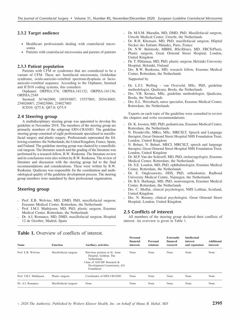
rs. N. Rooney, clinical psychologist, Great Ormond StreetHospital, London, United Kingdom2.5 Conflicts of interestAll members of the steering group declared their conflicts of
interest. An overview is given in Table 1.
Personal
financial
interests
Personal
relations
Externally
financed
research
Intellectual
interest
and reputation
Additional
interests
None None None None None
None None None None None
None None None None None
alf of Mutaz B. Habal, MD 2395

. (continued )
Name Function Ancillary activities
Personal
financial
interests
Personal
relations
Externally
financed
research
Intellectual
interest
and reputation
Additional
interests
Dr. M.S.M. Muradin Maxillofacial surgeon President of Auditing Committeeof Dutch Society of Cleft Palateand craniofacial surgery(NVSCA)
Treasurer of NVSCA(nov 2019)
Treasurer ECPCAInstructor Human Cadaver
Course, Implantcollege
None None None None None
Dr. R.H. Khonsari Maxillofacial surgeon None None None None None None
Dr. N.W. Bulstrode Plastic surgeon Will follow Will follow Will follow Will follow Will follow Will follow
Dr. T. Pihlamaa Plastic surgeon Private practice, Plasticsurgeon, PihlajalinnaTilkka Hospital & PlasticSurgery Center, Helsinki,Finland
None None None None None
Drs. R.W. Renkema Research fellow None None None None None None
Dr. L.F.J. Welling - vanOverveld
Guidelinemethodologist
None None None None None None
Drs. V.R. Krones Guideline methodologist None None None None None None
Prof. dr. K. Joosten Paediatric-intensivist None None None None None None
N. Prendeville Specialist speech andlanguage therapist
None None None None None None
N. Behari Specialist speech andlanguage therapist
None None None None None None
Dr. M.P. Van derSchroeff
Otolaryngologist None None None None None None
Dr. S.E. Loudon Paediatricophthalmologist
None None None None None None
Dr. E. Ongkosuwito Orthodontist Deputy director of thePostgraduate training inOrthodontics and section ofOrthodontics and CraniofacialBiology, Department ofDentistry, Radboud UniversityMedical Center, Nijmegen, theNetherlands
Private practice in Orthodontics,Orthopraktijk Capelle,Capelle aan den ljssel, TheNetherlands
None None None None None
Dr. B.S. Harhangi Neurosurgeon None None None None None None
Dr. C. Moffat Clinical psychologist Will follow Will follow Will follow Will follow Will follow Will follow
Dr. N. Rooney Clinical psychologist None None None None None None
Renkema et al The Journal of Craniofacial Surgery � Volume 31, Number 8S, November/December 2020
2396 # 2020 The Author(s). Published by Wolters Kluwer Health, Inc. on behalf of Mutaz B. Habal, MD

The Journal of Craniofacial Surgery � Volume 31, Number 8S, November/December 2020 European Guideline Craniofacial Microsomia
2.6 Patient perspectivesSince the guideline will be developed for patients and parents
of patients, the patient perspective will be of major importance inthis guideline. The perspective of patients was included byanalysing relevant bottlenecks from the online survey (seeChapter 2.8.1).
2.7 ImplementationThe implementation of the guideline and the practical feasibility
of the recommendations were taken into account during the differ-ent phases of guideline development. In doing so, explicit consid-eration was given to factors that could promote or hinder theimplementation of the guideline in practice.
2.8 Methods2.8.1 Bottleneck analysis
A draft list of bottlenecks from a professional perspective waswritten by the chair and vice chair. Members of the steering groupwere asked to give feedback on the draft bottleneck analysis. Thefirst set of bottlenecks were discussed during the first internationalconference in February 2019. No additional chapters were addedregarding the bottlenecks of the professionals.
All doctors included in the ERN-CRANIO, subgroup ‘craniofa-cial microsomia’, were asked to approach their CFM patients. Thisled to the identification of a group of 32 interviewees: 14 from Italy,13 from Germany, 4 from the Netherlands, and 1 from Sweden,including 9 patients and 23 parents of patients. An online surveywas set up with open and closed questions. All patients and parentsof patients were asked what difficulties they (had) experienced inthe healthcare process and in their lives. The questionnaire was builtup according to the proposed guideline chapters and the healthcareprocess, namely diagnosis and referral, organisation of care, com-munication and information, breathing difficulties, feeding diffi-culties or speech difficulties, surgical treatments, care for microtia,orthodontic treatment, vertebral anomalies, psychosocial aspects ofcare, and follow-up. Additionally, all patients were asked to namethe top three difficulties they experienced in the care process.Results were analysed by the research fellow (R.W. Renkema)and nurse specialist (E.L. Weissbach). Most frequently mentioneddifficulties included difficulties in receiving adequate informationon the diagnosis and the treatment, difficulties in getting referred toan experienced medical centre, and absence of psychological care.Based on the patient perspectives, a chapter on psychosocialdifficulties was added. Other relevant bottlenecks were includedin the chapter on organisation of care.
Bottlenecks of both patients and specialists were forwarded torelevant people in participating countries in March 2019, givingthem the opportunity to give feedback on the bottlenecks. After-wards, a draft framework of the guideline was set up and questionsand outcomes were formulated.
2.8.2 Questions and outcomesThe bottleneck analysis formed the basis for the questions for the
guideline. The questions were formed according to the PICOTFramework and presented in each guideline chapter. To maintain aclear and readable chapter, questions in the guideline were formu-lated in a broad and clinically relevant way. The terms for and morespecified questions to facilitate the literature search were specifiedin the summary of literature and Appendix 1, http://links.lww.com/SCS/B697.
The guideline is divided into a non-surgical and surgical part.Questions for the non-surgical chapters (Chapter 4 (4.1 to 4.8)) areformulated in a similar way. Likewise, questions for the surgical
# 2020 The Author(s). Published by Wolters Kluwer Health, Inc. on b
chapters (Chapter 5 (5.1 to 5.4)) are also formulated in asimilar way.
For questions in the surgical chapters specific patient outcomessuch as aesthetic results, quality of life, or complications wereformulated. The patient outcomes were described in the summary ofliterature. Patient outcomes were taken into account when formu-lating the recommendations. In addition, patients’ outcomes wereincluded in the summary of literature when they were reportedin literature.
2.8.3 Literature search and selection of literatureOne systematic search of literature was performed to identify all
available literature on craniofacial microsomia and synonyms. Thesearch was conducted in Embase, Pubmed/Medline Ovid. The fullsearch strategy is reported in the supplementary material.
Inclusion and exclusion criteria:
Type of studies
ehalf of Mutaz B. Habal, MD
- Original articles- Systematic review of sufficient
quality:- The question in thesystematic review matches thequestion of the guideline.- The search of the systematicreview was conducted in atleast two relevant databases,such as the Cochrane Library,Medline/Pubmed.- The full search strategy wasreported.- No relevant items weremissing in the search strategy.
Type of patients
- Patients with craniofacialmicrosomiaSubject
- Psychosocial functioning/difficulties, neurodevelopmentExclusion criteria
- Original studies with< 10included patients- Articles published before 1980- Case reports- Expert opinion- Letters- Editorials- Narrative reviews
A total of 1,747 articles were screened on title and abstract. Mostarticles (1,488) were excluded and 259 articles were reviewed onfull text. A total of 101 articles were included in the guideline.
The selected studies were categorised according to the frame-work of the guideline.
No narrative reviews were taken into account except for Chapter4.5, 5.2, and 5.3. Since there was hardly any evidence, the availablenarrative review was of importance.
For a couple of chapters, hardly no evidence was found. In thesecases an additional literature search was performed, regarding aspecific treatment in a different patient population for example. Thefull extra search strategy is reported in the supplementary material.Table 5 gives an overview of the chapters for which an additionalliterature search was performed. In a couple of chapters, additionalliterature is only included in the considerations and/or the rationale.
2397
Renkema et al The Journal of Craniofacial Surgery � Volume 31, Number 8S, November/December 2020
2.8.4 Quality assessment of individual studiesIndividual studies were systematically assessed, based on pre-
established methodological quality criteria, in order to estimate therisk of biased study results. The evidence table of all individualstudies is displayed in Appendix 6, http://links.lww.com/SCS/B697.
2.8.5 Summary of literatureThe most important findings from the literature were described
in the summary of literature. Literature with a high risk of bias wasfound for a number of chapters and hardly any evidence was foundfor a couple of chapters. Percentages were rounded in the conclu-sions. The steering group decided to include expert opinions for thechapters with hardly any evidence. Therefore, specific experts oneach topic of the guideline were consulted to review the chapter. Inaddition, experts were asked to write considerations and recom-mendations to initiate the discussion during the meeting inSeptember 2019. In the end, all written text was discussed duringthe meeting in September 2019.
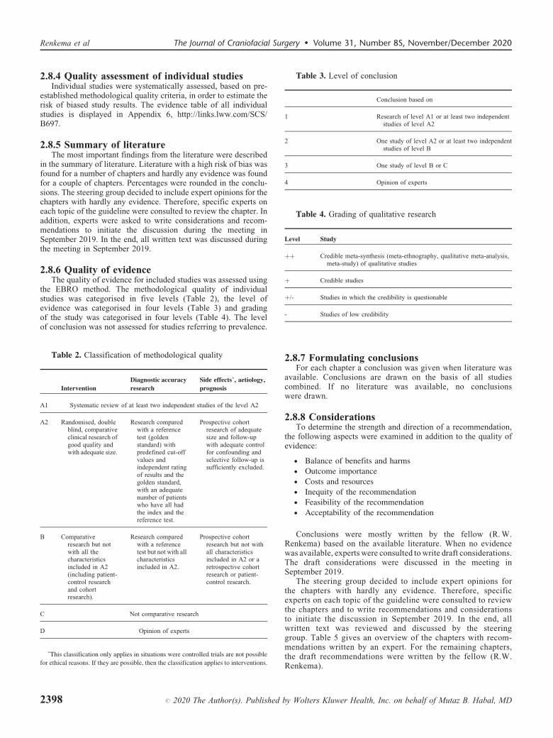
2.8.6 Quality of evidenceThe quality of evidence for included studies was assessed using
the EBRO method. The methodological quality of individualstudies was categorised in five levels (Table 2), the level ofevidence was categorised in four levels (Table 3) and gradingof the study was categorised in four levels (Table 4). The levelof conclusion was not assessed for studies referring to prevalence.
Table 2. Classification of methodological quality
2398
Intervention
Diagnostic accuracy
research
# 2020 T
Side effects�, aetiology,
prognosis
A1
Systematic review of at least two independent studies of the level A2A2
Randomised, doubleblind, comparativeclinical research ofgood quality andwith adequate size.Research comparedwith a referencetest (goldenstandard) withpredefined cut-offvalues andindependent ratingof results and thegolden standard,with an adequatenumber of patientswho have all hadthe index and thereference test.
Prospective cohortresearch of adequatesize and follow-upwith adequate controlfor confounding andselective follow-up issufficiently excluded.
B
Comparativeresearch but notwith all thecharacteristicsincluded in A2(including patient-control researchand cohortresearch).Research comparedwith a referencetest but not with allcharacteristicsincluded in A2.
Prospective cohortresearch but not withall characteristicsincluded in A2 or aretrospective cohortresearch or patient-control research.
C
Not comparative researchD
Opinion of experts�This classification only applies in situations were controlled trials are not possible
for ethical reasons. If they are possible, then the classification applies to interventions.
he Author(s). Published
Table 3. Level of conclusion
by Wolters Kluwer Health,
Conclusion based on
1
Research of level A1 or at least two independentstudies of level A22
One study of level A2 or at least two independentstudies of level B3
One study of level B or C4
Opinion of expertsTable 4. Grading of qualitative research
Level
Studyþþ
Credible meta-synthesis (meta-ethnography, qualitative meta-analysis,meta-study) of qualitative studiesþ
Credible studiesþ/-
Studies in which the credibility is questionable-
Studies of low credibility2.8.7 Formulating conclusionsFor each chapter a conclusion was given when literature was
available. Conclusions are drawn on the basis of all studiescombined. If no literature was available, no conclusionswere drawn.
2.8.8 ConsiderationsTo determine the strength and direction of a recommendation,
the following aspects were examined in addition to the quality ofevidence:
� B
alance of benefits and harms � O utcome importance � C osts and resources � I nequity of the recommendation � F easibility of the recommendation � A cceptability of the recommendationConclusions were mostly written by the fellow (R. W.Renkema) based on the available literature. When no evidencewas available, experts were consulted to write draft considerations.The draft considerations were discussed in the meeting inSeptember 2019.
The steering group decided to include expert opinions forthe chapters with hardly any evidence. Therefore, specificexperts on each topic of the guideline were consulted to reviewthe chapters and to write recommendations and considerationsto initiate the discussion in September 2019. In the end, allwritten text was reviewed and discussed by the steeringgroup. Table 5 gives an overview of the chapters with recom-mendations written by an expert. For the remaining chapters,the draft recommendations were written by the fellow (R.W.Renkema).
Inc. on behalf of Mutaz B. Habal, MD

3
44
4
44
4
44
4
44
4
44
4
44
4
44
4
4
4
4
5
5
5
5
5
5
55
The Journal of Craniofacial Surgery � Volume 31, Number 8S, November/December 2020 European Guideline Craniofacial Microsomia
Table 5. Overview of the source of information for each chapter.
Chapter
NY
Y
N
Y
Y
N
N
Y
Y
N
Y
N
A
NY
NY
NN
Y
Y
Y
Y
# 2020 The Author(s). Published by Wolters Kluwer Health, Inc. on
Literatureavailable?
D
N
D
behalf of Mutaz B. Hab
Draft considerationsand recommendationswritten by experts
al, MD
Additional searchperformed
3. Diagnostic criteria.1 Based on which criteria is a child or adult with craniofacial microsomia diagnosed?
Yes
4.1 Breathing difficulties.1.1 What is the type, prevalence and severity of breathing difficulties in craniofacial microsomia?.1.2 What is the policy for screening and monitoring of breathing difficulties (OSA) in patients with
craniofacial microsomia?.1.3 What are the indications and policy for treatment of breathing difficulties (OSA) in patients
with craniofacial microsomia?
Yes, except for type ofbreathing difficulties
oes, except for indications of
breathing difficulties. Thatquestion is answered by
including literature abouttreatment modalities.
Dr. K. Joosten
4.2 Feeding difficulties.2.1 What is the type, prevalence and severity of feeding difficulties in craniofacial microsomia?.2.2 What is the policy for screening and monitoring of feeding difficulties (OSA) in patients with
craniofacial microsomia?.2.3 What are the indications and policy for treatment of feeding difficulties (OSA) in patients with
craniofacial microsomia?
es
o
es
r. K. Joosten
4.3 Speech difficulties.3.1 What is the type, prevalence and severity of speech difficulties in craniofacial microsomia?.3.2 What is the policy for screening and monitoring of speech difficulties in patients with
craniofacial microsomia?.3.3 What are the indications and policy for treatment of speech difficulties in patients with
craniofacial microsomia?
es
o
o
N. Prendeville, MRes, MSc,MRCSLT
. Behari, MECI, MRCSLT
4.4 Hearing difficulties.4.1 What is the type, prevalence and severity of hearing difficulties in craniofacial microsomia?.4.2 What is the policy for screening and monitoring of hearing difficulties in patients with
craniofacial microsomia?.4.3 What are the indications and policy for treatment of hearing difficulties in patients with
craniofacial microsomia?
es
es
o
Dr. M.P. van der Schroeff
4.5 Eye anomalies.5.1 What is the type, prevalence and severity of eye anomalies in craniofacial microsomia?.5.2 What is the policy for screening and monitoring of eye anomalies in patients with craniofacial
microsomia?.5.3 What are the indications and policy for treatment of eye anomalies in patients with craniofacial
microsomia?
es
o
dditional literaturewas included
Dr. S.E. Loudon
Additional search onepibulbardermoids and variants4.6 Dental deformities.6.1 What is the type, prevalence and severity of dental deformities in craniofacial microsomia?.6.2 What is the policy for screening and monitoring of dental deformities in patients with
craniofacial microsomia?.6.3 What are the indications and policy for treatment of dental deformities in patients with
craniofacial microsomia?
Yesoes
Dr. E. Ongkosuwito
4.7 Vertebral anomalies.7.1 What is the type, prevalence and severity of vertebral deformities in craniofacial microsomia?.7.2 What is the policy for screening and monitoring of vertebral deformities in patients with
craniofacial microsomia?.7.3 What are the indications and policy for treatment of vertebral deformities in patients with
craniofacial microsomia?
Yesoes
Dr. B.S. Harhangi
4.8 Psychosocial difficulties.8.1 What is the type, prevalence and severity of psychosocial difficulties in craniofacial
microsomia?.8.2 What is the policy for screening and monitoring of psychosocial difficulties in patients with
craniofacial microsomia?.8.3 What are the indications and policy for treatment of psychosocial difficulties in patients with
craniofacial microsomia?
Yesoo
Drs. C. Moffatrs. N. Rooney
5.1 Mandible and Maxilla.1.1 What is the indication for surgical treatment of mandibular and maxillary deformity in patients
with craniofacial microsomia?.1.2 What is the most optimal treatment modality and its timing for mandibular/maxillary deformity in
craniofacial microsomia regarding severity, breathing problems, occlusal problems and aesthetics?
Yeses
Additional literaturesearch on thetreatment for retro/micrognathia wasperformed
5.2 Facial nerve.2.1 What is the indication for surgical treatment of facial nerve anomaly in patients with
craniofacial microsomia?.2.2 What is the most optimal treatment modality for facial nerve anomaly in patients with
craniofacial microsomia related to functional deficits and aesthetics?
Dutch guidelinewas used
es
Dutch guideline onperipheral facial palsywas additionallyincluded combinedwith an extra literaturesearch
5.3 Soft tissue.3.1 What is the indication for surgical treatment of soft tissue deficiency in patients with
craniofacial microsomia?.3.2 What is the most optimal treatment modality for soft tissue deficiency in patients with
craniofacial microsomia related to severity and its timing?
Noes
5.4 Microtia.4.1 What is the indication for surgical treatment of ear deformity in patients with CFM?.4.2 What is the most optimal treatment modality for ear deformity in patients with CFM related to its
timing?
Noes
Additional literaturesearch on earreconstruction wasperformed
2399
-
Renkema et al The Journal of Craniofacial Surgery � Volume 31, Number 8S, November/December 2020
2.8.9 Formulating recommendationsThe recommendations provide an answer to the basic question and
are based on the best available scientific evidence and the mostimportant considerations. The strength of the scientific evidence andthe weight that the working group assigns to the considerationstogether determine the strength of the recommendation. In accor-dance with the EBRO method, a low probative value of conclusions inthe systematic literature analysis does not exclude a strong recom-mendation in advance, and weak recommendations are also possiblewith a high probative value. The strength of the recommendation isalways determined by weighing all relevant arguments.
Recommendations were mostly written by the fellow (R.W.Renkema) based on the available literature. When no evidence wasavailable, experts were consulted to write draft recommendations.The draft recommendations were discussed in the meeting inSeptember 2019.
2.8.10 Conditions (organisation of care)The bottleneck analysis and the development of the guideline
explicitly take into account the aspects related to organisation ofcare. This contains all aspects that are preconditions for providingcare, such as coordination, communication, (financial) resources,manpower and infrastructure. More general, overarching, andadditional aspects of the organisation of care are discussed inChapter 6.
2.8.11 Knowledge gapDuring the development of this guideline a systematic literature
search was performed to answer the questions. For each question thesteering group investigated whether (additional) scientific researchis necessary. An overview of recommendations for further researchcan be found in Chapter 7.
2.8.12 Evaluation and authorisation phaseThe draft guideline was submitted to all craniofacial centres
involved in the care for patients with CFM and affiliated with theERN-CRANIO. The comments were collected and discussed withthe steering group. As a result of the comments, the draft guidelinewas adapted and finally adopted by the steering group.
CHAPTER 3. DIAGNOSTIC CRITERIA FORCRANIOFACIAL MICROSOMIA
IntroductionIn patients with craniofacial microsomia (CFM), the facial struc-
tures arising from the first and second pharyngeal arches may beunderdeveloped. The characteristic features are malformation, hypo-plasia or aplasia of the outer and inner ear, orbit, zygoma, maxilla,mandible, facial musculature, facial nerve, and/or soft tissue, butextracraniofacial anomalies may be present as well (1, 2). The varietyin type and severity of underdevelopment of these structures makeCFM a heterogeneous disorder. The diagnosis of CFM is based onclinical assessment, though no clear diagnostic criteria exist.
Various classification models have been proposed to categorisepatients with CFM based on its severity (3–8). The most commonlyused system is the O.M.E.N.S. classification, which describes thedegree of hypoplasia of the Orbit (O), Mandible (M), Ears (E),Facial Nerve (N), and Soft Tissue (S) (2, 3). Extracraniofacialanomalies, facial clefting, canting of the occlusal plane, anddetailed assessment of eye and ear anomalies is included in thisclassification as well (2, 9). A more detailed classification of themandibular deformity, based on radiography, was proposed byPruzansky and later subcategorised by Kaban (5, 10, 11). In this
2400 # 2020 The Author(s). Published
classification model the level of underdevelopment of the mandibleis graded as I, IIA, IIB, and III.
A wide range of terminology is used to refer to patients withCFM: hemifacial microsomia, Goldenhar syndrome, oculo-auri-culo-vertebral spectrum or dysplasia, first and second branchialarch syndrome, or facio-auriculo-vertebral syndrome or sequence.
The term CFM is preferred for two main reasons. Firstly, as slightlymore than 10% of the patients with CFM are bilaterally affected theterm hemifacial microsomia (HFM), which has been commonly used,is not considered to cover the extensiveness of the syndrome (9, 12).Secondly, the side that has the more significant hypoplasia causesdeformity on the contralateral side making this a condition that affectsboth sides. Therefore CFM is a more precise term.
Microtia is an important and common aspect of patients withCFM. Whether patients with isolated microtia fit the diagnosis CFMis still being debated (1, 4, 13, 14). Some authors advocate theinclusion of patients with isolated microtia as CFM, as the cause andtypes of ear malformations are similar in both groups. In addition,the incidence of ear malformations found in family members ofpatients with CFM is much higher than in the general population(15). However, no consensus has been reached.
Goldenhar syndrome was originally regarded as an associationof mandibular dysostosis, ear malformations and epibulbar der-moids (16). Although there are no clear diagnostic criteria forGoldenhar syndrome, currently the diagnosis is often seen asCFM with the additional presence of epibulbar dermoids andvertebral/cervical spine anomalies (the triad of Goldenhar). Someconsider the diagnosis Goldenhar syndrome to be an entity distinctfrom CFM, while others include Goldenhar syndrome as a variant ofCFM (15, 17). However, no consensus exists.
Another term used to describe patients with CFM is the oculo-auriculo-vertebral spectrum or dysplasia (OAVS) (18). OAVS isoften used to describe the expanded spectrum of CFM includingfacial malformations such as macrostomia or epibulbar dermoids,and extracraniofacial anomalies. Again, no consensus on diagnosticcriteria exists.
The use of different terminology and lack of diagnostic criteria isconfusing for patients and healthcare professionals. Therefore, thefollowing question was posed:
3.1 On which criteria is a child or adult withcraniofacial microsomia diagnosed?
Uniform terminology would be helpful to provide better infor-mation to patients and increase awareness of the disorder. Addi-tionally, it would be beneficial for the communication betweenprofessionals and patients, between healthcare professionals, and inresearch projects.
Literature searchA systematic search was performed to identify all available
literature on craniofacial microsomia and synonyms. The searchwas conducted in Embase, Pubmed/Medline Ovid. The full searchstrategy is reported in the supplementary material.
Inclusion and exclusion criteria:
Type of studies
by Wolters Kluwe
- Original articlesSystematic review of sufficient quality:- The question in the systematic review matches the question ofthe guideline.
- The search of the systematic review was conducted in at leasttwo relevant databases, such as the Cochrane Library,Medline/Pubmed.
- The full search strategy was reported.- No relevant items were missing in the search strategy.
Type of patients
- Patients with craniofacial microsomiar Health, Inc. on behalf of Mutaz B. Habal, MD
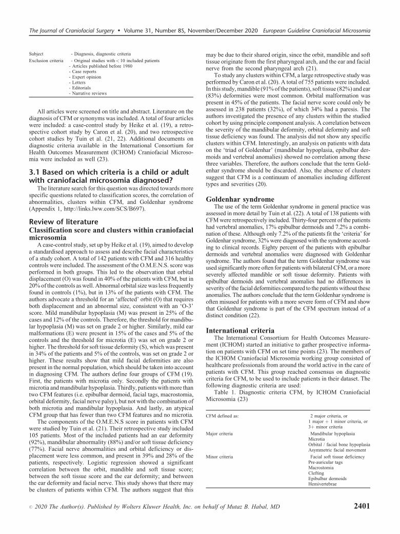
The Journal of Craniofacial Surgery � Volume 31, Number 8S, November/December 2020 European Guideline Craniofacial Microsomia
Subject
# 2020 The Auth
- Diagnosis, diagnostic criteria
Exclusion criteria
- Original studies with< 10 included patients- Articles published before 1980- Case reports- Expert opinion- Letters- Editorials- Narrative reviewsAll articles were screened on title and abstract. Literature on thediagnosis of CFM or synonyms was included. A total of four articleswere included: a case-control study by Heike et al. (19), a retro-spective cohort study by Caron et al. (20), and two retrospectivecohort studies by Tuin et al. (21, 22). Additional documents ondiagnostic criteria available in the International Consortium forHealth Outcomes Measurement (ICHOM) Craniofacial Microso-mia were included as well (23).
3.1 Based on which criteria is a child or adultwith craniofacial microsomia diagnosed?
The literature search for this question was directed towards morespecific questions related to classification scores, the correlation ofabnormalities, clusters within CFM, and Goldenhar syndrome(Appendix 1, http://links.lww.com/SCS/B697).
Review of literatureClassification score and clusters within craniofacialmicrosomia
A case-control study, set up by Heike et al. (19), aimed to developa standardised approach to assess and describe facial characteristicsof a study cohort. A total of 142 patients with CFM and 316 healthycontrols were included. The assessment of the O.M.E.N.S. score wasperformed in both groups. This led to the observation that orbitaldisplacement (O) was found in 40% of the patients with CFM, but in20% of the controls as well. Abnormal orbital size was less frequentlyfound in controls (1%), but in 13% of the patients with CFM. Theauthors advocate a threshold for an ‘affected’ orbit (O) that requiresboth displacement and an abnormal size, consistent with an ‘O-3’score. Mild mandibular hypoplasia (M) was present in 25% of thecases and 12% of the controls. Therefore, the threshold for mandibu-lar hypoplasia (M) was set on grade 2 or higher. Similarly, mild earmalformations (E) were present in 15% of the cases and 5% of thecontrols and the threshold for microtia (E) was set on grade 2 orhigher. The threshold for soft tissue deformity (S), which was presentin 34% of the patients and 5% of the controls, was set on grade 2 orhigher. These results show that mild facial deformities are alsopresent in the normal population, which should be taken into accountin diagnosing CFM. The authors define four groups of CFM (19).First, the patients with microtia only. Secondly the patients withmicrotia and mandibular hypoplasia. Thirdly, patients with more thantwo CFM features (i.e. epibulbar dermoid, facial tags, macrostomia,orbital deformity, facial nerve palsy), but not with the combination ofboth microtia and mandibular hypoplasia. And lastly, an atypicalCFM group that has fewer than two CFM features and no microtia.
The components of the O.M.E.N.S score in patients with CFMwere studied by Tuin et al. (21). Their retrospective study included105 patients. Most of the included patients had an ear deformity(92%), mandibular abnormality (88%) and/or soft tissue deficiency(77%). Facial nerve abnormalities and orbital deficiency or dis-placement were less common, and present in 39% and 28% of thepatients, respectively. Logistic regression showed a significantcorrelation between the orbit, mandible and soft tissue score;between the soft tissue score and the ear deformity; and betweenthe ear deformity and facial nerve. This study shows that there maybe clusters of patients within CFM. The authors suggest that this
or(s). Published by Wolters Kluwer Health, Inc. on b
may be due to their shared origin, since the orbit, mandible and softtissue originate from the first pharyngeal arch, and the ear and facialnerve from the second pharyngeal arch (21).
To study any clusters within CFM, a large retrospective study wasperformed by Caron et al. (20). A total of 755 patients were included.In this study, mandible (91% of the patients), soft tissue (82%) and ear(83%) deformities were most common. Orbital malformation waspresent in 45% of the patients. The facial nerve score could only beassessed in 238 patients (32%), of which 34% had a paresis. Theauthors investigated the presence of any clusters within the studiedcohort by using principle component analysis. A correlation betweenthe severity of the mandibular deformity, orbital deformity and softtissue deficiency was found. The analysis did not show any specificclusters within CFM. Interestingly, an analysis on patients with dataon the ‘triad of Goldenhar’ (mandibular hypoplasia, epibulbar der-moids and vertebral anomalies) showed no correlation among thesethree variables. Therefore, the authors conclude that the term Gold-enhar syndrome should be discarded. Also, the absence of clusterssuggest that CFM is a continuum of anomalies including differenttypes and severities (20).
Goldenhar syndromeThe use of the term Goldenhar syndrome in general practice was
assessed in more detail by Tuin et al. (22). A total of 138 patients withCFM were retrospectively included. Thirty-four percent of the patientshad vertebral anomalies, 17% epibulbar dermoids and 7.2% a combi-nation of these. Although only 7.2% of the patients fit the ‘criteria’ forGoldenhar syndrome, 32% were diagnosed with the syndrome accord-ing to clinical records. Eighty percent of the patients with epibulbardermoids and vertebral anomalies were diagnosed with Goldenharsyndrome. The authors found that the term Goldenhar syndrome wasused significantly more often for patients with bilateral CFM, or a moreseverely affected mandible or soft tissue deformity. Patients withepibulbar dermoids and vertebral anomalies had no differences inseverity of the facial deformities compared to the patients without theseanomalies. The authors conclude that the term Goldenhar syndrome isoften misused for patients with a more severe form of CFM and showthat Goldenhar syndrome is part of the CFM spectrum instead of adistinct condition (22).
International criteriaThe International Consortium for Health Outcomes Measure-
ment (ICHOM) started an initiative to gather prospective informa-tion on patients with CFM on set time points (23). The members ofthe ICHOM Craniofacial Microsomia working group consisted ofhealthcare professionals from around the world active in the care ofpatients with CFM. This group reached consensus on diagnosticcriteria for CFM, to be used to include patients in their dataset. Thefollowing diagnostic criteria are used:
Table 1. Diagnostic criteria CFM, by ICHOM CraniofacialMicrosomia (23)
CFM defined as:
ehalf of Mutaz B. Habal, MD
2 major criteria, or1 major þ 1 minor criteria, or3þ minor criteria
Major criteria
Mandibular hypoplasiaMicrotiaOrbital / facial bone hypoplasiaAsymmetric facial movementMinor criteria
Facial soft tissue deficiencyPre-auricular tagsMacrostomiaCleftingEpibulbar dermoidsHemivertebrae2401

Renkema et al The Journal of Craniofacial Surgery � Volume 31, Number 8S, November/December 2020
Conclusions
Level 3
2402
Correlation and clustersIn patients with craniofacial microsomia the mandibular
deformity, orbital deformity and soft tissue deficiency arecorrelated. However, no specific clusters of patient groupswithin craniofacial microsomia are present, indicatingcraniofacial microsomia is a continuum of anomalies.
Ref (20, 21)
Level 3
Goldenhar syndromeGoldenhar syndrome is not a separate diagnosis from craniofacialmicrosomia but is part of the phenotypic spectrum ofcraniofacial microsomia:� Clinically, patients diagnosed with Goldenhar syndrome
have a more severe facial deformity compared to patients withcraniofacial microsomia.� Objectively, by using the O.M.E.N.S. score, no
differences are present in the severity of facial anomalies inpatients with craniofacial microsomia compared to patientswith the triad of Goldenhar syndrome (craniofacialmicrosomia þ epibulbar dermoids þ vertebral anomalies).� No correlation exist between the triad of Goldenhar
syndrome (craniofacial microsomia þ epibulbar dermoids þvertebral anomalies).� A substantial number of patients with the triad of
Goldenhar syndrome (craniofacial microsomia þ epibulbardermoids þ vertebral anomalies) are not diagnosed withGoldenhar syndrome.
Ref (20, 22)
Considerations� Overall quality of evidenceTwo studies reported on correlations of facial deformities in
patients with CFM, based on the O.M.E.N.S. classification. Thecorrelation between the facial deformities was reported in bothstudies. The studies are descriptive retrospective cohort studies andwere therefore graded on level 3 (20, 21).
Concerning the conclusions on Goldenhar syndrome, two stud-ies were included as well. Both studies showed the absence of acorrelation between the triad of Goldenhar syndrome and concludedthat Goldenhar syndrome is part of CFM, instead of a variant. Thestudies are descriptive retrospective cohort studies and were there-fore graded on level 3 (20, 22).
The diagnostic criteria proposed by the ICHOM CraniofacialMicrosomia group were based on consensus between healthcareprofessionals (23). No research has been done to test the criteria.Since CFM is a clinical diagnosis and no specific criteria exist forCFM, verifying the criteria may be difficult. Since the criteria arebased on consensus and expert opinion, the evidence is graded onlevel 5.
� Balance of benefits and harmsNo harm is produced for the patients with CFM by applying the
diagnostic criteria. Unambiguous use of diagnostic criteria andterminology promotes clarity for both patients and healthcareprofessionals. Standardised use of the term CFM, by using cleardiagnostic criteria, helps patients receive the right diagnosis andtreatment, and improves access to information about the disease. Inresearch, clear use of terminology promotes the exchange of ideasand findings.
# 2020 The Author(s). Published
� Outcome importanceThe use of diagnostic criteria for CFM is considered to be of
importance to healthcare professionals to improve communication,research, and provision of information to patients. Several recentstudies confirm that CFM is a continuum of anomalies and dis-courage the use of the term Goldenhar syndrome. The consensus ofthis steering group is to solely use the term CFM.
� Costs and resourcesThe impact on costs and resources of the given recommenda-
tions will vary per member state, depending on the available careproviders and facilities. The recommendations concern the essentialrequirements for adequate treatment of patients with CFM andshould thus be implemented. Costs are lowest and resources aremost efficiently used when care for craniofacial disorders is cen-tralised in a limited number of expert centres per member state. Ageneral rule that can be applied is one expert centre per 10million inhabitants.
� Inequity of the recommendationThe goal of the European Reference Networks (ERNs) is to
eliminate inequality within Europe with regard to care for patientswith rare diseases. At present, not every member state offers an expertcentre for CFM, or the level of provided care does not (yet) meet all therequirements that are outlined in this guideline. By defining thebaseline of required care for CFM, this guideline will help thesemember states to reach the appropriate level. The ERN on craniofacialanomalies and ENT disorders (ERN-CRANIO) can guide a patient inEurope to the available centres of expertise (www.ern-cranio.eu) andcan support care providers with diagnosis and treatment advice.
� Feasibility of the recommendationRecommendations refer to the general requirements for deliv-
ering optimal healthcare and are discussed with members fromparticipating European countries. Quality of care was paramount inthe discussions. Centralisation is proposed as one of the core values.However, in some countries the national organisation of healthcaremight impede centralisation. National implementation of the ERNsthat fits the situation of each country is necessary. For the memberstates with the lowest number of inhabitants, the establishment of acraniofacial centre might not be feasible, and collaboration with acraniofacial centre in the surrounding countries can be an option.
� Acceptability of the recommendationIt is expected that all stakeholders want to apply and will be
applying the recommendations, because they are employed in ERN-acknowledged institutions. National implementation plans are nec-essary to ensure that recommendations fit the situation in eachcountry. In addition, not all countries participating in the ERN-CRANIO are represented in the guideline development group, andnew members will join within the coming years. For these countries,acceptance and implementation of ERN guidelines such as thisguideline on CFM is obligatory.
Rationale of the recommendationsSince the evidence suggests no difference between CFM and
Goldenhar syndrome, OAVS, or hemifacial microsomia, the steer-ing group advocate the use of a single term: CFM. To increaseawareness of this diagnosis and provide clarity for patients andhealthcare professionals, the diagnostic criteria agreed on by the‘ICHOM Craniofacial Microsomia’ working group are recom-mended for use in practice.
by Wolters Kluwer Health, Inc. on behalf of Mutaz B. Habal, MD

The Journal of Craniofacial Surgery � Volume 31, Number 8S, November/December 2020 European Guideline Craniofacial Microsomia
Recommendations
#
Terminology
� It is advised to exclusively use the term craniofacialmicrosomia. Discard the use of other terms such asGoldenhar syndrome, hemifacial microsomia or aur-
2020 T
iculo-oculo-vertebral spectrum.
Diagnostic criteria� It is advised to use the diagnostic criteria for craniofacial microsomia developed
by the ICHOM Craniofacial Microsomia group.
CFM is defined by:
13MOA
PMCEH
he Author(s). Published by Wolt
2 major criteria, ormajor þ 1 minor criteria, orþ minor criteria
Major criteria
Mandibular hypoplasiaicrotiarbital / facial bone hypoplasiasymmetric facial movementMinor criteria
Facial soft tissue deficiencyre-auricular tagsacrostomialeftingpibulbar dermoidsemivertebraeResearch gapNone
References
1. B
irgfeld CB, Heike C. Craniofacial microsomia. Semin PlastSurg. 2012;26(2):91-104.2. H
organ JE, Padwa BL, LaBrie RA, Mulliken JB. OMENS-Plus: analysis of craniofacial and extracraniofacial anomaliesin hemifacial microsomia. Cleft Palate Craniofac J.1995;32(5):405-12.3. V
ento AR, LaBrie RA, Mulliken JB. The O.M.E.N.S.classification of hemifacial microsomia. Cleft Palate Cranio-fac J. 1991;28(1):68-76; discussion 7.4. T
asse C, Bohringer S, Fischer S, Ludecke HJ, Albrecht B,Horn D, et al. Oculo-auriculo-vertebral spectrum (OAVS):clinical evaluation and severity scoring of 53 patients andproposal for a new classification. Eur J Med Genet.2005;48(4):397-411.5. P
ruzansky S. Not all dwarfed mandibles are alike. Birthdefects: Original Article Series. 1969;5:120-9.6. C
onverse JM, Wood-Smith D, McCarthy JG, Coccaro PJ,Becker MH. Bilateral facial microsomia. Diagnosis,classification, treatment. Plast Reconstr Surg. 1974;54(4):413-23.7. D
avid DJ, Mahatumarat C, Cooter RD. Hemifacial micro-somia: a multisystem classification. Plast Reconstr Surg.1987;80(4):525-35.8. L
auritzen C, Munro IR, Ross RB. Classification and treatmentof hemifacial microsomia. Scand J Plast Reconstr Surg.1985;19(1):33-9.9. B
irgfeld CB, Luquetti DV, Gougoutas AJ, Bartlett SP, LowDW, Sie KC, et al. A phenotypic assessment tool forcraniofacial microsomia. Plast Reconstr Surg. 2011;127(1):313-20.ers Kluwer Health, Inc. on b
10. K
ehalf
aban LB, Moses MH, Mulliken JB. Correction of hemifacialmicrosomia in the growing child: a follow-up study. CleftPalate J. 1986;23 Suppl 1:50-2.
11. K
aban LB, Mulliken JB, Murray JE. Three-dimensionalapproach to analysis and treatment of hemifacial microsomia.Cleft Palate J. 1981;18(2):90-9.12. X
u S, Zhang Z, Tang X, Yin L, Liu W, Shi L. The influence ofgender and laterality on the incidence of hemifacialmicrosomia. J Craniofac Surg. 2015;26(2):384-7.13. B
ennun RD, Mulliken JB, Kaban LB, Murray JE. Microtia: amicroform of hemifacial microsomia. Plast Reconstr Surg.1985;76(6):859-65.14. R
ollnick BR, Kaye CI, Nagatoshi K, Hauck W, Martin AO.Oculoauriculovertebral dysplasia and variants: phenotypiccharacteristics of 294 patients. Am J Med Genet.1987;26(2):361-75.15. R
ollnick BR, Kaye CI. Hemifacial microsomia and variants:pedigree data. Am J Med Genet. 1983;15(2):233-53.16. G
oldenhar M. Associations malformatives de l’oeil et del’oreille, en particulier le syndrome dermoıde epibulbaire -appendices auriculaires - fistula auris congenita et ses relationsavec la dysostose mandibulofaciale. J Genet Hum.1952;1:243-82.17. G
orlin RJ PJ. Syndromes of the head and neck. New York:McGraw-Hill. 1964:546-52.18. G
orlin RJ, Jue KL, Jacobsen U, Goldschmidt E. Oculoaur-iculovertebral Dysplasia. J Pediatr. 1963;63:991-9.19. H
eike CL, Wallace E, Speltz ML, Siebold B, Werler MM,Hing AV, et al. Characterizing facial features in individualswith craniofacial microsomia: A systematic approach forclinical research. Birth Defects Res A Clin Mol Teratol.2016;106(11):915-26.20. C
aron C, Pluijmers BI, Wolvius EB, Looman CWN, BulstrodeN, Evans RD, et al. Craniofacial and extracraniofacialanomalies in craniofacial macrosomia: A multicenter studyof 755 patients. J Craniomaxillofac Surg. 2017;45(8):1302-10.21. T
uin AJ, Tahiri Y, Paine KM, Paliga JT, Taylor JA, Bartlett SP.Clarifying the relationships among the different features of theOMENSþ classification in craniofacial microsomia. PlastReconstr Surg. 2015;135(1):149e-56e.22. T
uin J, Tahiri Y, Paliga JT, Taylor JA, Bartlett SP.Distinguishing Goldenhar Syndrome from CraniofacialMicrosomia. J Craniofac Surg. 2015;26(6):1887-92.23. M
easurement ICfHO. ICHOM Craniofacial Microsomia DataCollection Reference Guide. 2017.CHAPTER 4. SCREENING, MONITORING ANDINDICATION FOR TREATMENT
4.1 Breathing difficulties in craniofacialmicrosomia
IntroductionObstructive sleep disordered breathing (SDB) can be considered
to be a syndrome of upper airway dysfunction during sleep (1). It ischaracterised by snoring and/or increased respiratory effort due toincreased airway resistance and pharyngeal collapsibility (2, 3).Various clinical entities with intermittent upper airway obstructionare included in obstructive SDB, such as: primary snoring, upperairway resistance syndrome, obstructive hypoventilation, andobstructive sleep apnoea (OSA) (4, 5). Polysomnography (sleepstudy) is the golden standard to diagnose presence and severity ofsleep disordered breathing (SDB). Of the obstructive SDB entities,OSA in particular is known to be of clinical significance as it relates
of Mutaz B. Habal, MD 2403
-
------
Renkema et al The Journal of Craniofacial Surgery � Volume 31, Number 8S, November/December 2020
to failure to thrive, morning headache, excessive daytime sleepi-ness, decline of school achievements, feeding difficulties, upperairway infections, hypertension, and may lead to metabolic, cogni-tive and cardiovascular consequences (6). OSA is defined by adisruption of the normal oxygenation, ventilation and sleep patterndue to recurrent upper airway obstructions. The severity of OSA isdescribed by the Apnoea-Hypopnea Index (AHI).
The prevalence of OSA in children in the healthy population is2–4%, and is thought to be caused by adenotonsillar hypertrophy,obesity, or decrease in neuromuscular tone (7-10). Patients withcraniofacial microsomia (CFM) are at increased risk for obstructiveSDB and OSA due to the underdevelopment of the mandible, whichleads to obstruction of the upper airway. In addition, pharyngealand/or laryngeal anomalies may play a role in causing OSA inpatients with CFM (11, 12). Since OSA in patients with CFM relatesto congenital malformations, symptoms occur shortly after birth orduring childhood.
The European Respiratory Society provided a stepwiseapproach to diagnose, manage and treat obstructive SDB andOSA in children (1, 13). Treatment of OSA in patients with majorcraniofacial abnormalities, such as CFM, is reported as a priority.Obstructive SDB, including OSA, is treated by a stepwise approachand can require pharmacological treatment such as nasal cortico-steroids, non-surgical treatment by continuous positive airwaypressure (CPAP) or non-invasive positive-pressure ventilation(NPPV), or surgical treatment such as adenotonsillectomy orcraniofacial surgery. In addition to the regular treatment modali-ties, patients with CFM may require surgical correction of themandible by using mandibular distraction, costochondral graftingor osteotomy (11).
To describe the policy for breathing difficulties inpatients with CFM the following questions were included inthis chapter:
4.1.1 What is the type, prevalence and severity of breathingdifficulties in craniofacial microsomia?4.1.2 What is the policy for screening and monitoring ofbreathing difficulties (OSA) in patients with craniofacialmicrosomia?4.1.3 What are the indications and policy for treatment ofbreathing difficulties (OSA) in patients with craniofacialmicrosomia?
Describing the policy for breathing difficulties in patientswith CFM is of importance since these patients are at increasedrisk for OSA. Untreated OSA may lead to metabolic, cognitiveand/or cardiovascular pathology and can have a negative impacton the quality of life. Therefore, timely diagnosis and treatmentof OSA in patients with CFM is considered of importanceto patients.
DefinitionsThe definitions of apnoea’s, hypopnea and OSA severity are
displayed in Appendix 4, http://links.lww.com/SCS/B697. This isbased on the Manual for the Scoring of Sleep and Associated Eventsof the American Academy of Sleep Medicine (14). The severity ofOSA is graded according to the New Zealand Guideline forobstructive sleep apnoea syndrome in children (15).
Literature searchA systematic search of literature was performed to identify all
available literature on CFM and synonyms. The search was con-ducted in Embase, Pubmed/Medline Ovid. The full search strategyis reported in the supplementary material.
2404 # 2020 The Author(s). Published
Inclusion and exclusion criteria:
Type of studies
by Wolters Kluwe
- Original articlesSystematic review of sufficient quality:- The question in the systematic review matches the question ofthe guideline.
- The search of the systematic review was conducted in at leasttwo relevant databases, such as the Cochrane Library,Medline/Pubmed.
- The full search strategy was reported.- No relevant items were missing in the search strategy.
Type of patients
- Patients with craniofacial microsomiaSubject
- Breathing, airway, sleepExclusion criteria
- Original studies with< 10 included patientsArticles published before 1980Case reportsExpert opinionLettersEditorialsNarrative reviewsLiterature was screened on title and abstract. A total of eightarticles were included. One systematic review by Caron et al. wasincluded (11). Five studies were retrospective cohort studies:Szpalski et al. (6), Cohen et al. (16), Sculerati et al. (17), Sheret al. (18), and Caron et al. (19). The other two included studies werea cross-sectional study by D’Antonio et al. (12) and a case-controlstudy by Cloonan et al. (20).
4.1.1 What is the type, prevalence and severity of breathingdifficulties in craniofacial microsomia?
The literature search for this question was directed towards morespecific questions related to the type (primary snoring, upper airwayresistance syndrome, obstructive hypoventilation, and OSA), prev-alence and severity (mild, moderate or severe) of breathing diffi-culties in CFM (Appendix 1, http://links.lww.com/SCS/B697).Unfortunately, none of the included studies reported informationon the type of breathing difficulties in CFM. Studies only reportedon one of the types of breathing difficulties, OSA. Since thisquestion does not relate to interventions or diagnostics, no consid-erations, rationale or recommendations are provided.
Review of literatureA systematic review on obstructive sleep apnoea by Caron et al. was
published in 2015. The aim of the study was to report the prevalenceand treatment modalities of OSA in patients with CFM. Six studiesdescribing the prevalence of OSA in CFM were included in thesystematic review. However, one study included fewer than ten patientsand was not taken into account in this guideline (21). The prevalence ofOSA in CFM varied from 7% to 24% (12, 16-18, 20). The number ofpatients included in these studies varied from 38 to 124 patients. Moststudies were retrospective cohort studies and the diagnosis of OSA wasbased on chart review and polysomnography. Although some studiesdefined the used diagnostic criteria for OSA (16, 18), other studies didnot (12, 17, 20). Sculerati et al. included 41 patients of which 9 patientsrequired a tracheostomy (17). Therefore, the prevalence of OSA in thisstudy, which can be considered to be severe, is found to be 22%.However, no additional data is available on the presence of OSA in theother included patients. One of the other included studies analysed 124patients with CFM and 349 controls (20). Patients with CFM had ahigher frequency of SDB (23% vs 9%, OR: 3.0, CI: 1.5–5.1,p¼ 0.001), which includes OSA, compared to controls. None of theincluded studies reported information on the type of breathing diffi-culties in CFM. The severity of OSA and risk factors for OSA inpatients with CFM were assessed by multiple studies. Cloonan et al.reported that patients with moderate/severe CFM presented more oftenwith snoring and SDB than patients with mild CFM. In this study, thediagnosis mild, moderate or severe CFM was based on specialists’
r Health, Inc. on behalf of Mutaz B. Habal, MD
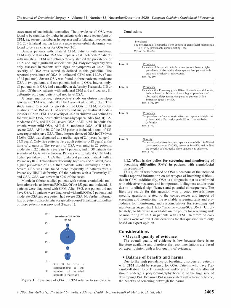
T
P
P
T
T
R
The Journal of Craniofacial Surgery � Volume 31, Number 8S, November/December 2020 European Guideline Craniofacial Microsomia
assessment of craniofacial anomalies. The prevalence of OSA wasfound to be significantly higher in patients with a more severe form ofCFM, i.e. severe mandibular hypoplasia and/or bilateral involvement(16, 20). Bilateral hearing loss or a more severe orbital deformity wasfound to be a risk factor for OSA too (16).
Besides patients with bilateral CFM, patients with unilateralCFM may be at risk for OSA too. Szpalski et al. included 62 patientswith unilateral CFM and retrospectively studied the prevalence ofOSA and any significant associations (6). Polysomnography wasonly assessed in patients with signs or symptoms of OSA. Theseverity of OSA was scored as defined in this guideline. Thereported prevalence of OSA in unilateral CFM was 11.3% (7 outof 62 patients). Severe OSA was found in three patients, moderateOSA in two patients, and two patients had mild OSA. Interestingly,all patients with OSA had a mandibular deformity Pruzansky IIB orhigher. Of the six patients with unilateral CFM and a Pruzansky IIIdeformity only one patient did not have OSA.
A large, multicentre, retrospective study on obstructive sleepapnoea in CFM was undertaken by Caron et al. in 2017 (19). Thisstudy aimed to report the prevalence of OSA in CFM, study therelationship of OSA and CFM severity and analyse treatment modal-ities for OSA in CFM. The severity of OSA in children was defined asfollows: mild OSA, obstructive apnoea-hypopnea index (oAHI) 1-5;moderate OSA, oAHI 5-24; severe OSA, oAHI >24. In adults thecriteria were: mild OSA, AHI 5-15; moderate OSA, AHI 15-30;severe OSA, AHI >30. Of the 755 patients included, a total of 133were reported to have OSA. Thus, the prevalence of OSA in CFM was17.6%. OSA was diagnosed at a median age of 2.4 years (range 0–25.8 years). Only five patients were adult patients (>18 years) at thetime of diagnosis. The severity of OSA was mild in 25 patients,moderate in 22 patients, severe in 48 patients, and in 38 patients theseverity of OSA was unknown. Patients with bilateral CFM had ahigher prevalence of OSA than unilateral patients. Patient with aPruzansky IIB/III mandibular deformity, both uni- and bilateral, had ahigher prevalence of OSA than patients with Pruzansky I or IIA.Severe OSA was thus found more frequently in patients with aPruzansky IIB/III deformity. Of the patients with a Pruzansky IIIand OSA, OSA was severe in 52% of the cases.
Moraleda-Cibrian studied patients with various craniofacial mal-formations who underwent PSG (22). Of the 153 patients included, 18patients were diagnosed with CFM. After PSG, one patient did nothave OSA, 11 patients were diagnosed with mild OSA, 5 patients hadmoderate OSA and one patient had severe OSA. No further informa-tion on patient characteristics or specification of breathing difficultiesof these patients was provided (Figure 1).
18
87
22
12
24
0
5
10
15
20
25
30Prevalence OSA in CFM
(in %)
Size o� he circle iscorresponding to thenumber ofi ncludedpa�ents in that study
Figure 1. Prevalence of OSA in CFM relative to sample size.
# 2020 The Author(s). Published by Wolters Kluwer Health, Inc. on b
Conclusions
-
ehalf of Mu
Prevalencehe prevalence of obstructive sleep apnoea in craniofacial microsomia
is 7–24%, presumably approximating 18%.ef (6, 12, 16–20)
RLevel 3
taz B
Prevalenceatients with bilateral craniofacial microsomia have a higher
prevalence of obstructive sleep apnoea than patients withunilateral craniofacial microsomia.
ef (16, 19)
RLevel 3
Prevalenceatients with a Pruzansky grade IIB or III mandibular deformity,both unilateral or bilateral, have a higher prevalence ofobstructive sleep apnoea compared to patients with aPruzansky grade I or IIA.
ef (6, 16, 19)
RLevel 3
Prevalencehe prevalence of severe obstructive sleep apnoea is higher inpatients with a Pruzansky grade IIB or III mandibulardeformity.
ef (6, 19)
RLevel 3
Severityhe severity of obstructive sleep apnoea was mild in 19–29% ofcases, moderate in 17–29%, severe in 36–43%, and in 29%the severity of obstructive sleep apnoea was unknown.
ef (6, 19)
4.1.2 What is the policy for screening and monitoring ofbreathing difficulties (OSA) in patients with craniofacialmicrosomia?
This question was focussed on OSA since none of the includedstudies reported information on other types of breathing difficul-ties in CFM. Additionally, OSA is a diagnosis that is confirmedwith objective measures and is important to diagnose and/or treatdue to its clinical significance and potential consequences. Theliterature search for this question was directed towards morespecific questions related to the consequences and impact ofscreening and monitoring, the available screening tests and pro-cedures for monitoring, and responsibilities for screening andmonitoring (Appendix 1, http://links.lww.com/SCS/B697). Unfor-tunately, no literature is available on the policy for screening and/or monitoring of OSA in patients with CFM. Therefore no con-clusions were written. Considerations for this question were onlybased on expert opinion.
Considerations� Overall quality of evidenceThe overall quality of evidence is low because there is no
literature available and therefore the recommendations are basedon expert opinion with a low quality of evidence.
� Balance of benefits and harmsDue to the high prevalence of breathing disorders all patients
with CFM should be screened for OSA. Patients who have Pru-zansky-Kaban IIb or III mandibles and/or are bilaterally affectedshould undergo a polysomnography because of the high risk ofdeveloping OSA. Because OSA is associated with adverse outcome,the benefits of screening outweigh the harms.
. Habal, MD 2405

Renkema et al The Journal of Craniofacial Surgery � Volume 31, Number 8S, November/December 2020
� Outcome importanceUntreated OSA may have severe consequences in growth, daily
functioning and (cognitive) development. Therefore, screening andmonitoring at regular times is necessary.
� Costs and resourcesThe impact on costs and resources of the given recommendations
will vary per member state, depending on the available care providersand facilities. The recommendations concern the essential require-ments for adequate treatment of patients with CFM and should thus beimplemented. Costs are lowest and resources are most efficientlyused when care for craniofacial disorders is centralised in a limitednumber of expert centres per member state. A general rule that can beapplied is one expert centre per 10 million inhabitants.
� Inequity of the recommendationThe goal of the European Reference Networks (ERNs) is to
eliminate inequality within Europe with regard to care for patientswith rare diseases. At present, not every member state offers anexpert centre for CFM, or the level of provided care does not (yet)meet all the requirements that are outlined in this guideline. Bydefining the baseline of required care for CFM, this guideline willhelp these member states to reach the appropriate level. The ERN oncraniofacial anomalies and ENT disorders (ERN-CRANIO) canguide a patient in Europe to the available centres of expertise(www.ern-cranio.eu) and can support care providers with diagnosisand treatment advice.
� Feasibility of the recommendationRecommendations refer to the general requirements for deliv-
ering optimal healthcare and are discussed with members fromparticipating European countries. Quality of care was paramount inthe discussions. Centralisation is proposed as one of the core values.However, in some countries the national organisation of healthcaremight impede centralisation. National implementation of the ERNsthat fits the situation of each country is necessary. For the memberstates with the lowest number of inhabitants, the establishment of acraniofacial centre might not be feasible, and collaboration with acraniofacial centre in the surrounding countries can be an option.
� Acceptability of the recommendationIt is expected that all stakeholders want to apply and will be
applying the recommendations, because they are employed in ERN-acknowledged institutions. National implementation plans are nec-essary to ensure that recommendations fit the situation in eachcountry. In addition, not all countries participating in the ERN-CRANIO are represented in the guideline development group, andnew members will join within the coming years. For these countries,acceptance and implementation of ERN guidelines such as thisguideline on CFM is obligatory.
Rationale of the recommendationsUndiagnosed or untreated OSA has been associated with learn-
ing impairment and behavioural problems such as daytime hyper-activity, aggression or attention deficit hyperactivity disorder(ADHD). More serious complications include neurologic anddevelopmental delay, failure to thrive, cardiovascular disease,including pulmonary hypertension and right ventricular hypertro-phy, and sudden death. Patients with OSA are at increased risk forfeeding difficulties (23). Several authors, including the EuropeanRespiratory Society, have emphasised early and accurate diagnosisof OSA by routinely screening patients who are at higher risk forOSA such as patients with CFM. Biannual screening should be
2406 # 2020 The Author(s). Published
performed by using the Paediatric Sleep Questionnaire (PSQ),which is agreed on in the ICHOM Standard Set for CraniofacialMicrosomia (24). Screening should be performed at least up to theage of six in the outpatient department. This questionnaire is free forall healthcare organisations and a license is not needed.
Recommendations
by
� All patients with craniofacial microsomia should bescreened with a questionnaire biannually, at least up tothe age of six, in the outpatient department for a clinicalhistory of obstructive sleep apnoea.
� If there is a suspicion of obstructive sleep apnoea basedon a questionnaire, a polysomnography (sleep study)
Wolte
has to be performed.
� All patients who have Pruzansky-Kaban IIb or IIImandibles and/or are bilaterally affected have toundergo a polysomnography (sleep study) to screen
for obstructive sleep apnoea in the first year of life.Research gapStudies on the consequences of mild, moderate or severe OSA in
patients with CFM are not available. Future studies on detailedassessment of PSG’s and the consequences of OSA in CFM areneeded.
4.1.3 What are the indications and policy for treatment ofbreathing difficulties (OSA) in patients with craniofacialmicrosomia?
The literature search for this question was directed towards morespecific research questions related to treatment options, symptoms,requirements, complications, and advantages and disadvantages oftreatment of breathing difficulties (Appendix 1, http://links.lww.-com/SCS/B697). Unfortunately, no literature on the indications totreat OSA based on its severity in patients with CFM is present.However, descriptive studies on treatment modalities used for OSAin patients with CFM are available. These studies may provide moreinsight on the indications and policy for treatment of OSA inpatients with CFM and are thus included in this chapter. Therefore,conclusions were only written for the policy for treatment ofbreathing difficulties. Considerations for these questions werebased on both literature and expert opinion.
Review of literatureA systematic review by Caron et al. included twelve studies
involving treatment modalities for OSA in CFM (11). Most of thesestudies were case reports or case series including fewer than tenpatients. Two of the included studies were included in this guide-line (17, 18). Sculerati et al. studied 41 patients, of which nineneeded tracheostomy (17). No additional information on follow-uptreatment of OSA was provided. In the study by Sher et al., 84patients with CFM were included; six patients were diagnosed withOSA (18). All patients with OSA underwent nasopharyngoscopicexamination of the upper airway. In three patients there was acircular constriction of the pharynx. Two of these patients requiredtracheotomy and one patient refused treatment. Obstruction of theupper airway due to posterior movement of the tongue to theposterior pharyngeal wall was observed in two patients. Bothpatients had a mandibular advancement. The last patient alsohad an obstruction of the upper airway due to movement of thetongue, but in this case the tongue compressed the soft palate
rs Kluwer Health, Inc. on behalf of Mutaz B. Habal, MD

The Journal of Craniofacial Surgery � Volume 31, Number 8S, November/December 2020 European Guideline Craniofacial Microsomia
causing closure in the oropharynx. This patient also requiredtracheostomy. Caron et al. concluded that good literature onnon-surgical and/or surgical treatment modalities for OSA inCFM is not available (11). Due to the lack of information no idealtreatment plan for OSA in patients with CFM could be proposed.
One of the aims of the retrospective study by Caron et al. was toanalyse the treatment modalities of OSA and their outcomes inpatients with CFM (19). Of the 133 patients with OSA included,102 required treatment, 20 patients were not treated, and of 11patients the treatment status was unknown. Different treatmentmodalities were chosen for initial treatment: adenotonsillectomy(ATE) (47%), tracheostomy (31%), continuous positive airwaypressure (CPAP) (13%), placement of a nasal pharyngeal airway(4%), prone positioning (1%), mandibular distraction osteogenesis(MDO) (1%), or other surgical procedures such as nasal septumcorrection (3%). In 84 of the 102 patients (80%) requiring treatment, asurgical procedure such as ATE, tracheostomy or MDO was treat-ment of first choice. Of the 48 patients receiving ATE as initialtreatment for OSA, 41 patients (85%) did not receive additionaltreatment. Thirty-two patients did not have a post-treatment PSG, ineight patients the post-treatment PSG was normal and in one patientthe ATE had no effect on the severity of OSA. Of the patients treatedby ATE, seven patients did receive additional treatment: CPAP inthree patients, placement of a nasal pharyngeal airway in two patients,one patient had a tracheotomy and one patient had other therapy notspecified. ATE was the initial therapy for most patients with mild andmoderate OSA but was applied in patients with severe OSA too.Tracheostomy was the initial therapy in 32 patients and the firstchoice of treatment for 67% of the patients with severe OSA. Eighteenpatients received additional treatment, nine patients with the trache-ostomy still in situ and nine patients without tracheostomy. Of allpatients with a tracheostomy, 51.4% were decannulated or their post-treatment PSG was normal. CPAP was the first choice of treatment inthirteen patients (13%) and the secondary choice of treatment inthirteen patients too. After additional therapy with CPAP, 46.2% ofthe patients needed additional therapy. Although eleven patients(unilateral) were treated by mandibular distraction osteogenesis, itwas the initial therapy in only one patient. The outcome of MDOon OSA was good in four patients, having a normal post-treatmentPSG. In five patients no post-treatment PSG was performed. Twopatients still required CPAP and one patient had a persistenttracheostomy.
Conclusions
Level 3
# 2020 The Auth
Treatment modalitiesTreatment modalities for obstructive sleep apnoea in craniofacial
microsomia are: prone positioning, nasal pharyngeal airwaytube, continuous positive airway pressure (CPAP),adenotonsillectomy (ATE), tracheostomy, mandibulardistraction osteogenesis, and mandibular osteotomy.
Ref (11, 18, 19)
Level 3
PolicyIn patients with mild to severe obstructive sleep apnoea andadenotonsillar hypertrophy, adenotonsillectomy (ATE) may be thetreatment of first choice. Most patients do not require additionaltherapy for their obstructive sleep apnoea.
Continuous positive airway pressure (CPAP) appears to be a goodtreatment for moderate to severe obstructive sleep apnoea incraniofacial microsomia.
In patients with severe obstructive sleep apnoea, tracheostomy is oftenconsidered the first choice of treatment.
Mandibular distraction osteogenesis (MDO) is often applied assecondary treatment for obstructive sleep apnoea caused byobstruction at the tongue base in craniofacial microsomia.Additional therapy after MDO is indicated in some patients.
Ref (19)
or(s). Published by Wolters Kluwer Health, Inc. on b
Considerations� Overall quality of evidenceThe treatment modalities for OSA in CFM were described in two
retrospective studies (18, 19) and the systematic review by Caronet al (11). The studies are descriptive retrospective cohort studiesand were therefore graded on level 3.
The choice of treatment for OSA in CFM was analysed in theretrospective study by Caron et al (19). Treatment modalities andoutcomes were retrospectively studied and no controls were used.The quality of evidence was graded on level 3.
� Balance of benefits and harmsUntreated OSA may lead to serious morbidity and worse out-
comes. Treatment of OSA will be beneficial for the child sufferingfrom OSA and outweigh the harms.
� Outcome importanceUntreated OSA will impair outcome, specially growth and
development. Therefore OSA has to be treated.
Costs and resourcesThe impact on costs and resources of the given recommenda-
tions will vary per member state, depending on the available careproviders and facilities. The recommendations concern the essentialrequirements for adequate treatment of patients with CFM andshould thus be implemented. Costs are lowest and resources aremost efficiently used when care for craniofacial disorders is cen-tralised in a limited number of expert centres per member state. Ageneral rule that can be applied is one expert centre per 10million inhabitants.
� Inequity of the recommendationThe goal of the European Reference Networks (ERNs) is to
eliminate inequality within Europe with regard to care for patientswith rare diseases. At present, not every member state offers anexpert centre for CFM, or the level of provided care does not (yet)meet all the requirements that are outlined in this guideline. Bydefining the baseline of required care for CFM, this guideline willhelp these member states to reach the appropriate level. The ERN oncraniofacial anomalies and ENT disorders (ERN-CRANIO) canguide a patient in Europe to the available centres of expertise(www.ern-cranio.eu) and can support care providers with diagnosisand treatment advice.
� Feasibility of the recommendationRecommendations refer to the general requirements for deliv-
ering optimal healthcare and are discussed with members fromparticipating European countries. Quality of care was paramount inthe discussions. Centralisation is proposed as one of the core values.However, in some countries the national organisation of healthcaremight impede centralisation. National implementation of the ERNsthat fits the situation of each country is necessary. For the memberstates with the lowest number of inhabitants, the establishment of acraniofacial centre might not be feasible, and collaboration with acraniofacial centre in the surrounding countries can be an option.
� Acceptability of the recommendationIt is expected that all stakeholders want to apply and will be
applying the recommendations, because they are employed in ERN-acknowledged institutions. National implementation plans are nec-essary to ensure that recommendations fit the situation in eachcountry. In addition, not all countries participating in the ERN-
ehalf of Mutaz B. Habal, MD 2407
Renkema et al The Journal of Craniofacial Surgery � Volume 31, Number 8S, November/December 2020
CRANIO are represented in the guideline development group, andnew members will join within the coming years. For these countries,acceptance and implementation of ERN guidelines such as thisguideline on CFM is obligatory.
Rationale of the recommendationsUntreated OSA has been associated with major impairments
to growth and (cognitive) development and may lead to seriouscomplications. Several authors, including the European Respira-tory Society, have emphasised the need to treat OSA. There is nouniform protocol or treatment algorithm available for treatmentof OSA in children with CFM. Non-surgical treatment forbreathing difficulties should always be considered in childrenwith OSA. Treatment modalities depend on the experience of acentre, the age of the child, the possibility to perform anendoscopy to detect the level of obstruction and the severityof symptoms. Therefore, because of the complexity of thetreatment, a multidisciplinary team has to discuss clinical find-ings and results of additional investigations to decide on theappropriate treatment. As described in the literature, MDOshould be considered to prevent a tracheostomy or aim fordecannulation in patients with severe OSA.
Recommendations
24
� Treatment of children with craniofacial microsomia andobstructive sleep apnoea has to be discussed in a
08
multidisciplinary team.
� Treatment of children with craniofacial microsomia andobstructive sleep apnoea depends on the age of thechild, the severity of symptoms and the level
of obstruction.� In older children with mild to severe obstructive sleepapnoea, adenotonsillectomy (ATE) may be the treat-
ment of first choice.� In young infants and children with craniofacialmicrosomia and obstructive sleep apnoea non-surgicalrespiratory support has to be considered to treat
obstructive sleep apnoea.� In children with craniofacial microsomia and severeobstructive sleep apnoea a tracheostomy has to be
considered at all ages.� Mandibular distraction osteogenesis (MDO) should beconsidered to treat patients with severe obstructive sleepapnoea who have a tracheostomy or to reduce the
necessity for a tracheostomy or respiratory support.# 2020 The Author(s). Published
Research gap
The literature on treatment modalities for OSA in CFM is
retrospective and no comparative studies have been done. Thepreferred type of treatment for mild, moderate or severe OSA inCFM has not been studied. Future studies on this subject, with long-term follow-up and patient-specific outcomes, are needed.References
1. K
by W
aditis AG, Alonso Alvarez ML, Boudewyns A, AlexopoulosEI, Ersu R, Joosten K, et al. Obstructive sleep disorderedbreathing in 2- to 18-year-old children: diagnosis andmanagement. Eur Respir J. 2016;47(1):69-94.
2. K
aditis A, Kheirandish-Gozal L, Gozal D. Algorithm for thediagnosis and treatment of pediatric OSA: a proposal of twopediatric sleep centers. Sleep Med. 2012;13(3):217-27.3. K
atz ES, D’Ambrosio CM. Pathophysiology of pediatricobstructive sleep apnea. Proc Am Thorac Soc. 2008;5(2):253-62.4. S
tandards and indications for cardiopulmonary sleep studiesin children. American Thoracic Society. Am J Respir Crit CareMed. 1996;153(2):866-78.5. D
ayyat E, Kheirandish-Gozal L, Gozal D. ChildhoodObstructive Sleep Apnea: One or Two Distinct DiseaseEntities? Sleep Med Clin. 2007;2(3):433-44.6. S
zpalski C, Vandegrift M, Patel PA, Appelboom G, Fisher M,Marcus J, et al. Unilateral Craniofacial Microsomia:Unrecognized Cause of Pediatric Obstructive Sleep Apnea.J Craniofac Surg. 2015;26(4):1277-82.7. A
nuntaseree W, Kuasirikul S, Suntornlohanakul S. Naturalhistory of snoring and obstructive sleep apnea in Thai school-age children. Pediatr Pulmonol. 2005;39(5):415-20.8. B
ixler EO, Vgontzas AN, Lin HM, Liao D, Calhoun S, Vela-Bueno A, et al. Sleep disordered breathing in children in ageneral population sample: prevalence and risk factors. Sleep.2009;32(6):731-6.9. L
umeng JC, Chervin RD. Epidemiology of pediatricobstructive sleep apnea. Proc Am Thorac Soc. 2008;5(2):242-52.10. I
shman SL. Evidence-based practice: pediatric obstructivesleep apnea. Otolaryngol Clin North Am. 2012;45(5):1055-69.11. C
aron CJJM, Pluijmers BI, Joosten KFM, Mathijssen IMJ,Van Der Schroeff MP, Dunaway DJ, et al. Obstructive sleepapnoea in craniofacial microsomia: A systematic review. Int JOral Maxillofac Surg. 2015;44(5):592-8.12. D
’Antonio LL, Rice Jr RD, Fink SC. Evaluation of pharyngealand laryngeal structure and function in patients with oculo-auriculo-vertebral spectrum. Cleft Palate-Craniofac J.1998;35(4):333-41.13. K
aditis AG, Alonso Alvarez ML, Boudewyns A, Abel F,Alexopoulos EI, Ersu R, et al. ERS statement on obstructivesleep disordered breathing in 1- to 23-month-old children. EurRespir J. 2017;50(6).14. B
erry RB, Budhiraja R, Gottlieb DJ, Gozal D, Iber C, KapurVK, et al. Rules for scoring respiratory events in sleep: updateof the 2007 AASM Manual for the Scoring of Sleep andAssociated Events. Deliberations of the Sleep ApneaDefinitions Task Force of the American Academy of SleepMedicine. J Clin Sleep Med. 2012;8(5):597-619.15. N
ew Zealand Guidelines Group Obstructive sleep apneasyndrome in children. 2006.16. C
ohen SR, Levitt CA, Simms C, Burstein FD. Airwaydisorders in hemifacial microsomia. Plast Reconstr Surg.1999;103(1):27-33.17. S
culerati N, Gottlieb MD, Zimbler MS, Chibbaro PD,McCarthy JG. Airway management in children with majorolters Kluwer Health, Inc. on behalf of Mutaz B. Habal, MD

The Journal of Craniofacial Surgery � Volume 31, Number 8S, November/December 2020 European Guideline Craniofacial Microsomia
# 20
craniofacial anomalies. Laryngoscope. 1998;108(12):1806-12.
18. S
her AE, Shprintzen RJ, Thorpy MJ. Endoscopic observationsof obstructive sleep apnea in children with anomalous upperairways: predictive and therapeutic value. Int J PediatrOtorhinolaryngol. 1986;11(2):135-46.19. C
aron CJJM, Pluijmers BI, Maas BDPJ, Klazen YP, Katz ES,Abel F, et al. Obstructive sleep apnoea in craniofacialmicrosomia: analysis of 755 patients. Int J Oral MaxillofacSurg. 2017;46(10):1330-7.20. C
loonan YK, Kifle Y, Davis S, Speltz ML, Werler MM, StarrJR. Sleep outcomes in children with hemifacial microsomiaand controls: A follow-up study. Pediatrics. 2009;124(2):e313-e21.21. L
una-Paredes C, Anton-Pacheco JL, Garcia Hernandez G,Martinez Gimeno A, Romance Garcia AI, Garcia R, II.Screening for symptoms of obstructive sleep apnea in childrenwith severe craniofacial anomalies: assessment in a multidis-ciplinary unit. Int J Pediatr Otorhinolaryngol. 2012;76(12):1767-1770.22. M
oraleda-Cibrian M, Edwards SP, Kasten SJ, Buchman SR,Berger M, O’Brien LM. Obstructive sleep apnea pretreatmentand posttreatment in symptomatic children with congenitalcraniofacial malformations. J Clin Sleep Med. 2015;11(1):37-43.23. C
aron C, Pluijmers BI, Joosten KFM, Dunaway D, Padwa BL,Wolvius EB, et al. Feeding difficulties in craniofacialmicrosomia: A multicenter retrospective analysis of 755patients. J Craniomaxillofac Surg. 2018;46(10):1777-82.24. M
easurement ICHOM. ICHOM Craniofacial MicrosomiaData Collection Reference Guide. 2017.CHAPTER 4. SCREENING, MONITORING ANDINDICATION FOR TREATMENT
4.2 Feeding difficulties in craniofacialmicrosomia
IntroductionBesides aesthetic malformations, patients with craniofacial
microsomia (CFM) also have functional difficulties. Characteristicfeatures of patients with CFM, such as mandibular hypoplasia,facial nerve and/or masticatory muscle weakness, or anomalies ofthe oropharynx and larynx may all play a contributing factor in thefeeding difficulties. Feeding difficulties may relate to difficultiesin suckling, mastication, dysphagia, and/or failure to thrive. In 15-22% of the cases a cleft lip/palate is diagnosed in patients withCFM, which may further enhance the risk for feeding difficulties(1, 2).
Potential factors that may cause feeding difficulties in patientswith CFM were discussed in a systematic review on this issue (3).Mandibular hypoplasia may cause a restricted mouth opening andlimited sucking efficiency, causing feeding difficulties. Facialnerve weakness and/or –musculature hypoplasia may lead tolimited cheek and lip movements, causing sucking and feedingdifficulties. Also, anomalies of the tongue, oropharynx, and/orlarynx may cause feeding difficulties in patients with CFM. Besidesthe facial malformations, extracraniofacial anomalies such as car-diac- or gastrointestinal anomalies may also play an important rolein the presence of feeding difficulties.
To describe the policy for feeding difficulties in patients withcraniofacial microsomia the following questions were included inthis chapter:
20 The Author(s). Published by Wolters Kluwer Health, Inc. on b
4.2.1 What is the type, prevalence and severity of feedingdifficulties in craniofacial microsomia?4.2.2 What is the policy for screening and monitoring offeeding difficulties in patients with craniofacial microsomia?4.2.3 What are the indications and policy for treatment offeeding difficulties in patients with craniofacial microsomia?
Diagnosis and treatment of feeding difficulties in an early phaseis essential to prevent further harm. Especially in children withCFM, who may be at increased risk for these difficulties, vigilancefor feeding difficulties is essential.
Literature searchA systematic search of literature was performed to identify all
available literature on craniofacial microsomia and synonyms. Thesearch was conducted in Embase, Pubmed/Medline Ovid. The fullsearch strategy is reported in the supplementary material.
Inclusion and exclusion criteria:
Type of studies
ehalf of Mutaz B
- Original articles- Systematic review of sufficient quality:
- The question in the systematic review matches thequestion of the guideline.- The search of the systematic review was conducted inat least two relevant databases, such as the CochraneLibrary, Medline/Pubmed.- The full search strategy was reported.- No relevant items were missing in the search strategy.
Type of patients
- Patients with Craniofacial MicrosomiaSubject
- Feeding, suckling, chewing, dysphagia, failureto thriveExclusion criteria
- Original studies with< 10 included patients- Articles published before 1980- Case reports- Expert opinion- Letters- Editorials- Narrative reviewsLiterature was screened on title and abstract. Six articles wereincluded in this chapter. A systematic review by Caron et al. (3) andcross-sectional study by Cohen et al. (4) were included. The otherfour articles were al retrospective cohort studies: Stromland et al.(5), Caron et al. (6), Van de Lande et al. (7), and Brotto et al. (8).
4.2.1 What is the type, prevalence and severity of feedingdifficulties in craniofacial microsomia?
The literature search for this question was directed towards morespecific questions related to the type (suckling, mastication, dys-phagia, and/or failure to thrive), prevalence and severity (mild,moderate or severe) of feeding difficulties in CFM (Appendix 1,http://links.lww.com/SCS/B697). Since this question is not relate tointerventions or diagnostics, no considerations, rationale or recom-mendations are provided.
Review of literatureType and prevalence
In 2015 a systematic review of literature was undertaken byCaron et al. to provide an overview of literature on the prevalenceand treatment of feeding difficulties in patients with CFM (3). Atotal of eight articles were included in this review. Six of the eightarticles were excluded in this chapter due to inclusion of less thanten patients or publication before 1980. The other studies included24 and 18 patients, respectively (4, 5). Of the 24 patients studied by
. Habal, MD 2409
Renkema et al The Journal of Craniofacial Surgery � Volume 31, Number 8S, November/December 2020
Cohen et al. (4), ten patients (42%) had feeding difficulties. Thetype of feeding difficulties were: swallowing dysfunction, feedingdifficulties not specified and/or failure to thrive. Of the patients withfeeding difficulties, three patients had bilateral CFM, seven patientshad unilateral CFM, and nine patients had extracraniofacial anom-alies. In addition, nine of the ten patients with feeding difficultieshad orofacial hypotonia, which was associated with poor expressivelanguage skills. A similar outcome was found by Stromland et al.(5). Of the eighteen patients included, twelve patients (63%) hadfeeding difficulties. Tube feeding was indicated in all patients.Three patients (25%) required gastrostomy. It is unclear whether thenine patients who were tube fed were also able to feed orally.Dysphagia was reported in nine patients of the twelve patients withfeeding difficulties. The type of feeding difficulties in the otherthree patients is unknown, although six patients complained ofdrooling. Of the nine patients with dysphagia, seven patients hadimpaired speech. All patients with cleft palate (n¼ 5) had feedingdifficulties. The authors state that dysphagia may be the result oforal motor impairment and orofacial dysmorphology, but cardiac,gastrointestinal, or breathing difficulties may also cause feedingdifficulties (5).
SeverityA large retrospective multicentre study to analyse feeding
difficulties in CFM was performed by Caron et al. (6). The authorsgraded the severity of feeding difficulties based on type of treatmentand scored as ‘mild’, ‘moderate’, or ‘severe’:
1. M
24
ild feeding difficulties: patients able to feed pureed or solidfoods orally regardless of minor adjustments such as the use of abottle or nipple.
2. M
oderate feeding difficulties: patients able to feed orally, butalso dependent on additional tube feeding.3. S
evere feeding difficulties: patients dependent on tube feedings,i.e. nasopharyngeal, percutaneous, or parenteral.A total of 755 patients with CFM were included. Feedingdifficulties were present in 26% of the patients (n¼ 199): 92patients (46%) with mild, 25 patients (13%) with moderate, and82 patients (41%) with severe feeding difficulties. Feedingdifficulties were diagnosed before the age of six months in60% of the patients. Patients presented with difficulties insuckling, swallowing, chewing, complaints of reflux and/or arestricted mouth opening. Of the 120 patients with a cleft lip/palate, 62 patients had feeding difficulties. Patients with obstruc-tive sleep apnoea (OSA) were significantly more frequentlydiagnosed with feeding difficulties compared to non-OSApatients (Pearson’s x2 (df1) ¼ 47.084; p< 0.001). The authorsstudied the risk factors for feeding difficulties and found thatpatients with cleft lip/palate, bilateral CFM, extracraniofacialanomalies such as heart or gastrointestinal anomalies, obstructivesleep apnoea, and/or more severe mandibular hypoplasia had ahigher risk for feeding difficulties. Of the patients with bilateralCFM, 49% had feeding difficulties, 50% of the patients withextracraniofacial anomalies had feeding difficulties, and 60% ofthe patients with obstructive sleep apnoea had feeding difficul-ties. Patients with macrostomia, which is part of the CFM‘spectrum’, did not have a higher risk for feeding difficultiescompared to patients without macrostomia. Patient-specific out-comes of feeding difficulties, such as growth or developmentaldelay, have not been assessed in this study. The authors concludethat all patients with CFM should be screened for feedingdifficulties by a speech and language therapist before the ageof one. After the age of one, patients should be monitored by afeeding specialist on a regular basis (6).
10 # 2020 The Author(s). Published
Swallowing difficultiesFeeding difficulties may be the result of swallowing difficulties.
To study swallowing difficulties and their incidence in patients withCFM, videofluoroscopic swallowing studies were evaluated by Vande Lande et al. (7). A total of 755 patients with CFM were included.Feeding difficulties were present in 208 patients (27.5%) and 102patients (13.5%) had swallowing difficulties. A total of 42 patientswith swallowing difficulties who underwent videofluoroscopicswallowing studies were analysed. swallowing studies were sepa-rated in an oral and pharyngeal phase. Patients younger than sixmonths (n¼ 11) showed difficulties in all phases, but especiallynasopharyngeal reflux (75%), bolus formation (62.5%), and aspi-ration (62.5%) were seen frequently. Patients older than six months(n¼ 31) mostly had difficulties in bolus formation (55%), delayed/variable swallow trigger (47.4%), and post-swallow stasis (47.1%).Aspiration occurred less frequently in patients older than six months(23.8%). Patients with a more severe mandibular deformity, Pru-zansky-Kaban III malformation, had difficulties in appropriatebolus formation significantly more often. In the pharyngeal phase,no difference was found between more and less severely affectedpatients. No difference in outcome of the videofluoroscopic swal-lowing studies was seen between unilateral and bilateral CFMpatients. The authors recommend screening for swallowing diffi-culties by a speech and language therapist in all patients with CFM.In addition, patients who have a Pruzansky-Kaban III mandibulardeformity or are at high risk for swallowing difficulties should bescreened using videofluoroscopic swallowing studies (7).
Hypoplasia and aplasiaSaliva is required for adequate swallowing function and digestion.
Loss of salivary flow may therefore contribute to feeding difficulties.Brotto et al. studied the rate of parotid and submandibular glandabnormalities in CFM (8). They found that 21 of their 25 includedpatients had hypoplasia or aplasia of the parotid gland. Aplasia wasreported in eight patients: three patients had bilateral aplasia and fivepatients unilateral aplasia of the parotid gland. Of the 21 patients withparotid gland hypoplasia/aplasia, six patients had submandibularsalivary gland hypoplasia too. No patient had isolated hypoplasiaof the submandibular salivary gland. A significant association wasfound between parotid gland hypoplasia/aplasia and ipsilateral tri-geminal and/or facial nerve abnormalities. Although most patientshad hypoplasia/aplasia of the salivary glands, no patients had com-plaints of decreased saliva production. Besides salivation deficits,other factors such as jaw deformities and oral apraxia presumablyplay a more significant role in feeding difficulties (8).
Conclusions
-
by Wolte
PrevalenceThe prevalence of feeding difficulties in craniofacial microsomia
varies from 26% to 63%, presumably approximating 26%.Ref (3–7)
Level 3
rs Kl
PrevalencePatients with craniofacial microsomia and obstructive sleep
apnoea are at increased risk for feeding difficulties(studied in 755 patients).
Ref (6)
Level 3
TypeType of feeding difficulties encountered in craniofacialmicrosomia are: difficulties in suckling, swallowing, chewing,complains of reflux, a restricted mouth opening, failure to thrive.
Ref (4–6)
uwer Health, Inc. on behalf of Mutaz B. Habal, MD

The Journal of Craniofacial Surgery � Volume 31, Number 8S, November/December 2020 European Guideline Craniofacial Microsomia
Level 3
# 2020 The
IncidenceThe incidence of feeding difficulties is higher in craniofacial
microsomia patients with:- Bilateral craniofacial microsomia- Cleft lip/palate- Cardiac or gastrointestinal anomalies- Obstructive sleep apnoea- Severe mandibular hypoplasia, i.e. Pruzansky-Kaban III- Orofacial hypotonia
Ref (4–7)
Level 3
SeveritySeverity� of feeding difficulties in patients with craniofacialmicrosomia (studied in 755 patients):- 46% of the patients had mild feeding difficulties- 13% of the patients had moderate feeding difficulties- 41% of the patients had severe feeding difficulties�based on the criteria defined by Caron et al (6).Ref (6)
Level 3
Auth
TypeSwallowing difficulties were reported in 13.5% of
patients with craniofacial microsomia.Videofluoroscopic swallowing studies showed no
differences in type of swallowing difficulties betweenunilateral and bilateral craniofacial microsomiapatients.
Hypoplasia or aplasia of the parotid or submandibularsalivary gland is common in craniofacial microsomia.Symptoms of decreased saliva production are notseen.
An association between facial- and/or trigeminal nerveabnormalities and parotid gland hypoplasia/aplasiawas found in patients with craniofacial microsomia.
Ref (7, 8)
4.2.2 What is the policy for screening and monitoring offeeding difficulties in patients with craniofacial microsomia?
The literature search for this question was directed towards morespecific questions related to the consequences and impact of screen-ing and monitoring, the available screening tests and procedures formonitoring, and responsibilities for screening and monitoring(Appendix 1, http://links.lww.com/SCS/B697). Unfortunately, noliterature is available on the policy for screening and/or monitoringof feeding difficulties in patients with CFM. Therefore, no conclu-sions were written. Considerations for this question were only basedon expert opinion.
Considerations� Overall quality of evidenceBecause there is no literature available the recommendations are
based on expert opinion with a low quality of evidence.
� Balance of benefits and harmsThe literature shows that feeding difficulties occur in 42–83%
of patients with CFM and are mostly the result of oral impairment ororofacial deformities. Difficulties with swallowing, chewing andchoking are the most frequently described types of feeding diffi-culties. Because most patients are diagnosed with feeding difficul-ties before the age of one and a significant number of patients withsevere feeding difficulties needed tube feeding, it is plausible torecommend to screen and monitor for feeding difficulties regularly.
or(s). Published by Wolters Kluwer Health, Inc. on b
� Outcome importanceIn none of the studies the indication for treatment, treatment
outcome and follow-up were described; however, this should be ofthe utmost importance in children because feeding difficulties canlead to impaired growth and development.
� Costs and resourcesThe impact on costs and resources of the given recommenda-
tions will vary per member state, depending on the available careproviders and facilities. The recommendations concern the essentialrequirements for adequate treatment of patients with CFM andshould thus be implemented. Costs are lowest and resources aremost efficiently used when care for craniofacial disorders is cen-tralised in a limited number of expert centres per member state. Ageneral rule that can be applied is one expert centre per 10million inhabitants.
� Inequity of the recommendationThe goal of the European Reference Networks (ERNs) is to
eliminate inequality within Europe with regard to care for patientswith rare diseases. At present, not every member state offers anexpert centre for CFM, or the level of provided care does not (yet)meet all the requirements that are outlined in this guideline. Bydefining the baseline of required care for CFM, this guideline willhelp these member states to reach the appropriate level. The ERN oncraniofacial anomalies and ENT disorders (ERN-CRANIO) canguide a patient in Europe to the available centres of expertise(www.ern-cranio.eu) and can support care providers with diagnosisand treatment advice.
� Feasibility of the recommendationRecommendations refer to the general requirements for deliv-
ering optimal healthcare and are discussed with members fromparticipating European countries. Quality of care was paramount inthe discussions. Centralisation is proposed as one of the core values.However, in some countries the national organisation of healthcaremight impede centralisation. National implementation of the ERNsthat fits the situation of each country is necessary. For the memberstates with the lowest number of inhabitants, the establishment of acraniofacial centre might not be feasible, and collaboration with acraniofacial centre in the surrounding countries can be an option.
� Acceptability of the recommendationIt is expected that all stakeholders want to apply and will be
applying the recommendations, because they are employed in ERN-acknowledged institutions. National implementation plans are nec-essary to ensure that recommendations fit the situation in eachcountry. In addition, not all countries participating in the ERN-CRANIO are represented in the guideline development group, andnew members will join within the coming years. For these countries,acceptance and implementation of ERN guidelines such as thisguideline on CFM is obligatory.
Rationale of the recommendationsConsidering the high prevalence of feeding difficulties in CFM
and potential negative consequences of these difficulties, biannualscreening for feeding difficulties is indicated in children with CFM.The role of the paediatrician, speech and language therapist anddietician is hardly described in existing research but should bestandard care in children with feeding difficulties and growthfailure. Paediatricians will screen for symptoms affecting thegeneral condition of patients, growth and development. TheWHO or national Growth Charts can be used to monitor growth
ehalf of Mutaz B. Habal, MD 2411

Renkema et al The Journal of Craniofacial Surgery � Volume 31, Number 8S, November/December 2020
and screen for feeding difficulties. In case of feeding difficulties,speech and language therapy should be the first step to improveoral-motor behaviour and swallowing difficulties. When childrenneed tube feeding, a speech and language therapist has to beinvolved in follow-up of oral-motor behaviour. Therefore, closecooperation between the paediatrician and speech and languagetherapist is required at all times for the treatment of feedingdifficulties in patients with CFM.
Recommendations
24
� Children with craniofacial microsomia should bescreened with a questionnaire biannually, at least upto the age of six, and monitored regularly for feeding
S
C
12
difficulties by a paediatrician or multidisciplinary team.
P
A
S
� The WHO or national Growth Charts can be used to
monitor growth and screen for feeding difficulties.� A speech and language therapist should be involved in
patients who require tube feeding.Research gapNo literature is available regarding the policy of screening and
monitoring of feeding difficulties in patients with craniofacialmicrosomia. Availability of studies could help to develop betterscreening and monitoring.
4.2.3 What are the indications and policy for treatment offeeding difficulties in patients with craniofacial microsomia?
The literature search for this question was directed towards morespecific research questions related to treatment options, symptoms,requirements, complications, and advantages and disadvantages oftreatment of feeding difficulties (Appendix 1, http://links.lww.com/SCS/B697). Literature was found on the indications of treatmentoptions.
Review of literatureTreatment modalities for feeding difficulties in patients with CFM
were further delineated in the retrospective study by Caron et al. (6).Table 1. shows the reported symptoms and treatment options inpatients with mild, moderate, and severe feeding difficulties. Resultswere based on retrospective evaluation without comparison betweentreatment modalities based on patients specific outcomes, such asgrowth or developmental delay. Patients with mild feeding difficul-ties presented with chewing difficulties more frequently than withswallowing difficulties. Of the patients with moderate feeding diffi-culties, most had a Pruzansky-Kaban III mandibular deformity.Patients receiving tube feeding needed this for a median durationof 11.7 months (range 1.1 month – 16.4 years). Patients with severefeeding difficulties were more frequently diagnosed with a Pru-zansky-Kaban III mandibular deformity too. Four patients neededparenteral feeding, which was always followed by tube feedings.Tube feeding in patients with severe feeding difficulties was indicatedfor a median duration of 7.1 months (range 2 days – 12.9 years). Theauthors discussed that in patients with bilateral CFM, and/or a severemandibular deformity (i.e. Pruzansky-Kaban III), the presence ofOSA, cleft lip/palate or extracraniofacial anomalies, surgical therapy
# 2020 The Author(s). Published
such as cleft lip repair or mandibular distraction osteogenesis, may beindicated to treat feeding difficulties. In other patients, conservativetreatment by using bottle/nipple feeding, supplemented (breast)milk,and/or antireflux medication, may be sufficient to treat feedingdifficulties (6).
Table 2. Feeding difficulties in CFM (6)
by Wolters K
Mild� feeding
difficulties (n¼ 92)
luwer Health, Inc. o
Moderate� feeding
difficulties (n¼ 25)
n behalf of Muta
Severe� feeding
difficulties (n¼ 82)
Cleft lip/palate
16 patients 8 patients 38 patientsOSA#
24 patients 14 patients 42 patientsSymptoms
Swallowing difficultiesuckling difficulties
hewing difficulties
estricted mouth opening
Not reported
Difficulties inswallowing
and/or reflux
R
Treatment
Bottle/nipple feedingureed foods
nti-reflux medication
peech and
language therapy
G
A
Tube feeding
astrostomy (n¼ 4)
djustment of oral
feeding by same
therapy as patients
with mild feeding
difficulties
P
G
Tube feeding
arenteral
feeding (n¼ 4)
astrostomy
(n¼ 11)
�based on the criteria applied by Caron et al (6). #OSA: obstructive sleep apnoea.
Conclusion
Level 3
IndicationsSurgical treatment, such as cleft lip/palate repair ormandibular distraction osteogenesis, may be indicatedfor treatment of feeding difficulties in craniofacialmicrosomia.
Conservative treatment, such as bottle/nipple feeding,supplemented milk, or anti-reflux medication may besufficient in some patient for treatment of feedingdifficulties in craniofacial microsomia.
Ref (6)
Considerations� Overall quality of evidenceData on treatment modalities was based on a single retrospective
study without comparison between treatment modalities based onpatient specific outcomes (6). The indication for a certain type oftreatment was not described in this study. Conclusions on CFM-specific factors of importance for a certain type of treatment couldnot be drawn. The quality of evidence was graded on level 3.
� Balance of benefits and harmsDifficulties with swallowing, with chewing and choking are the
most frequently described types of feeding difficulties necessitatingdifferent nutritional strategies depending on the severity of thefeeding difficulties. Because most patients are diagnosed withfeeding difficulties before the age of one and this is the periodof rapid growth and development, the benefits of appropriate andearly nutritional intervention outweigh the possible harms.
� Outcome importanceWith regard to the optimal growth and development of young
children it is of the utmost importance to focus on nutritionaltherapy, growth and follow-up.
z B. Habal, MD

The Journal of Craniofacial Surgery � Volume 31, Number 8S, November/December 2020 European Guideline Craniofacial Microsomia
� Costs and resourcesThe impact on costs and resources of the given recommendations
will vary per member state, depending on the available care providersand facilities. The recommendations concern the essential require-ments for adequate treatment of patients with CFM and should thus beimplemented. Costs are lowest and resources are most efficientlyused when care for craniofacial disorders is centralised in a limitednumber of expert centres per member state. A general rule that can beapplied is one expert centre per 10 million inhabitants.
� Inequity of the recommendationThe goal of the European Reference Networks (ERNs) is to
eliminate inequality within Europe with regard to care for patientswith rare diseases. At present, not every member state offers an expertcentre for CFM, or the level of provided care does not (yet) meet all therequirements that are outlined in this guideline. By defining thebaseline of required care for CFM, this guideline will help thesemember states to reach the appropriate level. The ERN on craniofacialanomalies and ENT disorders (ERN-CRANIO) can guide a patient inEurope to the available centres of expertise (www.ern-cranio.eu) andcan support care providers with diagnosis and treatment advice.
� Feasibility of the recommendationRecommendations refer to the general requirements for deliv-
ering optimal healthcare and are discussed with members fromparticipating European countries. Quality of care was paramount inthe discussions. Centralisation is proposed as one of the core values.However, in some countries the national organisation of healthcaremight impede centralisation. National implementation of the ERNsthat fits the situation of each country is necessary. For the memberstates with the lowest number of inhabitants, the establishment of acraniofacial centre might not be feasible, and collaboration with acraniofacial centre in the surrounding countries can be an option.
� Acceptability of the recommendationIt is expected that all stakeholders want to apply and will be
applying the recommendations, because they are employed in ERN-acknowledged institutions. National implementation plans are nec-essary to ensure that recommendations fit the situation in eachcountry. In addition, not all countries participating in the ERN-CRANIO are represented in the guideline development group, andnew members will join within the coming years. For these countries,acceptance and implementation of ERN guidelines such as thisguideline on CFM is obligatory.
Rationale of the recommendationsThe role of a multidisciplinary team to guide nutritional therapy
has been advocated by many societies to improve nutritional intakeand to optimise growth. In case of children with CFM a paediatri-cian or a paediatric gastroenterologist, speech and language thera-pist and dietician are involved in a multidisciplinary team. Whenchildren need tube feeding, a speech and language therapist has tobe involved in follow-up of oral-motor behaviour. The indicationfor a gastrostomy has to be discussed in close cooperation with apaediatric gastro-enterologist. After discharge from the hospitalmonitoring of appropriate growth is needed and it should be clearwho is responsible for growth and development in follow-up.
Recommendations
#
� Children with craniofacial microsomia with feedingdifficulties should be treated by a multidisciplinary
2020 T
team.
he Author(s). Published by Wolters Kluwer Health, Inc. on b
eh� Feeding strategies are guided by the severity of feeding
alf of
difficulties.
Research gapThe literature on treatment modalities for feeding difficulties in
CFM is retrospective and no comparative studies have been done.Future prospective studies on this subject, with long-term follow-upand patient specific outcomes, are needed. The ICHOM Craniofa-cial Microsomia standard set will help to create a good prospectivedataset from a large number of patients with CFM (9).
References
1. C
ousley RR, Calvert ML. Current concepts in the understand-ing and management of hemifacial microsomia. Br J Plast Surg.1997;50(7):536-51.2. A
dam MP AH, Pagon RA, et al., editors. GeneReviews[Internet]. Seattle (WA): University of Washington, Seattle;1993-2019.3. C
aron CJ, Pluijmers BI, Joosten KF, Mathijssen IM, van derSchroeff MP, Dunaway DJ, et al. Feeding difficulties incraniofacial microsomia: a systematic review. Int J OralMaxillofac Surg. 2015;44(6):732-7.4. C
ohen MS, Samango-Sprouse CA, Stern HJ, Custer DA,Vaught DR, Saal HM, et al. Neurodevelopmental profile ofinfants and toddlers with oculo-auriculo-vertebral spectrum andthe correlation of prognosis with physical findings. Am J MedGenet. 1995;60(6):535-40.5. S
tromland K, Miller M, Sjogreen L, Johansson M, JoelssonBM, Billstedt E, et al. Oculo-auriculo-vertebral spectrum:associated anomalies, functional deficits and possible develop-mental risk factors. Am J Med Genet A. 2007;143A(12):1317-25.6. C
aron C, Pluijmers BI, Joosten KFM, Dunaway D, Padwa BL,Wolvius EB, et al. Feeding difficulties in craniofacialmicrosomia: A multicenter retrospective analysis of 755patients. J Craniomaxillofac Surg. 2018;46(10):1777-82.7. v
an de Lande LS, Caron C, Pluijmers BI, Joosten KFM,Streppel M, Dunaway DJ, et al. Evaluation of Swallow Functionin Patients with Craniofacial Microsomia: A RetrospectiveStudy. Dysphagia. 2018;33(2):234-42.8. B
rotto D, Manara R, Vio S, Ghiselli S, Cantone E, Mardari R,et al. Salivary glands abnormalities in oculo-auriculo-vertebralspectrum. Clin Oral Investig. 2018;22(1):395-400.9. I
nternational Consortium for Health Outcomes Measurement.ICHOM Craniofacial Microsomia Data Collection ReferenceGuide. 2017.CHAPTER 4. SCREENING, MONITORING ANDINDICATION FOR TREATMENT
4.3 Speech and Language difficulties incraniofacial microsomia
IntroductionSpeech, language and communication are crucial to the devel-
opment of children and young people which impact on educationalachievement; emotional, social and mental wellbeing, and oppor-tunities in life. Individuals with speech, language and communica-tion needs (SLCN) present with a range of receptive and expressivedifficulties. These include: verbal, written or non-verbal expression;understanding spoken or written word, body language and facialexpressions; listening and remembering; expressing feelings and
Mutaz B. Habal, MD 2413
------
Renkema et al The Journal of Craniofacial Surgery � Volume 31, Number 8S, November/December 2020
emotions in an appropriate and adaptive way; or being able to relateto others in socially acceptable ways (1).
Oral-facial features of CFM such as mandibular hypoplasia,tongue anomalies, facial asymmetry, facial weakness, teeth anom-alies, dental malocclusion, cleft palate and macrosomia may allcontribute to speech difficulties, in particular articulation difficul-ties (2, 3). Presentation may range from mild difficulties affectingthe appearance of speech but not intelligibility to severe articulatoryerrors making speech unintelligible. Cleft lip/palate is reported in15-22% of the patients with CFM (3–5). This may result invelopharyngeal dysfunction (VPD) and atypical articulatory pat-terns knowns as cleft speech characteristics (CSCs), both of whichcan compromise speech intelligibility. VPD is characterised byhypernasal resonance and the nasal airflow features of nasal turbu-lence and emission, which may warrant surgical intervention ifintelligibility is compromised. In the absence of a cleft palate, thereremains a risk of VPD due to asymmetry and hypoplasticity on themicrosomic side which can affect the velopharyngeal anatomy andfunction (6). VPD has also been reported to occur with palatalparesis or paralysis (7).
Good hearing is essential for speech and language develop-ment. Patients with CFM are at an increased risk of hearingproblems due to external and/or internal ear abnormalities, audi-tory canal atresia, and eustachian tube dysfunction (8). Conclu-sions of literature and recommendations for care of hearingdifficulties in CFM is described in chapter 4.4. The condition ofCFM can predispose children to early language delay which maybe unrelated to hearing impairment. Language difficulties in olderchildren have been reported.
To gain a deeper understanding of the broad communicationprofile in CFM and to inform recommendations on screening,assessment and management, the following guiding questions areposed:
4.3.1 What is the type, prevalence and severity of speech andlanguage difficulties in craniofacial microsomia?4.3.2 What is the policy for screening and monitoring ofspeech and language difficulties in patients with craniofacialmicrosomia?4.3.3 What are the indications and policy for treatment ofspeech and language difficulties in patients with craniofacialmicrosomia?
Early detection of delays in the development of speech andlanguage is imperative to facilitate good communication, socialinteraction and improved quality of life (9, 10). As patients withCFM may be of increased risk for and language difficulties, earlyscreening is indicated.
Literature searchA systematic search of the evidence base was performed to
identify all available literature on craniofacial microsomia andappropriate synonyms. The search was conducted in Embase,Pubmed/Medline Ovid. The full search strategy is reported in thesupplementary material.
Inclusion and exclusion criteria:
Type of studies
2414
- Original articles- Systematic review of sufficient quality:
- The question in the systematic review matches the question ofthe guideline.
- The search of the systematic review was conducted in at leasttwo relevant databases, such as the Cochrane Library,Medline/Pubmed.
- The full search strategy was reported.- No relevant items were missing in the search strategy.
Type of patients
- Patients with craniofacial microsomia# 2020 The Author(s). Published
Subject
by Wolters Kluwe
- Speech, velopharyngeal dysfunction
Exclusion criteria
- Original studies with< 10 included patientsArticles published before 1980Case reportsExpert opinionLettersEditorialsNarrative reviewsLiterature was screened on title and abstract. Five articles wereincluded: a cross-sectional study by Funayama et al. (11), a retro-spective cohort study by Stromland et al. (12), a cross-sectionalstudy by D’Antonio et al. (13), a cross-sectional study by Cohenet al. (14), and a retrospective cohort study by Chen et al. (15).
4.3.1 What is the type, prevalence and severity of speechdifficulties in craniofacial microsomia?
The literature was specifically examined for evidence on thetype of speech and language difficulties, including resonancedisorders, articulation difficulties voice disorders, the prevalenceand severity (rating scales) in CFM (Appendix 1, http://links.lww.-com/SCS/B697). Since this question does not relate to interventionsor diagnostics, no considerations, rationale or recommendationsare provided.
REVIEW OF LITERATUREA range of communication difficulties and their associated preva-lence in CFM was reported inconsistently across the includedstudies, specifically language difficulties, velopharyngeal dysfunc-tion, articulation difficulties secondary to tongue abnormalities,facial nerve palsy, social communication difficulties and voicedisorder. Oral motor difficulties were referred to in one study,while a further paper reported on tracheostomy management forairway difficulties, with no reference to the impact on communica-tion. Many of these communication features were not systemati-cally reported using validated assessment tools, with inadequatedescriptive reporting as well as the use of subjective terms andratings. Furthermore, the description of the individual communica-tion difficulties was inaccurately described in a number of papers;e.g. non-verbal children described as having speech articulationdifficulties, when in fact, this is a language deficit. ‘Speechdifficulties’ is not defined in any study, making it unclear if thereported speech difficulties are structural, developmental or disor-dered. This is important in terms of the nature of the communicationdifficulty reported and the intervention that is recommended.
Receptive and expressive language skillsCohen et al. studied communication disorders in patients with
CFM (14). In this study, receptive- and expressive language skillswere found to be -2 standard deviations below the normal popula-tion in one-third of the cohort (n¼ 24). Receptive and expressivelanguage was assessed using the Early Language Milestone Scalesfor children aged one month to three. This is a norm-referencedlanguage screen that uses parental report, clinical observation anddirect testing. The Expressive One-Word Picture Vocabulary Testor Peabody Picture Vocabulary Test was used with older childrenaged three to five years to assess expressive and receptivevocabulary respectively.
The same study also found that the children with oral-facialhypotonia, diagnosed with neurological examination, had a signifi-cantly lower expressive language score with an average of 69 versus100 in the ‘‘normal tone’’ group. (14). Oral-facial hypotonia wasdiagnosed following a neurological examination, which was notfurther specified. In addition, receptive and expressive language
r Health, Inc. on behalf of Mutaz B. Habal, MD
The Journal of Craniofacial Surgery � Volume 31, Number 8S, November/December 2020 European Guideline Craniofacial Microsomia
scores scores were 2 s.d. below the mean of the normal population in32% (n¼ 6) and 37% (n¼ 7) of the patients respectively. Patientswith unilateral CFM scored significantly higher on the receptiveand expressive language scores compared to bilateral patients(respectively 100 versus 58, p¼ 0.003; 100 versus 64, p¼ 0.02)(14).
Speech articulation difficultiesMost facial structures are underdeveloped in patients with CFM,
including tongue anomalies which can contribute to atypical speecharticulation (15). Chen and colleagues’ (15) retrospective case-notereview of 167 patients revealed that only eight patients (4.8%) had atongue anomaly, e.g. ankyloglossia, tongue hypoplasia or a bifidtongue. Seven of these 8 cases had speech delay or deficits, thoughthe nature or severity of the articulation difficulties is not described.Resolution of these speech patterns occurred in seven cases, five ofwhom had speech therapy, while the eighth patient was non-verbal.Alternative communication modes for this patient were notreported. The study also included a prospective arm, in which 55patients with CFM underwent evaluation for tongue anomalies.Patients with a higher Mandible or Soft tissue score on theO.M.E.N.S. scale were found to be at increased risk of tongueanomalies: a correlation coefficient of 0.527, p¼ < 0.001 wasfound for the Mandible score while a correlation coefficient0.464, p¼ < 0.001 was confirmed for the Soft Tissue score. Thepresence of tongue anomalies was much higher in this prospectivesample, yielding a 44% prevalence. The type of speech evaluation isnot described. However, speech difficulties were reported in 21patients (38%) while four children were described as being deaf andnon-verbal. As nineteen patients were too young to be evaluated, theincidence of speech difficulties in this cohort may indeed be higher.It is unclear what proportion of children with a tongue deformityhad a speech deficit.
Stromland et al reported on oral-motor function and speechcharacteristics using the Mun-H-Center Observation chart (12).This observation chart is described as a record chart to collectclinical observations made not only about speech and oral motorfunction, but also orofacial morphology and odontology. This is nota validated assessment tool nor is reference made to the use of aformal speech assessment with a standardised speech sample,making it difficult to interpret speech findings and to appreciatethe impact upon intelligibility. Of the 15 patients aged between 2–17 years old, 53% were found to have articulation difficulties. Ageneral description of the severity of the speech difficulty isprovided, i.e. five children were non-verbal, one child had ‘severe’speech difficulties while two children had speech articulationdifficulties considered to be ‘slight’. No information is providedregarding the articulatory patterns or phonetic deviations is pro-vided, underscoring the limited detail of the nature of the speechsound disorder. The study alludes to a potential association betweenthese speech and language difficulties and the children’s cognitivefunction. Six children were reported to present with drooling, withno further information provided.
Velopharyngeal dysfunctionD’Antonio and colleagues (13) conducted a cross-sectional
study by means of a retrospective chart review and a prospectiveexamination of a portion of the same patients to examine thepharyngeal and laryngeal structure and function of patients withCFM. Cleft palate was a reported feature in 15% of the sample of 41patients, while 10% had a submucous cleft palate. Hypernasalitywas noted in 39% of the cohort with less than half of these having acleft palate (9 patients without a palatal cleft and 6 patients with acleft palate). Of cases without a cleft palate, 29% presented with
# 2020 The Author(s). Published by Wolters Kluwer Health, Inc. on b
hypernasal resonance or nasal emission. Two patients with cleftpalate had oral resonance.
In the prospective arm of the study (13), perceptual evaluationof velopharyngeal function for speech of 19 patients demonstratedhypernasality in five patients (26%) and hyponasality in threepatients (16%). Of the five patients with hypernasality, two had acleft palate. Twenty-three patients in this study arm underwentnasendoscopic evaluation of the velopharyngeal mechanism dur-ing speech, though four of these were later excluded due to theinability to interpret the speech findings as they were too young ornon-verbal at the time of investigation. Nasendoscopic viewrevealed asymmetric velopharyngeal function in 35% of thiscohort. 13% presented with incomplete velopharyngeal closureduring speech, with anteroposterior narrowing of the velopharynxreported in 9% of the sample. Aerodynamic evaluations were alsocompleted for 18 of the 23 cases, adding to the rigorous investiga-tive protocol. These aerodynamic evaluations supported the per-ceptual findings in all cases but one, which was felt to have beeninfluenced by the patient’s variable performance between tests.The study concludes that there may be a relationship VPD and theseverity of the mandibular deformity in this condition, given thatthe entire cohort with stage III mandibular deviation presented withasymmetrical velopharyngeal function. By contrast, less than afifth of the group with stage I mandibular dysmorphology had anasymmetrical velopharynx.
Articulation, resonance and intelligibility were reported usinga 7-point scale, however there is no reported validation of this,nor description of the scale (13). Impaired articulation wasreported in 74% of the sample, with five cases in the severerange, four considered to have moderate difficulty and fivehaving mild difficulty. However, no transcription of the phoneticor phonological deviations are provided. Intelligibility wasreported to be impaired in 56% of cases, with 5/23 cases ratedas severely unintelligible.
Funayama et al. studied 52 patients with CFM and reported onthe correlation between VPD and the velopharyngeal phenotypes(11). 48 patients with unilateral CFM and isolated microtia wereincluded, with an additional 4 patients with a repaired cleft palate inCFM evaluated for comparison. 50% (n¼ 24) of the patientswithout a cleft lip/palate had a unilateral hypodynamic palate,though interestingly, only seven of these 24 patients (29%) hadVPD. Velopharyngeal dysfunction was found to correlate withmandibular deficiency (Spearman’s rank correlation coefficient:1.00, p¼ < 0.01) and Soft Tissue score (Spearman’s rank correla-tion coefficient: 0.74, p¼ < 0.05) on the O.M.E.N.S. classification(orbit, mandible, ear, nerve, soft tissue) (11). Correlations betweenVPD and macrostomia (3 out of 7 patients had VPD, p¼ < 0.05)and cognitive function (5 of the 5 patients had VPD, p¼<0.01)were analysed. Of the 4 patients with cleft lip/palate, all presentedwith VPD. Their post mandibular distraction nasendoscopic inves-tigations revealed decreased unilateral velopharyngeal movementon the microsomic side. Two of these patients, both of whom hadUCLP, underwent secondary speech surgery to manage theirhypernasality.
VoiceD’Antonio and colleagues (13) reported hoarseness in 24% of
the retrospective cohort. This study examined laryngeal functionunder endoscopy in 22 patients in the prospective study arm. Arange of observations were reported, occurring in 86% of cases,including infantile larynges, anteroposterior narrowing, asymmet-rical arytenoidal movement, significant asymmetrical laryngealmovement. Vocal cord and epiglottic abnormalities were alsoreported (13). The authors did not report a relationship between
ehalf of Mutaz B. Habal, MD 2415
Renkema et al The Journal of Craniofacial Surgery � Volume 31, Number 8S, November/December 2020
these laryngeal pathologies and mandibular hypoplasia, though nostatistical testing is reported.
Impact of hearing difficulties oncommunication development
Chapter 4.4 Hearing focuses on hearing loss in CFM. However,two articles described that hearing loss may have an effect on speechsound and language development in patients with CFM. Hearing losswas observed in 22 of the 24 children with CFM in the Neurodeve-lopmental Profile Study by Cohen et al. (14). They hypothesized thatthis may have contributed to the low language scores. This howeverwas not analysed due to the small size of the cohort. In another studyby Stromland et al. (12), 15 of the 18 patients were documented ashaving a hearing loss, bilateral hearing loss being most prevalent(N¼ 11). Seven patients with bilateral hearing loss presented withspeech difficulties. Information on hearing intervention i.e. hearingaid use or surgical intervention was not included. No statistical testswere performed to assess the relationship between speech difficultiesand hearing defects. In this study, speech difficulties may not havearisen exclusively due to hearing loss, but were likely to have beeninfluenced by the comorbid factors of cognitive impairment and oralfacial malformation.
The authors of both studies recommend screening for hearingproblems early in childhood and repeat hearing tests if the resultsare inconclusive (12, 14).
Social communication difficultiesStromland et al. reported severe social communication difficul-
ties and limited communication, rendering a diagnosis of autisticspectrum disorder in 11% of children (12).
Conclusions
-
2416
Prevalence & TypeCommunication difficulties that may present in
craniofacial microsomia include: velopharyngealdysfunction; dysphonia; impaired speecharticulation; receptive and expressive languagedifficulties and social communication difficulties.
Prevalence rates of speech difficulties reported inCFM vary from 38% - 74%.
Receptive and expressive language difficulties arereported in 33% of the reported cohort of 24patients.
Dysphonia was reported in 24% of a retrospectivecohort of 41 patients.
Social Communication difficulties including autisticspectrum disorder, were reported in 11% of thecohort.
Ref (11–15)
-
PrevalenceThe reported prevalence of velopharyngealdysfunction in CFM without cleft palate is 15%. Ina sample of 48 patients, submucous cleft palate wasnot reported.
Velopharyngeal dysfunction was observed in 35% ofa cohort due to asymmetric velopharyngealclosure. No comparison with a healthy controlgroup was made.
Ref (11, 13)
# 2020 The Author(s). Published by W
4.3.2 What is the policy for screening and monitoring ofspeech and language difficulties in patients with craniofacialmicrosomia?
Review of literatureThe literature was specifically examined for evidence on the
consequences and impact of screening and monitoring, the availablescreening tests and procedures for monitoring, and responsibilitiesfor screening and monitoring (appendix 1, http://links.lww.com/SCS/B697). Unfortunately, no literature is available on the policyfor screening and/or monitoring of speech and language difficultiesin patients with CFM. Considerations for this question were basedon the evidence and expert opinion. Therefore no conclusions werewritten. Considerations for this question were only based onexpert opinion.
Considerations� Overall quality of evidenceThe evidence currently available on speech difficulties in CFM
is limited. Only five studies met the inclusion criteria, all of whichprofiled different speech and language characteristics. Three stud-ies had prospective designs (11, 12, 14), a further study had both aretrospective chart review and a prospective arm (15), while theremaining study was a retrospective cross-sectional chart review.A range of methodological weaknesses were found across thestudies, ranging from a lack of detail about consecutiveness andcomparison groups, to ill-defined and incorrect use of terminologyrelating to the communication aspects being profiled. Furthermore,all but one study did not report the use of a validated assessmenttool. These weaknesses are reflective of the inherent potential forbias within the studies. Thus, these studies represent LevelIII evidence.
� Balance of benefits and harmsChildren with CFM warrant early screening of their preverbal
and early language skills due to the risk of visual and hearingimpairment as well as oral-facial difference that can impact onearly social development and receptive and expressive languageskills. Children with the condition are at increased risk of receptiveand expressive language difficulties. Conti-Ramsden and collea-gues (16) found that when these language difficulties persist intoadolescence and adulthood in the non-CFM population, loweracademic and vocational qualifications than their peers werefound. Therefore, early screening and intervention for languagedifficulties is advised. Articulation difficulties, velopharyngealdysfunction and voice disorders and can all result in reducedintelligibility which impacts on self-esteem and compromises anindividual’s QoL.
� Outcome importanceTo date, patient reported outcomes for speech and language in
CFM have not been systematically reported in the literature.However, expert opinion indicates that patients attending CFMclinics report positive improvements in interactions with family,peers, and the wider community following speech and languageintervention. Increased communication confidence is reported,demonstrating the key strands of the ICF (International Classifica-tion of Functioning) of participation, functioning, impairment andwellbeing (17).
� Costs and resourcesThe impact on costs and resources of the given recommenda-
tions will vary per member state, depending on the available care
olters Kluwer Health, Inc. on behalf of Mutaz B. Habal, MD

The Journal of Craniofacial Surgery � Volume 31, Number 8S, November/December 2020 European Guideline Craniofacial Microsomia
providers and facilities. The recommendations concern the essentialrequirements for adequate treatment of patients with CFM andshould thus be implemented. Costs are lowest and resources aremost efficiently used when care for craniofacial disorders is cen-tralised in a limited number of expert centres per member state. Ageneral rule that can be applied is one expert centre per 10million inhabitants.
� Inequity of the recommendationThe goal of the European Reference Networks (ERNs) is to
eliminate inequality within Europe with regard to care for patientswith rare diseases. At present, not every member state offers anexpert centre for CFM, or the level of provided care does not (yet)meet all the requirements that are outlined in this guideline. Bydefining the baseline of required care for CFM, this guideline willhelp these member states to reach the appropriate level. The ERN oncraniofacial anomalies and ENT disorders (ERN-CRANIO) canguide a patient in Europe to the available centres of expertise(www.ern-cranio.eu) and can support care providers with diagnosisand treatment advice.
� Feasibility of the recommendationRecommendations refer to the general requirements for deliv-
ering optimal healthcare and are discussed with members fromparticipating European countries. Quality of care was paramount inthe discussions. Centralisation is proposed as one of the corevalues. However, in some countries the national organisation ofhealthcare might impede centralisation. National implementationof the ERNs that fits the situation of each country is necessary. Forthe member states with the lowest number of inhabitants, theestablishment of a craniofacial centre might not be feasible, andcollaboration with a craniofacial centre in the surrounding coun-tries can be an option.
� Acceptability of the recommendationIt is expected that all stakeholders want to apply and will be
applying the recommendations, because they are employed in ERN-acknowledged institutions. National implementation plans are nec-essary to ensure that recommendations fit the situation in eachcountry. In addition, not all countries participating in the ERN-CRANIO are represented in the guideline development group, andnew members will join within the coming years. For these countries,acceptance and implementation of ERN guidelines such as thisguideline on CFM is obligatory.
Rationale of the recommendationsIndividuals with CFM are at risk of communication difficulties
due to oral-facial anomalies, cleft palate and velopharyngeal dys-function, and/or hearing impairment. Other features that canadversely impact communication skills in CFM are: facial nervepalsy which can affect facial expression (18), velopharyngealfunction (7); airway difficulties requiring tracheostomy interven-tion and its impact on speech articulation, language developmentand voice; cognitive impairment ranging from neurotypical toseverely impaired with associated specific language difficulties(12, 14) some of the time; and visual impairment as it may affectplay, motor, cognitive, social and communication skills in children(12, 19).
Individuals with CFM are at risk of communication difficultiesdue to the following inter-related predisposing factors which areshown in the diagram below:
# 2020 The Author(s). Published by Wolters Kluwer Health, Inc. on b
Speech, Language and Communica�on
Deviant oral-facial
structuresCle� palate: VPD & cle�
speech characteris�cs
Hearing difficul�es
Facial nerve palsy
Airway difficul�es
Neurodevelopment/
Cogni�ve impairment
Visual impairment
At the age of two years, the child’s phonetic repertoire should beinitially assessed to examine his/her velopharyngeal function forspeech due to the potential risk of VPD related to a unilateralhypodynamic palate and/or cleft palate. If there are concerns aboutvelopharyngeal dysfunction (VPD), a referral to the specialistVelopharyngeal Investigations clinic attached to a cleft centre isrecommended from age three onwards. If there are concernsregarding cleft speech characteristics, therapy is recommended.The specialist Cleft-Craniofacial SLT may deliver this or supportthe community SLT in providing this intervention. After five years,bi-annual screening of velopharyngeal function for speech isrecommended until age 20. Speech screening should follow theguidelines set out in the Universal Parameters for Reporting SpeechOutcomes in Individuals with Cleft Palate (20). This consists ofobtaining a spontaneous speech sample, rote speech elicitation (e.g.counting, nursery rhyme), single words for younger children andsentence repetition for all speech sounds to examine the phoneticrepertoire and the features of resonance and nasal airflow (Table 1in Appendix 5, http://links.lww.com/SCS/B697). The parameters ofresonance and nasal airflow can be rated according to severity(Table 1 in Appendix 5, http://links.lww.com/SCS/B697).
In the case of non-cleft CFM, speech requires ongoing moni-toring throughout childhood as the child’s speech and languageskills mature, to ensure that any emerging velopharyngeal dysfunc-tion is detected. In later childhood, as the nasopharyngeal tract alterswith maturity and with adenoidal involution, previously absentfeatures of VPD, including hypernasality and nasal airflow errors,may emerge. It is thus important that the Speech and LanguageTherapist acknowledges the changing profile of resonance andspeech over time and does not discharge the patient in earlychildhood. Examination of oral-facial structure and function isstrongly recommended when evaluating speech. Features ofCFM such as tongue anomalies, orofacial hypotonia, macrostomia,dental malocclusion, mandibular hypoplasia contribute to articula-tion difficulties. Individuals with tracheostomy should be screenedfor speaking valve suitability or an augmentative and alternativecommunication system (communication methods used to supple-ment or replace speech or writing). Given the reported socialcommunication difficulties that may present in this population,
ehalf of Mutaz B. Habal, MD 2417
Renkema et al The Journal of Craniofacial Surgery � Volume 31, Number 8S, November/December 2020
either occurring in isolation or related to visual impairment, earlyscreening and monitoring of social interaction skills are recom-mended from the age of 9 months onwards. It is important toacknowledge that social communication difficulties may becomemore apparent in later childhood as abstract language features morein social interactions and the educational curriculum.
Recommendations
24
� Screen preverbal communication and babbling skills atthe age of nine months to decide if interventionis warranted.
� Evaluate receptive and expressive language skills at theage of two years and biannually until the age of eightyears in all patients with craniofacial microsomia.Those identified with difficulties should be referred totheir community speech and language therapist servicefor ongoing intervention.
� Oral-facial evaluation of structure and function isrecommended at each screening consultation to exam-ine any impact of asymmetry on speech production.This should include examination of facial symmetry,lips, dental occlusion and intra-oral examination oftongue movement, dentition, hard palate and soft palatemovement on sustained ‘ah’ vowel.
� Screen patients with tracheostomy for speaking valvesuitability or an augmentative and alternative commu-nication system.
� Social communication skills should be monitored intangent with all of the afore-mentioned communication
18
skills.
� Children with craniofacial microsomia and associatedcleft palate should be screened annually from 2–5 yearsby the Cleft-Craniofacial speech and language therapistand should follow the local Cleft Palate Protocol.Velopharyngeal dysfunction should be assessed fromthe age of two years or when verbal output has emerged.
� Children with craniofacial microsomia without a cleftpalate should also be screened at the age of two years toexamine for potential risk of velopharyngeal dysfunc-tion related to their asymmetrical structure. If velo-pharyngeal dysfunction is identified, these childrenshould follow the same pathway as children with a
cleft palate.Research gapNo literature is available regarding the policy of screening and
monitoring of speech difficulties in patients with craniofacialmicrosomia. Availability of studies could help to develop a betterscreening and monitoring.
4.3.3 What are the indications and policy for treatment ofspeech difficulties in patients with craniofacial microsomia?
The literature was specifically examined for evidence on treat-ment options, symptoms, requirements, complications, and advan-tages and disadvantages of treatment of speech problems (appendix 1,http://links.lww.com/SCS/B697). Unfortunately, no literature isavailable on the indications and policy for treatment of speechand language difficulties in patients with CFM. However, there is
# 2020 The Author(s). Published
evidence for the management of VPD in the cleft palate onlypopulation which can be applied to CFM. In addition, the localpolicies and guidelines that have been developed in different coun-tries (21) for the management of receptive and expressive languagedifficulties can also be used with individuals CFM. Considerations forthese questions were based on expert opinion and on the widerliterature on management of speech and language difficulties.
Considerations� Overall quality of evidenceBecause there is no literature available the recommendations are
based on expert opinion with a low quality of evidence.
� Balance of benefits and harmsEarly treatment of speech and language difficulties is crucial to
minimise lower school achievement and psychosocial sequelae(22). Early intervention can facilitate improved parent awarenessof their child’s communication difficulty, enabling them to supporttheir child appropriately in their everyday interactions.
� Outcome importanceEarly intervention and ongoing therapy for language, articula-
tion and social communication difficulties contributes to enhancedpatient QoL. Likewise, improving the child’s VP function forspeech, can contribute to this. The community SLT working witha child with CFM may not be aware of the management of VPfunction; therefore, regular review by a specialist craniofacial/cleftSLT will ensure good clinical practice.
� Costs and resourcesThe impact on costs and resources of the given recommendations
will vary per member state, depending on the available care providersand facilities. The recommendations concern the essential require-ments for adequate treatment of patients with CFM and should thus beimplemented. Costs are lowest and resources are most efficientlyused when care for craniofacial disorders is centralised in a limitednumber of expert centres per member state. A general rule that can beapplied is one expert centre per 10 million inhabitants.
� Inequity of the recommendationThe goal of the European Reference Networks (ERNs) is to
eliminate inequality within Europe with regard to care for patientswith rare diseases. At present, not every member state offers anexpert centre for CFM, or the level of provided care does not (yet)meet all the requirements that are outlined in this guideline. Bydefining the baseline of required care for CFM, this guideline willhelp these member states to reach the appropriate level. The ERN oncraniofacial anomalies and ENT disorders (ERN-CRANIO) canguide a patient in Europe to the available centres of expertise(www.ern-cranio.eu) and can support care providers with diagnosisand treatment advice.
� Feasibility of the recommendationRecommendations refer to the general requirements for deliv-
ering optimal healthcare and are discussed with members fromparticipating European countries. Quality of care was paramount inthe discussions. Centralisation is proposed as one of the core values.However, in some countries the national organisation of healthcaremight impede centralisation. National implementation of the ERNsthat fits the situation of each country is necessary. For the memberstates with the lowest number of inhabitants, the establishment of acraniofacial centre might not be feasible, and collaboration with acraniofacial centre in the surrounding countries can be an option.
by Wolters Kluwer Health, Inc. on behalf of Mutaz B. Habal, MD

The Journal of Craniofacial Surgery � Volume 31, Number 8S, November/December 2020 European Guideline Craniofacial Microsomia
� Acceptability of the recommendationIt is expected that all stakeholders want to apply and will be
applying the recommendations, because they are employed in ERN-acknowledged institutions. National implementation plans are nec-essary to ensure that recommendations fit the situation in eachcountry. In addition, not all countries participating in the ERN-CRANIO are represented in the guideline development group, andnew members will join within the coming years. For these countries,acceptance and implementation of ERN guidelines such as thisguideline on CFM is obligatory.
Rationale of the recommendationsIndividuals with CFM can present with a range of communica-
tion difficulties, not least, early language delay, articulation dis-orders and velopharyngeal dysfunction. These may be related to oneor a combination of the following: cleft palate, asymmetrical oral-facial structures, hearing difficulties and visual impairment. Inaddition, this population can also present with difficulties inreceptive and expressive language skills, requiring alternativecommunication modes such as signing in early childhood. Wherethe screening/evaluation has highlighted difficulties, any of thefollowing recommendations may apply and should be considered onan individual basis. The duration and intensity of treatment is notstipulated, as this will depend on the needs of the child, engagementof the child and family in the intervention process, availableresources and local policies for speech and language therapyservices. These recommendations have been made to facilitatethe best long-term outcome for the individual and to decreasethe burden of care for all agencies involved, as the literature showsthat poor outcomes for literacy, employability and mental health inadults (23).
Recommendations
#
� Recommend early language stimulation for delayedbabble onset from nine months.
� Facilitate receptive and expressive language develop-ment using a range of behavioural techniques such asmodelling, imitation, repetition and extension.
� Patients with cleft speech characteristics should havearticulation therapy when identified. Direct therapyusing an articulation approach is recommended fromage three onwards
� Monitor patients with tracheostomy and speaking valveuse on a regular base.
� Introduce low or high tech augmentative and alternativecommunication systems to children who are non-verbalor whose speech is unintelligible. These includegestures, signing, symbols, word boards, communica-tion boards and books, as well as Voice OutputCommunication Aids (VOCAs). A low-tech systemsuch as signing can be introduced from one.
� Intervention for social communication difficulties isrecommended; e.g. development of non-verbal com-munication skills (e.g. eye contact, turn-taking);conversational skills, recognitions of emotions and
2020 T
emotional regulation.
Research gapNo literature is available regarding indications and policy for
treatment of speech difficulties in patients with craniofacial micro-somia. Availability of studies could help determine an optimaltreatment strategy.
he Author(s). Published by Wolters Kluwer Health, Inc. on b
References
1. R
ehalf
CSLT. Resource Manual For Commissioning And PlanningServices For SLCN: Speech and Language Impairment.London: RCSLT. 2009.
2. F
an WS, Mulliken JB, Padwa BL. An association betweenhemifacial microsomia and facial clefting. J Oral MaxillofacSurg. 2005;63(3):330-4.3. C
aron C, Pluijmers BI, Wolvius EB, Looman CWN, BulstrodeN, Evans RD, et al. Craniofacial and extracraniofacialanomalies in craniofacial microsomia: a multicenter studyof 755 patients’. J Craniomaxillofac Surg. 2017;45(8):1302-10.4. C
ousley RR, Calvert ML. Current concepts in the understand-ing and management of hemifacial microsomia. Br J PlastSurg. 1997;50(7):536-51.5. A
dam MP AH, Pagon RA, et al., editors. GeneReviews[Internet]. Seattle (WA): University of Washington, Seattle;1993-2019.6. S
hprintzen RJ, Croft CB, Berkman MD, Rakoff SJ.Velopharyngeal insufficiency in the facio-auriculo-vertebralmalformation complex. Cleft Palate J. 1980;17(2):132-7.7. L
uce EA, McGibbon B, Hoopes JE. Velopharyngealinsufficiency in hemifacial microsomia. Plast Reconstr Surg.1977;60(4):602-6.8. M
itchell RM, Saltzman BS, Norton SJ, Harrison RG, HeikeCL, Luquetti DV, et al. Hearing Loss in Children WithCraniofacial Microsomia. Cleft Palate Craniofac J. 2017;54(6):656-63.9. B
irgfeld CB, Heike C. Craniofacial microsomia. Semin PlastSurg. 2012;26(2):91-104.10. R
oberts RM, Shute R. Children’s experience of living with acraniofacial condition: perspectives of children and parents.Clin Child Psychol Psychiatry. 2011;16(3):317-34.11. F
unayama E, Igawa HH, Nishizawa N, Oyama A, YamamotoY. Velopharyngeal insufficiency in hemifacial microsomia:analysis of correlated factors. Otolaryngol Head Neck Surg.2007;136(1):33-7.12. S
tromland K, Miller M, Sjogreen L, Johansson M, JoelssonBM, Billstedt E, et al. Oculo-auriculo-vertebral spectrum:associated anomalies, functional deficits and possible devel-opmental risk factors. Am J Med Genet A. 2007;143A(12):1317-25.13. D
’Antonio LL, Rice RD, Jr., Fink SC. Evaluation ofpharyngeal and laryngeal structure and function in patientswith oculo-auriculo-vertebral spectrum. Cleft Palate Cranio-fac J. 1998;35(4):333-41.14. C
ohen MS, Samango-Sprouse CA, Stern HJ, Custer DA,Vaught DR, Saal HM, et al. Neurodevelopmental profile ofinfants and toddlers with oculo-auriculo-vertebral spectrumand the correlation of prognosis with physical findings. Am JMed Genet. 1995;60(6):535-40.15. C
hen EH, Reid RR, Chike-Obi C, Minugh-Purvis N, WhitakerLA, Puchala J, et al. Tongue dysmorphology in craniofacialmicrosomia. Plast Reconstr Surg. 2009;124(2):583-9.16. C
onti-Ramsden G, Durkin K, Toseeb U, Botting N, Pickles A.Education and employment outcomes of young adults with ahistory of developmental language disorder. Int J LangCommun Disord. 2018;53(2):237-55.17. W
HO. Towards a Common Language for Functioning,Disability and Health ICF. 2002.18. H
ammal Z, Cohn JF, Wallace ER, Heike CL, Birgfeld CB,Oster H, et al. Facial Expressiveness in Infants With andWithout Craniofacial Microsomia: Preliminary Findings.Cleft Palate Craniofac J. 2018;55(5):711-20.of Mutaz B. Habal, MD 2419
Renkema et al The Journal of Craniofacial Surgery � Volume 31, Number 8S, November/December 2020
19. C
242
hen D. Visual impairment in young children: A review of theliterature with implications for working with families ofdiverse cultural and linguistic backgrounds. Early ChildhoodResearch Institute on Culturally and Linguistically Appropri-ate Services, University of Illinois at Urbana-Champaign.2001;Technical Report No 7:1-29.
20. H
enningsson G, Kuehn DP, Sell D, Sweeney T, Trost-Cardamone JE, Whitehill TL, et al. Universal parameters forreporting speech outcomes in individuals with cleft palate.Cleft Palate Craniofac J. 2008;45(1):1-17.21. D
ocument IPPaG. Supporting Children with DevelopmentalLanguage Disorder in Ireland. 2017.22. E
adie P, Conway L, Hallenstein B, Mensah F, McKean C,Reilly S. Quality of life in children with developmentallanguage disorder. Int J Lang Commun Disord. 2018;53(4):799-810.23. L
aw J, Rush R, Schoon I, Parsons S. Modeling developmentallanguage difficulties from school entry into adulthood:literacy, mental health, and employment outcomes. J SpeechLang Hear Res. 2009;52(6):1401-16.CHAPTER 4. SCREENING, MONITORING ANDINDICATION FOR TREATMENT
4.4 Hearing difficulties in craniofacialmicrosomia
IntroductionHearing is a sensory experience that facilitates communication
and social interaction. Hearing impairment may lead to difficultiesin learning, language and cognitive development, academicachievements, and can have a negative social impact on children(1). Hearing loss may be either conductive and/or sensorineural.Conductive hearing loss is based on any obstruction in the passageof sound through the outer ear, ear canal and/or middle ear. Theinner ear, with the cochlea and vestibulocochlear nerve, processesthe sound wave. Pathology in this system leads to sensorineuralhearing loss. Mixed hearing loss is a combination of sensorineuraland conductive hearing loss.
The degree of hearing loss is divided into mild, moderate, severeand profound hearing loss. Mild hearing loss is defined as a hearingloss of 26-40 decibels, moderate hearing loss as 41–60 decibels,severe hearing loss as 61–80 decibels, and profound hearing loss asover 81 decibels (2).
In the general population, the prevalence of hearing difficultiesin childhood is approximately 0.08% (3). The incidence of sensori-neural hearing loss is found to be 0.001% to 0.004%, but in patientswith craniofacial syndromes this increases to 3% to 4% (4).
Patients with craniofacial microsomia (CFM) may be atincreased risk for reduced hearing due to various congenital anom-alies, such as microtia, and outer and middle ear hypoplasia/dysplasia. Also, patients with CFM are prone to have Eustachiantube dysfunction and recurrent ear infections, which increases therisk for hearing loss too (5). Besides conductive hearing loss,patients with CFM may be at increased risk for sensorineuralhearing loss due to inner ear malformations.
Early detection and intervention of hearing loss is associatedwith improved outcomes in all children, hence the implementationof neonatal hearing screening programmes worldwide. To obtainmore knowledge on hearing loss and impairment in CFM and tooffer potential recommendations on (additional) screening andtreatment, the following questions were posed:
0 # 2020 The Author(s). Published by W
4.4.1 What is the type, prevalence and severity of hearingdifficulties in craniofacial microsomia?4.4.2 What is the policy for screening and monitoring ofhearing difficulties in patients with craniofacial microso-mia?4.4.3 What are the indications and policy for treatment ofhearing difficulties in patients with craniofacial microso-mia?
Literature searchA systematic search of literature was performed to identify allavailable literature on craniofacial microsomia and synonyms. Thesearch was conducted in Embase, Pubmed/Medline Ovid. The fullsearch strategy is reported in the supplementary material.
Inclusion and exclusion criteria:
Type of studies
olters Kluwer Health,
- Original articles- Systematic review of sufficient quality:
- The question in the systematicreview matches the question of theguideline.- The search of the systematic reviewwas conducted in at least two relevantdatabases, such as the CochraneLibrary, Medline/Pubmed.- The full search strategy wasreported.- No relevant items were missing inthe search strategy.
Type of patients
- Patients with craniofacial microsomiaSubject
- Hearing, ear anomaliesExclusion criteria
- Original studies with <10 includedpatients- Articles published before 1980- Case reports- Expert opinion- Letters- Editorials- Narrative reviews
Literature was screened on title and abstract. A total of eighteenarticles were included. Most were retrospective cohort studies:Bassila et al. (6), Carvalho et al. (4), Cousley et al. (7), Heikeet al. (8), Hennersdorf et al. (9), Jacobsson et al. (10), Llano-Rivaset al. (11), Mitchell et al. (5), Rahbar et al. (12), Stromland et al.(13), and Wan et al. (14). Three studies were case series: Bisdaset al. (15), Engiz et al. (16), and Rosa et al. (17). Four cross-sectional studies were included, by Cohen et al. (18), Davide andRenzo et al. (19), Goetze et al. (20), and Sleifer et al. (21). Noarticles on the indication or outcomes of treatment of hearingdifficulties in patients with CFM were available.
4.4.1 What is the type, prevalence and severity of hearingdifficulties in craniofacial microsomia?
The literature search for this question was directed towards morespecific questions related to the type of hearing loss (conductive,sensorineural) and type of congenital malformations (outer, middle,or inner ear anomalies), prevalence and severity (mild, moderate,severe or profound hearing loss) of hearing difficulties in CFM(Appendix 1, http://links.lww.com/SCS/B697). Since this questiondoes not relate to interventions or diagnostics, no considerations,rationale or recommendations are provided.
Inc. on behalf of Mutaz B. Habal, MD
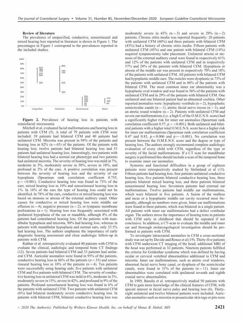
The Journal of Craniofacial Surgery � Volume 31, Number 8S, November/December 2020 European Guideline Craniofacial Microsomia
Review of literatureThe prevalence of unspecified, conductive, sensorineural and
mixed hearing loss reported in literature is shown in Figure 1. Thepercentages in Figure 1 correspond to the prevalences reported inthe included studies.
82%86%
20%17%
50%
30%
1%6%
66%
50%
3%10%
67% 73%
10%
75% 74%
16%
79%
17%
0%
10%
20%
30%
40%
50%
60%
70%
80%
90%
100%
Unspecified Conduc�ve Sensorineural Mixed
Figure 2. Prevalence of hearing loss in patients withcraniofacial microsomia.
Mitchell et al. evaluated facial malformations and hearing loss inpatients with CFM (5). A total of 79 patients with CFM wereincluded: 39 patients had bilateral CFM and 40 patients hadunilateral CFM. Microtia was present in 94% of the patients andhearing loss in 82% (n¼ 65) of the patients. Of the patients withhearing loss, twelve patients had bilateral hearing loss and 53patients had unilateral hearing loss. Interestingly, two patients withbilateral hearing loss had a normal ear phenotype and two patientshad unilateral microtia. The severity of hearing loss was mild in 7%,moderate in 3%, moderately severe in 30%, severe in 10%, andprofound in 2% of the ears. A positive correlation was presentbetween the severity of hearing loss and the severity of earhypoplasia (Spearman rank correlation coefficient 0.755,p¼<0.001). Conductive hearing loss was found in 73% of theears, mixed hearing loss in 10% and sensorineural hearing loss in1%. In 16% of the ears the type of hearing loss could not beidentified. In 78% of the ears, conductive or mixed hearing loss wasbased on stenosis or atresia of the external auditory canal. Othercauses for conductive or mixed hearing loss were middle eareffusion (n¼ 4), negative pressure (n¼ 2), or tympanic membraneperforations (n¼ 1). Hearing loss was frequently associated withipsilateral hypoplasia of the ear or mandible, although 8% of thepatients had contralateral hearing loss. Of the patients with man-dibular hypoplasia and microtia, 90% had hearing loss, whereas inpatients with mandibular hypoplasia and normal ears, only 33.5%had hearing loss. The authors emphasise the importance of earlydiagnostic hearing assessment and close audiologic follow-up inpatients with CFM.
Rahbar et al. retrospectively evaluated 40 patients with CFM toevaluate the clinical, audiologic and temporal bone CT findings(12). Seven patients had bilateral CFM and 33 patients had unilat-eral CFM. Auricular anomalies were found in 95% of the patients,conductive hearing loss in 86% of the patients (n¼ 35) and senso-rineural hearing loss in 10% of the patients (n¼ 4). Ten patientswere successfully using hearing aids: five patients with unilateralCFM and five patients with bilateral CFM. The severity of conduc-tive hearing loss in unilateral CFM was mild in 6%, moderate in 3%,moderately severe in 15%, severe in 42%, and profound in 9% of thepatients. Profound sensorineural hearing loss was found in 6% ofthe patients with unilateral CFM. Two patients with unilateral CFM(6%) had bilateral moderately severe conductive hearing loss. Inpatients with bilateral CFM, bilateral conductive hearing loss was
# 2020 The Author(s). Published by Wolters Kluwer Health, Inc. on b
moderately severe in 43% (n¼ 3) and severe in 29% (n¼ 2)patients. Chronic otitis media was reported frequently: 20 patientswith unilateral CFM (60%) and three patients with bilateral CFM(43%) had a history of chronic otitis media. Fifteen patients withunilateral CFM (45%) and one patient with bilateral CFM (14%)required tympanostomy tube placement. Unilateral atresia or ste-nosis of the external auditory canal were found in respectively 61%and 12% of the patients with unilateral CFM and in respectively57% and 28% of the patients with bilateral CFM. Hypoplasia oratresia of the middle ear was present in respectively 70% and 18%of the patients with unilateral CFM. All patients with bilateral CFMhad hypoplastic middle ears. The ossicles were dysplastic in 73% ofthe patients with unilateral CFM and in 86% of the patients withbilateral CFM. The most common inner ear abnormality was ahypoplastic oval window and was found in 36% of the patients withunilateral CFM and in 29% of the patients with bilateral CFM. Oneunilateral and one bilateral patient had an abnormal cochlea. Otherreported anomalies were: hypoplastic vestibule (n¼ 2), hypoplasticsemicircular canals (n¼ 1), atretic facial nerve recess (n¼ 1), andan atretic round window (n¼ 2). Patients with unilateral CFM andsevere ear malformations (i.e. a high E of the O.M.E.N.S. score) hada significantly higher risk for inner ear anomalies (Spearman rankcorrelation coefficient 0.57, p¼<0.001). Both unilateral and bilat-eral patients with a higher total O.M.E.N.S. score have a higher riskfor inner ear malformations (Spearman rank correlation coefficient0.47 and 0.83, p¼ 0.006 and p¼<0.001). No correlation waspresent between the O.M.E.N.S. score and the degree or type ofhearing loss. The authors strongly recommend complete audiologicevaluation of every child with CFM, regardless of the type orseverity of the facial malformations. If a CT scan for mandibularsurgery is performed this should include a scan of the temporal boneto examine inner ear anomalies.
Systemic and functional difficulties in a group of eighteenpatients were retrospectively studied by Stromland et al (13).Fifteen patients had hearing loss: four patients unilateral conductivehearing loss, five patients bilateral conductive hearing loss, threepatients bilateral mixed hearing loss, and three patients bilateralsensorineural hearing loss. Seventeen patients had external earmalformations. Twelve patients had middle ear malformations,which were bilateral in five patients. A fusion of the malleusand incus or a hypoplastic middle ear cavity occurred most fre-quently, although no numbers were given. Inner ear malformationswere found in three patients, which was bilateral in a single patient.All patients with inner ear malformations had a defect vestibularorgan. The authors stress the importance of hearing tests in patientswith CFM early in childhood that should be repeated if notconclusive. In addition, a CT-scan to evaluate the middle and innerear and thorough otolaryngological investigation should be per-formed in patients with CFM.
To investigate intracranial anomalies in CFM a cross-sectionalstudy was set up by Davide and Renzo et al (19). Thirty-five patientswith CFM underwent CT imaging of the head; additional MRI ofthe head was performed in 32 patients. Nineteen patients fulfilledthe criteria for Goldenhar syndrome which was defined by havingocular or cervical vertebral abnormalities additional to CFM andmicrotia. Inner ear malformations, such as atretic oval windows,abnormal facial nerve bony canal, or dysplasia of the semicircularcanals, were found in 31% of the patients (n¼ 11). Inner earabnormalities were correlated with ipsilateral seventh and eighthcranial nerve abnormalities.
In 1989, Bassila et al. retrospectively studied 50 patients withCFM to gain more knowledge of the clinical features of CFM, withspecial interest in facial nerve palsy and hearing loss (6). Thirty-eight unilateral and twelve bilateral patients were included. Auric-ular anomalies such as microtia or preauricular skin tags or pits were
ehalf of Mutaz B. Habal, MD 2421
Renkema et al The Journal of Craniofacial Surgery � Volume 31, Number 8S, November/December 2020
present in 92% of the patients. Facial nerve paralysis or paresis wasreported in eleven patients. A total of eight patients had sensori-neural hearing loss (prevalence of 16%). In six patients the senso-rineural hearing loss was accompanied by facial nerve palsy and intwo patients it was not. All eight patients with sensorineural hearingloss had unilateral CFM. Interestingly, four of the eight patientswith hearing loss had bilateral sensorineural hearing loss. In twopatients with sensorineural hearing loss a CT scan was obtainedwhich showed an absent cochlea and abnormal vestibulum in onepatient, and a normal cochlea and vestibulum in the other patient.
The relationship between auditory and facial nerve dysfunctionwas assessed by Carvalho et al. too, in a retrospective cohort studyof 99 patients with CFM (4). Thirty patients had bilateral CFM and69 unilateral CFM. Auricular anomalies, including microtia andear tags/pits, were present in 91 patients. A total of 75% of thepatients (n¼ 74) had hearing loss. Fourteen of the 74 patients withhearing loss (19%) required rehabilitation with auditory amplifi-cation. Conductive hearing loss was found in 74% (n¼ 73)patients. Patients with auricular anomalies had conductive hearingloss twice as often (p¼ 0.03). Sensorineural hearing loss waspresent in eleven patients, ten of these patients had conductivehearing loss too. In eight of the eleven patients, the sensorineuralhearing loss was unilateral and in three patients it was bilateral.Patients with auricular anomalies had a higher incidence of sen-sorineural hearing loss compared to patients without auricularanomalies (p¼ 0.04). Twenty-two patients had facial nerve dys-function (all unilateral CFM). No correlation was found betweenfacial nerve dysfunction and sensorineural hearing loss or auricularanomalies. Patients with a more severe form of CFM or bilateralCFM were not found to have a higher risk for sensorineural hearingloss or facial nerve dysfunction. The authors strongly recommend acomplete audiologic evaluation of every child in which the diag-nosis of CFM is considered.
Wan et al. retrospectively studied the clinical, temporal bone andaudiological findings in 70 patients with CFM (14). Seventeenpatients (24%) had bilateral CFM and 53 patients (76%) unilateralCFM. Almost all patients (99%) had auricular deformities. Hearingaids were used by 19 patients (27%). A total of 87 ears wereevaluated: conductive hearing loss in 82%, mixed hearing loss in15%, sensorineural hearing loss in 3% of the ears. The externalauditory canal was normal in 8%, stenotic in 10% and atretic in 81%of the 87 ears. The middle ear was hypoplastic in 67% and atretic in7% of the 87 ears. The ossicles were fused in 59% and absent in 23%of the 87 ears. The inner ear was abnormal in 8% of the 87 ears. Thefacial nerve was displaced in 40% and not identified in 15% ofthe 87 ears. The severity of the facial malformations, displayed bythe total O.M.E.N.S. score, was correlated with the malformation onradiography of the temporal bone (p¼<0.001). The severity andtype of hearing loss was not related to the total O.M.E.N.S. score orthe malformations on temporal bone radiography. This emphasisesthe importance of audiological testing in all patients with CFM.
A cross-sectional study of 89 patients with CFM was undertakenby Cohen et al. to study the type and frequency of maxillofacial andsystemic malformations in CFM (18). All patients were evaluatedfor hearing impairment. External ear anomalies (microtia or anotia)were present in 83% of the patients of which 15% had bilateral earmalformations. Atresia of the auditory canal was found in 58% ofthe patients, and 21% of the patients had stenosis of the auditorycanal. Hearing impairment, which is associated with auditory canalmalformations, was present in 79% (n¼ 69) of the patients. Bilat-eral deafness due to cochlea or acoustic nerve damage was found infive patients (6%).
Cousley et al. included 50 CFM patients in a retrospective cohortstudy to compare two classification systems (SAT and O.M.E.N.S.)(7). Data on hearing difficulties was included in this study: 66% of
2422 # 2020 The Author(s). Published
the patients had hearing difficulties. Three patients had unilateralconductive hearing loss combined with bilateral sensorineuralhearing loss. No further data on hearing loss or auricular anomalieswas provided.
A large retrospective cohort study was set up by Heike et al. todevelop a standardised approach to assess and describe facialcharacteristics in patients with CFM and controls (8). A total of142 patients with CFM and 290 controls without a craniofacialsyndrome were included. Sixty-seven percent (n¼ 91) of thepatients with CFM and four percent (n¼ 9) were reported to havehearing difficulties. Hearing devices were used by 21% of thepatients with CFM and 0.3% of the controls. No further informationon hearing difficulties was available in this study.
Engiz et al. studied 31 patients with CFM to describe thephenotypic features (16). In 21 patients the hearing status wasknown. Eighteen of these patients had hearing loss: conductive intwelve patients, mixed- in two patients, and sensorineural hearingloss in a single patient. Three patients with an atretic ear had normalhearing in the other ear.
A cross-sectional study on hearing difficulties in a group of tenpatients with CFM was performed by Goetze et al (20). All includedpatients had microtia. Two patients had unilateral conductivehearing loss, respectively mild and moderate. One patient hadmoderate bilateral conductive hearing loss. Sensorineural hearingloss was present in two patients, both bilateral. One patient hadprofound sensorineural hearing loss in both ears and the otherpatient had moderate and mild sensorineural hearing loss.
Sleifer et al. studied audiological findings in CFM in a cross-sectional study (21). All ten included patients with CFM underwenttonal and vocal hearing evaluations. Conductive hearing loss waspresent in 40% of the left ears and 60% of the right ears. Sensori-neural hearing loss was found in five ears, mild (n¼ 2), moderate(n¼ 1), and profound (n¼ 2). It is unclear if any patient had isolatedsensorineural hearing loss. External ear malformations were foundin 70% of the left ears (n¼ 7) and in 80% of the right ears (n¼ 8).No correlation was found between the severity of ear malformationand hearing loss.
To assess inner ear anomalies in Goldenhar syndrome, a retro-spective review of temporal bone changes in 21 patients withGoldenhar syndrome was undertaken by Hennersdorf et al (9).Ninety percent (n¼ 19) of the patients had atresia or stenosis of theexternal auditory canal. Dysplasia of the ossicles was present in90% of the patients; absence of ossicles was found in one patient(5%). A narrowed or opacified tympanic cavity was found in 67%(n¼ 14) of the patients. The antrum mastoidium was opacified in57% (n¼ 12) of the cases. Thirty-three percent of the patients hadinner ear anomalies of the cochlea, semicircular canals or vestibule.Cochlear hypoplasia and a common cavity deformity were bothfound in a single patient. Anomalies of the semicircular canals werefound in five patients (24%) and were associated with dysplasia ofthe vestibule, which was seen in four patients (19%). Information onhearing difficulties was available for seven patients: four patientshad isolated severe conductive hearing loss and three patients hadcombined hearing loss, which was correlated to severe inner earmalformations in a single patient. Interestingly, the other twopatients with mixed hearing loss had no apparent inner ear mal-formations on imaging. The authors state that inner ear deformitiesare not always correlated to external and middle ear malformations,which stresses the importance of a high-resolution CT or MRI of thetemporal bone in all CFM patients.
Jacobsson et al. studied the clinical appearance of syndromesthat affect the structures originated from the first and secondpharyngeal arches (10). This retrospective cohort study included26 patients with unilateral CFM. Twenty-five patients had atreticexternal ear canals. One patient with unilateral CFM had bilateral
by Wolters Kluwer Health, Inc. on behalf of Mutaz B. Habal, MD
T
R
I
R
T
The Journal of Craniofacial Surgery � Volume 31, Number 8S, November/December 2020 European Guideline Craniofacial Microsomia
middle ear deformities. Dysplasia of the ossicles was common in thestudied population: malleus malformation in 25 ears, malleus fusionin three ear, incus malformation in 24 ears, incus fusion in threeears, stapes malformation in 21 ears, and a missing stapes in fiveears. Two patients had a cochlear malformation, one of thesepatients had sensorineural hearing loss. No information on hearingin the other patients was available.
A case series of twelve patients with CFM to describe earanomalies was initiated by Rosa et al (17). All twelve patients hadexternal ear anomalies, eight patients had middle ear anomalies andthree patients inner ear anomalies. Type of middle ear anomalies:displacement (n¼ 2), opacification (n¼ 2), and malformation (n¼ 2)of the ossicles. Agenesis of the inner ear canal was found in twopatients. No information about hearing difficulties was reported.
A case series of fourteen patients with Goldenhar syndrome waspresented by Bisdas et al. to study inner ear malformations inpatients with Goldenhar syndrome (15). No clear criteria were usedto include patients; no information on laterality of Goldenharsyndrome was available. Atresia of the external auditory canalwas present unilaterally in ten patients and bilaterally in twopatients. Inner ear malformations were seen in five (36%) patients:dysplastic or absent ossicles in 13 patients, absence or dysplasia ofthe semicircular canals in four patients, enlarged vestibule in fourpatients, enlarged internal auditory canal in two patients, and acommon cavity deformity in one patient. No information abouthearing difficulties in these patients was provided.
Llano-Rivas et al. included 87 patients with isolated microtiaand 58 patients with CFM, to study the clinical and geneticbehaviour of microtia (11). A normal external auditory canalwas present in 48% of the patients with CFM (n¼ 28); 47%(n¼ 27) of the patients had a unilateral atretic external auditorycanal and 5% (n¼ 3) of the patients had a bilateral atretic externalauditory canal. The prevalence of a normal or unilateral atreticexternal auditory canal was equal in patients with CFM and isolatedmicrotia. However, patients with isolated microtia were found tohave a higher prevalence of bilateral atretic external auditory canal(16% versus 5%, p¼ 0.05). Data on hearing loss was only availablefor all 145 patients and not specified for patients with CFM. Eighty-five percent of the patients had conductive hearing loss (n¼ 123).Five patients (3%) had sensorineural hearing loss and six patients(4%) had mixed hearing loss.
The types of ear anomalies and its prevalence found in patientswith CFM are displayed in Table 1.
Table 3. Types of ear anomalies.
# 2020 Th
Type of malformation
e Author(s). Published by W
Prevalence
olters Kluw
Reference
R
Outer ear Atresia or stenosis externalauditory canal90%
(9)Atresia external auditory canal
52% - 96% (10–12, 14, 15, 18)Stenosis external auditory canal
10% - 35% (12, 14, 18)S
Middle ear Dysplastic or absent ossicles 93% (15)Dysplastic ossicles
17% - 90% (9, 12, 17)A
Agenesis ossicles 5% - 23% (9, 14)Opacification of ossicles
17% (17)Displacement of ossicles
17% (17)N
Fusion of ossicles 59% (14)Malleus malformation
96% (10)Malleus fusion
12% (10)P
Incus malformation 92% (10)Incus fusion
12% (10)Stapes malformation
81% (10) R Stapes aplasia 19% (10)Hypoplastic oval window
35% (12)er Health, Inc. on b
. (continued )
ehalf of M
Type of malformation
utaz B. Habal, MD
Prevalence
ReferenceAgenesis round window
5% (12)Agenesis facial nerve recess
3% (12)Narrowed or opacified tympaniccavity
67%
(9)Opacification of antrummastoideum
57%
(9)Hypoplastic middle ear
67% (14)Agenesis middle ear
7% (14)Inner ear
Inner ear malformations notspecified8% - 31%
(14, 19)Cochlear anomaly
5% - 10% (9, 12)Dysplastic or absent semicircularcanals
3% - 29%
(9, 12, 15)Vestibule deformity
5% - 29% (9, 12, 15)Enlarged internal auditory canal
14% (15)Common cavity deformity
7% (15)Agenesis inner ear canal
17% (17)Conclusions
-
Prevalencehe prevalence of hearing difficulties in patients with craniofacialmicrosomia is:- Unspecified hearing loss: 50% - 82%- Conductive hearing loss: 30% - 86%- Sensorineural hearing loss: 1% - 20%- Mixed hearing loss: 6% - 17%
ef (4–8, 12–14, 16, 18, 20, 21)
Level 3
Prevalencen 6% - 10% of the patients with unilateral craniofacialmicrosomia, hearing difficulties can be present bilaterally orsolely on the contralateral side.
ef (5, 6, 12)
Level 3
Typehe most common anomalies of the ear in patients withcraniofacial microsomia are:- Outer ear: Atresia or stenosis of the external auditory canal- Middle ear: Dysplastic or absent ossicles- Inner ear: vestibule deformity or semicircular canal anomalies
ef (9–12, 14, 15, 17, 18)
Level 3
Risk factorsevere microtia or anotia is positively correlated with hearingloss.lthough a higher total O.M.E.N.S. score was found to be
correlated with inner ear malformation, no correlation waspresent between the total O.M.E.N.S. score and the degree ortype of hearing loss.
o correlation was present between facial nerve dysfunction andsensorineural hearing loss or auricular anomalies. However,facial and vestibulocochlear nerve dysfunction werecorrelated with ipsilateral inner ear anomalies.
atients with bilateral craniofacial microsomia were not found tohave a higher risk for hearing difficulties compared to patientswith unilateral craniofacial microsomia.
ef (4, 5, 12, 14, 19)
2423
Renkema et al The Journal of Craniofacial Surgery � Volume 31, Number 8S, November/December 2020
24
4.4.2 What is the policy for screening and monitoring ofhearing difficulties in patients with craniofacial microso-mia?
The literature search for this question was directed towards morespecific questions related to the consequences and impact ofscreening and monitoring, the available screening tests and proce-dures for monitoring, and responsibilities for screening and moni-toring (Appendix 1, http://links.lww.com/SCS/B697).
Review of literatureVarious manuscripts included in Chapter 4.4.1. conclude that
every child diagnosed with CFM should have complete audiologicevaluation (4, 5, 9, 11–14, 20). Imaging of the temporal bone byusing high-resolution CT or MRI to assess middle and inner earanomalies is recommended by various authors as well (9, 12, 13,17). Although screening is recommended, timing and frequencywere not further specified.
ConclusionNo conclusions based on CFM-specific literature could be made.
Considerations� Overall quality of evidenceBecause there is no applicable literature available the recom-
mendations are based on expert opinion with a low qualityof evidence.
� Balance of benefits and harmsThe steering group reasons that most parents of patients want
their children to be screened for hearing difficulties, to ensureoptimal speech and language development.
� Outcome importanceA delay in detecting or failure to detect hearing loss could have
considerable consequences for speech, and language development(bilateral hearing loss) and/or social interaction (unilateral orbilateral hearing loss), while a hearing test is a low burden forthe patient or parents. The Joint Committee on Infant Hearingemphasise the importance of early detection of hearing difficulties.A guideline for early hearing detection and intervention waspublished in 2007 (22).
� Costs and resourcesThe impact on costs and resources of the given recommenda-
tions will vary per member state, depending on the available careproviders and facilities. The recommendations concern the essentialrequirements for adequate treatment of patients with CFM andshould thus be implemented. Costs are lowest and resources aremost efficiently used when care for craniofacial disorders is cen-tralised in a limited number of expert centres per member state. Ageneral rule that can be applied is one expert centre per 10million inhabitants.
� Inequity of the recommendationThe goal of the European Reference Networks (ERNs) is to
eliminate inequality within Europe with regard to care for patientswith rare diseases. At present, not every member state offers anexpert centre for CFM, or the level of provided care does not (yet)meet all the requirements that are outlined in this guideline. Bydefining the baseline of required care for CFM, this guideline willhelp these member states to reach the appropriate level. The ERN oncraniofacial anomalies and ENT disorders (ERN-CRANIO) can
24 # 2020 The Author(s). Published
guide a patient in Europe to the available centres of expertise(www.ern-cranio.eu) and can support care providers with diagnosisand treatment advice.
� Feasibility of the recommendationRecommendations refer to the general requirements for deliv-
ering optimal healthcare and are discussed with members fromparticipating European countries. Quality of care was paramount inthe discussions. Centralisation is proposed as one of the core values.However, in some countries the national organisation of healthcaremight impede centralisation. National implementation of the ERNsthat fits the situation of each country is necessary. For the memberstates with the lowest number of inhabitants, the establishment of acraniofacial centre might not be feasible, and collaboration with acraniofacial centre in the surrounding countries can be an option.
� Acceptability of the recommendationIt is expected that all stakeholders want to apply and will be
applying the recommendations, because they are employed in ERN-acknowledged institutions. National implementation plans are nec-essary to ensure that recommendations fit the situation in eachcountry. In addition, not all countries participating in the ERN-CRANIO are represented in the guideline development group, andnew members will join within the coming years. For these countries,acceptance and implementation of ERN guidelines such as thisguideline on CFM is obligatory.
Rationale of the recommendationsAll new-born infants that do not pass the neonatal hearing test
should have an auditory brainstem response (ABR) test included aspart of their screening. Referral to an otolaryngologist experiencedin paediatric hearing loss should be made to have complete screen-ing of both ears. Complete audiological evaluation should be donewithin three months of age in patients who do not pass the initialhearing test. Audiologic intervention should be initiated before theage of six months in these patients. Children who pass the neonatalhearing test but have a higher risk for hearing difficulties should bere-evaluated in 24 to 30 months. Since patients with hearing loss areat risk for a delay in speech and language development, regularmonitoring for speech and language development at six monthsintervals should be initiated.
Screening for hearing loss will incur costs but outweighs the riskof not screening for hearing difficulties. The main aspects indetermining these risks are the age of the patients and severityof CFM. Overall, all young patients in whom speech and languageskills are developing should be examined by a hearing test yearly.
Recommendations
by
� Perform neonatal hearing test in all new-borns withcraniofacial microsomia. If indicated, complete audio-logical evaluation in an experienced audiology centreshould be performed before the age of three months toensure timely treatment.
� Re-evaluate hearing tests in patients with craniofacialmicrosomia by the age of 24–30 months.
� Regularly perform otoscopy and audiometry in patientswith craniofacial microsomia including microtia and/orcleft palate by the ENT doctor/otolaryngologist.
� Audiologic intervention should be initiated before theage of six months in patients with congenital
Wolte
hearing loss.
rs Kluwer Health, Inc. on behalf of Mutaz B. Habal, MD
The Journal of Craniofacial Surgery � Volume 31, Number 8S, November/December 2020 European Guideline Craniofacial Microsomia
Research gap
The available literature mainly focuses on the prevalence of
hearing loss but provides little information on the severity ofhearing loss, types of treatment, and results of treatment.4.4.3 What are the indications and policy for treatment ofhearing difficulties in patients with craniofacial microso-mia?
The literature search for this question was directed towards morespecific research questions related to treatment options, symptoms,requirements, complications, and advantages and disadvantages oftreatment of hearing difficulties (Appendix 1, http://links.lww.com/SCS/B697). Unfortunately, no literature is available on the indica-tions and policy for treatment of hearing difficulties in patients withCFM. Therefore no conclusions were written. Considerations forthis question were only based on expert opinion.
Considerations� Overall quality of evidenceBecause there is no literature available the recommendations are
based on expert opinion with a low quality of evidence.
� Balance of benefits and harmsThe steering group reasons that most parents of patients want
their children to be screened for hearing difficulties, to ensureoptimal speech and language development.
� Outcome importanceUntreated hearing loss could have significant consequences for
the well-being of the patient, his or her speech and languagedevelopment, and his or her social participation.
� Costs and resourcesThe impact on costs and resources of the given recommenda-
tions will vary per member state, depending on the available careproviders and facilities. The recommendations concern the essentialrequirements for adequate treatment of patients with CFM andshould thus be implemented. Costs are lowest and resources aremost efficiently used when care for craniofacial disorders is cen-tralised in a limited number of expert centres per member state. Ageneral rule that can be applied is one expert centre per 10million inhabitants.
� Inequity of the recommendationThe goal of the European Reference Networks (ERNs) is to
eliminate inequality within Europe with regard to care for patientswith rare diseases. At present, not every member state offers anexpert centre for CFM, or the level of provided care does not (yet)meet all the requirements that are outlined in this guideline. Bydefining the baseline of required care for CFM, this guideline willhelp these member states to reach the appropriate level. The ERN oncraniofacial anomalies and ENT disorders (ERN-CRANIO) canguide a patient in Europe to the available centres of expertise(www.ern-cranio.eu) and can support care providers with diagnosisand treatment advice.
� Feasibility of the recommendationRecommendations refer to the general requirements for deliv-
ering optimal healthcare and are discussed with members fromparticipating European countries. Quality of care was paramount inthe discussions. Centralisation is proposed as one of the core values.However, in some countries the national organisation of healthcaremight impede centralisation. National implementation of the ERNs
# 2020 The Author(s). Published by Wolters Kluwer Health, Inc. on b
that fits the situation of each country is necessary. For the memberstates with the lowest number of inhabitants, the establishment of acraniofacial centre might not be feasible, and collaboration with acraniofacial centre in the surrounding countries can be an option.
� Acceptability of the recommendationIt is expected that all stakeholders want to apply and will be
applying the recommendations, because they are employed in ERN-acknowledged institutions. National implementation plans are nec-essary to ensure that recommendations fit the situation in eachcountry. In addition, not all countries participating in the ERN-CRANIO are represented in the guideline development group, andnew members will join within the coming years. For these countries,acceptance and implementation of ERN guidelines such as thisguideline on CFM is obligatory.
Rationale of the recommendationsThe Joint Committee on Infant Hearing states that the initiation
of early intervention for hearing loss should begin as soon aspossible after the diagnosis of hearing loss but no later than theage of six months (22). Intervention before the age of six monthsgives significantly better outcomes on language, speech and social-emotional development compared to later-identified children. Per-sonal amplification devices should be provided by an experiencedaudiologist soon after the diagnosis of hearing loss. Surgicaltreatment may be indicated in some patients to reconstruct mal-formed outer and/or middle ears and includes bone-anchoredhearing aids. Cochlear implantation should be considered inpatients who have limited benefits from (bone-anchored)hearing aids.
The International Microtia and Atresia Workgroup advises thatall patients with microtia and external auditory atresia should beseen in a multidisciplinary team (23). It is important for patientswith bilateral aural atresia to be treated with bone conductiondevices or cochlear implants to support language and speechdevelopment (23). Timing of placement should be discussed witha microtia surgeon to ensure the possibility of microtia reconstruc-tion in the future (23).
Recommendations
eh
� Treat moderate to severe hearing loss, either with non-surgical or surgical options.
� Coordinate surgical approach and timing in a multidis-ciplinary team regarding hearing augmentation andother surgical procedures including ear reconstruction
alf of
and mandibular surgeries.
Research gapNo literature is available regarding indications and policy for
treatment of hearing difficulties in patients with craniofacial micro-somia. Availability of studies could help determine an optimaltreatment strategy.
References
1. T
onn CR, Grundfast KM. What an otolaryngologist shouldknow about evaluation of a child referred for delay in speechdevelopment. JAMA Otolaryngol Head Neck Surg. 2014;140(3):259-65.2. E
tienne Krug AC, Shelly Chadha, Laura Sminkey, RicardoMartinez, Gretchen Stevens, Karl White, Katrin Neumann,Bolajoko Olusanya, Paige Stringer, Mohan Kameswaran,Glyn Vaughan, Ruth Warick, Andrea Bohnert, LillianMutaz B. Habal, MD 2425
Renkema et al The Journal of Craniofacial Surgery � Volume 31, Number 8S, November/December 2020
242
Henderson, Irving Basanez, Maeva LeGeoff, Vincent Fougner,Tess Bright, Sebastian Brown. Childhood hearing loss:strategies for prevention and care. WHO Library Catalogu-ing-in-Publication Data. 2016.
3. L
in CY, Tseng YC, Guo HR, Lai DC. Prevalence of childhoodhearing impairment of different severities in urban and ruralareas: a nationwide population-based study in Taiwan. BMJOpen. 2018;8(3):e020955.4. C
arvalho GJ, Song CS, Vargervik K, Lalwani AK. Auditoryand facial nerve dysfunction in patients with hemifacialmicrosomia. Arch Otolaryngol Head Neck Surg. 1999;125(2):209-12.5. M
itchell RM, Saltzman BS, Norton SJ, Harrison RG, HeikeCL, Luquetti DV, et al. Hearing Loss in Children WithCraniofacial Microsomia. Cleft Palate Craniofac J.2017;54(6):656-63.6. B
assila MK, Goldberg R. The association of facial palsy and/or sensorineural hearing loss in patients with hemifacialmicrosomia. Cleft Palate J. 1989;26(4):287-91.7. C
ousley RR. A comparison of two classification systems forhemifacial microsomia. Br J Oral Maxillofac Surg.1993;31(2):78-82.8. H
eike CL, Wallace E, Speltz ML, Siebold B, Werler MM,Hing AV, et al. Characterizing facial features in individualswith craniofacial microsomia: A systematic approach forclinical research. Birth Defects Res A Clin Mol Teratol.2016;106(11):915-26.9. H
ennersdorf F, Friese N, Lowenheim H, Tropitzsch A,Ernemann U, Bisdas S. Temporal bone changes in patientswith Goldenhar syndrome with special emphasis on inner earabnormalities. Otol Neurotol. 2014;35(5):826-30.10. J
acobsson C, Granstrom G. Clinical appearance of spontane-ous and induced first and second branchial arch syndromes.Scand J Plast Reconstr Surg Hand Surg. 1997;31(2):125-36.11. L
lano-Rivas I, Gonzalez-del Angel A, del Castillo V, Reyes R,Carnevale A. Microtia: a clinical and genetic study at theNational Institute of Pediatrics in Mexico City. Arch Med Res.1999;30(2):120-4.12. R
ahbar R, Robson CD, Mulliken JB, Schwartz L, Dicanzio J,Kenna MA, et al. Craniofacial, temporal bone, and audiologicabnormalities in the spectrum of hemifacial microsomia. ArchOtolaryngol Head Neck Surg. 2001;127(3):265-71.13. S
tromland K, Miller M, Sjogreen L, Johansson M, JoelssonBM, Billstedt E, et al. Oculo-auriculo-vertebral spectrum:associated anomalies, functional deficits and possible devel-opmental risk factors. Am J Med Genet A. 2007;143A(12):1317-25.14. W
an J, Meara JG, Kovanlikaya A, Nelson MD, Don D.Clinical, radiological, and audiological relationships inhemifacial microsomia. Ann Plast Surg. 2003;51(2):161-6.15. B
isdas S, Lenarz M, Lenarz T, Becker H. Inner earabnormalities in patients with Goldenhar syndrome. OtolNeurotol. 2005;26(3):398-404.16. E
ngiz O, Balci S, Unsal M, Ozer S, Oguz KK, Aktas D. 31cases with oculoauriculovertebral dysplasia (Goldenharsyndrome): clinical, neuroradiologic, audiologic and cyto-genetic findings. Genet Couns. 2007;18(3):277-88.17. R
osa RF, Silva AP, Goetze TB, Bier Bde A, Almeida ST,Paskulin GA, et al. Ear abnormalities in patients with oculo-auriculo-vertebral spectrum (Goldenhar syndrome). Braz JOtorhinolaryngol. 2011;77(4):455-60.18. C
ohen N, Cohen E, Gaiero A, Zecca S, Fichera G, Baldi F,et al. Maxillofacial features and systemic malformations inexpanded spectrum Hemifacial Microsomia. Am J Med GenetA. 2017;173(5):1208-18.6 # 2020 The Author(s). Published
19. D
by W
avide B, Renzo M, Sara G, Elisa L, Rodica M, Irene T, et al.Oculo-auriculo-vertebral spectrum: going beyond the first andsecond pharyngeal arch involvement. Neuroradiology.2017;59(3):305-16.
20. G
oetze TB, Sleifer P, Rosa RF, da Silva AP, Graziadio C, ZenPR. Hearing characterization in oculoauriculovertebral spec-trum: A prospective study with 10 patients. Am J Med GenetA. 2017;173(2):309-14.21. S
leifer P, Gorsky Nde S, Goetze TB, Rosa RF, Zen PR.Audiological findings in patients with oculo-auriculo-verte-bral spectrum. Int Arch Otorhinolaryngol. 2015;19(1):5-9.22. A
merican Academy of Pediatrics JCoIH. Year 2007 positionstatement: Principles and guidelines for early hearingdetection and intervention programs. Pediatrics. 2007;120(4):898-921.23. Z
hang TY, Bulstrode N, Chang KW, Cho YS, Frenzel H, JiangD, et al. International Consensus Recommendations onMicrotia, Aural Atresia and Functional Ear Reconstruction.J Int Adv Otol. 2019;15(2):204-8.CHAPTER 4. SCREENING, MONITORING ANDINDICATION FOR TREATMENT
4.5 Eye anomalies in craniofacial microsomiaIntroductionOriginally, the triad of Goldenhar syndrome, now believed to be
part of the craniofacial microsomia (CFM) ‘spectrum’, consisted ofmandibular dysostosis, ear malformations and epibulbar dermoids(1). This illustrates the prominent role eye anomalies have in CFM.Patients with CFM regularly present with epibulbar dermoids orother eye anomalies, such as colobomata. This chapter focusses oneye anomalies in CFM. Orbital malformations will not be takeninto account.
Colobomata are caused by incomplete closure of embryonicfissures and can clinically present as gaps or notches of the eyelids,lens, and/or choroid and retina. Epibulbar dermoids are benign,ectopic, congenital neoplasms that vary in composition (dermoid orlipodermoid) or location (corneal or limbal) (2). It is the mostcommon congenital benign tumour of the eye, occurring in 1–3 per10,000 births in the general population (3). The aetiology ofepibulbar dermoids remains unclear.
Epibulbar dermoids have been classified in three grades (4, 5):
Grade I:
olters Kluwe
Superficial dermoids measuring less than 5 mm,located on the limbus
Grade II:
Limbal dermoid covering most of the cornea,down into the stroma up to Descemet’smembrane, without involving the membrane.Grade III:
Large dermoids covering the whole corneaextending up to the pigmented epithelium of theiris.To develop recommendations on screening, monitoring andtreatment of eye anomalies in CFM the following questions wereposed:
4.5.1 What is the type, prevalence and severity of eye anomaliesin craniofacial microsomia?4.5.2 What is the policy for screening and monitoring of eyeanomalies in patients with craniofacial microsomia?4.5.3 What are the indications and policy for treatment of eyeanomalies in patients with craniofacial microsomia?
r Health, Inc. on behalf of Mutaz B. Habal, MD
The Journal of Craniofacial Surgery � Volume 31, Number 8S, November/December 2020 European Guideline Craniofacial Microsomia
An overview of the type, prevalence and severity of eyeanomalies seen in patients with CFM makes it possible to developrecommendations on screening and treatment of these anomalies.Commonly seen eye anomalies in patients with CFM, such asepibulbar dermoids and colobomata, can lead to various patientrelevant consequences. Limbal dermoids, including grade I der-moids, can cause amblyopia, difficulties with eyelid closure, irrita-tion, corneal erosion, and/or aesthetic difficulties. This may be anindication for surgical resection. Other eye anomalies, as colobo-mata, can lead to exposure keratopathy, corneal ulceration, retinaldetachment and/or cataract (6). Since timely treatment is importantto maintain optimal vision, recommendations on screening andmonitoring should be made.
Literature searchA systematic search of literature was performed to identify all
available literature on craniofacial microsomia and synonyms. Thesearch was conducted in Embase, Pubmed/Medline Ovid. The fullsearch strategy is reported in the supplementary material. Theinclusion and exclusion criteria are as follows:
Inclusion and exclusion criteria:
Type of studies
# 2020 The Author(s). Pu
- Original articles- Systematic review of sufficient quality:
- The question in the systematicreview matches the question of theguideline.- The search of the systematic reviewwas conducted in at least two relevantdatabases, such as the CochraneLibrary, Medline/Pubmed.- The full search strategy wasreported.- No relevant items were missing inthe search strategy.
Type of patients
- Patients with craniofacialmicrosomia- Patients with epibulbar dermoidsSubject
- Eye anomalies, epibulbar dermoids,colobomaExclusion criteria
- Original studies with <10 includedpatients- Articles published before 1980- Case reports- Expert opinion- Letters- Editorials
Literature was screened on title and abstract. All literature oneye anomalies in CFM that was available was included in thischapter. A total of five manuscripts were included: a large retro-spective cohort study by Caron et al. (7), a cross-sectional study byCohen et al. (8), and four retrospective cohort studies by Hertle et al.(9), Ewart-Toland et al. (10), Stromland et al. (11), and Mansouret al (3).
Since no literature on the indication for treatment was available,an additional search was done. This search contained epibulbardermoids and variants and was performed in Medline/Pubmed.Literature on indications for treatment for epibulbar dermoids ingeneral was included in this chapter. The full search strategy isincluded in the supplementary material. A total of 254 articles wereidentified; exclusion based on title and abstract led to the inclusionof two articles. Although narrative reviews were excluded in
blished by Wolters Kluwer Health, Inc. on b
previous chapters, a review by Pirouzian et al. (5) was includedin this chapter due to the extensive nature of the review and the goodlink with the research question. A case series by Matsuo wasincluded too (12).
4.5.1 What is the type, prevalence and severity of eyeanomalies in craniofacial microsomia?
The literature search for this question was directed towards morespecific questions related to the type (epibulbar dermoids andcolobomata), prevalence and severity (mild, moderate or severe)of eye anomalies in CFM (Appendix 1, http://links.lww.com/SCS/B697). Since this question does not relate to interventions ordiagnostics, no considerations, rationale or recommendationsare provided.
Review of literatureIn 1985, Mansour et al. aimed to report ocular findings in a large
cohort of patients with CFM (3). A total of 57 patients wereincluded in this retrospective cohort study. Half of the patients(51%) had ocular anomalies: 32% (n¼ 18) epibulbar (lipo)der-moids, 19% (n¼ 11) ocular motility disorders, 12% (n¼ 7) ble-pharoptosis, 11% (n¼ 6) eyelid coloboma, and 11% (n¼ 6)lacrimal duct or gland anomalies. Two-thirds of the epibulbar(lipo)dermoids were unilateral, the others were bilateral. Allpatients with an eyelid coloboma had epibulbar (lipo)dermoidstoo, although in one case the coloboma was located in the othereye than the (lipo)dermoid. Ocular motility disorders includedesotropia in six patients, exotropia in three patients, a single patientwith Duane’s syndrome, a patient with abduction deficiency, and apatient with unspecified strabismus. It is unclear whether the ocularmotility disorders were caused by epibulbar (lipo)dermoids. Inter-estingly, data on anomalies of the posterior pole of the eye waspresent in nineteen patients and three patients were found to haveanomalies: a patient with a choroidal coloboma, one patient with alarge peripapillary hypopigmentation, and a patient with a retinalvascular tortuosity.
Hertle et al. reported the ocular and adnexal findings of 49patients with CFM (44 unilateral and 5 bilateral CFM) who all hadophthalmologic evaluation (9). Two-thirds (67%) of the patientshad some ocular or adnexal anomaly. Frequently seen ocularanomalies included: astigmatism (18%), amblyopia (16%) andanisometropia (8%). Amblyopia was caused by multiple factorsincluding strabismus, refractive errors, and ptosis. Another fre-quently seen ocular anomaly was strabismus (22%, n¼ 11). Six ofthese patients had noncomitant strabismus (Duane syndrome(n¼ 2), sixth nerve palsies (n¼ 2) and bilateral superior obliquepalsies (n¼ 2)). Comitant exotropia was reported in three patientsand esotropia in two patients. No globe or optic nerve anomalieswere seen, although a single patient had tortuous retinal vasculaturewith normal vision in both eyes. Lid or adnexal anomalies were seenin 41% of the patients and included: dacryostenosis (15% of allpatients), blepharoptosis (12% of all patients), canthal dystopia (8%of all patients), lagophthalmos (8% of all patients), entropion (4% ofall patients), lipodermoids (4% of all patients), lid retraction (2% ofall patients), and telecanthus (2% of all patients). This study shows ahigh frequency of ocular and adnexal anomalies in patients withCFM, which according to the authors necessitates ophthalmologicexamination for all patients (9). It is important to diagnose and treatstrabismus, amblyopia, and adnexal anomalies early to preventvisual loss (9).
Caron et al. reviewed 755 patients with CFM retrospectively toanalyse typical phenotypes within CFM (7). Most patients hadunilateral CFM (88.6%) and 11.4% had bilateral CFM. A total of13.4% of patients had ophthalmic anomalies such as epibulbardermoids and/or colobomata. The location of the colobomata were
ehalf of Mutaz B. Habal, MD 2427
Renkema et al The Journal of Craniofacial Surgery � Volume 31, Number 8S, November/December 2020
not further specified. Patients with bilateral CFM had a significantlyhigher risk for ocular anomalies (Pearson X2¼ 27.191,p¼<0.001). Epibulbar dermoids were present in 81 patients(10.7%): 74% of these patients had unilateral CFM and 26%patients had bilateral CFM. Colobomata were less frequently seen:in eleven patients (1.5%) colobomata were present together withepibulbar dermoids and nine patients (1.2%) solely had colobomata.No further information on ocular anomalies was reported.
Other studies reported briefly on ocular anomalies. Cohen et al.reported in a cross-sectional study of 89 patients with CFM that 41patients (46%) had ocular anomalies: 23 dermoid cysts (56%), 15blepharoptosis (37%), 13 ocular and/or eyelid coloboma (32%), 6dermolipoma (15%), and 12 other eye defects (29%) (8). A micro-phtalmic eye was present in 29 patients (71%) and two patients hadanophtalmia (5%). Ewart-Toland et al. studied 21 patients withCFM (10). Two patients had epibulbar dermoids; other anomaliesreported in single patients: bilateral conjunctival dermoids, cornealopacity, eye lipoma, microphthalmus, and hypoplastic tear duct.Stromland et al. retrospectively studied 18 patients with CFM (11).Eleven patients (61%) had lipodermal dermoids of the eye and eightpatients (44%) had epibulbar dermoids. All patients with epibulbardermoids had lipodermal dermoids too. Three patients had unilat-eral microphthalmus and one patient bilateral microphthalmus.Other eye anomalies that were reported: upper eyelid coloboma(n¼ 3), unilateral ptosis (n¼ 1), and distichiasis (two rows of eyelashes) (n¼ 1).
Conclusions
2
Type and prevalence
Reported eye anomalies in craniofacialmicrosomia and its prevalence:
- Lipodermal dermoids
428 # 2
- prevalence: 4% - 61%
- Epibulbar dermoids
- prevalence: 10% - 56%- Colobomata (eyelid)
- prevalence: 3% - 32%- Blepharoptosis
- prevalence: 9% - 37%- Microphthalmus
- prevalence: 5% - 71%- Lacrimal duct or gland anomalies
- prevalence: 5% - 11%- Strabismus
- prevalence: 12% - 22%-
Reported eye anomalies in CFMin a single study- Astigmatism
- prevalence: 18%- Amblyopia
- prevalence: 16%- Anisometropia
- prevalence: 8%- Anomalies of the posterior poleof the eye
prevalence: 5%
- Anophtalmia
- prevalence: 5%Colobomata are often accompaniedby epibulbar dermoids in patients withcraniofacial microsomia.
Ref (3, 7–11)
Level 3
PrevalencePatients with bilateral craniofacial microsomiahave a significant higher risk for eyeanomalies (studied in 755 patients,p¼<0.001).
Ref (7)
020 The Author(s). Published
Level 3
by Wolters Kluwer H
SeverityVisual loss could be seen in 8% of the patients
with craniofacial microsomia, although thiswas reported in a single study with smallsample size (n¼ 49).
Ref (9)
4.5.2 What is the policy for screening and monitoring of eyeanomalies in patients with craniofacial microsomia?
The literature search for this question was directed towards morespecific questions related to the consequences and impact of screen-ing and monitoring, the available screening tests and procedures formonitoring, and responsibilities for screening and monitoring(Appendix 1, http://links.lww.com/SCS/B697). Unfortunately, noliterature on screening and/or monitoring of eye anomalies in patientswith CFM is available.
Considerations� Overall quality of evidenceBecause there is no literature available the recommendations are
based on expert opinion with a low quality of evidence.
� Balance of benefits and harmsCFM is found to be associated with a high percentage of ocular
anomalies (67%). The main concern is the risk of permanent visualimpairment caused by a treatable obstacle/anomaly. Therefore, allchildren with CFM should be screened regarding visual develop-ment at an early stage. Persistent amblyopia causes a reducedquality of life especially, if the better eye is lost later in life anda significant (financial) burden on society. Therefore, the benefits ofscreening outweigh the harms.
� Outcome importanceThe reduced quality of life in patients with poor visual acuity
requires adequate screening, monitoring and treatment, especially ifthe visual acuity in the better eye is lost later in life.
� Costs and resourcesThe impact on costs and resources of the given recommenda-
tions will vary per member state, depending on the available careproviders and facilities. The recommendations concern the essentialrequirements for adequate treatment of patients with CFM andshould thus be implemented. Costs are lowest and resources aremost efficiently used when care for craniofacial disorders is cen-tralised in a limited number of expert centres per member state. Ageneral rule that can be applied is one expert centre per 10million inhabitants.
� Inequity of the recommendationThe goal of the European Reference Networks (ERNs) is to
eliminate inequality within Europe with regard to care for patientswith rare diseases. At present, not every member state offers anexpert centre for CFM, or the level of provided care does not (yet)meet all the requirements that are outlined in this guideline. Bydefining the baseline of required care for CFM, this guideline willhelp these member states to reach the appropriate level. The ERN oncraniofacial anomalies and ENT disorders (ERN-CRANIO) canguide a patient in Europe to the available centres of expertise(www.ern-cranio.eu) and can support care providers with diagnosisand treatment advice.
ealth, Inc. on behalf of Mutaz B. Habal, MD
The Journal of Craniofacial Surgery � Volume 31, Number 8S, November/December 2020 European Guideline Craniofacial Microsomia
� Feasibility of the recommendationRecommendations refer to the general requirements for deliv-
ering optimal healthcare and are discussed with members fromparticipating European countries. Quality of care was paramount inthe discussions. Centralisation is proposed as one of the core values.However, in some countries the national organisation of healthcaremight impede centralisation. National implementation of the ERNsthat fits the situation of each country is necessary. For the memberstates with the lowest number of inhabitants, the establishment of acraniofacial centre might not be feasible, and collaboration with acraniofacial centre in the surrounding countries can be an option.
� Acceptability of the recommendationIt is expected that all stakeholders want to apply and will be
applying the recommendations, because they are employed in ERN-acknowledged institutions. National implementation plans are nec-essary to ensure that recommendations fit the situation in eachcountry. In addition, not all countries participating in the ERN-CRANIO are represented in the guideline development group, andnew members will join within the coming years. For these countries,acceptance and implementation of ERN guidelines such as thisguideline on CFM is obligatory.
Rationale of the recommendationsThe main purpose of screening children with CFM is to diagnose
risk factors that negatively influence visual development and provideoptimal treatment. Therefore, screening of all children with CFM byan orthoptist and ophthalmologist in a specialised centre at an earlystage in the visual development, i.e. age three or four, is necessary; or,in case there is an obvious anomaly obstructing the visual pathway,patients should be referred at an earlier age. Depending on the results,follow-up visits need to be scheduled on a regular basis.
Recommendations
#
� All patients with craniofacial microsomia should bescreened at least once during the visual development(before the age of five) by an orthoptist andophthalmologist. Depending on the results, follow-up
2020 T
visits need to be scheduled on a regular basis.
Research gapNo literature is available regarding the policy of screening and
monitoring of eye anomalies in patients with craniofacial micro-somia. Availability of studies could help to develop better screeningand monitoring.
4.5.3 What are the indications and policy for treatment of eyeanomalies in patients with craniofacial microsomia?
The literature search for this question was directed towards morespecific research questions related to treatment options, symptoms,requirements, complications, and advantages and disadvantages oftreatment of eye anomalies (Appendix 1, http://links.lww.com/SCS/B697). Unfortunately, no literature on the indications and policy fortreatment of eye anomalies in patients with CFM is available.However, since epibulbar dermoids/lipodermoids is the most com-monly seen eye anomaly in patients with CFM, literature on treatmentof these anomalies in non-CFM patients was included in this chapter.
Review of literatureMatsuo (12) described the process of clinical decision making in
ocular dermoids or dermolipomas by retrospectively studyingthirteen patients with epibulbar dermoids. Eight of the thirteen
he Author(s). Published by Wolters Kluwer Health, Inc. on b
patients underwent surgical resection of the dermoids and the otherfive patients were observed. None of the patients had complaints ofirritation or redness of the eye. The indication for surgery was notreported. Of the eight patients that had surgical resection, sixpatients had elevated, dome-shaped limbal dermoids and twopatients had conjunctival fornix dermolipomas. No complicationswere seen after surgery, although all six patients that had the limbaldermoid removed showed superficial flat corneal opacification andminimal neovascularization at the place of removal. The patientsand families were satisfied with the surgical outcome. The degree ofpostoperative astigmatism was 0–7 diopters (median 2.9) inpatients with limbal dermoids and 0–1 diopters in the two patientswith conjunctival fornix dermolipomas. Surgical resection of thelimbal dermoids did not significantly influence the degree ofastigmatism (p¼>0.05). Of the five patients that did not undergosurgery, three had flat limbal dermoids, one a dome-shaped limbaldermoid, and one patient a conjunctival fornix dermolipoma. In theobservation group, the patient with the dome-shaped limbal der-moids showed astigmatism. No changes in size of the dermoids wasfound during observation. The authors conclude that dome-shapedlimbal dermoids were associated with a large degree of astigmatismand were more frequently resected, and that flat-shaped limbaldermoids and conjunctival fornix dermolipomas were not associ-ated with astigmatism and were more frequently observed. Allpatients with ocular surface dermoids or dermolipomas should havea visual acuity check-up yearly. Dermoids and dermolipomas notaffecting the visual axis and not impairing visual acuity can beobserved at first. Surgical resection may also be indicated becauseof cosmetic appearance. Timing of surgery could be based onpatients’ preferences, although combination with other surgicalprocedures such as skin tag removal if indicated, should be consid-ered in mind to reduce the risk of general anaesthesia. Ultrasoundimaging could help in decision making and conducting surgery inpatients with ocular dermoids that do not affect the visual axis.
A review of literature on medical and surgical management ofpaediatric limbal dermoids was published by Pirouzian in 2013 (5).Removal of dermoids may lead to scarring and development ofpseudopterygium. Therefore, it is discouraged to remove small,asymptomatic grade I limbal dermoids. These patients should bemonitored on a regular basis, once every two-three months, whenthe visual acuity and absence or presence of amblyopia should beexamined. If the patient develops amblyopia or anisometropia,dermoid growth resulting in harm or defects of the cornea, and/or aesthetic difficulties, surgical removal of the dermoid may beindicated. In Table 1 the indications for surgical removal of grade Ilimbal dermoids in children proposed by the authors are shown. Forgrade II or grade III limbal dermoids surgical removal is commonlyindicated since these dermoids often cause amblyopia. In somedermoids a tail extending into the orbit may be present, in whichcase simple excision can result in perforation of the globe orincomplete resection (13). Careful evaluation by using imagingis indicated in dermoids suspected of posterior tail extension.
Table 1. Indications for surgical removal of grade I limbaldermoids, from Pirouzian (5).
Chronic eye rubbing due to irritation and recurrent conjunctivitis
Amblyopia unresponsive to medical management
Progressive dellen, with corneal surface decompensation
Growth and encroaching into pupillary area or optical zone
Aesthetic considerations
Induction of irregular astigmatism
Inadequate lid closure
ehalf of Mutaz B. Habal, MD 2429
Renkema et al The Journal of Craniofacial Surgery � Volume 31, Number 8S, November/December 2020
Simple excision of corneal dermoids can lead to scar formation,persistent epithelial defects, or corneal vascularisation at the der-moid excision site, presumably due to focal limbal stem celldeficiency at the resection site. To prevent complications of simpleexcision, various additional techniques have been proposed. Use ofautologous limbal stem cell appear to prevent complications (14). Inpatients with proven limbal stem cell deficiency with a partiallimbal deficiency, amniotic membrane transplantation was found tobe superior to allograft limbal transplantation (15). Except for twoof the 31 studied eyes, all amniotic membrane-covered defectsshowed rapid epithelialisation and reduced inflammation, scarringand vascularisation (15). A pericardial patch graft after grade IIlimbal dermoid excision can be used too and is found to give goodcosmetic results (16). The use of fibrin glue-assisted multi-layeredamniotic membranes for reconstruction of the defect after lamellardissection in grade I perilimbal dermoids, has been shown to beclinically successful (17). The membrane leads to volumetric fillingof the defective area and should be in height with the surroundingcorneal tissue. In patients with a grade III dermoid, in which thecornea and anterior chamber are involved, reconstruction of theanterior segment of the eye should be anticipated. Enucleation orevisceration with orbital reconstruction has been proposed formicrophtalmic patients. A summary of the proposed treatmentoptions is presented in Table 2.
Table 2. Recommendations for surgical removal of oculardermoids. From Pirouzian (5).
Grade of paediatric limbal/corneal dermoid
2430
Recommended techniques
Grade I: <50 mm thicknessand <1 mm diameter
Simple excision
Grade I: <100 mm thicknessand <1 mm diameter
Keratectomy þ AMT þ ALSCA
Grade II and deeper Grade I
Keratectomy þ AMT þ LSCA þPPGversus anterior or deep anteriorlamellar keratoplasty � AMT
Grade III
Total anterior segmentreconstructionAbbreviations: AMT, amniotic membrane transplantation (multilayered); ALSCA,
autologous limbal stem cell allograft; PPG, pericardial patch graft.
Conclusions
Level 3
Indications and PolicySurgical resection of grade I limbal dermoids may beindicated if complaints of eye irritation,conjunctivitis, growth, inadequate eyelid closure,corneal erosion, astigmatism, or aestheticdifficulties start to occur.
Simple excision of corneal dermoids may lead to scarformation, epithelial defects, or cornealvascularization. Surgical resection of small,asymptomatic grade I limbal dermoids with noaesthetic complaints is discouraged.
Ref (5, 12)
Level 3
PolicyGrade II or grade III limbal dermoids and/or dome-shaped limbal dermoids lead to astigmatism morefrequently. Therefore, surgical resection of thesedermoids is commonly indicated.
Ref (5, 12)
# 2020 The Author(s). Published
Level 3
by Wolters Kluwer H
PolicyDermoids suspected of for having a tail extending into
the orbit should be carefully evaluated by usingimaging before surgical resection to avoidincomplete resection or globe perforation.
Ref (5, 12)
PolicySurgical removal of ocular dermoids:
Grade of paediatric limbal/corneal dermoid
ealth, Inc. on beha
Recommended techniques
Grade I: <50 mm thicknessand <1 mm diameter
Simple excision
Level 4
Grade I: <100 mm thicknessand <1 mm diameterKeratectomy þ AMT þALSCA
Grade II and deeper Grade I
Keratectomy þ AMT þLSCA þ PPGversus anterior or deep anteriorlamellar keratoplasty �AMT
Grade III
Total anterior segmentreconstructionAbbreviations: AMT, amniotic membrane transplantation(multilayered); ALSCA, autologous limbal stem cellallograft; PPG, pericardial patch graft.
Ref (5)
Considerations� Overall quality of evidenceThe quality of evidence is graded on level 3 or 4 as it is based on
non-comparative studies: a case series by Matsuo et al. (12) and anarrative review by Pirouzian et al. (5). In addition, the literature isregarded as indirect as no patients with CFM were included.
� Balance of benefits and harmsThe risks of removing of the dermoid should be carefully
evaluated before surgical removal as even ‘simple excision’ cancause severe complications. Ultrasound imaging needs to be con-ducted in case extension posteriorly and into the orbit is suspected.When refractive errors, astigmatism and/or anisometropia, arepresent this should be corrected at all times with optimal spectaclecorrection and occlusion of the better eye in case of amblyopia.Persistent amblyopia causes a significant burden on society, finan-cially as well as in terms of a reduced quality of life especially if thebetter eye is lost later in life.
� Outcome importanceThe primary purpose of treatment is to achieve optimal visual
outcome and cosmetic appearance. If left untreated, patients withpoor visual acuity are a significant concern for society, financiallyas well as in terms of a reduced quality of life; especially if thevisual acuity in the better eye is lost later in life.
� Costs and resourcesThe impact on costs and resources of the given recommenda-
tions will vary per member state, depending on the available careproviders and facilities. The recommendations concern the essential
lf of Mutaz B. Habal, MD
The Journal of Craniofacial Surgery � Volume 31, Number 8S, November/December 2020 European Guideline Craniofacial Microsomia
requirements for adequate treatment of patients with CFM andshould thus be implemented. Costs are lowest and resources aremost efficiently used when care for craniofacial disorders is cen-tralised in a limited number of expert centres per member state. Ageneral rule that can be applied is one expert centre per 10million inhabitants.
� Inequity of the recommendationThe goal of the European Reference Networks (ERNs) is to
eliminate inequality within Europe with regard to care for patientswith rare diseases. At present, not every member state offers anexpert centre for CFM, or the level of provided care does not (yet)meet all the requirements that are outlined in this guideline. Bydefining the baseline of required care for CFM, this guideline willhelp these member states to reach the appropriate level. The ERN oncraniofacial anomalies and ENT disorders (ERN-CRANIO) canguide a patient in Europe to the available centres of expertise(www.ern-cranio.eu) and can support care providers with diagnosisand treatment advice.
� Feasibility of the recommendationRecommendations refer to the general requirements for deliv-
ering optimal healthcare and are discussed with members fromparticipating European countries. Quality of care was paramount inthe discussions. Centralisation is proposed as one of the core values.However, in some countries the national organisation of healthcaremight impede centralisation. National implementation of the ERNsthat fits the situation of each country is necessary. For the memberstates with the lowest number of inhabitants, the establishment of acraniofacial centre might not be feasible, and collaboration with acraniofacial centre in the surrounding countries can be an option.
� Acceptability of the recommendationIt is expected that all stakeholders want to apply and will be
applying the recommendations, because they are employed in ERN-acknowledged institutions. National implementation plans are nec-essary to ensure that recommendations fit the situation in eachcountry. In addition, not all countries participating in the ERN-CRANIO are represented in the guideline development group, andnew members will join within the coming years. For these countries,acceptance and implementation of ERN guidelines such as thisguideline on CFM is obligatory.
Rationale of the recommendationsThe main purpose of treating children with CFM is to optimise
visual acuity outcome and cosmetic appearance. Therefore, chil-dren with ocular disturbances need to be evaluated by a specialisedorthoptist and ophthalmologist during the visual development(before the age of five). In addition, amblyopia should be treatedbefore the age of six. Surgery has to be discussed in a multidisci-plinary team, especially in the case of young children in whomvision is still developing.
Recommendations
#
� Children with ocular disturbances need to be evaluatedby a specialised orthoptist and ophthalmologist duringthe visual development (before the age of five).
� Optimal spectacle correction should be provided in caseof a refractive error.
� Amblyopia should be treated before the age of six.� When surgery is considered this has to be discussed in a
multidisciplinary team, carefully evaluating the harms
2020 T
he Author(s). Published by Wolters Kluwer Health, Inc. on behand the benefits, especially in the case of young childrenin whom vision is still developing.
� Ultrasound imaging of the ocular dermoid needs to beconducted if extension posteriorly and into the orbit
alf of
is suspected.
Research gapAs only indirect literature was available, future studies on eye
anomalies in CFM studying treatment outcomes with long-termfollow-up and patient specific outcomes, are needed. The ICHOMCraniofacial Microsomia standard set will help to create a goodprospective dataset from a large number of patients with CFM (18).
References
1. G
orlin RJ, Jue KL, Jacobsen U, Goldschmidt E. Oculoaur-iculovertebral Dysplasia. J Pediatr. 1963;63:991-9.2. W
alker BA, Saltzman BS, Herlihy EP, Luquetti DV.Phenotypic characterization of epibulbar dermoids. IntOphthalmol. 2017;37(3):499-505.3. M
ansour AM, Wang F, Henkind P, Goldberg R, Shprintzen R.Ocular findings in the facioauriculovertebral sequence (Gold-enhar-Gorlin syndrome). Am J Ophthalmol. 1985;100(4):555-559.4. I
M. Developmental Abnormalities of the Eye. Cambridge,UK: Cambridge University Press. 1937.5. P
irouzian A. Management of pediatric corneal limbaldermoids. Clin Ophthalmol. 2013;7:607-14.6. S
chubert HD. Structural organization of choroidal colobomasof young and adult patients and mechanism of retinaldetachment. Trans Am Ophthalmol Soc. 2005;103:457-72.7. C
aron C, Pluijmers BI, Wolvius EB, Looman CWN, BulstrodeN, Evans RD, et al. Craniofacial and extracraniofacialanomalies in craniofacial microsomia: a multicenter studyof 755 patients’. J Craniomaxillofac Surg. 2017;45(8):1302-10.8. C
ohen N, Cohen E, Gaiero A, Zecca S, Fichera G, Baldi F,et al. Maxillofacial features and systemic malformations inexpanded spectrum Hemifacial Microsomia. Am J Med GenetA. 2017;173(5):1208-18.9. H
ertle RW, Quinn GE, Katowitz JA. Ocular and adnexalfindings in patients with facial microsomias. Ophthalmology.1992;99(1):114-9.10. E
wart-Toland A, Yankowitz J, Winder A, Imagire R, Cox VA,Aylsworth AS, et al. Oculoauriculovertebral abnormalities inchildren of diabetic mothers. Am J Med Genet. 2000;90(4):303-9.11. S
tromland K, Miller M, Sjogreen L, Johansson M, JoelssonBM, Billstedt E, et al. Oculo-auriculo-vertebral spectrum:associated anomalies, functional deficits and possible devel-opmental risk factors. Am J Med Genet A. 2007;143A(12):1317-25.12. M
atsuo T. Clinical decision upon resection or observation ofocular surface dermoid lesions with the visual axis unaffectedin pediatric patients. Springerplus. 2015;4:534.13. B
ourne RA. Epibulbar dermoid tumours of the corneal limbustreated by lamellar keratoplasty. Trans Can Opthalmolog Soc.1961;24:153-8.14. H
ong S, Kim EJ, Seong GJ, Seo KY. Limbal Stem CellTransplantation for Limbal Dermoid. Ophthalmic Surg LasersImaging. 2010:1-2.15. T
seng SC, Prabhasawat P, Barton K, Gray T, Meller D.Amniotic membrane transplantation with or without limbalallografts for corneal surface reconstruction in patients withMutaz B. Habal, MD 2431

Renkema et al The Journal of Craniofacial Surgery � Volume 31, Number 8S, November/December 2020
243
limbal stem cell deficiency. Arch Ophthalmol. 1998;116(4):431-41.
16. L
azzaro DR, Coe R. Repair of limbal dermoid with excisionand placement of a circumlimbal pericardial graft. EyeContact Lens. 2010;36(4):228-9.17. P
irouzian A, Holz H, Merrill K, Sudesh R, Karlen K. Surgicalmanagement of pediatric limbal dermoids with suturelessamniotic membrane transplantation and augmentation.J Pediatr Ophthalmol Strabismus. 2012;49(2):114-9.18. M
easurement ICHOM. ICHOM Craniofacial MicrosomiaData Collection Reference Guide. 2017.CHAPTER 4. SCREENING, MONITORING ANDINDICATION FOR TREATMENT
4.6 Dental deformities in craniofacialmicrosomia
IntroductionThe prevalence of teeth agenesis in the general population is
around 6.9% (range 1.4% – 11.3%), excluding third molar agenesis(1). Patients with craniofacial microsomia (CFM) are presumably atincreased risk for teeth agenesis or other dental deformities. Arelation with mandibular hypoplasia or other characteristics of CFMmay be present.
To obtain more knowledge on dental deformities in CFM andoffer potential recommendations on screening, monitoring, andtreatment the following questions were posed:
4.6.1 What is the type, prevalence and severity of dentaldeformities in craniofacial microsomia?4.6.2 What is the policy for screening and monitoring ofdental deformities in patients with craniofacial microsomia?4.6.3 What are the indications and policy for treatment ofdental deformities in patients with craniofacial microsomia?
Awareness of dental deformities in patients with CFM is essen-tial to identify problems in an early phase and start treatment ifneeded. Occlusal problems may occur due to dental anomalies,which could lead to oral health damage and/or feeding difficulties.Orthodontic treatment may be indicated to treat these deformities.
Literature searchA systematic search of literature was performed to identify all
available literature on craniofacial microsomia and synonyms. Thesearch was conducted in Embase, Pubmed/Medline Ovid. The fullsearch strategy is reported in the supplementary material. Theinclusion and exclusion criteria are as follows:
Inclusion and exclusion criteria:
Type of studies
2
- Original articles- Systematic review of sufficient quality:
- The question in the systematic review matches thequestion of the guideline.
- The search of the systematic review was conducted in atleast two relevant databases, such as the CochraneLibrary, Medline/Pubmed.
- The full search strategy was reported.- No relevant items were missing in the search strategy.
Type of patients
- Patients with craniofacial microsomiaSubject
- Dental anomaliesExclusion criteria
- Original studies with <10 included patients- Articles published before 1980- Case reports- Expert opinion- Letters- Editorials- Narrative reviews# 2020 The Author(s). Published
Literature was screened on title and abstract. All available
literature on dental anomalies in CFM was included in this chapter.A total of eight manuscripts were included: retrospective cohortstudies by Ahiko et al. (2), by Farias et al. (3), by Jacobsson et al.(4), by Maruko et al. (5), and by Silvestri et al. (6), two retrospectivecohort studies with a healthy control group by Seow et al. (7) and byOngkosuwito et al. (8), and lastly a case series by Silvestri et al. (9).4.6.1 What is the type, prevalence and severity of dentaldeformities in craniofacial microsomia?
The literature search for this question was directed towards morespecific questions related to the type, prevalence and severity of dentaldeformities in CFM (Appendix 1, http://links.lww.com/SCS/B697).Since this question does not relate to interventions or diagnostics, noconsiderations, rationale or recommendations are provided.
Review of literatureThe dental development in patients with CFM was studied by Ahiko
et al. by comparing the affected side with the unaffected side (2). A totalof 24 patients with unilateral CFM were included. Third molars wereexcluded from the analysis. Frontal and lateral cephalogram analysisshowed that the included patients had a short mandibular ramus and –body, a steep mandibular plane angle, large gonial angle, and a slightlyconvex facial structure. Orthopantomograms showed an upward cant-ing of the occlusal plane toward the affected side in 87.5% of thepatients. Eight patients (33.3%) had a total of fourteen missing teeth.The lateral incisor of the mandible, on both the affected and unaffectedside, was missing most frequently. No significant difference in toothagenesis on the affected- versus unaffected side was present. However,a delay in tooth calcification of the first and second mandibular molarson the affected side compared to the unaffected side was found. Thismay be related to the mandibular hypoplasia.
Seow et al. studied dental casts of fifty patients with unilateralCFM to analyse dentitions and compared this with control subjectthat were matched for dental age (7). Twenty patients had casts ofprimary dentition (40%), 15 patients of mixed dentition (30%) and15 patients of permanent dentition (30%). The mesiodistal diameterof the primary second molar and permanent first molar was found tobe significantly smaller on the affected side compared to theunaffected side in patients with CFM (p<0.05). There were nodifferences in faciolingual dimensions. Patients with CFM hadsignificantly smaller first and second molars in both maxilla andmandible compared to controls, on both the affected and unaffectedside (p<0.05). Although teeth on the unaffected side were smallercompared to controls, this was to a lesser extent compared to teethon the affected side. No changes in teeth size of the anterior regionof the dental arches were present.
Tooth size and morphology in patients with CFM was also studiedby Farias et al (3). Plaster casts of 40 patients with CFM, age 8-21,were studied. Patients with severe CFM had significantly smallermandibular first molars and canines on the affected side compared topatients with less severe CFM. All other teeth were not significantlydifferent in size. The authors suspect that tooth size is closely relatedto the degree of mandibular hypoplasia. Also, morphological abnor-malities of the mandibular first molar, such as cusp asymmetry, wasseen more frequently on the affected side compared to the unaffectedside (p<0.001), and mostly in patients with severe CFM.
Silvestri et al. retrospectively studied the incidence of teethagenesis and failure of teeth eruption in 63 patients with CFM (6).The presence of third molars was included in the analysis forpatients age > eight. Almost all included patients had unilateralCFM (n¼ 61); two patients had bilateral CFM. A total of 17.4%(n¼ 11) had teeth agenesis of in total 29 teeth. All eleven patientshad a tooth missing on the affected side and eight patients had amissing tooth on the contralateral side. If third molars were
by Wolters Kluwer Health, Inc. on behalf of Mutaz B. Habal, MD
The Journal of Craniofacial Surgery � Volume 31, Number 8S, November/December 2020 European Guideline Craniofacial Microsomia
excluded from the analysis, the prevalence of teeth agenesis was7.9%, missing 13 teeth. The teeth missing most frequently was thethird molar (16 of the 29), followed by the first and second premolar(7/29), and inferior lateral incisor (3/29). Agenesis of premolarsappeared more frequently in patients with severe CFM. Six patients(7.9%) had a failure of eruption of teeth, which was associated withdental agenesis in two patients.
Jacobsson et al. studied the clinical appearances of 26 patientswith CFM (4). Eight patients (30.8%) had teeth agenesis. Teethmissing most frequently were the first premolar (n¼ 5), secondpremolar (n¼ 4), and the third molar (n¼ 2).
Maruko et al. retrospectively studied 125 patients with CFM todescribe the patterns of teeth agenesis in CFM and to compare itsprevalence with healthy persons (5). Panoramic radiographs werereviewed, and teeth were reported as present if any part of the toothbud, follicle, or tooth was visible. Third molars were excluded fromthe analysis. To study missing teeth in CFM, only patients age >4years with presurgical panoramic radiographs were included. A totalof 76 patients met these criteria. A third (32.9%) of the 76 patients hadat least one missing tooth, 17.1% had bilateral absence of teeth, 13.2%had missing teeth on the ipsilateral side of the mandibular deformity,and 2.6% of the patients had contralateral absence of teeth. Patientswith severe mandibular hypoplasia had a significantly higher preva-lence of teeth agenesis (p¼<0.024). A small difference was presentbetween the prevalence of mandibular teeth agenesis (26%) andmaxillary teeth agenesis (24.6%). The number of missing teeth variedfrom one to ten; most patients (24%) had one missing tooth. The mostcommon missing tooth was the second premolar (11 mandibular, 5maxillary), followed by the second molar (7 mandibular, 7 maxillary)and lateral incisor (6 mandibular, 4 maxillary). To determine theprevalence of teeth agenesis in CFM, only patients age >8 wereincluded since by this age the enamel has formed in the permanentsecond molars. A total of 52 patients with CFM and 45 healthycontrols were included for this analysis. The prevalence of teethagenesis in patients with CFM was 26.9%, whereas none of thecontrols had teeth agenesis (p¼<0.001).
To compare the dental development of the affected and unaffectedmandibular sides in patients with CFM, a retrospective cohort studywith a healthy control group was initiated by Ongkosuwito et al (8). Atotal of 84 patients with unilateral CFM and 451 healthy controls wereincluded. Patients with two or more missing mandibular teeth wereexcluded because no dental maturity score could be determined inthose patients. Patients with severe mandibular hypoplasia, Pru-zansky-Kaban III, showed a significant delay in dental developmenton the affected side (p<0.05). However, no significant difference indental development between the affected and unaffected side waspresent in the other patients with CFM. The mean dental age wassignificantly younger in patients with Pruzansky-Kaban I and sig-nificantly older in patients with Pruzansky-Kaban IIb and III, com-pared to the healthy controls (p<0.05). Initially, patients withPruzanksy-Kaban IIb/IIII do have a delay in dental development,which is followed by a catch-up in growth.
Conclusions
Level 3
# 2020 The A
Prevalence and typeTooth agenesisThe prevalence of tooth agenesis in craniofacial microsomia
varies from 8% to 33%, excluding third molar agenesis,and is thus more frequent than in the general population(range 1% - 11%).
Most frequently missing teeth in patients with craniofacialmicrosomia are the first and second premolars, followedby the second molar and lateral incisor.
Ref (2, 4–6)
uthor(s). Published by Wolters Kluwer Health, Inc. on b
Level 3
ehalf of Muta
Prevalence and severityAffected versus unaffected side in patients with unilateral
craniofacial microsomiaNo significant difference in the prevalence of tooth agenesis
was found between the affected and unaffected side inpatients with unilateral craniofacial microsomia.
Patients with a Pruzansky-Kaban type III mandible deformityshowed a significant delay in dental development on theaffected side compared to the unaffected side. Thisdifference in dental development between sides was notpresent in all other patients with craniofacial microsomia(p<0.05).
Patients with severe craniofacial microsomia (Pruzansky-Kaban type IIb/III) showed a delay in dental developmentcompared to milder patients (Pruzansky-Kaban type I/IIa)and to healthy children (p<0.05).
Ref (2, 5, 8)
Level 3
Severity and prevalenceTooth sizeMandibular first molars and canines were significantlysmaller in patients with severe craniofacial microsomiacompared to mild craniofacial microsomia, and smaller onthe affected side compared to the unaffected side(p<0.05). All other teeth were not significantly differentin size in patients with craniofacial microsomia.
The first and second mandibular and maxillary molars onboth the affected and unaffected side were found to besignificantly smaller in patients with craniofacialmicrosomia compared to healthy controls (p< 0.05).
Morphological abnormalities of the mandibular first molar,such as cusp asymmetry, were significantly morefrequently seen on the affected side compared to theunaffected side, and mostly in patients with severecraniofacial microsomia (p< 0.001).
Ref (3, 7)
4.6.2 What is the policy for screening and monitoring ofdental deformities in patients with craniofacial microsomia?
The literature search for this question was directed towards morespecific questions related to the consequences and impact of screen-ing and monitoring, the available screening tests and procedures formonitoring, and responsibilities for screening and monitoring(Appendix 1, http://links.lww.com/SCS/B697). Unfortunately, noliterature is available on the policy for screening and/or monitoringof dental deformities in patients with CFM. Therefore no conclusionswere written. Considerations for this questions were only based onexpert opinion.
Considerations� Overall quality of evidenceBecause there is no literature available the recommendations are
based on expert opinion with a low quality of evidence.
� Balance of benefits and harmsScreening and monitoring for dental deformities in patients with
CFM is needed as the prevalence of these deformities in thesepatients is high. Diagnosing dental deformities early helps make atimely treatment plan to optimise future treatment. The harms arelimited as the assessment consists of intra-oral inspection andan orthopantomogram.
z B. Habal, MD 2433

Renkema et al The Journal of Craniofacial Surgery � Volume 31, Number 8S, November/December 2020
� Outcome importanceScreening of outcomes through standard dental records and oral
health-related quality of life questionnaires should be performed tomonitor growth and treatment. Therefore a follow-up and takingorthodontic records (5/6, 9, 12, 15 and 18 years) to evaluate growth,timing of treatment and dental deformities are needed.
� Costs and resourcesThe impact on costs and resources of the given recommenda-
tions will vary per member state, depending on the available careproviders and facilities. Intra-oral inspection and orthopantomo-gram will provide a great deal of insight while the cost is relativelylow. The recommendations concern the essential requirements foradequate treatment of patients with CFM and should thus beimplemented. Early diagnosis by means of an orthopantomogramto diagnose dental malformations should be done; this diagnostictool is widely available and at relatively low cost. In the case that aCBCT is taken for other diagnostic reasons, the CBCT should besufficient and no additional OPT should be taken. However, the useof 3D stereophotogrammetry brings additional costs. Costs arelowered and resources are most efficiently used when care forcraniofacial disorders is centralised in a limited number of expertcentres per member state. A general rule that can be applied is oneexpert centre per 10 million inhabitants.
� Inequity of the recommendationThe goal of the European Reference Networks (ERNs) is to
eliminate inequality within Europe with regard to care for patientswith rare diseases. At present, not every member state offers an expertcentre for CFM, or the level of provided care does not (yet) meet all therequirements that are outlined in this guideline. By defining thebaseline of required care for CFM, this guideline will help thesemember states to reach the appropriate level. The ERN on craniofacialanomalies and ENT disorders (ERN-CRANIO) can guide a patient inEurope to the available centres of expertise (www.ern-cranio.eu) andcan support care providers with diagnosis and treatment advice.
� Feasibility of the recommendationRecommendations refer to the general requirements for deliv-
ering optimal healthcare and are discussed with members fromparticipating European countries. Quality of care was paramount inthe discussions. Centralisation is proposed as one of the core values.However, in some countries the national organisation of healthcaremight impede centralisation. National implementation of the ERNsthat fits the situation of each country is necessary. For the memberstates with the lowest number of inhabitants, the establishment of acraniofacial centre might not be feasible, and collaboration with acraniofacial centre in the surrounding countries can be an option.
� Acceptability of the recommendationIt is expected that all stakeholders want to apply and will be
applying the recommendations, because they are employed in ERN-acknowledged institutions. National implementation plans are nec-essary to ensure that recommendations fit the situation in eachcountry. In addition, not all countries participating in the ERN-CRANIO are represented in the guideline development group, andnew members will join within the coming years. For these countries,acceptance and implementation of ERN guidelines such as thisguideline on CFM is obligatory.
Rationale of the recommendationsIn order to diagnose dental deformities, an intra oral inspection
should be combined with the assessment of an orthopantomogram
2434 # 2020 The Author(s). Published
preferably by an orthodontist and/or a special care dentist who hasaffinity with craniofacial malformations. Since these dental defor-mities including growth may alter over time, the craniofacialorthodontist needs to do a follow-up over time. Both the firstscreening and follow-up should be done in a multidisciplinary teamsetting. Standard dental records should be obtained for orthodonticmanagement. Screening of outcomes through standard dentalrecords and oral health-related quality of life questionnaires shouldbe performed to monitor growth and treatment. Therefore a follow-up and taking orthodontic records (5–6, 9–10, 12, 15 and 17–18years) to evaluate growth, timing of treatment and dental deformi-ties are needed. Screening should start by the age of six as at this ageeruption of the first molars occur.
Patients with craniofacial deformities, including CFM, shouldhave routine dental care, as these patients are at increased risk fordental decay.
Recommendations
by
� Patients with craniofacial microsomia should haveroutine dental care.
� Patients with craniofacial microsomia should be seenfrom age five by an orthodontist within a multidisci-plinary team to diagnose dental deformities.
� Perform screening for dental deformities by intra-oralinspection and standard dental records.
� Take orthodontic records in a structured schedule, at 5–
Wolte
6, 9–10, 12, 15 and 17–18 years of age.
Research gapResearch should focus on following CFM patients with 3D
examinations to determine whether progressiveness is a problemfor growth over time. To understand growth and other dentaldeformities in CFM patients better, research should at least use adesign that involves a control group. CFM does not involve themandible only, therefore research should also look into the com-plete craniofacial complex. The exact impact on having additionaldental malformations in CFM patients is unclear compared to CFMpatients that don’t have these additional dental malformations.Therefore, a component should be included in any oral health-related quality of life research in CFM patients related to the dentalmalformations described above.
The ICHOM initiative has produced a manual for specific datacollection for CFM patients. In these patients they also recommendgathering quality of life data. However, much work is still to bedone and it would be worthwhile to investigate which parts of theoverall treatment have the most impact on the OHrQol of CFMpatients. This can be combined with taking the orthodontic records.
4.6.3 What are the indications and policy for treatment ofdental deformities in patients with craniofacial microsomia?
The literature search for this question was directed towards morespecific research questions related to treatment options, symptoms,requirements, complications, and advantages and disadvantages oftreatment of dental deformities (Appendix 1, http://links.lww.com/SCS/B697). Only one study that reported on treatment options fordental deformities in CFM and met the inclusion criteria was foundin the literature. No literature based conclusions on the policy fortreatment of dental deformities could be drawn.
Review of literatureSilvestri et al. presented a case series of sixteen children with
unilateral CFM who were treated with an asymmetrical functionalactivator (AFA) (9). Before and after therapy, electromyography
rs Kluwer Health, Inc. on behalf of Mutaz B. Habal, MD
The Journal of Craniofacial Surgery � Volume 31, Number 8S, November/December 2020 European Guideline Craniofacial Microsomia
was performed to evaluate neuromuscular deficits of the mastica-tory muscles on the affected and unaffected sides. Treatment out-comes were evaluated based on panoramic radiographs. Functionaltherapy, by using an AFA, gave additional mandibular and maxil-lary growth on the affected side and increased neuromuscularfunction in seven patients with mild CFM. No additional surgerywas needed in these patients. Another five patients with moderateCFM underwent functional therapy followed by conventionalorthodontics which induced a growth response. Four of thesepatients required surgical mandibular repositioning around theage of ten. In another four patients with severe CFM, functionaltherapy and orthodontic treatment with fixed appliances was initi-ated to prepare for surgical intervention around the age of ten. Theauthors conclude that it is possible to induce balanced maxilla-mandibular growth by using an AFA in children with unilateralCFM. In mild cases, the use of an AFA can induce mandibulargrowth. Additional orthodontic treatment with fixed appliances canbe used to perfect the occlusion. In patients with severe CFM, theuse of an AFA is limited. Nevertheless, the authors advocate the useof an AFA in these patients too, to correct dento-alveolar disordersand prepare the neuromuscular system for the new spatial positionafter surgery. The aim of functional therapy in patients with severeCFM is to lay the basis for surgery and not to stimulate growth.
Conclusions
Level 3
# 2020 The A
IndicationsDentofacial orthopaedic treatment has been proposed but due
to study limitations no conclusions on mandibular growtheffect can be drawn.
Ref (9)
Considerations� Overall quality of evidenceThe conclusion on treatment with an asymmetrical functional
activator was based on a single case series, which studied a verysmall patient sample (n¼ 16) (9). The reported outcome of treat-ment, mandibular growth, can be questioned since it was based oncomparison of panoramic radiographs. The duration of treatment isthus unknown. The quality of evidence is graded on level 3 becausethe conclusion was based on a non-comparative study, but isconsidered to be very weak. No conclusions on the indicationsand policy for treatment of dental deformities in CFM can be drawn.
� Balance of benefits and harmsTreatment of dental deformities leads to a better occlusal
relationship and is therefore beneficial for the patient. Somepatients might require treatment with an activator, which may havea negative influence on the quality of life of the patient, althoughthis hasn’t been studied in patients with CFM.
� Outcome importanceTreatment of dental deformities may be needed to achieve good
oral health in the patient and should always be discussed in amultidisciplinary team. It is important to distinguish between afunctional and a non-functional condyle or mild versus severe CFM.In mild cases an activator treatment can be started in dialogue withpatient and parents. In more severe cases treatment planning shouldaim to get congruent arches by orthodontic treatment trying to avoidlater surgical expansion or implant placement when orthodonticclosure would have been an option in collaboration withthe surgeon.
uthor(s). Published by Wolters Kluwer Health, Inc. on b
� Costs and resourcesThe impact on costs and resources of the given recommenda-
tions will vary per member state, depending on the available careproviders and facilities. The recommendations concern the essentialrequirements for adequate treatment of patients with CFM andshould thus be implemented. Costs are lowest and resources aremost efficiently used when care for craniofacial disorders is cen-tralised in a limited number of expert centres per member state. Ageneral rule that can be applied is one expert centre per 10million inhabitants.
� Inequity of the recommendationThe goal of the European Reference Networks (ERNs) is to
eliminate inequality within Europe with regard to care for patientswith rare diseases. At present, not every member state offers anexpert centre for CFM, or the level of provided care does not (yet)meet all the requirements that are outlined in this guideline. Bydefining the baseline of required care for CFM, this guideline willhelp these member states to reach the appropriate level. The ERN oncraniofacial anomalies and ENT disorders (ERN-CRANIO) canguide a patient in Europe to the available centres of expertise(www.ern-cranio.eu) and can support care providers with diagnosisand treatment advice.
� Feasibility of the recommendationRecommendations refer to the general requirements for deliv-
ering optimal healthcare and are discussed with members fromparticipating European countries. Quality of care was paramount inthe discussions. Centralisation is proposed as one of the core values.However, in some countries the national organisation of healthcaremight impede centralisation. National implementation of the ERNsthat fits the situation of each country is necessary. For the memberstates with the lowest number of inhabitants, the establishment of acraniofacial centre might not be feasible, and collaboration with acraniofacial centre in the surrounding countries can be an option.
� Acceptability of the recommendationIt is expected that all stakeholders want to apply and will be
applying the recommendations, because they are employed in ERN-acknowledged institutions. National implementation plans are nec-essary to ensure that recommendations fit the situation in eachcountry. In addition, not all countries participating in the ERN-CRANIO are represented in the guideline development group, andnew members will join within the coming years. For these countries,acceptance and implementation of ERN guidelines such as thisguideline on CFM is obligatory.
Rationale of the recommendationsSince the CFM phenotype is so highly variable, it is very
difficult to generalise a study over the whole CFM patient group.Therefore it is advised to use a classification system like thePruzansky/Kaban or the O.M.E.N.S. Clinicians can then distinguishbetween cases that have a functional condyle at the affected sideversus a non-functional or absent condyle. In very mild cases inwhich no surgical correction is expected to be necessary at a laterage, mild corrections may be gained by an activator. However, theactivator should then be activated in a sagittal direction but also in acranial-caudal direction. The last direction is best addressed duringthe largest development of the jaws. This is the period in whichpermanent teeth erupt and can gain height. This also means a longtreatment duration with the activator and may be less (cost) efficientin more severe cases. Therefore this should only be done in mildcases, while more severe cases profit more from not providing
ehalf of Mutaz B. Habal, MD 2435

Renkema et al The Journal of Craniofacial Surgery � Volume 31, Number 8S, November/December 2020
activator treatment, when surgical correction may be necessary inthe future. Whether long-duration activator treatment or a policy of‘wait and see’ may have a different outcome in terms of OHrQolremains to be answered. Another reason not to use a prolongedactivator treatment is that the development of soft tissue is ques-tionable in CFM cases and may affect speech. Additionally, it isimportant not to lose cooperation of the patient due to prolongedtreatment before the moment you really need cooperation. In bothmild and severe cases, the solution and planning to solve theproblem of dental malformations should be determined early anda clear treatment goal should be described.
The most important aspect is avoiding use of orthodonticresources and the risk of overtreatment and thereby loss of cooper-ation in the process. Interaction with all members of the multidis-ciplinary team is needed to provide the best overall treatment.
Recommendations
24
� Dentofacial orthopaedic treatment can be consideredappropriate in very mild craniofacial microsomia cases.In severe craniofacial microsomia patients, currentevidence does not promote activator treatment.
� Orthodontic treatment should be discussed and coordi-nated in a multidisciplinary team depending on the
36
decision to conduct orthognathic surgery or not.
Research gapThe ICHOM initiative has produced a manual for specific data
collection for CFM patients. In these patients they also recom-mend gathering quality of life data. However, much work is stillto be done and it would be worthwhile to investigate which partsof the overall treatment have the most impact on the OHrQol ofCFM patients. This can be combined with taking the orthodonticrecords.
References
1. T
akehiko Shimizu TM. Prevalence and genetic basis of toothagenesis. Japanese Dental Science Review. 2009;45(1):52-8.2. A
hiko N, Baba Y, Tsuji M, Suzuki S, Kaneko T, Kindaichi J,et al. Investigation of maxillofacial morphology and dentaldevelopment in hemifacial microsomia. Cleft Palate CraniofacJ. 2015;52(2):203-9.3. F
arias M, Vargervik K. Tooth size and morphology inhemifacial microsomia. Int J Paediatr Dent. 1998;8(3):197-201.4. J
acobsson C, Granstrom G. Clinical appearance of spontaneousand induced first and second branchial arch syndromes. Scand JPlast Reconstr Surg Hand Surg. 1997;31(2):125-36.5. M
aruko E, Hayes C, Evans CA, Padwa B, Mulliken JB.Hypodontia in hemifacial microsomia. Cleft Palate Craniofac J.2001;38(1):15-9.6. S
ilvestri A, Natali G, Fadda MT. Dental agenesis in hemifacialmicrosomia. Pediatr Dent. 1996;18(1):48-51.7. S
eow WK, Urban S, Vafaie N, Shusterman S. Morphometricanalysis of the primary and permanent dentitions in hemifacialmicrosomia: a controlled study. J Dent Res. 1998;77(1):27-38.8. O
ngkosuwito EM, de Gijt P, Wattel E, Carels CE, Kuijpers-Jagtman AM. Dental development in hemifacial microsomia. JDent Res. 2010;89(12):1368-72.9. S
ilvestri A, Natali G, Iannetti G. Functional therapy inhemifacial microsomia: therapeutic protocol for growingchildren. J Oral Maxillofac Surg. 1996;54(3):271-8; discussion8-80.# 2020 The Author(s). Published
CHAPTER 4. SCREENING, MONITORING ANDINDICATION FOR TREATMENT
4.7 Vertebral anomalies in craniofacialmicrosomia
IntroductionThe prevalence of vertebral anomalies in the general population
is considered to be 0.1% to 2% (1, 2). In patients with craniofacialmicrosomia (CFM) these anomalies are more frequently observed.Originally, patients with the facial characteristics of CFM andvertebral anomalies were considered to have Goldenhar syndromeor oculo-auriculo-vertebral spectrum/disorder. These syndromesare now considered to be a variant of CFM. Various types ofvertebral anomalies have been reported in patients with CFM, suchas hemivertebrae, blockvertebrae, or scoliosis (3–5).
To obtain more knowledge on vertebral anomalies in CFM andoffer potential recommendations on screening, monitoring andtreatment the following questions were posed:
4.7.1 What is the type, prevalence and severity of vertebralanomalies in craniofacial microsomia?4.7.2 What is the policy for screening and monitoring ofvertebral anomalies in patients with craniofacial micro-somia?4.7.3 What are the indications and policy for treatment ofvertebral anomalies in patients with craniofacial micro-somia?
Vertebral anomalies may have several negative consequencesand be present without symptoms (6, 7). Dysplastic cervical verte-brae can lead to cervical spine instability (6). Manipulation of thecervical spine in patients with cervical spine instability can causecompression of the spinal cord or vertebral artery, which can havevarious neurological consequences (7–9). Another risk of vertebralanomalies is the development of progressive scoliosis or fracturesof ankylosed vertebrae. This may be caused by incorrect formationor fusion of vertebrae (6). Progressive scoliosis should be preventedin patients with early-onset scoliosis. This type of scoliosis occursbefore the age of ten (10, 11). It is relevant to optimise thedevelopment of the spine, chest wall and lungs, which couldpotentially be achieved by surgical treatment. Early identificationand treatment of vertebral anomalies is relevant to prevent thesepotential harms.
Literature searchA systematic search of literature was performed to identify all
available literature on craniofacial microsomia and synonyms. Thesearch was conducted in Embase, Pubmed/Medline Ovid. The fullsearch strategy is reported in the supplementary material. Theinclusion and exclusion criteria are as follows:
Inclusion and exclusion criteria:
Type of studies
by Wolters Kluwer H
- Original articles- Systematic review of sufficient quality:
- The question in the systematic review matches thequestion of the guideline.- The search of the systematic review wasconducted in at least two relevant databases, suchas the Cochrane Library, Medline/Pubmed.- The full search strategy was reported.- No relevant items were missing in the searchstrategy.
Type of patients
- Patients with craniofacial microsomiaSubject
- Vertebral anomaliesealth, Inc. on behalf of Mutaz B. Habal, MD

The Journal of Craniofacial Surgery � Volume 31, Number 8S, November/December 2020 European Guideline Craniofacial Microsomia
Exclusion criteria
# 2020 The Author
- Original studies with <10 included patients- Articles published before 1980- Case reports- Expert opinion- Letters- Editorials- Narrative reviews
Literature was screened on title and abstract. All literatureavailable on vertebral anomalies in CFM was included in thischapter. A retrospective cohort study on craniofacial and extra-craniofacial anomalies in CFM by Caron et al. (12) was excludedbecause the same cohort was studied by Renkema et al. (13), withmore detail on vertebral anomalies. A total of six manuscripts wereincluded, five on the prevalence and types of vertebral anomaliesand one article on treatment: a cross-sectional study by Cohen et al.(14), a systematic review by Renkema et al. (15), and four retro-spective cohort studies by Pegler et al. (16), Davide et al. (17),Renkema et al. (13), and Connell et al. (18).
4.7.1 What is the type, prevalence and severity of vertebralanomalies in craniofacial microsomia?
The literature search for this question was directed towards morespecific questions related to the type (block vertebrae, hemiverteb-rae, scoliosis, and rib anomalies), prevalence and severity ofvertebral anomalies in CFM (Appendix 1, http://links.lww.com/SCS/B697). Since this question does not relate to interventions ordiagnostics, no considerations, rationale or recommendationsare provided.
Review of literatureThe prevalence and type of vertebral anomalies in CFM reported
in literature was examined in a systematic review by Renkema et al.(15). A systematic search of literature was performed and studies onpatients with isolated microtia or case-reports were excluded. Atotal of 31 articles were included in this review: 26 articlesdescribed the prevalence of vertebral anomalies in CFM and 22articles the type of vertebral anomalies in CFM. In two of the 31included studies fewer than ten patients were analysed; both studiesonly assessed the type of vertebral anomalies in CFM. The preva-lence of vertebral anomalies in CFM varied from 8% to 79%. Thislarge variability was due to differences in sample size, studycharacteristics, patient selection and the level of spine investigation.Studies with larger sample size tended to show prevalences around25%. Vertebral anomalies were most frequently reported in thecervical and thoracic spine and were less often seen in the lumbarspine. Hemivertebrae, block vertebrae and scoliosis were most oftendescribed. Rib anomalies, such as fusion, aplasia, hypoplasia, orextra ribs were commonly seen and reported in ten studies. Somestudies found a higher prevalence of additional extracraniofacialanomalies in other tracts in patients with CFM and vertebralanomalies (19, 20). Patients with CFM and cervical spine anomaliesalso had lower cognitive, fine motor, and expressive languagescores (21). A more severe facial deformity, by using the sum ofthe O.M.E.N.S. score, increased the risk for the presence of extra-craniofacial, and thus vertebral, anomalies (22). The authors con-clude that vertebral anomalies are present in a substantial proportionof patients with CFM. To prevent harmful consequences, carefuland extensive physical and neurological examination of eachpatient with CFM is indicated to diagnose these anomalies at anearly stage.
Four studies on vertebral anomalies in CFM were published afterpublication of the systematic review. Davide et al. found a preva-lence of 40% of vertebral anomalies in 35 studied patients (17). Nofurther information on these anomalies was provided. Cohen et al.
(s). Published by Wolters Kluwer Health, Inc. on b
reported a prevalence of 47% (n¼ 40) of vertebral anomalies in theirstudied cohort of 89 patients (14). The type of vertebral anomalieswas hemivertebrae (n¼ 20), scoliosis (n¼ 18), rib anomalies(n¼ 10), vertebral fusion/block vertebrae (n¼ 9), butterfly vertebrae(n¼ 8), vertebral schisis (n¼ 8), and spina bifida (n¼ 3). No corre-lation between the severity of extracraniofacial anomalies and facialdeformities by using the O.M.E.N.S. score was present. Pegler et al.studied 41 patients with CFM and 65.9% (n¼ 27) had vertebralanomalies (16). Similar vertebral anomalies were reported: scoliosis(n¼ 13), hemivertebrae (n¼ 10), block vertebrae (n¼ 9), incompletefusion of vertebral arches (n¼ 4), sacral dimple (n¼ 5), spina bifida(n¼ 4), and transitional vertebrae (n¼ 4).
A large, multicentre, retrospective cohort study aiming to studythe prevalence, types and symptoms of vertebral anomalies in CFMwas performed by Renkema et al. (13). Patients with isolatedmicrotia were excluded in this study. A total of 991 patients wereincluded. Vertebral anomalies were present in 28% (n¼ 275) of thepatients. Patients with bilateral CFM had a higher prevalence ofvertebral anomalies compared to unilateral patients (Pearson x2
(df1)¼15.93, odds ratio¼2.21, p¼<0.001). Furthermore, patientswith a more severe facial deformity had a higher risk for vertebralanomalies. A correlation between the Orbit, Mandible, Nerve, andSoft tissue scores on the O.M.E.N.S. scale and the presence ofvertebral anomalies was found. The Mandible score on theO.M.E.N.S. scale was based on the Pruzansky-Kaban score in thisstudy. Vertebral anomalies were present in all sections of the spine:cervical spine (n¼ 158), thoracic spine (n¼ 187), lumbar spine(77), and/or in multiple segments of the spine (n¼ 131). Theanomalies reported most frequently were block vertebrae, hemi-vertebrae, rib anomalies, and scoliosis. The curvature of the scoli-osis extended to multiple segments in 83 patients. Specificanomalies of the cervical spine such as cervical ribs (n¼ 12),cervical spine instability (n¼ 7), occipitalisation of the atlas(n¼ 6), rotated subluxation of C1-C2 (n¼ 4), or os odontoideum(n¼ 2) were seen in multiple patients. Clinical symptoms ofvertebral anomalies were present in 44% (n¼ 122) of the 275patients with vertebral anomalies. The symptoms most frequentlyreported were torticollis (n¼ 40), back or neck pain (n¼ 28), andlimited neck movement (n¼ 22). These symptoms were mostfrequently reported by patients with vertebral anomalies, but alsoby some patients without vertebral anomalies. Besides vertebralanomalies, other extracraniofacial anomalies were often reported inthe studied cohort. A total of 462 (47%) patients had extracranio-facial anomalies. Of the patients with vertebral anomalies, 61% hadadditional extracraniofacial anomalies in other tracts. A signifi-cantly higher risk for extracraniofacial anomalies in patients withvertebral anomalies was found for almost all tracts: central nervoussystem (odds ratio¼ 4.46), spinal cord anomalies (oddsratio¼ 77.84), cardiac anomalies (odds ratio¼ 3.01), gastrointesti-nal anomalies (odds ratio¼ 3.67), and urogenital anomalies (oddsratio¼ 3.41). Although the prevalence of pulmonary anomalies washigher in patients with vertebral anomalies and a trend towardssignificance could be observed, no statistically significant differ-ence was found (odds ratio¼ 2.17, p¼ 0.055). The authors advisedto screen all patients with CFM for vertebral anomalies by perform-ing physical and neurological examination. Patients suspected ofhaving vertebral anomalies or patients with a higher risk forvertebral anomalies should be assessed by using standard uprightposterior-anterior and lateral radiographs. Flexion-extension radio-graphs of the cervical spine should be made in patients with cervicalspine anomalies to rule out cervical spine instability. Since patientswith vertebral anomalies are at increased risk for having additionalextracraniofacial anomalies, cardiac, renal, and neurological eva-luations are indicated in patients with vertebral anomalies, accord-ing to the authors.
ehalf of Mutaz B. Habal, MD 2437
Renkema et al The Journal of Craniofacial Surgery � Volume 31, Number 8S, November/December 2020
Conclusions
-
2438
PrevalenceThe prevalence of vertebral anomalies in
craniofacial microsomia is 8 - 79%,presumably approximating 28%.
Ref (13–17)
Level 3
TypeVertebral anomalies in craniofacialmicrosomia are seen in all segments of thespine, but more frequently in the cervicaland thoracic spine than in the lumbar spine.
The types of vertebral anomalies that are mostfrequently seen in patients withcraniofacial microsomia: block vertebrae,hemivertebrae, scoliosis, and ribanomalies.
Ref (13–16)
Level 3
SymptomsAlmost half of the patients (44% of 991patients studied) with craniofacialmicrosomia show symptoms of vertebralanomalies: torticollis, back or neck pain,and/or limited neck movement.
Ref (13)
Level 3
Risk factorsPatients with bilateral craniofacialmicrosomia have a higher risk for havingvertebral anomalies compared to patientswith unilateral craniofacial microsomia.
Patients with a high Pruzansky–Kaban score,or a high orbit, nerve, and/ or soft tissuescore on the O.M.E.N.S. scale have ahigher risk for having vertebral anomalies.
Patients with craniofacial microsomia andvertebral anomalies have a higher risk foradditional extracraniofacial anomalies inother tracts.
Ref (13, 15)
4.7.2 What is the policy for screening and monitoring ofvertebral anomalies in patients with craniofacial micro-somia?
The literature search for this question was directed towardsmore specific questions related to the consequences and impact ofscreening and monitoring, the available screening tests and pro-cedures for monitoring, and responsibilities for screening andmonitoring (Appendix 1, http://links.lww.com/SCS/B697). Unfor-tunately, no literature on screening and/or monitoring of vertebralanomalies in patients with CFM is available. Therefore no con-clusions were written. Considerations for this question were onlybased on expert opinion.
# 2020 The Author(s). Published
Considerations� Overall quality of evidenceBecause there is no literature available the recommendations are
based on expert opinion with a low quality of evidence.
� Balance of benefits and harmsTo the best of our knowledge evidence on the harms and benefits
of screening for vertebral anomalies do not exist. Some evidence onthe harms of screening for adolescent idiopathic scoliosis is avail-able. It has been reported that false-positive screening may causeimportant potential harms, including unnecessary surgery or brac-ing. Rates of potential harms are reported from 0.8% to 21.5% (23).The direct harms of screening are subject to debate. Potential directharms of false-positive results include multiple unnecessary follow-up visits, unnecessary radiation exposure with an increased cancerrisk, overtreatment and exposure treatment-related (iatrogenic)risks, or psychosocial effects including hospitalisation and thediagnosis of clinically non-significant scoliosis (23). Children,especially those younger than five years, are not only more proneto radiation-induced malignancies due to increased radio-sensitivityof certain organs but also due to the fact that they have a longerlatency period to develop a cancer (24). The use of CT for paediatricspine should be limited because of the 50% increase in meanradiation dose to the spine in paediatric patients relative to conven-tional radiography (25). It has been reported that lifetime cancermortality risks attributable to the radiation exposure from a CT ofthe cervical spine for a one year old is approximately 0.07 to 0.18%which is much higher than that for adults who are exposed to a CT ofthe cervical spine (26).
� Outcome importanceThe main reason to perform screening of the spine in patients
with CFM is the early detection of spine anomalies and toprevent scoliosis and/or neurological deficits including spinalcord injury. In patients who are asymptomatic, neurological andradiological imaging should be performed. Patients who developsymptoms should be referred to a neurologist or orthopaedicsurgeon.
� Costs and resourcesThe impact on costs and resources of the given recommenda-
tions will vary per member state, depending on the available careproviders and facilities. The recommendations concern the essentialrequirements for adequate treatment of patients with CFM andshould thus be implemented. Costs are lowest and resources aremost efficiently used when care for craniofacial disorders is cen-tralised in a limited number of expert centres per member state. Ageneral rule that can be applied is one expert centre per 10million inhabitants.
� Inequity of the recommendationThe goal of the European Reference Networks (ERNs) is to
eliminate inequality within Europe with regard to care for patientswith rare diseases. At present, not every member state offers anexpert centre for CFM, or the level of provided care does not (yet)meet all the requirements that are outlined in this guideline. Bydefining the baseline of required care for CFM, this guideline willhelp these member states to reach the appropriate level. The ERN oncraniofacial anomalies and ENT disorders (ERN-CRANIO) canguide a patient in Europe to the available centres of expertise(www.ern-cranio.eu) and can support care providers with diagnosisand treatment advice.
by Wolters Kluwer Health, Inc. on behalf of Mutaz B. Habal, MD
The Journal of Craniofacial Surgery � Volume 31, Number 8S, November/December 2020 European Guideline Craniofacial Microsomia
� Feasibility of the recommendationRecommendations refer to the general requirements for deliv-
ering optimal healthcare and are discussed with members fromparticipating European countries. Quality of care was paramount inthe discussions. Centralisation is proposed as one of the core values.However, in some countries the national organisation of healthcaremight impede centralisation. National implementation of the ERNsthat fits the situation of each country is necessary. For the memberstates with the lowest number of inhabitants, the establishment of acraniofacial centre might not be feasible, and collaboration with acraniofacial centre in the surrounding countries can be an option.
� Acceptability of the recommendationIt is expected that all stakeholders want to apply and will be
applying the recommendations, because they are employed in ERN-acknowledged institutions. National implementation plans are nec-essary to ensure that recommendations fit the situation in eachcountry. In addition, not all countries participating in the ERN-CRANIO are represented in the guideline development group, andnew members will join within the coming years. For these countries,acceptance and implementation of ERN guidelines such as thisguideline on CFM is obligatory.
Rationale of the recommendationsIn CFM a variety of structural developmental anomalies of the
vertebral column may occur (block vertebrae, hemivertebrae, sco-liosis including rib anomalies). Often, these anomalies are simplewith minimal clinical consequence, but sometimes they are or maybecome complex with structural and neurologic implications.
For screening and monitoring of vertebral anomalies we have toconsider some general aspects of the normal spine. It is known thatchildren under the age of eight are more susceptible to injury to thecraniocervical junction (C0-C2) due to specific features of theiranatomic development (27). First of all the paediatric body isrelatively smaller than the head which may increase the forcemoment on the CVJ in trauma. Secondly, the cervical musculatureis not strong enough to stabilise the head appropriately. Thirdly, dueto the increased laxity of the ligaments there is a greater mobility ofthe upper cervical spine as compared to the spinal cord. Hencespinal cord injury can occur without radiographic abnormalities(SCIWORA). For screening of vertebral abnormalities in CFMpatients knowledge about the normal age-specific anatomy isimportant to distinguish these anomalies from trauma (bipartiteatlas versus Jefferson fracture, butterfly vertebra versus burstfracture) especially in acute situations.
Almost half of the patients with CFM show symptoms ofvertebral anomalies (13). Trenga et al. have shown a high preva-lence of cord abnormalities associated with congenital spinalanomalies (28). Approximately 55% of children with congenitalbony spinal deformities are associated with intraspinal neural axisdefects. From these two point estimates we may conclude that oneout of four CFM patients will have spinal cord anomalies. Trengaet al recommend to order MRIs of the brainstem and spinal cord forall patients with congenital vertebral malformation. The timing ofthe MRI remains controversial. Due to the high incidence ofunderlying neural defects, all patients with congenital spinal defor-mities should have regular examinations including a neurologicalsurvey. For screening of vertebral anomalies in CFM no consensusexists on when, how and why screening should be performed. Foradolescent idiopathic scoliosis existing screening guidelines areavailable (29). To summarise, these guidelines recommend visualinspection of the spine to look for asymmetry of the shoulders,shoulder blades, and hips. In the United States, the forward bend testis commonly used to screen for idiopathic scoliosis. The clinician
# 2020 The Author(s). Published by Wolters Kluwer Health, Inc. on b
visually inspects the spine of a patient while the patient is standingupright. Next, the patient stands with feet together and bendsforward at the waist with arms hanging and palms touching whilevisual inspection is repeated. A scoliometer, which measures theangle of trunk rotation, may be used during the forward bend test.An angle of trunk rotation of 58 to 78 is often the threshold forreferral for radiography to confirm the diagnosis and to quantify theCobb’s angle.
Screening questions and clinical examinations related to neck/back symptoms should be undertaken at initial consultation and aspart of pre-operative workup. Patients should be referred appropri-ately and attention should be payed when patients are undergoinggeneral anaesthesia.
Recommendations
eh
� Screening questions and clinical examinations relatedto neck/back symptoms should be undertaken at initialconsultation and as part of pre-operative workup.
� All patients with craniofacial microsomia who haveneurologic symptoms (e.g., paraesthesia, numbness, orweakness) or neck pain suggestive of neuronal injuryshould be evaluated as soon as possible by a (paediatric)neurologist.
� Patients should be referred appropriately and attentionto the cervical spine should be payed when patients are
alf of
undergoing general anaesthesia.
Research gapNo literature on screening anomalies in patients with CFM is
available. Since the incidence of CFM is rare with the knowledgethat not every CFM patient has vertebral anomalies, prospectiverandomised controlled screening studies are difficult but alsomaybe unethical to perform. Controlled trials that allow for com-parison of screened and non-screened patients will provide insighton the effect of screening but are likely to be unethical. Studies onthe potential harms of screening and treatment are also needed andmore feasible to perform. Registries with information on long-termoutcomes would be helpful. For idiopathic scoliosis, a studyregarding benefits and harms has been published. The study bythe US Preventive Services Task Force concludes that the currentevidence is insufficient to assess the balance of benefits and harmsof screening for adolescent idiopathic scoliosis in children andadolescents aged 10 to 18 (29). Since RCTs are not very feasible andunethical a multi-centre prospective study with centre-based allo-cation with and without screening will provide some more insightson screening for vertebral anomalies in patients with CFM. Theclinical relevance of vertebral anomalies in CFM remains unclearand should be studied.
4.7.3 What are the indications and policy for treatment ofvertebral anomalies in patients with craniofacial micro-somia?
The literature search for this question was directed towards morespecific research questions related to treatment options, symptoms,requirements, complications, and advantages and disadvantages oftreatment of vertebral anomalies (Appendix 1, http://links.lww.-com/SCS/B697).
Review of literatureConnell et al. studied the effect of growth-friendly surgery for
early-onset scoliosis associated with CFM (18). The radiographicresults and complications of this surgery were evaluated. A total of17 patients with CFM and scoliosis were included. Ten patients
Mutaz B. Habal, MD 2439

Renkema et al The Journal of Craniofacial Surgery � Volume 31, Number 8S, November/December 2020
were treated with growth-friendly surgery, three patients withfusion of vertebrae, one patient was treated with a brace, and threepatients were observed. The preoperative scoliosis of the threepatients who had fusion was 698, which improved to 608 two yearspostoperatively. The single patient who had a brace had a scoliosisof 118 at start of the treatment. And the three patients who wereobserved had a scoliosis of 388 (range 308-448) at start, which wasunchanged at two-year follow-up with a mean of 418 (range 368-518). The ten patients who received growth-friendly surgery wereassessed in more detail. Nine patients had rib-based distraction(including vertical expandable prosthetic titanium rib) and onepatient was treated with traditional growth-friendly rods. The meanage of the first surgery was 4.6 years (range 10 months - 8 years).The mean scoliosis improved from 648 (318-988) to 528 (p¼ 0.15)and remained constant over two-year follow-up (508, p¼ 0.09). Themean maximum kyphosis increased a bit after treatment from 368(138-508) pre-surgery, to 388 (p¼ 0.40) post-surgery and 42.48(p¼ 0.08) at two-year follow-up, although the results were notsignificant. At two-year follow-up post-surgery a slight, non-sig-nificant increase in high of the spine (T1-S1) was seen (23.6 cm to27.3 cm, p¼ 0.06); an increase of convex hemithoracic height(7.9 cm to 12.8 cm, p¼ 0.05); a non-significant increase in concavehemithoracic height (8.8 cm to 9.9 cm, p¼ 0.30); a non-significantincrease in right hemithoracic width (7.2 cm to 7.9 cm, p¼ 0.07);and a non-significant increase of left hemithoracic width (7.2 cm to7.9 cm, p¼ 0.43). The average number of lengthening surgeries perpatient was 3.0 (range 2-4). Eight of the ten patients had complica-tion of surgery, such as infection (n¼ 4), migration (n¼ 3), pneu-monia (n¼ 2), and instrumentation failure (n¼ 2). No severecomplications were noted. The authors conclude that these resultsshow that growth-friendly surgical intervention for early-onsetscoliosis in CFM tends to improve scoliosis and spine height,but significantly improves convex hemithoracic height.
Conclusions
Level 3
2440
PolicyGrowth-friendly surgical intervention for
early-onset scoliosis in craniofacialmicrosomia may improve the scoliosis,spine height, and hemithoracic width andheight (studied in 17 patients).
Ref (18)
Considerations� Overall quality of evidenceData on treatment modalities was based on a single retrospective
study, with a small patient sample, and without comparison betweentreatment modalities (18). No strong conclusions on the indicationsand policy for treatment of vertebral anomalies in CFM can bemade. The quality of evidence was graded on level 3.
� Balance of benefits and harmsAn important benefit of surgery is to keep patients mobile and to
prevent neurological injury. Later surgery may reduce rib promi-nence and diminish spinal deformity. It could lead to a cosmeticimprovement for some patients. Whenever surgery is indicated thebenefit should clearly outweigh the risks of being conservative.Patients undergoing spinal surgery are at significant risk for thedevelopment of complications including unplanned readmissionand reoperations. These outcomes are unwanted for patients anddoctors and will increase costs for the healthcare system in general.
# 2020 The Author(s). Published
Results of surgery may vary. Mortality and complications likeinfection, blood loss, nerve damage, wrong screw placement,CSF leakage, and bowel and bladder problems are some of thepotential risks of any type of spinal surgery, including spinal fusionfor the treatment of scoliosis. Additional complications that may beassociated with surgery include loss of proper spinal balance,pseudoarthrosis (no fusion), instrumentation failure and vertebraldegeneration in the levels adjacent to the fused section.
Data on risks and benefits are scarce. Recently the database ofthe American College of Surgeons National Surgical QualityImprovement Program (ACS NSQIP), a multicentre paediatricdatabase that aims to collect short-term data on paediatric patients,was used to determine clinical outcomes in paediatric patientsundergoing spinal arthrodesis (30). The objectives were to analysethe short-term mortality rate, complications, reoperation rates andreadmission rates and to identify significant patient risk factorsassociated with these risks. A total of 4420 paediatric patients whounderwent spinal fusion were identified. Common indications forsurgical intervention included acquired/idiopathic scoliosis orkyphoscoliosis (71.2%) and genetic/syndromic scoliosis (10.7%).They found that for paediatric patients undergoing spinal arthrode-sis for all causes, there was a 3.6% unplanned reoperation rate, a3.96% unplanned readmission rate, and a 9.0% complication rate.Furthermore, the presence of pulmonary comorbidities and femalesex represent significant risk factors for reoperation. By using riskstratification these data will allow the surgeons to identify high-riskgroups to optimise shared decision making and improve outcomes.
The benefit of bracing is that it is easily applied, non-invasiveand will prevent surgery in some cases. The potential harms ofbracing include pressure ulcers, chest pain, physical and sociallimitations, anxiety, and depression. Since bracing improves out-comes in patients with idiopathic scoliosis it is likely to have thesame effect in patients with CFM.
� Outcome importanceTo achieve optimal outcome in care and treatment of vertebral
anomalies in CFM, risk-balanced proper surgical indication and theavoidance of complications are the main goals for physicians andtheir patients. Not only from a surgical point of view but also from ascreening and conservative point of few the goals are comparable.Surgical and non-surgical treatment, even in the best hands, hascomplications. Benefit to the patient should be the primary concernin the attempt to avoid negative outcomes and legal issues. Com-plications will have detrimental effects and will always increase thecosts of treatment. Hence, a multidisciplinary approach is warrantedto optimise outcomes for these patients.
� Costs and resourcesThe impact on costs and resources of the given recommenda-
tions will vary per member state, depending on the available careproviders and facilities. Treatment of spine anomalies may reducecosts by preventing neurological deterioration. The recommenda-tions concern the essential requirements for adequate treatment ofpatients with CFM and should thus be implemented. The costs ofbracing are low and will prevent expensive surgery in some cases.Since the costs of spinal cord injury are high, prevention of neuronaldamage is essential and will be cost effective in the long term.Surgical prevention of (neurological) deterioration including spinalcord injury will prevent additional care being necessary, especiallywhen tertraparesis occurs (31). Costs are lowest and resources aremost efficiently used when care for craniofacial disorders is cen-tralised in a limited number of expert centres per member state. Ageneral rule that can be applied is one expert centre per 10million inhabitants.
by Wolters Kluwer Health, Inc. on behalf of Mutaz B. Habal, MD
The Journal of Craniofacial Surgery � Volume 31, Number 8S, November/December 2020 European Guideline Craniofacial Microsomia
� Inequity of the recommendationThe goal of the European Reference Networks (ERNs) is to
eliminate inequality within Europe with regard to care for patientswith rare diseases. At present, not every member state offers anexpert centre for CFM, or the level of provided care does not (yet)meet all the requirements that are outlined in this guideline. Bydefining the baseline of required care for CFM, this guideline willhelp these member states to reach the appropriate level. The ERN oncraniofacial anomalies and ENT disorders (ERN-CRANIO) canguide a patient in Europe to the available centres of expertise(www.ern-cranio.eu) and can support care providers with diagnosisand treatment advice.
� Feasibility of the recommendationRecommendations refer to the general requirements for deliv-
ering optimal healthcare and are discussed with members fromparticipating European countries. Quality of care was paramount inthe discussions. Centralisation is proposed as one of the corevalues. However, in some countries the national organisation ofhealthcare might impede centralisation. National implementationof the ERNs that fits the situation of each country is necessary. Forthe member states with the lowest number of inhabitants, theestablishment of a craniofacial centre might not be feasible, andcollaboration with a craniofacial centre in the surrounding coun-tries can be an option.
� Acceptability of the recommendationIt is expected that all stakeholders want to apply and will be
applying the recommendations, because they are employed in ERN-acknowledged institutions. National implementation plans are nec-essary to ensure that recommendations fit the situation in eachcountry. In addition, not all countries participating in the ERN-CRANIO are represented in the guideline development group, andnew members will join within the coming years. For these countries,acceptance and implementation of ERN guidelines such as thisguideline on CFM is obligatory.
Rationale of the recommendationsIn the most recent recommendation Statement of the USPSTF
(29) there is evidence from five studies that treatment with bracingmay decrease curvature progression in adolescents with mild ormoderate curvature severity (an intermediate outcome). However,no adequate evidence on the association between reduction in spinalcurvature in adolescence and long-term health outcomes in adult-hood was found. We may extrapolate these findings to scoliosis inpatients with CFM.
If the spine is unstable, surgical fusion and/or bracing may benecessary to obtain spinal stability and to prevent secondary injuryof the spinal structures, including spinal cord. When conservativetreatment fails, surgical intervention is usually necessary. Whensurgery is considered, adequate radiological imaging includingMRI, CT and CT-Angiography is warranted. Surgery is less feasiblein children especially at a younger age and smaller size. Awarenessof age-related anatomy is indispensable in the surgical treatment ofthese patients. A multidisciplinary approach and shared decision-making should be part of treatment strategies. The choice for aspecific surgical technique should be based on its benefits andharms and the surgeon’s experience. Every surgical treatmenttechnique has its own harms and benefits which are specific tothe individual patient and the level of instability. For example, whencervical stabilisation is considered, detailed information about thevertebral arteries is warranted whereas for scoliosis informationabout tethering of the spinal cord is more important. The importantobjective of surgery for vertebral anomalies including scoliosis in
# 2020 The Author(s). Published by Wolters Kluwer Health, Inc. on b
patients with CFM is to prevent neurological damage and progres-sion of spinal deformity and to maintain spine, chest wall, and lungdevelopment. Intraoperative neuromonitoring is considered indis-pensable for these types of surgery. Evidence on when, what andhow surgery should be performed is often lacking. One of theevidence-based techniques available is the growth-friendly surgicalintervention for early-onset scoliosis in patients with CFM. Thistechnique improves convex hemithoracic height significantly andhas a tendency to improve scoliosis (18).
Recommendations
eh
� Surgical fusion and/or bracing in patients with vertebralanomalies may be necessary to obtain spinal stabilityand to prevent secondary injury of the spinal structures.
� A multidisciplinary approach in treatment and timing is
alf of
warranted to optimise outcomes for these patients.
Research gapLow-quality data make the use of evidence-based medicine in
decisions on surgery for vertebral anomalies in CFM difficult. Dataon the natural history, imaging, treatment, and outcomes of CFMpatients with vertebral paediatric spondylolysis are scarce andtreatment is mostly based on expert opinions. In most surgicalcases the technique used is mainly dependent on the surgeon’spreference rather than based on evidence. Large registries willprovide more insight into the best treatment strategies and couldbe of help with making evidence-based medical decisions.
References
1. S
hands AR, Jr., Eisberg HB. The incidence of scoliosis in thestate of Delaware; a study of 50,000 minifilms of the chestmade during a survey for tuberculosis. J Bone Joint Surg Am.1955;37-A(6):1243-9.2. A
lexander PG, Tuan RS. Role of environmental factors inaxial skeletal dysmorphogenesis. Birth Defects Res C EmbryoToday. 2010;90(2):118-32.3. A
nderson PJ, David DJ. Spinal anomalies in Goldenharsyndrome. Cleft Palate Craniofac J. 2005;42(5):477-80.4. B
arisic I, Odak L, Loane M, Garne E, Wellesley D, CalzolariE, et al. Prevalence, prenatal diagnosis and clinical features ofoculo-auriculo-vertebral spectrum: a registry-based study inEurope. Eur J Hum Genet. 2014;22(8):1026-33.5. A
von SW, Shively JL. Orthopaedic manifestations of Gold-enhar syndrome. J Pediatr Orthop. 1988;8(6):683-6.6. C
han G, Dormans JP. Update on congenital spinal deformities:preoperative evaluation. Spine (Phila Pa 1976). 2009;34(17):1766-74.7. W
ills BP, Dormans JP. Nontraumatic upper cervical spineinstability in children. J Am Acad Orthop Surg. 2006;14(4):233-45.8. T
sou PM, Yau A, Hodgson AR. Embryogenesis and prenataldevelopment of congenital vertebral anomalies and theirclassification. Clin Orthop Relat Res. 1980(152):211-31.9. M
orin B, Poitras B, Duhaime M, Rivard CH, Marton D.Congenital kyphosis by segmentation defect: etiologic andpathogenic studies. J Pediatr Orthop. 1985;5(3):309-14.10. D
avid L. Skaggs TG, Ron El-Hawary, John Emans, MichaelMendelow, John Smith,. Early Onset Scoliosis ConsensusStatement, SRS Growing Spine Committee, 2015,. SpineDeformity,. 2015;3(2):107.11. Y
ang S, Andras LM, Redding GJ, Skaggs DL. Early-OnsetScoliosis: A Review of History, Current Treatment, and FutureDirections. Pediatrics. 2016;137(1).Mutaz B. Habal, MD 2441
Renkema et al The Journal of Craniofacial Surgery � Volume 31, Number 8S, November/December 2020
12. C
244
aron C, Pluijmers BI, Wolvius EB, Looman CWN, BulstrodeN, Evans RD, et al. Craniofacial and extracraniofacialanomalies in craniofacial microsomia: a multicenter studyof 755 patients’. J Craniomaxillofac Surg. 2017;45(8):1302-10.
13. R
enkema RW, Caron C, Wolvius EB, Rooijers W, SchipperJAM, Dunaway DJ, et al. Vertebral anomalies in craniofacialmicrosomia: a retrospective analysis of 991 patients. Int J OralMaxillofac Surg. 2018;47(11):1365-72.14. C
ohen N, Cohen E, Gaiero A, Zecca S, Fichera G, Baldi F,et al. Maxillofacial features and systemic malformations inexpanded spectrum Hemifacial Microsomia. Am J Med GenetA. 2017;173(5):1208-18.15. R
enkema RW, Caron C, Mathijssen IMJ, Wolvius EB,Dunaway DJ, Forrest CR, et al. Vertebral anomalies incraniofacial microsomia: a systematic review. Int J OralMaxillofac Surg. 2017;46(10):1319-29.16. P
egler JR, Soares DC, Quaio CR, Fernandes N, Oliveira LA,Honjo RS, et al. Clinical description of 41 Brazilian patientswith oculo-auriculo-vertebral dysplasia. Rev Assoc Med Bras(1992). 2016;62(3):202-6.17. D
avide B, Renzo M, Sara G, Elisa L, Rodica M, Irene T, et al.Oculo-auriculo-vertebral spectrum: going beyond the first andsecond pharyngeal arch involvement. Neuroradiology.2017;59(3):305-16.18. C
onnell B, Oore JJ, Pahys JM, Thompson GH, St Hilaire T,Flynn T, et al. Growth-Friendly Surgery Is Effective atTreating Early-Onset Scoliosis Associated With GoldenharSyndrome. Spine Deform. 2018;6(3):327-33.19. B
eleza-Meireles A, Hart R, Clayton-Smith J, Oliveira R, ReisCF, Venancio M, et al. Oculo-auriculo-vertebral spectrum:clinical and molecular analysis of 51 patients. Eur J MedGenet. 2015;58(9):455-65.20. R
ollnick BR, Kaye CI, Nagatoshi K, Hauck W, Martin AO.Oculoauriculovertebral dysplasia and variants: phenotypiccharacteristics of 294 patients. Am J Med Genet. 1987;26(2):361-75.21. C
ohen MS, Samango-Sprouse CA, Stern HJ, Custer DA,Vaught DR, Saal HM, et al. Neurodevelopmental profile ofinfants and toddlers with oculo-auriculo-vertebral spectrumand the correlation of prognosis with physical findings. Am JMed Genet. 1995;60(6):535-40.22. H
organ JE, Padwa BL, LaBrie RA, Mulliken JB. OMENS-Plus: analysis of craniofacial and extracraniofacial anomaliesin hemifacial microsomia. Cleft Palate Craniofac J.1995;32(5):405-12.23. D
unn J, Henrikson NB, Morrison CC, Blasi PR, Nguyen M,Lin JS. Screening for Adolescent Idiopathic Scoliosis:Evidence Report and Systematic Review for the US PreventiveServices Task Force. JAMA. 2018;319(2):173-87.24. F
rush DP, Donnelly LF, Rosen NS. Computed tomography andradiation risks: what pediatric health care providers shouldknow. Pediatrics. 2003;112(4):951-7.25. A
delgais KM, Grossman DC, Langer SG, Mann FA. Use ofhelical computed tomography for imaging the pediatriccervical spine. Acad Emerg Med. 2004;11(3):228-36.26. B
renner D, Elliston C, Hall E, Berdon W. Estimated risks ofradiation-induced fatal cancer from pediatric CT. AJR Am JRoentgenol. 2001;176(2):289-96.27. A
kbarnia BA. Pediatric spine fractures. Orthop Clin NorthAm. 1999;30(3):521-36, x.28. T
renga AP, Singla A, Feger MA, Abel MF. Patterns ofcongenital bony spinal deformity and associated neuralanomalies on X-ray and magnetic resonance imaging. J ChildOrthop. 2016;10(4):343-52.2 # 2020 The Author(s). Published
29. F
by W
orce USPST, Grossman DC, Curry SJ, Owens DK, Barry MJ,Davidson KW, et al. Screening for Adolescent IdiopathicScoliosis: US Preventive Services Task Force Recommenda-tion Statement. JAMA. 2018;319(2):165-72.
30. A
bu-Bonsrah N, Goodwin CR, Ortega G, Abdullah F,Cornwell E, De la Garza-Ramos R, et al. Risk factorsassociated with short-term complications and mortality afterpediatric spinal arthrodesis. Neurosurg Focus. 2017;43(4):E7.31. M
cDaid D, Park AL, Gall A, Purcell M, Bacon M.Understanding and modelling the economic impact of spinalcord injuries in the United Kingdom. Spinal Cord. 2019.CHAPTER 4. SCREENING, MONITORING ANDINDICATION FOR TREATMENT
4.8 Psychosocial difficulties in craniofacialmicrosomia
IntroductionAtypical facial appearance can lead to poor social acceptance by
others and cause psychosocial difficulties (1, 2). Social comparisonbegins in primary school and increases at least into adolescence,with comparisons to peers and those portrayed in the media, as wellas the influence of comments from peers and parents, transformingbeliefs and stereotypes about attractiveness into self-evaluationconcerning appearance (1). Patients with an unusual facial appear-ance or craniofacial syndromes can have difficulties with psycho-logical adjustment and experience teasing. Various forms of teasingor bullying have been described, such as staring, whispering,making inappropriate comments, aggression, emotional distress,and/or social withdrawal (3–5). Teasing was found to be associatedwith a higher risk for depression, social difficulties, and aggressionin patients with microtia (6, 7). Teasing is common in these patients:30% to 100% of patients with microtia reported being teased,starting in early childhood (8, 9).
Although patients with craniofacial syndromes are at increasedrisk for psychosocial problems, parents/caregivers of these patientsalso experience difficulties. Raising a child with a craniofacialanomaly is demanding and leads to stress in parents due to variousreasons, such as multiple medical appointments, functional pro-blems, teasing, psychosocial problems, or learning difficulties(10–14).
Intellectual abilities, language or speech development, or otherneurodevelopmental processes are important for adequate psycho-social functioning. Although some craniofacial syndromes areassociated with neurodevelopmental delay, this is less clear forpatients with craniofacial microsomia (15).
To obtain more knowledge regarding psychosocial difficulties incraniofacial microsomia (CFM) and offer potential recommenda-tions on screening, monitoring, and treatment, the following ques-tions were posed:
4.8.1 What is the type, prevalence and severity ofpsychosocial difficulties in craniofacial microsomia?4.8.2 What is the policy for screening and monitoring ofpsychosocial difficulties in patients with craniofacial micro-somia?4.8.3 What are the indications and policy for treatment ofpsychosocial difficulties in patients with craniofacial micro-somia?
Some characteristics of patients with CFM may increase the riskfor psychosocial difficulties. Difficulties in facial expressiveness oreye contact may be caused by facial nerve deficits in some patients
olters Kluwer Health, Inc. on behalf of Mutaz B. Habal, MD
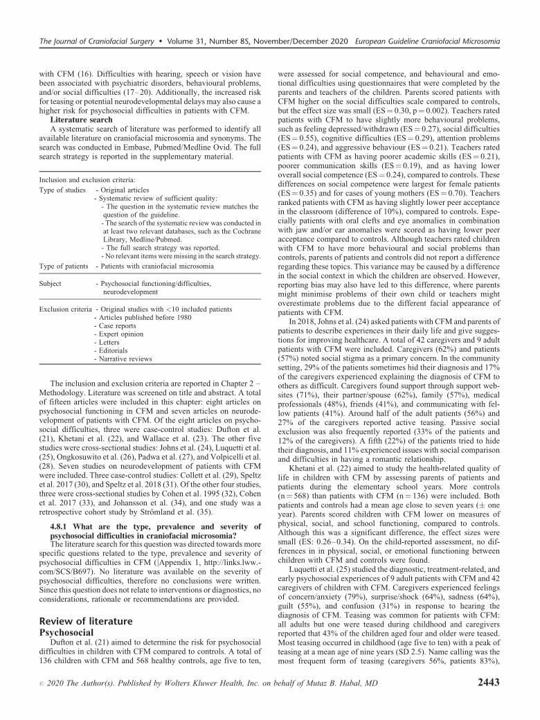
The Journal of Craniofacial Surgery � Volume 31, Number 8S, November/December 2020 European Guideline Craniofacial Microsomia
with CFM (16). Difficulties with hearing, speech or vision havebeen associated with psychiatric disorders, behavioural problems,and/or social difficulties (17–20). Additionally, the increased riskfor teasing or potential neurodevelopmental delays may also cause ahigher risk for psychosocial difficulties in patients with CFM.
Literature searchA systematic search of literature was performed to identify all
available literature on craniofacial microsomia and synonyms. Thesearch was conducted in Embase, Pubmed/Medline Ovid. The fullsearch strategy is reported in the supplementary material.
Inclusion and exclusion criteria:
Type of studies
# 2020 The Auth
- Original articles- Systematic review of sufficient quality:
- The question in the systematic review matches thequestion of the guideline.- The search of the systematic review was conducted inat least two relevant databases, such as the CochraneLibrary, Medline/Pubmed.- The full search strategy was reported.- No relevant items were missing in the search strategy.
Type of patients
- Patients with craniofacial microsomiaSubject
- Psychosocial functioning/difficulties,neurodevelopmentExclusion criteria
- Original studies with <10 included patients- Articles published before 1980- Case reports- Expert opinion- Letters- Editorials- Narrative reviewsThe inclusion and exclusion criteria are reported in Chapter 2 –Methodology. Literature was screened on title and abstract. A totalof fifteen articles were included in this chapter: eight articles onpsychosocial functioning in CFM and seven articles on neurode-velopment of patients with CFM. Of the eight articles on psycho-social difficulties, three were case-control studies: Dufton et al.(21), Khetani et al. (22), and Wallace et al. (23). The other fivestudies were cross-sectional studies: Johns et al. (24), Luquetti et al.(25), Ongkosuwito et al. (26), Padwa et al. (27), and Volpicelli et al.(28). Seven studies on neurodevelopment of patients with CFMwere included. Three case-control studies: Collett et al. (29), Speltzet al. 2017 (30), and Speltz et al. 2018 (31). Of the other four studies,three were cross-sectional studies by Cohen et al. 1995 (32), Cohenet al. 2017 (33), and Johansson et al. (34), and one study was aretrospective cohort study by Stromland et al. (35).
4.8.1 What are the type, prevalence and severity ofpsychosocial difficulties in craniofacial microsomia?
The literature search for this question was directed towards morespecific questions related to the type, prevalence and severity ofpsychosocial difficulties in CFM (jAppendix 1, http://links.lww.-com/SCS/B697). No literature was available on the severity ofpsychosocial difficulties, therefore no conclusions were written.Since this question does not relate to interventions or diagnostics, noconsiderations, rationale or recommendations are provided.
Review of literaturePsychosocial
Dufton et al. (21) aimed to determine the risk for psychosocialdifficulties in children with CFM compared to controls. A total of136 children with CFM and 568 healthy controls, age five to ten,
or(s). Published by Wolters Kluwer Health, Inc. on b
were assessed for social competence, and behavioural and emo-tional difficulties using questionnaires that were completed by theparents and teachers of the children. Parents scored patients withCFM higher on the social difficulties scale compared to controls,but the effect size was small (ES¼ 0.30, p¼ 0.002). Teachers ratedpatients with CFM to have slightly more behavioural problems,such as feeling depressed/withdrawn (ES¼ 0.27), social difficulties(ES¼ 0.55), cognitive difficulties (ES¼ 0.29), attention problems(ES¼ 0.24), and aggressive behaviour (ES¼ 0.21). Teachers ratedpatients with CFM as having poorer academic skills (ES¼ 0.21),poorer communication skills (ES¼ 0.19), and as having loweroverall social competence (ES¼ 0.24), compared to controls. Thesedifferences on social competence were largest for female patients(ES¼ 0.35) and for cases of young mothers (ES¼ 0.70). Teachersranked patients with CFM as having slightly lower peer acceptancein the classroom (difference of 10%), compared to controls. Espe-cially patients with oral clefts and eye anomalies in combinationwith jaw and/or ear anomalies were scored as having lower peeracceptance compared to controls. Although teachers rated childrenwith CFM to have more behavioural and social problems thancontrols, parents of patients and controls did not report a differenceregarding these topics. This variance may be caused by a differencein the social context in which the children are observed. However,reporting bias may also have led to this difference, where parentsmight minimise problems of their own child or teachers mightoverestimate problems due to the different facial appearance ofpatients with CFM.
In 2018, Johns et al. (24) asked patients with CFM and parents ofpatients to describe experiences in their daily life and give sugges-tions for improving healthcare. A total of 42 caregivers and 9 adultpatients with CFM were included. Caregivers (62%) and patients(57%) noted social stigma as a primary concern. In the communitysetting, 29% of the patients sometimes hid their diagnosis and 17%of the caregivers experienced explaining the diagnosis of CFM toothers as difficult. Caregivers found support through support web-sites (71%), their partner/spouse (62%), family (57%), medicalprofessionals (48%), friends (41%), and communicating with fel-low patients (41%). Around half of the adult patients (56%) and27% of the caregivers reported active teasing. Passive socialexclusion was also frequently reported (33% of the patients and12% of the caregivers). A fifth (22%) of the patients tried to hidetheir diagnosis, and 11% experienced issues with social comparisonand difficulties in having a romantic relationship.
Khetani et al. (22) aimed to study the health-related quality oflife in children with CFM by assessing parents of patients andpatients during the elementary school years. More controls(n¼ 568) than patients with CFM (n¼ 136) were included. Bothpatients and controls had a mean age close to seven years (� oneyear). Parents scored children with CFM lower on measures ofphysical, social, and school functioning, compared to controls.Although this was a significant difference, the effect sizes weresmall (ES: 0.26–0.34). On the child-reported assessment, no dif-ferences in in physical, social, or emotional functioning betweenchildren with CFM and controls were found.
Luquetti et al. (25) studied the diagnostic, treatment-related, andearly psychosocial experiences of 9 adult patients with CFM and 42caregivers of children with CFM. Caregivers experienced feelingsof concern/anxiety (79%), surprise/shock (64%), sadness (64%),guilt (55%), and confusion (31%) in response to hearing thediagnosis of CFM. Teasing was common for patients with CFM:all adults but one were teased during childhood and caregiversreported that 43% of the children aged four and older were teased.Most teasing occurred in childhood (age five to ten) with a peak ofteasing at a mean age of nine years (SD 2.5). Name calling was themost frequent form of teasing (caregivers 56%, patients 83%),
ehalf of Mutaz B. Habal, MD 2443

Renkema et al The Journal of Craniofacial Surgery � Volume 31, Number 8S, November/December 2020
although questions about their appearance or nonverbal negativereactions of mimicking or running away occurred too (caregivers22%, patients 17%). In response to teasing most caregivers (45%)and patients (67%) said to ignore the teasing. Being stared at wasalso frequently noted by patients. Although some patients did notcare (13%), others described feeling shy or having negative emo-tional responses (caregivers 13%, patients 38%). Most patients hadadaptive coping responses to staring (caregivers 26%, patients38%), others ignored staring (caregivers 6%, patients 13%), or feltreminded about their diagnosis of CFM (caregivers 3%, patients25%).
Stress in parents of children with CFM and their coping styleswere studied by Ongkosuwito et al. (26). Questionnaires on parentalstress and on cognitive emotion-regulation were completed by 31parents of patients with CFM. Learning difficulties and psychoso-cial difficulties were associated with an increase in parental stress(r¼ 0.71, r¼ 0.63, both p¼<.001). No relation between hearingdifficulties (r¼ 0.32, p¼ 0.08), articulation difficulties (r¼ 0.34,p¼ 0.06), or the facial appearance of the child (r¼ 0.07, p¼ 0.72)and parental stress was found. The coping strategies that had apositive correlation with parental stress were acceptance and posi-tive reappraisal (r¼ 0.50, p¼ 0.01 and r¼ 0.54, p¼ 0.002). Multi-variate regression analysis showed that learning difficulties and theuse of acceptance as a coping strategy explained a large proportionof parental stress. Interventions for parents of children that addresslearning difficulties and coping strategies could potentially bebeneficial to lower parental stress.
Padwa et al. (27) studied psychosocial adjustment in childrenwith craniofacial anomalies to examine the relationships betweensymmetry, functional impairments, and social adaptation. Of the 30patients included, 11 had CFM. Most patients (80%) with CFMwere able to socialise in a positive way by joining the group, whichwas higher when compared to other craniofacial anomalies (52%-57%). Low self-esteem and negative self-concept were reported by35–40% of all children; no subgroup data of patients with CFM wasreported. Patients with symmetrical craniofacial anomalies had asignificantly higher prevalence of low mood. Patients with sym-metrical and non-CFM craniofacial anomalies had lower socialcompetences scores. This suggests a higher prevalence of lowmood, behavioural problems, and socialising problems were presentin this group when compared to patients with asymmetrical cranio-facial disorders, and to patients with CFM. The authors debatewhether children with facial asymmetry may be viewed as having anormal face with imperfections, while patients with a symmetricalcraniofacial anomaly could be seen as having a severe medicalcondition or syndrome. Patients with an asymmetric deformity maybe more acceptable to themselves and experience fewer psychoso-cial difficulties. The study concludes that early surgery in patientswith symmetrical craniofacial deformities may help to preventpsychosocial difficulties. However, no evidence supporting thistheory is provided. Additionally, it is important to note the smallsample size of patients with CFM in this study.
The timing of paediatric craniofacial surgery and its effect onpsychosocial development was further examined by Volpicelli et al.(28). They aimed to study differences in psychosocial outlook inchildren with craniofacial anomalies under active treatment. A totalof 99 patients with a craniofacial disorder such as cleft lip/palate orcraniofacial syndrome, age 8–17 years, were included. Of these 99patients, 22 patients had CFM (21.6%). Younger patients (age 8–10) were found to have significantly higher anxiety, depression, andanger scores, and lower peer relationship scores, compared to olderpatients (age 11–13 and/or age 14–17) and to the U.S. averagescores. The authors argue that more prevention and interventionmethods may be needed for families and schools to help youngerpatients (age 8–10).
2444 # 2020 The Author(s). Published
Wallace et al. (23) studied psychosocial adjustment in adoles-cents with CFM and compared this with controls. A total of 142patients with CFM and 316 healthy controls were included, with amean age of 13.4 years (SD 1.4 years, range 10–17 years). Bothpatients with CFM and controls reported similar levels of beha-vioural problems. However, lower social and school functioningscores were reported by patients with CFM and parents of patientsdue to various reasons such as teasing, not being able to participatewith other teens, or missing school due to hospital visits. Althoughpatients with CFM had slightly more difficulties in social function-ing compared to controls, fewer problems with externalising behav-iour were reported compared to controls. The authors discuss thatthe higher frequency of social difficulties in patients with CFM maybe the result of teasing, rejection, or stigmatisation, instead of apersonal trait. No strong evidence for differences in behaviouraladaptation or socialisation based on the severity of the facialmalformations was found.
NeurodevelopmentSpeltz et al. (2017) (30) studied the intelligence and academic
achievements of adolescents with CFM. A total of 142 patients withCFM and 316 controls were included (mean age 13 years, range 11–17 years). Patients with CFM scored significantly lower on IQ andacademic achievements (ES¼�0.01 to �0.3, p-values range 0.01to 0.92). It is important to note that this effect size is relativelysmall. Learning difficulties occurred in 25% of the controls and in38% of the patients with CFM (adjusted OR¼ 1.5, 95% CI 0.9–2.4). Patients with mandibular hypoplasia and microtia had signifi-cantly lower IQ and achievement scores as did patients with isolatedmicrotia, when compared to controls. Although 70% of the patientswith CFM had hearing difficulties compared to 1% of the controls,an effect of hearing difficulties on IQ and achievements scores wasnot found. The authors conclude that these findings should beviewed ‘primarily as hypothesis-generating, rather than as firmconclusions’. According to the authors, developmental surveillanceof children and adolescents with CFM should be undertaken.
In another study by Speltz et al. (2018) (31) the neurodevelop-mental profile of infants with CFM was assessed and compared withcontrols. A total of 108 patients with CFM and 84 controls, all age12–24 months, were included. Developmental delay was found in21% of the patients with CFM and in 16% of the controls. However,no differences in terms of developmental delay were present afteradjustment for demographic factors (OR¼ 0.68, 95% CI 0.29–1.61). No differences in development were found based on the CFMphenotype. The authors conclude that no evidence for neurodeve-lopmental differences between infants with CFM (age one to two)and controls could be found.
Collett et al. (29) aimed to determine the risk for neurodevelop-mental delay in 136 children with CFM compared to 568 healthycontrols. Lower verbal, nonverbal, and academic skills were seen inpatients with CFM compared to controls. Patients with CFM had asignificantly higher risk for receptive language delays and visuo-motor skills delay compared to controls. Worse outcomes on theneurodevelopmental profile were seen in patients born to motherswho were 25 or younger at time of birth. The authors emphasise theneed to carry out routine neurodevelopmental screening for patientswith CFM.
A smaller, cross-sectional study by Cohen et al. (1995) (32)included 24 patients with CFM to examine their neurodevelop-mental profile. No significant difference in mean IQ compared tothe general population was found. A delay was found in gross (38%)and fine (20%) motor development and in receptive (32%) andexpressive (37%) language development compared to the generalpopulation. Receptive and expressive language development weresignificantly lower in patients with bilateral CFM compared to
by Wolters Kluwer Health, Inc. on behalf of Mutaz B. Habal, MD

The Journal of Craniofacial Surgery � Volume 31, Number 8S, November/December 2020 European Guideline Craniofacial Microsomia
unilateral CFM. In a larger cross-sectional study by Cohen et al.(2017) (33) on facial and systemic malformations in a cohort of 89patients with CFM, 5% (n¼ 4) were found to have an intellectualdisability. No further information on developmental delay wasprovided in this study.
Johansson et al. (34) aimed to study autism spectrum disorder in20 patients with CFM (mean age 8 years, range 8 months – 17years). Eleven patients (55%) were found to have normal intelli-gence and nine patients (45%) had learning disabilities, which weremild in four patients (20%), severe in three patients (15%), andprofound in two patients (10%). Autism spectrum disorder wasdiagnosed in two patients (10%), autism-like condition in onepatient (5%), and autistic traits in five patients (25%). Behaviouraldisturbances, such as aggressive behaviour or hyperactivity, weresolely seen in patients with autism spectrum disorders.
Stromland et al. (35) also studied autism spectrum disorder inCFM. One of their eighteen included patients with CFM had autismand two met the criteria for childhood autism/autistic-like disorder.Six other patients had learning disabilities (30%): three had severe(17%) and three had mild (17%) learning disabilities.
Conclusions
Level 3
# 2020 The A
TypePatients and/or parents of patients with craniofacial
microsomia have a higher risk of experiencing socialstigma, low self-esteem and active or passive bullying,especially in childhood.
Parental stress in parents of patients with craniofacialmicrosomia is linked to their child’s learning difficulties,and parental acceptance. The facial appearance of thechild does not appear to be an important influential factorin parental stress.
Ref (24–27)
Level 3
Risk factorsPatients with symmetrical craniofacial anomalies mayexperience more behavioural problems, depressedfeelings, and socializing problems compared to patientswith an asymmetrical craniofacial anomaly.
Younger children (age 8–10) with a craniofacial anomalymay experience more problems with anxiety, low mood,anger, and peer relationships compared to older children(age 11–17).
Ref (27, 28)
Level 3
Type - NeurodevelopmentalPatients with craniofacial microsomia may have a slightlylower IQ score and more learning difficulties compared tohealthy controls.
The neurodevelopmental profile of infants with craniofacialmicrosomia (age 1–2 years) is not different from healthycontrols.
Patients with craniofacial microsomia might have a higherrisk for language- and motor skills delays compared tohealthy controls.
Ref (29–35)
4.8.2 What is the policy for the screening and monitoring ofpsychosocial difficulties in patients with craniofacial micro-somia?
The literature search for this question was directed towards morespecific questions related to the consequences and impact ofscreening and monitoring, the available screening tests and
uthor(s). Published by Wolters Kluwer Health, Inc. on b
procedures for monitoring, and responsibilities for screening andmonitoring (Appendix 1, http://links.lww.com/SCS/B697). Unfor-tunately, no literature is available on the policy for screening and/ormonitoring of psychosocial difficulties in patients with CFM.Therefore no conclusions were written. Considerations for thisquestion were only based on expert opinion.
Considerations� Overall quality of evidenceBecause of the lack of evidence, the recommendations on
screening and monitoring of psychosocial difficulties in CFMare based on expert opinion with a low quality of evidence.
� Balance of benefits and harmsHaving access to a psychologist and a team specialising in CFM
will have obvious biopsychosocial benefits for patients and theirfamilies including providing a high standard of care, improvingpatient quality of life and potentially reducing hospital staysand communications.
� Outcome importanceThe main reason to perform screening is to help to reduce
emotional distress experienced by patients and their families,promote information sharing and a holistic approach, and pre-emptpsychological difficulties developing as a result of the patient’sphysical health condition.
� Costs and resourcesThe impact on costs and resources of the given recommenda-
tions will vary per member state, depending on the available careproviders and facilities. The recommendations concern the essentialrequirements for adequate treatment of patients with CFM andshould thus be implemented. Costs are lowest and resources aremost efficiently used when care for craniofacial disorders is cen-tralised in a limited number of expert centres per member state. Ageneral rule that can be applied is one expert centre per 10million inhabitants.
� Inequity of the recommendationThe goal of the European Reference Networks (ERNs) is to
eliminate inequality within Europe with regard to care for patientswith rare diseases. At present, not every member state offers anexpert centre for CFM, or the level of provided care does not (yet)meet all the requirements that are outlined in this guideline. Bydefining the baseline of required care for CFM, this guideline willhelp these member states to reach the appropriate level. The ERN oncraniofacial anomalies and ENT disorders (ERN-CRANIO) canguide a patient in Europe to the available centres of expertise(www.ern-cranio.eu) and can support care providers with diagnosisand treatment advice.
� Feasibility of the recommendationRecommendations refer to the general requirements for deliv-
ering optimal healthcare and are discussed with members fromparticipating European countries. Quality of care was paramount inthe discussions. Centralisation is proposed as one of the core values.However, in some countries the national organisation of healthcaremight impede centralisation. National implementation of the ERNsthat fits the situation of each country is necessary. For the memberstates with the lowest number of inhabitants, the establishment of acraniofacial centre might not be feasible, and collaboration with acraniofacial centre in the surrounding countries can be an option.
ehalf of Mutaz B. Habal, MD 2445
Renkema et al The Journal of Craniofacial Surgery � Volume 31, Number 8S, November/December 2020
� Acceptability of the recommendationIt is expected that all stakeholders want to apply and will be
applying the recommendations, because they are employed in ERN-acknowledged institutions. National implementation plans are nec-essary to ensure that recommendations fit the situation in eachcountry. In addition, not all countries participating in the ERN-CRANIO are represented in the guideline development group, andnew members will join within the coming years. For these countries,acceptance and implementation of ERN guidelines such as thisguideline on CFM is obligatory.
Rationale of the recommendationsThe rationale for having psychological input in a specialist
team for CFM is to pre-empt any psychological issues experi-enced by the child and family that could have an impact on thepatient journey, and to ensure that the team approach is holistic(36). It may involve mediating on behalf of the family withmedical teams or schools, or between child and parent if thereis a difference in opinion regarding intervention. The overall aimis to reduce distress and to enhance and promote psychologicalwellbeing. The amount of distress does not always seem to beproportionate to objective appearance, as it is a psychologicalprocess that determines response to differences in appearance.Regular review by the specialist team offers the patient and familyholistic and up-to-date help. Regular contact fosters good rela-tionships between professionals and the family and allows theteam to discuss intervention in an age-appropriate manner. It alsoallows the family to ask about new developments in the field andif necessary, to discuss psychosocial, educational and hearingissues which may arise. Being provided with this information andsupport promotes parental adjustment and acceptance and enablesthem to communicate positive themes of normalisation andreassurance, which children mirror in their own responses andadjustment (37). The ICHOM Standard Set for CraniofacialMicrosomia advises to see patients and their family at age 2,5, 8, and 22 to measure psychosocial wellbeing and quality of lifeusing the ClefQ, YP-CORE, HADS, and CFEQ. It is advised touse the Distress Thermometer for Parents to measurefamily stress.
It is good practise to advise families of active voluntary sectorsupport groups and relevant charities. Support groups and charitiescan help children and their families deal with difference, andpromote confidence, resilience and positive self-image. Healthcareproviders play a key role in family adjustment by clarifyingmisinformation, providing education regarding treatment options,modelling acceptance, screening for psychosocial concerns, pro-viding resources and connecting families to support networks. Withregards to visible differences, patient coping and level of concern isrelated to personality, family responses, school support and devel-opmental stage. Predictors of good psychosocial outcomes aresocial and family support, perceptions of severity and socialinteraction skills, and clinicians (with regular contact and workingwith the system) can to have a positive influence on these factors.
Recommendations
24
� All craniofacial microsomia patients should have accessto a clinical psychology service with appropriateprofessional expertise and knowledge of craniofacialmicrosomia.
� Time points for reviews and screening should observekey life transitions such as birth, starting school,
46
transition to secondary school, etc.
# 2020 The Author(s). Published
by� To measure psychosocial wellbeing and family stress,validated self-reported psychological outcome mea-sures should be obtained from to all craniofacialmicrosomia patients as a matter of routine to screen forthe presence of behavioural, emotional, social and/orlearning difficulties. This includes the CleftQ, CFEQ,YP-CORE, HADS and Distress Thermometer forParents and should be performed at age 2, 5, 8 and22. Elevated scores should alert clinicians to thepotential need for further assessment or support.Standardised measures should assess levels of emo-tional distress as well as evaluate difficulties related to
Wolte
visible differences.
Research gapIt is recognised that there is a lack of quantitative and qualitative
research looking at the psychological impact of CFM, and a need forlongitudinal studies to further inform psychological assessments andinterventions. Use of measures pre- and post-operatively providesvaluable data for audit and research as well as quickly identifying anypsychological difficulties that can potentially be addressed, andthemes and issues identified in clinics directly from patients andtheir families could be assessed using qualitative methods.
4.8.3 What are the indications and policies for the treatmentof psychosocial difficulties in patients with craniofacialmicrosomia?
The literature search for this question was directed towards morespecific research questions related to treatment options, symptoms,requirements, complications, and advantages and disadvantages oftreatment of psychosocial difficulties (Appendix 1, http://links.lww.com/SCS/B697). Unfortunately, no literature is availableon the policy for treatment of psychosocial difficulties in patientswith CFM. Therefore no conclusions were written. Considerationsfor this question were only based on expert opinion.
Considerations� Overall quality of evidenceBecause of the lack of evidence, the recommendations on
treatment of psychosocial difficulties in CFM are based on expertopinion with a low quality of evidence.
� Balance of benefits and harmsPsychological therapy can be time consuming, but interventions
are individualised and planned collaboratively with the patient andtheir family, which increases acceptability and adherence. Manag-ing the patient ‘system’ can be heavy work for clinicians, requiringcommunications and meetings with family members, school, othermedical teams etc.
� Outcome importancePsychosocial support and psychological intervention are
designed to promote quality of life, which is important to patients,family, and clinicians. Early psychological intervention wouldreduce the risk of patients developing long-term mental healthdifficulties and their associated sequela, thus reducing the strainon healthcare resources in future years.
� Costs and resourcesThe impact on costs and resources of the given recommenda-
tions will vary per member state, depending on the available care
rs Kluwer Health, Inc. on behalf of Mutaz B. Habal, MD

The Journal of Craniofacial Surgery � Volume 31, Number 8S, November/December 2020 European Guideline Craniofacial Microsomia
providers and facilities. The recommendations concern the essentialrequirements for adequate treatment of patients with CFM and shouldthus be implemented. Costs are lowest and resources are mostefficiently used when care for craniofacial disorders is centralisedin a limited number of expert centres per member state. A general rulethat can be applied is one expert centre per 10 million inhabitants.
� Inequity of the recommendationThe goal of the European Reference Networks (ERNs) is to
eliminate inequality within Europe with regard to care for patientswith rare diseases. At present, not every member state offers an expertcentre for CFM, or the level of provided care does not (yet) meet all therequirements that are outlined in this guideline. By defining thebaseline of required care for CFM, this guideline will help thesemember states to reach the appropriate level. The ERN on craniofacialanomalies and ENT disorders (ERN-CRANIO) can guide a patient inEurope to the available centres of expertise (www.ern-cranio.eu) andcan support care providers with diagnosis and treatment advice.
� Feasibility of the recommendationRecommendations refer to the general requirements for deliv-
ering optimal healthcare and are discussed with members fromparticipating European countries. Quality of care was paramount inthe discussions. Centralisation is proposed as one of the core values.However, in some countries the national organisation of healthcaremight impede centralisation. National implementation of the ERNsthat fits the situation of each country is necessary. For the memberstates with the lowest number of inhabitants, the establishment of acraniofacial centre might not be feasible, and collaboration with acraniofacial centre in the surrounding countries can be an option.
� Acceptability of the recommendationIt is expected that all stakeholders want to apply and will be
applying the recommendations, because they are employed in ERN-acknowledged institutions. National implementation plans are nec-essary to ensure that recommendations fit the situation in eachcountry. In addition, not all countries participating in the ERN-CRANIO are represented in the guideline development group, andnew members will join within the coming years. For these countries,acceptance and implementation of ERN guidelines such as thisguideline on CFM is obligatory.
Rationale of the recommendationsWe suggest that including a psychologist in the specialist team
or having access to paediatric or adult health psychologistsimproves overall psychosocial outcomes for people with CFM.Interventions can be targeted to the patient, family or the widersystem, to enhance psychological wellbeing, acceptance and adjust-ment. Having a psychologist present enables psychosocial difficul-ties to be identified, with the aim being early intervention. Supportgroups and charities play a key role in psychosocial wellbeing,helping children and their families to deal with difference andpromote confidence, resilience and positive self-image.
Healthcare providers play a key role in individual coping andadjustment, for example, by promoting positive and assertivestrategies when dealing with the reactions of others, focusing onpersonal achievements, and encouraging participation in social andphysical activities, which in turn can lead to greater self-acceptance.They also play a key role in supporting parents/carers of childrenwith CFM in terms of making decisions regarding whether or not toproceed with surgical reconstruction, which can feel like a hugeresponsibility for a parent/carer to have (38).
In terms of involving the child in decision making surroundingsurgery, we suggest that (where possible) there is a strong argument
# 2020 The Author(s). Published by Wolters Kluwer Health, Inc. on b
for waiting until the child is old enough, with the necessary level ofabstract conceptual thinking and emotional maturity, to enableinformed decision making. A study that interviewed adolescentswith CFM found that the children were glad that their parents hadallowed them to be involved in the decision-making process sur-rounding surgery (39). They also did not feel that concern regardingcurrent or potential bullying was a good reason to have surgery at ayoung age, and that promoting self-acceptance and resilience were farmore important. They were asked to list positive aspects of growingup with a craniofacial condition (39). Positive changes in personalityincluded being more open-minded, less judgemental, kinder, andmore independent. Participants also expressed the belief that they arestrong individuals who are able to overcome challenges. When askedwhat advice they would give younger children growing up withsimilar conditions, participants said they would advise them to beopen and honest and not be ashamed of their visible differences.
If indicated, a psychologist should be involved when proceedingwith surgery at preadmission stage to assess psychological readiness forsurgery, including identifying any procedural anxiety that will impacton the surgery itself, and psychosocial issues that may affect treatmentadherence post-operatively. Assessment of readiness for surgeryshould occur before or at the preadmission clinic. A semi-structuredinterview format should be used to establish current emotional state,family functioning, mental and physical health history, significant lifeevents, risk factors for poor psychological adjustment post-surgery –including unrealistic expectations, concerns regarding the process ofhospital admission, surgery, post-operative pain etc. As well as collat-ing feedback for the medical team, the aim should be to attempt toascertain if surgical reconstruction will contribute to long-term psy-chological wellbeing. Post-operative follow-up should occur followingcompletion of surgical reconstruction to assess emotional state, satis-faction with surgical process, and body image issues. It is important thatthe patient understands the nature of the healing process and itspotential impact on their body image. With patients often waiting along time before the surgery can be performed and with the cosmeticresult not fully visible for a number of weeks due to bruising andswelling, expectations must be carefully managed. Whilst it can beseveral weeks before patients feel fully physically recovered fromreconstructive surgery, it could be argued that the bigger challenge atthis time will be the need to adapt to the look and feel of their newappearance. Revealing the newly reconstructed area for the first timemust be done with sensitivity and in a way that allows them somecontrol over how and when it is seen, as well as who will see it.
Recommendations
eh
Parental adjustment and support
� Parents of newly diagnosed children with craniofacialmicrosomia should have access to a specialist clinicalpsychology service with expertise and knowledge ofthe condition.
� Information on support groups and organisations should
alf of
be provided, both at initial contact and at regular review.
Behavioural and/or learning difficulties
� When appropriate, clinicians should liaise with localservices and schools to discuss the child’s support needs.
� Cognitive assessment may be offered if warranted
Mutaz B. Habal, MD 2447
Renkema et al The Journal of Craniofacial Surgery � Volume 31, Number 8S, November/December 2020
24
Coping with visible difference
� Patients with craniofacial microsomia should have accessto specialist psychological support, particularly thosewho are presenting with low self-esteem, depression/lowmood, anxiety, appearance- or treatment-related con-cerns, including adjustment difficulties or trauma as aresult of surgical/medical interventions.
� Clinicians with appropriate professional expertise incraniofacial microsomia should consider liaising withlocal schools to offer advice on how to support childrenwho have visible differences.
� Information about support groups and organisationsshould be provided.
� Psychological input is required pre- and post- facialsurgery to monitor expectation and acceptance.
� The psychologist is part of the coordinated care in themultidisciplinary team. See recommendations in
48
Chapter 6.
Research gapAs described in Chapter 4.8.2.References
1. D
ion K, Berscheid, E. Physical attractiveness and peerperception among children. Sociometry. 1974;37(1):1-12.2. G
ertner BL, Rice ML, Hadley PA. Influence of communica-tive competence on peer preferences in a preschool classroom.J Speech Hear Res. 1994;37(4):913-23.3. R
umsey N, Harcourt D. Body image and disfigurement: issuesand interventions. Body Image. 2004;1(1):83-97.4. T
opolski TD, Edwards TC, Patrick DL. Quality of life: how doadolescents with facial differences compare with otheradolescents? Cleft Palate Craniofac J. 2005;42(1):25-32.5. M
asnari O, Landolt MA, Roessler J, Weingaertner SK,Neuhaus K, Meuli M, et al. Self- and parent-perceivedstigmatisation in children and adolescents with congenital oracquired facial differences. J Plast Reconstr Aesthet Surg.2012;65(12):1664-70.6. J
iamei D, Jiake C, Hongxing Z, Wanhou G, Yan W, Gaifen L.An investigation of psychological profiles and risk factors incongenital microtia patients. J Plast Reconstr Aesthet Surg.2008;61 Suppl 1:S37-43.7. B
rent B. The pediatrician’s role in caring for patients withcongenital microtia and atresia. Pediatr Ann. 1999;28(6):374-83.8. J
ohns AL, Lucash RE, Im DD, Lewin SL. Pre and post-operative psychological functioning in younger and olderchildren with microtia. J Plast Reconstr Aesthet Surg.2015;68(4):492-7.9. J
ohns AL, Lewin SL, Im DD. Teasing in younger and olderchildren with microtia before and after ear reconstruction. JPlast Surg Hand Surg. 2017;51(3):205-9.10. S
peltz ML, Endriga MC, Hill S, Maris CL, Jones K, OmnellML. Cognitive and psychomotor development of infants withorofacial clefts. J Pediatr Psychol. 2000;25(3):185-90.11. H
unt O, Burden D, Hepper P, Stevenson M, Johnston C. Parentreports of the psychosocial functioning of children with cleftlip and/or palate. Cleft Palate Craniofac J. 2007;44(3):304-11.12. C
ollett BR, Speltz ML. A developmental approach to mentalhealth for children and adolescents with orofacial clefts.Orthod Craniofac Res. 2007;10(3):138-48.# 2020 The Author(s). Published
13. S
by W
arimski K. Children with Apert syndrome: behaviouralproblems and family stress. Dev Med Child Neurol.1998;40(1):44-9.
14. N
elson P, Glenny AM, Kirk S, Caress AL. Parents’experiences of caring for a child with a cleft lip and/orpalate: a review of the literature. Child Care Health Dev.2012;38(1):6-20.15. M
athijssen IM. Guideline for Care of Patients With theDiagnoses of Craniosynostosis: Working Group on Cranio-synostosis. J Craniofac Surg. 2015;26(6):1735-807.16. R
ives Bogart K, Matsumoto D. Facial mimicry is notnecessary to recognize emotion: Facial expression recognitionby people with Moebius syndrome. Soc Neurosci. 2010;5(2):241-51.17. B
rown R, Hobson RP, Lee A, Stevenson J. Are there ‘‘autistic-like’’ features in congenitally blind children? J Child PsycholPsychiatry. 1997;38(6):693-703.18. F
ellinger J, Holzinger D, Sattel H, Laucht M, Goldberg D.Correlatesofmentalhealthdisordersamongchildrenwithhearingimpairments. Dev Med Child Neurol. 2009;51(8):635-41.19. M
cAlpine LM, Moore, C. The development of socialunderstanding in children with visual impairments. Journalof Visual Impairment and Blindness. 1993;89:349–58.20. v
an Daal J, Verhoeven L, van Balkom H. Behaviour problemsin children with language impairment. J Child PsycholPsychiatry. 2007;48(11):1139-47.21. D
ufton LM, Speltz ML, Kelly JP, Leroux B, Collett BR, WerlerMM. Psychosocial outcomes in children with hemifacialmicrosomia. J Pediatr Psychol. 2011;36(7):794-805.22. K
hetani MA, Collett BR, Speltz ML, Werler MM. Health-related quality of life in children with hemifacial microsomia:parent and child perspectives. J Dev Behav Pediatr. 2013;34(9):661-8.23. W
allace ER, Collett BR, Heike CL, Werler MM, Speltz ML.Behavioral-Social Adjustment of Adolescents with Craniofa-cial Microsomia. Cleft Palate Craniofac J. 2018;55(5):664-75.24. J
ohns AL, Luquetti DV, Brajcich MR, Heike CL, Stock NM. InTheir Own Words: Caregiver and Patient Perspectives onStressors, Resources, and Recommendations in CraniofacialMicrosomia Care. J Craniofac Surg. 2018;29(8):2198-205.25. L
uquetti DV, Brajcich MR, Stock NM, Heike CL, Johns AL.Healthcare and psychosocial experiences of individuals withcraniofacial microsomia: Patient and caregivers perspectives.Int J Pediatr Otorhinolaryngol. 2018;107:164-75.26. O
ngkosuwito E, van der Vlies L, Kraaij V, Garnefski N, vanNeck H, Kuijpers-Jagtman AM, et al. Stress in Parents of aChild with Hemifacial Microsomia: The Role of ChildCharacteristics and Parental Coping Strategies. Cleft PalateCraniofac J. 2018;55(7):959-65.27. P
adwa BL, Evans CA, Pillemer FC. Psychosocial adjustmentin children with hemifacial microsomia and other craniofacialdeformities. Cleft Palate Craniofac J. 1991;28(4):354-9.28. V
olpicelli EJ, Pfaff MJ, Hakimi K, Bradley JP, Solem RC, LeeJC. Age-Related Differences in Psychosocial Function ofChildren with Craniofacial Anomalies. Plast Reconstr Surg.2017;140(4):776-84.29. C
ollett BR, Speltz ML, Cloonan YK, Leroux BG, Kelly JP,Werler MM. Neurodevelopmental outcomes in children withhemifacial microsomia. Arch Pediatr Adolesc Med. 2011;165(2):134-40.30. S
peltz ML, Wallace ER, Collett BR, Heike CL, Luquetti DV,Werler MM. Intelligence and Academic Achievement ofAdolescents with Craniofacial Microsomia. Plast ReconstrSurg. 2017;140(3):571-80.olters Kluwer Health, Inc. on behalf of Mutaz B. Habal, MD

The Journal of Craniofacial Surgery � Volume 31, Number 8S, November/December 2020 European Guideline Craniofacial Microsomia
31. S
# 20
peltz ML, Kapp-Simon KA, Johns AL, Wallace ER, CollettBR, Magee L, et al. Neurodevelopment of Infants with andwithout Craniofacial Microsomia. J Pediatr. 2018;198:226-33 e3.
32. C
ohen MS, Samango-Sprouse CA, Stern HJ, Custer DA,Vaught DR, Saal HM, et al. Neurodevelopmental profile ofinfants and toddlers with oculo-auriculo-vertebral spectrumand the correlation of prognosis with physical findings. Am JMed Genet. 1995;60(6):535-40.33. C
ohen N, Cohen E, Gaiero A, Zecca S, Fichera G, Baldi F,et al. Maxillofacial features and systemic malformations inexpanded spectrum Hemifacial Microsomia. Am J Med GenetA. 2017;173(5):1208-18.34. J
ohansson M, Billstedt E, Danielsson S, Stromland K, MillerM, Granstrom G, et al. Autism spectrum disorder andunderlying brain mechanism in the oculoauriculovertebralspectrum. Dev Med Child Neurol. 2007;49(4):280-8.35. S
tromland K, Miller M, Sjogreen L, Johansson M, JoelssonBM, Billstedt E, et al. Oculo-auriculo-vertebral spectrum:associated anomalies, functional deficits and possible devel-opmental risk factors. Am J Med Genet A. 2007;143A(12):1317-25.36. U
K Care Standards for the Management of Patients withMicrotia and Atresia37. J
ohns AL, Im DD, Lewin SL. Early Familial Experiences WithMicrotia: Psychosocial Implications for Pediatric Providers.Clin Pediatr (Phila). 2018;57(7):775-82.38. H
amlet CaH, D. Adults’ experiences of living with Microtia.A report for Microtia UK.39. H
amilton KV, Ormond KE, Moscarello T, Bruce JS,Bereknyei Merrell S, Chang KW, et al. Exploring the Medicaland Psychosocial Concerns of Adolescents and Young AdultsWith Craniofacial Microsomia: A Qualitative Study. CleftPalate Craniofac J. 2018;55(10):1430-9.CHAPTER 5. SURGICAL TREATMENT
5.1 Mandible & MaxillaIntroductionMandibular hypoplasia is seen in 73% to 91% of patients with
CFM with varying severity (1, 2). The Pruzansky classification,later modified by Kaban, has been developed to characterise themandibular deformity in patients with CFM based on radiographicassessment (3–5). A small but normal shaped mandibular ramusand temporomandibular joint (TMJ) is classified as type I. In typeIIa, the mandibular ramus is malformed in size and shape but theTMJ is adequately positioned. Type IIb is characterised by anabnormal shape, size, and location of the mandibular ramus andTMJ. In type III, the ramus, condyle and TMJ are absent. Mostpatients with CFM have a type I or IIa deformity (respectively 41%and 26%), although severe mandibular hypoplasia, i.e. type IIb orIII, is also seen in a considerable number of patients (respectively14% and 10% of all patients with CFM) (2).
Maxillary hypoplasia in patients with CFM could be secondaryto the mandibular deformity. A deviation of the mandible to theaffected side is seen in patients with CFM, causing facial asymme-try and canting of the occlusal plane (6). Treatment of the maxillo-mandibular complex to achieve facial symmetry and restore occlu-sion may be needed.
Besides occlusal problems, other functional problems may occurdue to the mandibular/maxillary deformity in CFM. The risk forobstructive sleep apnoea (OSA) increases in patients with mandib-ular hypoplasia due to obstruction of the upper airway at tonguebase level (7). Although OSA may be treated non-surgically, as is
20 The Author(s). Published by Wolters Kluwer Health, Inc. on b
described in Chapter 4.1 – Breathing problems, surgical correctionof the mandibular deformity may be needed. Besides functionalproblems, psychosocial problems may occur due to aesthetic con-cerns related to the facial asymmetry, which may requiresurgical correction.
To study indications for surgical treatment of the mandibular/maxillary deformity in patients with CFM and offer recommenda-tions for treatment, the following questions were posed:
5.1.1 What is the indication for surgical treatment ofmandibular and maxillary deformity in patients withcraniofacial microsomia?5.1.2 What is the most optimal treatment modality and itstiming for mandibular/maxillary deformity in craniofacialmicrosomia regarding severity, breathing problems, occlu-sal problems and aesthetics?
The variety of indications for treatment, differences in severity,and diversity in types of treatment make it difficult to standardisecare. Nevertheless, recommendations for the indications and typesof treatment would help to achieve standardisation in the careprocess and enable surgeons and patients to make better, well-informed decisions.
Literature searchA systematic search of literature was performed to identify all
available literature on CFM and synonyms. The search was con-ducted in Embase, Pubmed/Medline Ovid. The full search strategyis reported in the supplementary material.
Inclusion and exclusion criteria:
Type of studies
ehalf of Mutaz B
- Original articles- Systematic review of sufficient quality:
- The question in the systematic review matches thequestion of the guideline.- The search of the systematic review was conducted inat least two relevant databases, such as the CochraneLibrary, Medline/Pubmed.- The full search strategy was reported.- No relevant items were missing in the search strategy.
Type of patients
- Patients with craniofacial microsomiaSubject
- Treatment for mandible/maxilla deformityExclusion criteria
- Original studies with <10 included patients- Articles published before 1980- Case reports- Expert opinion- Letters- Editorials- Narrative reviewsLiterature was screened on title and abstract. A total of 31studies were included that addressed (surgical) treatment of man-dible or maxilla deformity in patients with CFM. Three systematicreviews were included: Nagy et al. (8), Pluijmers et al. (9), and Vande Lande et al. (10). Two case-control studies by Meazzini et al.were included (11, 12). Nineteen retrospective cohort studies wereincluded: Ascenco et al. (13), Gui et al. (14), Kearns et al. (15), Koet al. (16), Caron et al. (17), Lam et al. (18), Pluijmers et al. (2019)(19), Suh et al. (20), Wang et al. (21), Weichman et al. (22), Zhanget al. (23), Bertin et al. (24), Padwa et al. (25), Tahiri et al. (26),Fattah et al. (27), Yamaguchi et al. (28), and Pluijmers et al. (2018)(29). And seven case series were included: Kaban et al. (1986) (4),Kaban et al. (1988) (30), Rachmiel et al. (2000) (31), Rachmiel et al.2014 (32), Santamarıa et al. (33), Liu et al. (34), and Polley et al.(35).
. Habal, MD 2449

Renkema et al The Journal of Craniofacial Surgery � Volume 31, Number 8S, November/December 2020
An additional search on the treatment for mandibular of maxil-lary hypoplasia in all types of patients was performed to include anyadditional literature that might be relevant for patients with CFM.All articles that described outcomes or complications of treatmentof mandibular/maxillary hypoplasia were included. Only system-atic reviews or meta-analysis were included. The full searchstrategy is shown in the addendum. A total of 53 articles wereidentified and seven articles met the inclusion criteria. An addi-tional two articles were found trough a reference check, leading tothe inclusion of a total of nine systematic reviews: Breik et al.(2016) (36), Breik et al. (2016) (37), Breik et al. (2016) (38), Owand Cheung (39), Paes et al. (40), Tsui et al. (41), Verlinden et al.(42), Master et al. (43), and Tahiri et al. (44).
5.1.1 What is the indication for surgical treatment ofmandibular and maxillary deformity in patients withcraniofacial microsomia?
The literature search for this question was directed towards morespecific research questions related to clinical problems, symptoms,treatment options, consequences of not treating, and requirementsfor surgical treatment of mandibular and maxillary deformity(Appendix 1, http://links.lww.com/SCS/B697). This chapter wasdivided into psychosocial/aesthetic problems, breathing problems,and occlusal/feeding problems.
Review of literatureThe indication for surgical treatment of mandibular/maxillary
deformity in patients with CFM is patient specific and is based onthe clinical consequences caused by the deformity, which includespsychosocial/aesthetic problems, breathing problems, or occlusal/feeding problems.
Psychosocial / aesthetic problemsNo comparative studies assessing the outcomes of surgical treat-
ment of mandibular/maxillary deformity in patients with CFMregarding psychosocial problems have been performed. It is debatedin literature whether treatment of the mandibular deformity should beperformed in children with CFM without functional complaints,solely to prevent potential negative psychosocial effects. Patientswith CFM may experience psychosocial problems, as is discussed in‘Chapter 4.8 – Psychosocial problems’. It is unclear whether treat-ment of the mandibular/maxillary deformity improves or prevents thepsychosocial problems related to CFM. Some authors advocate earlycorrection of the mandibular deformity to prevent potential psycho-social problems (13, 23–25). The potential beneficial psychosocialeffects of treatment should outweigh the risk of complications and re-interventions, which may be the case in patients with severe man-dibular hypoplasia (9, 13). If the aim of surgery is to restore symmetrythroughout growth, it is important to clearly explain the risks ofsurgery and the need for subsequent procedures (12). The potentialbeneficial psychosocial effects of surgical treatment should be ‘eval-uated and justified’ by psychologists and not by surgeons or ortho-dontists, according to Meazzini et al. (12).
Breathing problemsBilateral mandibular distraction osteogenesis (MDO) is a pre-
ferred method for treatment of respiratory distress / obstructive sleepapnoea in patients (<18 years) with mandibular hypoplasia who areunresponsive to non-surgical treatment. Rachmiel et al. (2014)included 37 patients who underwent MDO for the treatment ofOSA over a period of nine year. Fourteen of these patients hadCFM, fifteen patients had Pierre Robin sequence and eight patientsTreacher Collins. Of the 37 patients included, 21 patients hadrespiratory distress and 16 patients were tracheostomy dependent
2450 # 2020 The Author(s). Published
(32). No information on other additional therapy, such as CPAP, wasprovided. The authors found that mandibular distraction led toimproved airway patency in all 21 patients with respiratory distress.All 16 patients who were tracheotomy dependent could be decan-nulated after MDO (32). No specific outcomes of the includedpatients with CFM could be extracted. Lam et al. performed aretrospective cohort study to assess the success of MDO on airwayproblems and included 123 patients who underwent MDO (including11 patients with CFM) (18). The authors studied all patients whounderwent initial MDO between 1995 to 2009. The indication fortreatment was to improve upper airway obstruction. Surgical successwas defined as the avoidance of tracheostomy due to MDO beingperformed first or as successful decannulation in patients with atracheostomy after MDO. The median age at time of MDO was21 months (IQR 2.2-48.2 months). Patients treated with MDO firsthad a significant lower mean age compared to tracheostomy first (5.1versus 30 months, p<0.001). No specific results of the includedpatients with CFM could be extracted. Two thirds (n¼ 42, 67.7%) ofthe patients with a tracheostomy could be decannulated after MDOand 83.6% of the patients (n¼51) did not need a tracheostomy afterMDO. The surgical success of MDO in patients who had a tracheos-tomy first was found to be the lowest in patients with CFM comparedto other syndromic conditions including Pierre Robin sequence orTreacher Collins syndrome (OR: 0.05 (CI 0.005–0.43), p¼0.007;adjusted for sex and age). Due to these lower odds of success, theauthors recommend that patients with CFM and respiratory distresswho are unresponsive to non-surgical treatment should have atracheostomy as an initial procedure and MDO secondarily to achievedecannulation (no specific age for treatment was proposed) (18).MDO as treatment for OSA in patients with CFM and unilateralmandibular hypoplasia was found to have a much lower success rateof 36.4% as only four of the eleven treated patients had normal PSGafter MDO. The reason for this low success rate was not reported (e.g.bad timing or quality of the MDO). It is relevant to be aware thatdecannulation may not be achieved (17).
The additional search on MDO in non-CFM patients led to theinclusion of five systematic reviews on the treatment of MDO toimprove respiratory distress. Breik et al. included 51 articles onMDO outcomes in their meta-analysis and studied 490 patients withmicrognathia who underwent bilateral MDO (mean age: 10.4months, range 5 days to 8 years) (37). All patients were treatedfor upper airway obstruction and had unsuccessfully undergonenon-surgical therapy. A successful outcome of MDO was defined asthe relief of upper airway obstruction or prevention of tracheos-tomy. They found that MDO was successful in 95.5% of the patientsand that 4.5% (22 patients) required a tracheostomy. Most patientswith a failed outcome had undiagnosed lower airway anomaliesincluding laryngomalacia and tracheal or subglottic stenosis, orcentral apnoeas (19/22). This suggests an incomplete pre-operativework-up as these factors should have been noted before startingMDO. Of the patients with a tracheostomy (n¼ 152), 80.3%(n¼ 122) could be decannulated after MDO. The follow-up variedbetween the studies included, although all studies had a minimumfollow-up of one year. The authors state that, due to the variation infollow-up, the outcomes could only be interpreted as short-term (upto one year). All patients considered for MDO should have poly-somnography and nasoendoscopy to confirm the upper airwayobstruction and rule out lower airway anomalies, according toBreik et al. (37). The success rate of MDO to achieve decannulationis higher in patients age<24 months compared to older patients. Nodifferences in age on the success rate of primary MDO were found(37). The rate of successful MDO in patients with micrognathia asprevention of tracheostomy was 89% - 91.3%; and the rate ofdecannulation after MDO was 78.4% - 94% in the other systematicreviews included (39, 40, 44). The follow-up after treatment varied
by Wolters Kluwer Health, Inc. on behalf of Mutaz B. Habal, MD

The Journal of Craniofacial Surgery � Volume 31, Number 8S, November/December 2020 European Guideline Craniofacial Microsomia
in the studies included: Tahiri et al. reported a mean follow-up timeof 2.4 years, Paes et al. reported a follow-up time >12 months innine of their twelve included articles, and Ow and Cheung did notreport the time of follow-up (39, 40, 44). Tsui et al. found asignificant improvement in apnoea-hypoapnoea index scores(AHI) after MDO, which successfully corrected the OSA in 90–100% of the patients (41).
Occlusal / feeding problemsThe prevalence of feeding problems in CFM is 26% to 63% and is
further analysed in ‘Chapter 4.2 – Feeding problems’ (45, 46). Manyfactors may lead to feeding problems, including chewing problemswhich may be caused by mandibular hypoplasia. However, the preciserole of mandibular hypoplasia in feeding problems in CFM is notdescribed in literature. Liu et al. and Fattah et al. emphasise theimportance of orthognathic surgery to restore facial symmetry andimprove the occlusal cant in patients with CFM (27, 34). Tahiri et al.advises to restore significant occlusal abnormalities in patients withCFM and severe mandibular hypoplasia (Pruzansky IIB, III) with acostochondral graft (26). Nevertheless, no literature is available onthe exact indication for surgical treatment of occlusal problemsin CFM.
Breik et al. studied 21 articles, including 300 non-CFM patientswith micrognathia and feeding problems (mean age seven months,range five days – six years) (38). After treatment with bilateral MDO,82% of the children (n¼ 246) were able to feed completely orallywithin twelve months of treatment (mean follow-up three years, rangeone to seven years). These patients did no longer need feeding adjunctand could be fed exclusively orally. The authors hypothesise that thisis due to an increase in upper airway continuity and possibly due toimproved lip closure creating a better-quality suckling reflex. Addi-tionally, four articles on gastro-intestinal reflux disease (GERD) werestudied and the authors found that 66 of the 70 patients (94%) withGERD (based on pH monitoring) had no reflux after treatment withbilateral MDO. The authors conclude that early MDO is successful intreating feeding problems in patients with micrognathia. However,they note that upper airway obstruction should first be treated withnon-surgical management, which could be followed by surgicaltreatment if non-invasive techniques fail (38).
Conclusions
Level 3
# 2020 Th
Indications for surgical treatment of the mandibular and/or maxillarydeformity in patients with craniofacial microsomia include:� Breathing problems� Feeding / occlusal problems.� Psychosocial / aesthetic considerations
Ref (9, 12, 13, 17, 18, 23–27, 34)
Level 3
Bilateral mandibular distraction osteogenesis (MDO) appears to be aneffective treatment in patients with micrognathia, respiratory distressand a tracheostomy (mean age range two months to five years).� Most patients with a tracheostomy can be decannulated afterbilateral MDO: 78% - 94% (‘tracheostomy first’ group).� Some patients still require a tracheostomy after initial treatment
with bilateral MDO: 5% - 11% (‘MDO first’ group).Large differences in outcomes of MDO in patients with craniofacial
microsomia for treatment of breathing problems are observed:� The success rate of mandibular distraction osteogenesis for
obstructive sleep apnoea in patients with unilateral craniofacialmicrosomia appears to be low (36.4%).� The success rate of decannulation after mandibular distraction
osteogenesis may be lower in patients with craniofacial microsomiacompared to other craniofacial syndromes including Pierre Robinsequence or Treacher Collins syndrome (reported in a single studyincluding 62 patients (7 with CFM) in the tracheostomy first group; OR0.05, CI 0.005–0.43).
Ref (17, 18, 37, 39–41, 44)
e Author(s). Published by Wolters Kluwer Health, Inc. on b
Level 3
ehalf of Mutaz B
Bilateral mandibular distraction osteogenesis (MDO)appears to be an effective treatment in patients withmicrognathia and feeding problems:� 82% of the patients (n¼ 246/300) in need of
feeding adjuncts were able to feed exclusively orallywithin twelve months after bilateral MDO.
Ref (38)
Considerations� Overall quality of evidenceThe conclusions on patients with CFM were based on studies
on retrospective cohort studies, case series or a systematic reviewof these types of studies. Since all studies were non-comparativestudies, the quality of evidence was graded on level 3. Theconclusions which were based on the systematic reviews(included after an additional search) were graded on level 3,since almost all studies included in the systematic reviews wereretrospective cohort studies and other non-comparative studies. Inaddition, it should be kept in mind that this evidence is based onpatients with micrognathia and not specifically on patients withCFM. This indirectness of evidence also leads to a lower qualityof evidence.
� Balance of benefits and harmsNo literature on the psychosocial consequence of the mandibular
deformity specifically is available. However, some patients experi-ence difficulties due to their different facial appearance comparedto other children/persons (47). Treatment of the mandibular/maxil-lary deformity may help to improve self-esteem or prevent psycho-social problems. Additionally, mandibular hypoplasia can causebreathing and feeding/occlusal problems. The potential harms ofthese problems and need for therapy are further explained in‘Chapter 4.1 – Breathing problems’ and ‘Chapter 4.2 – Feedingproblems’. The burden of surgical treatment of the mandibular/maxillary deformity and its potential risks are important toacknowledge. The benefits of (early) treatment should outweighthe risks. The risk for re-surgery, nerve or dental damage, scarformation, insufficient bone quality, infection, etc. should be takeninto account. Therefore, conservative treatment for breathing/feed-ing problems in CFM is considered the first choice of treatment,which can be followed by surgical treatment if conservativetherapy fails.
� Outcome importanceNo literature is available on the psychosocial consequences of
mandibular and maxillary deformity in patients with CFM. Never-theless, aesthetic difficulties could cause psychosocial problemswhich may be prevented or treated by surgical correction of themandibular/maxillary deformity. Besides aesthetic concerns,breathing or feeding problems could make surgical interventionnecessary. Mandibular hypoplasia could lead to obstructive sleepapnoea, which may have severe consequences for growth, dailyfunctioning and (cognitive) development. Feeding problems canlead to impaired growth and development. Presence of thesefunctional problems calls for surgical treatment of the mandibu-lar/maxillary deformity in patients with CFM, especially if conser-vative treatment fails.
� Costs and resourcesThe impact on costs and resources of the given recommenda-
tions will vary per member state, depending on the available care
. Habal, MD 2451
Renkema et al The Journal of Craniofacial Surgery � Volume 31, Number 8S, November/December 2020
providers and facilities. The recommendations concern the essentialrequirements for adequate treatment of patients with CFM andshould thus be implemented. Costs are lowest and resources aremost efficiently used when care for craniofacial disorders is cen-tralised in a limited number of expert centres per member state. Ageneral rule that can be applied is one expert centre per 10million inhabitants.
� Inequity of the recommendationThe goal of the European Reference Networks (ERNs) is to
eliminate inequality within Europe with regard to care for patientswith rare diseases. At present, not every member state offers anexpert centre for CFM, or the level of provided care does not (yet)meet all the requirements that are outlined in this guideline. Bydefining the baseline of required care for CFM, this guideline willhelp these member states to reach the appropriate level. The ERN oncraniofacial anomalies and ENT disorders (ERN-CRANIO) canguide a patient in Europe to the available centres of expertise(www.ern-cranio.eu) and can support care providers with diagnosisand treatment advice.
� Feasibility of the recommendationRecommendations refer to the general requirements for deliv-
ering optimal healthcare and are discussed with members fromparticipating European countries. Quality of care was paramount inthe discussions. Centralisation is proposed as one of the corevalues. However, in some countries the national organisation ofhealthcare might impede centralisation. National implementationof the ERNs that fits the situation of each country is necessary. Forthe member states with the lowest number of inhabitants, theestablishment of a craniofacial centre might not be feasible, andcollaboration with a craniofacial centre in the surrounding coun-tries can be an option.
� Acceptability of the recommendationIt is expected that all stakeholders want to apply and will be
applying the recommendations, because they are employed in ERN-acknowledged institutions. National implementation plans are nec-essary to ensure that recommendations fit the situation in eachcountry. In addition, not all countries participating in the ERN-CRANIO are represented in the guideline development group, andnew members will join within the coming years. For these countries,acceptance and implementation of ERN guidelines such as thisguideline on CFM is obligatory.
Rationale of the recommendationsA patient-specific approach is needed for surgical treatment of
mandibular/maxillary deformity in patients with CFM due to thevarious indications for treatment, including psychosocial/aes-thetic, breathing problems and feeding/occlusal problems. Provid-ing information about the treatment options and their benefits andharms is important for patients to make a well-informed decisionand have a realistic view of what can be expected, especially inpatients with aesthetic concerns. For all patients with aestheticdifficulties, as mentioned in other chapters of this guideline,psychosocial help is important before starting with (surgical)treatment. In patients with functional problems (i.e. breathing,feeding/occlusal), there might be a strong indication for surgicalcorrection of the mandibular/maxillary deformity to prevent worseoutcomes, especially if conservative treatment fails. The choice fortreatment is based on clinical assessment and should be discussedin a multidisciplinary team, taking all other (future planned)procedures into account.
2452 # 2020 The Author(s). Published
Recommendations
by
� Consider surgical management (tracheostomy, adeno-tonsillectomy, mandibular and/or maxillary surgery) inpatients with craniofacial microsomia for the treatmentof breathing problems if non-surgical therapy fails or toend non-surgical therapy.
� Inform patients and parents about of the uncertainty ofrespiratory outcomes following mandibular and/ormaxillary surgery for OSA in patients with CFM.
� If surgical treatment of the mandibular/maxillarydeformity in patients with craniofacial microsomia isindicated to prevent or treat psychosocial problems, it isimportant to inform the patient about the potentialbenefits and harms and to ensure that the patients/parents have a realistic view of what can be expected.
� It is advised to integrate the (surgical) treatment of themandibular/maxillary deformity in patients with cra-niofacial microsomia in the planning of other surgeries,especially for those that affect facial symmetry, palsy,soft tissue augmentation and treatment of atresia
Wolte
or microtia.
Research gapFuture studies on the indication for mandibular distraction in
patients with CFM could help determine an optimal treatmentstrategy. Literature on the impact of mandibular/maxillary surgeryon psychosocial problems due to aesthetic concerns is missing.Multicentre prospective cohort studies with long-term outcomemeasures and an assessment of the impact of surgery on psychoso-cial health could offer insight into the indication for MDO.
5.1.2 What is the most optimal treatment modality and itstiming for mandibular/maxillary deformity in patients withcraniofacial microsomia regarding severity, breathingproblems, occlusal problems and aesthetics?
The literature search for this question was directed towards morespecific research questions related to treatment options, complica-tions, advantages and disadvantages, short and long term results andtreatment burden for treating mandibular/maxillary deformity inpatients with CFM (Appendix 1, http://links.lww.com/SCS/B697).This review of literature addresses the most optimal treatmentmodalities related to the severity of mandibular/maxillary defor-mity, breathing problems, occlusal problems and aesthetics.Included patient outcomes were related to breathing problems,occlusal problems and aesthetics. This chapter was subdivided intothe type of mandibular/maxillary surgery in patients with CFM,bilateral mandibular distraction osteogenesis (MDO), unilateralMDO, internal or external distractor for MDO, complications ofMDO, mandibular reconstruction with bone graft, free bone flap,orthognathic surgery, and temporomandibular joints (TMJ)implants. Additional literature on complications of MDO anddistractor type for MDO was included through an additional searchon non-CFM patients.
Review of literatureTypes of surgery
In a systematic review, Pluijmers et al. studied the outcomes ofsurgical correction of the mandible in patients with unilateral CFM(9). A total of 19 articles were included with a mean follow-up of4.3 years after treatment. Mandibular reconstruction (43.9%) anddistraction (13.7%) with a costochondral bone graft, or less fre-quently used iliac crest or rib grafts, were the most common types of
rs Kluwer Health, Inc. on behalf of Mutaz B. Habal, MD
The Journal of Craniofacial Surgery � Volume 31, Number 8S, November/December 2020 European Guideline Craniofacial Microsomia
surgery used in patients with severe mandibular hypoplasia (Pru-zansky type IIB and III). These patients often needed reconstructionof the mandibular ramus and/or temporomandibular joint. Patientswith mild mandibular hypoplasia (Pruzansky I and IIA) weregenerally treated with distraction osteogenesis (30.4%) or osteo-tomies (2.1%). The indications for surgery were not reported. Mostof the studies on mandibular distraction and bone grafts that wereincluded in the systematic review by Pluijmers et al. showed arecurrence in facial asymmetry after treatment in patients withsevere mandibular hypoplasia (Pruzansky II-III), which may be dueto resorption, relapse, or asymmetric growth (9). More stable resultswere seen in patients with mild mandibular hypoplasia (PruzanskyI, IIA), receiving distraction osteogenesis or osteotomies. Distrac-tion osteotomies with bone grafts led to the most stable long-termresults for both mild and severe mandibular hypoplasia. The authorsconclude that the outcomes of mandibular correction in patientswith unilateral CFM are ‘not so much treatment-dependent, butmore patient-dependent’. In patients with mild mandibular hypo-plasia, a single-stage surgical correct might be sufficient to correctthe facial asymmetry, compared to patients with severe mandibularhypoplasia who benefit from a multi-stage treatment protocol. Thepsychosocial benefits of treatment must outweigh the risks oftreatment. Therefore, Pluijmers et al. advise to perform single-stagecorrection after the permanent dentition phase or in skeletal maturepatients (9).
A large retrospective cohort study of 565 patients with CFMshowed that 33.5% of all patients with CFM had mandibular surgery(19). The percentage of patients that needed mandibular surgeryincreased with the severity of mandibular hypoplasia: 5% of thepatients with Pruzansky I, 22% of the patients with Pruzansky IIA,51% of the patients with Pruzansky IIB, and 68% of the patientswith Pruzansky III had mandibular surgery. The types of surgeryalso varied with the severity: osteotomies, genioplasties and MDOwere most frequently performed in patients with mild mandibularhypoplasia (Pruzansky I and IIA), whereas MDO followed bymandibular reconstruction with a bone graft or initial reconstructionof the condyle with a bone graft were most often performed inpatients with severe mandibular hypoplasia (Pruzansky IIB and III).Complications occurred in 33% of the patients who had mandibularreconstructions and included graft infections, postoperativehypoesthesia, (graft) ankylosis, infection, or malposition. Addi-tional surgeries were most frequently needed in patients with severemandibular hypoplasia and often included bimaxillary or sagittalsplit osteotomies and genioplasties. Correction of the maxilla wasneeded in 13% of all patients with CFM and was always performedat the end of puberty. Treatment included bimaxillary osteotomiesand Le Fort 1 procedures. Patients with bilateral CFM had a highernumber of surgical interventions compared to patients with unilat-eral CFM (average 2.44 versus 1.85, p¼ 0.007). The total numberof surgeries is influenced by the age at which the first surgicalintervention was performed. Linear regression analysis showed ahigher number of operations for every year decrease in age,independent of the severity of mandibular hypoplasia. Accordingto the authors this suggests it is beneficial to perform mandibularcorrections at an older age, unless functional problems warrantearlier treatment (19).
Bilateral mandibular distraction osteogenesisBilateral MDO is considered to be an effective treatment
modality for breathing problems in patients with micrognathia,as is further described in the previous Chapter 5.1.1. – Indicationsfor mandibular surgery. In the literature on MDO, the highestquality of evidence is based on large systematic reviews on patientswith retrognathia/micrognathia. Although this evidence is indirect
# 2020 The Author(s). Published by Wolters Kluwer Health, Inc. on b
as it is based on patients with micrognathia and not CFM, studiesthat included patients with CFM showed similar results (17, 18, 32).As described in the previous chapter, results of MDO for thetreatment of OSA in patients with unilateral CFM may be associatedwith lower success rates (17).
Unilateral mandibular distraction osteogenesisThe long-term stability of MDO after early osteodistraction in
patients with unilateral CFM was studied in a systematic review byNagy et al. (8). A total of 13 studies (155 patients) on unilateralMDO aimed to correct asymmetry in patients with CFM that wasperformed before the age of 16 were included. The mean age at thetime of MDO was 7.8� 2.6 years, with a mean follow-up of 4� 3.5years. The effectiveness of MDO was based on the long-termstability of mandibular dimensions. Although seven studies showedstable results after MDO and six studies did not, most studies couldnot prove long-term stability or were based on non-objectiveevaluation methods. Especially in patients with a Pruzansky IIBor III deformity, the facial asymmetry and affected ramus height areunpredictable after unilateral MDO.
Nagy et al. found no evidence for long-term stability of unilat-eral MDO in patients with CFM (8). Revisional surgery is oftenindicated after early MDO to maintain facial symmetry duringgrowth, indicating unstable long-term results (8, 19). This was alsofound in the systematic review by Pluijmers et al., which showed anincrease in facial asymmetry after MDO and placement of bonegrafts in Pruzansky II-III patients. However, patients with mildmandibular hypoplasia (Pruzansky I-IIA) showed more stableresults (9). Ascenco et al. found that 90% (n¼ 30) of their studiedpatients had recurrence of their facial asymmetry after unilateralMDO (13).
Nagy et al. concluded that literature shows that CFM is a non-progressive disease (8). The facial and mandibular asymmetry inpatients with CFM does not increase over time. However, someauthors advocate early unilateral MDO to stimulate midfacialgrowth of the affected side, which would lead to better facialsymmetry and fewer surgical interventions during development(4, 15, 30). Kaban et al. showed that creating an open bite onthe affected side during childhood could lead to vertical growth ofthe maxilla and alveolar bone (4, 30). Additional surgery was notneeded in some patients with mild mandibular hypoplasia (4 of the17 patients). Early unilateral MDO was also advocated by Weich-man et al. (22). They studied the long-term outcomes of unilateralMDO (16.5� 4.2 years) in patients with mild to moderate unilateralCFM (Pruzansky I and IIA). Their retrospective analysis of 19patients showed satisfactory results (based on randomised photo-graphic judgement by a panel) in 12 patients and unsatisfactoryresults in 7 patients. Patients with satisfactory results appeared to betreated at a younger age compared to the unsatisfactory group (4.7years versus 7.5 years, p¼ 0.07). Additionally, the average per-centage of overcorrection was greater in the satisfactory cohortcompared to the unsatisfactory cohort (41.3% compared to 1.8%)(22). However, based on these data it cannot be concluded thatovercorrection leads to better outcomes. Overcorrecting has beenproposed to compensate for any relapse or lack of growth on theaffected side. However, this has its limits since it creates a contra-lateral crossbite leading to occlusal problems, it may increase facialasymmetry by overcorrecting the position of the chin, and it doesnot prevent the need for additional surgery (8).
Ko et al. compared the long-term facial growth of 20 patientswith unilateral CFM: 9 patients had early MDO (age 5–9 years) and11 patients without early MDO or other reported treatment (meanfollow-up 13.08 years) (16). All patients had a Pruzansky grade IImandible deformity. The indication for MDO was to correct facial
ehalf of Mutaz B. Habal, MD 2453

Renkema et al The Journal of Craniofacial Surgery � Volume 31, Number 8S, November/December 2020
asymmetry, to treat occlusal problems, or to treat psychological/aesthetic difficulties. The growth of the mandible was lower on theaffected side compared to the unaffected side and showed a similargrowth pattern to that of untreated patients. The mandibular ramuslength reduced from 90% direct after MDO to 69% after follow-up.Nevertheless, early MDO may lead to a smaller chin deviation in thelong term (8.42 mm versus 12.7 mm) (16). A retrospective study bySuh et al. with 10-year follow-up showed no differences in man-dibular, maxillary, and occlusal tilting after early unilateral MDO(n¼ 26 patients) (20). The changes of mandibular dimensions aftertreatment gradually returned asymmetrical with growth in alltreated patients (20). Meazzini et al. showed in a comparativestudy of 24 patients with unilateral CFM (all Pruzansky I and II)that the results of early MDO were temporary and the ramus lengthreturned to values almost identical as prior to treatment (12).However, adding functional orthodontic stimulation after unilateralMDO may lead to less canting of the occlusal plane although nodifferences in ramus length were seen (11). This conclusion is basedon the results of a study by Meazinni et al. with a very small samplesize (ten patients treated with functional orthodontic stimulationand seven without) (11).
Most patients with CFM need orthognathic surgery after earlyMDO to treat the recurrent facial asymmetry and/or occlusalproblems (13). The benefits of early MDO, which may yieldpositive psychosocial effects, should be balanced with the needfor later surgery (13). Zhang et al. compared the need for orthog-nathic surgery in 17 patients who had early MDO and in 21 patientswithout MDO (23). The severity of the mandibular deformity inboth groups was similar. No differences between the MDO and non-MDO group were seen with regard to the need for orthognathicsurgery: 59% of the MDO group and 38% of the non-MDO groupneeded orthognathic surgery (p¼ 0.203). Patients who neededrepeated MDO (six patients, 35%) appeared to have a higher rateof additional orthognathic surgery 83.3%) compared to patientswho had single MDO (45.5%) or no MDO (38.8%) (p¼ 0.162) (23).
Internal or external distractorThe outcomes of the use of an external and internal distractor for
bilateral MDO in patients with CFM were studied by Rachmiel et al.in two different case series, including 37 patients in total and 14patients with CFM (31, 32). The age at time of treatment variedfrom 6 months to 14 years. Outcomes of treatment, defined bymandibular advancement and improved airway, were successful inall patients treated with both external and internal distractors.Placement of external distractors was considered simpler andpermitted longer distraction compared to internal distractors (31,32). However, loosening of the pins (22.5% of the patients), pintract infections (16.7% - 27.5% of the patients), and earlier removalof the pins (15% of the patients) was reported in some patients.Relapse after distraction was higher after external versus internaldistraction (23.5% versus 13.3%), which may be due to bending ofthe pins. Additionally, scars on the buccal skin were notable afterexternal distraction. Internal distractors led to stable distraction withprecise and predictable vector of lengthening in most patients,although one patient (5.8%) had a device failure which led to anadditional operation to remove the device. No facial nerve damagewas seen after external distraction, although internal distraction ledto transient damage to the mandibular branch of the facial nerve in14.7% of the patients. Local infection of internal distractors wasseen in two patients (5.8%). In addition, two patients had acontralateral open bite after internal distraction due to reducedvector control, which had to be corrected with orthodontic treat-ment. Another important aspect of internal distraction is the needfor a second operation under general anaesthesia to remove the
2454 # 2020 The Author(s). Published
distractor. The authors advocate the use of internal distractorswhenever possible due to less discomfort and less visible scars,and a lower risk of pin loosening and relapse. Nevertheless, externaldistraction could be used in patients with severely hypoplasticmandibles or when optimal vector control is necessary (31, 32).
Potential differences in outcomes of MDO with internal orexternal distractors were studied in a systematic review by Breiket al., which was included after our additional search (36). A total of43 articles on surgical outcomes of bilateral MDO in patients withmicrognathia were included. A similar number of patients wastreated with internal (n¼ 207) and external (n¼ 206) distractors.The age of treatment and potential differences in timing of treat-ment with either internal or external distractors was not reported.The number of device-related failures was significantly lower inpatients treated with internal distractors (two failures) compared toexternal distractors (thirteen failures) (p¼ 0.012). Failure of thedevice was often caused by loosening of the pins during distraction.A higher number of complications occurred in patients treated withexternal distractors (33%) compared to internal distractors (23%),which was also observed in a systematic review by Tahiri et al.(22.1% external distractor versus 8.3% internal distractors) (44).Significant scarring (9.4% versus 0.61%, p¼ 0.006) and technicalfailures (8.72% versus 3.05%), p¼ 0.39) were more frequently seenin patients treated with external distractors compared to internaldistractors (36). However, all cases with infections that neededsurgical drainage (n¼ 3) were seen in the internal distractor group.
Complications of mandibular distractionosteogenesis
Wang et al. studied complications of MDO in 71 patients withunilateral CFM (21). All distractors were internal and placed inpatients aged 5 to 14 years (mean follow up: 2.75 years). The overallcomplication rate was 36.6%. Minor complications, which could beresolved without invasive treatment, were seen in 18.3% of thepatients and included local soft tissue infections (n¼ 11) andneurapraxia (n¼ 2). Moderate complications were reported in12.7% of all patients and required invasive treatment. This includedtemporary resorption of the temporal bone (n¼ 5), loosening of thedevice (n¼ 3), and hypertrophic scars (n¼ 1). Temporary resorp-tion of the bone was considered a remarkable complication and mayhave been caused by an elongated distractor, which led to theformation of a hole in the temporal bone. Major complications,which not could be resolved with invasive therapy, were seen in 4patients (5.6%). This included tooth or tooth germ damage (n¼ 2)and long-term limitations of mouth opening due to ankylosis (n¼ 1)and coronoid process hyperplasia (n¼ 1). Tooth germ damage wasexpected to occur and was considered inevitable due to an extremeshort mandibular ramus. Complications of MDO were seen in 4(23.5%) of the 17 patients with CFM studied by Zhang et al. andincluded ankylosis, distractor replacement due to malfunction, andre-surgery due to premature fusion of the mandible (23).
The additional search to identify literature on distraction osteo-genesis in non-CFM patients led to the inclusion of four systematicreviews that described the risk of complications after bilateral MDO.Verlinden et al. found an overall complication rate of 34.4% in 1,258treated patients (42). Again, the age at which treatment was initiatedwas not reported. Most complications resolved spontaneously orcould be treated (15% of all patients). This included temporaryhypoesthesia, local infection, dehiscence, pin loosening, or incorrectvector. Technical complications, such as device failure, pin looseningor incomplete osteotomy were seen in in 4% of the patients. Perma-nent disabling complications or unsatisfactory results were reportedin 7.9% of all patients and included permanent teeth damage or facialnerve palsy, TMJ ankylosis, non-union, hypertrophic scars, and a
by Wolters Kluwer Health, Inc. on behalf of Mutaz B. Habal, MD

The Journal of Craniofacial Surgery � Volume 31, Number 8S, November/December 2020 European Guideline Craniofacial Microsomia
skeletal open bite. In another systemic review on complications ofMDO, Master et al. reported a complication rate of 20.5% to 25.6%(43). Most seen complications were: relapse (64.8%), tooth injury(22.5%), hypertrophic scars (15.6%), nerve injury (11.4%), infection(9.5%), incorrect vector (8.8%), device failure (7.9%), non/malunion(2.4%), and TMJ injury (0.7%) (43).
Tahiri reported a slightly lower complication rate of 23.8% in atotal of 711 patients, which mainly included abscess formation,nerve injury, hypertrophic scar formation, and a skeletal open bite(44). Interestingly, the authors found that complications mainlyoccurred in patients who were treated at an older age. The mean ageof patients who experienced complications was 1.5 years older thanthe mean age of all patients (36.9 months versus 18.1 months).
Breik et al. found in their systematic review that patients treatedwith a higher distraction rate of 2 mm/day or 1.1- to 1.9 mm/day,had a lower incidence of complications compared to 1 mm/daydistraction (respectively 28.6% and 26.5% versus 36.7% complica-tions, number of patients per distraction rate respectively 199, 113,and 143) (36). The mean age of patients treated with a distractionrate of 2 mm/day was significantly younger (9.9 months) comparedto patients with a 1 mm/day distraction rate (38.6 months). Thetypes of complications, which included infection, ankylosis, scar-ring, dental injury, and premature ossification were similar betweenthe three distraction rates groups. However, facial nerve injurieswere less frequently observed in patients with a distraction rate of>2 mm/day compared to 1 mm/day (p<0.018) (36). The risk fortransient hypoesthesia of the inferior alveolar nerve was found begreater in patients with a distraction rate of >1 mm/day (19.5%versus 2.4% in patients with 1 mm/day distraction) (43, 48). Theauthors concluded that rapid distraction with 2 mm/day is favour-able and safe in children younger than 12 months of age, due to theshorter period of treatment (36).
Mandibular reconstruction with bone graftThe systematic review of Pluijmers et al. on mandibular recon-
struction in CFM showed that patients with severe mandibularhypoplasia often need reconstruction of the ramus and TMJ (9).The most popular graft was the costochondral graft, follow by iliaccrest, rib graft, and fibular free flap. In patients with mild mandibu-lar hypoplasia (Pruzansky I and IIA), bone grafts were sometimesused in combination with MDO or as interposed bone grafts formandibular lengthening (9). More than 50% of the patients treatedwith bone grafts showed a recurrence of facial asymmetry, whichoften led to additional surgery. In a retrospective cohort study of 33patients with CFM treated with costochondral grafts, Padwa et al.showed that patients with a lower O.M.E.N.S. score had betteroutcomes of treatment (25). These patients had symmetrical andequivalent growth of the reconstructed ramus compared to thehealthy side, in contrast to more severely affected patients whodid not (25).
Tahiri et al. studied 22 patients with CFM who had mandibularreconstruction with a costochondral graft (26). The mean age attreatment was 7 years (range 2 - 15 years) and follow-up was 6.75years (range 7 months - 15 years). No failures of the bone grafts, nomalunion or non-union, and no donor-site complications werereported. Adequate, symmetrical growth was seen in 81.8% ofthe hemimandibles, whereas 18.2% (n¼ 6) of the costochondralgrafts grew insufficiently. These patients required additional dis-traction osteogenesis to correct mandibular asymmetry. Ankylosisof a rib graft was seen in one patient, which was in retrospect theonly patient who was treated with an osseous rib graft withoutcartilage. This led to a modification of the technique by alwaysusing a costochondral graft with a cartilage cap. The authorsadvised to perform mandibular reconstruction with rib grafts from
# 2020 The Author(s). Published by Wolters Kluwer Health, Inc. on b
the age of five, since the quality of the rib is sufficient at this time(26), or during mid-mixed dentition phase (8 to 10 years) accordingto Nagy et al. and Padwa et al. (8, 25).
Mandibular occlusal canting was improved in all patients aftertreatment with a costochondral graft in a retrospective cohort studyof 39 patients with CFM (treated at a mean age of 13 years) (24).However, a slight recurrence of mandibular asymmetry was seen atfollow-up. Aesthetic outcomes were reported to be good. Compli-cations of costochondral grafting included post-surgical infections(n¼ 2), lower labial facial nerve paresis (n¼2), hypertrophic scar-ring (n¼ 1), and costochondral graft overgrowth (n¼ 2). Secondarysurgery due to occlusal canting relapse or chin deviation was neededin 23% of the patients (24).
Complications after mandibular reconstruction occurred in33.3% (n¼ 21) of the 63 patients retrospectively studied by Pluij-mers et al. (19). This included autologous graft infections (n¼ 12),postoperative hypoesthesia (n¼ 9), graft ankylosis (n¼ 8), ankylo-sis (n¼ 8), wound infection (n¼ 6), malunion/non-union (n¼ 5),rib overgrowth (n¼ 3), and graft dehiscence and hypertrophic scars(n¼ 1).
Free bone flapMandibular reconstruction with a free flap may be an option for
patients with severe mandibular hypoplasia (Pruzansky IIB and III)in which MDO is not feasible. Santamarıa et al. reported successfulmandibular reconstruction with a vascularised fibula flap in 9 of the10 included patients (mean age 7.2 years, range 3–10) (33). Facialsymmetry, mastication and patient satisfaction improved aftertreatment (mean follow-up 3.75 years, range 1–8 years). Twopatients had minor donor site complications including dehiscenceand partial skin paddle necrosis. No extremity weakness, anklestiffness or gait disturbances were observed. Half of the patients hadprior reconstruction with bone grafts that failed. The ideal age ofreconstruction was considered to be between 5 and 7 years, sinceharvesting of the fibula and the microsurgical procedure is morechallenging in younger patients.
Orthognathic surgeryOrthognathic surgery, including bimaxillary osteotomies, Le
Fort I osteotomies, sagittal split osteotomies, and genioplasties,is often indicated in patients with CFM. This may be as additionaltreatment in patients with severe mandibular hypoplasia or bilateralCFM, or as initial treatment in patients with mild mandibularhypoplasia (19). The aim of treatment is to level the occlusal cant,achieve a good occlusal relationship, and align the dental midline tothe facial midline (19, 27). Since CFM is considered to be a non-progressive disorder, treatment may be delayed until skeletalmaturity to achieve stable results (27). Facial symmetry and occlu-sal outcomes improved after orthognathic surgery in 10 patientswith CFM studied by Fattah et al. and remained stable at long-termfollow-up (3 years, range 1.1 - 7.75 years). Bertin et al. reported thatall their 39 included patients who had mandibular reconstructionneeded restoration of the occlusal canting at skeletal maturity (24).Orthognathic surgery may be combined with fat grafting whenperformed in adulthood, to improve the facial deformity in patientswith CFM (28). Especially in patients with a notable soft tissuedeficiency, orthognathic surgery alone may not be sufficient toachieve facial symmetry (28).
Surgical correction of the maxillary deformity in patients withunilateral CFM was studied in a systematic review by Van de Landeet al. (10). Seven articles including a total of 57 patients, with atleast 6 months follow-up after treatment were included. Patients hadeither a bimaxillary osteotomy (20 patients, mean age of treatment19.7 years) or a Le Fort I osteotomy with MDO (37 patients, mean
ehalf of Mutaz B. Habal, MD 2455

Renkema et al The Journal of Craniofacial Surgery � Volume 31, Number 8S, November/December 2020
age of treatment 19.9 years). No surgical correction of exclusivelythe maxilla was reported. Bimaxillary osteotomies were morefrequently performed in patients with severe mandibular hypopla-sia, whereas a Le Fort I osteotomy with MDO was more oftenreported in patients with mild mandibular hypoplasia. Similarresults were seen in a retrospective cohort study by Pluijmerset al., who showed that nearly half of the patients who had abimaxillary osteotomy had prior mandibular surgery, especiallypatients with severe mandibular hypoplasia (Pruzansky III) (29).
All of the included studies in the systematic review by Van deLande et al. showed improvement of the occlusal cant after maxil-lary surgery, although four articles reported a slight residual cant ofa maximum of 2.3 degrees (10). Improvement of facial symmetryand aesthetic outcomes, based on patients’ or surgeon’s opinion,was reported in four studies. Most studies advised to surgicallycorrect the maxillo-mandibular asymmetry combined with ortho-dontic pre-treatment at skeletal maturity or permanent dentition, onthe condition that no functional or psychological problems arepresent (10, 24, 27, 29).
Temporomandibular joint (TMJ) implantsPolley et al. presented a case series of ten patients with CFM and
a Pruzansky III mandibular deformity who had custom-madetitanium TMJ implants placed at skeletal maturity (35). The fol-low-up was six months to four years. Treatment led to stabilisationof the occlusal relationship, maintained or improved mouth open-ing, and improved facial aesthetics. No permanent facial nerveparesis, no infection, and no implant incidents or dislocation wasrecorded. Placement of TMJ implants at skeletal maturity appears tobe a solution for patients with failed autogenous mandibularreconstructions (35).
Conclusions
Level 3
2456
Mandibular surgeryA third of all patients with craniofacial microsomia require
mandibular surgery.Mandibular and facial asymmetry in patients with craniofacial
microsomia is non-progressive.Most stable long-term results of mandibular surgery in patients
with craniofacial microsomia are seen in patients with mild-moderate mandibular hypoplasia (Pruzansky I-IIA) comparedto severe mandibular hypoplasia (Pruzansky IIB-III).
The total number of mandibular/maxillary surgeries increases ifpatients are treated at a younger age, independent of theseverity of the mandibular hypoplasia.
Ref (4, 8, 9, 19, 30)
Level 3
Mandibular distraction osteogenesis (MDO)MDO is often (approximately 30%) performed incraniofacial microsomia patients with mild-moderatemandibular hypoplasia (Pruzansky I - IIA).
Bilateral MDO in patients with micrognathia andrespiratory distress has a high success rate and mayprevent tracheostomy or lead to decannulation.Success rates may be lower in patients with unilateralCFM. (See Chapter 5.1.1)
The outcomes of early unilateral MDO in patients withcraniofacial microsomia and severe mandibularhypoplasia (Pruzansky IIB-III) regarding facialasymmetry and affected ramus height areunpredictable.
The long-term stability of early unilateral MDO(performed<16 years) in patients with CFM appears tobe poor. Facial asymmetry often reoccurs, necessitatingsecondary (orthognathic) surgery.
Ref (8, 9, 12, 13, 16, 19, 20, 22, 23)
# 2020 The Author(s). Published
Level 2
by Wolters Kluwe
Internal or external distractor for bilateral MDO (basedon studies on patients with micrognathia)
Internal distractors are, compared to external distractors,associated with a significantly lower number ofcomplications (22% to 33% versus 8% to 23%;p¼ 0.006–0.012).
External distractors may, compared to internaldistractors, be associated with more relapse, a highernumber of technical failures, and scarring of theskin.
More optimal vector control is seen when using externaldistractors, which may especially be needed inpatients with severe mandibular hypoplasia.
Ref (31, 32, 36, 44)
Level 3
Complications of mandibular distraction osteogenesis(based on studies on patients with micrognatia)The overall complication rate of mandibular distractionosteogenesis is 20% to 34%, which is similar forpatients with craniofacial microsomia (24% - 37%).
Minor, temporary complications occurred in 15% - 18%of the patients and included hypoesthesia, localinfections, pin loosening, dehiscence, or incorrectvector.
Moderate complications which required invasivetreatment occurred in 4% - 12% of the patients andincluded technical complications such as loosening ofthe device or pins.
Major, permanent complications occurred in 5% - 8% ofthe patients and included teeth damage, facial nervepalsy, TMJ ankylosis, non/malunion, hypertrophicscars, or a skeletal open bite.
Treatment with MDO at a younger age (<2 years) may beassociated with a lower risk of complications (meanfollow up 2 years).
A higher distraction rate (>1 mm/day) is associated witha lower risk of complications compared to aconventional distraction rate of 1 mm/day, and isapplied in significantly younger patients (10 monthscompared to 39 months).
Ref (21, 23, 36, 42–44)
Level 3
Mandibular reconstruction with bone graftsMandibular reconstruction with non-vascularised bonegrafts is most commonly performed in craniofacialmicrosomia patients with severe mandibularhypoplasia (Pruzansky IIB - III).
Secondary surgery to restore the remaining or recurrenceof mandibular asymmetry after mandibularreconstruction with costochondral grafts is needed in18% - 23% of the patients.
Complications occur in 0–33% of the patients withcraniofacial microsomia who had mandibularreconstructions with bone grafts.
Complications of mandibular reconstruction incraniofacial microsomia include graft infections,postoperative hypoesthesia, (graft) ankylosis,infection, malposition, or graft overgrowth.
Mandibular reconstruction with a free vascularised fibulaflap may be an option for patients with craniofacialmicrosomia and severe mandibular hypoplasia whohave no other therapeutic options.
Ref (9, 19, 24–26, 33)
r Health, Inc. on behalf of Mutaz B. Habal, MD
The Journal of Craniofacial Surgery � Volume 31, Number 8S, November/December 2020 European Guideline Craniofacial Microsomia
Level 3
# 2020 The Auth
Orthognathic surgeryOrthognatic surgery is often performed as primairy
treatment in patients with mild mandibular hypoplasiaor as secondary treatment in patients with severemandibular hypoplasia or bilateral craniofacialmicrosomia.
Orthognathic surgery is performed to improve occlusalcanting and facial symmetry at skeletal maturity.
Ref (9, 10, 19, 24, 27–29)
Level 3
Temporomandibular joint implantsPlacement of custom-made TMJ implants at skeletalmaturity may be a good solution for patients withfailed autogenous mandibular reconstructions.Improvement in occlusal relationship, mouth openingand aesthetics without complications were seen in tenstudied patients with craniofacial microsomia (followup six months to four years).
Ref (35)
Considerations� Overall quality of evidenceAll conclusions that are based on literature that included patients
with CFM are graded as level 3 evidence as all papers, besides twoarticles by Meazzini et al. (11, 12), are non-comparative studies.Conclusions on the type of distractors are based on large meta-analysis that showed consistent results and are based on compara-tive studies. This leads to a level 2 quality of evidence. Complica-tions of mandibular distraction osteogenesis were also based onlarge meta-analysis, showing consistent results. Results are basedon descriptive data and no comparison can be made. Therefore, thisconclusion was graded as level 3 evidence.
� Balance of benefits and harmsThe balance of benefits and harms is hard to assess due to the
variance in indications for surgical treatment of mandibular/maxil-lary deformity in patients with CFM. In addition, the indication forsurgical treatment may be strong in some cases, such as in patientswith severe OSA, but less strong in other patients, such as patientswith aesthetic concerns. Patients and their parents should be able tomake a well-informed decision for surgical treatment. Therefore, itis important to inform the patients on the potential benefits andharms of treatment. This may include treatment of OSA, betterocclusion or mouth opening leading to fewer or no feeding pro-blems, better aesthetics, or fewer psychosocial problems. Using aninternal distractor necessitates secondary surgery under generalanaesthesia to remove the device, whereas the extraoral pins ofan external distractor can be removed under local anaesthesia.Equally important is the need to inform patients on the risks oftreatment, the burden of treatment, the risks of complications, andthe need for potential additional surgeries in the future. In somecases, the benefits easily outbalance the potential harm. However,in other cases, such as in patients with aesthetic problems, thepotential benefits and harms may be more difficult to assess.Patients and their parents should be well informed to optimisethe process of shared decision making.
� Outcome importanceThe importance of treatment for OSA is further delineated in
Chapter 4.1 – Breathing problems. Occlusal problems or a limitedmouth opening may lead to feeding problems. Besides, occlusalproblems should be treated to minimise oral health damage. The
or(s). Published by Wolters Kluwer Health, Inc. on b
severity of occlusal problems determines the need for and timing oftreatment. Treatment of the mandibular/maxillary deformity toreduce psychosocial problems that are related to aesthetic concernsis based on the balance of benefits and harms. Treatment shouldonly be considered after psychosocial support by a psychologist.
� Costs and resourcesThe impact on costs and resources of the given recommendations
will vary per member state, depending on the available care providersand facilities. The recommendations concern the essential require-ments for adequate treatment of patients with CFM and should thus beimplemented. The use of custom-made TMJ implants is expensive.Additionally, developments in 3D planning could lead to an increasein costs. Costs are lowest and resources are most efficiently usedwhen care for craniofacial disorders is centralised in a limited numberof expert centres per member state. A general rule that can be appliedis one expert centre per 10 million inhabitants.
� Inequity of the recommendationThe goal of the European Reference Networks (ERNs) is to
eliminate inequality within Europe with regard to care for patientswith rare diseases. At present, not every member state offers anexpert centre for CFM, or the level of provided care does not (yet)meet all the requirements that are outlined in this guideline. Bydefining the baseline of required care for CFM, this guideline willhelp these member states to reach the appropriate level. The ERN oncraniofacial anomalies and ENT disorders (ERN-CRANIO) canguide a patient in Europe to the available centres of expertise(www.ern-cranio.eu) and can support care providers with diagnosisand treatment advice.
� Feasibility of the recommendationRecommendations refer to the general requirements for deliv-
ering optimal healthcare and are discussed with members fromparticipating European countries. Quality of care was paramount inthe discussions. Centralisation is proposed as one of the core values.However, in some countries the national organisation of healthcaremight impede centralisation. National implementation of the ERNsthat fits the situation of each country is necessary. For the memberstates with the lowest number of inhabitants, the establishment of acraniofacial centre might not be feasible, and collaboration with acraniofacial centre in the surrounding countries can be an option.
� Acceptability of the recommendationIt is expected that all stakeholders want to apply and will be
applying the recommendations, because they are employed in ERN-acknowledged institutions. National implementation plans are nec-essary to ensure that recommendations fit the situation in eachcountry. In addition, not all countries participating in the ERN-CRANIO are represented in the guideline development group, andnew members will join within the coming years. For these countries,acceptance and implementation of ERN guidelines such as thisguideline on CFM is obligatory.
Rationale of the recommendationsTreatment of mandibular/maxillary deformity may be indi-
cated for various reasons, including obstructive sleep apnoea,occlusal problems, and aesthetics. The preferred treatment optionis based on several factors, including the severity of mandibularhypoplasia, age of the patient, risk of complications, otheroptional treatment modalities, and so forth. There are varioustreatment options for correction of mandibular/maxillary defor-mity in patients with CFM.
ehalf of Mutaz B. Habal, MD 2457

Renkema et al The Journal of Craniofacial Surgery � Volume 31, Number 8S, November/December 2020
Nevertheless, based on the available literature, conclusions andrecommendations can be made to improve care for patients withCFM. Recommendations on the optimal types of treatment for thepreviously reported indications for treatment (in Chapter 5.1.1.)were provided.
Obstructive sleep apnoea should be treated with non-surgicaltherapy if possible, to prevent the risks that are associated withsurgery, which includes complications and a high burden for thepatient. In patients with severe OSA, tracheostomy or mandibulardistraction osteogenesis may be indicated. Primary mandibulardistraction could prevent placement of a tracheostomy, but maynot be suitable for all patients. Mandibular distraction could also beperformed after placement of a tracheostomy for decannulation.The selection of a type of treatment is patient specific and should bebased on multidisciplinary assessment. MDO may be indicated toend non-surgical treatment for sleep apnoea (e.g. CPAP), to allowdecannulation, or to prevent tracheostomy. There is a difference inpreference of timing of MDO. If possible, it is advised to performmandibular distraction after the age of six because at this age, thefirst permanent teeth start to erupt and occlusal problems could betreated with a combined surgical and orthodontic approach. Thebenefits of early mandibular distraction should outweigh the needfor additional surgery later in life, since Pluijmers et al. found thatthe total number of surgeries in patients with CFM is influenced bythe age at which the first surgical intervention was performed (19).
Mandibular distraction osteogenesis could be performed inyoung, infant patients, which is associated with a lower risk ofcomplications (44), but a bigger likelihood of repeat surgery (19).Additionally, a rapid distraction rate could be used in young patientsunder the age of twelve months, which is also associated with alower risk of complications (36). Literature shows that the use of aninternal distractor is preferable in most patients due to fewerrelapses, lower complications rates, fewer technical failures andless scarring compared to an external distractor. However, second-ary surgery under general anaesthesia is needed in patients with aninternal distractor to remove the device. The use of an externaldistractor is preferred in patients with severe mandibular hypopla-sia, when a high level of vector control is needed. Besides theseaspects, the surgeon should be experienced with bothtreatment modalities.
Severe occlusal problems in patients with CFM could be treatedwith mandibular surgery/distraction during childhood. The ortho-dontist should be included in the team to optimise the treatmentplan. If surgery can be postponed, it is advised to perform orthog-nathic surgery with or without mandibular distraction in patientswho are skeletally mature to ensure stable results and to limit thenumber of procedures with the associated complication risks (27).Early surgery in patients with CFM to achieve facial symmetry isfrequently debated in literature. Today, literature shows that CFM isa non-progressive disorder, meaning the facial and mandibularasymmetry in CFM does not increase over time (8). Psychosocialproblems may be present due to aesthetic issues. Therefore, it isessential to start with psychosocial support during childhood.Surgical correction of the facial asymmetry with unilateral man-dibular distraction or other treatment options could be helpful toprevent or treat psychosocial problems. However, the positivepsychosocial effects of treatment should outweigh the risks ofsurgery and the risks of the need for additional surgeries (13). Ifsurgical treatment is considered, the patients should be seen by apsychologist pre- and post-operatively to monitor expectation andacceptance. The risk for additional surgeries is important, sinceliterature shows that most patients with CFM need additionalsurgery if they are treated at a young age (19). Therefore, it isadvised to postpone mandibular/maxillary surgery for correction offacial asymmetry until skeletal maturity.
2458 # 2020 The Author(s). Published
Mandibular reconstruction with a bone graft may be indicated inpatients with severe mandibular hypoplasia and absence of thetemporal mandibular joint. Various types of bone grafts have beenreported in literature, but the costochondral graft is the mostfrequently used graft (19). It is advised to perform mandibularreconstruction with a costochondral graft during mid-dentitionphase (after the age of six) to ensure good quality of the rib. Inselected cases there might be an indication for free vascularisedbone grafts. Reconstruction with a temporomandibular joint pros-thesis shows promising results in a small group in a single centreand could be implanted in patients before or after skeletal maturity.This may be indicated in patients with failed previous mandibularreconstruction or (graft) ankylosis.
3D planning is gaining in popularity and results in improvedoutcomes in orthognatic surgery (49). Therefore, it might help toimprove planning and outcomes of mandibular/maxillary surgeriesin patients with CFM. However, it requires facilities and imposesadditional costs and might therefore not be applicable in allcountries. The steering group considers access to 3D planningfor treatment of the mandibular/maxillary deformity in patientswith CFM as obligatory to ensure good quality of care.
Recommendations
by
Obstructive sleep apnoea
� Start with non-surgical treatment for the management(e.g. oxygen, CPAP) of mild-moderate OSA in infantswith craniofacial microsomia. See Chapter 4.1 –Breathing difficulties for recommendations.
� Perform a tracheostomy or mandibular distractionosteogenesis in infants with mandibular hypoplasia andsevere OSAwho do not respond to non-surgical treatment.
� If the aim of surgical treatment is to end non-surgicaltreatment (e.g. CPAP), perform elective mandibulardistraction osteogenesis.
� Mandibular reconstruction with costochondral bone
Wolte
grafts should be performed after the age of six.
Occlusal problems
� For patients with craniofacial microsomia and severeocclusal problems, perform mandibular distractionosteogenesis in mixed dentition phase.
� A combined orthodontic and orthognathic surgery planis mandatory to achieve and optimise stable long-term outcomes.
� Perform secondary orthognathic surgery to correct
occlusion at skeletal maturity.Aesthetic problems
� Postpone surgical correction of the mandibular/maxil-lary deformity for aesthetic reasons in patients withcraniofacial microsomia until skeletal maturity.
� The implications of early surgery (i.e. repeat surgery)for psychosocial reasons should be discussed within themultidisciplinary team and with patient and caregivers.
� Psychological input is required pre- and post-opera-
tively to monitor expectation and acceptance.rs Kluwer Health, Inc. on behalf of Mutaz B. Habal, MD
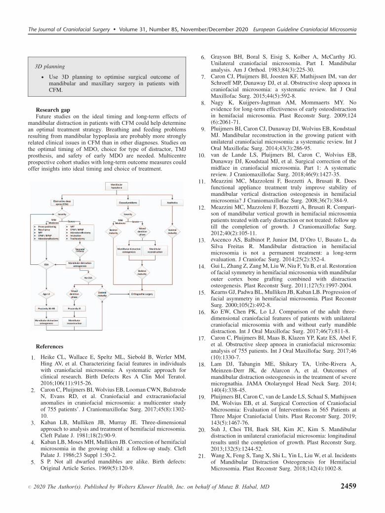
The Journal of Craniofacial Surgery � Volume 31, Number 8S, November/December 2020 European Guideline Craniofacial Microsomia
#
3D planning
� Use 3D planning to optimise surgical outcome ofmandibular and maxillary surgery in patients with
2020 T
CFM.
Research gapFuture studies on the ideal timing and long-term effects of
mandibular distraction in patients with CFM could help determinean optimal treatment strategy. Breathing and feeding problemsresulting from mandibular hypoplasia are probably more stronglyrelated clinical issues in CFM than in other diagnoses. Studies onthe optimal timing of MDO, choice for type of distractor, TMJprosthesis, and safety of early MDO are needed. Multicentreprospective cohort studies with long-term outcome measures couldoffer insights into ideal timing and choice of treatment.
References
1. H
eike CL, Wallace E, Speltz ML, Siebold B, Werler MM,Hing AV, et al. Characterizing facial features in individualswith craniofacial microsomia: A systematic approach forclinical research. Birth Defects Res A Clin Mol Teratol.2016;106(11):915-26.2. C
aron C, Pluijmers BI, Wolvius EB, Looman CWN, BulstrodeN, Evans RD, et al. Craniofacial and extracraniofacialanomalies in craniofacial microsomia: a multicenter studyof 755 patients’. J Craniomaxillofac Surg. 2017;45(8):1302-10.3. K
aban LB, Mulliken JB, Murray JE. Three-dimensionalapproach to analysis and treatment of hemifacial microsomia.Cleft Palate J. 1981;18(2):90-9.4. K
aban LB, Moses MH, Mulliken JB. Correction of hemifacialmicrosomia in the growing child: a follow-up study. CleftPalate J. 1986;23 Suppl 1:50-2.5. S
P. Not all dwarfed mandibles are alike. Birth defects:Original Article Series. 1969(5):120-9.he Author(s). Published by Wolters Kluwer Health, Inc. on b
6. G
ehalf
rayson BH, Boral S, Eisig S, Kolber A, McCarthy JG.Unilateral craniofacial microsomia. Part I. Mandibularanalysis. Am J Orthod. 1983;84(3):225-30.
7. C
aron CJ, Pluijmers BI, Joosten KF, Mathijssen IM, van derSchroeff MP, Dunaway DJ, et al. Obstructive sleep apnoea incraniofacial microsomia: a systematic review. Int J OralMaxillofac Surg. 2015;44(5):592-8.8. N
agy K, Kuijpers-Jagtman AM, Mommaerts MY. Noevidence for long-term effectiveness of early osteodistractionin hemifacial microsomia. Plast Reconstr Surg. 2009;124(6):2061-71.9. P
luijmers BI, Caron CJ, Dunaway DJ, Wolvius EB, KoudstaalMJ. Mandibular reconstruction in the growing patient withunilateral craniofacial microsomia: a systematic review. Int JOral Maxillofac Surg. 2014;43(3):286-95.10. v
an de Lande LS, Pluijmers BI, Caron C, Wolvius EB,Dunaway DJ, Koudstaal MJ, et al. Surgical correction of themidface in craniofacial microsomia. Part 1: A systematicreview. J Craniomaxillofac Surg. 2018;46(9):1427-35.11. M
eazzini MC, Mazzoleni F, Bozzetti A, Brusati R. Doesfunctional appliance treatment truly improve stability ofmandibular vertical distraction osteogenesis in hemifacialmicrosomia? J Craniomaxillofac Surg. 2008;36(7):384-9.12. M
eazzini MC, Mazzoleni F, Bozzetti A, Brusati R. Compari-son of mandibular vertical growth in hemifacial microsomiapatients treated with early distraction or not treated: follow uptill the completion of growth. J Craniomaxillofac Surg.2012;40(2):105-11.13. A
scenco AS, Balbinot P, Junior IM, D’Oro U, Busato L, daSilva Freitas R. Mandibular distraction in hemifacialmicrosomia is not a permanent treatment: a long-termevaluation. J Craniofac Surg. 2014;25(2):352-4.14. G
ui L, Zhang Z, Zang M, Liu W, Niu F, Yu B, et al. Restorationof facial symmetry in hemifacial microsomia with mandibularouter cortex bone grafting combined with distractionosteogenesis. Plast Reconstr Surg. 2011;127(5):1997-2004.15. K
earns GJ, Padwa BL, Mulliken JB, Kaban LB. Progression offacial asymmetry in hemifacial microsomia. Plast ReconstrSurg. 2000;105(2):492-8.16. K
o EW, Chen PK, Lo LJ. Comparison of the adult three-dimensional craniofacial features of patients with unilateralcraniofacial microsomia with and without early mandibledistraction. Int J Oral Maxillofac Surg. 2017;46(7):811-8.17. C
aron C, Pluijmers BI, Maas B, Klazen YP, Katz ES, Abel F,et al. Obstructive sleep apnoea in craniofacial microsomia:analysis of 755 patients. Int J Oral Maxillofac Surg. 2017;46(10):1330-7.18. L
am DJ, Tabangin ME, Shikary TA, Uribe-Rivera A,Meinzen-Derr JK, de Alarcon A, et al. Outcomes ofmandibular distraction osteogenesis in the treatment of severemicrognathia. JAMA Otolaryngol Head Neck Surg. 2014;140(4):338-45.19. P
luijmers BI, Caron C, van de Lande LS, Schaal S, MathijssenIM, Wolvius EB, et al. Surgical Correction of CraniofacialMicrosomia: Evaluation of Interventions in 565 Patients atThree Major Craniofacial Units. Plast Reconstr Surg. 2019;143(5):1467-76.20. S
uh J, Choi TH, Baek SH, Kim JC, Kim S. Mandibulardistraction in unilateral craniofacial microsomia: longitudinalresults until the completion of growth. Plast Reconstr Surg.2013;132(5):1244-52.21. W
ang X, Feng S, Tang X, Shi L, Yin L, Liu W, et al. Incidentsof Mandibular Distraction Osteogenesis for HemifacialMicrosomia. Plast Reconstr Surg. 2018;142(4):1002-8.of Mutaz B. Habal, MD 2459

Renkema et al The Journal of Craniofacial Surgery � Volume 31, Number 8S, November/December 2020
22. W
246
eichman KE, Jacobs J, Patel P, Szpalski C, Shetye P,Grayson B, et al. Early Distraction for Mild to ModerateUnilateral Craniofacial Microsomia: Long-Term Follow-Up,Outcomes, and Recommendations. Plast Reconstr Surg.2017;139(4):941e-53e.
23. Z
hang RS, Lin LO, Hoppe IC, Swanson JW, Taylor JA,Bartlett SP. Early Mandibular Distraction in CraniofacialMicrosomia and Need for Orthognathic Correction at SkeletalMaturity: A Comparative Long-Term Follow-Up Study. PlastReconstr Surg. 2018;142(5):1285-93.24. B
ertin H, Mercier J, Cohen A, Giordanetto J, Cohen N, LeeSH, et al. Surgical correction of mandibular hypoplasia inhemifacial microsomia: A retrospective study in 39 patients. JCraniomaxillofac Surg. 2017;45(6):1031-8.25. P
adwa BL, Mulliken JB, Maghen A, Kaban LB. Midfacialgrowth after costochondral graft construction of the mandib-ular ramus in hemifacial microsomia. J Oral Maxillofac Surg.1998;56(2):122-7; discussion 7-8.26. T
ahiri Y, Chang CS, Tuin J, Paliga JT, Lowe KM, Taylor JA,et al. Costochondral grafting in craniofacial microsomia. PlastReconstr Surg. 2015;135(2):530-41.27. F
attah AY, Caro C, Khechoyan DY, Tompson B, Forrest CR,Phillips JH. Cephalometric outcomes of orthognathic surgeryin hemifacial microsomia. J Craniofac Surg. 2014;25(5):1734-9.28. Y
amaguchi K, Lonic D, Ko EW, Lo LJ. An integrated surgicalprotocol for adult patients with hemifacial microsomia:Methods and outcome. PLoS One. 2017;12(8):e0177223.29. P
luijmers BI, van de Lande LS, Caron C, Wolvius EB,Dunaway DJ, Padwa BL, et al. Part 2: Is the maxillary cantingand its surgical correction in patients with CFM correlated tothe mandibular deformity? J Craniomaxillofac Surg.2018;46(9):1436-40.30. K
aban LB, Moses MH, Mulliken JB. Surgical correction ofhemifacial microsomia in the growing child. Plast ReconstrSurg. 1988;82(1):9-19.31. R
achmiel A, Aizenbud D, Eleftheriou S, Peled M, Laufer D.Extraoral vs. intraoral distraction osteogenesis in the treatmentof hemifacial microsomia. Ann Plast Surg. 2000;45(4):386-94.32. R
achmiel A, Nseir S, Emodi O, Aizenbud D. External versusInternal Distraction Devices in Treatment of Obstructive SleepApnea in Craniofacial Anomalies. Plast Reconstr Surg GlobOpen. 2014;2(7):e188.33. S
antamaria E, Morales C, Taylor JA, Hay A, Ortiz-MonasterioF. Mandibular microsurgical reconstruction in patients withhemifacial microsomia. Plast Reconstr Surg. 2008;122(6):1839-49.34. L
iu H, Zhang X, Liu L, Chen Q, Shao J, Luo E. CombinedBimaxillary Distraction Osteogenesis Associated withOrthognathic Surgery for Hemifacial Microsomia in Adults.Aesthetic Plast Surg. 2017;41(3):650-60.35. P
olley JW, Girotto JA, Fahrenkopf MP, Dietze-Fiedler ML,Kelley JP, Taylor JC, et al. Salvage or Solution: AlloplasticReconstruction in Hemifacial Microsomia. Cleft PalateCraniofac J. 2019;56(7):896-901.36. B
reik O, Tivey D, Umapathysivam K, Anderson P. Does theRate of Distraction or Type of Distractor Affect the Outcomeof Mandibular Distraction in Children With Micrognathia? JOral Maxillofac Surg. 2016;74(7):1441-53.37. B
reik O, Tivey D, Umapathysivam K, Anderson P. Mandibulardistraction osteogenesis for the management of upper airwayobstruction in children with micrognathia: a systematicreview. Int J Oral Maxillofac Surg. 2016;45(6):769-82.0 # 2020 The Author(s). Published
38. B
by W
reik O, Umapathysivam K, Tivey D, Anderson P. Feedingand reflux in children after mandibular distraction osteogene-sis for micrognathia: A systematic review. Int J PediatrOtorhinolaryngol. 2016;85:128-35.
39. O
w AT, Cheung LK. Meta-analysis of mandibular distractionosteogenesis: clinical applications and functional outcomes.Plast Reconstr Surg. 2008;121(3):54e-69e.40. P
aes EC, Mink van der Molen AB, Muradin MS, Speleman L,Sloot F, Kon M, et al. A systematic review on the outcome ofmandibular distraction osteogenesis in infants suffering Robinsequence. Clin Oral Investig. 2013;17(8):1807-20.41. T
sui WK, Yang Y, Cheung LK, Leung YY. Distractionosteogenesis as a treatment of obstructive sleep apneasyndrome: A systematic review. Medicine (Baltimore).2016;95(36):e4674.42. V
erlinden CR, van de Vijfeijken SE, Jansma EP, Becking AG,Swennen GR. Complications of mandibular distractionosteogenesis for congenital deformities: a systematic reviewof the literature and proposal of a new classification forcomplications. Int J Oral Maxillofac Surg. 2015;44(1):37-43.43. M
aster DL, Hanson PR, Gosain AK. Complications ofmandibular distraction osteogenesis. J Craniofac Surg.2010;21(5):1565-70.44. T
ahiri Y, Viezel-Mathieu A, Aldekhayel S, Lee J, Gilardino M.The effectiveness of mandibular distraction in improvingairway obstruction in the pediatric population. Plast ReconstrSurg. 2014;133(3):352e-9e.45. C
aron C, Pluijmers BI, Joosten KFM, Dunaway D, Padwa BL,Wolvius EB, et al. Feeding difficulties in craniofacialmicrosomia: A multicenter retrospective analysis of 755patients. J Craniomaxillofac Surg. 2018;46(10):1777-82.46. C
aron CJ, Pluijmers BI, Joosten KF, Mathijssen IM, van derSchroeff MP, Dunaway DJ, et al. Feeding difficulties incraniofacial microsomia: a systematic review. Int J OralMaxillofac Surg. 2015;44(6):732-7.47. D
ion K B, E. Physical attractiveness and peer perceptionamong children. Sociometry. 1974;37(1):1-12.48. M
ofid MM, Manson PN, Robertson BC, Tufaro AP, Elias JJ,Vander Kolk CA. Craniofacial distraction osteogenesis: areview of 3278 cases. Plast Reconstr Surg. 2001;108(5):1103-14; discussion 15-7.49. H
su SS, Gateno J, Bell RB, Hirsch DL, Markiewicz MR,Teichgraeber JF, et al. Accuracy of a computer-aided surgicalsimulation protocol for orthognathic surgery: a prospectivemulticenter study. J Oral Maxillofac Surg. 2013;71(1):128-42.CHAPTER 5. SURGICAL TREATMENT
5.2 Facial nerveIntroductionPalsy of the facial nerve can be seen in patients with cranio-
facial microsomia (CFM). The prevalence of facial palsy inpatients with CFM is 22% - 53% and may be unilateral orbilateral (1–4). In the O.M.E.N.S. classification, which is com-monly used to grade the severity of patients with CFM, the Nervescore categorises the loss of function of the upper (N1), lower(N2), or total facial nerve (N3) (5).
After exiting the brain stem, the facial nerve progresses throughthe skull and branches to the chorda tympani, posterior auricularnerve and to the digastric and stylohyoid muscles. The mainbranches of the facial nerve originate in the parotid and includethe frontal, zygomatic, buccal, marginal mandibular, and cervical
olters Kluwer Health, Inc. on behalf of Mutaz B. Habal, MD

The Journal of Craniofacial Surgery � Volume 31, Number 8S, November/December 2020 European Guideline Craniofacial Microsomia
branch. Palsy of the facial nerve, which is due to congenitalunderdevelopment in patients with CFM, may cause problems witheye closure, articulation of speech, oral continence, or asymmetricfacial mimics and smile (6).
Since a considerable number of patients with CFM experiencefacial palsy, recommendations on the indications for treatment andthe most optimal treatment modalities are needed. Therefore, thefollowing questions were posed:
5.2.1 What is the indication for surgical treatment of facialnerve anomaly in patients with craniofacial microsomia?5.2.2 What is the most optimal treatment modality for facialnerve anomaly in patients with craniofacial microsomiarelated to functional deficits and aesthetics?
The answers to these questions are important to patients sincevarious functional and aesthetic problems may occur due to facialpalsy. For example, palsy of the upper branches of the facial nervecan lead to an inability to close the eye (lagophthalmos). Awarenessof the consequences of lagophthalmos is important, since cornealexposure can lead to keratitis, ulceration, and eventuallyendophthalmitis which can cause blindness. Palsy of the lowereyelid may also lead to ptosis and lagophthalmos.
Besides insufficient eye closure, ptosis of the eyebrow due toparalysis of the frontalis muscle may also be seen in patients withfacial palsy. Since ptosis may lead to vision loss, surgical treatmentmay be indicated. In addition, facial palsy may also cause oralrelated problems, such as drooling, dysphagia, or speech- or feedingproblems, due to lack of lower lip support and oral sphinctericincompetence (7).
Inadequate functioning of facial mimics can lead to socialproblems and psychological stress (8). Although patients withCFM commonly experience more facial differences besides facialpalsy, the impact on psychosocial functioning may be significant.
Literature searchA systematic search of literature was performed to identify all
available literature on CFM and synonyms. The search was con-ducted in Embase, Pubmed/Medline Ovid. The full search strategyis reported in the supplementary material.
Inclusion and exclusion criteria:
Type of studies
# 2020 The Author(s). Pu
- Original articles- Systematic review of sufficient quality:
- The question in the systematic reviewmatches the question of the guideline.- The search of the systematic review wasconducted in at least two relevantdatabases, such as the Cochrane Library,Medline/Pubmed.- The full search strategy was reported.- No relevant items were missing in thesearch strategy.
Type of patients
- Patients with craniofacial microsomia- Patients with facial palsySubject
- Facial palsyExclusion criteria
- Original studies with <10 included patients- Articles published before 1980- Case reports- Expert opinion- Letters- EditorialsLiterature was screened on title and abstract. No articles on theindication or outcomes of treatment of facial nerve deformity inpatients with CFM were available. Therefore, the Dutch Guideline
blished by Wolters Kluwer Health, Inc. on b
on peripheral facial palsy was included in this chapter (9). Sincethis guideline was published in 2009 and included literature until2007, an additional search, based on the search strategy used in theDutch Guideline, was performed. The full search strategy is shownin the appendix. All literature on indication and/or treatmentoutcomes of facial palsy from 2007 to the present was included.A total of 888 articles was screened on title and abstract. Since thequality of evidence of the identified literature was considered to below, narrative reviews were also included. Besides the DutchGuideline on peripheral facial palsy, 27 other articles wereincluded: two systematic reviews/meta-analyses by Luijmeset al. (10) and Bos et al. (11); eleven retrospective-cohort studiesby Hontanilla et al. (12), Loyo et al. (13), Terzis et al. (2010) (14),Chen et al. (15), Lindsay et al. (16), Roy et al. (7), Chuang et al.(17), Gousheh et al. (18), Harrison et al. (19), Hontanilla et al. (20),and MacQuillan et al. (21); seven case series by Baheerathan et al.(22), Banks et al. (23), Bianchi et al. (24), Panossian et al. (25),Terzis et al.(2009) (26), and Terzis et al. (2009) (27); and sevennarrative reviews by Heike et al. (5), Reddy et al. (6), Volk et al.(28), Rahman et al. (29), Majid et al. (30), Barr et al. (31), andTerzis et al. (2008) (32).
5.2.1 What is the indication for surgical treatment offacial nerve anomaly in patients with craniofacialmicrosomia?
The literature search for this question was directed towards morespecific research questions related to clinical problems, symptoms,treatment options, consequences of not treating, and requirementsfor treating facial nerve anomalies (Appendix 1, http://links.lww.-com/SCS/B697). Unfortunately, no literature on the indication fortreatment of lagophthalmos in patients with CFM was found.Therefore, the Dutch Guideline on peripheral facial palsy wasincluded in this question.
Review of literatureThe Dutch guideline on peripheral facial palsy advises to start
lubrication of the eye if the patient has complaints of the eye(dryness, irritation, tears, photophobia), difficulties with full closureof the eyelid, a positive Bell’s phenomenon, or irritation of theconjunctiva. Surgical treatment of the eyelid is, according to theDutch guideline, indicated if conservative treatment (lubrication) isno longer sufficient. To prevent blindness, eyelid surgery is indi-cated in patients with a corneal ulcer (9). Surgical treatment offacial palsy in patients with CFM may also be indicated to restorefacial animations and improve aesthetics. Various techniques havebeen described in literature, static or dynamic, to restore the smile inpatients with facial palsy. However, no literature on the indicationfor surgical treatment to restore facial function, eyebrow ptosis ororal incompetence was found.
Birgfeld and Heike state that the timing of facial reanima-tion surgery in patients with CFM should be based onpatient’s preferences and other surgical interventions. Microtiareconstruction and major craniofacial or orthognathic surgeryshould ideally be performed prior to facial reanimationsurgery (5).
In their systematic review on the quality of life in patients withperipheral facial palsy, Luijmes et al. found that patients had asignificant increase of the quality of life after (surgical) treatmentof their facial palsy (mean FaCE score 43.38; 95% CI: 40.48–46.27; p<0.001) (10). Quality of life was measured using variousquestionnaires and various treatment modalities, such as botu-linum toxin injections, free flaps, or static procedures, wereincluded. It should be noted that patients with CFM have multiple,often complex, facial deformities, making comparison of the out-comes of treatment on the quality of life of other patientgroups difficult.
ehalf of Mutaz B. Habal, MD 2461
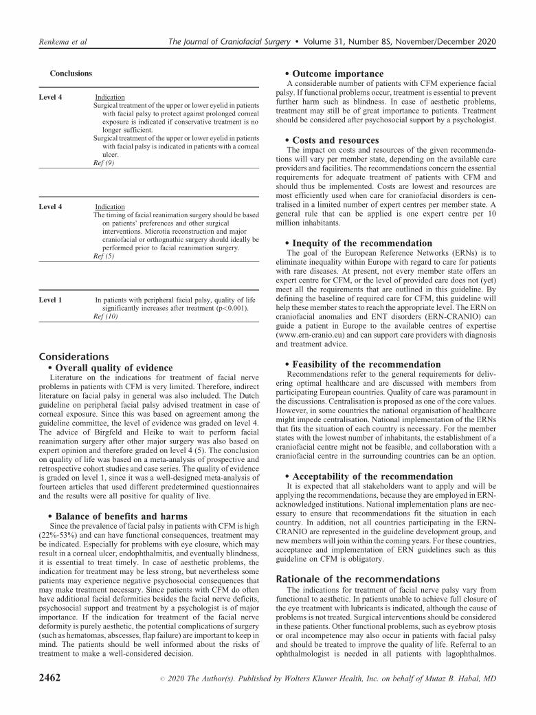
Renkema et al The Journal of Craniofacial Surgery � Volume 31, Number 8S, November/December 2020
Conclusions
Level 4
2462
IndicationSurgical treatment of the upper or lower eyelid in patients
with facial palsy to protect against prolonged cornealexposure is indicated if conservative treatment is nolonger sufficient.
Surgical treatment of the upper or lower eyelid in patientswith facial palsy is indicated in patients with a cornealulcer.
Ref (9)
Level 4
IndicationThe timing of facial reanimation surgery should be basedon patients’ preferences and other surgicalinterventions. Microtia reconstruction and majorcraniofacial or orthognathic surgery should ideally beperformed prior to facial reanimation surgery.
Ref (5)
Level 1
In patients with peripheral facial palsy, quality of lifesignificantly increases after treatment (p<0.001).Ref (10)
Considerations� Overall quality of evidenceLiterature on the indications for treatment of facial nerve
problems in patients with CFM is very limited. Therefore, indirectliterature on facial palsy in general was also included. The Dutchguideline on peripheral facial palsy advised treatment in case ofcorneal exposure. Since this was based on agreement among theguideline committee, the level of evidence was graded on level 4.The advice of Birgfeld and Heike to wait to perform facialreanimation surgery after other major surgery was also based onexpert opinion and therefore graded on level 4 (5). The conclusionon quality of life was based on a meta-analysis of prospective andretrospective cohort studies and case series. The quality of evidenceis graded on level 1, since it was a well-designed meta-analysis offourteen articles that used different predetermined questionnairesand the results were all positive for quality of live.
� Balance of benefits and harmsSince the prevalence of facial palsy in patients with CFM is high
(22%-53%) and can have functional consequences, treatment maybe indicated. Especially for problems with eye closure, which mayresult in a corneal ulcer, endophthalmitis, and eventually blindness,it is essential to treat timely. In case of aesthetic problems, theindication for treatment may be less strong, but nevertheless somepatients may experience negative psychosocial consequences thatmay make treatment necessary. Since patients with CFM do oftenhave additional facial deformities besides the facial nerve deficits,psychosocial support and treatment by a psychologist is of majorimportance. If the indication for treatment of the facial nervedeformity is purely aesthetic, the potential complications of surgery(such as hematomas, abscesses, flap failure) are important to keep inmind. The patients should be well informed about the risks oftreatment to make a well-considered decision.
# 2020 The Author(s). Published
� Outcome importanceA considerable number of patients with CFM experience facial
palsy. If functional problems occur, treatment is essential to preventfurther harm such as blindness. In case of aesthetic problems,treatment may still be of great importance to patients. Treatmentshould be considered after psychosocial support by a psychologist.
� Costs and resourcesThe impact on costs and resources of the given recommenda-
tions will vary per member state, depending on the available careproviders and facilities. The recommendations concern the essentialrequirements for adequate treatment of patients with CFM andshould thus be implemented. Costs are lowest and resources aremost efficiently used when care for craniofacial disorders is cen-tralised in a limited number of expert centres per member state. Ageneral rule that can be applied is one expert centre per 10million inhabitants.
� Inequity of the recommendationThe goal of the European Reference Networks (ERNs) is to
eliminate inequality within Europe with regard to care for patientswith rare diseases. At present, not every member state offers anexpert centre for CFM, or the level of provided care does not (yet)meet all the requirements that are outlined in this guideline. Bydefining the baseline of required care for CFM, this guideline willhelp these member states to reach the appropriate level. The ERN oncraniofacial anomalies and ENT disorders (ERN-CRANIO) canguide a patient in Europe to the available centres of expertise(www.ern-cranio.eu) and can support care providers with diagnosisand treatment advice.
� Feasibility of the recommendationRecommendations refer to the general requirements for deliv-
ering optimal healthcare and are discussed with members fromparticipating European countries. Quality of care was paramount inthe discussions. Centralisation is proposed as one of the core values.However, in some countries the national organisation of healthcaremight impede centralisation. National implementation of the ERNsthat fits the situation of each country is necessary. For the memberstates with the lowest number of inhabitants, the establishment of acraniofacial centre might not be feasible, and collaboration with acraniofacial centre in the surrounding countries can be an option.
� Acceptability of the recommendationIt is expected that all stakeholders want to apply and will be
applying the recommendations, because they are employed in ERN-acknowledged institutions. National implementation plans are nec-essary to ensure that recommendations fit the situation in eachcountry. In addition, not all countries participating in the ERN-CRANIO are represented in the guideline development group, andnew members will join within the coming years. For these countries,acceptance and implementation of ERN guidelines such as thisguideline on CFM is obligatory.
Rationale of the recommendationsThe indications for treatment of facial nerve palsy vary from
functional to aesthetic. In patients unable to achieve full closure ofthe eye treatment with lubricants is indicated, although the cause ofproblems is not treated. Surgical interventions should be consideredin these patients. Other functional problems, such as eyebrow ptosisor oral incompetence may also occur in patients with facial palsyand should be treated to improve the quality of life. Referral to anophthalmologist is needed in all patients with lagophthalmos.
by Wolters Kluwer Health, Inc. on behalf of Mutaz B. Habal, MD

The Journal of Craniofacial Surgery � Volume 31, Number 8S, November/December 2020 European Guideline Craniofacial Microsomia
Aesthetic problems are considered to be another reason to treatfacial nerve palsy in patients with CFM, although no literature onthis topic is available. The involvement of a psychologist is essen-tial during childhood for patients with CFM to learn to cope withtheir facial differences. The ICHOM Standard Set for CraniofacialMicrosomia advises to assess smile and facial movement by usingthe CleftQ appearance questionnaire at age 8, 12, and 22. Treatmentof facial nerve palsy for aesthetic reasons should be based on well-informed patient preferences and be integrated with otherplanned surgeries.
Recommendations
#
Indications for treatment
� Provide all patients with craniofacial microsomia withpsychosocial support.
� Refer all craniofacial microsomia patients withlagophthalmos to an ophthalmologist.
� Surgical treatment of the upper or lower eyelids shouldbe considered in patients with craniofacial microsomiaand loss of function of the upper facial nerve branches.
� Coordinate the timing of facial reanimation surgery inpatients with craniofacial microsomia in the planning ofother major surgeries.
� Facial movement should by assessed with the CleftQ
2020 T
Appearance at age 8, 12, and 22.
Research gapNo literature on the indication for treatment of facial palsy in
CFM was available. Literature on the (psychological) consequencesof facial palsy in CFM could help determine the indication fortreatment.
5.2.2 What is the most optimal treatment modality for facialnerve anomaly in patients with craniofacial microsomiarelated to functional deficits and aesthetics?
The literature search for this question was directed towards morespecific research questions related to treatment options, complica-tions, advantages and disadvantages, short and long term results andtreatment burden for treating facial nerve anomalies (Appendix 1,http://links.lww.com/SCS/B697). Patient outcomes that are rele-vant for this chapter are functional deficits and aesthetics. Thisreview of literature addresses the most optimal treatment modalitiesrelated to the functional and aesthetic consequences of facial palsy.The functional problems and their treatment modalities in thischapter are subdivided into problems with eye closure, oral pro-blems, and dynamic facial reanimation.
Review of literatureEye
Various treatment modalities are available to treat the inabilityto close the eye in patients with facial palsy. The guideline onperipheral facial palsy advises to start conservative treatment usinglubrication in all patients with facial palsy and complaints of theeye. In patients with CFM, the facial palsy is congenital and morepermanent solutions should be striven for. The goal of surgery of theupper eyelid is to improve vision, prevent corneal erosion andmaintain or improve aesthetics (20).
The upper eyelid can be lowered using botulinum toxin, muscletransfers, or placing a gold weight. Injection of botulin toxin hasbeen found to be effective by establishing a ptosis by temporarilyparalysing the levator palpebrae superioris muscle (28). The dura-tion of the effect is approximately three months and there is a
he Author(s). Published by Wolters Kluwer Health, Inc. on b
chance that therapy is unsuccessful due to injecting too little or toomuch botulinum toxin (9).
Lengthening of the levator palpebrae superioris muscle byaponeurosis interposition could be performed to achieve full eyeclosure, although this technique is less frequently described inliterature. The technique is complex compared to botulinum toxinor placement of a gold weight, but may be have a better cosmeticoutcome compared to gold weights (9).
Placement of a gold weight in the upper eyelid is a populartechnique to correct lagophthalmos. A predetermined gold weightis implanted in the eyelid that supports closure of the eye. Othermaterials such as a platinum chain are currently used too (13).Although placement of gold weights is reported to be the preferredmethod for several surgeons due to the predictable results, goodaesthetic outcome, reversibility, relatively easy technique the factthat and it can be placed under local anaesthesia in adults, variouscomplications may occur (20). This includes migration or extru-sion of the gold weight, soreness, or persistent lagophthalmos (22).The incidence of extrusion of the gold weight was 2.6% in 2,000studied patients, which may be prevented by attaching the goldweight on the tarsal plate using sutures (22, 29). In a case series byBaheerathan that described the experience with gold weights in 16patients, one patient reported extrusion of the gold weight after fivemonths and one patient needed a heavier gold weight due toresidual lagophthalmos (22). No infections, implant migration,visual problems or keratopathy occurred. All patients had adequateeyelid closure at the last follow-up and fifteen of the sixteenpatients were satisfied with the outcome (22). Surgical revisionafter placement of a upper eyelid weight was needed in 18 of the 95patients (19%) studied by Loyo et al. (13). One patient had implantextrusion, two patients bulging/superficial implants, two patientsimplant migration, five patients had persistent lagophthalmos, andnine patients eyelid ptosis. In a retrospective cohort study byHontanilla et al. 58 patients had gold weights implanted (20).All patients had complaints of dry eyes. Five patients (8.6%) hadextrusion of the gold weight, which was due to material intolerancein two patients and exchanged for a different weight in the otherthree patients without complications. The complaints of eye dry-ness improved in 90.7% of the patients (n¼ 49). Corneal ulcers,which were present in 14 patients, resolved without further treat-ment in 85.7% of the patients (n¼ 12) (20). It is important to notethat all these studies included adult patients only and no literatureon children is available.
Facial palsy may also lead to laxity or ectropion of the lowereyelid which disrupts physiological tear drainage (20, 28). Thisinability to achieve full closure of the eye may again lead to keratitisor corneal erosion. The aim of surgery of the lower eyelid is toimprove tear drainage and correct the ectropion or ptosis (20).Tarsorrhaphy has been the golden standard to achieve this, but othertechniques such as lateral tarsal suspension or the use of a fascia latastrip, which also provide corneal coverage but produce betteraesthetic appearance, have replaced this (20). In addition, tarsor-rhaphy may cause loss of peripheral vision and its use is thereforediscouraged (29). In the retrospective study by Loyo et al. lowereyelid surgery was performed in 47% of the patients with paralyticlagophthalmos (47/101 patients) (13). All patients had proceduresthat addressed the lateral canthal tendon and 10 of the 47 patientsunderwent medial canthopexy. Of these patients, 29% (14/47patients) required additional lower eyelid procedures. Hontanillaet al. retrospectively studied 43 patients with lower eyelid suspen-sion with a tendon sling (20). Successful correction of the ectropion/ptosis and epiphora was achieved in respectively 93.9% and 91.9%.Eight patients (18.6%) needed additional surgery to increase tendontension. No complications that required surgical interventionoccurred. The authors discourage the use of a dynamic technique
ehalf of Mutaz B. Habal, MD 2463
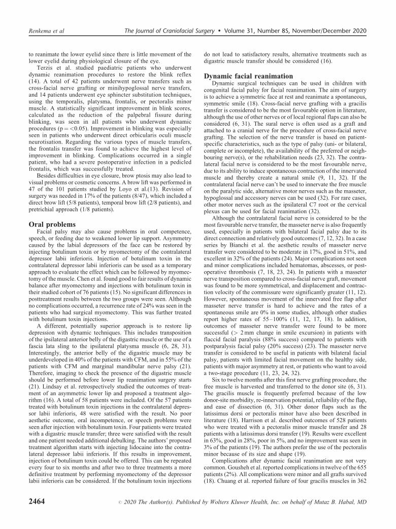
Renkema et al The Journal of Craniofacial Surgery � Volume 31, Number 8S, November/December 2020
to reanimate the lower eyelid since there is little movement of thelower eyelid during physiological closure of the eye.
Terzis et al. studied paediatric patients who underwentdynamic reanimation procedures to restore the blink reflex(14). A total of 42 patients underwent nerve transfers such ascross-facial nerve grafting or minihypoglossal nerve transfers,and 14 patients underwent eye sphincter substitution techniques,using the temporalis, platysma, frontalis, or pectoralis minormuscle. A statistically significant improvement in blink scores,calculated as the reduction of the palpebral fissure duringblinking, was seen in all patients who underwent dynamicprocedures (p¼<0.05). Improvement in blinking was especiallyseen in patients who underwent direct orbicularis oculi muscleneurotisation. Regarding the various types of muscle transfers,the frontalis transfer was found to achieve the highest level ofimprovement in blinking. Complications occurred in a singlepatient, who had a severe postoperative infection in a pedicledfrontalis, which was successfully treated.
Besides difficulties in eye closure, brow ptosis may also lead tovisual problems or cosmetic concerns. A brow lift was performed in47 of the 101 patients studied by Loyo et al.(13). Revision ofsurgery was needed in 17% of the patients (8/47), which included adirect brow lift (5/8 patients), temporal brow lift (2/8 patients), andpretrichial approach (1/8 patients).
Oral problemsFacial palsy may also cause problems in oral competence,
speech, or feeding due to weakened lower lip support. Asymmetrycaused by the labial depressors of the face can be restored byinjecting botulinum toxin or by myomectomy of the contralateraldepressor labii inferioris. Injection of botulinum toxin in thecontralateral depressor labii inferioris can be used as a temporaryapproach to evaluate the effect which can be followed by myomec-tomy of the muscle. Chen et al. found good to fair results of dynamicbalance after myomectomy and injections with botulinum toxin intheir studied cohort of 76 patients (15). No significant differences inposttreatment results between the two groups were seen. Althoughno complications occurred, a recurrence rate of 24% was seen in thepatients who had surgical myomectomy. This was further treatedwith botulinum toxin injections.
A different, potentially superior approach is to restore lipdepression with dynamic techniques. This includes transpositionof the ipsilateral anterior belly of the digastric muscle or the use of afascia lata sling to the ipsilateral platysma muscle (6, 28, 31).Interestingly, the anterior belly of the digastric muscle may beunderdeveloped in 40% of the patients with CFM, and in 55% of thepatients with CFM and marginal mandibular nerve palsy (21).Therefore, imaging to check the presence of the digastric muscleshould be performed before lower lip reanimation surgery starts(21). Lindsay et al. retrospectively studied the outcomes of treat-ment of an asymmetric lower lip and proposed a treatment algo-rithm (16). A total of 58 patients were included. Of the 57 patientstreated with botulinum toxin injections in the contralateral depres-sor labii inferioris, 48 were satisfied with the result. No pooraesthetic outcome, oral incompetence, or speech problems wereseen after injection with botulinum toxin. Four patients were treatedwith a digastric muscle transfer; three were satisfied with the resultand one patient needed additional debulking. The authors’ proposedtreatment algorithm starts with injecting lidocaine into the contra-lateral depressor labii inferioris. If this results in improvement,injection of botulinum toxin could be offered. This can be repeatedevery four to six months and after two to three treatments a moredefinitive treatment by performing myomectomy of the depressorlabii inferioris can be considered. If the botulinum toxin injections
2464 # 2020 The Author(s). Published
do not lead to satisfactory results, alternative treatments such asdigastric muscle transfer should be considered (16).
Dynamic facial reanimationDynamic surgical techniques can be used in children with
congenital facial palsy for facial reanimation. The aim of surgeryis to achieve a symmetric face at rest and reanimate a spontaneous,symmetric smile (18). Cross-facial nerve grafting with a gracilistransfer is considered to be the most favourable option in literature,although the use of other nerves or of local regional flaps can also beconsidered (6, 31). The sural nerve is often used as a graft andattached to a cranial nerve for the procedure of cross-facial nervegrafting. The selection of the nerve transfer is based on patient-specific characteristics, such as the type of palsy (uni- or bilateral,complete or incomplete), the availability of the preferred or neigh-bouring nerve(s), or the rehabilitation needs (23, 32). The contra-lateral facial nerve is considered to be the most favourable nerve,due to its ability to induce spontaneous contraction of the innervatedmuscle and thereby create a natural smile (9, 11, 32). If thecontralateral facial nerve can’t be used to innervate the free muscleon the paralytic side, alternative motor nerves such as the masseter,hypoglossal and accessory nerves can be used (32). For rare cases,other motor nerves such as the ipsilateral C7 root or the cervicalplexus can be used for facial reanimation (32).
Although the contralateral facial nerve is considered to be themost favourable nerve transfer, the masseter nerve is also frequentlyused, especially in patients with bilateral facial palsy due to itsdirect connection and relatively good outcomes (7, 12, 32). In a caseseries by Bianchi et al. the aesthetic results of masseter nervetransfer were considered to be moderate in 17%, good in 51%, andexcellent in 32% of the patients (24). Major complications not seenand minor complications included hematomas, abscesses, or post-operative thrombosis (7, 18, 23, 24). In patients with a masseternerve transposition compared to cross-facial nerve graft, movementwas found to be more symmetrical, and displacement and contrac-tion velocity of the commissure were significantly greater (11, 12).However, spontaneous movement of the innervated free flap aftermasseter nerve transfer is hard to achieve and the rates of aspontaneous smile are 0% in some studies, although other studiesreport higher rates of 55–100% (11, 12, 17, 18). In addition,outcomes of masseter nerve transfer were found to be moresuccessful (> 2 mm change in smile excursion) in patients withflaccid facial paralysis (88% success) compared to patients withpostparalysis facial palsy (20% success) (23). The masseter nervetransfer is considered to be useful in patients with bilateral facialpalsy, patients with limited facial movement on the healthy side,patients with major asymmetry at rest, or patients who want to avoida two-stage procedure (11, 23, 24, 32).
Six to twelve months after this first nerve grafting procedure, thefree muscle is harvested and transferred to the donor site (6, 31).The gracilis muscle is frequently preferred because of the lowdonor-site morbidity, re-innervation potential, reliability of the flap,and ease of dissection (6, 31). Other donor flaps such as thelatissimus dorsi or pectoralis minor have also been described inliterature (18). Harrison et al. described outcomes of 528 patientswho were treated with a pectoralis minor muscle transfer and 28patients with a latissimus dorsi transfer (19). Results were excellentin 63%, good in 28%, poor in 5%, and no improvement was seen in3% of the patients (19). The authors prefer the use of the pectoralisminor because of its size and shape (19).
Complications after dynamic facial reanimation are not verycommon. Gousheh et al. reported complications in twelve of the 655patients (2%). All complications were minor and all grafts survived(18). Chuang et al. reported failure of four gracilis muscles in 362
by Wolters Kluwer Health, Inc. on behalf of Mutaz B. Habal, MD

The Journal of Craniofacial Surgery � Volume 31, Number 8S, November/December 2020 European Guideline Craniofacial Microsomia
patients (1%), which were successfully replaced in all cases (17).The long-term outcomes of free-muscle transfer were studied byTerzis et al. (26, 27). A total of 32 children with a follow-up of atleast five years were included (26). Almost all patients (91%)needed additional surgical revisions, which included debulking,defatting, reanchoring the muscle graft, or a mini-temporalis trans-fer to the oral commissure. All flaps survived and all patientsshowed improved function and symmetry at two-year follow-up(p¼<0.001). This improvement remained over time. Muscle func-tion and aesthetic outcomes showed further improvement over time(p¼<0.001). These results were also seen in a different study bythe same authors that addressed the long-term outcomes of free-muscle transfers in adults (27). The authors conclude that these datasupport the use of free-muscle transfer in children and the techniqueis effective to restore a coordinated functional and symmetric smile(26, 27).
The use of regional flaps can be considered in patients inwhom a cross-facial nerve graft or free-muscle transfers is not anoption (18). Regional flaps cause shorter hospital stay andoperative times, and limited donor-side morbidity compared tocross-facial nerve grafting (25). Regional temporalis or masseterflaps are less commonly used in children due to the bulky aspectof the masseter, temporal hollowing, and the inability torespond to emotions and form a natural smile (28, 31). Althoughno spontaneous smile can be achieved, commissuredisplacement after lengthening temporalis myoplasty was foundto be 8.2 mm (range 5–13 mm) in the 14 patients studied byPanossian et al. (25).
Conclusions
Level 3
# 2020 The Auth
Eye
Lowering of the upper eyelid to prevent keratitis or
corneal erosion can be achieved by injection of
botulinum toxin, lengthening of the levator palpebrae
superioris muscle or placement of a gold weight in the
upper eyelid.
Placement of a gold or platinum weight to correct
lagophthalmos is a preferred method due to good
aesthetic outcome, predictable results, reversibility,
easy technique, and the fact that it can be placed under
local anaesthesia in adults.
Surgical revision after placement of a gold weight is
needed in up to 19% of patients, due to extrusion or
migration of the implant, persistent lagophthalmos, or
ptosis.
Ref (9, 13, 20, 22, 29)
Level 3
EyeEye sphincter substitution techniques by using muscle
transfers or cross-facial nerve grafting can lead to a
significant improvement in blinking (p¼<0.05).
The highest level of improvement in blinking is seen after
transfer of the frontalis muscle compared to the use of
the platysma, frontalis, or pectoralis minor muscle.
Surgical correction of ptosis of the eyebrow in patients
with facial palsy may be indicated to treat visual or
cosmetic problems.
Ref (13, 14)
or(s). Published by Wolters Kluwer Health, Inc. on b
Level 3
ehalf of Mutaz B
OralSymmetric lip depression may be restored by repeated
injections of botulinum toxin or myomectomy of thenon-affected depressor labii inferioris, or by dynamicreconstruction.
Results after botulinum toxin injections or myomectomyof the non-affected depressor labii inferioris areconsidered to be fair to good and no significantdifference in post treatment results of the twotreatment options are seen.
Dynamic reconstruction of lower lip depression includestransposition of the ipsilateral anterior belly of thedigastric muscle or the use of a fascia lata sling to theipsilateral platysma muscle.
Due to the high prevalence of agenesis of the anteriorbelly of the digastric muscle in patients withcraniofacial microsomia (55%), imaging of the muscleshould be performed before reanimation surgery of thelower lip is undertaken.
Ref (6, 9, 15, 16, 21, 28, 31)
Level 4
OralProposed treatment algorithm for the asymmetric lowerlip:- Start with injection of botulinum toxin incontralateral depressor- Repeat treatment if results are satisfactory- After 2–3 treatments and good results, myomectomycan be considered- No satisfactory results: consider digastric muscletransfer
Ref (16)
Level 3
EyeCorrection of the lower eyelid can be achieved by tendonsling, lateral tarsal suspension, or by a fascia lata strip.Successful correction can be achieved in >90% ofpatients.
The use of tarsorrhaphy is discouraged due to poorcosmetic outcome and risk of peripheral vision loss.
The rate of surgical revisions after a tendon sling ormedial canthopexy to correct paralytic lagophthalmosvaries from 18 to 29%.
Ref (9, 13, 20, 29)
Level 4
Dynamic facial reconstruction / aestheticsTo restore facial animation and a spontaneous smile,cross-facial nerve grafting is considered to be thepreferred treatment for unilateral craniofacialmicrosomia.
Other motor nerves, such as the masseter, hypoglossal,accessory or cervical nerves can be used if cross-facialnerve grafting is not possible.
The gracilis muscle is often preferred due to the lowdonor-site morbidity, re-innervation potential,reliability of the flap, and ease of dissection. Other freeflaps, such as the latissimus dorsi or pectoralis minorcould also be used.
Ref (6, 9, 11, 18, 19, 23, 31, 32)
. Habal, MD 2465
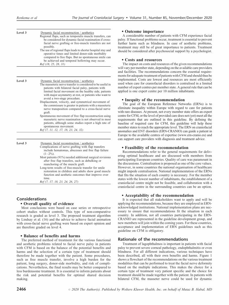
Renkema et al The Journal of Craniofacial Surgery � Volume 31, Number 8S, November/December 2020
Level 3
2466
Dynamic facial reconstruction / aestheticsRegional flaps, such as temporalis muscle transfers, can
be considered for dynamic facial reanimation if cross-facial nerve grafting or free-muscle transfers are notpossible.
The use of regional flaps leads to shorter hospital stay andoperative times and limited donor-side morbiditycompared to free flaps. But no spontaneous smile canbe achieved and temporal hollowing may occur.
Ref (18, 25, 28, 31)
Level 3
Dynamic facial reconstruction / aestheticsThe masseteric nerve transfer is considered to be useful inpatients with bilateral facial palsy, patients withlimited facial movement on the healthy side, patientswith major asymmetry at rest, or patients who want toavoid a two-stage procedure.
Displacement, velocity, and symmetrical movement ofthe commissure is greater in patients with a massetericnerve transposition compared to a cross-facial nervegraft.
Spontaneous movement of free flap reconstruction usingmasseteric nerve reanimation is not observed in mostpatients, although some studies report a high rate ofspontaneous movement.
Ref (7, 11, 12, 17, 18, 23, 24, 32)
Level 3
Dynamic facial reconstruction / aestheticsComplications of nerve grafting with flap transfersinclude hematomas, abscesses and free flap failure(1–2%).
Most patients (91%) needed additional surgical revisionsafter free flap transfers, such as debulking orreanchoring of the muscle graft.
Long-term results of free-muscle transfer for smilerestoration in children and adults show good musclefunction and aesthetic outcomes that improve overtime.
Ref (7, 17, 18, 23, 24, 26, 27)
Considerations� Overall quality of evidenceMost conclusions were based on case series or retrospective
cohort studies without controls. This type of non-comparativeresearch is graded as level 3. The proposed treatment algorithmby Lindsay et al. (16) and the advice to achieve facial animationwith cross-facial nerve grafting were based on expert opinion andare therefore graded on level 4.
� Balance of benefits and harmsThe preferred method of treatment for the various functional
and aesthetic problems related to facial nerve palsy in patientswith CFM is based on the balance of the potential benefits andharms and the selection of a certain type of treatment shouldtherefore be made together with the patient. Some procedures,such as free muscle transfer, involve a high burden for thepatient, long surgery, donor-side morbidity, and risk of compli-cations. Nevertheless, the final results may be better compared toless burdensome treatment. It is essential to inform patients aboutthe risk and potential benefits for optimal shared decisionmaking.
# 2020 The Author(s). Published
� Outcome importanceA considerable number of patients with CFM experience facial
palsy. If functional problems occur, treatment is essential to preventfurther harm such as blindness. In case of aesthetic problems,treatment may still be of great importance to patients. Treatmentshould be considered after psychosocial support by a psychologist.
� Costs and resourcesThe impact on costs and resources of the given recommendations
will vary per member state, depending on the available care providersand facilities. The recommendations concern the essential require-ments for adequate treatment of patients with CFM and should thus beimplemented. Costs are lowest and resources are most efficientlyused when care for craniofacial disorders is centralised in a limitednumber of expert centres per member state. A general rule that can beapplied is one expert centre per 10 million inhabitants.
� Inequity of the recommendationThe goal of the European Reference Networks (ERNs) is to
eliminate inequality within Europe with regard to care for patientswith rare diseases. At present, not every member state offers an expertcentre for CFM, or the level of provided care does not (yet) meet all therequirements that are outlined in this guideline. By defining thebaseline of required care for CFM, this guideline will help thesemember states to reach the appropriate level. The ERN on craniofacialanomalies and ENT disorders (ERN-CRANIO) can guide a patient inEurope to the available centres of expertise (www.ern-cranio.eu) andcan support care providers with diagnosis and treatment advice.
� Feasibility of the recommendationRecommendations refer to the general requirements for deliv-
ering optimal healthcare and are discussed with members fromparticipating European countries. Quality of care was paramount inthe discussions. Centralisation is proposed as one of the core values.However, in some countries the national organisation of healthcaremight impede centralisation. National implementation of the ERNsthat fits the situation of each country is necessary. For the memberstates with the lowest number of inhabitants, the establishment of acraniofacial centre might not be feasible, and collaboration with acraniofacial centre in the surrounding countries can be an option.
� Acceptability of the recommendationIt is expected that all stakeholders want to apply and will be
applying the recommendations, because they are employed in ERN-acknowledged institutions. National implementation plans are nec-essary to ensure that recommendations fit the situation in eachcountry. In addition, not all countries participating in the ERN-CRANIO are represented in the guideline development group, andnew members will join within the coming years. For these countries,acceptance and implementation of ERN guidelines such as thisguideline on CFM is obligatory.
Rationale of the recommendationsTreatment of lagophthalmos is important in patients with facial
palsy to prevent severe corneal pathology, endophthalmitis or evenblindness. For all different indications, various techniques havebeen described, all with their own benefits and harms. Figure 1shows a flowchart of the recommendations on the various treatmentmodalities that can be performed to treat the facial nerve deformitybased on the multiple indications. This makes the choice for acertain type of treatment very patient specific and the choice fortreatment should be made together with the patient. In patients withbilateral CFM, the masseter nerve could be used for dynamic
by Wolters Kluwer Health, Inc. on behalf of Mutaz B. Habal, MD
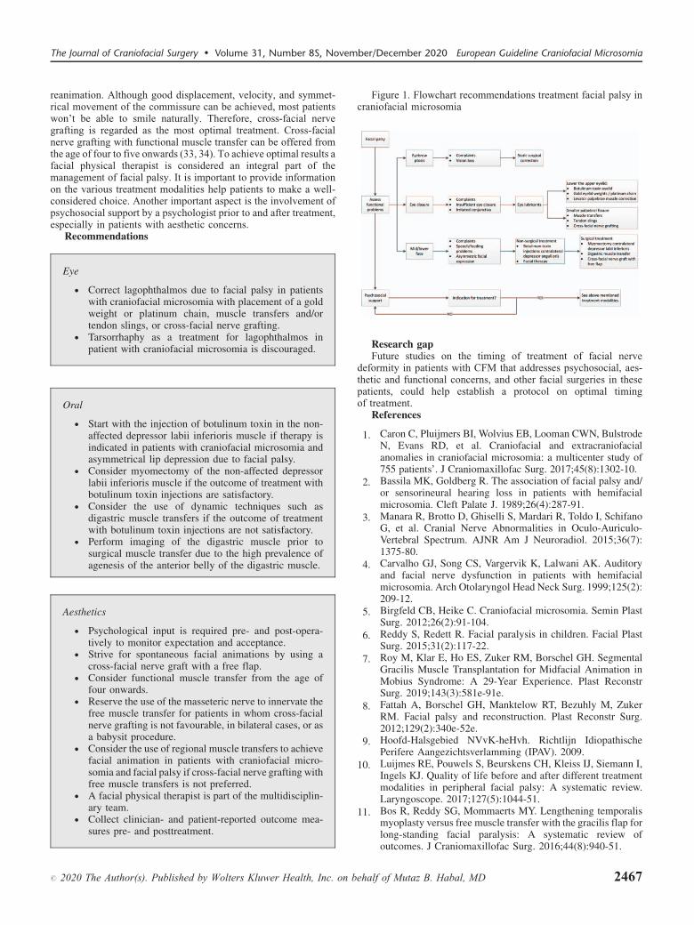
The Journal of Craniofacial Surgery � Volume 31, Number 8S, November/December 2020 European Guideline Craniofacial Microsomia
reanimation. Although good displacement, velocity, and symmet-rical movement of the commissure can be achieved, most patientswon’t be able to smile naturally. Therefore, cross-facial nervegrafting is regarded as the most optimal treatment. Cross-facialnerve grafting with functional muscle transfer can be offered fromthe age of four to five onwards (33, 34). To achieve optimal results afacial physical therapist is considered an integral part of themanagement of facial palsy. It is important to provide informationon the various treatment modalities help patients to make a well-considered choice. Another important aspect is the involvement ofpsychosocial support by a psychologist prior to and after treatment,especially in patients with aesthetic concerns.
Recommendations
#
Eye
� Correct lagophthalmos due to facial palsy in patientswith craniofacial microsomia with placement of a goldweight or platinum chain, muscle transfers and/ortendon slings, or cross-facial nerve grafting.
� Tarsorrhaphy as a treatment for lagophthalmos in
2020 T
patient with craniofacial microsomia is discouraged.
Oral
� Start with the injection of botulinum toxin in the non-affected depressor labii inferioris muscle if therapy isindicated in patients with craniofacial microsomia andasymmetrical lip depression due to facial palsy.
� Consider myomectomy of the non-affected depressorlabii inferioris muscle if the outcome of treatment withbotulinum toxin injections are satisfactory.
� Consider the use of dynamic techniques such asdigastric muscle transfers if the outcome of treatmentwith botulinum toxin injections are not satisfactory.
� Perform imaging of the digastric muscle prior tosurgical muscle transfer due to the high prevalence of
agenesis of the anterior belly of the digastric muscle.Aesthetics
� Psychological input is required pre- and post-opera-tively to monitor expectation and acceptance.
� Strive for spontaneous facial animations by using across-facial nerve graft with a free flap.
� Consider functional muscle transfer from the age offour onwards.
� Reserve the use of the masseteric nerve to innervate thefree muscle transfer for patients in whom cross-facialnerve grafting is not favourable, in bilateral cases, or asa babysit procedure.
� Consider the use of regional muscle transfers to achievefacial animation in patients with craniofacial micro-somia and facial palsy if cross-facial nerve grafting withfree muscle transfers is not preferred.
� A facial physical therapist is part of the multidisciplin-ary team.
� Collect clinician- and patient-reported outcome mea-
sures pre- and posttreatment.he Author(s). Published by Wolters Kluwer Health, Inc. on b
Figure 1. Flowchart recommendations treatment facial palsy incraniofacial microsomia
Research gapFuture studies on the timing of treatment of facial nerve
deformity in patients with CFM that addresses psychosocial, aes-thetic and functional concerns, and other facial surgeries in thesepatients, could help establish a protocol on optimal timingof treatment.
References
1. C
ehalf
aron C, Pluijmers BI, Wolvius EB, Looman CWN, BulstrodeN, Evans RD, et al. Craniofacial and extracraniofacialanomalies in craniofacial microsomia: a multicenter study of755 patients’. J Craniomaxillofac Surg. 2017;45(8):1302-10.
2. B
assila MK, Goldberg R. The association of facial palsy and/or sensorineural hearing loss in patients with hemifacialmicrosomia. Cleft Palate J. 1989;26(4):287-91.3. M
anara R, Brotto D, Ghiselli S, Mardari R, Toldo I, SchifanoG, et al. Cranial Nerve Abnormalities in Oculo-Auriculo-Vertebral Spectrum. AJNR Am J Neuroradiol. 2015;36(7):1375-80.4. C
arvalho GJ, Song CS, Vargervik K, Lalwani AK. Auditoryand facial nerve dysfunction in patients with hemifacialmicrosomia. Arch Otolaryngol Head Neck Surg. 1999;125(2):209-12.5. B
irgfeld CB, Heike C. Craniofacial microsomia. Semin PlastSurg. 2012;26(2):91-104.6. R
eddy S, Redett R. Facial paralysis in children. Facial PlastSurg. 2015;31(2):117-22.7. R
oy M, Klar E, Ho ES, Zuker RM, Borschel GH. SegmentalGracilis Muscle Transplantation for Midfacial Animation inMobius Syndrome: A 29-Year Experience. Plast ReconstrSurg. 2019;143(3):581e-91e.8. F
attah A, Borschel GH, Manktelow RT, Bezuhly M, ZukerRM. Facial palsy and reconstruction. Plast Reconstr Surg.2012;129(2):340e-52e.9. H
oofd-Halsgebied NVvK-heHvh. Richtlijn IdiopathischePerifere Aangezichtsverlamming (IPAV). 2009.10. L
uijmes RE, Pouwels S, Beurskens CH, Kleiss IJ, Siemann I,Ingels KJ. Quality of life before and after different treatmentmodalities in peripheral facial palsy: A systematic review.Laryngoscope. 2017;127(5):1044-51.11. B
os R, Reddy SG, Mommaerts MY. Lengthening temporalismyoplasty versus free muscle transfer with the gracilis flap forlong-standing facial paralysis: A systematic review ofoutcomes. J Craniomaxillofac Surg. 2016;44(8):940-51.of Mutaz B. Habal, MD 2467
Renkema et al The Journal of Craniofacial Surgery � Volume 31, Number 8S, November/December 2020
12. H
246
ontanilla B, Olivas J, Cabello A, Marre D. Cross-Face NerveGrafting versus Masseteric-to-Facial Nerve Transposition forReanimation of Incomplete Facial Paralysis: A ComparativeStudy Using the FACIAL CLIMA Evaluating System. PlastReconstr Surg. 2018;142(2):179e-91e.
13. L
oyo M, Jones D, Lee LN, Collar RM, Molendijk J, BoaheneKD, et al. Treatment of the periocular complex in paralyticlagophthalmos. Ann Otol Rhinol Laryngol. 2015;124(4):273-9.14. T
erzis JK, Karypidis D. The outcomes of dynamic proceduresfor blink restoration in pediatric facial paralysis. PlastReconstr Surg. 2010;125(2):629-44.15. C
hen CK, Tang YB. Myectomy and botulinum toxin forparalysis of the marginal mandibular branch of the facialnerve: a series of 76 cases. Plast Reconstr Surg. 2007;120(7):1859-64.16. L
indsay RW, Edwards C, Smitson C, Cheney ML, HadlockTA. A systematic algorithm for the management of lower lipasymmetry. Am J Otolaryngol. 2011;32(1):1-7.17. C
huang DC, Lu JC, Chang TN, Laurence VG. Comparison ofFunctional Results After Cross-Face Nerve Graft-, SpinalAccessory Nerve-, and Masseter Nerve-Innervated Gracilisfor Facial Paralysis Reconstruction: The Chang GungExperience. Ann Plast Surg. 2018;81(6S Suppl 1):S21-S9.18. G
ousheh J, Arasteh E. Treatment of facial paralysis: dynamicreanimation of spontaneous facial expression-apropos of 655patients. Plast Reconstr Surg. 2011;128(6):693e-703e.19. H
arrison DH, Grobbelaar AO. Pectoralis minor muscletransfer for unilateral facial palsy reanimation: an experienceof 35 years and 637 cases. J Plast Reconstr Aesthet Surg.2012;65(7):845-50.20. H
ontanilla B, Marre D. Eyelid reanimation with gold weightimplant and tendon sling suspension: evaluation of excursionand velocity using the FACIAL CLIMA system. J PlastReconstr Aesthet Surg. 2013;66(4):518-24.21. M
acQuillan A, Biarda FU, Grobbelaar A. The incidence ofanterior belly of digastric agenesis in patients with hemifacialmicrosomia. Plast Reconstr Surg. 2010;126(4):1285-90.22. B
aheerathan N, Ethunandan M, Ilankovan V. Gold weightimplants in the management of paralytic lagophthalmos. Int JOral Maxillofac Surg. 2009;38(6):632-6.23. B
anks CA, Jowett N, Iacolucci C, Heiser A, Hadlock TA. Five-Year Experience with Fifth-to-Seventh Nerve Transfer forSmile. Plast Reconstr Surg. 2019;143(5):1060e-71e.24. B
ianchi B, Ferri A, Ferrari S, Copelli C, Salvagni L, SesennaE. The masseteric nerve: a versatile power source in facialanimation techniques. Br J Oral Maxillofac Surg. 2014;52(3):264-9.25. P
anossian A. Lengthening Temporalis Myoplasty for Single-Stage Smile Reconstruction in Children with Facial Paralysis.Plast Reconstr Surg. 2016;137(4):1251-61.26. T
erzis JK, Olivares FS. Long-term outcomes of free muscletransfer for smile restoration in children. Plast Reconstr Surg.2009;123(2):543-55.27. T
erzis JK, Olivares FS. Long-term outcomes of free-muscletransfer for smile restoration in adults. Plast Reconstr Surg.2009;123(3):877-88.28. V
olk GF, Pantel M, Guntinas-Lichius O. Modern concepts infacial nerve reconstruction. Head Face Med. 2010;6:25.29. R
ahman I, Sadiq SA. Ophthalmic management of facial nervepalsy: a review. Surv Ophthalmol. 2007;52(2):121-44.30. M
ajid OW. Clinical use of botulinum toxins in oral andmaxillofacial surgery. Int J Oral Maxillofac Surg. 2010;39(3):197-207.8 # 2020 The Author(s). Published
31. B
by W
arr JS, Katz KA, Hazen A. Surgical management of facialnerve paralysis in the pediatric population. J Pediatr Surg.2011;46(11):2168-76.
32. T
erzis JK, Konofaos P. Nerve transfers in facial palsy. FacialPlast Surg. 2008;24(2):177-93.33. G
hali S, MacQuillan A, Grobbelaar AO. Reanimation of themiddle and lower face in facial paralysis: review of theliterature and personal approach. J Plast Reconstr AesthetSurg. 2011;64(4):423-31.34. L
eckenby J, Grobbelaar A. Smile restoration for permanentfacial paralysis. Arch Plast Surg. 2013;40(5):633-8.CHAPTER 5. SURGICAL TREATMENT
5.3 Soft tissuesIntroductionOne of the relevant factors leading to asymmetry in patients with
craniofacial microsomia (CFM) is soft tissue deficiency. This ismainly characterised by a lack of subcutaneous fat or a deficiency ofthe musculature. A large retrospective study of 755 patients withCFM reported a soft tissue deficiency in 82% of all patients: mild(S1) in 43%, moderate (S2) in 30%, and severe (S3) in 9% ofpatients (1). In the O.M.E.N.S. classification, which is often used tocategorise patients with CFM, the soft tissue score (S) is originallygraded from S0 – no obvious deficiency, to S1 – minimal subcu-taneous/muscle deficiency, S2 – moderate, between S1 and S3, andS3 – severe soft tissue deficiency due to subcutaneous and muscularhypoplasia (2). The phenotypic assessment tool by Birgfeld et al.translated this to S0 – normal, S1 – minimal soft tissue deficiency,S2 – moderate soft tissue deficiency, and S3 – severe soft tissuedeficiency (3).
The aim of treatment is to restore a normal shape of the face andstrive for symmetry (4). Various types of treatment are currentlyused to treat soft tissue deficiency, including fat grafting, pediclesflaps, free tissue transfers, or alloplastic implants. The main factorsin the selection of a type of treatment are the severity of the softtissue deficiency, the presence of other (bony) facial deformities,and the patient’s age.
To make recommendations on the indications for treatment andthe optimal treatment strategy to address soft tissue deficiency inpatients with CFM, the following questions were posed:
5.3.1 What is the indication for surgical treatment of softtissue deficiency in patients with craniofacial microsomia?5.3.2 What is the most optimal treatment modality for softtissue deficiency in patients with craniofacial microsomiarelated to severity and its timing?
Recommendations on the indications and optimal treatmentstrategy for soft tissue deficiency in patients with CFM are impor-tant to optimise outcomes. It is essential to inform the patient on thepotential harms and benefits of treatment and to ensure that thepatient has a realistic view on what can be expected, especially if theindication for treatment is patient specific and based on aestheticconcerns. Shared decision making is only possible if the patient iswell informed. The aim of these questions is to guide surgeons andto help patients in the choice for treatment.
Literature searchA systematic search of literature was performed to identify all
available literature on CFM and synonyms. The search was con-ducted in Embase, Pubmed/Medline Ovid. The full search strategyis reported in the supplementary material.
olters Kluwer Health, Inc. on behalf of Mutaz B. Habal, MD

The Journal of Craniofacial Surgery � Volume 31, Number 8S, November/December 2020 European Guideline Craniofacial Microsomia
Inclusion and exclusion criteria:
Type of studies
# 2020 The Author(s). Pu
- Original articles- Systematic review of sufficient quality:
- The question in the systematic reviewmatches the question of the guideline.- The search of the systematic review wasconducted in at least two relevantdatabases, such as the Cochrane Library,Medline/Pubmed.- The full search strategy was reported.- No relevant items were missing in thesearch strategy.
Type of patients
- Patients with craniofacial microsomiaSubject
- Treatment of soft tissue deficiencyExclusion criteria
- Original studies with <10 included patients- Articles published before 1980- Case reports- Expert opinion- Letters- EditorialsLiterature was screened on title and abstract. Ten studies thataddressed (surgical) treatment of soft tissue deficiency in patientswith CFM were included. Seven of these articles were included in asystematic review by Sinclair et al. (4). The other two studiesincluded were a narrative review by Birgfeld and Heike (5) and aprospective cohort study on autologous free fat grafting by Denadaiet al. (6). The systematic review of Sinclair et al. studied the surgicaltechniques used to treat soft tissue deficiency in patients with CFM(4). Case reports, case control studies, retrospective cohort studies,and non-randomised prospective studies that addressed treatment ofsoft tissue deficiency in patients with CFM were included. A total of38 articles were included. Although 27 of the 38 original articles inthis systematic review included fewer than 10 patients, eachtreatment modality described in the review included >10 patientsdue to combining of data. Therefore, the full systematic review wasincluded in this chapter.
5.3.1 What is the indication for surgical treatment of softtissue deficiency in patients with craniofacial microsomia?
The literature search for this question was directed towards morespecific research questions related to clinical problems, symptoms,treatment options, consequences of not treating, and requirementsfor treating soft tissue deficiency (Appendix 1, http://links.lww.com/SCS/B697). Unfortunately, no literature on the indication forsurgical treatment of soft tissue deficiency in patients with CFM isavailable. Therefore no conclusions were written. Considerationswere only based on expert opinion.
Considerations� Overall quality of evidenceBecause there is no literature available the recommendations are
based on expert opinion with a low quality of evidence.
� Balance of benefits and harmsA considerable number of patients with CFM have a deficiency
of the soft tissue. This deficiency, in combinations with other(aesthetic) concerns that may be present in patients with CFM,may lead to psychosocial problems. The aesthetic differences withother, ‘healthy’ persons can lead to low self-esteem, bullying, ornegative feelings. This underlines the importance of psychosocialsupport for all patients with CFM, which should start duringchildhood.
blished by Wolters Kluwer Health, Inc. on b
� Outcome importanceNo literature is available on the importance of correcting soft
tissue deficiency in patients with CFM. Patients with CFM oftenexperience multiple difficulties and the facial deficiency/asymme-try is usually not solely due to soft tissue deficiency. Nevertheless, adeficiency of the facial soft tissue, mild or severe, can have a bigimpact on the patient. Aesthetic concerns could lead to psychosocialproblems in some patients. Treatment of soft tissue deficiency mayhelp patients to feel less ‘atypical’.
� Costs and resourcesThe impact on costs and resources of the given recommenda-
tions will vary per member state, depending on the available careproviders and facilities. The recommendations concern the essentialrequirements for adequate treatment of patients with CFM andshould thus be implemented. Costs are lowest and resources aremost efficiently used when care for craniofacial disorders is cen-tralised in a limited number of expert centres per member state. Ageneral rule that can be applied is one expert centre per 10million inhabitants.
� Inequity of the recommendationThe goal of the European Reference Networks (ERNs) is to
eliminate inequality within Europe with regard to care for patientswith rare diseases. At present, not every member state offers anexpert centre for CFM, or the level of provided care does not (yet)meet all the requirements that are outlined in this guideline. Bydefining the baseline of required care for CFM, this guideline willhelp these member states to reach the appropriate level. The ERN oncraniofacial anomalies and ENT disorders (ERN-CRANIO) canguide a patient in Europe to the available centres of expertise(www.ern-cranio.eu) and can support care providers with diagnosisand treatment advice.
� Feasibility of the recommendationRecommendations refer to the general requirements for deliv-
ering optimal healthcare and are discussed with members fromparticipating European countries. Quality of care was paramount inthe discussions. Centralisation is proposed as one of the core values.However, in some countries the national organisation of healthcaremight impede centralisation. National implementation of the ERNsthat fits the situation of each country is necessary. For the memberstates with the lowest number of inhabitants, the establishment of acraniofacial centre might not be feasible, and collaboration with acraniofacial centre in the surrounding countries can be an option.
� Acceptability of the recommendationIt is expected that all stakeholders want to apply and will be
applying the recommendations, because they are employed in ERN-acknowledged institutions. National implementation plans are nec-essary to ensure that recommendations fit the situation in eachcountry. In addition, not all countries participating in the ERN-CRANIO are represented in the guideline development group, andnew members will join within the coming years. For these countries,acceptance and implementation of ERN guidelines such as thisguideline on CFM is obligatory.
Rationale of the recommendationsThe indication for treatment of soft tissue deficiency in patients
with CFM is based on aesthetic problems. The aim of treatment is tostrive for facial symmetry. Since the indication for treatment islargely aesthetic and psychosocial with few functional problems, itis essential that the patient is well-informed about the potential risks
ehalf of Mutaz B. Habal, MD 2469

Renkema et al The Journal of Craniofacial Surgery � Volume 31, Number 8S, November/December 2020
of therapy. Psychosocial help or therapy may help patients to learnto cope with their aesthetic differences. Additionally, psychosocialhelp enhances the ability to make a well-considered decision tochoose whether or not to have treatment. All patients with CFMshould be provided with psychosocial support, as is recommendedin Chapter 5.3.2. The ICHOM Standard Set for Craniofacial Micro-somia advises to assess any difficulties with facial form/asymmetryby using the CleftQ appearance questionnaire at age 8, 12, and 22.
Recommendations
24
� The indication for surgical treatment of soft tissuedeficiency in patients with craniofacial microsomia ismainly aesthetic. Inform the patient about the potentialbenefits and harms to ensure that the patient has arealistic view of what can be expected.
� Patients’ difficulties with facial form/asymmetry shouldbe assessed with the CleftQ Appearance at age 8, 12,
70
and 22.
Research gapThere is a lack of knowledge on the effectiveness of and
patients’ reported satisfaction with soft tissue augmentation inpatients with CFM. Future research on this topic could help doctorsto support patients in the decision-making process for potential softtissue augmentation. Patient outcome measures, such as FaceQkids, could be used to evaluate patients perspective on their softtissue deficiency.
5.3.2 What is the most optimal treatment modality for softtissue deficiency in patients with craniofacial microsomiarelated to severity and its timing?
The literature search for this question was directed towards morespecific research questions related to treatment options, complica-tions, advantages and disadvantages, short and long term results andtreatment burden for treating soft tissue (Appendix 1, http://links.lww.com/SCS/B697). The surgical treatment modalities weredivided into five groups: pedicled flaps, functional reconstruction,alloplastic reconstruction, free flaps, and structural fat grafting.Timing refers to the age of the patient. Included patient outcomeswere (psychological) impact of the treatment, complications andsatisfaction with the result. Literature on alloplastic reconstructionswas scarce and no conclusions on this treatment modality couldbe drawn.
Review of literatureIn 2019, a systematic review by Sinclair et al. was published that
studied the surgical techniques used to treat soft tissue deficiency inpatients with CFM (4).
Pedicled flapFive articles described a total of thirteen patients with CFM who
were treated with a pedicled flap to correct the facial soft tissuedeficiency. The mean age of treatment with a pedicled flap was 12.3years (range 3-27 years). Various pedicled flaps were used: sub-mental flap (n¼ 3), a composite osteomyocutaneous flap based offof the thoracodorsal artery (n¼ 4), a pedicled parietal osteofascialflap (n¼ 2), and a pedicled temporoparietal fascial flap (n¼ 4). Theoutcomes of treatment were specified in three articles that wereincluded in the review, and were reported to be ‘acceptable’,‘satisfactory’ or a ‘good result’. No quantitative outcomes orcomplication rates were specified and no information about theneed for revisional surgery to improve final aestheticswas available.
# 2020 The Author(s). Published
Alloplastic reconstructionThe systematic review by Sinclair et al. included one article by
Liu et al. that assessed the use of alloplastic materials for soft tissueaugmentation, which included thirteen patients with CFM (7).However, no outcomes, complications, severity of the soft tissuedeficiency, or location of the implant were reported.
Free flapsThe use of free flaps was studied in 24 articles including 129
patients with CFM. The severity of the soft tissue deficiency wasspecified in only one of the 24 studies, which reported a ‘severe softtissue deficiency’ in the treated patients. Therefore, the indicationfor treatment was unknown. The mean age of treatment was 19.0years (range 2.5–29 years). Most frequently used flaps were theparascapular fasciocutaneous flap (n¼ 67) and the inframammaryextended circumflex scapular flap (n¼ 18), although in more recentliterature perforator flaps were often preferred. The parascapularfasciocutaneous flap is often used due to its larger size, reliableharvest, and long pedicle (4). Follow-up varied from seven monthsto seven years and most studies did not report the length of follow-up. Outcomes were satisfactory or positive in most studies. Mostfrequently reported complications included hematoma (n¼ 7),partial flap loss (n¼ 2) or donor site infection (n¼ 1). One patienthad a total flap loss. Debulking procedures were needed in 28 flaps(20%). Two studies mentioned that fat grafting was indicated afterfree flap reconstruction. The indication for fat grafting in thesepatients, which may be to correct a low volume of the free flap orsagging of the flap, was not provided in the manuscript.
According to Birgfeld and Heike (5), patients with severe softtissue deficiencies can best be treated with an adipofascial free flap,such as the scapula, parascapular, groin, omentum, anterolateralthigh, and deep inferior epigastric perforator flap. Drawbacks of theuse of free flaps are the need for microsurgical skills, length of theprocedure, possible need for debulking, and the donor-site morbid-ity and scarring (5). Free tissue flaps may sag over time or disruptnormal facial movement.
Fat graftingStructural fat grafting to correct the soft tissue deficiency in
patients with CFM was addressed in eight articles included in thesystematic review. The mean age of treatment was 16.3 years(range 3 - 25 years) and patients needed a mean of 2.7 fatgrafting sessions (range 1-6 sessions). Three studies used quan-titative analysis to study the outcomes of fat grafting and all threestudies reported a (statistically significant) increase in facialsymmetry (facial symmetry from 75–86% to 95–99%,p<0.003). Complications of treatment were specified in onestudy, which reported a 5% complication rate which includedinfections and contour irregularities.
Birgfeld and Heike (5) argued that the advantages of fatgrafting include minimal scarring and donor-site morbidity, pre-cision of the treated location, and it is less likely to droop ordisrupt normal facial movement compared to free tissue grafts.However, multiple sessions may be needed to achieve optimalresults. Birgfeld and Heike prefer to use fat grafting to improvefacial symmetry during childhood and adolescence and combinefat grafting with other procedures.
To assess fat graft retention in patients with craniofacial contourdeformities, a prospective cohort study was undertaken by Denadaiet al. (6). Pre- and postoperative soft tissue thickness was measuredusing ultrasound linear measurements. A total of 142 patients wereincluded; 46 of these patients had CFM. No complications werereported. After the first three months of treatment, a significantand progressive reduction of soft tissue thickness was found
by Wolters Kluwer Health, Inc. on behalf of Mutaz B. Habal, MD
The Journal of Craniofacial Surgery � Volume 31, Number 8S, November/December 2020 European Guideline Craniofacial Microsomia
(p¼>0.05). However, these rates stabilised from three to twelvemonths postoperatively (p¼>0.05). Confounding factors such aspostoperative swelling and persistent inflammation may have con-tributed to the reduction of soft tissue thickness in the first threemonths. The retention rate of the fat graft was 67.7%. Multivariateanalysis showed that patients’ age, previous craniofacial bonesurgery at the site of fat grafting, and the grafted volume are allindependent negative predictors of fat graft retention (p¼<0.05).The higher retention rates in paediatric patients compared to adultsadvocate fat grafting in young patients to achieve facial symmetry,according to the authors (6).
In the systematic review by Sinclair et al., the complication rate offree tissue transfers was compared to fat grafting (4). Patients who hada free tissue transfer had significantly more complications comparedto patients who underwent fat grafting (27.1% vs 4.2%, p¼<0.001,odds ratio 6.7). However, the authors state that it is difficult tocompare the effectiveness of different reconstructive modalitiesdue to differences in reported data, soft tissue deficiency, volumeof the flap reconstruction, or quantity of fat grafted. One of theincluded studies, by Tanna et al. (8), compared free tissue transfer toserial fat grafting and included 31 patients with CFM. Patients withCFM who had fat grafting reported fewer complications (5% vs 12%)and had a posttreatment symmetry score of 99% (�5.4%) comparedto 121% (�7.9%) in patients with a free tissue transfer. The post-reconstruction symmetry score was significantly closer to 100% inpatients with CFM who had fat grafting compared to a free tissuetransfer (p¼ 0.05%). However, no statistically significant differencewas found in patient and physician satisfaction between the twogroups. The symmetry analysis and patient and physician satisfactionwere measured in the follow-up period, the length of which was notspecified. Free tissue transfers achieved a higher (in fact too high)amount of volume augmentations compared to fat grafting (p¼ 0.05).Tanna et al. concluded that free tissue transfers are considered to bethe appropriate management in adolescent patients with significantsoft tissue hypoplasia and that fat grafting can be used in growingpatients combined with other surgical interventions to improve thecraniofacial contour (8). This conclusion was underlined by theauthors of the systematic review. Sinclair et al. (4) stated that aminor to moderate soft tissue deficiency can be treated by serial fatgrafting, but major soft tissue deficiencies should be treated with afasciocutaneous free tissue transfer. This may be followed withsubsequent fat grafting if indicated. Additionally, surgeons shouldbe aware of the possibility of alterations in vessel anatomy in case offree tissue transfers in patients with CFM.
Conclusions
Level 3
# 2020 The Auth
Pedicled flapsPedicled flaps have been used to treat soft tissue
deficiency in patients with craniofacial microsomia ina very small number of patients (n¼ 13) and thereforeit is difficult to comment on results.
Ref (4)
Level 3
Free flapsThe results of free tissue transfers to treat soft tissuedeficiency in patients with craniofacial microsomiaare considered to be satisfactory or positive in moststudies.
Complications after free tissue transfer occur in 27% ofpatients and include hematoma, partial or total flaploss, infections, or sagging.
Debulking procedures of the free tissue graft are neededin 20% of the patients with craniofacial microsomia.
Ref (4, 5)
or(s). Published by Wolters Kluwer Health, Inc. on b
Level 3
ehalf of Mutaz B
Free tissue transfer versus fat graftingThe severity and rate of complications of free tissue
transfers is significantly higher compared to fatgrafting in patients with craniofacial microsomia(27% vs 4%, p¼<0.001).
Fat grafting leads to a higher level of post-treatmentsymmetry in patients with craniofacial microsomiacompared to free tissue transfers.
Ref (4, 8)
Level 3
Fat graftingSerial fat grafting to correct soft tissue deficiency inpatients with craniofacial microsomia causes astatistically significant increase in facial symmetry(facial symmetry from 75–86% to 95–99%,p<0.003).
The complication rate of fat grafting in patients withcraniofacial microsomia is 4–5% and includesinfections and contour irregularities.
The retention rate of the fat graft is 68–82%, whichstabilises three months after treatment.
Paediatric patients have a higher fat graft retention ratecompared to adults.
Ref (4–6)
Considerations� Overall quality of evidenceThe conclusions from the literature are mainly based on the
systematic review by Sinclair et al. (4), which included 38 articles.The studies included were case reports, case series, or retrospectivereviews, all with a level of evidence of 3 to 4 (according to thePRISMA statement (9)). The quality of evidence of conclusionsbased on this systematic review were therefore graded as level 3.Denadai et al. (6) performed a prospective cohort study withoutcontrols which was graded as level 3. The study by Tanna et al. (8)was a retrospective cohort study that compared the outcomes of twodifferent groups. The indication for a type of treatment and thelength of follow-up were unclear in this study. The quality ofevidence was graded as level 3.
� Balance of benefits and harmsThe preferred method of treatment for the aesthetic problems
related to soft tissue deficiency in patients with CFM is based onthe balance of the potential benefits and harms, and the selectionof a certain type of treatment should therefore be made togetherwith the patient. Some procedures, such as free tissue transfers,involve a high burden for the patient, long surgery, donor-sidemorbidity, and a higher of complications compared to fat grafting.Other drawbacks of this surgery that remain underreported inliterature are sagging of the flap in the long run and loss of facialexpression. In addition, if complications occur after free tissuetransfer these are often severe and involve a high burden for thepatient. Free tissue transfers are a suitable solution for patientswith a severe soft tissue deficiency, but a substantial volumeaugmentation can also be achieved with multiple sessions of fatgrafting. Limited literature is available on the use of alloplasticmaterials for soft tissue augmentation in patients with CFM.Complication rates of alloplastic implants such as silicone orpolyethylene are similar (4.7% and 4.8%) and include infection,hematoma, displacement or prominence (10, 11). It is essential toinform patients about the risk and potential benefits for optimalshared decision making.
. Habal, MD 2471
Renkema et al The Journal of Craniofacial Surgery � Volume 31, Number 8S, November/December 2020
� Outcome importanceIt is important to consider timing of treatment and optimal
treatment strategy to correct soft tissue deficiency in patients withCFM, because the indication for treatment may vary depending onthe patient’s age. It is important that patients who prefer treatment tocorrect the soft tissue deficiency during childhood, adolescence, oradulthood know what types or treatments are available, how thesemethods might interfere with each other, and what the benefits andharms of these various treatment modalities are. True shareddecision making between doctor and patients is only possible ifthe patient is well informed.
� Costs and resourcesThe impact on costs and resources of the given recommenda-
tions will vary per member state, depending on the available careproviders and facilities. The recommendations concern the essentialrequirements for adequate treatment of patients with CFM andshould thus be implemented. Costs are lowest and resources aremost efficiently used when care for craniofacial disorders is cen-tralised in a limited number of expert centres per member state. Ageneral rule that can be applied is one expert centre per 10million inhabitants.
� Inequity of the recommendationThe goal of the European Reference Networks (ERNs) is to
eliminate inequality within Europe with regard to care for patientswith rare diseases. At present, not every member state offers anexpert centre for CFM, or the level of provided care does not (yet)meet all the requirements that are outlined in this guideline. Bydefining the baseline of required care for CFM, this guideline willhelp these member states to reach the appropriate level. The ERN oncraniofacial anomalies and ENT disorders (ERN-CRANIO) canguide a patient in Europe to the available centres of expertise(www.ern-cranio.eu) and can support care providers with diagnosisand treatment advice.
� Feasibility of the recommendationRecommendations refer to the general requirements for deliv-
ering optimal healthcare and are discussed with members fromparticipating European countries. Quality of care was paramount inthe discussions. Centralisation is proposed as one of the corevalues. However, in some countries the national organisation ofhealthcare might impede centralisation. National implementationof the ERNs that fits the situation of each country is necessary. Forthe member states with the lowest number of inhabitants, theestablishment of a craniofacial centre might not be feasible, andcollaboration with a craniofacial centre in the surrounding coun-tries can be an option.
� Acceptability of the recommendationIt is expected that all stakeholders want to apply and will be
applying the recommendations, because they are employed in ERN-acknowledged institutions. National implementation plans are nec-essary to ensure that recommendations fit the situation in eachcountry. In addition, not all countries participating in the ERN-CRANIO are represented in the guideline development group, andnew members will join within the coming years. For these countries,acceptance and implementation of ERN guidelines such as thisguideline on CFM is obligatory.
Rationale of the recommendationsThe outcomes of fat grafting in literature are considered to be
good with relatively low complication rates and a low burden for
2472 # 2020 The Author(s). Published
the patient. This makes fat grafting the first choice of treatmentfor most patients in which treatment of soft tissue deficiency isindicated. Especially patients with a mild soft tissue deficiencyand all children or adolescents are preferred to be treated with fatgrafting. Free tissue transfers are considered to be a goodtreatment modality for patients with severe soft tissue deformitiesdue to the high volume of soft tissue augmentation. Nevertheless,this type of treatment has more impact on the patient: highercomplication rates compared to fat grafting (27%), a longerprocedure, and donor-site morbidity and scarring. Patients oftenneed additional debulking procedures due to overcorrection or fatgrafting to optimise the results. The risk for drooping or otherlong-term effects of free tissue grafts is not reported in literature.This suggests the use of free tissue grafts should be limited; theyshould solely be used in the treatment of patients with CFM and asevere soft tissue deficiency, particularly if the patients haveinsufficient fat at the donor sites. Although literature on allo-plastic implants for soft tissue augmentation in patients withCFM is very limited, alloplastic implants such as silicone orpolyethylene, could be a preferred method to treat soft tissuedeficiency in patients with CFM, especially for the mandibularangle. The long-term effects of alloplastic implants for soft tissueaugmentation are unknown due to the limited follow-up in moststudies. Therefore, alloplastic implants for soft tissue augmenta-tion should not be used in growing patients and placement shouldbe postponed until the patient is eighteen years or older. Ifplacement of alloplastic implants is considered, it is advisedto postpone fat grafting procedures after placement of alloplasticimplants since it changes facial symmetry. The flowchart of therecommendations for treatment of soft tissue deformity inpatients with CFM is shown in Figure 1.
Recommendations
by
� Psychological input is required pre- and post-opera-tively to monitor expectations and acceptance.
� Reconstruct soft tissue deficiencies in patients withcraniofacial microsomia with fat grafting from child-hood.
� Free tissue transfer is only considered in patients with avery severe soft tissue deficiency.
� Alloplastic implants to correct soft tissue deficiency inpatients with craniofacial microsomia are ideallyperformed at skeletal maturity.
� The use of pedicled flaps for correction of soft tissuedeficiency in patients with craniofacial microsomia isstrongly discouraged.
� Coordinate the timing of surgical treatment of softtissue deficiency in patients with craniofacial micro-somia with the planning of other surgeries, especiallyfor surgeries that affect facial symmetry such as
Wolte
mandibular surgeries or placement of facial implants.
Research gapFuture studies on the long-term effects of soft tissue augmenta-
tion in patients with CFM could help determine an optimal treat-ment strategy. Especially treatment with alloplastic implants andfree tissue transfers should be studied with special attention to thelong-term outcomes. Patient outcome measures, such as FaceQkids, could be used to evaluate of the patients’ perspective on theirsoft tissue deficiency. 3D stereometry is a good tool to studyoutcomes of treatment.
rs Kluwer Health, Inc. on behalf of Mutaz B. Habal, MD
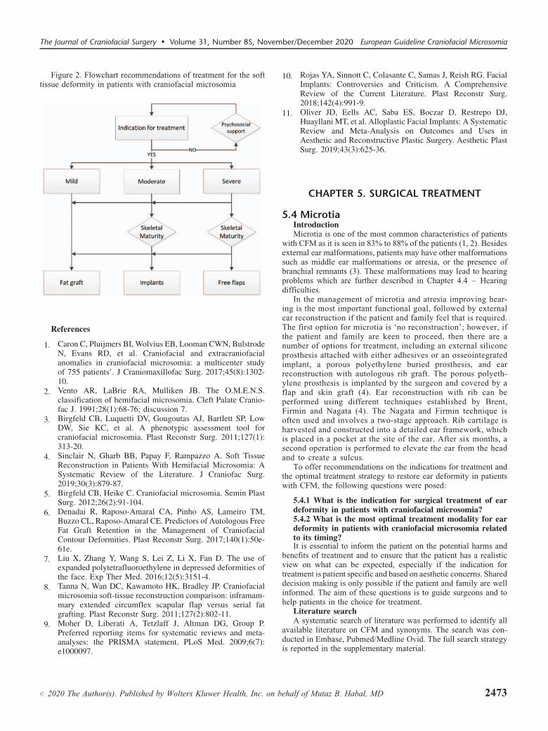
The Journal of Craniofacial Surgery � Volume 31, Number 8S, November/December 2020 European Guideline Craniofacial Microsomia
Figure 2. Flowchart recommendations of treatment for the softtissue deformity in patients with craniofacial microsomia
References
1. C
# 20
aron C, Pluijmers BI, Wolvius EB, Looman CWN, BulstrodeN, Evans RD, et al. Craniofacial and extracraniofacialanomalies in craniofacial microsomia: a multicenter studyof 755 patients’. J Craniomaxillofac Surg. 2017;45(8):1302-10.
2. V
ento AR, LaBrie RA, Mulliken JB. The O.M.E.N.S.classification of hemifacial microsomia. Cleft Palate Cranio-fac J. 1991;28(1):68-76; discussion 7.3. B
irgfeld CB, Luquetti DV, Gougoutas AJ, Bartlett SP, LowDW, Sie KC, et al. A phenotypic assessment tool forcraniofacial microsomia. Plast Reconstr Surg. 2011;127(1):313-20.4. S
inclair N, Gharb BB, Papay F, Rampazzo A. Soft TissueReconstruction in Patients With Hemifacial Microsomia: ASystematic Review of the Literature. J Craniofac Surg.2019;30(3):879-87.5. B
irgfeld CB, Heike C. Craniofacial microsomia. Semin PlastSurg. 2012;26(2):91-104.6. D
enadai R, Raposo-Amaral CA, Pinho AS, Lameiro TM,Buzzo CL, Raposo-Amaral CE. Predictors of Autologous FreeFat Graft Retention in the Management of CraniofacialContour Deformities. Plast Reconstr Surg. 2017;140(1):50e-61e.7. L
iu X, Zhang Y, Wang S, Lei Z, Li X, Fan D. The use ofexpanded polytetrafluoroethylene in depressed deformities ofthe face. Exp Ther Med. 2016;12(5):3151-4.8. T
anna N, Wan DC, Kawamoto HK, Bradley JP. Craniofacialmicrosomia soft-tissue reconstruction comparison: inframam-mary extended circumflex scapular flap versus serial fatgrafting. Plast Reconstr Surg. 2011;127(2):802-11.9. M
oher D, Liberati A, Tetzlaff J, Altman DG, Group P.Preferred reporting items for systematic reviews and meta-analyses: the PRISMA statement. PLoS Med. 2009;6(7):e1000097.20 The Author(s). Published by Wolters Kluwer Health, Inc. on b
10. R
ehalf
ojas YA, Sinnott C, Colasante C, Samas J, Reish RG. FacialImplants: Controversies and Criticism. A ComprehensiveReview of the Current Literature. Plast Reconstr Surg.2018;142(4):991-9.
11. O
liver JD, Eells AC, Saba ES, Boczar D, Restrepo DJ,Huayllani MT, et al. Alloplastic Facial Implants: A SystematicReview and Meta-Analysis on Outcomes and Uses inAesthetic and Reconstructive Plastic Surgery. Aesthetic PlastSurg. 2019;43(3):625-36.CHAPTER 5. SURGICAL TREATMENT
5.4 MicrotiaIntroductionMicrotia is one of the most common characteristics of patients
with CFM as it is seen in 83% to 88% of the patients (1, 2). Besidesexternal ear malformations, patients may have other malformationssuch as middle ear malformations or atresia, or the presence ofbranchial remnants (3). These malformations may lead to hearingproblems which are further described in Chapter 4.4 – Hearingdifficulties.
In the management of microtia and atresia improving hear-ing is the most important functional goal, followed by externalear reconstruction if the patient and family feel that is required.The first option for microtia is ‘no reconstruction’; however, ifthe patient and family are keen to proceed, then there are anumber of options for treatment, including an external siliconeprosthesis attached with either adhesives or an osseointegratedimplant, a porous polyethylene buried prosthesis, and earreconstruction with autologous rib graft. The porous polyeth-ylene prosthesis is implanted by the surgeon and covered by aflap and skin graft (4). Ear reconstruction with rib can beperformed using different techniques established by Brent,Firmin and Nagata (4). The Nagata and Firmin technique isoften used and involves a two-stage approach. Rib cartilage isharvested and constructed into a detailed ear framework, whichis placed in a pocket at the site of the ear. After six months, asecond operation is performed to elevate the ear from the headand to create a sulcus.
To offer recommendations on the indications for treatment andthe optimal treatment strategy to restore ear deformity in patientswith CFM, the following questions were posed:
5.4.1 What is the indication for surgical treatment of eardeformity in patients with craniofacial microsomia?5.4.2 What is the most optimal treatment modality for eardeformity in patients with craniofacial microsomia relatedto its timing?
It is essential to inform the patient on the potential harms andbenefits of treatment and to ensure that the patient has a realisticview on what can be expected, especially if the indication fortreatment is patient specific and based on aesthetic concerns. Shareddecision making is only possible if the patient and family are wellinformed. The aim of these questions is to guide surgeons and tohelp patients in the choice for treatment.
Literature searchA systematic search of literature was performed to identify all
available literature on CFM and synonyms. The search was con-ducted in Embase, Pubmed/Medline Ovid. The full search strategyis reported in the supplementary material.
of Mutaz B. Habal, MD 2473
Renkema et al The Journal of Craniofacial Surgery � Volume 31, Number 8S, November/December 2020
Inclusion and exclusion criteria:
Type of studies
2474
- Original articles- Systematic review of sufficient quality:
- The question in the systematic reviewmatches the question of the guideline.- The search of the systematic review wasconducted in at least two relevantdatabases, such as the Cochrane Library,Medline/Pubmed.- The full search strategy was reported.- No relevant items were missing in thesearch strategy.
Type of patients
- Patients with craniofacial microsomiaSubject
- Indication and/or treatment for earreconstructionExclusion criteria
- Original studies with <10 included patients- Articles published before 1980- Case reports- Expert opinion- Letters- Editorials- Narrative reviewsOnly literature that described indications, outcomes or compli-cations of ear reconstruction was included. The aim of this chapteris to provide patients and clinicians with guidance on the availabletreatment options for ear reconstruction in patients with CFM.Therefore, articles that studied surgical techniques in detail wereexcluded, as this is beyond the scope of this chapter.
Literature was screened on title and abstract. A total of threeretrospective cohort studies were included that addressed outcomeson ear reconstruction in patients with CFM: by Mandelbaum et al.(5), Park et al. (6), and Qian et al. (7)
The ‘UK Care Standards for the Management of Patients withMicrotia and Atresia’ (2018) was also included in this chapter (4).This UK guideline on microtia/atresia makes clinically relevantrecommendations to improve care, but is not been based on thesame methodological criteria as this guideline on CFM. Hence theUK guideline on microtia/atresia did not provide information onthe literature search, the inclusion/exclusion criteria, and quality ofevidence of the individual studies and conclusions. Nevertheless,the guideline is considered to be clinically relevant, especially sincethe general quality of evidence of literature on auricular reconstruc-tion is low (8).
An additional literature search was performed to include liter-ature on auricular reconstruction in patients with microtia or atresia.All articles that described indications, outcomes or complicationson treatment modalities for auricular reconstruction were included.The full search strategy is shown in the addendum. A total of 369articles were identified and ten articles met the inclusion criteria andwere included in this chapter. One systematic review by Long et al.was included (9), and two cross-sectional studies by Younis et al.(10) and Braun et al. (11), five retrospective cohort studies byConstantine et al. (12), Fu et al. (13), Roos et al. (14), Al Kadah et al.(15), and Si et al. (16), and two case series by Vijverberg et al. (17)and Wright et al. (18)
5.4.1 What is the indication for surgical treatment of eardeformity in patients with craniofacial microsomia?
Auricular deformities in patients with CFM are often accompa-nied by other congenital facial malformations. These deformitiescan cause aesthetic or psychosocial concerns and may be anindication for surgical reconstruction. As patients with CFM oftenhave multiple facial malformations, the indication for treatment
# 2020 The Author(s). Published
may be more complex compared to patients with isolated microtia/anotia. Therefore, only literature on patients with CFM wasincluded in this chapter.
Review of literatureThe characteristics of patients with CFM and external ear and
hearing problems were studied by Mandelbaum et al. (5) Theirretrospective analysis included 68 patients with CFM. Most of thesepatients had auricular abnormalities (91%), which were mostfrequently characterised as grade III microtia (in 75% of thepatients). Data on complications, surgical revisions, aesthetics,and psychosocial function were collected. Psychosocial problemsor deficits in school functioning were seen in 30.7% of the patients.However, no control group was included, which makes this per-centage hard to interpret. Data on psychosocial problems werebased on retrospective review of medical records and were gradedas ‘good’ or ‘poor’. Regression analysis showed that any treatmentof hearing loss (which led to significantly better hearing results aftertreatment) was a significant predictor of good psychosocial out-come (OR, 4.889; 95% CI, 1.459–16.381; p¼ 0.010). No statisti-cally significant influences of auricular reconstruction onpsychosocial functioning were found.
Conclusion
Level 3
by Wolters Kluwe
One study did not find a difference in psychosocialfunctioning after ear reconstruction in patients withcraniofacial microsomia (n¼ 68).
Ref (5)
Considerations� Overall quality of evidenceThe quality of evidence on the indication for treatment of
auricular deformity in patients with CFM was graded on level 3,since the study of Mandelbaum et al. was a retrospective cohortstudy (5). It is important to keep in mind the retrospective nature ofthe study and the imperfect method of determining psychosocialproblems (by chart review) in assessing the quality of the conclu-sion. Due to this methodological weakness, the conclusion isconsidered less strong.
� Balance of benefits and harmsMicrotia can have negative psychosocial consequences due to
aesthetic difficulties and the visibility of the disorder. Patients oftencover their disfigurement by wearing their hair down, avoiding wethair or windy weather, avoiding physical activities, social situa-tions, mirrors, photographs and so on (4). A higher prevalence ofsocial problems, aggressive behaviour, and mood disorders are seenin patients with microtia compared to healthy controls (19). Theaesthetic differences with other, ‘healthy’ persons could lead to lowself-esteem, bullying, or negative feelings. This underlines theimportance of psychosocial support for all patients with CFM,which should start during childhood. Ear reconstruction may bebeneficial for patients to feel less ‘atypical’ and prevent thepreviously mentioned potential harms.
� Outcome importanceNo literature is available on the importance of correction of the
auricular deformity in patients with CFM. Patients with CFM oftenexperience multiple facial differences compared to healthy controls.Nevertheless, it has been shown that the management of microtia or
r Health, Inc. on behalf of Mutaz B. Habal, MD
The Journal of Craniofacial Surgery � Volume 31, Number 8S, November/December 2020 European Guideline Craniofacial Microsomia
anotia has significant positive impact for the patient (20). Aestheticconcerns could lead to psychosocial problems in some patients.Auricular reconstruction may help patients feel less ‘atypical’. Toassess the benefits of ear reconstruction, the use of a new PROMEar-Q was agreed at the International Society for Auricular Recon-struction ISAR congress in Beijing in 2017. All surgeons should usethe Ear-Q pre- and post operatively.
� Costs and resourcesThe impact on costs and resources of the given recommenda-
tions will vary per member state, depending on the available careproviders and facilities. Treatment with porous polyethylene pros-thesis (Medpore) especially could contribute to an increase in costs.Health insurance reimbursement and the willingness to cover thesecosts may differ between countries. The recommendations concernthe essential requirements for adequate treatment of patients withCFM and should thus be implemented. Costs are lowest andresources are most efficiently used when care for craniofacialdisorders is centralised in a limited number of expert centres permember state. A general rule that can be applied is one expert centreper 10 million inhabitants.
� Inequity of the recommendationThe goal of the European Reference Networks (ERNs) is to
eliminate inequality within Europe with regard to care for patientswith rare diseases. At present, not every member state offers anexpert centre for CFM, or the level of provided care does not (yet)meet all the requirements that are outlined in this guideline. Bydefining the baseline of required care for CFM, this guideline willhelp these member states to reach the appropriate level. The ERN oncraniofacial anomalies and ENT disorders (ERN-CRANIO) canguide a patient in Europe to the available centres of expertise(www.ern-cranio.eu) and can support care providers with diagnosisand treatment advice.
� Feasibility of the recommendationRecommendations refer to the general requirements for deliv-
ering optimal healthcare and are discussed with members fromparticipating European countries. Quality of care was paramount inthe discussions. Centralisation is proposed as one of the core values.However, in some countries the national organisation of healthcaremight impede centralisation. National implementation of the ERNsthat fits the situation of each country is necessary. For the memberstates with the lowest number of inhabitants, the establishment of acraniofacial centre might not be feasible, and collaboration with acraniofacial centre in the surrounding countries can be an option.
� Acceptability of the recommendationIt is expected that all stakeholders want to apply and will be
applying the recommendations, because they are employed in ERN-acknowledged institutions. National implementation plans are nec-essary to ensure that recommendations fit the situation in eachcountry. In addition, not all countries participating in the ERN-CRANIO are represented in the guideline development group, andnew members will join within the coming years. For these countries,acceptance and implementation of ERN guidelines such as thisguideline on CFM is obligatory.
Rationale of the recommendationsThe indication for treatment of microtia is primarily aesthetic
but has been shown to have large psychosocial benefits in patientswith isolated microtia (21). The aim of treatment is to improve thepatient’s quality of life. Since the indication for treatment is
# 2020 The Author(s). Published by Wolters Kluwer Health, Inc. on b
aesthetic, it is essential that the patient is well-informed aboutthe potential risks of therapy. Hearing problems should be assessedand treated, as is explained in Chapter 4.4 – Hearing difficulties.Psychosocial help or therapy may help patients learn to cope withtheir different appearance. In an even earlier phase, parents ofpatients should receive information on the possible treatmentoptions for the ear deformity. The multidisciplinary craniofacialteam should assess the psychosocial problems and reconstructiveoptions in an individualised manner (4). If treatment is considered,it is essential to inform patients and parents on the expectedoutcomes and the burden of treatment such as the frequency andlength of hospital stays, and risk of complications.
Recommendations
eh
� The indication for auricular reconstruction in patientswith craniofacial microsomia is aesthetic and psycho-social. Inform the patient about the potential benefitsand harms to ensure that the patient has a realistic viewof what can be expected.
� Provide all patients with craniofacial microsomia withpsychosocial support.
� Use the PROM Ear-Q pre- and postoperatively to assess
alf of
benefit of treatment.
Research gapThere is a lack of knowledge on the effectiveness of and patients
reported satisfaction with auricular reconstruction in patients withCFM. Future research on this topic could help doctors supportpatients in the decision-making process. It is hoped that the use ofthe new PROM Ear-Q will reduce this gap.
5.4.2 What is the most optimal treatment modality for eardeformity in patients with craniofacial microsomia relatedto its timing?
The literature search for this question was directed towards morespecific research questions related to treatment options, complica-tions, advantages and disadvantages, short and long term results andtreatment burden for treatment of microtia/anotia in patients withCFM (Appendix 1, http://links.lww.com/SCS/B697). Besides lit-erature on CFM, the ‘UK Care Standards for the Management ofPatients with Microtia and Atresia’ (2018) and literature identifiedfrom the additional search on auricular reconstruction was included(4). Treatment modalities were divided into four groups: externalsilicone prosthesis attached with adhesives, external silicone pros-thesis attached on implants, porous polyethylene implants (Med-pore), and auricular reconstruction with autologous rib grafts.Included patient outcomes were (psychological) impact of thetreatment, complications and satisfaction with the result.
Review of literatureCraniofacial microsomia
Mendelbaum et al. retrospectively studied 68 patients withCFM, all age >13 (5). Microtia was present in 91% of the patients,and was characterised as type III microtia in 75% of the patients(n¼ 58). Total auricular reconstruction with rib grafts was per-formed in 56 ears, starting at a mean age of 8.5 years (range 5.5-18.7years). Aesthetic outcomes were assessed by three independentobservers on a scale of 1 to 3. The mean aesthetic outcome scorewas 5.56 (range 3-9). Patients with severe microtia required moresurgeries to achieve optimal results and had lower aesthetic out-come ratings after reconstruction compared to less severe cases(respectively IRR, 4.350; 95% CI, 2.140–8.843; p < 0.001, and
Mutaz B. Habal, MD 2475
Renkema et al The Journal of Craniofacial Surgery � Volume 31, Number 8S, November/December 2020
IRR, 0.763; 95% CI, 0.658–0.886; p < 0.001). Complicationsoccurred in 18% of the patients and were all secondary to woundcomplications. Most patients (n¼ 46) needed revisions after com-pletion of ear reconstruction such as deepening of the helical root,tragus, or concha, revision of the lobule, or projection of the newear. No correlation between the complication rate and the severityof microtia was observed (p¼ 0.193).
Microtia in patients with CFM is difficult to treat due toadditional abnormalities such as facial asymmetry, a low hairline,depression of the temporomandibular joint or temporal bone, andlow ear vestige. These characteristics can lead to a deficiency in theskin volume, making covering of the auricular reconstruct difficult(6). Three types of coverage techniques in microtia reconstructionin patients with CFM were studied by Park et al., who included 52patients with CFM with a mean follow-up of 40 months (range6 months to 10 years) (6). All patients had auricular reconstructionwith rib and were treated with three types of coverage techniques.Results on the comparison of these coverage techniques wereconsidered to be too surgically detailed for this chapter and werenot included. However, results on aesthetic outcomes and compli-cations were considered relevant and extracted from the article. Theaesthetic outcome of treatment was rated on a 1–10 scale, based onsenior author assessment. The mean aesthetic score varied from6.30 to 8.26. Poor postoperative outcomes were due to large size ofthe reconstructed ears (n¼ 11), partial resorption of the cartilageframework (n¼ 11), mismatched skin colour (n¼ 8), different earaxis (n¼ 7) or shape (n¼ 5), ear convolution (n¼ 2), or bad-lookingscars (n¼ 2). The severity of microtia had no effect on the aestheticoutcome (6).
Qian et al. studied 111 patients with CFM to study the effect ofthe expanded two-flap method (7). Results on this specific surgicaltechnique were considered to be too detailed for this chapter andwere not included. General outcomes of ear reconstruction with ribgraft were included. The mean age of the patients was 9 years (range5 to 27 years) and mean follow-up was 8.3 months (range 5 to 20months). Aesthetic outcomes of treatment were rated on a 0–15scale, and were satisfactory in 92.8% of the patients, partiallysatisfactory in 6.3% of the patients, and unsatisfactory in onepatient (0.9%). Nine patients (8.1%) had surgical complications:tissue expander-related complications and hematoma (1.8%). Threepatients had complications in the second treatment stage: cartilageframework exposure in two patients (1.8%) and poor skin graftsurvival in one patient (0.9%).
External silicone prosthesis attached with adhesivesThe UK Guideline Microtia and Anotia considers external
silicone ear prosthesis as a good treatment modality for microtia,which can achieve an excellent shape and colour (4). The prosthesismay be attached with adhesives or with implants. No literature onthe outcomes or complications of treatment with external siliconeprosthesis with adhesives was identified. The benefits and disad-vantages of the use of this type of external prosthesis are discussedin the considerations.
External silicone prosthesis attached onosseointegrated implants
The additional search on non-CFM patients with microtia led tothe inclusion of five articles on external prosthesis. Vijverberg et al.studied the outcomes and patient satisfaction of osseointegratedauricular (VXI) implants (17). A total of 31 implants were placed in11 patients, with a mean age of 44 years (range 13–85 years) and aminimal follow-up of 7 months (mean follow-up 2.6 years). Softtissue reaction was observed in 32.2% of the implants, which allresolved after topical treatment with antibiotic/corticosteroid
2476 # 2020 The Author(s). Published
ointment. All patients reported an improvement in quality of lifeafter receiving the implants with prosthesis, although two patients(18%) reported a slight physical disadvantage using the prosthesis(17). Si et al. also reported the troublesome complication of skinreaction (16). All their studied patients (n¼ 24, 25 ears) whoreceived osseointegrated auricular implants for an external pros-thesis had skin reaction. The degree of skin redness, moist, orgranulation tissue gradually decreased over time. Almost allpatients (95.8%) wore the prosthesis for more than eight hoursper day (16).
Wright et al. studied the survival rate of auricular implants in 16patients and 39 implants. The average age at placement was 40 years(range 6 to 76 years) and the mean follow-up was 45 months (range6 months to 17 years). The survival rate was 100% as no implantfailures occurred. Most patients (n¼ 13) had soft tissue inflamma-tion, which resolved in 54% of the patients with hygiene support orsoft tissue reduction (18).
Younis et al. examined the patient satisfaction in a cross-sectional study of 20 patients who completed prosthetic ear recon-struction (average age 37 years, range 17–56 years) (10). Theindication for ear prosthesis was due to a congenital deformity(n¼ 10), trauma (n¼ 9) or malignancy (n¼ 1). Most patients(n¼ 14) had previous failed autologous reconstruction. Skin pro-blems, such as granulation tissue (n¼ 14) or skin infection (n¼ 10),were often reported. Nevertheless, 14 patients wore the prosthesisdaily. The aesthetic appearance of the prosthesis was consideredexcellent or very good by 17 of the 20 patients. Only 12 of the 20patients were satisfied with their prosthesis. The authors considerthe high rate of skin complications to be the reason for the lowoverall satisfaction rate (10).
Osseointegrated implant-supported externalsilicone prosthesis versus porous polyethyleneimplants (Medpore)
Al Kadah et al. reported their experience with silicone or porouspolyethylene ear prosthesis in 39 patients (43 implants) with aminimal follow-up of two years (15). Eighteen patients had porouspolyethylene implants placed (mean age 19 years, range 8–56years) and 21 patients had silicone ear protheses fixed on implants(mean age 41 years, range 6–84 years). None of the 18 patients witha porous polyethylene implant saw the implant rejected, althoughfour patients needed revisions due to keloid (n¼ 1), retroauricularadhesion (n¼ 2), and local infection leading to partial resection ofthe implant (n¼ 1). In the silicone prosthesis group, 18 patients(85%) wore the prosthesis for at least 10 hours a day. No extrusionof the implants for the external prosthesis was observed. Revisionsurgeries were needed in five patients due to: exposure of theimplants (n¼ 2), infection (n¼ 1), loose screw (n¼ 1), tumourrecurrence (n¼ 1). Local skin reaction at the abutment was seenin 71.4% of the patients (n¼ 15) during follow-up. No skin reac-tions in the porous polyethylene implant group were reported.
Porous polyethylene implants (Medpore)Porous polyethylene implants could yield good aesthetic results
with lower burden for the patient compared with autologousreconstruction, although complications such as extrusion or fractureof the implant could be seen in up to 13–15% of the patientsaccording to the UK microtia/anotia guideline (4). Unfortunately,no literature was provided to validate these numbers and themethodological quality of the articles.
In the study by Al Kadah et al. (described above), no extrusionof the alloplastic implants was observed, although one of theeighteen patients needed partial resection of the implant after localinfection (15).
by Wolters Kluwer Health, Inc. on behalf of Mutaz B. Habal, MD
The Journal of Craniofacial Surgery � Volume 31, Number 8S, November/December 2020 European Guideline Craniofacial Microsomia
Braun et al. studied the patient-reported outcome after ear recon-struction with porous polyethylene implants (Medpore) in a cross-sectional study (11). A total of 65 patients were included, who allreceived a questionnaire. Most patients (84.6%) were treated due tocongenital microtia and 15 patients were found to have CFM. Themedian age at time of treatment was 18 years (range 4-68 years), andthe mean follow-up was 2.9 years (range 0.5–6.0 years). Adultpatients (n¼ 45) reported an overall increase in quality of life afterauricular reconstruction (p<0.0001). Most of these patients (72.7%)were satisfied with the aesthetic outcome. Of the 27.3% (n¼ 12) ofthe patients who were not satisfied, eight would again decide to havethe operation. A negative score, indicating an adverse effect, wasreported by 17.8% of the adults. Most frequently reported complaintsby patients include difficulties with scars (n¼ 18, 40%), feeling(n¼ 17, 37%), shape of the ear (n¼ 16, 36%), numbness (n¼ 11,24%), skin colour (n¼ 8, 18%) or position of the ear (n¼ 4, 9%). Nofailures leading to removal of the prosthesis were reported. Two thirdsof the patients were not limited in their physical activities due to thereconstructed ear and 62.8% of the patients could sleep on thereconstructed ear (11). Similar results were seen in the paediatriccohort (n¼ 20). An increase in the health-related quality of life wasobserved (p¼ 0.01). Most patients (85%) and parents of patients(73.7%) were satisfied with the aesthetic results, and all patients andparents who were not satisfied would again decide to have theoperation. The main complaints were similar to the adults, includingscars and feeling. Complaints regarding the position, shape andcolour were less frequently reported compared to the adult population(no numbers were reported). One patient psychologically rejected thenew ear. Most children had no physical limitations (75%) and couldsleep on the reconstructed ear (85%) (11).
Porous polyethylene implants (Medpore) versus earreconstruction with autologous rib graft
The outcomes of porous polyethylene implants and autologous earreconstruction with rib were compared by Constantine et al (12).Their retrospective analysis included 35 patients (36 ears) who hadcomplete microtia repair: 17 polyethylene implants (mean age 6.9years, SD 3.0), 17 reconstructions with rib (mean age 8.0 years, SD3.8), and 2 with both. The follow-up ranged from 2 to 11 years. Twoblinded observers rated the aesthetic outcomes of treatment. Themean number of operations was higher in the rib reconstruction group(4.88, SD 1.54) compared to the polyethylene implant group (3.35,SD 1.27, p¼ 0.004). One complication occurred in the rib recon-struction group: cartilage exposure. Two complications occurred inthe implant group: one infection and one extrusion of the implant.And in the two patients who had both types of reconstruction, twoinfections and two extrusions of the polyethylene implants occurred.A trend toward more infections and extrusions in the polyethylenegroup compared to the rib reconstruction group may be present.Concerning the aesthetic outcomes, the rib reconstruction group hadsignificantly better outcomes regarding the definition, shape, andcolour match compared to polyethylene implants (p¼ 0.05, p¼ 0.08,p¼ 0.05 respectively). The size of the ear was better in the implantgroup (p¼ 0.05). No statistically significant differences in ear pro-trusion and location were observed.
Ear reconstruction with autologous rib graftEar reconstruction with autologous rib cartilage has been con-
sidered the golden standard in microtia reconstruction for years (4).In the first operation, performed at age nine to ten, the rib cartilage isharvested and constructed into a framework, which is then placed ina pocket at the site of the ear. Six months later, the second procedureis performed in which the ear is elevated from the head. Thisreconstruction is considered to be durable, showing no softening or
# 2020 The Author(s). Published by Wolters Kluwer Health, Inc. on b
shrinkage of the cartilage and having minimal long-term complica-tions (4). The newly reconstructed ears with rib cartilage were foundto grow as much as normal ears after reconstruction (after a medianfollow-up of 5.6 years, range 2.1–10.6 years) (14).
A systematic review on the complications of autologous ear recon-struction with rib was conducted by Long et al. in 2013 (9). A total of 60articles, including 9,415 patients, were included in the analysis. Theoverall complication incidence was 16.2% (range 0–72.9%). Compli-cations included: dissatisfying final results (8.5%), hypertrophic scar(6.3%), asymmetry (2.1%), wire or suture extrusion (1.6%), flap venouscongestion (1.6%), cartilage absorption (1.3%), facial nerve injury(1.1%), frame exposure (1.0%), infection (0.9%), hematoma (0.3%),and grafted skin necrosis (0.4%). Leakage or exposure of the tissueexpander has an incidence of respectively 1.4% and 3.5%. Complica-tions at the donor site included atelectasis, pleural tear, chest walldeformity, thoracic scoliosis, and hypertrophic scar. The overall inci-dence of these complications varied from 5.6% to 36.1%.
A recent retrospective cohort study of 470 ear reconstructions withrib grafts by Fu et al. (13) reported a complication rate of 9.43%. Allpatients included (n¼ 429) were treated in the same centre, by thesame surgeon. The mean follow-up was 3.7 years (�2.5 years). Age,sex, and laterality were not associated with the complications(p>0.05). The risk of complications was significantly higher inpatients treated with the Nagata technique (12.2%, 38/311) comparedwith the Brent technique (2.98%, 4/134) (OR 6.14, 95% CI 1.63–23.19; p< 0.01). The authors suggest that this is presumably the resultof a more complicated and stereoscopic technique in the Nagatatechnique, with a higher risk for vascular compromise of the skin flapcompared to the Brent technique (13). Despite the higher risk ofcomplications when using the Nagata technique, the authors advisethe use of this technique since it requires fewer surgical stages (twostages) compared to the Brent technique (three to four stages).
Conclusions
Level 3
ehalf of Muta
External silicone prosthesisNon-craniofacial microsomia patients who wear an
osseointegrated implant- supported external siliconeprosthesis are often satisfied with the result (placed at amean age of 37–44 years).
Implants for an external prosthesis often lead to local skinreactions (in 32–100% of patients), which may decreasethe satisfaction with the prosthesis. Treatment withantibiotic/corticosteroid ointment or increased hygiene isoften successful.
No conclusion on revision- or failure rates of implants can bedrawn, as studies were based on small patient sampleswith limited follow-up.
Implants for external silicone prosthesis were placed at amean age of 19–44 years (range 8–85 years) in non-craniofacial microsomia patients.
Ref (4, 10, 15–18)
Level 3
Porous polyethylene implants (Medpore) (inserted at meanage of 6–18)Most patients (73%) report an increase in quality of life andare satisfied with the aesthetic result (73–75%) after earreconstruction with porous polyethylene implants.
Dissatisfying results were mainly due to complaintsregarding scars, hair loss, and numbness.
Extrusion or fracture of the implant was seen in up to 13 -15% of patients.
Polyethylene implants have better aesthetic outcomesregarding the size of the ear compared to reconstructionwith rib, whereas rib-reconstructed ears show betteroutcomes regarding definition, shape, and colour match.
Ref (4, 11, 12, 15)
z B. Habal, MD 2477
Renkema et al The Journal of Craniofacial Surgery � Volume 31, Number 8S, November/December 2020
Level 3
2478
Ear reconstruction with rib in patients with craniofacialmicrosomia
Patients with craniofacial microsomia and severemicrotia required more operations to achieve optimalresults compared to patients with less severe microtia.
Evidence on the effect of the severity of microtia on theaesthetic outcome of ear reconstruction in patientswith craniofacial microsomia is contradictory and noconclusions can be drawn.
Reasons for poor postoperative outcomes after earreconstruction in patients with craniofacialmicrosomia include: mismatch in size, skin colour, earaxis, shape, or partial resorption of the cartilageframework.
Ref (5, 6)
Level 3
Covering techniques for ear reconstruction with rib inpatients with craniofacial microsomiaVarious covering techniques can be used for earreconstruction in patients with craniofacialmicrosomia:- The embedded technique is associated with goodaesthetic outcomes.- The fascia flap technique is associated with goodaesthetic outcomes and is often used in patients withsevere craniofacial microsomia. A mismatch in skincolour is more frequently observed in patients treatedwith this technique compared to others.- The expansion technique is associated with goodaesthetic outcomes, but has worse outcomes comparedto the other techniques.
Ref (6, 7)
Level 3
Ear reconstruction with ribEar reconstruction with rib is considered a durabletreatment option with minimal long-termcomplications. The reconstructed ears show a similargrowth pattern to the normal ear.
Ear reconstruction with rib is usually performed at ageeight to ten.
The overall complications incidence of ear reconstructionwith rib is 16%.
Reported complications include dissatisfying final results(9%), hypertrophic scar (6%), asymmetry (2%), wireor suture extrusion (2%), flap venous congestion (2%),cartilage absorption (1%), facial nerve injury (1%).
Donor site complications include pain, atelectasis, pleuraltear, chest wall deformity, thoracic scoliosis, andhypertrophic scar.
Ref (4, 9, 13, 14)
Considerations� Overall quality of evidenceThe levels of evidence were considered to be level 3 due to the
non-comparative nature of most studies. The systematic review byLong et al. on complications of ear reconstruction with ribexcluded case reports and case series with fewer than 15 patients.This study is descriptive and non-comparative, leading to a level 3quality of evidence. Al Kadah et al. and Constantine et al. made aretrospective comparison between two groups: polyethyleneimplants versus reconstruction with rib (12) and polyethyleneimplants versus silicone prosthesis (15). Park et al. retrospectivelycompared three different covering methods for the reconstructedear (6). Although these studies are comparative, the level ofevidence is graded as level 3 since the studies were not randomised
# 2020 The Author(s). Published
and no other comparative studies could be included that supportedthe conclusions.
� Balance of benefits and harmsExternal silicone prosthesis on adhesivesThe prosthesis can be attached with adhesives at a young age,
although this is controversial. Early use of the prosthesis to letthe child get used to it, could make the child feel less-consciousof the prosthesis when school starts. However, no evidence isavailable that supports this theory. Additionally it may preventteasing, although no literature is available to support this theoryeither. Early treatment is likely to be a reaction to parentalanxiety rather than clinical need. The use of an external pros-thesis in young children could also imply that the deformed earneeds to be camouflaged and could lead to the association ofnegative feelings (4).
External silicone prosthesis on implantsAdvantages of an external silicone prosthesis are the realistic
looks and its relatively simple use/placement. Disadvantagesinclude the need for replacement of the implants, cleaning of thepin sites, risk of losing the prosthesis during physical activities, skincolour changes, or problems with the implants (e.g. infection orover-granulation) (4).
If implants are placed for prosthesis attachment, the microticremnant often needs to be removed. It is important that the patientand family are aware that other forms of ear reconstruction areimpaired after this treatment (4).
Alloplastic implantsReconstruction with Medpore implants is quicker and has a
lower burden for the patient compared to autologous ear recon-struction, as no rib cartilage has to be harvested using this technique.In addition, reconstruction with Medpore is less technically chal-lenging compared to autologous reconstruction, although technicalexpertise is still required to achieve good results (4).
Medpore implants can be placed at a young age (three to fiveyears), although the size of the implants has to be bigger toaccount for future growth of the contralateral ear (4). At this age,the child is not aware of its facial differences, which makes theindication for this invasive treatment debatable. Also, if animplant is placed, future autologous reconstruction with rib isimpaired given the scarring of the local soft tissues. Anotherimportant aspect is the risk of complications, especially if theimplant is placed in children and if the child engages in contactsports. The complications, such as extrusions or fractures of theimplants, are serious and make future reconstruction with othermethods challenging. Therefore, it is advised to be cautious intreating children with Medpore implants. If reconstruction withMedpore is indicated, it is advised to postpone treatment untilafter the age of six since at this age the width of the normal ear isapproaching its mature size (22).
Ear reconstruction with ribThe aesthetic results of ear reconstruction with autologous rib
grafts are considered to be good in the hands of experiencedsurgeons (4, 12). The main benefit of this treatment is the long-term outcome. The cartilage responds naturally to trauma and nochanges in the cartilage over time are observed. Long-term com-plications are minimal, leading to lower costs and burden for thepatient compared to other techniques.
The surgical technique of ear reconstruction with rib grafts istechnically challenging with a steep learning curve. Treatment byspecialised surgeons who perform this type of surgery on a regularbasis is preferred to achieve the best results. Other drawbacks of thissurgery include the burden for the patient, which is higher comparedto other treatments due to harvesting the rib graft (4, 5).
by Wolters Kluwer Health, Inc. on behalf of Mutaz B. Habal, MD
The Journal of Craniofacial Surgery � Volume 31, Number 8S, November/December 2020 European Guideline Craniofacial Microsomia
� Outcome importanceThe choice for one of the treatment options for ear recon-
struction is based on the potential outcomes, benefits and harmsof treatment. Patients and parents should be able to make theirchoice based on the available information and weigh the advan-tages and disadvantages of treatment options and should besupported if they decide that no reconstruction is the correctoption for them. The conclusions and recommendations in thischapter aim to help for patients and doctors in the process ofchoosing a type of treatment.
� Costs and resourcesThe impact on costs and resources of the given recommenda-
tions will vary per member state, depending on the available careproviders and facilities. Treatment with porous polyethylene pros-thesis (Medpore) could contribute to an increase in costs. Healthinsurance reimbursement and the willingness to cover these costsmay differ between countries. Costs are lowest and resources aremost efficiently used when care for craniofacial disorders is cen-tralised in a limited number of expert centres per member state. Ageneral rule that can be applied is one expert centre per 10million inhabitants.
� Inequity of the recommendationThe goal of the European Reference Networks (ERNs) is to
eliminate inequality within Europe with regard to care for patientswith rare diseases. At present, not every member state offers anexpert centre for CFM, or the level of provided care does not (yet)meet all the requirements that are outlined in this guideline. Bydefining the baseline of required care for CFM, this guideline willhelp these member states to reach the appropriate level. The ERN oncraniofacial anomalies and ENT disorders (ERN-CRANIO) canguide a patient in Europe to the available centres of expertise(www.ern-cranio.eu) and can support care providers with diagnosisand treatment advice.
� Feasibility of the recommendationRecommendations refer to the general requirements for deliv-
ering optimal healthcare and are discussed with members fromparticipating European countries. Quality of care was paramount inthe discussions. Centralisation is proposed as one of the corevalues. However, in some countries the national organisation ofhealthcare might impede centralisation. National implementationof the ERNs that fits the situation of each country is necessary. Forthe member states with the lowest number of inhabitants, theestablishment of a craniofacial centre might not be feasible, andcollaboration with a craniofacial centre in the surrounding coun-tries can be an option.
� Acceptability of the recommendationIt is expected that all stakeholders want to apply and will be
applying the recommendations, because they are employed in ERN-acknowledged institutions. National implementation plans are nec-essary to ensure that recommendations fit the situation in eachcountry. In addition, not all countries participating in the ERN-CRANIO are represented in the guideline development group, andnew members will join within the coming years. For these countries,acceptance and implementation of ERN guidelines such as thisguideline on CFM is obligatory.
Rationale of the recommendationsThere is no strongly preferred treatment option to treat
microtia in patients with CFM. The selected treatment should
# 2020 The Author(s). Published by Wolters Kluwer Health, Inc. on b
be based on patients’ preferences, which is only possible if thesurgeon informs the patient (and parents) on all the advantagesand disadvantages of the various options. Autologous reconstruc-tion with rib grafts is considered the most durable treatmentoptions with good long-term outcomes. This is especially rele-vant in patients with congenital microtia, since these patientsneed a life-long treatment solution. The timing of treatmentdepends on the psychosocial development of the child, theavailability of cartilage for reconstruction with rib, the size ofthe natural ear, and the ability of the child to be part of thedecision-making process and to cooperate with the surgicalmanagement. Ear reconstruction with an autologous rib graftcan be performed from the age of eight since by then enoughcartilage can be harvested to reconstruct the ear. It is advised toplace Medpore implants after the age of six, because by then, thewidth of the ear approaches its mature size (22). Treatment withexternal silicone prosthesis attached with adhesives may beinitiated in children. However, treatment at a young age isdiscouraged since this emphasises that the child is ‘differentfrom normal’. Treatment of the ear deformity at a young age istherefore often done to comfort the parents, which makes theindication for treatment debatable. Their worries about theirchild being bullied should be taken seriously and support shouldbe provided, but surgery will not be the solution. Psychosocialsupport by a psychologist prior to and after treatment is impor-tant, especially in patients with aesthetic concerns. Given all theadvantages and disadvantages of all options, ear reconstructionwith rib grafts is considered to be the gold standard. An externalprosthesis on osseointegrated implants is ideally used as asecondary option for salvage cases. Treatment should be dis-cussed within the multidisciplinary team to ensure optimaltiming in the planning of other surgeries. Figure 1 shows aflowchart of the recommendations on the various treatmentmodalities that can be performed to treat microtia in patientswith CFM.
Recommendations
eh
� Patients should be treated within a multidisciplinaryteam setting.
� Discuss the advantages and disadvantages of the varioustreatment modalities with the patient and base thechoice for treatment on patients’ preferences.
� Psychological input is required pre- and postoperativelyto monitor expectation and acceptance.
� Ear reconstruction with rib grafts is the first choiceof treatment.
� Perform ear reconstruction with rib from the age ofeight onwards.
� Treatment before the age of eight is not recommended,but if chosen, use external silicone prosthesis attachedwith adhesives.
� If chosen, place polyethylene implants (Medpore) fromthe age of six onwards.
� Osseointegrated implants are an option for salvageprocedures.
� Outcome measures should be obtained pre- and
alf of
postoperatively with all techniques and interventions.
Research gapMeasurement of an agreed set of outcomes should be done
before and after surgery in order to assess and compare the benefitof the procedure to allow benchmarking.
Mutaz B. Habal, MD 2479

Renkema et al The Journal of Craniofacial Surgery � Volume 31, Number 8S, November/December 2020
Figure 3. Flowchart of recommendations for treatment of micro-tia in craniofacial microsomia
References
1. C
248
aron C, Pluijmers BI, Wolvius EB, Looman CWN, BulstrodeN, Evans RD, et al. Craniofacial and extracraniofacialanomalies in craniofacial microsomia: a multicenter studyof 755 patients’. J Craniomaxillofac Surg. 2017;45(8):1302-10.
2. T
uin J, Tahiri Y, Paliga JT, Taylor JA, Bartlett SP.Distinguishing Goldenhar Syndrome from CraniofacialMicrosomia. J Craniofac Surg. 2015;26(6):1887-92.3. B
irgfeld CB, Heike C. Craniofacial microsomia. Semin PlastSurg. 2012;26(2):91-104.4. U
K Care Standards for the Management of Patients withMicrotia and Atresia.5. M
andelbaum RS, Volpicelli EJ, Martins DB, Park SH, DubinaE, Ishiyama A, et al. Evaluation of 4 Outcomes Measures inMicrotia Treatment: Exposures, Infections, Aesthetics, andPsychosocial Ramifications. Plast Reconstr Surg Glob Open.2017;5(9):e1460.6. P
ark JY, Park C. Microtia Reconstruction in HemifacialMicrosomia Patients: Three Framework Coverage Techni-ques. Plast Reconstr Surg. 2018;142(6):1558-70.7. Q
ian J, Li Z, Liu T, Zhou X, Zhang Q. AuricularReconstruction in Hemifacial Microsomia with an ExpandedTwo-Flap Method. Plast Reconstr Surg. 2017;139(5):1200-9.8. L
i Y, Zhang R, Li D. A Systematic Review of Levels ofEvidence in Auricular Defects Literature: How Far Has ItBeen During the Past Three Decades? J Craniofac Surg.2015;26(7):2062-6.9. L
ong X, Yu N, Huang J, Wang X. Complication rate ofautologous cartilage microtia reconstruction: a systematicreview. Plast Reconstr Surg Glob Open. 2013;1(7):e57.10. Y
ounis I, Gault D, Sabbagh W, Kang NV. Patient satisfactionand aesthetic outcomes after ear reconstruction with aBranemark-type, bone-anchored, ear prosthesis: a 16 yearreview. J Plast Reconstr Aesthet Surg. 2010;63(10):1650-5.11. B
raun T, Gratza S, Becker S, Schwentner I, Stelter K,Patscheider M, et al. Auricular reconstruction with porouspolyethylene frameworks: outcome and patient benefit in65 children and adults. Plast Reconstr Surg. 2010;126(4):1201-12.12. C
onstantine KK, Gilmore J, Lee K, Leach J, Jr. Comparison ofmicrotia reconstruction outcomes using rib cartilage vs porouspolyethylene implant. JAMA Facial Plast Surg. 2014;16(4):240-4.0 # 2020 The Author(s). Published
13. F
by W
u YY, Li CL, Zhang JL, Zhang TY. Autologous cartilagemicrotia reconstruction: Complications and risk factors. Int JPediatr Otorhinolaryngol. 2019;116:1-6.
14. R
oos EW, S.O. Oberg, M. Growth of ears reconstructed fromautologous rib cartilage. Eur J Plast Surg. 2015(38):443–8.15. A
l Kadah B, Naumann A, Schneider M, Schick B,Linxweiler M, Papaspyrou G. Auricular reconstructionwith polyethylene implants or silicone prosthesis: A singleinstitution experience. J Craniomaxillofac Surg. 2018;46(12):2150-6.16. S
i Y, Fan SC, Sun W, Chen YB, Zhang ZG. Osseointegrationtechnique in patients with acquired auricular deformities andfailed previous reconstruction: a retrospective study of long-term follow-up and Chinese experience. ORL J Otorhinolar-yngol Relat Spec. 2012;74(3):129-35.17. V
ijverberg MA, Verhamme L, van de Pol P, Kunst HPM,Mylanus EAM, Hol MKS. Auricular prostheses attached toosseointegrated implants: multidisciplinary work-up andclinical evaluation. Eur Arch Otorhinolaryngol. 2019;276(4):1017-27.18. W
right RF, Zemnick C, Wazen JJ, Asher E. Osseointegratedimplants and auricular defects: a case series study. JProsthodont. 2008;17(6):468-75.19. L
i D, Chin W, Wu J, Zhang Q, Xu F, Xu Z, et al. Psychosocialoutcomes among microtia patients of different ages andgenders before ear reconstruction. Aesthetic Plast Surg.2010;34(5):570-6.20. A
kter F, Mennie JC, Stewart K, Bulstrode N. Patient reportedoutcome measures in microtia surgery. J Plast ReconstrAesthet Surg. 2017;70(3):416-24.21. S
oukup B, Mashhadi SA, Bulstrode NW. Health-relatedquality-of-life assessment and surgical outcomes for auricularreconstruction using autologous costal cartilage. PlastReconstr Surg. 2012;129(3):632-40.22. F
arkas LG, Posnick JC, Hreczko TM. Anthropometric growthstudy of the ear. Cleft Palate Craniofac J. 1992;29(4):324-9.CHAPTER 6. ORGANISATION OF CARE
Minimal care standards and monitoringoutcomes
IntroductionCraniofacial microsomia (CFM) is a condition involving various
congenital disorders which requires the involvement of multiplehealthcare professionals. A multidisciplinary team is needed toprovide and align the complex, long-lasting care for patientswith CFM.
Since CFM is a relatively rare congenital disorder, with anincidence of 1:3,000 to 1:5,000 live births, centralisation of care inspecialised craniofacial centres is preferred since this makes itpossible to build experience in treating these patients, improvethe quality of care, and to perform clinically relevant scientificresearch.
Multidisciplinary care requires good coordination and commu-nication among healthcare professionals, but also with patients andparents of patients. The responsibility and division of tasks of allprofessionals should be clear for all team members. Centralisationof care and formation of multidisciplinary craniofacial teams makesit possible to perform comparative studies between centres. Com-paring outcomes of the various craniofacial centres would have apositive effect on the quality of care.
To make recommendations on the minimal care standards for thetreatment of patients with CFM, the following question was posed:
olters Kluwer Health, Inc. on behalf of Mutaz B. Habal, MD

The Journal of Craniofacial Surgery � Volume 31, Number 8S, November/December 2020 European Guideline Craniofacial Microsomia
# 2
3.1 What are the minimal care standards to treat patientswith craniofacial microsomia and how should outcomes ofcare be monitored?
Defining the minimal care standards to treat patients with CFMis important to guarantee good quality of treatment for all patientswith CFM. Patients of the hospitals participating in the ERN-CRANIO were asked to identify current problems in the careprocess (patient bottlenecks). Patients reported problems in gettingreferred to a specialised centre, the lack of a multidisciplinaryapproach and long travel distance. Additionally, patients advisedappointing a care provider as point of contact within the craniofacialteam, who can be contacted by patients, parents of patients andhealthcare professionals.
Literature searchA systematic search of literature was performed to identify all
available literature on CFM and synonyms. The search was con-ducted in Embase, Pubmed/Medline Ovid. The full search strategyis reported in the supplementary material.
Inclusion and exclusion criteria:
Type of studies
020 The Author(s). Pu
- Original articles- Systematic review of sufficient quality:
- The question in the systematic reviewmatches the question of the guideline.- The search of the systematic review wasconducted in at least two relevantdatabases, such as the Cochrane Library,Medline/Pubmed.- The full search strategy was reported.- No relevant items were missing in thesearch strategy.
Type of patients
- Patients with craniofacial microsomiaSubject
- Organisation of care- Patient reported bottleneck in care process- Outcome measuresExclusion criteria
- Original studies with <10 included patients- Articles published before 1980- Case reports- Expert opinion- Letters- Editorials- Narrative reviewsLiterature was screened on title and abstract. One article, byJohns et al., was included which discussed the experiences ofpatients with CFM during medical care (1).
6.1 What are the minimal care standards to treat patientswith craniofacial microsomia and how should outcomes ofcare be monitored?
The literature search for this chapter was directed towardsbottlenecks mentioned by patients regarding communication, infor-mation, referral, and collaboration. Results of the bottleneck anal-ysis that was performed by the authors of this guideline aredisplayed in the appendix. The review of literature and conclusionswill be based on the article of Johns et al (1). The considerations andrecommendations in this chapter will be based on both expertopinion, patient preferences and literature.
Review of literatureJohns et al. approached patients with CFM and caregivers of
patients with CFM from the USA to share their views and experi-ences regarding their situation at home, school, community andmedical care. A total of 42 caregivers and 9 adult patients were
blished by Wolters Kluwer Health, Inc. on b
included. When asked about the difficulties in medical care, 25%of the caregivers reported a lack of knowledge about CFM amonghealthcare professionals along the whole healthcare pathway.Additionally, 16% of the caregivers and 20% of the patientsreported that there was little guidance during treatment. Thecoordination of care was troublesome for some patients: 13% ofthe caregivers reported difficulties accessing treatment; unfortu-nately it was not clarified what the reasons for these difficultieswere (e.g. organisational or insurance problems). Another 13% ofthe patients received conflicting medical recommendations, and9% found it difficult to coordinate care with the multiplehealthcare providers.
A third of the caregivers and half of the adult patients suggestedimproving treatment communication and coordination during thecare process. Caregivers recommended providing clear informationabout the diagnosis CFM (16%) and stated they would like to bemore informed about CFM (13%). Appropriate referral to specialistcare (13%), parent support (10%), help with insurance coverage(6%) and support of patient organisations (17%) were other issuesthat caregivers and patients mentioned. A patient-centred approachand involving the family in decision making regarding treatmentwas recommended by the patients.
Conclusions
Level 3
ehalf of Mutaz B
The most frequently reported difficulties in care forpatients and parents of patients with craniofacialmicrosomia include healthcare professionals’ lack ofknowledge regarding the diagnosis craniofacialmicrosomia, lack of guidance during treatment andunclear coordination of care.
Ref (1)
Level 3
Communication about and coordination of the treatmentduring the care process should be improved tooptimise treatment for patients with craniofacialmicrosomia. A patient-centred approach andinvolvement of the family is recommended bypatients.Ref (1)
Considerations� Overall quality of evidenceThe quality of evidence of the conclusions was graded as level 3
since the study by Johns et al. is a cross-sectional, non-comparable study.
� Balance of benefits and harmsInformationPatients mentioned that they would prefer to have more knowl-
edge about the condition, about possible treatments, and aboutspecialised professionals and institutions in their own or neighbour-ing country. Information about CFM is scarce in most countries. Inaddition, patients mentioned that they were forced to find their owninformation on the internet and in books. CFM might result inlifelong medical treatment or disability of the patient. Information israrely available throughout the healthcare process. Similarly, doc-tors in urban hospitals are not always aware where CFM is besttreated in their country. Clear protocols and patient informationare necessary.
. Habal, MD 2481
Renkema et al The Journal of Craniofacial Surgery � Volume 31, Number 8S, November/December 2020
Developing information for patients, parents and professionalstakes effort, but it will eventually help both patients, parents andprofessionals. This guideline will point out gaps in knowledge onwhich further research can focus.
CommunicationPatients mentioned that different teams in various hospitals
might come up with different treatment options. This is quitedifficult for patients and parents to understand. Patients prefer tohave a contact person each centre; a care coordinator who willfunction as a point of reference for the patient, parents and profes-sionals and will clarify and facilitate communication betweendifferent institutions and within her/his own institution. This rolecould be played, for example, by a (specialist) nurse.
Communication between and within teams is necessary tofacilitate the best possible treatment and help patients and parentsin the best way possible. This might require an extra effort from theteam but it will make communication for parents, patients andprofessionals much better.
ReferralPatients mentioned that they were often referred after many
months. In addition, there was unclearness about the city or countryof specialists. Timely referral to the correct centre of expertisefacilitates the whole healthcare process for patients and parents.Optimal referral will result in less overtreatment, receiving thewrong treatment, and less waiting time for the patient and theprofessional. It will make the healthcare path smoother and willresult in less unnecessary healthcare.
Collaboration and conditionsPatients mentioned that it sometimes felt as if people from
different disciplines worked individually instead of functioningas a multidisciplinary team. Specialists often worked in differenthospitals, which would not be an issue if optimal communicationcould be arranged. Multidisciplinary care requires good coordi-nation and communication within the team itself, towards practi-tioners involved outside the center and with the patient andparents. This can be difficult. The responsibility and divisionof tasks for the various care providers within the team shouldtherefore be clearly described. This is a responsibility of thecentre of expertise.
Treating patients with CFM requires input from several experts;therefore, coordination of healthcare is important. A multidisciplin-ary team should meet the following requirements:
1. C
248
omposition of and collaboration within a multidisciplinaryteam
2. E
xperience with treatment of CFM3. A
ccess to the necessary facilities:a. (3D)photography, roentgen, CT, MRI, 3D-planning facility
b. Paediatric ICU
c. Sleep study facility
d. Audiological evaluation
e. Dental lab
2
4.
Good accessibility of care
5. C
ore team working in one facility:a. Maxillofacial surgeon
b. Plastic surgeon
c. ENT/audiology
d. Psychology
e. Orthodontics
f. Ophthalmologist
g. Paediatric anaesthesiologists
h. Team coordinator
# 2020 The Author(s). Published
6. a
by W
Access to a wider team (even outside the hospital):
. Paediatrician
b. Clinical geneticist
c. Paediatric intensivist
d. Neurosurgeon and/or orthopaedic surgeon for spinalanomalies
e. Paediatric radiologist
f. Social worker
g. Speech therapists
h. Pedagogical worker
i. (Facial) physical therapist
j. Prosthetist
k. Respiratory team
olte
7.
C(
ontinuity of care for patients who reach adulthoodtransitional care)
8. S
ystematic evaluation of outcomes and implementing changesthat are the result of these evaluations9. I
nnovation and scientific research (congresses and publica-tions)10. A
dditional and continuing training of all team members11. U
pdated information for patients and caregiversCentralisationHealthcare for patients with CFM requires a multidisciplinary
approach, given the complex care these patients need. Centralisa-tion results in more expertise, higher quality of care and morepossibilities for scientific research to improve care.
Healthcare for patients with CFM is centralised in expertisecentres in different countries in Europe. However, patients arenot always aware of this. In addition, not every European countrywill have an specialist centre for CFM. Centralisation mightresult in less expertise being present in other institutions and alonger travel distance for patients. Because a large part of thehealthcare for CFM concerns elective care this rarely leads tomedically undesirable situations. Centralisation is the only wayto achieve good evaluation of new treatment methods andcomparative studies.
Professionals treating CFM are already connected in EuropeanReference Networks (ERNs) with a focus on complex craniofacialanomalies and ear, nose and throat (ENT) disorders (ERN-CRA-NIO).
European Reference Networks (ERNs) are virtual networks ofhealthcare providers from across Europe. The networks aim to pooltogether expertise on complex and rare diseases and concentrateknowledge and resources. There are 24 ERNs, each focusing on aparticular disease area. A general rule of thumb is that one expertisecentre is required for a specific group of diagnoses for every 10million inhabitants.
ERN-CRANIO seeks to facilitate cooperation between mul-tidisciplinary experts across Europe to support the provision ofhigh-quality care. Initially there are 29 European hospitalsinvolved in ERN-CRANIO, from 11 member states. It is amultidisciplinary network of highly specialised healthcare pro-fessionals. Further information can be found on the websitehttps://ern-cranio.eu.
By encouraging cooperation between countries, patients in needof specialised treatment or patients with rare diseases are offered thepossibility to choose from a wider range of healthcare providers andto access alternative or specialised treatment abroad more easily.
MonitoringCurrent practice varies in different European countries. In
addition to developing a guideline with standards of care, it isrelevant to monitor the outcome of these standards of care in the
rs Kluwer Health, Inc. on behalf of Mutaz B. Habal, MD

The Journal of Craniofacial Surgery � Volume 31, Number 8S, November/December 2020 European Guideline Craniofacial Microsomia
future with the participating institutions. Monitoring standards ofcare should encourage institutions to change their current prac-tice if results of treatment are falling short. In addition, institu-tions can learn from each other and can help each other byimplementing optimal organisation of care. It is important tomention that monitoring standards of care will not be set up tocriticise each other but to improve the quality of healthcaretogether.
Monitoring standards of care might require some effort from theteam but can result in many valuable (international) results that willimprove quality of care.
� Outcome importancePatients mentioned in the bottleneck analysis that they prefer
a care coordinator, clear referral, a multidisciplinary approach,more cooperation between several specialists, and more knowl-edge about the disease, about possible treatments, and aboutspecialised professionals and institutions in their own neighbour-ing country.
� Costs and resourcesThe impact on costs and resources of the given recommenda-
tions will vary per member state, depending on the available careproviders and facilities. The recommendations concern the essentialrequirements for adequate treatment of patients with CFM andshould thus be implemented. Costs are lowest and resources aremost efficiently used when care for craniofacial disorders is cen-tralised in a limited number of expert centres per member state. Ageneral rule that can be applied is one expert centre per 10million inhabitants.
� Inequity of the recommendationThe goal of the European Reference Networks (ERNs) is to
eliminate inequality within Europe with regard to care for patientswith rare diseases. At present, not every member state offers anexpert centre for CFM, or the level of provided care does not (yet)meet all the requirements that are outlined in this guideline. Bydefining the baseline of required care for CFM, this guideline willhelp these member states to reach the appropriate level. The ERN oncraniofacial anomalies and ENT disorders (ERN-CRANIO) canguide a patient in Europe to the available centres of expertise(www.ern-cranio.eu) and can support care providers with diagnosisand treatment advice.
� Feasibility of the recommendationRecommendations refer to the general requirements for deliv-
ering optimal healthcare and are discussed with members fromparticipating European countries. Quality of care was paramount inthe discussions. Centralisation is proposed as one of the corevalues. However, in some countries the national organisation ofhealthcare might impede centralisation. National implementationof the ERNs that fits the situation of each country is necessary. Forthe member states with the lowest number of inhabitants, theestablishment of a craniofacial centre might not be feasible, andcollaboration with a craniofacial centre in the surrounding coun-tries can be an option.
� Acceptability of the recommendationIt is expected that all stakeholders want to apply and will
be applying the recommendations, because they are employed inERN-acknowledged institutions. National implementation plans arenecessary to ensure that recommendations fit the situation ineach country. In addition, not all countries participating in the
# 2020 The Author(s). Published by Wolters Kluwer Health, Inc. on b
ERN-CRANIO are represented in the guideline development group,and new members will join within the coming years. For thesecountries, acceptance and implementation of ERN guidelines suchas this guideline on CFM is obligatory.
Rationale of the recommendationsThe guiding principle of the recommendations is to offer the
most optimal care to children with CFM and their parents. Theassessment and treatment of this condition is complex and has alifelong impact on the patients. This necessitates a dedicationfrom healthcare providers to continue training in all aspects ofthis care.
eh
Information
� The multidisciplinary team should provide informationregarding the condition and treatment options based onthe present craniofacial microsomia guideline in their
alf of
own language.
Recommendations
Referral
� Patients should be referred to the multidisciplinary
craniofacial microsomia team in a timely manner.Collaboration
� Care for patients with craniofacial microsomia shouldbe delivered by the multidisciplinary team.
� The clinical pathway based on this guideline should
be followed.Communication
� Communication between and within teams (also indifferent hospitals) should be initiated to facilitate thebest possible treatment. A contact person in each centre- a care coordinator - clarify and facilitate communica-tion between different institutions and within her/his
own institution.Conditions
� A craniofacial centre has the following care providers:
� Maxillofacial surgeon
� Plastic surgeon
� ENT/audiology
� Psychology
� Orthodontics
� Ophthalmologist
� Paediatric anaesthesiologists
� Team coordinator
Mu
taz B. Habal, MD 2483
Renkema et al The Journal of Craniofacial Surgery � Volume 31, Number 8S, November/December 2020
24
� Paediatrician
� Clinical geneticist
� Paediatric intensivist
� Neurosurgeon and/or orthopaedic surgeon forspinal anomalies
� Paediatric radiologist
� Social worker
� Speech therapists
� Pedagogical worker
� (Facial) physical therapist
� Prosthetist
� Respiratory team
�A craniofacial centre has access to the following carefacilities:
� (3D)photography, roentgen, CT, MRI, 3D-planningfacility
� Paediatric ICU
� Sleep study facility
� Audiological evaluation
� Dental lab
84
Transitional care
� Continuity of care should be ensured for patients with
craniofacial microsomia who reach adulthood.# 2020 The Author(s). Published by
Centralisation
� Patients with craniofacial microsomia are only treatedfor craniofacial microsomia-related difficulties in acentres that meets the criteria (including volume of
Wolte
care) defined by the ERN-CRANIO.
Monitoring
� Patient measure should be performed as stated ineach chapter.
� Adhere to the ERN-CRANIO registry.
Research gapResearch can be directed towards the following gaps: distribu-
tion of information on CFM for patients, parents and professionals;optimising referral to specialised care centres; optimising coordi-nation and communication between specialist within and betweendifferent hospitals; and optimising the role of psychosocial care forpatients with CFM. All patient reported outcome measures shouldbe studied for patients with CFM and be translated and validated inall ERN-CRANIO languages.
References
1. J
ohns AL, Luquetti DV, Brajcich MR, Heike CL, Stock NM. InTheir Own Words: Caregiver and Patient Perspectives onStressors, Resources, and Recommendations in CraniofacialMicrosomia Care. J Craniofac Surg 2018;29:2198-205.rs Kluwer Health, Inc. on behalf of Mutaz B. Habal, MD