Effect of ZrO2 addition on the mechanical properties of porous TiO2 bone scaffolds
10
Effect of TiO 2 scaffolds coated with alginate hydrogel containing a proline-rich peptide on osteoblast growth and differentiation in vitro M. Rubert, 1 H. Pullisaar, 2 M. G omez-Florit, 1 J. M. Ramis, 1 H. Tiainen, 2 H. J. Haugen, 2 S. P. Lyngstadaas, 2 M. Monjo 1 1 Group of Cell Therapy and Tissue Engineering, Research Institute on Health Sciences (IUNICS), University of Balearic Islands, Palma de Mallorca, Spain 2 Department of Biomaterials, Institute for Clinical Dentistry, University of Oslo, Oslo, Norway Received 29 August 2012; revised 20 September 2012; accepted 21 September 2012 Published online in Wiley Online Library (wileyonlinelibrary.com). DOI: 10.1002/jbm.a.34458 Abstract: The aim of this study was to investigate the effect of TiO 2 scaffold (SC) coated with an alginate hydrogel con- taining a proline-rich peptide (P2) on osteoblast proliferation and differentiation in vitro. Peptide release was evaluated and a burst release was observed during the first hours of incubation, and then progressively released overtime. No changes were observed in the cytotoxicity after 48 h of seed- ing MC3T3-E1 cells on the coated and uncoated TiO 2 SC. The amount of cells after 7 days was higher on uncoated TiO 2 SC than on alginate-coated TiO 2 SC, measured by DNA content and scanning electron microscope imaging. In addition, while lower expression of integrin beta1 was detected for alginate- coated TiO 2 SC at this time point, similar gene expression was observed for other integrins, fibronectin-1, and several osteoblast differentiation markers. After 21 days, gene expression of integrin beta3, fibronectin-1, osterix, and colla- gen-I was increased in alginate-coated compared to TiO 2 SC. Moreover, increased gene expression of integrin alpha8, bone morphogenetic protein 2, interleukin-6, and collagen-I was found on P2 alginate-coated TiO 2 SC compared to algi- nate-coated TiO 2 SC. In conclusion, our results indicate that alginate-coated TiO 2 SC can act as a matrix for delivery of proline-rich peptides increasing osteoblast differentiation. V C 2012 Wiley Periodicals, Inc. J Biomed Mater Res Part A: 00A:000– 000, 2012. Key Words: TiO 2 scaffolds, alginate, proline-rich peptide, cell attachment, osteoblast differentiation How to cite this article: Rubert M, Pullisaar H, G omez-Florit M, Ramis JM, Tiainen H, Haugen HJ, Lyngstadaas SP, Monjo M. 2012. Effect of TiO 2 scaffolds coated with alginate hydrogel containing a proline-rich peptide on osteoblast growth and differentiation in vitro. J Biomed Mater Res Part A 2012:00A:000–000. INTRODUCTION Natural bone tissue formation from osteogenic cells with the aid of a three-dimensional scaffold (SC) offers an alter- native to autografts and allografts to repair and regenerate lost bone. A well-constructed SC provides a suitable surface for cells to attach and adhere with a porous and well inter- connected network guiding the development of new bone, supporting migration, proliferation, and differentiation of bone-forming cells, and vascularization of the ingrowth tis- sue. 1–3 Although several polymers and bioceramics have been developed for their use in bone tissue engineering, their low mechanical properties have limited their use for load-bearing applications. 4,5 Titanium dioxide (TiO 2 ) is a biocompatible material, which has also been reported to have bioactive properties and a certain degree of bacteriostatic effect. 6–8 Therefore, ce- ramic TiO 2 has been studied as a material for bone tissue en- gineering purposes. 6,9,10 High porous and well-interconnected TiO 2 SC with high mechanical strength achieving values of 90% of porosity and of 1.63–2.67 MPa of compressive strength have been recently developed 10 and their biocom- patibility and osteoconductive properties have been demon- strated in in vitro 11 and in vivo 12,13 studies. In vitro, TiO 2 SCs provided a suitable surface for osteoblast cell attachment and cell differentiation and cells were well-distributed through the entire 3D structure over time. 11 Further, the formation of new mineralized bone tissue and vascularization of the ingrowth tissue has been observed in vivo. 12,13 Proline-rich proteins guide the deposition and growth of hydroxyapatite (Hap) into endoskeletal mineralized tissues Correspondence to: M. Monjo; e-mail: [email protected] Contract grant sponsor: Norwegian Research Council; contract grant number: 171058 Contract grant sponsor: Conselleria de Comerc ¸ Industria i Energia de les Illes Balears; contract grant number: BA-2009-CALT-0001-PY Contract grant sponsor: Conselleria d’Innovaci o, Interior i Justicia de les Illes Balears; contract grant number: AAEE0044/09 Contract grant sponsor: Ministerio de Ciencia e Innovaci on del Gobierno de Espa~ na (Torres Quevedo contract to MR, JMR, MGF, and Ram on y Cajal contract to MM) Contract grant sponsors: Eureka-Eurostars Project Application E!5069 NewBone. Interempresas Internacional Program from the Centre for the Development of Industrial Technology (CDTI); contract grant number: CIIP20101024 V C 2012 WILEY PERIODICALS, INC. 1
Transcript of Effect of ZrO2 addition on the mechanical properties of porous TiO2 bone scaffolds

Effect of TiO2 scaffolds coated with alginate hydrogel containing aproline-rich peptide on osteoblast growth and differentiation in vitro
M. Rubert,1 H. Pullisaar,2 M. G�omez-Florit,1 J. M. Ramis,1 H. Tiainen,2 H. J. Haugen,2
S. P. Lyngstadaas,2 M. Monjo1
1Group of Cell Therapy and Tissue Engineering, Research Institute on Health Sciences (IUNICS), University of Balearic
Islands, Palma de Mallorca, Spain2Department of Biomaterials, Institute for Clinical Dentistry, University of Oslo, Oslo, Norway
Received 29 August 2012; revised 20 September 2012; accepted 21 September 2012
Published online in Wiley Online Library (wileyonlinelibrary.com). DOI: 10.1002/jbm.a.34458
Abstract: The aim of this study was to investigate the effect
of TiO2 scaffold (SC) coated with an alginate hydrogel con-
taining a proline-rich peptide (P2) on osteoblast proliferation
and differentiation in vitro. Peptide release was evaluated
and a burst release was observed during the first hours of
incubation, and then progressively released overtime. No
changes were observed in the cytotoxicity after 48 h of seed-
ing MC3T3-E1 cells on the coated and uncoated TiO2 SC. The
amount of cells after 7 days was higher on uncoated TiO2 SC
than on alginate-coated TiO2 SC, measured by DNA content
and scanning electron microscope imaging. In addition, while
lower expression of integrin beta1 was detected for alginate-
coated TiO2 SC at this time point, similar gene expression
was observed for other integrins, fibronectin-1, and several
osteoblast differentiation markers. After 21 days, gene
expression of integrin beta3, fibronectin-1, osterix, and colla-
gen-I was increased in alginate-coated compared to TiO2 SC.
Moreover, increased gene expression of integrin alpha8,
bone morphogenetic protein 2, interleukin-6, and collagen-I
was found on P2 alginate-coated TiO2 SC compared to algi-
nate-coated TiO2 SC. In conclusion, our results indicate that
alginate-coated TiO2 SC can act as a matrix for delivery of
proline-rich peptides increasing osteoblast differentiation.
VC 2012 Wiley Periodicals, Inc. J Biomed Mater Res Part A: 00A:000–
000, 2012.
Key Words: TiO2 scaffolds, alginate, proline-rich peptide, cell
attachment, osteoblast differentiation
How to cite this article: Rubert M, Pullisaar H, G�omez-Florit M, Ramis JM, Tiainen H, Haugen HJ, Lyngstadaas SP, Monjo M. 2012.Effect of TiO2 scaffolds coated with alginate hydrogel containing a proline-rich peptide on osteoblast growth and differentiation invitro. J Biomed Mater Res Part A 2012:00A:000–000.
INTRODUCTION
Natural bone tissue formation from osteogenic cells withthe aid of a three-dimensional scaffold (SC) offers an alter-native to autografts and allografts to repair and regeneratelost bone. A well-constructed SC provides a suitable surfacefor cells to attach and adhere with a porous and well inter-connected network guiding the development of new bone,supporting migration, proliferation, and differentiation ofbone-forming cells, and vascularization of the ingrowth tis-sue.1–3 Although several polymers and bioceramics havebeen developed for their use in bone tissue engineering,their low mechanical properties have limited their use forload-bearing applications.4,5
Titanium dioxide (TiO2) is a biocompatible material,which has also been reported to have bioactive properties
and a certain degree of bacteriostatic effect.6–8 Therefore, ce-ramic TiO2 has been studied as a material for bone tissue en-gineering purposes.6,9,10 High porous and well-interconnectedTiO2 SC with high mechanical strength achieving values of90% of porosity and of 1.63–2.67 MPa of compressivestrength have been recently developed10 and their biocom-patibility and osteoconductive properties have been demon-strated in in vitro11 and in vivo12,13 studies. In vitro, TiO2 SCsprovided a suitable surface for osteoblast cell attachment andcell differentiation and cells were well-distributed throughthe entire 3D structure over time.11 Further, the formation ofnew mineralized bone tissue and vascularization of theingrowth tissue has been observed in vivo.12,13
Proline-rich proteins guide the deposition and growth ofhydroxyapatite (Hap) into endoskeletal mineralized tissues
Correspondence to: M. Monjo; e-mail: [email protected]
Contract grant sponsor: Norwegian Research Council; contract grant number: 171058
Contract grant sponsor: Conselleria de Comerc Industria i Energia de les Illes Balears; contract grant number: BA-2009-CALT-0001-PY
Contract grant sponsor: Conselleria d’Innovaci�o, Interior i Justicia de les Illes Balears; contract grant number: AAEE0044/09
Contract grant sponsor: Ministerio de Ciencia e Innovaci�on del Gobierno de Espa~na (Torres Quevedo contract to MR, JMR, MGF, and Ram�on y
Cajal contract to MM)
Contract grant sponsors: Eureka-Eurostars Project Application E!5069 NewBone. Interempresas Internacional Program from the Centre for the
Development of Industrial Technology (CDTI); contract grant number: CIIP20101024
VC 2012 WILEY PERIODICALS, INC. 1

by serving as a SC to control Hap crystal assembly and bindto protein domains frequently involved in signalingevents.14,15 We have previously designed synthetic proline-rich peptides of 25 amino acid length based on polyprolineconsensus sequences of hard tissue extracellular matrix pro-teins and have demonstrated their ability to stimulate osteo-blast differentiation in vitro either in mouse pre-osteoblastlike cells (MC3T3-E1)16 or in human umbilical cord derivedmesenchymal stem cells (hUCMSCs)17 and to promoteosseointegration in vivo.18 Further, we have shown that 2%alginate hydrogels are a suitable formulation for local deliv-ery of synthetic proline-rich peptides.19
In the present study, the best variant of our designedproline-rich peptides tested earlier in vitro was selected tocoat TiO2 SCs with an alginate hydrogel containing thesepeptides, and their effect investigated in vitro with MC3T3-E1 osteoblasts. Our hypothesis was that the combination ofthe osteoconductive properties of TiO2 SC with the osteo-genic effects of proline-rich peptides could represent a newstrategy for bone tissue regeneration in load-bearing appli-cations. Osteoblast cell viability was evaluated by determi-nation of lactate dehydrogenase (LDH) activity and cell pro-liferation by quantification of DNA content. Relative mRNAlevels of cell adhesion and of osteoblasts markers were alsoinvestigated by using real-time reverse transcription poly-merase chain reaction (RT-PCR). The behavior of cells intothis 3D structure was visualized by scanning electron micro-scope (SEM). The release profile of the peptide to the me-dium was as well monitored by UV spectroscopy.
MATERIAL AND METHODS
Preparation of synthetic peptide 2Synthetic proline-rich peptide 2 (P2, 2HNAPLVPSQPLVPSQPLVPSQPQPPLPPACOOH) was purchased from Eurogentec(Seraing, Belgium). One vial containing 7.2 mg of theselected synthetic peptide was delivered in a freeze-driedpellet form and dissolved to 10 mg/mL in 0.1% acetic acidin phosphate-buffered saline (PBS) (PAA LaboratoriesGmbH, Pasching, Austria). Aliquots to avoid repeated freeze-thaw cycles were prepared and stored at �20�C until use.
Preparation of 2% alginate gel containing peptide 2Sodium alginate (Pronova UP LVGVR )—a low viscosity algi-nate where minimum 60% of monomers are guluronate—was purchased from NovaMatrix (FMC BioPolymer AS, Nor-way). The sodium alginate was used without further purifi-cation. Quantity (2%, wt/vol) of sodium alginate was dis-solved in distilled water by stirring for 3 h at roomtemperature to get a homogenous alginate solution. A fixedconcentration (50 lg/mL) of P2 was added to the solutionand stirred for 1 h.
Fabrication of TiO2 scaffolds coated with 2% alginategel containing P2The porous TiO2 SCs were produced by polymer spongereplication as previously described by Tiainen et al.,10 witha size of 9 mm of diameter and 8 mm high. Total surfacearea of the SCs produced was 20,295 cm2. Then, SCs were
coated with one layer of 2% alginate gel with or withoutP2. Briefly, all TiO2 SCs were submerged into 25 mL of 2%alginate solution (50 lg/mL) with or without P2 under agi-tation at 100 rpm on an orbital shaker (IKA Vibrax VXR ba-sic, Staufen, Germany) for 1 h at room temperature. SCswere then centrifuged at 252�g for 1 min, to remove theexcess of alginate solution containing or not P2. Sampleswere then immersed into 50 mM CaCl2 for 1 h to allow ge-lation. SCs were then rinsed with dH2O to remove theexcess of CaCl2. Finally, samples were let to dry overnight atroom temperature. SCs coated with one layer of 2% alginategel (control alginate SC), were used as control group,whereas uncoated TiO2 SCs (without alginate, SC) were alsoused as control group.
Peptide 2 release profile from TiO2 scaffolds coatedwith 2% alginate gelTiO2 SCs coated with 2% alginate containing P2 (P2-algi-nate-coated SC) were placed into 48-well plates (NuncGmBh & Co. Kg, Langenselbold, Germany) containing 1 mLdistilled water (pH 7.4). In order to mimic cell culture con-ditions, the samples were agitated on an orbital shaker at200 rpm (IKAVR Schuttler MTS 2, Germany) for 6 h at 37�Cand in humidity conditions (using a distilled water con-tainer). Then, samples were maintained at 37�C in a humidi-fied atmosphere for up to 21 days. At prefixed time points(2 days, 5 days, 7 days, 9 days, 12 days, 14 days, 16 days,19 days, and 21 days), distilled water was collected and 1mL of fresh distilled water was added into each well. Sam-ple absorbances were analyzed by UV–vis spectrophotome-ter (PerkinElmerVR Lambda 25 UV/Vis Systems, USA) at awavelength of 206 nm to determine the amount of peptidereleased. In parallel, SCs coated with one layer of 2% algi-nate gel were used as control to subtract absorbance valuesobtained from degradation products from alginate. Relativeabsorbance units were correlated with the amount of pep-tide released using a linear standard curve for each timepoint and the cumulative P2 released was then calculated.The experiment was performed in triplicate.
Cell culture of MC3T3-E1 on coated and uncoated TiO2
scaffoldsTiO2 SC uncoated and coated with 2% alginate with or with-out peptide (P2 and control (�)) were placed into 48-wellplates (Nunc GmbH & CO. KG, Langenselbold, Germany) insterile conditions. Cells were seeded at a density of 200,000cells/SC and maintained in a-minimum essential media(MEM; PAA Laboratories, Pasching, Austria) supplementedwith 10% fetal bovine serum (FBS; PAA Laboratories,Pasching, Austria) and 100 U penicillin/mL and 100 lgstreptomycin/mL antibiotics (PAA Laboratories, Pasching,Austria). In order to guarantee a homogenous cell distribu-tion inside the SC, an agitated seeding method was used.20
Briefly, after adding 1 mL of cell suspension to the SCs,plates were agitated on an orbital shaker (Unitron, InforsHT, Basel, Switzerland) for 6 h at 180 rpm at 37�C and inhumidity conditions. Then, cells were maintained at 37�C ina humidified atmosphere of 5% CO2 for up to 21 days.
2 RUBERT ET AL. OSTEOBLAST RESPONSE TO ALGINATE-COATED TiO2 SCAFFOLDS WITH POLYPROLINE PEPTIDE

Culture media (1 mL) was refreshed every other day. Aspreviously described by G�omez-Florit et al., 2012,11 after 24h of seeding, half of the cells initially seeded were attachedto the SCs while the rest remained on the tissue cultureplastic (TCP) surface. To ensure that the cell characteriza-tion was done only on the cells growing onto the SCs, sam-ples were moved to a new plate or tube prior to cell num-ber quantification and gene expression analysis.
Culture media was collected after 48 h of treatment totest cytotoxicity (LDH activity). To assess the ability of cellproliferation into this 3D system, the number of cells after 7days was also studied by DNA quantification using Hoechststaining. In parallel, the cell attachment of MC3T3-E1 intothe SC was also visualized by SEM after 7 and 21 days ofculture. Expression of markers related to osteoblast cellmaturation and differentiation after 7 and 21 days of cellculture was assessed by real-time RT-PCR.
SEM visualization of 2% alginate-coated TiO2 scaffoldsMorphology of alginate-coated TiO2 SC was observed usinga SEM (Hitachi S-3400N, Hitachi High-Technologies EuropeGmbH, Krefeld, Germany). SEM was further used to visualizethe cell adhesion onto the TiO2 SC structure after 7 and 21days of culture. Briefly, cells were washed twice with PBSand fixed with glutaraldehyde 4% in PBS for 2 h. Then thefixative solution was removed and the cells were washedwith distilled water twice. At 30 min intervals, the cellswere dehydrated by the addition of 50%, 70%, 90%, and100% ethanol solutions. Ethanol was removed and the cellswere left at room temperature to evaporate the remainingethanol. SCs were observed at 10 kV and 40 Pa using backscattered and secondary electrons detector. Images pre-sented are from a representative area.
Cell viabilityThe LDH activity determined in the culture media after 48h was taken as an indicator of cell survival. The activity ofthe cytosolic enzyme was determined according to the man-ufacturer’s kit instructions (Roche Diagnostics, Mannheim,Germany). Results from SCwere presented relative to theLDH activity in the medium from cells cultured on TCP (0%cell death, low control) and of cells cultured on TCP andtreated with 1% Triton X-100 (100% cell death, High con-trol). Values represent the mean 6 SEM.
Cell number determinationCells growing on the 3D SCs were lysed by a freeze-thawmethod in deionized distilled water.21 Cell lysates wereused for determination of DNA quantity using Hoechst33258 fluorescence assay. Samples were mixed with 20 lg/mL of Hoechst 33258 fluorescence stain (Sigma, St. QuentinFallavier, France) in TNE buffer (100 mM Tris; 2.0 M NaCl;10 mM EDTA; pH 7.4), and the intensity of fluorescencewas measured at excitation and emission wavelengths of356/465 nm using a multifunction microplate reader (CaryEclipse fluorescence spectrophotometer, Agilent Technolo-gies, Santa Clara, USA). Relative fluorescence units were cor-related with the cell number using a linear standard curve.
RNA isolation and real-time RT-PCR analysisTotal RNA was isolated using TripureVR (Roche Diagnostics,Mannheim, Germany), according to the manufacturers proto-col. Total RNA was quantified at 260 nm using a Nanodropspectrophotometer (NanoDrop Technologies, Wilmington,DE, USA). The same amount of total RNA (850 ng) wasreverse transcribed to cDNA at 42�C for 60 min using HighCapacity RNA-to-cDNA kit (Applied Biosystems, Foster City,CA), according to the protocol of the supplier. Aliquots ofeach cDNA were frozen (�20�C) until the PCR reactionswere carried out.
Real-time PCR was performed in the Lightcycler 480VR
(Roche Diagnostics, Mannheim, Germany) using SYBR greendetection. Real-time PCR was done for two reference genes(18SrRNA and glyceraldehyde-3-phosphate dehydrogenase(Gapdh)) and 12 target genes (integrin alpha8 (Itga8), integ-rin beta1 (Itgb1), integrin beta3 (Itgb3), fibronectin 1 (Fn1),osterix (Osx), bone morphogenetic protein 2 (Bmp2), colla-gen-I (Coll-I), interleukin-6 (Il-6), bone sialoprotein (Bsp),alkaline phosphatase (Alp), osteocalcin (Oc), and osteopontin(Opn)). The primer sequences are detailed in Table I.
Each reaction contained 7 lL Lightcycler-FastStart DNAMasterPLUS SYBR Green I (containing Fast Start Taq poly-merase, reaction buffer, dNTPs mix, SYBRGreen I dye andMgCl2), 0.5 lM of each, the sense and the antisense specificprimers and 3 lL of the cDNA dilution in a final volume of10 lL. The amplification program consisted of a preincuba-tion step for denaturation of the template cDNA (10 min,95�C), followed by 45 cycles consisting of a denaturationstep (10 s, 95�C), an annealing step (8–10 s, 60�C, exceptfor Osx that was 5 s, 68�C and Alp that was 8 s, 65�C) andan extension step (10 s, 72�C). After each cycle, fluorescencewas measured at 72�C (kex 470 nm, kem 530 nm). A nega-tive control without cDNA template was run in each assay.
Real-time efficiencies were calculated from the givenslopes in the LightCycler 480 software using serial dilutions,showing all the investigated transcripts high real-time PCRefficiency rates, and high linearity when different concentra-tions are used. PCR products were subjected to a meltingcurve analysis on the LightCycler and subsequently 2% aga-rose/tris-acetate-EDTA (TAE) gel electrophoresis to confirmamplification specificity, Tm and amplicon size, respectively.
Relative quantification after PCR was calculated bydividing the concentration of the target gene in each sampleby the mean of the concentration of the two reference genesin the same sample using the Advanced relative quantifica-tion method provided by the LightCycler 480 analysis soft-ware version 1.5 (Roche Diagnostics, Mannheim, Germany).
StatisticsAll data are presented as mean values 6 SEM. A Kolmo-gorov–Smirnov test was done to assume parametric or non-parametric distributions for the normality tests, differencesbetween groups were assessed by Mann–Whitney-test or byStudent t-test depending on their normal distribution.SPSSVR program for Windows (Chicago, IL), version 17.0 wasused. Results were considered statistically significant at p-values � 0.05.
ORIGINAL ARTICLE
JOURNAL OF BIOMEDICAL MATERIALS RESEARCH A | MONTH 2012 VOL 00A, ISSUE 00 3
RESULTS
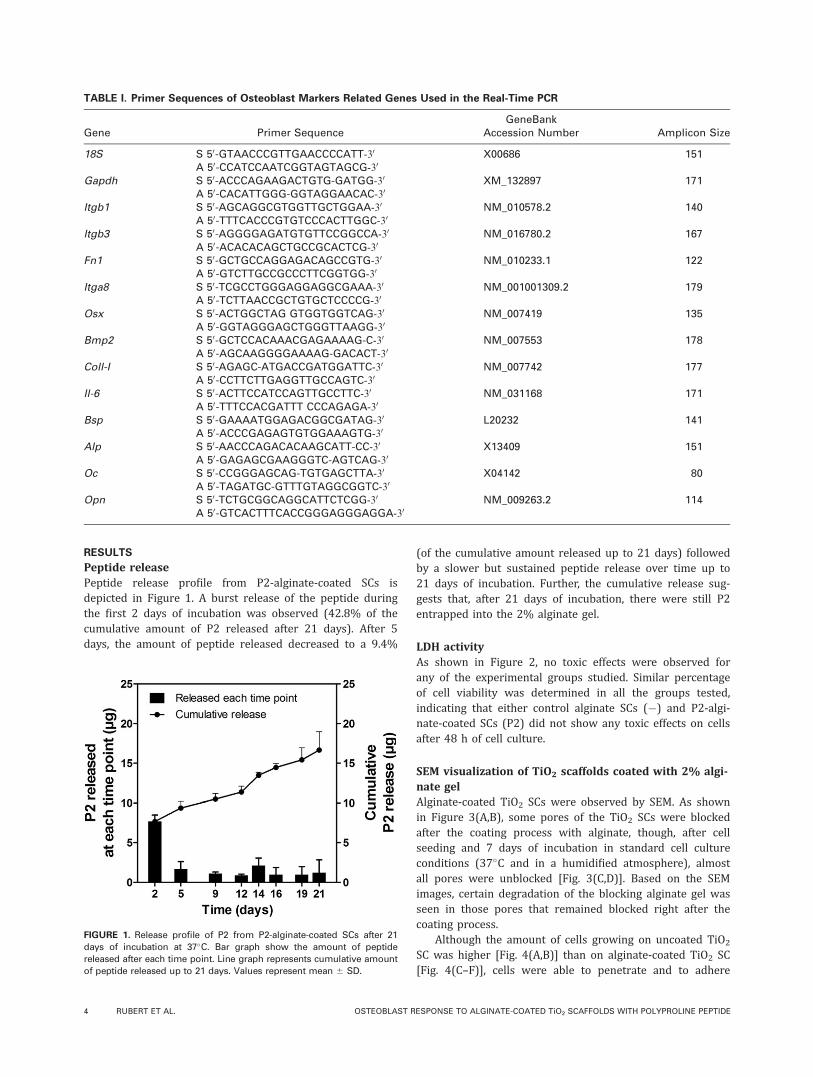
Peptide releasePeptide release profile from P2-alginate-coated SCs isdepicted in Figure 1. A burst release of the peptide duringthe first 2 days of incubation was observed (42.8% of thecumulative amount of P2 released after 21 days). After 5days, the amount of peptide released decreased to a 9.4%
(of the cumulative amount released up to 21 days) followedby a slower but sustained peptide release over time up to21 days of incubation. Further, the cumulative release sug-gests that, after 21 days of incubation, there were still P2entrapped into the 2% alginate gel.
LDH activityAs shown in Figure 2, no toxic effects were observed forany of the experimental groups studied. Similar percentageof cell viability was determined in all the groups tested,indicating that either control alginate SCs (�) and P2-algi-nate-coated SCs (P2) did not show any toxic effects on cellsafter 48 h of cell culture.
SEM visualization of TiO2 scaffolds coated with 2% algi-nate gelAlginate-coated TiO2 SCs were observed by SEM. As shownin Figure 3(A,B), some pores of the TiO2 SCs were blockedafter the coating process with alginate, though, after cellseeding and 7 days of incubation in standard cell cultureconditions (37�C and in a humidified atmosphere), almostall pores were unblocked [Fig. 3(C,D)]. Based on the SEMimages, certain degradation of the blocking alginate gel wasseen in those pores that remained blocked right after thecoating process.
Although the amount of cells growing on uncoated TiO2
SC was higher [Fig. 4(A,B)] than on alginate-coated TiO2 SC[Fig. 4(C–F)], cells were able to penetrate and to adhere
TABLE I. Primer Sequences of Osteoblast Markers Related Genes Used in the Real-Time PCR
Gene Primer SequenceGeneBank
Accession Number Amplicon Size
18S S 50-GTAACCCGTTGAACCCCATT-30 X00686 151A 50-CCATCCAATCGGTAGTAGCG-30
Gapdh S 50-ACCCAGAAGACTGTG-GATGG-30 XM_132897 171A 50-CACATTGGG-GGTAGGAACAC-30
Itgb1 S 50-AGCAGGCGTGGTTGCTGGAA-30 NM_010578.2 140A 50-TTTCACCCGTGTCCCACTTGGC-30
Itgb3 S 50-AGGGGAGATGTGTTCCGGCCA-30 NM_016780.2 167A 50-ACACACAGCTGCCGCACTCG-30
Fn1 S 50-GCTGCCAGGAGACAGCCGTG-30 NM_010233.1 122A 50-GTCTTGCCGCCCTTCGGTGG-30
Itga8 S 50-TCGCCTGGGAGGAGGCGAAA-30 NM_001001309.2 179A 50-TCTTAACCGCTGTGCTCCCCG-30
Osx S 50-ACTGGCTAG GTGGTGGTCAG-30 NM_007419 135A 50-GGTAGGGAGCTGGGTTAAGG-30
Bmp2 S 50-GCTCCACAAACGAGAAAAG-C-30 NM_007553 178A 50-AGCAAGGGGAAAAG-GACACT-30
Coll-I S 50-AGAGC-ATGACCGATGGATTC-30 NM_007742 177A 50-CCTTCTTGAGGTTGCCAGTC-30
Il-6 S 50-ACTTCCATCCAGTTGCCTTC-30 NM_031168 171A 50-TTTCCACGATTT CCCAGAGA-30
Bsp S 50-GAAAATGGAGACGGCGATAG-30 L20232 141A 50-ACCCGAGAGTGTGGAAAGTG-30
Alp S 50-AACCCAGACACAAGCATT-CC-30 X13409 151A 50-GAGAGCGAAGGGTC-AGTCAG-30
Oc S 50-CCGGGAGCAG-TGTGAGCTTA-30 X04142 80A 50-TAGATGC-GTTTGTAGGCGGTC-30
Opn S 50-TCTGCGGCAGGCATTCTCGG-30 NM_009263.2 114A 50-GTCACTTTCACCGGGAGGGAGGA-30
FIGURE 1. Release profile of P2 from P2-alginate-coated SCs after 21
days of incubation at 37�C. Bar graph show the amount of peptide
released after each time point. Line graph represents cumulative amount
of peptide released up to 21 days. Values represent mean 6 SD.
4 RUBERT ET AL. OSTEOBLAST RESPONSE TO ALGINATE-COATED TiO2 SCAFFOLDS WITH POLYPROLINE PEPTIDE

into the coated SCs with 2% alginate gel either with orwithout P2. An increase from day 7 to day 21 in the num-ber of cells growing on the SCs was seen for all the experi-mental groups.
Cell numberDNA quantification was used to determine the number ofcells growing on the TiO2 SC after 7 days of culture (Fig. 5).In accordance to SEM images, after 7 days of culture thenumber of cells was significantly lower in any of the algi-nate-coated TiO2 SC compared to TiO2 SC. Thus, comparedto SC, a 61% and a 49% reduction in cell number wasfound on alginate-coated SCs without and with P2, respec-tively. Although data did not reach statistical significance,SCs coated with P2 showed 32% more cells than the algi-nate control SC (�).
Gene expression of cell adhesion-related markersAs shown in Figure 6, relative mRNA levels of Itgb1 weresignificantly decreased in cells growing onto alginate-coatedSC (either with or without P2) compared to TiO2 SC after 7days of culture. Nevertheless, after 21 days of cell cultureno differences were observed among groups. After 21 days,Itgb3 mRNA levels were increased in cells growing on algi-nate-coated SC (either with or without P2) compared toTiO2 SC. Higher mRNA levels of Fn1 were found in cellsgrowing on 2% alginate-coated SC after 21 days, and in
FIGURE 2. LDH activity measured from culture media collected after
48 h of culture. Results from SCs were presented relative to the LDH
activity in the medium from cells cultured on TCP (0% cell death, low
control) and of cells cultured on TCP and treated with 1% Triton X-100
(100% cell death, High control). Values represent the mean 6 SEM.
FIGURE 3. SEM visualization of 2% alginate-coated TiO2 SCs (control alginate SC) at 10 kV and 40 Pa. Figures (A) and (B) show the microstruc-
ture of TiO2 SCs right after the coating process with one layer of 2% alginate gel at 50� (A) and 300� (B) of magnification. Figures (C) and (D)
show cells cultured on control alginate SCs after 7 days of culture at 50� (C) and 300� (D) of magnification.
ORIGINAL ARTICLE
JOURNAL OF BIOMEDICAL MATERIALS RESEARCH A | MONTH 2012 VOL 00A, ISSUE 00 5

cells growing on P2-alginate-coated SC compared touncoated SC, although for the last group data did not reachstatistical significance. Itga8 mRNA was significantlyincreased in cells growing on P2-alginate-coated SC com-pared to control alginate SC after 21 days of culture.
Gene expression of several osteoblast differentiationmarkersFigure 7 shows relative mRNA levels for several osteoblastdifferentiation marker genes. After 21 days of culture,osterix mRNA levels were increased in cells growing on algi-nate-coated SC (either with or without P2) compared touncoated SC. Bmp-2 and Il-6 mRNA levels were significantlyincreased in cells cultured on P2-alginate-coated SC com-pared to both uncoated SC and alginate-coated SC after 21days of cell culture. Coll-I mRNA levels, a marker relatedwith cell proliferation,22 were significantly increased in cellscultured on P2-alginate-coated SC compared to alginate-coated SC after 7 days of cell culture. After 21 days of cul-ture Coll-I was significantly increased in both alginate-coated SC and P2-alginate-coated SC compared to uncoatedSC. No significant differences were observed in Opn, Bsp,Alp, and Oc mRNA expression levels among experimentalgroups at any of the time points studied.
DISCUSSION
In the present study, we have combined the improvedreported biocompatibility and osteoconductivity of TiO2 SCwith the suitability of alginate gel as a carrier for the deliv-ery of a proline-rich peptide, for their use in load-bearingbone tissue applications to promote bone formation andmineralization. TiO2 SC have been reported to have strength
up to 2.6 MPa in compressive strength10 and showed excel-lent mechanical resistance in a pig in vivo study.13
In bone tissue engineering, the structure of the SC mustprovide an optimal microenvironment for osteogenesis.10
The SC porosity, pore network interconnectivity, the surface-area-to-volume ratio, and the physico-chemical properties ofthe surface determines cell migration and differentiation,bone ingrowth, vascularization, and mass transfer between
FIGURE 4. SEM visualization of MC3T3-E1 cells growing on regular SCs (A, B), control alginate SCs (C, D) and P2-alginate-coated SCs (E, F) after
7 (A, C, E) and 21 (B, D, F) days of culture. SCs were observed by SEM at 10 kV, 40 Pa, and 50� of magnification.
FIGURE 5. Number of cells growing on the SCs after 7 days of cul-
ture. DNA content was analyzed by Hoechst fluorescence staining and
correlated to a linear standard curve. Values represent the mean 6
SEM. Values represent the mean 6 SEM. Mann–Whitney test: (a) p �0.05 versus regular SC.
6 RUBERT ET AL. OSTEOBLAST RESPONSE TO ALGINATE-COATED TiO2 SCAFFOLDS WITH POLYPROLINE PEPTIDE

the cells and the environment.23 The use of highly porousTiO2 SC using an agitated cell seeding method has provedto achieve a good attachment and distribution of mousepre-osteoblastic cells.11 It has been reported that, pore sizegreater than 300 lm and with a porosity of 70% allow vas-cularization and new bone formation.24 Here, TiO2 SC witha pore diameter about to 392 lm at 85–90% of porositywere coated with 2% alginate. As revealed by the gray valueintensity in the SEM images by using a back-scattered elec-tron detector (BSE), alginate infiltrated into the three-dimensional structure and covered the whole surface.Although a few pores remained blocked right after the coat-ing process, almost all pores were unblocked after 7 days ofincubation at 37�C due to the loss of mechanical propertiesof alginate over time in vitro,25 thus providing opening win-dows for cells to penetrate and migrate into the structure.Degradation properties of alginate might be due to an out-ward flux of calcium ions into the surrounding medium,thus decreasing crosslinking overtime. In this study, no dif-
ferences on cell viability were observed between coated anduncoated TiO2 SC, in agreement with previous reportsshowing cell safety and biocompatibility of TiO2 SC7 and ofalginate gels.19,26–28 Alginate has been widely used for deliv-ery of bioactive molecules27,29 and constitute a suitable car-rier for delivery of synthetic peptides at the local site.19 Toavoid serum proteins or phenol interference from the cellculture media, peptide release was performed in distilledwater in the present study, although in an early study,28 P2labeled with fluorescein isothiocyanate (FITC) was used andmeasured in the cell culture media showing a similarrelease profile. Thus, we found a burst release of P2 duringthe first hours of incubation followed by progressive andsustained release during the 21-days period.
Consistent with previous studies,6,9,11 TiO2 SCs providedan appropriate surface for osteoblasts to adhere, migrate,and proliferate. Although the amount of cells into alginate-coated TiO2 SCs was lower than in uncoated TiO2 SC, SCcoated with alginate supported cell progression and
FIGURE 6. Relative mRNA expression levels of Itgb1(A), Itgb3 (B), Fn1(C), and Itga8 (D) in MC3T3-E1 cells cultured on TiO2 SCs for 7 (&) and 21
days (n). Regular SCs were used as reference group. Data represent fold changes of target genes normalized with reference genes (Gapdh and
18S), expressed as a percentage of cells cultured on regular SCs at day 7, which were set to 100%. Values represent the mean 6 SEM. Student
t- test: (a) p � 0.05 versus regular SC and (b) versus control alginate SC (�).
ORIGINAL ARTICLE
JOURNAL OF BIOMEDICAL MATERIALS RESEARCH A | MONTH 2012 VOL 00A, ISSUE 00 7

differentiation. These results are in accordance with previ-ous studies reporting that the alginate is an inert substratefor cell attachment30 and that synthetic peptides rich in pro-line sequences increase properties for cell attachment of thealginate hydrogel.19 Thus, although not significantly, TiO2 SCcoated with 2% alginate containing synthetic P2 showed atrend to improve cell attachment (þ32%) after 7 days com-pared to alginate-coated TiO2 SC.
It has been reported that biomaterial composition regu-lates cell attachment and cytoskeletal organization withlong-term effects on osteoblast cell maturation and minerali-zation.31 In accordance to the efficiency in cell attachmentobserved by SEM and DNA quantification onto the differentgroups, Itgb1 mRNA levels were decreased in cells growingon alginate-coated TiO2 SC compared to those growing onuncoated SC. Further, Itgb3 and Fn1 mRNA levels (whichare highly expressed at early stages of osteogenesis andreduced through the cellular maturation process11,32,33)were significantly increased in cells growing into alginate-coated TiO2 SC compared to the uncoated SCs after 21 daysof culture. Moreover, expression of Itga8, an integrin thatplays a role during the mineralization stage through thebinding to osteopontin, was induced by P2-alginate-coatedSC compared to alginate-coated SC, suggesting that P2 mightinfluence mineralization processes, as previously reportedwith the increased mineralization in MC3T3-E1 andhUCMSCs after P2 treatment.16 Integrins are not onlyinvolved in the attachment of cells to the material sur-face34–38 but also mediate signal transduction pathwaysinducing bone formation and mineralization.33,39 Interest-ingly, expression of genes like Itgb3, Fn1, Coll-I, and Osx thatare related to early stages of osteoblast differentiation, andwhich are normally upregulated at short-term and downre-gulated thereafter, were increased in the later time pointstudied (21 days) in cells grown into alginate-coated TiO2
SC compared to cells growing on uncoated SC. It is possiblethat the temporal sequence of early markers related toosteoblast differentiation varies when MC3T3-E1 cells aregrowing on uncoated SC or on alginate-coated SC, so thatcells growing onto alginate-coated surfaces showed animproved cell differentiation over proliferation compared touncoated TiO2 SC, probably due to the initial difficulties ofcell adhesion onto the alginate. Although alginate coatingseems to impair cell adhesion and proliferation on the SC,the acquisition of a mature and organized matrix (ECM)competent for mineralization was confirmed by a markedincrease in Alp and Bsp mRNA levels from day 7 to 21 daysfor any of the groups. Once the synthesis, organization, andmaturation of the ECM has finalized, Oc expression is upreg-ulated leading to mineralization.40,41 Our results showed aslight increase in Oc mRNA levels after 21 days of culture,therefore, we can conclude that cells were just at the begin-ning of the mineralization process. Moreover, in accordanceto our results with gene expression levels of Opn and Bsp,the increased relation Bsp/Opn mRNA in osteoblastic cellscould be indicative for the stimulation of ECM mineraliza-tion, as previously reported with MC3T3-E1 cells seeded onuncoated TiO2 SC.11
It is important to highlight that the addition of P2 to al-ginate improved the properties for cell proliferation and dif-ferentiation compared to alginate-coated SC, as it can beappreciated by the amount of cells measured by DNA con-tent and the higher expression levels of Bmp2, Coll-I and Il-6. However, P2 did not induce expression of the samemarkers in osteoblastic cells growing on 2% alginate in a2D system in an early study.19 Differences between these
FIGURE 7. Relative mRNA expression levels of (A) osterix (Osx), (B)
bone morphogenetic protein 2 (Bmp2), (C) collagen-I (Coll-I), (D) inter-
leukin 6 (Il-6), (E) osteopontin (Opn), (F) bone sialoprotein (Bsp), (G)
alkaline phosphatase (Alp), and (H) osteocalcin (Oc) in MC3T3-E1 cells
cultured on TiO2 SCs for 7 (&) and 21 days (n). Regular SCs were
used as reference group. Data represent fold changes of target genes
normalized with reference genes (Gapdh and 18S), expressed as a
percentage of cells cultured on regular SCs at day 7, which were set
to 100%. Values represent the mean 6 SEM. Student t-test: (a) p �0.05 versus regular SC and (b) versus control alginate SC (�).
8 RUBERT ET AL. OSTEOBLAST RESPONSE TO ALGINATE-COATED TiO2 SCAFFOLDS WITH POLYPROLINE PEPTIDE

studies could be related to that temporal osteoblast geneexpression is retarded when cells grow in a 3D structure.Thus, while P2 could influence more on the expression ofmarkers associated to early stages of differentiation likeBmp2 and Coll-I in a 3D system, late markers like osteocal-cin has been shown to be modulated by P2 in a 2D systemwhen giving the peptide in solution.16,17 So far the syntheticpeptides rich in polyproline sequences have repetitivelyshowed an increase in osteocalcin mRNA levels, both invitro16,17 and in in vivo studies where titanium implantswere coated with the peptide,18 and further when loadedinto an alginate hydrogel for their use as a carrier for localdelivery.19 Thus, taken together our previous studies andthe results from the present study, this allows us to suggestthat P2-alginate-coated SCs would promote higher cell dif-ferentiation and mineralization in an in vivo environment.
CONCLUSION
In conclusion, our results demonstrate that alginate-coatedTiO2 SC can act as a matrix for delivery of synthetic peptiderich in proline sequences inducing osteoblast cell differen-tiation. The combination of the physical and osteoconductiveproperties of TiO2 SC with the osteogenic effects of syn-thetic proline-rich peptides on bone formation and minerali-zation may represent a new strategy for bone tissue regen-eration in load-bearing applications.
ACKNOWLEDGMENTS
The authors are especially thankful for the excellent technicalsupport from Ferran Hierro (Serveis Cientıfico-Tecnics, Univer-sity of Balearic Islands).
REFERENCES
1. Jokinen M, Patsi M, Rahiala H, Peltola T, Ritala M, Rosenholm JB.
Influence of sol and surface properties on in vitro bioactivity of
sol–gel-derived TiO2 and TiO2-SiO2 films deposited by dip-coating
method. J Biomed Mater Res 1998;42:295–302.
2. Lee N, Oh H, Hong C, Suh H, Hong S. Comparison of the syn-
thetic biodegradable polymers, polylactide (PLA), and polylactic-
co-glycolic acid (PLGA) as scaffolds for artificial cartilage. Biotech-
nol Bioprocess Eng 2009;14:180–186.
3. Thonmson RC, Shung AK, Yaszemski MJ, Mikos AG. Scaffolds
processing. In: Lanza R, Langer R, Vacanti J, editors. Principles of
Tissue Engineering. San Diego: Academic Press; 2000. p252–259.
4. Bueno Rde B, Adachi P, Castro-Raucci LM, Rosa AL, Nanci A, Oli-
veira PT. Oxidative nanopatterning of titanium surfaces promotes
production and extracellular accumulation of osteopontin. Braz
Dent J 2011;22:179–84.
5. Khan Y, Yaszemski MJ, Mikos AG, Laurencin CT. tissue engineer-
ing of bone: material and matrix considerations. J Bone Joint
Surg Am 2008;90 (Suppl. 1):36–42.
6. Fostad G, Hafell B, Førde A, Dittmann R, Sabetrasekh R, Will J,
Ellingsen JE, Lyngstadaas SP, Haugen HJ. Loadable TiO2 scaf-
folds-A correlation study between processing parameters, micro
CT analysis and mechanical strength. J Eur Ceram Soc 2009;29:
2773–2781.
7. Nygren H, Tengvall P, Lundstrom I. The initial reactions of TiO2
with blood. J Biomed Mater Res 1997;34:487–492.
8. Rincon JC, Xiao Y, Young WG, Bartold PM. Enhanced prolifera-
tion, attachment and osteopontin expression by porcine peri-
odontal cells exposed to Emdogain. Arch Oral Biol 2005;50:
1047–54.
9. Sabetrasekh R, Tiainen H, Reseland JE, Will J, Ellingsen JE, Lyng-
stadaas SP, Haugen HJ. Impact of trace elements on biocompati-
bility of titanium scaffolds. Biomed Mater 2010;5:15003.
10. Tiainen H, Lyngstadaas SP, Ellingsen JE, Haugen HJ. Ultra-porous
titanium oxide scaffold with high compressive strength. J Mater
Sci Mater Med 2010;21:2783–2792.
11. G�omez-Florit M, Rubert M, Ramis JM, Tianinen H, Haugen HJ,
Lyngstadaas SP, Monjo M. TiO2 scaffolds sustain differentiation
of MC3T3-E1 cells. J Biomater Tissue Eng. 2012; doi: 10.1166/
jbt.2012.1055.
12. Haugen HJ, Monjo M, Rubert M, Verket A, Lyngstadaas SP, Elling-
sen JE, Ronold HJ, Wohlfahrt JC. Porous ceramic titanium diox-
ide scaffolds promote bone formation in rabbit peri-implant
cortical defect model. Acta Biomater. 2012; doi: 10.1016/
j.actbio.2012.09.009.
13. Tiainen H, Wohlfahrt JC, Verket A, Lyngstadaas SP, Haugen HJ.
Bone formation in TiO2 bone scaffolds in extraction sockets of
minipigs. Acta Biomater 2012;8:2384–2391.
14. Ball LJ, Kuhne R, Schneider-Mergener J, Oschkinat H. Recogni-
tion of proline-rich motifs by protein–protein-interaction domains.
Angew Chem Int Ed Engl 2005;44:2852–2869.
15. Jin T, Ito Y, Luan X, Dangaria S, Walker C, Allen M, Kulkarni A,
Gibson C, Braatz R, Liao X, Diekwisch TG. Elongated polyproline
motifs facilitate enamel evolution through matrix subunit com-
paction. PLoS Biol 2009;7:e1000262.
16. Rubert M, Ramis JM, Vondrasek J, Gay�a A, Lyngstadaas SP,
Monjo M. Synthetic peptides analogue to enamel proteins pro-
mote osteogenic differentiation of MC3T3-E1 and mesenchymal
stem cells. J Biomater Tissue Eng 2011;1:198–209.
17. Ramis JM, Rubert M, Vondrasek J, Gay�a A, Lyngstadaas SP,
Monjo M. Effect of enamel matrix derivative and of proline-rich
synthetic peptides on the differentiation of human mesenchymal
stem cells towards the osteogenic lineage. Tissue Eng Part A
2012;18:1–11.
18. Petzold C, Monjo M, Rubert M, Reinholt FP, Gomez-Florit M,
Ramis JM, Ellingsen JE, Lyngstadaas SP. Effect of coated tita-
nium implants with a proline-rich synthetic peptide on bone heal-
ing. Oral Craniofac Tissue Eng 2012;2:35–43.
19. Rubert M, Monjo M, Lyngstadaas SP, Ramis JM. Effect of alginate
hydrogel containing polyproline-rich peptides on osteoblast dif-
ferentiation. Biomed Mater 2012;7:055003.
20. Takahashi Y, Tabata Y. Homogeneous seeding of mesenchymal
stem cells into nonwoven fabric for tissue engineering. Tissue
Eng 2003;9:931–938.
21. Rage R, Mitchen J, Wilding G. DNA fluorometric assay in 96-well
tissue culture plates using Hoechst 33258 after cell lysis by freez-
ing in distilled water. Anal Biochem 1990;191:31–34.
22. Stein GS, Lian JB, Stein JL, Van Wijnen AJ, Montecino M. Tran-
scriptional control of osteoblast growth and differentiation. Phys-
iol Rev 1996;76:593–629.
23. Hutmacher DW. Scaffolds in tissue engineering bone and carti-
lage. Biomaterials 2000;21:2529–2543.
24. Karageorgiou V, Kaplan D. Porosity of 3D biomaterial scaffolds
and osteogenesis. Biomaterials 2005;26:5474–5491.
25. Rowley JA, Madlambayan G, Mooney DJ. Alginate hydrogels as
synthetic extracellular matrix materials. Biomaterials 1999;20:
45–53.
26. Barbetta A, Barigelli E, Dentini M. Porous alginate hydrogels: Syn-
thetic methods for tailoring the porous texture. Biomacromole-
cules 2009;10:2328–2337.
27. Lee JY, Choo JE, Park HJ, Park JB, Lee SC, Jo I, Lee SJ, Chung
CP, Park YJ. Injectable gel with synthetic collagen-binding peptide
for enhanced osteogenesis in vitro and in vivo. Biochem Biophys
Res Commun 2007;357:68–74.
28. Rubert M, Alonso-Sande M, Monjo M, Ramis JM. Evaluation of
alginate and hyaluronic acid for their use in bone tissue engineer-
ing. Biointerphases 2012;7:1–11.
29. Kolambkar YM, Dupont KM, Boerckel JD, Huebsch N, Mooney DJ,
Hutmacher DW, Guldberg RE. An alginate-based hybrid system
for growth factor delivery in the functional repair of large bone
defects. Biomaterials 2011;32:65–74.
ORIGINAL ARTICLE
JOURNAL OF BIOMEDICAL MATERIALS RESEARCH A | MONTH 2012 VOL 00A, ISSUE 00 9

30. Alsberg E, Anderson KW, Albeiruti A, Franceschi RT, Mooney DJ.
Cell-interactive alginate hydrogels for bone tissue engineering. J
Dent Res 2001;80:2025–2029.
31. Shah AK, Lazatin J, Sinha RK, Lennox T, Hickok NJ, Tuan RS.
Mechanism of BMP-2 stimulated adhesion of osteoblastic cells to
titanium alloy. Biol Cell 1999;91:131–142.
32. Cheng S-L, Lai C-F, Blystone SD, Avioli LV. Bone mineralization
and osteoblast differentiation are negatively modulated by integ-
rin avb3. J Bone Miner Res 2001;16:277–288.
33. Moursi AM, Globus RK, Damsky CH. Interactions between integrin
receptors and fibronectin are required for calvarial osteoblast dif-
ferentiation in vitro. J Cell Sci 1997;110:2187–2196.
34. El-Amin SF, Attawia M, Lu HH, Shah AK, Chang R, Hickok NJ,
Tuan RS, Laurencin CT. Integrin expression by human osteoblasts
cultured on degradable polymeric materials applicable for tissue
engineered bone. J Orthop Res 2002;20:20–28.
35. Gronowicz G, McCarthy MB. Response of human osteoblasts to
implant materials: Integrin-mediated adhesion. J Orthop Res
1996;14:878–887.
36. Lynch MP, Stein JL, Stein GS, Lian JB. The influence of type I col-
lagen on the development and maintenance of the osteoblast
phenotype in primary and passaged rat calvarial osteoblasts:
Modification of expression of genes supporting cell growth, adhe-
sion, and extracellular matrix mineralization. Exp Cell Res 1995;
216:35–45.
37. Siebers MC, ter Brugge PJ, Walboomers XF, Jansen JA. Integrins
as linker proteins between osteoblasts and bone replacing materi-
als. A critical review. Biomaterials 2005;26:137–146.
38. Steele JG, Dalton BA, Johnson G, Underwood PA. Polystyrene
chemistry affects vitronectin activity: An explanation for cell
attachment to tissue culture polystyrene but not to unmodified
polystyrene. J Biomed Mater Res 1993;27:927–940.
39. Hynes RO. Integrins: Versatility, modulation, and signaling in cell
adhesion. Cell 1992;69:11–25.
40. Sodek J, Chen J, Nagata T, Kasugai S, Todescan R Jr, Li IW, Kim
RH. Regulation of osteopontin expression in osteoblasts. Ann NY
Acad Sci 1995;760:223–241.
41. McKee MD, Nanci A. Osteopontin and the bone remodeling
sequence. Ann NY Acad Sci 1995;760:177–189.
10 RUBERT ET AL. OSTEOBLAST RESPONSE TO ALGINATE-COATED TiO2 SCAFFOLDS WITH POLYPROLINE PEPTIDE