Synovial cytokine expression in ankle osteoarthritis depends on age and stage
ORIGINAL RESEARCH ARTICLEpublished: 02 August 2012
doi: 10.3389/fimmu.2012.00231
Effect of arthritic synovial fluids on the expression ofimmunomodulatory factors by mesenchymal stem cells:an explorative in vitro studyMaarten J. C. Leijs1, Gerben M. van Buul 1,2, Erik Lubberts3, Pieter K. Bos1, Jan A. N. Verhaar 1,Martin J. Hoogduijn4 and Gerjo J. V. M. van Osch1,5*1 Department of Orthopaedics, Erasmus MC University Medical Center, Rotterdam, The Netherlands2 Department of Radiology, Erasmus MC University Medical Center, Rotterdam, The Netherlands3 Department of Rheumatology, Erasmus MC University Medical Center, Rotterdam, The Netherlands4 Department of Internal Medicine, Erasmus MC University Medical Center, Rotterdam, The Netherlands5 Department of Otorhinolaryngology, Erasmus MC University Medical Center, Rotterdam, The Netherlands
Edited by:Frank J. M. F. Dor, Erasmus MCUniversity Medical Center Rotterdam,The Netherlands
Reviewed by:Meindert Crop, Albert SchweitzerZiekenhuis Dordrecht, TheNetherlandsDina Sabry, Cairo University, Egypt
*Correspondence:Gerjo J. V. M. van Osch, ConnectiveTissue Cells and Repair Group,Departments of Orthopaedics andOtorhinolaryngology, Erasmus MCUniversity Medical Center Rotterdam,Dr. Molewaterplein 50, Room Ee1655,3015 GE Rotterdam, P.O. Box 1738,3000 DR Rotterdam,The Netherlands.e-mail: [email protected]
Background: In diseased joints, the catabolic environment results in progressive joint dam-age. Mesenchymal stem cells (MSCs) can have immunomodulatory effects by secretinganti-inflammatory factors. To exert these effects, MSCs need to be triggered by pro-inflammatory cytokines. To explore the potential of MSCs as a treatment for diseasedjoints, we studied the effect of synovial fluid (SF) from donors with different joint diseasesand donors without joint pathology on the immunomodulatory capacities of human MSCsin vitro. We hypothesized that SF of diseased joints influences the immunomodulatoryeffects of MSCs. Materials and Methods: MSCs were cultured in medium with SF ofsix osteoarthritis (OA) or six rheumatoid arthritis (RA) donors and three donors withoutjoint pathology were used as control. Gene expressions of IL-6, HGF, TNFa, TGFb1, andindoleamine 2,3-dioxygenase (IDO) were analyzed. L-kynurenine concentration in condi-tioned medium (CM) by MSCs with SF was determined as a measure of IDO activity byMSCs. Furthermore, the effect of CM with SF on proliferation of activated lymphocyteswas analyzed. Results: Addition of SF significantly up-regulated the mRNA expressionof IL-6 and IDO in MSCs. SF(OA) induced significantly higher expression of IDO thanSF(control), although no difference in IDO activity of the MSCs could be shown with aL-kynurenine assay. Medium conditioned by MSCs with SF(OA or RA) suppressed acti-vated lymphocyte proliferation in vitro more than medium conditioned by MSCs withoutSF or with SF(control). Discussion: SF can influence the expression of genes involved inimmunomodulation by MSCs and the effect on lymphocyte proliferation. We found indica-tions for disease-specific differences between SFs but the variation between donors, evenwithin one disease group was high. These data warrant further research to examine thepotential application of MSC therapy in arthritic joints.
Keywords: MSC, osteoarthritis, rheumatoid arthritis, synovial fluid, immunomodulation
INTRODUCTIONOsteoarthritis (OA) and rheumatoid arthritis (RA) are highprevalent forms of arthritis. OA is mainly characterized by pro-gressive functional loss and cartilage degeneration. Main factorsinvolved in cartilage degeneration are a variety of matrix degrad-ing enzymes and pro-inflammatory cytokines (Goldring, 2000;Goldring and Marcu, 2009). It is possible to treat the symp-toms of OA with lifestyle changes, analgesics, non-steroidal anti-inflammatory drugs (NSAIDs), or intra-articular injections withcorticosteroids or hyaluronic acid and the ultimate treatment forend stage OA is joint replacement. A treatment to cure OA, how-ever, is still not available. RA is an auto-immune disease initiatedby immune complexes that together with cytokines, complement,and metalloproteinases (Weissmann, 2006) cause an inflamma-tory and catabolic environment in the joint (Goldring and Marcu,
2009). It is a systemic disease characterized by persistent synovi-tis, systemic inflammation, and auto-antibodies which eventuallycause joint damage with progressive cartilage degeneration andbone alterations. There is a wide range of therapeutic optionsfor RA like analgesics, NSAIDs, disease-modifying anti rheumaticdrugs (DMARDs), and biologicals (Lee and Weinblatt, 2001; Scottet al., 2010). However, to date there is no treatment available tocure RA.
Human mesenchymal stem cells (MSCs), the progenitors ofconnective tissue cells, are able to differentiate into different celltypes including chondrocytes (Caplan, 1991, 1994; Solchaga et al.,2004; Caplan and Dennis, 2006). This has attracted the interestof many people working in the area of cartilage repair. Besidesthe ability to reconstruct tissues, MSCs also have the ability tomodulate the environment by secreting many immunomodulating
www.frontiersin.org August 2012 | Volume 3 | Article 231 | 1
Leijs et al. The effect of synovial fluids on immunomodulation by MSCs
and trophic factors like cytokines, chemokines, and growth fac-tors (Deans and Moseley, 2000; Minguell et al., 2001; Kim et al.,2005; Caplan and Dennis, 2006; Chen et al., 2006; Schinkotheet al., 2008; Hoogduijn et al., 2010; Meisel et al., 2011). These fac-tors have potent immunomodulatory capacity as demonstratedin vitro by inhibition of T-lymphocyte proliferation after addingMSCs in mixed lymphocyte reactions (Hoogduijn et al., 2010;Landgraf et al., 2011). MSCs also inhibit the antibody produc-tion of B lymphocytes and inhibit the generation and function ofantigen presenting cells (Sze et al., 2007; Chen et al., 2008; Hoog-duijn et al., 2010). The stimulation of MSC by pro-inflammatorycytokines like TNFa and IFNg strongly enhances the immunosup-pressive function of MSCs (Klyushnenkova et al., 2005; Schinkotheet al., 2008; Siegel et al., 2009; Eggenhofer et al., 2010; Hoogduijnet al., 2010).
In a healthy joint environment, a balance exists between an ana-bolic and catabolic state. In a situation of inflammation or chronicdamage, i.e., OA or RA, the environment becomes more catabolic(Findlay and Haynes, 2005; Goldring and Marcu, 2009). All jointtissues are exposed to synovial fluid (SF) and in OA and RA inflam-matory factors are secreted into the SF. The aim of the present studywas to investigate whether SF of donors with OA, RA, or no jointpathology triggers MSCs to become immunomodulatory. Sinceinflammation plays a large role in RA and OA, we hypothesizedthat MSCs will be triggered to become immunomodulatory. Weexplored this by studying the effect of SF of OA and RA patientsas well as SF of non-pathological(control) donors on MSCs. Ourhypothesis was that MSCs conditioned in SF(RA) will express alarge anti-inflammatory effect compared to SF(control) due to thehigh inflammation state of RA patients and MSCs conditionedwith SF(OA) will express a mild anti-inflammatory effect com-pared to SF(control) as a reaction to a less inflamed environmentin joints of OA patients.
We evaluated the effect of SF on expression of genes of MSCs forimmunomodulatory factors. Furthermore, we performed a func-tional assay to study the capacity of factors secreted by MSCs inresponse of SF to inhibit proliferation of activated lymphocytes.
MATERIALS AND METHODSSYNOVIAL FLUIDSFifteen SF samples were obtained from six OA patients, six RApatients, and three donors without any joint pathology. SFs(OA)were obtained from patients undergoing total knee replacementsurgery. All patients implicitly consented to the use of these fluidsfor scientific research (with approval by Erasmus MC medical ethi-cal committee protocol # MEC-2004-322). SFs(RA) were obtainedfrom RA patients with active inflammation of the knee during con-sultation at the rheumatology outpatient clinic (with approval byErasmus MS medical ethical committee protocol # MEC-236.904-2003-255). SFs(control) were purchased from SF donors withoutjoint diseases, post mortem within 24 h of death (Articular Engi-neering, Northbrook, IL, USA). After aspiration, all SF samplesfrom the joints of all donors were centrifuged to remove debris.Supernatant was stored at−80˚C.
To evaluate the inflammatory aspects of the different SFs wedid amplified enzyme linked immunosorbent assays (ELISA) toquantify cytokines IL-6, TNFa (R&D Systems, Minneapolis, MN,
USA), and IFNg (Invitrogen, Carlsbad, CA, USA). Measurementsof IL-6, TNFa, and IFNg were performed in duplicate. All SFs weretreated with 1:3 hyaluronidase (1000 U/ml PBS, 10 min at 37˚C)prior to ELISA measurements. ELISAs were carried out accordingto the manufacturer’s instructions by means of a multilabel platereader (VersaMax™, Molecular Devices, Sunnyvale, CA, USA).
MSC ISOLATIONMesenchymal stem cells were isolated from heparinized femoral-shaft marrow aspirate of patients undergoing total hip arthroplasty(with informed consent after approval by Erasmus MC med-ical ethical committee protocol # MEC-2004-142). About 5–10 mlmarrow was harvested with a sterile Jamshidi needle into sterile10 ml syringes containing 0.5 ml of heparin (1000 U/ml). About30–100× 106 mononuclear cells were plated in a T175 flask in25 ml expansion medium (Dulbecco’s Modified Eagle Medium(DMEM) low glucose (Invitrogen, Carlsbad, CA, USA) containing15% heat inactivated fetal calf serum (Lonza, Verviers, Belgium,selected batch), 1.5 µg/ml fungizone (All Invitrogen, Carlsbad,CA, USA), 50 µg/ml gentamicin (Invitrogen, Carlsbad, CA, USA),1 ng/ml fibroblast growth factor-2 (Instruchemie B.V., Delfzijl, TheNetherlands), and 0.1 mM of l-ascorbic acid 2-phosphate (vit-amin C; Sigma, St. Louis, MO, USA). After 24 h, non-adherentcells and erythrocytes were removed by washing three times with2% FCS in 1× PBS (Invitrogen, Carlsbad, CA, USA). Remainingadherent cells were cultured in expansion medium at 37˚C and5% carbon dioxide (CO2). Expansion media were renewed twice aweek. At subconfluent cells were trypsinized with a 0.25% trypsinsolution containing 0.01% EDTA (Invitrogen, Carlsbad, CA, USA)and plated at a density of 2300 cells/cm2.
MSC CULTURE WITH SFCryopreserved MSCs of passage two were used for the experi-ments. After thawing, MSCs were seeded in a T175 flask at a densityof 2300 cells/cm2, expanded for one passage and subsequentlyplated in six well plates at a density of 4000 cells/cm2 for the exper-imental conditions. At 70% confluence the existing medium wasdiscarded and the cells were washed three times using PBS (Invit-rogen, Carlsbad, CA, USA). Subsequently 0.8 ml of DMEM lowglucose containing 9 µg/ml fungizone and 50 µg/ml gentamicin,was applied per well. The different SFs(OA, RA, and control) wereadded in triplicates to the media in a concentration of 20%. Inpreliminary tests MSCs were cultured in 0, 10, or 25% SF of fourOA donors, Gene expression was not significantly different in 10and 25% SF. Based on this and taking into account the availabilityof the SF (from SF(control) we obtained maximal 1 ml per donor)we decided to use 20% SF for all further experiments. All condi-tions contained a total concentration of 1% ITS (BD Bioscience,Bedford, MA, USA). Nine wells with only medium plus 1% ITSwere used as negative controls for unstimulated MSCs. After 48 hof incubation, MSCs were harvested for gene expression analy-ses and the conditioned medium (CM) was harvested and storedat−80˚C.
GENE EXPRESSION ANALYSISAfter 48 h of incubation total RNA from MSCs was isolatedusing RNeasy® microkit (Qiagen, Hilden, Germany) with RNeasy
Frontiers in Immunology | Alloimmunity and Transplantation August 2012 | Volume 3 | Article 231 | 2
Leijs et al. The effect of synovial fluids on immunomodulation by MSCs
MinElute spin columns. After quantification of nucleic acids byspectrophotometry (NanoDrop 2000, Thermo Scientific, IsogenLife Science, IJsselstein, The Netherlands) the RNA was reversetranscribed using a First Strand cDNA Synthesis kit (RevertAid™;MBI Fermentas, St. Leon-Rot, Germany). Amplifications were per-formed as 20 µl reactions with real-time PCR. Thermocycler con-ditions comprised an initial holding at 95˚C for 10 min, followedby one step at 95˚C for 15 s and 60˚C for 60 s for 40 cycles. A disso-ciation stage was added at the end using 95˚C for 15 s, 60˚C for 20 s,and 95˚C for 15 s. For UBC, IL-6, HGF, TNF-α, qPCR™MastermixPlus for SYBR® Green I (Eurogentec, Nederland B.V., Maastricht,The Netherlands) was used. For GAPDH, HPRT, IDO, and TGF-β1TaqMan Master Mix (ABI, Branchburg, NJ, USA) was used. Sets ofprimers and probes used in this study: GAPDH (NM_002046.3)Fw: ATGGGGAAGGTGAAGGTCG Rv: TAAAAGCAGCCCTG-GTGACC Probe: Fam-CGCCCAATACGACCAAATCCGTTGAC;HPRT (NM_000194.2) Fw: TATGGACAGGACTGAACGTCTTGRv: CACACAGAGGGCTACAATGTG Probe: Fam-AGATGTGATGAAGGAGATGGGAGGCCA; UBC (NM_021009.5) Fw: ATTTGGGTCGCGGTTCTTG Rv: TGCCTTGACATTCTCGATGGT; IL-6 (NM_000600.3) Fw: TCGAGCCCACCGGGAACGAA Rv:GCAGGGAAGGCAGCAGGCAA; HGF (NM_000601.4) Fw:GGCTGGGGCTACACTGGATTG Rv: CCACCATAATCCCCCT-CACAT; TNF-aplha (NM_000594.2) Fw: GCCGCATCGC-CGTCTCCTAC Rv: AGCGCTGAGTCGGTCACCCT; TGF-beta1(NM_000660.4) Fw: GTGACAGCAGGGATAACACACTG Rv:CATGAATGGTGGCCAGGTC Probe: Fam-ACATCAACGGGTTCACTACCGGC. IDO was detected using a taqman assay on demand(Applied Biosystems, Capelle a/d IJssel, The Netherlands) of whichthe primer sequence is not known to us. Data were collected andquantitatively analyzed on an ABI Prism 7000 Sequence DetectionSystem (SDS) with SDS software, version 1.2.3 (Applied Biosys-tems, Capelle a/d IJssel, The Netherlands). Gene expressions ofthe cytokines and IDO in MSCs were calculated by cycle threshold(CT) values. CT values of 36 and higher were considered as non-expressed and set to 100 for further calculations. The CT values ofthe housekeeper genes GAPDH, HPRT, and UBC were averagedby using geometric averaging of every sample. This average is thebest keeper index (BKI) for every single sample. All separate CTvalues were corrected to the BKI by using the 2−∆CT formula.
L-KYNURENIN ASSAYIn order to evaluate whether SF influenced IDO activity inMSCs, we measured the concentration of l-kynurenine in theCM and SFs. To correct for possible l-kynurenine in SF, theSFs were diluted in the same concentration and the samemedia as the CM and values were subtracted from the CMvalues. Values of one of the OA donors could not be usedsince no remaining SF was available for correction. Thirtypercent trichloroacetic acid was added to the samples in a1:3 ratio and after 30 min incubation at 50˚C the sampleswere centrifuged at 12000 rpm for 5 min. Supernatant of allconditions were diluted 1:1 in Ehrlich reagent (200 µg 4-dimethylaminobenzaldehyde (Sigma, St. Louis, MO, USA) in10 ml of glacial acetic acid) in duplicate in a 96-wells flat bottomplate and absorbance was determined at 490 nm in a multil-abel plate reader (VersaMax™, Molecular Devices, Sunnyvale, CA,
USA). l-kynurenine (Sigma, St. Louis, MO, USA) was used asstandard.
PBMC PROLIFERATION ASSAYPeripheral blood mononuclear cells (PBMCs) were isolated frombuffy coats (Sanquin, Rotterdam, The Netherlands) of healthy vol-unteers using Ficoll-Paque™Plus (GE Healthcare, Uppsala, Swe-den) separation and stored at −135˚C until use. PBMCs werethawed and centrifuged at 2000 rpm for 5 min. Viable cells werecounted using trypan blue exclusion test. PBMCs were seeded inalpha-modified Minimum Essential Medium (aMEM; Invitrogen,Carlsbad, CA, USA) supplemented with 20% heat inactivated FCS(Lonza, Verviers, Belgium, selected batch), 2% pen-strep (Peni-cillin 10,000 UI/ml, Streptomycin 10,000 UI/ml, Lonza, Verviers,Belgium), and 2% l-glutamine (200 mM, Lonza, Verviers, Bel-gium) and activated with anti-CD3 and anti-CD28 linked withlinker goat-anti-mouse antibody (BD Pharmingen, San Diego, CA,USA). 5× 10⁴ PBMCs in 100 µl expansion medium were seededper well in round-bottom 96-well plates (Nunc, Roskilde, Den-mark) and incubated for 5 days. The immunosuppressive capacityof factors secreted by MSCs was evaluated by using the CM fromthe MSC culture conditions described earlier. One hundred micro-liters of CM of MSCs incubated with each of the SFs, except one ofthe OA donors were no SF was left, was added in triplicate to thePBMCs for 5 days. CM of MSCs without SF and unconditionedmedium, identical to the medium used in the CM except for thefact that it had not been in contact with MSCs, were added intriplicate as a control. To correct for direct effects of the SF presentin the CM on the PBMCs we added controls of medium not con-ditioned by MSCs with similar concentration of SF of each of thedonors. At day four of incubation, 3H-thymidine (0.5 µCi/well;Perkin Elmer, Inc., San Jose, CA, USA) was added. At day five, after16 h of incorporation of 3H-thymidine, PBMCs were harvested,and 3H-thymidine incorporation measured using a β-plate reader(Wallac 1450 MicroBeta TriLux Liquid Scintillation Counter andLuminometer, Perkin Elmer, Inc., San Jose, CA, USA).
DATA ANALYSISStatistical difference in gene expression by MSCs conditionedwith SF(OA), SF(RA), and SF(control) was analyzed by using amixed linear model in which condition(SF of OA, RA, or no jointpathology donors) was considered a fixed factor and the differ-ent SF donors for all conditions a random factor. Values for thegenes IDO, TNFa, and TGFb were log-transformed to approacha normal distribution. Statistical differences of inhibitory capac-ity of the different CM was analyzed by using a mixed linearmodel in which condition (CM by MSCs incubated with OA,RA, or control SF) was considered a fixed factor, different donorsa random factor and Sidak was used as adjustment for multi-ple comparisons. Inhibitory effects of CM with SF compared toSF only were explored by statistical analyses with the Wilcoxonsigned ranks test. Data are presented as the mean± standarddeviation and 2.5–97.5 percentile. P-value of ≤0.05 was con-sidered statistical significant; ∗P < 0.05, ∗∗P < 0.001. Analyseswere performed using SPSS 17.0 Statistics (SPSS, Inc., Chicago,IL, USA).
www.frontiersin.org August 2012 | Volume 3 | Article 231 | 3
Leijs et al. The effect of synovial fluids on immunomodulation by MSCs
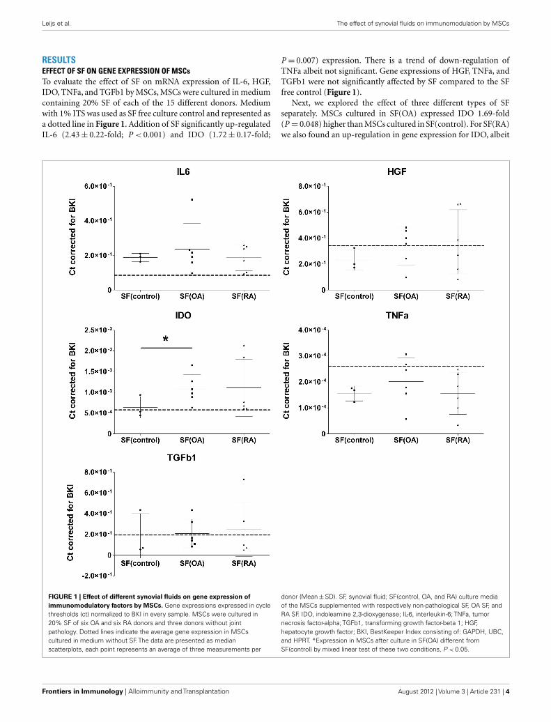
RESULTSEFFECT OF SF ON GENE EXPRESSION OF MSCsTo evaluate the effect of SF on mRNA expression of IL-6, HGF,IDO, TNFa, and TGFb1 by MSCs, MSCs were cultured in mediumcontaining 20% SF of each of the 15 different donors. Mediumwith 1% ITS was used as SF free culture control and represented asa dotted line in Figure 1. Addition of SF significantly up-regulatedIL-6 (2.43± 0.22-fold; P < 0.001) and IDO (1.72± 0.17-fold;
P = 0.007) expression. There is a trend of down-regulation ofTNFa albeit not significant. Gene expressions of HGF, TNFa, andTGFb1 were not significantly affected by SF compared to the SFfree control (Figure 1).
Next, we explored the effect of three different types of SFseparately. MSCs cultured in SF(OA) expressed IDO 1.69-fold(P = 0.048) higher than MSCs cultured in SF(control). For SF(RA)we also found an up-regulation in gene expression for IDO, albeit
FIGURE 1 | Effect of different synovial fluids on gene expression ofimmunomodulatory factors by MSCs. Gene expressions expressed in cyclethresholds (ct) normalized to BKI in every sample. MSCs were cultured in20% SF of six OA and six RA donors and three donors without jointpathology. Dotted lines indicate the average gene expression in MSCscultured in medium without SF. The data are presented as medianscatterplots, each point represents an average of three measurements per
donor (Mean±SD). SF, synovial fluid; SF(control, OA, and RA) culture mediaof the MSCs supplemented with respectively non-pathological SF, OA SF, andRA SF. IDO, indoleamine 2,3-dioxygenase; IL-6, interleukin-6; TNFa, tumornecrosis factor-alpha; TGFb1, transforming growth factor-beta 1; HGF,hepatocyte growth factor; BKI, BestKeeper Index consisting of: GAPDH, UBC,and HPRT. *Expression in MSCs after culture in SF(OA) different fromSF(control) by mixed linear test of these two conditions, P < 0.05.
Frontiers in Immunology | Alloimmunity and Transplantation August 2012 | Volume 3 | Article 231 | 4
Leijs et al. The effect of synovial fluids on immunomodulation by MSCs
not significant which is probably caused by the large variationbetween the six different RA donors. No further significant dif-ferences in gene expression of IL-6, TNFa, TGFb1, and HGF werefound between MSCs cultured in the three different SFs (Figure 1).IDO activity of MSCs was analyzed by an l-kynurenine assay onall different CM with SF corrected for l-kynurenine content in SFof that donor. No significant differences of IDO activity by MSCscultured in SF of different donors were found (data not shown).
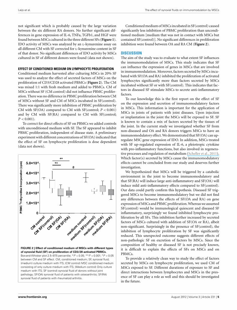
EFFECT OF CONDITIONED MEDIUM ON LYMPHOCYTE PROLIFERATIONConditioned medium harvested after culturing MSCs in 20% SFwas used to analyze the effect of secreted factors of MSCs on theproliferation of CD3/CD28 activated PBMCs (Figure 2). The CMwas mixed 1:1 with fresh medium and added to PBMCs. CM ofMSCs without SF (CM control) did not influence PBMC prolifer-ation. There was no difference in PBMC proliferation between CMof MSCs without SF and CM of MSCs incubated in SF(control).There was significantly more inhibition of PBMC proliferation byCM with SF(OA) compared to CM with SF(control; P < 0.001)and by CM with SF(RA) compared to CM with SF(control;P < 0.001).
To correct for direct effects of SF on PBMCs we added controlswith unconditioned medium with SF. The SF appeared to inhibitPBMC proliferation, independent of disease state. A preliminaryexperiment with different concentrations of SF(OA) indicated thatthe effect of SF on lymphocyte proliferation is dose dependent(data not shown).
FIGURE 2 | Effect of conditioned medium of MSCs with different typesof synovial fluid (SF) on proliferation of CD3/28 activated PBMCs.Box-and-Whisker plot 2.5–97.5 percentile; *P < 0.05; **P < 0.001; #P < 0.05between CM and SF effect. CM, conditioned medium; SF, synovial fluid;(medium) culture medium with ITS; (CM control) MSC conditioned mediumconsisting of only culture medium with ITS; (Medium control) Only culturemedium with ITS; SF (control) synovial fluid of donors without jointpathology; SF(OA) synovial fluid of patients with osteoarthritis; SF(RA)synovial fluid of patients with rheumatoid arthritis.
Conditioned medium of MSCs incubated in SF(control) causedsignificantly less inhibition of PBMC proliferation than uncondi-tioned medium [medium that was not in contact with MSCs butcontained SF(control)]. No significant differences in proliferationinhibition were found between OA and RA CM (Figure 2).
DISCUSSIONThe aim of the study was to evaluate to what extent SF influencesthe immunomodulation of MSCs. This study indicates that SFcan influence the expression of genes in MSCs that are involvedin immunomodulation. Moreover, factors secreted by MSCs incu-bated with SF(OA and RA) inhibited the proliferation of activatedlymphocytes significantly more than factors secreted by MSCsincubated without SF or with SF(control). This indicates that fac-tors in diseased SF stimulate MSCs to secrete anti-inflammatoryfactors.
To our knowledge this is the first report on the effect of SFon the expression and secretion of immunomodulatory factorsin MSCs. This information is important for the application ofMSCs in joints of patients with joint diseases. Upon injectionor implantation in the joint the MSCs will be exposed to SF. SFis known to contain a mix of factors secreted by the tissues ofthe joint. In the current study we investigated whether SF fromnon-diseased and OA and RA donors triggers MSCs to have animmunomodulatory effect. We demonstrated that SF(OA) can up-regulate MSC gene expression of IDO. In addition, MSCs treatedwith SF up-regulated expression of IL-6, a pleiotropic cytokinewith pro-inflammatory functions, but also involved in regenera-tive processes and regulation of metabolism (Scheller et al., 2011).Which factor(s) secreted by MSCs cause the immunomodulatoryeffects cannot be concluded from our study and deserves furtherinvestigation.
We hypothesized that MSCs will be triggered by a catabolicenvironment in the joint to become immunomodulatory andthat SF(RA) will induce large anti-inflammatory and SF(OA) willinduce mild anti-inflammatory effects compared to SF(control).Our data could partly confirm this hypothesis. Diseased SF trig-gered MSCs to become immunomodulatory but we did not findany differences between the effects of SF(OA and RA) on geneexpression of MSCs and PBMC proliferation. Whereas we assumedSF(control) would be immunological quiescent and diseased SFinflammatory, surprisingly we found inhibited lymphocyte pro-liferation by all SFs. This inhibition further increased by secretedfactors of MSCs cultured with addition of SF(OA or RA), albeitnon-significant. Surprisingly in the presence of SF(control), theinhibition of lymphocyte proliferation by SF was significantlyreduced. This unexpected outcome suggests different effects ofnon-pathologic SF on excretion of factors by MSCs. Since thecomposition of healthy or diseased SF is not precisely known,it is difficult to explain the effects of SFs on MSCs and onPBMCs.
To provide a relatively clean way to study the effect of factorssecreted by MSCs on lymphocyte proliferation, we used CM ofMSCs exposed to SF. Different durations of exposure to SF anddirect interactions between lymphocytes and MSCs in the pres-ence of SF can play a role as well and this should be investigatedin the future.
www.frontiersin.org August 2012 | Volume 3 | Article 231 | 5

Leijs et al. The effect of synovial fluids on immunomodulation by MSCs
We here demonstrate that MSCs can be differently influencedby exposure to SF from diseased and non-pathological joints,but the effects were small compared to commonly used stimu-lation with TNFa and IFNg (Crisostomo et al., 2008; Hemedaet al., 2010; Hoogduijn et al., 2010). This might explain whyresident MSCs in joints cannot prevent disease development;they might not be properly activated by the environment. Itcan also be regarded as somewhat disappointing in respect tothe application of MSCs in the diseased joint since exposure toSF might not be sufficient to stimulate the healing activity ofthe MSCs.
Although it is unknown which factor in SF does stimulateMSCs, we performed ELISA on SFs. SF(RA) has a higher con-centration of IL-6 compared to SF(OA; 7380 vs. 525.4 pg/ml;P = 0.009) and SF(control; 7380 vs. 22.4 pg/ml; P = 0.024), con-firming previous reports (Kokebie et al., 2011). TNFa was mea-surable in only one OA donor and two RA donors and IFNgwas measurable in only one OA and one RA SF donor (datanot shown). Neither of these cytokines correlated with the effectsof SF on MSCs or PBMCs but we can not exclude that otherfactors evoke an effect on MSCs. Moreover, in vivo, direct con-tact with MSCs and inflamed synovial tissue, immune cells inthe synovium, or degenerated cartilage might, however, activatethe MSCs. Finally, it should be noted that we have selecteda limited number of immunomodulatory factors to evaluatethe effect on MSCs and we cannot exclude that SF stimulatesother processes in MSCs that can effect healing of the diseasedjoint.
Since the SFs were considered as redundant materials, ethi-cal regulations preclude the availability of patient-specific infor-mation. It is very likely that the OA and RA patients usedmedication that might have influenced the compositions of theSFs. It has been demonstrated that analgesic drugs, NSAIDs,and DMARDs can change concentrations of immunomodula-tory factors in SF (Bianchi et al., 2003, 2007; Alvarez-Soriaet al., 2006). Use of different types of medication within donorgroups could be a cause for the high variations within thegroups.
Moreover, this explorative study was performed with SF of sixOA donors, six RA donors, and three donors without joint pathol-ogy. To gain sufficient power the study should be repeated withlarger numbers of pathological and non-pathological SFs. SF wasused in a concentration of 20% for 48 h in analyses on MSCs. Itremains unknown how MSCs will react on 100% SF over a longerperiod of time, which eventually will be the environment for MSCswhen they are injected in a joint.
Although MSCs appear a promising therapy for degenerativejoint diseases, the working mechanisms are not entirely clear. Inanimal studies it is possible to track MSCs injected in the joint.It was demonstrated that some of the injected MSCs stayed inthe joint and adhered to the synovium or affected areas (Qi et al.,2011; Sato et al., 2012) from where they could exert a modulatingeffect and decrease the inflammatory or catabolic environment indiseased joints. The immunomodulatory capacity of MSCs can beuseful for patients with OA and RA. Good therapeutic options forRA are already available, such as DMARDs and biologicals. How-ever MSCs are capable of secreting many different factors, possiblyfor a prolonged time, which can influence many different mecha-nisms and are not restricted to one single target, unlike for exampleanti-TNFα. This explorative study shows that (1) SF can influencethe expression of genes by MSCs involved in immunomodulationand (2) factors in CM by MSCs cultured with arthritic SF inhibitlymphocyte proliferation more than factors in CM by MSCs cul-tured without SF or with SF(control). These results warrant furtherresearch to examine the potential application of MSC therapy inarthritic joints.
ACKNOWLEDGMENTSThe authors are grateful to N. Kops, J. L. M. Koevoet, and F. K.F. Mensah (Erasmus MC, University Medical Centre Rotterdam,The Netherlands) for technical assistance; to A.M.C. Mus (Eras-mus MC, University Medical Centre Rotterdam, The Netherlands)for kindly providing synovial fluids of patients with rheuma-toid arthritis. Furthermore, the authors gratefully acknowledgethe financial support of the NIRM (Netherlands Institute ofRegenerative Medicine).
REFERENCESAlvarez-Soria, M. A., Largo, R., San-
tillana, J., Sanchez-Pernaute, O.,Calvo, E., Hernandez, M., Egido, J.,and Herrero-Beaumont, G. (2006).Long term NSAID treatmentinhibits COX-2 synthesis in theknee synovial membrane of patientswith osteoarthritis: differentialproinflammatory cytokine profilebetween celecoxib and aceclofenac.Ann. Rheum. Dis. 65, 998–1005.
Bianchi, M., Broggini, M., Balzarini,P., Baratelli, E., Ferrario, P., Pan-erai, A. E., and Sacerdote, P.(2003). Effects of tramadol onsynovial fluid concentrations ofsubstance P and interleukin-6 inpatients with knee osteoarthritis:comparison with paracetamol. Int.Immunopharmacol. 3, 1901–1908.
Bianchi, M., Broggini, M., Balzarini,P., Franchi, S., and Sacerdote, P.(2007). Effects of nimesulide on painand on synovial fluid concentra-tions of substance P, interleukin-6 and interleukin-8 in patientswith knee osteoarthritis: compari-son with celecoxib. Int. J. Clin. Pract.61, 1270–1277.
Caplan, A. I. (1991). Mesenchymal stemcells. J. Orthop. Res. 9, 641–650.
Caplan, A. I. (1994). The mesen-genic process. Clin. Plast. Surg. 21,429–435.
Caplan, A. I., and Dennis, J. E. (2006).Mesenchymal stem cells as trophicmediators. J. Cell. Biochem. 98,1076–1084.
Chen, L., Tredget, E. E., Wu, P. Y.,and Wu, Y. (2008). Paracrine fac-tors of mesenchymal stem cells
recruit macrophages and endothe-lial lineage cells and enhancewound healing. PLoS ONE 3, e1886.doi:10.1371/journal.pone.0001886
Chen, X., Armstrong, M. A., and Li, G.(2006). Mesenchymal stem cells inimmunoregulation. Immunol. CellBiol. 84, 413–421.
Crisostomo, P. R., Wang, Y., Markel, T.A., Wang, M., Lahm, T., and Mel-drum, D. R. (2008). Human mes-enchymal stem cells stimulated byTNF-alpha, LPS, or hypoxia pro-duce growth factors by an NF kappaB- but not JNK-dependent mech-anism. Am. J. Physiol. Cell Physiol.294, C675–C682.
Deans, R. J., and Moseley, A. B. (2000).Mesenchymal stem cells: biology andpotential clinical uses. Exp. Hematol.28, 875–884.
Eggenhofer, E., Steinmann, J. F., Ren-ner, P., Slowik, P., Piso, P., Geissler, E.K., Schlitt, H. J., Dahlke, M. H., andPopp, F. C. (2010). Mesenchymalstem cells together with mycophe-nolate mofetil inhibit antigen pre-senting cell and T cell infiltrationinto allogeneic heart grafts. Transpl.Immunol. 24, 157–163.
Findlay,D. M.,and Haynes,D. R. (2005).Mechanisms of bone loss in rheuma-toid arthritis. Mod. Rheumatol. 15,232–240.
Goldring, M. B. (2000). Therole of the chondrocyte inosteoarthritis. Arthritis Rheum. 43,1916–1926.
Goldring, M. B., and Marcu, K.B. (2009). Cartilage homeostasisin health and rheumatic diseases.Arthritis Res. Ther. 11, 224.
Frontiers in Immunology | Alloimmunity and Transplantation August 2012 | Volume 3 | Article 231 | 6
Leijs et al. The effect of synovial fluids on immunomodulation by MSCs
Hemeda, H., Jakob, M., Ludwig, A.K., Giebel, B., Lang, S., and Bran-dau, S. (2010). Interferon-gammaand tumor necrosis factor-alpha dif-ferentially affect cytokine expressionand migration properties of mes-enchymal stem cells. Stem Cells Dev.19, 693–706.
Hoogduijn, M. J., Popp, F., Verbeek, R.,Masoodi, M., Nicolaou, A., Baan,C., and Dahlke, M. H. (2010).The immunomodulatory propertiesof mesenchymal stem cells andtheir use for immunotherapy. Int.Immunopharmacol. 10, 1496–1500.
Kim, D. H., Yoo, K. H., Choi, K. S., Choi,J., Choi, S. Y., Yang, S. E., Yang, Y. S.,Im, H. J., Kim, K. H., Jung, H. L.,Sung, K. W., and Koo, H. H. (2005).Gene expression profile of cytokineand growth factor during differenti-ation of bone marrow-derived mes-enchymal stem cell. Cytokine 31,119–126.
Klyushnenkova, E., Mosca, J. D., Zer-netkina, V., Majumdar, M. K.,Beggs, K. J., Simonetti, D. W.,Deans, R. J., and Mcintosh, K. R.(2005). T cell responses to allo-geneic human mesenchymal stemcells: immunogenicity, tolerance,and suppression. J. Biomed. Sci. 12,47–57.
Kokebie, R., Aggarwal, R., Lidder, S.,Hakimiyan, A. A., Rueger, D. C.,Block, J. A., and Chubinskaya,S. (2011). The role of synovial
fluid markers of catabolismand anabolism in osteoarthritis,rheumatoid arthritis and asympto-matic organ donors. Arthritis Res.Ther. 13, R50.
Landgraf, K., Brunauer, R., Lep-perdinger, G., and Grubeck-Loebenstein, B. (2011). Thesuppressive effect of mesenchymalstromal cells on T cell proliferationis conserved in old age. Transpl.Immunol. 25, 167–172.
Lee, D. M., and Weinblatt, M. E. (2001).Rheumatoid arthritis. Lancet 358,903–911.
Meisel, R., Brockers, S., Heseler, K.,Degistirici, O., Bulle, H., Woite, C.,Stuhlsatz, S., Schwippert, W., Jager,M., Sorg, R., Henschler, R., Seissler,J., Dilloo, D., and Daubener, W.(2011). Human but not murine mul-tipotent mesenchymal stromal cellsexhibit broad-spectrum antimicro-bial effector function mediatedby indoleamine 2,3-dioxygenase.Leukemia 25, 648–654.
Minguell, J. J., Erices, A., and Con-get, P. (2001). Mesenchymal stemcells. Exp. Biol. Med. (Maywood) 226,507–520.
Qi,Y., Feng, G., andYan,W. (2011). Mes-enchymal stem cell-based treatmentfor cartilage defects in osteoarthritis.Mol. Biol. Rep. 39, 5683–5689.
Sato, M., Uchida, K., Nakajima, H.,Miyazaki, T., Rodriguez Guerrero,A., Watanabe, S., Roberts, S., and
Baba, H. (2012). Direct transplan-tation of mesenchymal stem cellsinto the knee joints of Hartleystrain guinea pig with spontaneousosteoarthritis. Arthritis Res. Ther. 14,R31.
Scheller, J., Chalaris, A., Schmidt-Arras,D., and Rose-John, S. (2011). Thepro- and anti-inflammatory prop-erties of the cytokine interleukin-6. Biochim. Biophys. Acta 1813,878–888.
Schinkothe, T., Bloch, W., and Schmidt,A. (2008). In vitro secreting profileof human mesenchymal stem cells.Stem Cells Dev. 17, 199–206.
Scott, D. L., Wolfe, F., and Huizinga,T. W. (2010). Rheumatoid arthritis.Lancet 376, 1094–1108.
Siegel, G., Schafer, R., and Dazzi,F. (2009). The immunosuppressiveproperties of mesenchymal stemcells. Transplantation 87, S45–S49.
Solchaga, L. A., Welter, J. F., Lennon, D.P., and Caplan, A. I. (2004). Gener-ation of pluripotent stem cells andtheir differentiation to the chondro-cytic phenotype. Methods Mol. Med.100, 53–68.
Sze, S. K., De Kleijn, D. P., Lai, R. C., KhiaWay Tan, E., Zhao, H.,Yeo, K. S., Low,T. Y., Lian, Q., Lee, C. N., Mitchell,W., El Oakley, R. M., and Lim, S.K. (2007). Elucidating the secretionproteome of human embryonic stemcell-derived mesenchymal stem cells.Mol. Cell Proteomics 6, 1680–1689.
Weissmann, G. (2006). The pathogen-esis of rheumatoid arthritis. Bull.NYU Hosp. Jt. Dis. 64, 12–15.
Conflict of Interest Statement: Theauthors declare that the research wasconducted in the absence of any com-mercial or financial relationships thatcould be construed as a potential con-flict of interest.
Received: 29 March 2012; paper pend-ing published: 25 April 2012; accepted:14 July 2012; published online: 02 August2012.Citation: Leijs MJC, van Buul GM, Lub-berts E, Bos PK, Verhaar JAN, Hoog-duijn MJ and van Osch GJVM (2012)Effect of arthritic synovial fluids on theexpression of immunomodulatory factorsby mesenchymal stem cells: an explorativein vitro study. Front. Immun. 3:231. doi:10.3389/fimmu.2012.00231This article was submitted to Frontiersin Alloimmunity and Transplantation, aspecialty of Frontiers in Immunology.Copyright © 2012 Leijs, van Buul, Lub-berts, Bos, Verhaar, Hoogduijn and vanOsch. This is an open-access article dis-tributed under the terms of the CreativeCommons Attribution License, whichpermits use, distribution and reproduc-tion in other forums, provided the originalauthors and source are credited and sub-ject to any copyright notices concerningany third-party graphics etc.
www.frontiersin.org August 2012 | Volume 3 | Article 231 | 7
Copyright © 2022 FDOKUMEN