The new IL1 family member IL1F8 stimulates production of inflammatory mediators by synovial...
11
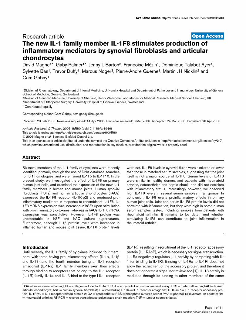
Open Access Available online http://arthritis-research.com/content/8/3/R80 Page 1 of 11 (page number not for citation purposes) Vol 8 No 3 Research article The new IL-1 family member IL-1F8 stimulates production of inflammatory mediators by synovial fibroblasts and articular chondrocytes David Magne 1 *, Gaby Palmer 1 *, Jenny L Barton 3 , Francoise Mézin 1 , Dominique Talabot-Ayer 1 , Sylvette Bas 1 , Trevor Duffy 1 , Marcus Noger 3 , Pierre-Andre Guerne 1 , Martin JH Nicklin 2 and Cem Gabay 1 1 Division of Rheumatology, Department of Internal Medicine, University Hospital and Department of Pathology and Immunology, University of Geneva School of Medicine, Geneva, Switzerland 2 Division of Genomic Medicine, University of Sheffield, Henry Wellcome Laboratories for Medical Research, Medical School, Sheffield, UK 3 Department of Orthopedic Surgery, University Hospital of Geneva, Geneva, Switzerland * Contributed equally Corresponding author: Cem Gabay, [email protected] Received: 28 Feb 2005 Revisions requested: 14 Apr 2005 Revisions received: 8 Mar 2006 Accepted: 24 Mar 2006 Published: 28 Apr 2006 Arthritis Research & Therapy 2006, 8:R80 (doi:10.1186/ar1946) This article is online at: http://arthritis-research.com/content/8/3/R80 © 2006 Magne et al.; licensee BioMed Central Ltd. This is an open access article distributed under the terms of the Creative Commons Attribution License (http://creativecommons.org/licenses/by/2.0 ), which permits unrestricted use, distribution, and reproduction in any medium, provided the original work is properly cited. Abstract Six novel members of the IL-1 family of cytokines were recently identified, primarily through the use of DNA database searches for IL-1 homologues, and were named IL-1F5 to IL-1F10. In the present study, we investigated the effect of IL-1F8 on primary human joint cells, and examined the expression of the new IL-1 family members in human and mouse joints. Human synovial fibroblasts (hSFs) and human articular chondrocytes (hACs) expressed the IL-1F8 receptor (IL-1Rrp2) and produced pro- inflammatory mediators in response to recombinant IL-1F8. IL- 1F8 mRNA expression was increased in hSFs upon stimulation with proinflammatory cytokines, whereas in hACs IL-1F8 mRNA expression was constitutive. However, IL-1F8 protein was undetectable in hSF and hAC culture supernatants. Furthermore, although IL-1β protein levels were increased in inflamed human and mouse joint tissue, IL-1F8 protein levels were not. IL-1F8 levels in synovial fluids were similar to or lower than those in matched serum samples, suggesting that the joint itself is not a major source of IL-1F8. Serum levels of IL-1F8 were similar in healthy donors, and patients with rheumatoid arthritis, osteoarthritis and septic shock, and did not correlate with inflammatory status. Interestingly however, we observed high IL-1F8 levels in several serum samples in all groups. In conclusion, IL-1F8 exerts proinflammatory effects in primary human joint cells. Joint and serum IL-1F8 protein levels did not correlate with inflammation, but they were high in some human serum samples tested, including samples from patients with rheumatoid arthritis. It remains to be determined whether circulating IL-1F8 can contribute to joint inflammation in rheumatoid arthritis. Introduction Until recently, the IL-1 family of cytokines included four mem- bers, with three having pro-inflammatory effects (IL-1α, IL-1β and IL-18) and the fourth member being an IL-1 receptor antagonist (IL-1Ra). IL-1 family members exert their effects through binding to receptors that belong to the IL-1 receptor (IL-1R) family. IL-1α and IL-1β bind to the type I IL-1 receptor (IL-1RI), resulting in recruitment of the IL-1 receptor accessory protein (IL-1RAcP), which is necessary for signal transduction. IL-1Ra negatively regulates IL-1 activity by competing with IL- 1 for binding to IL-1RI. Binding of IL-1Ra to IL-1RI does not allow the recruitment of the accessory protein, and therefore it does not generate a signal (for review see [1]). IL-18 activity is mediated through its binding to other members of the same BSA = bovine serum albumin; CIA = collagen-induced arthritis; ELISA = enzyme-linked immunosorbent assay; FCS = foetal calf serum; hAC = human articular chondrocyte; hSF = human synovial fibroblast; IL = interleukin; IL-1Ra = IL-1 receptor antagonist; IL-1RacP = IL-1 receptor accessory pro- tein; IL-1Rrp2 = IL-1 receptor related protein 2; OA = osteoarthritis; PBS = phosphate-buffered saline; PMA = phorbol 13-myristate 12-acetate; RA = rheumatoid arthritis; RT-PCR = reverse transcriptase polymerase chain reaction; TNF = tumour necrosis factor.
Transcript of The new IL1 family member IL1F8 stimulates production of inflammatory mediators by synovial...
Available online http://arthritis-research.com/content/8/3/R80
Open AccessVol 8 No 3Research articleThe new IL-1 family member IL-1F8 stimulates production of inflammatory mediators by synovial fibroblasts and articular chondrocytesDavid Magne1*, Gaby Palmer1*, Jenny L Barton3, Francoise Mézin1, Dominique Talabot-Ayer1, Sylvette Bas1, Trevor Duffy1, Marcus Noger3, Pierre-Andre Guerne1, Martin JH Nicklin2 and Cem Gabay1
1Division of Rheumatology, Department of Internal Medicine, University Hospital and Department of Pathology and Immunology, University of Geneva School of Medicine, Geneva, Switzerland2Division of Genomic Medicine, University of Sheffield, Henry Wellcome Laboratories for Medical Research, Medical School, Sheffield, UK3Department of Orthopedic Surgery, University Hospital of Geneva, Geneva, Switzerland* Contributed equally
Corresponding author: Cem Gabay, [email protected]
Received: 28 Feb 2005 Revisions requested: 14 Apr 2005 Revisions received: 8 Mar 2006 Accepted: 24 Mar 2006 Published: 28 Apr 2006
Arthritis Research & Therapy 2006, 8:R80 (doi:10.1186/ar1946)This article is online at: http://arthritis-research.com/content/8/3/R80© 2006 Magne et al.; licensee BioMed Central Ltd. This is an open access article distributed under the terms of the Creative Commons Attribution License (http://creativecommons.org/licenses/by/2.0), which permits unrestricted use, distribution, and reproduction in any medium, provided the original work is properly cited.
Abstract
Six novel members of the IL-1 family of cytokines were recentlyidentified, primarily through the use of DNA database searchesfor IL-1 homologues, and were named IL-1F5 to IL-1F10. In thepresent study, we investigated the effect of IL-1F8 on primaryhuman joint cells, and examined the expression of the new IL-1family members in human and mouse joints. Human synovialfibroblasts (hSFs) and human articular chondrocytes (hACs)expressed the IL-1F8 receptor (IL-1Rrp2) and produced pro-inflammatory mediators in response to recombinant IL-1F8. IL-1F8 mRNA expression was increased in hSFs upon stimulationwith proinflammatory cytokines, whereas in hACs IL-1F8 mRNAexpression was constitutive. However, IL-1F8 protein wasundetectable in hSF and hAC culture supernatants.Furthermore, although IL-1β protein levels were increased ininflamed human and mouse joint tissue, IL-1F8 protein levels
were not. IL-1F8 levels in synovial fluids were similar to or lowerthan those in matched serum samples, suggesting that the jointitself is not a major source of IL-1F8. Serum levels of IL-1F8were similar in healthy donors, and patients with rheumatoidarthritis, osteoarthritis and septic shock, and did not correlatewith inflammatory status. Interestingly however, we observedhigh IL-1F8 levels in several serum samples in all groups. Inconclusion, IL-1F8 exerts proinflammatory effects in primaryhuman joint cells. Joint and serum IL-1F8 protein levels did notcorrelate with inflammation, but they were high in some humanserum samples tested, including samples from patients withrheumatoid arthritis. It remains to be determined whethercirculating IL-1F8 can contribute to joint inflammation inrheumatoid arthritis.
IntroductionUntil recently, the IL-1 family of cytokines included four mem-bers, with three having pro-inflammatory effects (IL-1α, IL-1βand IL-18) and the fourth member being an IL-1 receptorantagonist (IL-1Ra). IL-1 family members exert their effectsthrough binding to receptors that belong to the IL-1 receptor(IL-1R) family. IL-1α and IL-1β bind to the type I IL-1 receptor
(IL-1RI), resulting in recruitment of the IL-1 receptor accessoryprotein (IL-1RAcP), which is necessary for signal transduction.IL-1Ra negatively regulates IL-1 activity by competing with IL-1 for binding to IL-1RI. Binding of IL-1Ra to IL-1RI does notallow the recruitment of the accessory protein, and therefore itdoes not generate a signal (for review see [1]). IL-18 activity ismediated through its binding to other members of the same
Page 1 of 11(page number not for citation purposes)
BSA = bovine serum albumin; CIA = collagen-induced arthritis; ELISA = enzyme-linked immunosorbent assay; FCS = foetal calf serum; hAC = human articular chondrocyte; hSF = human synovial fibroblast; IL = interleukin; IL-1Ra = IL-1 receptor antagonist; IL-1RacP = IL-1 receptor accessory pro-tein; IL-1Rrp2 = IL-1 receptor related protein 2; OA = osteoarthritis; PBS = phosphate-buffered saline; PMA = phorbol 13-myristate 12-acetate; RA = rheumatoid arthritis; RT-PCR = reverse transcriptase polymerase chain reaction; TNF = tumour necrosis factor.
Arthritis Research & Therapy Vol 8 No 3 Magne et al.
receptor family, namely IL-18 receptor (IL-18R) and the IL-18Raccessory protein [2].
Six new members of the IL-1 family were recently identified,primarily through the use of DNA database searches for homo-logues of IL-1 [3-10]. These proteins were named IL-1F5 to IL-1F10 [11]. In humans all of the new genes map to less than300 kb of chromosome 2, where they are flanked by IL-1α, IL-1β and IL-1Ra. Sequence alignments and some physical datapredict that the secondary structure of all of the new homo-logues is characterized by a 12-stranded β-trefoil structureshared with IL-1α, IL-1β and IL-1Ra [12]. IL-1F5 was recentlycharacterized at high resolution [13].
Expression patterns and the biological functions of the six newIL-1 family members have not yet been well characterized. Ithas been reported that IL-1F7 forms a complex with IL-18binding protein, which might bind to and sequester IL-18Raccessory protein, thus inhibiting the effects of IL-18 [14]. Inaddition, adenoviral overexpression of IL-1F7 in mouse wasshown to have anti-tumour effects by an undefined mecha-nism, even though rodents appear to lack the IL-1F7 gene[15]. IL-1F10 has been described as a low affinity, nonagonis-tic ligand for IL-1RI [7]. Debets and coworkers [5] have shownthat IL-1F9 activates nuclear factor-κB in Jurkat cells that over-express IL-1 receptor related protein 2 (IL-1Rrp2) and that thisactivation is blocked by IL-1F5, suggesting that IL-1F5 mightbe an IL-1F9 antagonist. Recently, Towne and coworkers [16]reported that, in addition to IL-1F9, IL-1F6 and IL-1F8 alsoactivated nuclear factor-κB and showed that signallingrequired IL-1RAcP. Inhibition of IL-1F6-, IL-1F8-, or IL-1F9-mediated activation of nuclear factor-κB by IL-1F5 wasdescribed as incomplete and inconsistent. In that study, usingan epithelial cell line that expresses both IL-1Rrp2 and IL-1RAcP, the three homologues activated an IL-8 promoterreporter gene construct and secretion of IL-6, even though therequired IL-1F concentrations were much higher than thosenecessary for IL-1β activity.
Rheumatoid arthritis (RA) is characterized by chronic inflam-mation of the synovial tissue in multiple joints that leads to jointdestruction. Major hypotheses have involved dysfunction ofantigen-presenting cells; B cells and autoantibody production;T cell reactivity; and, recently, cytokines (for review, see [17]).Indeed, it is widely recognized that tumour necrosis factor(TNF)-α and IL-1 play key roles in mediating the pathophysio-logical processes that underlie the inflammation and tissuedestruction that occur in RA. The role of the four 'classical' IL-1 family members (for instance, IL-1α, IL-1β, IL-1Ra and IL-18)in the pathogenesis and development of RA was illustrated inmouse models of arthritis, particularly by the spontaneousarthritis that develops in IL-1α transgenic mice [18] as well asin IL-1Ra deficient mice [19]. It was also highlighted by the sig-nificant protection against collagen-induced arthritis (CIA) that
characterizes overexpression of IL-1Ra [20,21] and geneticdeficiency in IL-1α, IL-1β [22], or IL-18 [23].
In the present study we investigated the effects of the new IL-1 family member IL-1F8 on primary human synovial fibroblasts(hSFs) and human articular chondrocytes (hACs), and exam-ined the expression of the new IL-1 homologues in human andmouse joints.
Materials and methodsMaterialsCell culture reagents were obtained from Invitrogen Life Tech-nologies (Basel, Switzerland). Recombinant human IL-1β,recombinant human IL-1F8 and goat polyclonal anti-human IL-1Rrp2, as well as anti-human and anti-mouse IL-1F8 antibod-ies, were purchased from R&D Systems (Abington, UK). Trizolreagent and dNTP were obtained from Invitrogen. Taq DNApolymerase was obtained from Qiagen AG (Basel, Switzer-land). DNase I, AMV-RT (avian myeloblastosis virus-reversetranscriptase), random primers, recombinant ribonucleaseinhibitor and DNA 100 bp ladder were purchased fromPromega (Wallisellen, Switzerland). DNA Master SYBR greenI or Fast Start DNA Master SYBR green I kits were obtainedfrom Roche Molecular Biochemicals (Rotkreuz, Swizerland).
Cell cultureSynovium and articular cartilage were obtained from patientsundergoing joint replacement (knee or hip prosthetic surgery)for osteoarthritis (OA) or broken femoral neck (normal adultarticular cartilage). hSFs and hACs were isolated by colla-genase digestion, as reported previously [24], and cultured inDulbecco's modified Eagle medium supplemented with l-glutamine, streptomycin, penicillin and 10% heat-inactivatedfoetal calf serum (FCS) at 37°C in a humidified atmospherecontaining 5% CO2. Primary hACs were used directly afterisolation from cartilage and hSFs were used between pas-sages 2 and 8. To reduce the nonspecific effects of agonistspresent in FCS, cells were incubated overnight in low-serum(0.5% FCS) medium before the various treatments.
RNA isolationFor RNA isolation, hSFs and hACs were seeded in 25 or 75cm2 flasks. After the indicated incubation times, media wereremoved and cells were lyzed in Trizol. Total RNA was pre-pared according to the manufacturer's instructions. Briefly,homogenization of tissues in Trizol was followed by centrifuga-tion at 10,000 rpm (4°C) for 15 minutes in the presence ofchloroform. The upper aqueous phase was collected and totalRNA was precipitated by addition of isopropanol and centrifu-gation at 7,500 rpm (4°C) for 5 minutes. RNA pellets werewashed with 75% ethanol, dried, reconstituted with sterilewater and quantified by spectrometry.
Page 2 of 11(page number not for citation purposes)
Available online http://arthritis-research.com/content/8/3/R80
Reverse transcription and polymerase chain reactionFor analysis of mRNA levels by RT-PCR and real-time PCR, 1–3 µg total RNA were used. After DNase I digestion, RNA sam-ples were reverse transcribed using AMV-RT (avian myelob-lastosis virus-reverse transcriptase) and random primers in atotal volume of 30–50 µl. Template cDNAs (2.5 µl) wereamplified in a typical 25 µl PCR reaction containing 20 mmol/l Tris-HCl (pH 8.4), 50 mmol/l KCl, 1 µmol/l of the respectiveprimers (Table 1), 2 mmol/l MgCl2, 200 µmol/l dNTP and 2.5units Taq DNA polymerase. The absence of DNA contamina-tion in RNA preparations was tested by including RNA sam-ples that had not been reverse transcribed. Amplificationswere carried out in an Eppendorf Master Cycler (Dr. VaudauxAG, Schonenbuch, Switzerland) under the following condi-tions: denaturation for 3 minutes at 94°C followed by cycles of30 seconds denaturation at 94°C, 30 seconds annealing atthe primer-specific temperature, and 45 seconds elongation at72°C. Amplifications of IL-1Rrp2 (primer pair A) and IL-1F8were performed with 45 cycles, whereas amplification of β-actin was performed with 25 cycles. PCR products were visu-alized on 2% agarose gels containing ethidium bromide. AllPCR products were cloned into the pCRII-TOPO® vector (LifeTechnologies) and their identity was checked by sequencing.
Quantitative real-time polymerase chain reaction analysisExpression of 28S ribosomal RNA, human IL-1Rrp2 (primerpair B), IL-1F8 and IL-1β mRNAs was determined by quantita-tive real-time PCR on reverse-transcribed samples using alight cycler (Roche Diagnostics, Rotkreuz, Swizerland) withthe DNA Master SYBR green I or Fast Start DNA MasterSYBR green I kits as appropriate. Template cDNAs (2 µl) wereamplified in a typical 10 µl PCR reaction containing 0.25
µmol/l of the respective primers (Table 1). The absence ofDNA contamination in RNA preparations was tested by includ-ing RNA samples that had not been reverse transcribed.Primer sequences and conditions for each PCR reaction aredetailed in Table 1. Expression of IL-1Rrp2, IL-1F8, or IL-1βmRNA was corrected for 28S ribosomal RNA levels.
Preparation of human IL-1F8 and IL-1F5 recombinant proteinsRecombinant human IL-1F8 and IL-1F5 were produced inEscherichia coli, as previously reported [3]. To remove endo-toxin contamination, protein samples were treated with poly-myxin B-agarose beads (Sigma, Buchs, Switzerland).Moreover, in order to check that the effects of IL-1F8 were dueto the protein itself and not to endotoxin contamination, insome experiments IL-1F8 was heat-inactivated at 95°C for 5minutes before use. Commercial human recombinant IL-1F8(R&D Systems) was used for comparison in some experimentsand similar data were obtained with our recombinant proteinand with commercial IL-1F8.
Determination of IL-6, IL-8 and nitric oxide levelsFor determination of IL-6, IL-8 and nitric oxide production,hSFs and primary hACs were plated in 96-well plates at a den-sity of 40,000 cells per well. Cells were treated for 48 hourswith the indicated concentrations of IL-1β, IL-1F5 and/or IL-1F8. In some experiments cells were preincubated for 1 hourwith anti-IL-1Rrp2 antibodies (10 µg/ml) before stimulationwith IL-1F8 or IL-1β. Levels of IL-6 and IL-8 in cell superna-tants, as well as IL-6 levels in human serum, were assessedusing enzyme-linked immunosorbent assay (ELISA) kits fromR&D Systems. Production of nitric oxide was assessed, as
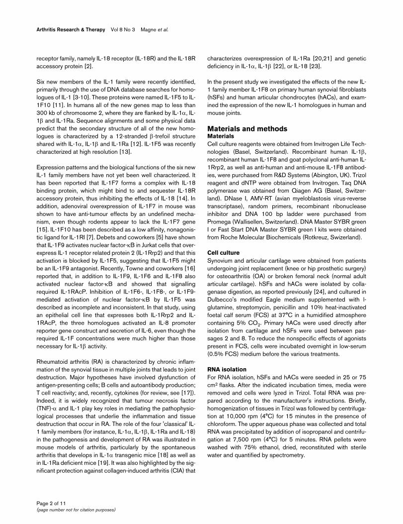
Table 1
Summary of primers used
cDNA Forward and reverse primers Ta (°C) Product (bp) GenBank
IL-1Rrp2 (A) F: 5'-AGCAAAATCCCAGTGTCCAAA-3' 60 291 [AF284434]
R: 5'-ACCCAAAACACAACTCTTCGG-3'
IL-1Rrp2 (B) F: 5'-AGCAAAATCCCAGTGTCCAAA-3' 60 147 [AF284434]
R: 5'-GGTTTACATGTATTCTATGACAG-3'
IL-1F8 F: 5'-ACCAAGGAGAGAGGCATAACTAAT-3' 60 147 [NM173178]
R: 5'-AGTGAACTCAGTCGCATAATGATC-3'
IL-1β F: 5'-GCTGAGGAAGATGCTGGTTC-3' 57 146 [NM000756]
R: 5'-GTGATCGTACAGGTGCATCG-3'
β-actin F: 5'-CCAAGGCCAACCGCGAGAAGATGAC-3' 55 579 [M10277]
R: 5'-AGGGTACATGGTGGTGCCGCCAGAC-3'
28S F: 5'-TTGAAAATCCGCGGGAGA-3' 54 100 [U13369]
R: 5'-ACATTGTTCCAACATGCCAG-3'
Shown are the primer sequences, annealing temperatures (Ta), lengths of the corresponding PCR products, and GenBank accession numbers of the DNA sequences. F, forward; IL, interleukin; IL-1Rrp2, IL-1 receptor related protein 2; R, reverse; PCR, polymerase chain reaction.
Page 3 of 11(page number not for citation purposes)

Arthritis Research & Therapy Vol 8 No 3 Magne et al.
previously described [24], by the Griess reaction using aNaNO2 standard.
Human and mouse tissue samplesSynovial biopsies from patients with OA or inflammatoryarthritides (two patients with RA, one with Lyme disease, onewith sacroid arthritis and one with seronegative arthritis) wereobtained by knee arthroscopy. All samples were immediatelyfrozen in liquid nitrogen. Samples were obtained after appro-priate informed consent, and their use for research wasapproved by the Ethics Committee of the University Hospitalof Geneva.
For induction of CIA, male DBA/1 mice aged between 8 and10 weeks (Janvier, Le Genest-St-Isle, France) were immunizedwith 100 µg native bovine collagen type II (Morwell Diagnos-tics, Zumikon, Switzerland), emulsified in complete Freund'sadjuvant containing 5 mg/ml Mycobacterium tuberculosis(Difco, Basel, Switzerland), by intradermal injection at the baseof tail. On day 21, a booster injection of 100 µg collagen typeII in incomplete Freund's adjuvant was given at the base of the
tail. From day 15 after the first immunization onward, micewere examined daily for the onset of clinical arthritis. Micewere killed at various time points after disease onset andarthritic knees were removed and immediately frozen in liquidnitrogen. Control knees were obtained from naïve DBA/1 miceand from immunized DBA/1 mice without clinical signs ofarthritis. Skin was obtained from phorbol 13-myristate 12-ace-tate (PMA; Sigma, Buchs, Switzerland) treated and controlDBA/1 mice. PMA (1 µg in 200 µl acetone), or acetone (200µl) for control mice, was applied to the dorsal surfaces ofshaved mice. Application of PMA plus acetone or acetonealone was repeated 24 and 48 hours later. Mice were killed 48hours after the last application and small pieces of skin wereimmediately frozen in liquid nitrogen. Institutional approval wasobtained for all animal experiments.
Determination of IL-1F8 protein levels by enzyme-linked immunosorbent assayFor determination of IL-1F8 production, hSFs and primaryhACs were plated in 96-well plates at a density of 40,000 cellsper well. Cells were treated (or not treated) for 48 or 72 hours
Figure 1
IL-1Rrp2 expression by hSFs and hACsIL-1Rrp2 expression by hSFs and hACs. The left panels show a RT-PCR analyses of IL-1Rrp2 expression by (a) hSFs and (b) hACs treated or not treated for 8 hours by IL-1β (1 ng/ml) and/or TNF-α (10 ng/ml), as detailed under Materials and method and in Table 1. The images show represent-ative agarose gel electrophoresis of PCR products. The right panels show real-time PCR analysis of IL-1Rrp2 mRNA levels in hSFs and hACs stim-ulated (black columns) or not stimulated (white columns) for 8 hours with IL-1β (1 ng/ml) and TNF-α (10 ng/ml). The amount of 28S rRNA was monitored as an internal control. The expression of IL-1Rrp2 mRNA was corrected for 28S rRNA levels and the IL-1Rrp2/28S ratios were normal-ized to the maximal value observed in each experiment, which was set to 100%. The results shown represent the mean ± standard error of data obtained with samples from three (hSFs) or four (hACs) independent cultures. IL, interleukin; IL-1Rrp2, IL-1 receptor related protein 2; hAC, human articular chondrocyte; hSF, human synovial fibroblast; RT-PCR, reverse transcriptase polymerase chain reaction; TNF, tumour necrosis factor.
Page 4 of 11(page number not for citation purposes)

Available online http://arthritis-research.com/content/8/3/R80
with 1 ng/ml IL-1β before supernatants were collected.Human and mouse tissue samples were homogenized in ice-cold TNT buffer (50 mmol/l Tris [pH 7.4], 150 mmol/l NaCl, 1mmol/l PMSF, 0.5% Triton X-100) and the lysates werecleared by centrifugation at 13,000 rpm (4°) for 15 minutes.Protein concentration in the lysates was assessed using theBiorad DC protein assay kit (Bio-Rad Laboratories, Hercules,CA, USA). Human serum and synovial fluid samples wereobtained from patients with RA or OA, and control serum wasobtained from healthy blood donors. Serum samples frompatients with septic shock were kindly provided by Dr Pugin(Department of Intensive Care, University Hospital of Geneva,Geneva, Switzerland). For determination of IL-1F8 levels inculture supernatants, human tissue lysates, human serum orsynovial fluids, 96-well plates were coated with a polyclonalanti-human IL-1F8 antibody (R&D Systems), diluted to 1 µg/mlin phosphate-buffered saline (PBS). Wells were then washedwith PBS containing 0.05% Tween-20 and blocked with 1%bovine serum albumin (BSA) in PBS. Samples were applied tothe wells for two hours at room temperature. After washing, abiotin-conjugated polyclonal anti-human IL-1F8 antibody (R&DSystems) was added at a dilution of 1/1000 in PBS and 1%BSA, and incubated for 2 hours at room temperature. Boundantibody was detected by incubation with streptavidin-horse-radish peroxidase (dilution 1/1000 in PBS and 1% BSA) for20 minutes. Colour was developed using tetramethylbenzidineand H2O2, the reaction was stopped with H2SO4 2N, and opti-cal density was assessed at 450 nm. Recombinant human IL-F8 was used for the standard curve. To detect mouse IL-1F8,a similar ELISA was set up using a polyclonal anti-mouse IL-1F8 antibody, a biotin-conjugated polyclonal anti-mouse IL-1F8 antibody and recombinant mouse IL-1F8 (R&D Systems).The detection limit of these assays was 19 pg/ml.
Statistical analysisThe significance of differences was calculated by analysis ofvariance or Mann-Whitney test as appropriate. A differencebetween experimental groups was considered statistically sig-nificant when the P value was below 0.05.
ResultsAs a first approach to investigate expression of new IL-1 familymembers during arthritis, we examined IL-1F5 to IL-1F10mRNA expression by RT-PCR in joints of mice with CIA and insynovial biopsies from patients with RA or OA. IL-1F8 was theonly new IL-1 family member for which we detected mRNAexpression both in human synovial biopsies and in mousejoints. In addition, we also observed IL-1F9 mRNA expressionin mouse joints, whereas expression of IL-1F6 and IL-1F7mRNA was detected in some human synovial samples (datanot shown).
A recent study [16] reported that IL-1F8 signalling requires thepresence of both IL-1Rrp2 and IL-1RAcP. Therefore, we inves-tigated IL-1Rrp2 mRNA expression by hSFs and hACs,
because IL-1RAcP expression in these cells has already beenreported [25] and is further demonstrated by their well estab-lished responsiveness to IL-1β. As shown in Figure 1, bothhSFs and hACs expressed basal levels of IL-1Rrp2 mRNA,which were not upregulated by IL-1β and/or TNF-α. In con-trast, we did not observe IL-1Rrp2 expression in THP-1 andJurkat cell lines (data not shown), confirming previous findings[16,25].
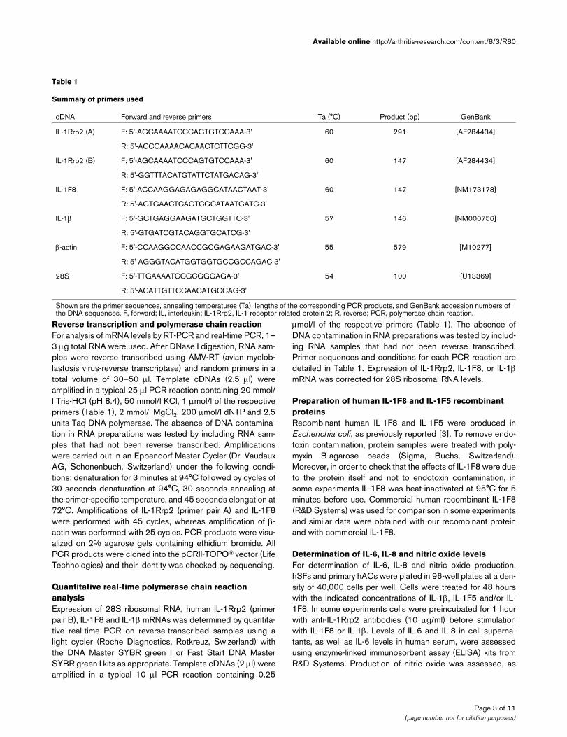
Because hSFs and hACs express IL-1Rrp2 mRNA, wehypothesized that these cells should be able to respond to IL-1F8 without need for receptor over-expression. As indicatedby Figure 2, IL-1F8 stimulated both IL-6 and IL-8 production inhSFs and hACs. The response was stronger in hACs, with asignificant increase in IL-6 production with 500 ng/ml of IL-1F8. In addition, IL-1F8 also stimulated nitric oxide productionby hACs (Figure 2e), suggesting that its effects might be sim-ilar to those exhibited by IL-1β. There was no synergy betweenIL-1β and IL-1F8 for the stimulation of IL-6 production byhACs, and the effect of 5 µg/ml IL-1F8 was additive with thatof low doses of IL-1β (1–10 pg/ml; data not shown). Further-more, the effects of IL-1F8 were indeed due to the proteinitself and not to endotoxin contamination because heat-inacti-vated IL-1F8 failed to stimulate IL-6 production in hACs (Fig-ure 3a). The effects of IL-1F8 were mediated by IL-1Rrp2 andcould be completely blocked in presence of a polyclonal anti-IL-1Rrp2 antibody (Figure 3b). Finally, the reported correlationbetween IL-1F8 responsiveness and IL-1Rrp2 expression [16]was supported by our observation that C28/I2 and SW1353'chondrocyte-like' cell lines and human dermal fibroblasts inwhich levels of IL-1Rrp2 mRNA were very low or absent didnot produce IL-6 in response to 5 µg/ml IL-1F8. In contrast,incubation of these cells with 1 ng/ml of IL-1β stimulated IL-6production (data not shown).
Debets and coworkers [5] reported that IL-1F5 could antago-nize the effects of IL-1F9 when it was added at equimolar con-centrations; we therefore tested the ability of recombinanthuman IL-1F5 concentrations from 50 ng/ml to 5 µg/ml toinhibit the effects of 5 µg/ml IL-1F8 on IL-6 production inhACs. In these conditions, antagonism by IL-1F5 of the effectsof IL-1F8 was incomplete and not reproducible (data notshown).
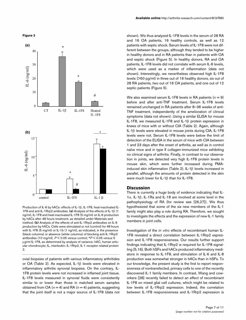
We then screened various cell types present in the inflamedjoint for endogenous IL-1F8 mRNA expression in vitro. By RT-PCR, IL-1F8 expression was observed in hSFs when theywere treated for eight hours with IL-1β, TNF-α, or both (Figure4a). By real-time PCR, we confirmed increased IL-1F8 mRNAexpression after 8 hours stimulation of hSFs with IL-1β alone,IL-1β plus TNF-α, or IL-1α alone (Figure 4b). The increase inIL-1F8 mRNA levels was strongest with stimulation by 1 ng/mlIL-1β, as compared with 0.1 and 10 ng/ml (data not shown).The steady state levels of IL-1F8 mRNA expression peaked at8 hours (Figure 4c) in response to 1 ng/ml IL-1β, which is sim-
Page 5 of 11(page number not for citation purposes)

Arthritis Research & Therapy Vol 8 No 3 Magne et al.
ilar to the time course of induction of endogenous IL-1β mRNAby IL-1β in hSFs. In hACs expression of IL-1F8 mRNA wasconstitutive and IL-1F8 levels were not affected by stimulationof cells with IL-1β and TNF-α for 8 hours, whereas this treat-ment consistently induced IL-1β gene expression (Figure 4d).We also observed that the THP-1 monocyte cell line and Jur-kat T-cell line expressed basal levels of IL-1F8 mRNA, but real-time PCR experiments failed to detect IL-1F8 mRNA upregu-
lation in response to various stimuli, including IL-1β, TNF-α, IL-4 and PMA (data not shown).
Next, we assessed IL-1F8 protein levels by ELISA in culturesupernatants of hSFs and hACs stimulated (or not stimulated)with IL-1β for 48 or 72 hours. IL-1F8 protein levels were belowthe limit of detection of the ELISA (19 pg/ml) in all samples.We also measured IL-1F8 and IL-1β protein expression in syn-
Figure 2
Production of IL-6, IL-8 and nitric oxide by hSFs and hACs: effects of IL-1F8 and IL-1βProduction of IL-6, IL-8 and nitric oxide by hSFs and hACs: effects of IL-1F8 and IL-1β. Shown is an analysis of the effects of IL-1F8 and IL-1β on production of (a) IL-6 and (c) IL-8 by hSFs, and of (b) IL-6, (d) IL-8 and (e) nitric oxide by hACs. Cells were treated with the indicated cytokine con-centrations for 48 hours, as detailed under Materials and method. *P < 0.05 versus control; #P < 0.05 versus 0.1 ng/ml IL-1β; &P < 0.05 versus 500 ng/ml IL-1F8, determined using analysis of variance. IL, interleukin; hAC, human articular chondrocyte; hSF, human synovial fibroblast.
Page 6 of 11(page number not for citation purposes)
Available online http://arthritis-research.com/content/8/3/R80
ovial biopsies of patients with various inflammatory arthritidesor OA (Table 2). As expected, IL-1β levels were elevated ininflammatory arthritis synovial biopsies. On the contrary, IL-1F8 protein levels were not increased in inflamed joint tissue.IL-1F8 levels measured in synovial fluids were consistentlysimilar to or lower than those in matched serum samplesobtained from OA (n = 4) and RA (n = 4) patients, suggestingthat the joint itself is not a major source of IL-1F8 (data not
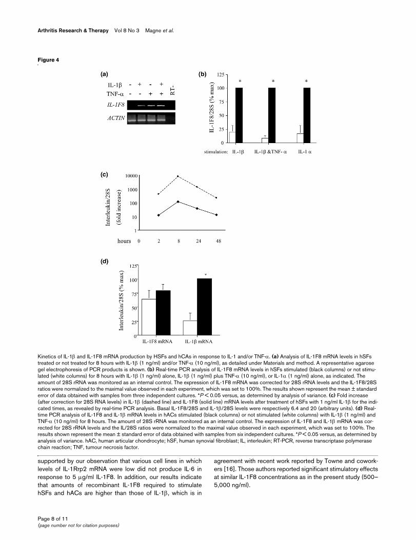
shown). We thus analyzed IL-1F8 levels in the serum of 28 RAand 16 OA patients, 16 healthy controls, as well as 12patients with septic shock. Serum levels of IL-1F8 were not dif-ferent between the groups, although they tended to be higherin healthy donors and in RA patients than in patients with OAand septic shock (Figure 5). In healthy donors, RA and OApatients, IL-1F8 levels did not correlate with serum IL-6 levels,which were used as a marker of inflammation (data notshown). Interestingly, we nevertheless observed high IL-1F8levels (>50 pg/ml) in three out of 16 healthy donors, six out of28 RA patients, two out of 16 OA patients, and one out of 12septic patients (Figure 5).
We also examined serum IL-1F8 levels in RA patients (n = 9)before and after anti-TNF treatment. Serum IL-1F8 levelsremained unchanged in RA patients after 8–36 weeks of anti-TNF treatment, independently of the amelioration of clinicalsymptoms (data not shown). Using a similar ELISA for mouseIL-1F8, we measured IL-1F8 and IL-1β protein expression inknees of mice with or without CIA (Table 2). Again, althoughIL-1β levels were elevated in mouse joints during CIA, IL-1F8levels were not. Serum IL-1F8 levels were below the limit ofdetection of the ELISA in the serum of mice with CIA between1 and 23 days after the onset of arthritis, as well as in controlnaïve mice and in type II collagen-immunized mice exhibitingno clinical signs of arthritis. Finally, in contrast to our observa-tion in joints, we detected very high IL-1F8 protein levels inmouse skin, which were further increased during PMA-induced skin inflammation (Table 2). IL-1β levels increased inparallel, although the amounts of protein detected in the skinwere much lower for IL-1β than for IL-1F8.
DiscussionThere is currently a huge body of evidence indicating that IL-1α, IL-1β, IL-1Ra and IL-18 are involved at some level in thepathophysiology of RA (for review see [26,27]). We thushypothesized that some of the six new members of the IL-1family might also play a role during RA. Therefore, we soughtto investigate the effects and the expression of new IL-1 familymembers in joint cells.
Investigation of the in vitro effects of recombinant human IL-1F8 revealed a direct correlation between IL-1Rrp2 expres-sion and IL-1F8 responsiveness. Our results further supportfindings indicating that IL-1Rrp2 is required for IL-1F8 signal-ling [5,16]. Both hSFs and hACs produced inflammatory medi-ators in response to IL-1F8, and stimulation of IL-6 and IL-8production was somewhat stronger in hACs than in hSFs. Toour knowledge, the present study is the first to report respon-siveness of nontransfected, primary cells to one of the recentlydiscovered IL-1 family members. In contrast, Wang and cow-orkers [28] recently failed to detect an effect of recombinantIL-1F8 on mixed glial cell cultures, which might be related tolow levels of IL-1Rrp2 expression. Indeed, the correlationbetween IL-1F8 responsiveness and IL-1Rrp2 expression is
Figure 3
Production of IL-6 by hACs: effects of IL-1β, IL-1F8, heat-inactivated IL-1F8 and anti-IL-1Rrp2 antibodiesProduction of IL-6 by hACs: effects of IL-1β, IL-1F8, heat-inactivated IL-1F8 and anti-IL-1Rrp2 antibodies. (a) Analysis of the effects of IL-1β (1 ng/ml), IL-1F8 and heat-inactivated IL-1F8 (5 mg/ml) on IL-6 production by hACs after 48 hours treatment, as detailed under Materials and method. (b) Analysis of the effects of anti-IL-1Rrp2 antibodies on IL-6 production by hACs. Cells were stimulated or not (control) for 48 hours with IL-1F8 (5 mg/ml) or IL-1b (1 ng/ml), as indicated, in the presence (black columns) or absence (white columns) of blocking anti-IL-1Rrp2 antibodies (10 mg/ml). P < 0.05 versus control; #P < 0.05 versus 5 µg/ml IL-1F8, as determined by analysis of variance. hAC, human artic-ular chondrocyte; IL, interleukin; IL-1Rrp2, IL-1 receptor related protein 2.
Page 7 of 11(page number not for citation purposes)
Arthritis Research & Therapy Vol 8 No 3 Magne et al.
supported by our observation that various cell lines in whichlevels of IL-1Rrp2 mRNA were low did not produce IL-6 inresponse to 5 µg/ml IL-1F8. In addition, our results indicatethat amounts of recombinant IL-1F8 required to stimulatehSFs and hACs are higher than those of IL-1β, which is in
agreement with recent work reported by Towne and cowork-ers [16]. Those authors reported significant stimulatory effectsat similar IL-1F8 concentrations as in the present study (500–5,000 ng/ml).
Figure 4
Kinetics of IL-1β and IL-1F8 mRNA production by HSFs and hCAs in response to IL-1 and/or TNF-αKinetics of IL-1β and IL-1F8 mRNA production by HSFs and hCAs in response to IL-1 and/or TNF-α. (a) Analysis of IL-1F8 mRNA levels in hSFs treated or not treated for 8 hours with IL-1β (1 ng/ml) and/or TNF-α (10 ng/ml), as detailed under Materials and method. A representative agarose gel electrophoresis of PCR products is shown. (b) Real-time PCR analysis of IL-1F8 mRNA levels in hSFs stimulated (black columns) or not stimu-lated (white columns) for 8 hours with IL-1β (1 ng/ml) alone, IL-1β (1 ng/ml) plus TNF-α (10 ng/ml), or IL-1α (1 ng/ml) alone, as indicated. The amount of 28S rRNA was monitored as an internal control. The expression of IL-1F8 mRNA was corrected for 28S rRNA levels and the IL-1F8/28S ratios were normalized to the maximal value observed in each experiment, which was set to 100%. The results shown represent the mean ± standard error of data obtained with samples from three independent cultures. *P < 0.05 versus, as determined by analysis of variance. (c) Fold increase (after correction for 28S RNA levels) in IL-1β (dashed line) and IL-1F8 (solid line) mRNA levels after treatment of hSFs with 1 ng/ml IL-1β for the indi-cated times, as revealed by real-time PCR analysis. Basal IL-1F8/28S and IL-1β/28S levels were respectively 6.4 and 20 (arbitrary units). (d) Real-time PCR analysis of IL-1F8 and IL-1β mRNA levels in hACs stimulated (black columns) or not stimulated (white columns) with IL-1β (1 ng/ml) and TNF-α (10 ng/ml) for 8 hours. The amount of 28S rRNA was monitored as an internal control. The expression of IL-1F8 and IL-1β mRNA was cor-rected for 28S rRNA levels and the IL/28S ratios were normalized to the maximal value observed in each experiment, which was set to 100%. The results shown represent the mean ± standard error of data obtained with samples from six independent cultures. *P < 0.05 versus, as determined by analysis of variance. hAC, human articular chondrocyte; hSF, human synovial fibroblast; IL, interleukin; RT-PCR, reverse transcriptase polymerase chain reaction; TNF, tumour necrosis factor.
Page 8 of 11(page number not for citation purposes)

Available online http://arthritis-research.com/content/8/3/R80
The need for high concentrations of recombinant IL-1F8 is notunderstood, and thus far no biological effect of any of the newIL-1 family members has been reported at below about 10-7
mol/l, as compared with about 10-11 mol/l for IL-1β and about10-9 mol/l for IL-18. Interestingly, we recently observed thattransfection of IL-1Rrp2 expressing C20A4 chondrocytic cellswith an expression vector for human IL-1F8, which led to theproduction of moderate quantities of IL-1F8 (50–200 pg/ml inculture supernatants after 48 hours), efficiently induced IL-6secretion in these cells, as compared with empty vector trans-fected control cells (GP, FM and CG; unpublished observa-tions). These observations suggest that endogenouslyexpressed IL-1F8 is active at much lower doses than recom-binant IL-1F8, although the reason for this discrepancy is stillunknown and is currently under investigation. One hypothesisis that post-translational modifications of the IL-1F8 proteinmight be important for its biological activity and might be lack-ing in recombinant IL-1F8 produced in E. coli. Levels of IL-1F8detected for RA patients ranged up to 347 pg/ml in serum andup to 176 pg/ml in synovial fluid, and according to our obser-vations in C20A4 cells such concentrations of endogenouslyproduced IL-1F8 might be sufficient to trigger biologicaleffects in joint cells.
There is some controversy concerning the putative antagonisteffects of IL-1F5 [5,16]. Debets and coworkers [5] have
shown that IL-1F5 inhibits IL-1F9 induced nuclear factor-κBactivation in Jurkat T cells overexpressing IL-1Rrp2 [5], butTowne and coworkers [16] did not observe consistent inhibi-tory effects of IL-1F5 on IL-1F6-, IL-1F8-, or IL-1F9-inducedactivation of nuclear factor-κB in the same cells. Although insome experiments we observed antagonistic effects of IL-1F5on the inflammatory action of IL-1F8 on hACs and hSFs, thisantagonism was inconsistent and incomplete. We currentlyhave no explanation for these nonreproducible effects. Theuse of primary cultures may account for such findings in ourstudy but not in that of Towne and coworkers [16], who usedcell lines. It is also possible that recombinant IL-1F5 lacks con-formational stability or post-translational modification, and thatthis may alter its activity. The possible role played by IL-1F5therefore remains unknown.
Investigation by RT-PCR of their expression in joints of micewith CIA and in synovial tissue from patients with RA revealedthat, among the newly cloned IL-1 family members, only IL-1F8was expressed in both mouse and human joints. QuantitativePCR experiments demonstrated a significant upregulation ofIL-1F8 mRNA levels in cultured hSFs in response to IL-1β and/or TNF-α. In contrast, IL-1F8 mRNA expression was constitu-tive in hACs and was not affected by inflammatory stimuli. Sim-ilarly, although monocyte and T-lymphocyte cell lines expressIL-1F8 mRNA to some extent, IL-1F8 levels were not increasedin response to a panel of stimuli. It has been reported that Tcells, either stimulated with anti-CD3 and/or anti-CD28 or leftunstimulated, do not express IL-1F8 mRNA, whereas lipopoly-saccharide-treated monocytes do [10]. Despite IL-1F8 mRNAexpression, IL-1F8 protein expression was below the limit of
Figure 5
IL-1F8 protein levels in control individuals, patients with RA, OA and septic shockIL-1F8 protein levels in control individuals, patients with RA, OA and septic shock. Shown are serum IL-1F8 protein levels in healthy donors (n = 16), patients with RA (n = 28) or OA (n = 16) patients, and patients with septic shock (n = 12), as determined by ELISA. Individual values (grey dots) and mean (stippled lines) ± standard error (black lines) are shown. Differences between the groups were not significant. ELISA, enzyme-linked immunosorbent assay; IL, interleukin; OA, oste-oarthritis; RA, rheumatoid arthritis.
Table 2
IL-1F8 protein levels in mouse and human joint samples and in mouse skin
Samples Patients/animals IL-1F8 (pg/mg protein)
IL-1β (pg/mg protein)
Human synovium
OA (n = 4) 3.7 ± 1.4 3.6 ± 0.3
Inflammatory arthritis (n = 5)
3.9 ± 1.0 11.6 ± 5.7a
Mouse joint Naïve (n = 1) 2.8 2.3
Nonarthritic (n = 1) 2.7 4.5
CIA earlyb (n = 3) 2.2 ± 0.3 16.6 ± 1.2c
CIA lateb (n = 3) 4.5 ± 1.4 8.8 ± 0.8
Mouse skin Control (n = 3) 761.7 ± 313.4 16 ± 3
PMA (n = 3) 14569.1 ± 3632.5d
28 ± 3d
IL-1F8 protein levels were determined using enzyme-linked immunosorbent assay. aP < 0.05 versus osteoparthritis (OA), as assessed using the Mann-Whitney test. bCollagen-induced arthritis (CIA) early: days 1–7 after the onset of arthritis; CIA late: days 8–21 after onset of arthritis. cP < 0.05 versus late CIA, as assessed using analysis of variance. dP < 0.05 versus control, as assessed using analysis of variance.
Page 9 of 11(page number not for citation purposes)

Arthritis Research & Therapy Vol 8 No 3 Magne et al.
detection of our assay in hSF and hAC culture supernatants.In human OA and normal mouse joint tissue, IL-1F8 proteinexpression levels were similar to those of IL-1β. However,although IL-1β protein levels were increased in inflamed joints,IL-1F8 levels were not. Interestingly, a very different situationapplied to mouse skin samples, in which IL-1F8 levels werevery high and further increased with inflammation. Further-more, IL-1F8 levels in synovial fluids were similar to or lowerthan those measured in matched serum samples, suggestingthat the joint itself is not a major source of IL-1F8. Indeed, inthe case of IL-6, for instance, which is produced in the joint,synovial fluid concentrations are 100-fold to 1000-fold higherthan those measured in serum [29]. Serum levels of IL-1F8 didnot differ between healthy donors, and patients with RA, OAand septic shock, and did not correlate with inflammatory sta-tus. Interestingly, however, we observed high IL-1F8 levels inseveral serum samples in all of these groups. The cause ofsuch high serum IL-F8 levels and the source of circulating IL-1F8 are as yet unknown.
ConclusionIL-1F8 exerts proinflammatory effects in primary human jointcells. However, although IL-1F8 mRNA is expressed in hSFand hAC, joint cells are not a major source of IL-1F8 protein.Joint and serum IL-1F8 protein levels did not correlate withinflammation, but IL-1F8 was elevated in some human serumsamples tested, including several samples from RA patients. Itremains to be determined whether, in some cases, circulatingIL-1F8 can contribute to joint inflammation in RA.
Competing interestsThe authors declare that they have no competing interests.
Authors' contributionsDM, GP, FM and DT-A performed the experiments concerningthe in vitro effects of IL-1F8, as well as the mRNA and proteinexpression studies. SB, TD and MN collected and providedhuman tissue, synovial fluid and serum samples. JLB andMJHN produced the recombinant IL-1F proteins. DM, GP, SB,PAG, MJHN and CG participated in the design of the study,data analysis, and drafting and reviewing of the manuscript. Allauthors read and approved the final manuscript.
AcknowledgementsThis work was supported the Swiss National Science Foundation (grants 3200-107592/1 to CG and 3100-064123.00/1 to PAG).
References1. Arend WP, Malyak M, Guthridge CJ, Gabay C: Interleukin-1
receptor antagonist: role in biology. Annu Rev Immunol 1998,16:27-55.
2. Born TL, Thomassen E, Bird TA, Sims JE: Cloning of a novelreceptor subunit, AcPL, required for interleukin-18 signaling. JBiol Chem 1998, 273:29445-29450.
3. Barton JL, Herbst R, Bosisio D, Higgins L, Nicklin MJ: A tissuespecific IL-1 receptor antagonist homolog from the IL-1 clus-ter lacks IL-1, IL-1ra, IL-18 and IL-18 antagonist activities. EurJ Immunol 2000, 30:3299-3308.
4. Busfield SJ, Comrack CA, Yu G, Chickering TW, Smutko JS, ZhouH, Leiby KR, Holmgren LM, Gearing DP, Pan Y: Identification andgene organization of three novel members of the IL-1 familyon human chromosome 2. Genomics 2000, 66:213-216.
5. Debets R, Timans JC, Homey B, Zurawski S, Sana TR, Lo S, Wag-ner J, Edwards G, Clifford T, Menon S, et al.: Two novel IL-1 fam-ily members, IL-1 delta and IL-1 epsilon, function as anantagonist and agonist of NF-kappa B activation through theorphan IL-1 receptor-related protein 2. J Immunol 2001,167:1440-1446.
6. Kumar S, McDonnell PC, Lehr R, Tierney L, Tzimas MN, GriswoldDE, Capper EA, Tal-Singer R, Wells GI, Doyle ML, Young PR:Identification and initial characterization of four novel mem-bers of the interleukin-1 family. J Biol Chem 2000,275:10308-10314.
7. Lin H, Ho AS, Haley-Vicente D, Zhang J, Bernal-Fussell J, PaceAM, Hansen D, Schweighofer K, Mize NK, Ford JE: Cloning andcharacterization of IL-1HY2, a novel interleukin-1 familymember. J Biol Chem 2001, 276:20597-20602.
8. Mulero JJ, Pace AM, Nelken ST, Loeb DB, Correa TR, Drmanac R,Ford JE: IL1HY1: a novel interleukin-1 receptor antagonistgene. Biochem Biophys Res Commun 1999, 263:702-706.
9. Pan G, Risser P, Mao W, Baldwin DT, Zhong AW, Filvaroff E,Yansura D, Lewis L, Eigenbrot C, Henzel WJ, et al.: IL-1H, aninterleukin 1-related protein that binds IL-18 receptor/IL-1Rrp.Cytokine 2001, 13:1-7.
10. Smith DE, Renshaw BR, Ketchem RR, Kubin M, Garka KE, SimsJE: Four new members expand the interleukin-1 superfamily.J Biol Chem 2000, 275:1169-1175.
11. Sims JE, Nicklin MJ, Bazan JF, Barton JL, Busfield SJ, Ford JE,Kastelein RA, Kumar S, Lin H, Mulero JJ, et al.: A new nomencla-ture for IL-1-family genes. Trends Immunol 2001, 22:536-537.
12. Taylor SL, Renshaw BR, Garka KE, Smith DE, Sims JE: Genomicorganization of the interleukin-1 locus. Genomics 2002,79:726-733.
13. Dunn EF, Gay NJ, Bristow AF, Gearing DP, O'Neill LA, Pei XY:High-resolution structure of murine interleukin 1 homologueIL-1F5 reveals unique loop conformations for receptor bindingspecificity. Biochemistry 2003, 42:10938-10944.
14. Bufler P, Azam T, Gamboni-Robertson F, Reznikov LL, Kumar S,Dinarello CA, Kim SH: A complex of the IL-1 homologue IL-1F7b and IL-18-binding protein reduces IL-18 activity. ProcNatl Acad Sci USA 2002, 99:13723-13728.
15. Gao W, Kumar S, Lotze MT, Hanning C, Robbins PD, GambottoA: Innate immunity mediated by the cytokine IL-1 homologue4 (IL-1H4/IL-1F7) induces IL-12-dependent adaptive and pro-found antitumor immunity. J Immunol 2003, 170:107-113.
16. Towne JE, Garka KE, Renshaw BR, Virca GD, Sims JE: Interleukin(IL)-1F6, IL-1F8, and IL-1F9 signal through IL-1Rrp2 and IL-1RAcP to activate the pathway leading to NF-kappaB andMAPKs. J Biol Chem 2004, 279:13677-13688.
17. Arend WP, Gabay C: Cytokines in the rheumatic diseases.Rheum Dis Clin North Am 2004, 30:41-67. v-vi.
18. Niki Y, Yamada H, Seki S, Kikuchi T, Takaishi H, Toyama Y,Fujikawa K, Tada N: Macrophage- and neutrophil-dominantarthritis in human IL-1 alpha transgenic mice. J Clin Invest2001, 107:1127-1135.
19. Horai R, Saijo S, Tanioka H, Nakae S, Sudo K, Okahara A, Ikuse T,Asano M, Iwakura Y: Development of chronic inflammatoryarthropathy resembling rheumatoid arthritis in interleukin 1receptor antagonist-deficient mice. J Exp Med 2000,191:313-320.
20. Ma Y, Thornton S, Boivin GP, Hirsh D, Hirsch R, Hirsch E: Alteredsusceptibility to collagen-induced arthritis in transgenic micewith aberrant expression of interleukin-1 receptor antagonist.Arthritis Rheum 1998, 41:1798-1805.
21. Palmer G, Talabot-Ayer D, Szalay-Quinodoz L, Maret M, ArendWP, Gabay C: Mice transgenic for intracellular interleukin-1receptor antagonist type 1 are protected from collagen-induced arthritis. Eur J Immunol 2003, 33:434-440.
22. Saijo S, Asano M, Horai R, Yamamoto H, Iwakura Y: Suppressionof autoimmune arthritis in interleukin-1-deficient mice inwhich T cell activation is impaired due to low levels of CD40ligand and OX40 expression on T cells. Arthritis Rheum 2002,46:533-544.
Page 10 of 11(page number not for citation purposes)
http://www.ncbi.nlm.nih.gov/entrez/query.fcgi?cmd=Retrieve&db=PubMed&dopt=Abstract&list_uids=9792649
http://www.ncbi.nlm.nih.gov/entrez/query.fcgi?cmd=Retrieve&db=PubMed&dopt=Abstract&list_uids=9792649
http://www.ncbi.nlm.nih.gov/entrez/query.fcgi?cmd=Retrieve&db=PubMed&dopt=Abstract&list_uids=9778220

Available online http://arthritis-research.com/content/8/3/R80
23. Wei XQ, Leung BP, Arthur HM, McInnes IB, Liew FY: Reducedincidence and severity of collagen-induced arthritis in micelacking IL-18. J Immunol 2001, 166:517-521.
24. Guicheux J, Palmer G, Relic B, Mezin F, Caverzasio J, ApostolidesP, Gauchat JF, Gabay C, Guerne PA: Primary human articularchondrocytes, dedifferentiated chondrocytes, and synovio-cytes exhibit differential responsiveness to interleukin-4: cor-relation with the expression pattern of the common receptorgamma chain. J Cell Physiol 2002, 192:93-101.
25. Attur MG, Dave M, Cipolletta C, Kang P, Goldring MB, Patel IR,Abramson SB, Amin AR: Reversal of autocrine and paracrineeffects of interleukin 1 (IL-1) in human arthritis by type II IL-1decoy receptor. Potential for pharmacological intervention. JBiol Chem 2000, 275:40307-40315.
26. Dayer JM: The pivotal role of interleukin-1 in the clinical mani-festations of rheumatoid arthritis. Rheumatology (Oxford)2003, 42(Suppl 2):ii3-ii10.
27. Liew FY, Wei XQ, McInnes IB: Role of interleukin 18 in rheuma-toid arthritis. Ann Rheum Dis 2003, 62(Suppl 2):ii48-ii50.
28. Wang P, Meinhardt B, Andre R, Renshaw BR, Kimber I, RothwellNJ, Pinteaux E: The interleukin1-related cytokine IL-1F8 isexpressed in glial cells, but fails to induce IL-1beta signallingresponses. Cytokine 2005, 29:245-250.
29. Bas S, Gauthier BR, Spenato U, Stingelin S, Gabay C: CD14 is anacute-phase protein. J Immunol 2004, 172:4470-4479.
Page 11 of 11(page number not for citation purposes)