Quantitative gait analysis as a method to assess mechanical hyperalgesia modulated by...
7
Open Access Available online http://arthritis-research.com/content/9/5/R91 Page 1 of 7 (page number not for citation purposes) Vol 9 No 5 Research article Quantitative gait analysis as a method to assess mechanical hyperalgesia modulated by disease-modifying antirheumatoid drugs in the adjuvant-induced arthritic rat Shabana Usman Simjee 1 , Huma Jawed 1 , Javeria Quadri 2 and Sheikh Arshad Saeed 2 1 HEJ Research Institute of Chemistry, International Centre for Chemical and Biological Sciences, University of Karachi, Karachi 75270, Pakistan 2 Dr Panjwani Centre for Molecular Medicine and Drug Research, International Centre for Chemical and Biological Sciences, University of Karachi, Karachi 75270, Pakistan Corresponding author: Shabana Usman Simjee, [email protected] Received: 23 May 2007 Revisions requested: 10 Aug 2007 Revisions received: 21 Aug 2007 Accepted: 11 Sep 2007 Published: 11 Sep 2007 Arthritis Research & Therapy 2007, 9:R91 (doi:10.1186/ar2290) This article is online at: http://arthritis-research.com/content/9/5/R91 © 2007 Simjee et al., licensee BioMed Central Ltd. This is an open access article distributed under the terms of the Creative Commons Attribution License (http://creativecommons.org/licenses/by/2.0 ), which permits unrestricted use, distribution, and reproduction in any medium, provided the original work is properly cited. Abstract In the present study, azothioprine, chloroquine, D-penicillamine, methotrexate and sodium aurothiomalate (gold salt) were evaluated for possible disease-modifying effects in the adjuvant- induced arthritis model of human rheumatoid arthritis in rats. Gait analysis was used to examine the role of disease-modifying antirheumatic drugs in the development of pain. Body weights were also measured to monitor the progression of disease and the systemic antiarthritic effects of the test compounds used in this study, as well as their systemic toxicity. Our results showed that azothioprine (5 mg/kg/day), chloroquine (12.5 mg/kg/day), sodium aurothiomalate (2.5 mg/kg/day) and methotrexate (1 mg/kg/week) not only inhibited the macroscopic changes such as erythema and swelling of limbs, but also exhibited significant reversal of gait deficits seen in the untreated or saline-treated arthritic rats. No reduction in the body weights were observed in the arthritic rats treated with azothioprine, chloroquine, sodium aurothiomalate and methotrexate. D-Penicillamine (12.5 mg/kg/ day), however, showed a significant reduction (P < 0.03) in the body weights of the arthritic rats over a period of 22 days; furthermore, it was unable to show any reduction in arthritic score (P < 0.1). In earlier experiments, chloroquine and methotrexate failed to suppress carageenan-induced edema, suggesting that the mode of antiarthritic action may be different from those of nonsteroidal anti-inflammatory agents. Since these disease-modifying antirheumatic drugs are reported to have an immunomodulatory role, especially the gold salt, which influences the monocyte–macrophage system, it is suggested that the observed antiarthritic effects of disease-modifying antirheumatic drugs may be partly attributed to their immunomodulatory activity. Introduction Chronic pain, a devastating and widespread problem, is a syn- drome that cuts across traditionally defined disciplinary boundaries within the health sciences. Patients with chronic pain, compared with all other medical conditions, suffer dra- matic reductions in physical, psychological and social wellbe- ing [1-3]. Within this group of patients, arthritis is the second largest self-reported cause of pain [4]. Although there is no rheumatoid arthritis cure, there are effective treatments that can alleviate the symptoms and improve the quality of life. The nonsteroidal anti-inflammatory drugs and glucocorticoids are largely used for treatment of rheumatoid arthritis in spite of their systemic, gastric and renal toxicities [5-11]. These cur- rently available analgesic and anti-inflammatory drugs are clearly not adequate therapy. In addition to these classical available therapies, there are several reports regarding the use of disease-modifying antirheumatic drugs (DMARDs), which act as potentially effective therapies for rheumatoid arthritis [12,13]. DMARD treatment is currently based on symptomatic relief of pain and inflammation associated with arthritis to increase joint function and mobility. In order to study the effects of DMARDs in the progression of disease, we have used the adjuvant-induced arthritis model in the rat. This model has biochemical and pathological features similar to rheumatic disease in human, and merits investigation AZ = azothioprine; CQ = chloroquine; D-PEN = D-penicillamine; DMARD = disease-modifying antirheumatic drug; GS = sodium aurothiomalate (gold salt); MTX = methotrexate.
Transcript of Quantitative gait analysis as a method to assess mechanical hyperalgesia modulated by...
Available online http://arthritis-research.com/content/9/5/R91
Open AccessVol 9 No 5Research articleQuantitative gait analysis as a method to assess mechanical hyperalgesia modulated by disease-modifying antirheumatoid drugs in the adjuvant-induced arthritic ratShabana Usman Simjee1, Huma Jawed1, Javeria Quadri2 and Sheikh Arshad Saeed2
1HEJ Research Institute of Chemistry, International Centre for Chemical and Biological Sciences, University of Karachi, Karachi 75270, Pakistan2Dr Panjwani Centre for Molecular Medicine and Drug Research, International Centre for Chemical and Biological Sciences, University of Karachi, Karachi 75270, Pakistan
Corresponding author: Shabana Usman Simjee, [email protected]
Received: 23 May 2007 Revisions requested: 10 Aug 2007 Revisions received: 21 Aug 2007 Accepted: 11 Sep 2007 Published: 11 Sep 2007
Arthritis Research & Therapy 2007, 9:R91 (doi:10.1186/ar2290)This article is online at: http://arthritis-research.com/content/9/5/R91© 2007 Simjee et al., licensee BioMed Central Ltd. This is an open access article distributed under the terms of the Creative Commons Attribution License (http://creativecommons.org/licenses/by/2.0), which permits unrestricted use, distribution, and reproduction in any medium, provided the original work is properly cited.
Abstract
In the present study, azothioprine, chloroquine, D-penicillamine,methotrexate and sodium aurothiomalate (gold salt) wereevaluated for possible disease-modifying effects in the adjuvant-induced arthritis model of human rheumatoid arthritis in rats.Gait analysis was used to examine the role of disease-modifyingantirheumatic drugs in the development of pain. Body weightswere also measured to monitor the progression of disease andthe systemic antiarthritic effects of the test compounds used inthis study, as well as their systemic toxicity. Our results showedthat azothioprine (5 mg/kg/day), chloroquine (12.5 mg/kg/day),sodium aurothiomalate (2.5 mg/kg/day) and methotrexate (1mg/kg/week) not only inhibited the macroscopic changes suchas erythema and swelling of limbs, but also exhibited significantreversal of gait deficits seen in the untreated or saline-treatedarthritic rats. No reduction in the body weights were observed in
the arthritic rats treated with azothioprine, chloroquine, sodiumaurothiomalate and methotrexate. D-Penicillamine (12.5 mg/kg/day), however, showed a significant reduction (P < 0.03) in thebody weights of the arthritic rats over a period of 22 days;furthermore, it was unable to show any reduction in arthriticscore (P < 0.1). In earlier experiments, chloroquine andmethotrexate failed to suppress carageenan-induced edema,suggesting that the mode of antiarthritic action may be differentfrom those of nonsteroidal anti-inflammatory agents. Since thesedisease-modifying antirheumatic drugs are reported to have animmunomodulatory role, especially the gold salt, whichinfluences the monocyte–macrophage system, it is suggestedthat the observed antiarthritic effects of disease-modifyingantirheumatic drugs may be partly attributed to theirimmunomodulatory activity.
IntroductionChronic pain, a devastating and widespread problem, is a syn-drome that cuts across traditionally defined disciplinaryboundaries within the health sciences. Patients with chronicpain, compared with all other medical conditions, suffer dra-matic reductions in physical, psychological and social wellbe-ing [1-3]. Within this group of patients, arthritis is the secondlargest self-reported cause of pain [4]. Although there is norheumatoid arthritis cure, there are effective treatments thatcan alleviate the symptoms and improve the quality of life. Thenonsteroidal anti-inflammatory drugs and glucocorticoids arelargely used for treatment of rheumatoid arthritis in spite oftheir systemic, gastric and renal toxicities [5-11]. These cur-
rently available analgesic and anti-inflammatory drugs areclearly not adequate therapy. In addition to these classicalavailable therapies, there are several reports regarding the useof disease-modifying antirheumatic drugs (DMARDs), whichact as potentially effective therapies for rheumatoid arthritis[12,13]. DMARD treatment is currently based on symptomaticrelief of pain and inflammation associated with arthritis toincrease joint function and mobility.
In order to study the effects of DMARDs in the progression ofdisease, we have used the adjuvant-induced arthritis model inthe rat. This model has biochemical and pathological featuressimilar to rheumatic disease in human, and merits investigation
Page 1 of 7(page number not for citation purposes)
AZ = azothioprine; CQ = chloroquine; D-PEN = D-penicillamine; DMARD = disease-modifying antirheumatic drug; GS = sodium aurothiomalate (gold salt); MTX = methotrexate.
Arthritis Research & Therapy Vol 9 No 5 Simjee et al.
[14-19]. In this model, disease is characterized by joint pain,joint stiffness, joint swelling and tenderness, and muscle wast-ing leading to weight loss [20-22]. The aim of the presentstudy was to identify the antiarthritic and antinociceptiveeffects of the DMARDs azothioprine (AZ), chloroquine (CQ),D-penicillamine (D-PEN), sodium aurothiomalate (gold salt(GS)) and methotrexate (MTX), and to measure any effect ofthese drugs on gait. Gait analysis allows highly sensitive, non-invasive detection and evaluation of many pathophysiologicalfeatures, such as those occurring in Alzheimer's disease,arthritis, pain, Parkinson's disease, neuromuscular and skeletalmuscle diseases. In addition, the method of gait analysisshowed good evidence of reproducibility and reliability in ourearlier studies [23,24]. It is suggested that changes in gaitmay be considered a potential marker of chronic pain.
Materials and methodsAnimalsFemale Sprague–Dawley rats weighing 215–230 g (8–10weeks) were used in the study. The animals were kept at 21 ±2°C on a 12-hour light/dark cycle with free access to standardlaboratory rat food pellets and water. The ethical guidelines ofthe International Association for the Study of Pain in consciousanimals were followed [25]. Rats were randomly distributed toeach treatment group of six animals. The group size was deter-mined as the minimum number of rats for valid statistical anal-yses based on a pilot study by our group. The group size of sixhas an 80% power to detect differences in the means.
Induction of arthritisLyophilized Mycobacterium tuberculosis H37Ra (MT H37Ra;DIFCO Laboratories, Detroit, MI, USA) was used as an adju-vant to induce arthritis. Fresh adjuvant was prepared on thesame day as arthritis was induced. A volume of 0.1 ml of a 1mg suspension of MT H37Ra was injected intradermally at thebase of the tail using a sterile hypodermic needle underanesthesia with a combination of ketamine/xylazine in the doseof 20 mg/kg/5 mg/kg (intraperitoneal). Treatment was initiatedon the same day of arthritis induction.
DrugsThe reference drug indomethacine and the DMARDs (AZ, CQ,D-PEN, GS and MTX) were purchased from Sigma ChemicalCompany (St Louis, MO, USA). The vehicle (saline) and drugswere administered intraperitoneally – except for GS, whichwas administered subcutaneously. The doses of drugs used inthe present study were selected by perusal of the literatureand preliminary dose-finding studies to obtain regimens thathad no effect on gait in nonarthritic rats. In addition, concurrenttest control rats were administered only with saline.
Clinical assessment of collagen-induced arthritisRats were evaluated on alternate days for arthritis using a mac-roscopic scoring system, where 0 = no signs of arthritis, 1 =swelling and/or redness of the paw or one digit, 2 = two joints
involved, 3 = more than two joints involved, and 4 = severearthritis of the entire paw and all digits [26,27]. The arthritisseverity score for each rat was calculated by adding thescores for each individual paw.
Measurement of hind paw hyperalgesia and edemaThe method for measuring hyperalgesia has been describedpreviously [28]. The tendency of normal (naive), control andarthritic rats to vocalize following flexion of the tarsotibial jointsof both hind paws was tested daily for 22 days starting fromday 0. Hyperalgesia is reported as the mean ± standard errorof the mean number of vocalizations following five flexions ofthe hind limb tarsotibial joints, considering maximal hyperalge-sia (value = 1) when five vocalizations were obtained followingfive flexions of the paws.
The clinical severity of arthritis was also determined by quanti-tating the change in the paw volume (as an indicator of edema)with a plethysmometer (model 7140; Ugo Basile, Varese, Italy)following the hyperalgesia test. The advantage of using thismethod over diameter measurements of tibiotarsal joint is thatit measures the limb in three dimensions and therefore takesinto account any variability of the pattern of swelling of individ-ual limbs. The volume of a hind paw is reported as the mean ±standard error of the mean in milliliters. All measurements weremade at the same time of day. The body weight and hind pawvolumes were measured in both the control and test groups ondays 0 and then on alternate days until day 22 when the exper-iment ended.
Gait analysisLocomotion was recorded in test and control groups at thebeginning of an experiment and was used as the baselinereading (day 0). The apparatus used for this purpose was theTreadScan system (Clever Sys. Inc., Reston, VA, USA). Thissystem records a video of an animal (mouse or rat) running ona transparent treadmill as the input. A mirror is placed at anangle of 45° below the belt section of the chamber, whichallows the viewing of floor/paw contacts. The video essentiallycaptures the footprints of the animal during exercises on thetreadmill. The software provided with this system (TreadScan)can analyze the video, and can determine various characteris-tic parameters that are related to the pathophysiological con-ditions. The parameters measured in this study include thestance time (paw in contact with the floor), the swing time(paw in the air), the stride length and the running speed.
Statistical analysisStatistical Package for the Social Sciences software (SPSSInc. Chicago, IL, USA) was used to analyze the data. Through-out the study, the mean ± standard error of means was usedto describe the data in the figures. The data were analyzedusing two-way analysis of variance. Bonferroni's post-hoc testwas used to determine which group means differ. With thistest the SPSS Inc. software automatically adjusts the
Page 2 of 7(page number not for citation purposes)
Available online http://arthritis-research.com/content/9/5/R91
significant level for the multiple comparisons to avoid spurioussignificant differences being identified (any values <0.05 wereconsidered significant). The Mann–Whitney U test was usedfor nonparametric data (inflammation scores).
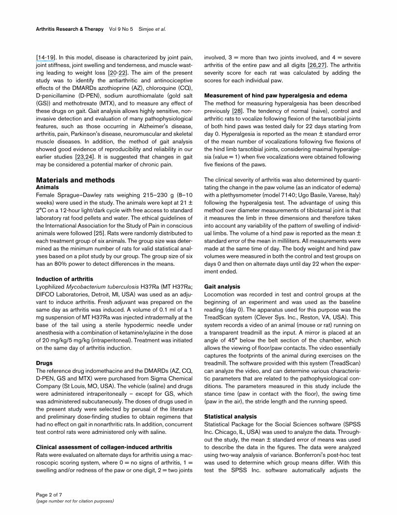
ResultsEffects of DMARDs on clinical signs of adjuvant-induced arthritisAfter day 10, animals began to show evidence of clinicalinflammation in one or both hind paws. The first manifestationof disease was erythema of one or more ankle joints, followedby involvement of the metatarsal and interphalangeal joints.The typical time course for the development and progressionof disease, as assessed by the mean arthritis severity scoreand the paw volume, is shown in Figures 1 and 2. Signs of anarthritic score of 3 in untreated arthritic rats, in arthritic ratstreated with saline only or in arthritic rats treated with D-PENwere evident at day 10. The arthritic rats treated with AZ, CQ,GS, MTX and indomethacine, however, showed a score of 2on day 10. The disease was progressive, with joint recruitmentfollowing the same pattern: tarsal, metatarsophalangeal andthen interphalangeal. In the vehicle and nontreated arthriticgroup, the incidence of disease was 100% (that is, all animalsin the group were affected) at day 12, and remained as suchthroughout the duration of the experiment. In contrast, treat-ment with indomethacine, AZ, CQ, GS and MTX exerted a sig-nificant attenuation in the incidence of adjuvant-inducedarthritis: 80% with GS treatment (P < 0.02), 70% with AZtreatment (P < 0.05), 60% with CQ and indomethacine treat-
ment (P < 0.05) and 50% with MTX treatment (P < 0.05).Hyperalgesia, the increase in vocalization in response toforced flexion of the tarsotibial joints, was evident from day 11in arthritic animals, reaching a maximum value by the end of theexperiment. Once the treatment was started, the animals in thearthritic groups treated with AZ, with CQ, with GS, with MTXand with indomethacine showed a marked decrease in thevocalization compared with their concurrent arthritic controlanimals.
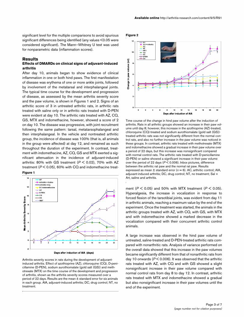
A large increase was observed in the hind paw volume ofuntreated, saline-treated and D-PEN-treated arthritic rats com-pared with nonarthritic rats. Analysis of variance performed onthe overall data showed that this increase in the paw volumesbecame significantly different from that of nonarthritic rats fromday 10 onwards (P < 0.006). It was observed that the arthriticrats treated with AZ, with CQ and with GS showed a slightnonsignificant increase in their paw volume compared withnormal control rats from day 8 to day 12. In contrast, arthriticrats treated with MTX and indomethacine showed a gradualbut also nonsignificant increase in their paw volumes until theend of the experiment.
Figure 1
Arthritis severity scores in rats during the development of adjuvant-induced arthritisArthritis severity scores in rats during the development of adjuvant-induced arthritis. Effect of azothioprine (AZ), chloroquine (CQ), D-peni-cillamine (D-PEN), sodium aurothiomalate (gold salt (GS)) and meth-otrexate (MTX) on the time course of the development and progression of arthritis, shown as the arthritis severity scores measured over a period of 22 days. Results are the mean ± standard error for six animals in each group. AIA, adjuvant-induced arthritis; DC, drug control; NT, no treatment.
Figure 2
Time course of the change in hind paw volume after the induction of arthritisTime course of the change in hind paw volume after the induction of arthritis. Rats in all arthritic groups showed an increase in their paw vol-ume until day 8; however, this increase in the azothioprine (AZ)-treated, chloroquine (CQ)-treated and sodium aurothiomalate (gold salt (GS))-treated arthritic rats was not significantly different from the normal con-trol rats, and also no further increase in the paw volume was noticed in these groups. In contrast, arthritic rats treated with methotrexate (MTX) and indomethacine showed a gradual increase in their paw volume over a period of 22 days, but this increase was nonsignificant compared with normal control rats. The arthritic rats treated with D-penicillamine (D-PEN) or saline showed a significant increase in their paw volume over the period of 22 days (P < 0.006). Inbox pictures, difference between the arthritic rat paw and the normal rat paw. Results expressed as mean ± standard error (n = 6). AC, arthritic control; AIA, adjuvant-induced arthritis; DC, drug control; NT, no treatment; Sal + Art, saline and arthritis.
Page 3 of 7(page number not for citation purposes)
Arthritis Research & Therapy Vol 9 No 5 Simjee et al.
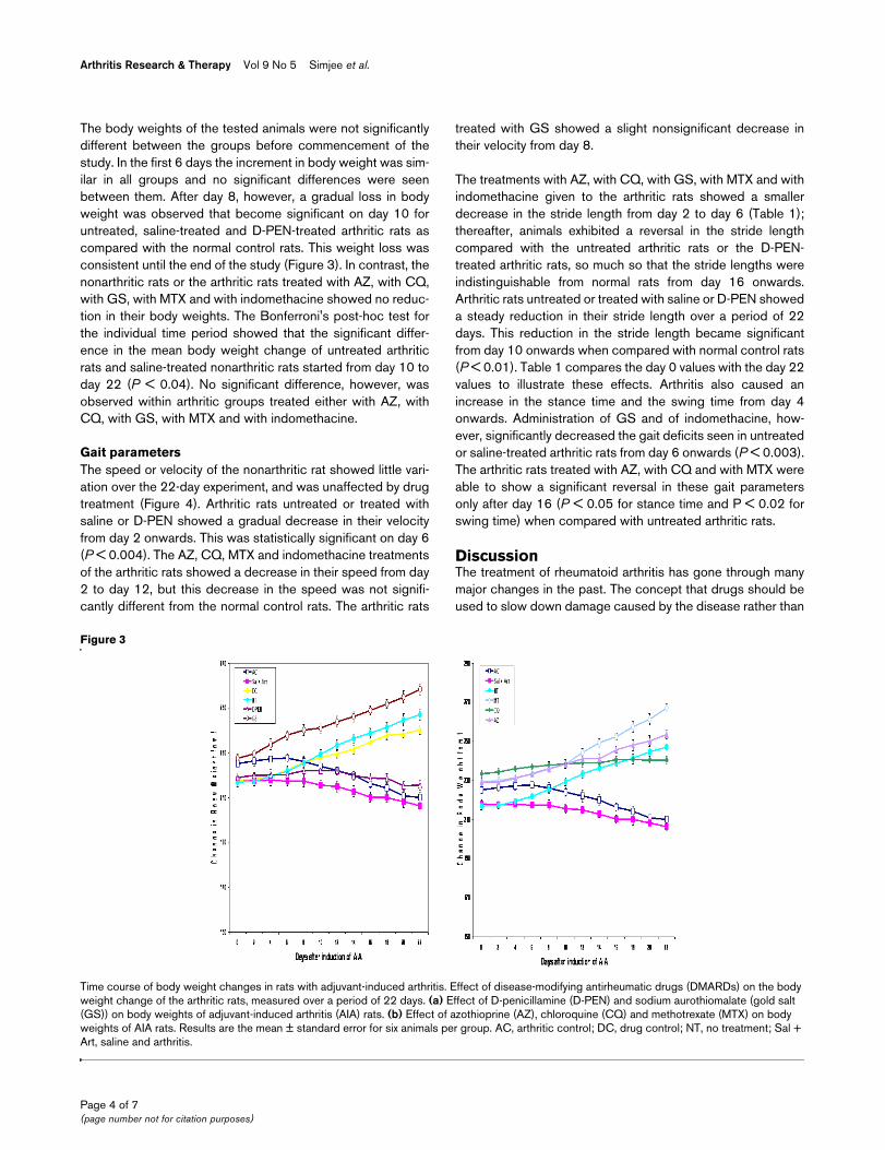
The body weights of the tested animals were not significantlydifferent between the groups before commencement of thestudy. In the first 6 days the increment in body weight was sim-ilar in all groups and no significant differences were seenbetween them. After day 8, however, a gradual loss in bodyweight was observed that become significant on day 10 foruntreated, saline-treated and D-PEN-treated arthritic rats ascompared with the normal control rats. This weight loss wasconsistent until the end of the study (Figure 3). In contrast, thenonarthritic rats or the arthritic rats treated with AZ, with CQ,with GS, with MTX and with indomethacine showed no reduc-tion in their body weights. The Bonferroni's post-hoc test forthe individual time period showed that the significant differ-ence in the mean body weight change of untreated arthriticrats and saline-treated nonarthritic rats started from day 10 today 22 (P < 0.04). No significant difference, however, wasobserved within arthritic groups treated either with AZ, withCQ, with GS, with MTX and with indomethacine.
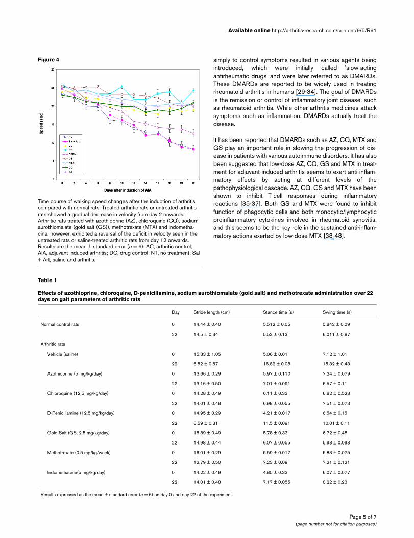
Gait parametersThe speed or velocity of the nonarthritic rat showed little vari-ation over the 22-day experiment, and was unaffected by drugtreatment (Figure 4). Arthritic rats untreated or treated withsaline or D-PEN showed a gradual decrease in their velocityfrom day 2 onwards. This was statistically significant on day 6(P < 0.004). The AZ, CQ, MTX and indomethacine treatmentsof the arthritic rats showed a decrease in their speed from day2 to day 12, but this decrease in the speed was not signifi-cantly different from the normal control rats. The arthritic rats
treated with GS showed a slight nonsignificant decrease intheir velocity from day 8.
The treatments with AZ, with CQ, with GS, with MTX and withindomethacine given to the arthritic rats showed a smallerdecrease in the stride length from day 2 to day 6 (Table 1);thereafter, animals exhibited a reversal in the stride lengthcompared with the untreated arthritic rats or the D-PEN-treated arthritic rats, so much so that the stride lengths wereindistinguishable from normal rats from day 16 onwards.Arthritic rats untreated or treated with saline or D-PEN showeda steady reduction in their stride length over a period of 22days. This reduction in the stride length became significantfrom day 10 onwards when compared with normal control rats(P < 0.01). Table 1 compares the day 0 values with the day 22values to illustrate these effects. Arthritis also caused anincrease in the stance time and the swing time from day 4onwards. Administration of GS and of indomethacine, how-ever, significantly decreased the gait deficits seen in untreatedor saline-treated arthritic rats from day 6 onwards (P < 0.003).The arthritic rats treated with AZ, with CQ and with MTX wereable to show a significant reversal in these gait parametersonly after day 16 (P < 0.05 for stance time and P < 0.02 forswing time) when compared with untreated arthritic rats.
DiscussionThe treatment of rheumatoid arthritis has gone through manymajor changes in the past. The concept that drugs should beused to slow down damage caused by the disease rather than
Figure 3
Time course of body weight changes in rats with adjuvant-induced arthritisTime course of body weight changes in rats with adjuvant-induced arthritis. Effect of disease-modifying antirheumatic drugs (DMARDs) on the body weight change of the arthritic rats, measured over a period of 22 days. (a) Effect of D-penicillamine (D-PEN) and sodium aurothiomalate (gold salt (GS)) on body weights of adjuvant-induced arthritis (AIA) rats. (b) Effect of azothioprine (AZ), chloroquine (CQ) and methotrexate (MTX) on body weights of AIA rats. Results are the mean ± standard error for six animals per group. AC, arthritic control; DC, drug control; NT, no treatment; Sal + Art, saline and arthritis.
Page 4 of 7(page number not for citation purposes)
Available online http://arthritis-research.com/content/9/5/R91
simply to control symptoms resulted in various agents beingintroduced, which were initially called 'slow-actingantirheumatic drugs' and were later referred to as DMARDs.These DMARDs are reported to be widely used in treatingrheumatoid arthritis in humans [29-34]. The goal of DMARDsis the remission or control of inflammatory joint disease, suchas rheumatoid arthritis. While other arthritis medicines attacksymptoms such as inflammation, DMARDs actually treat thedisease.
It has been reported that DMARDs such as AZ, CQ, MTX andGS play an important role in slowing the progression of dis-ease in patients with various autoimmune disorders. It has alsobeen suggested that low-dose AZ, CQ, GS and MTX in treat-ment for adjuvant-induced arthritis seems to exert anti-inflam-matory effects by acting at different levels of thepathophysiological cascade. AZ, CQ, GS and MTX have beenshown to inhibit T-cell responses during inflammatoryreactions [35-37]. Both GS and MTX were found to inhibitfunction of phagocytic cells and both monocytic/lymphocyticproinflammatory cytokines involved in rheumatoid synovitis,and this seems to be the key role in the sustained anti-inflam-matory actions exerted by low-dose MTX [38-48].
Figure 4
Time course of walking speed changes after the induction of arthritis compared with normal ratsTime course of walking speed changes after the induction of arthritis compared with normal rats. Treated arthritic rats or untreated arthritic rats showed a gradual decrease in velocity from day 2 onwards. Arthritic rats treated with azothioprine (AZ), chloroquine (CQ), sodium aurothiomalate (gold salt (GS)), methotrexate (MTX) and indometha-cine, however, exhibited a reversal of the deficit in velocity seen in the untreated rats or saline-treated arthritic rats from day 12 onwards. Results are the mean ± standard error (n = 6). AC, arthritic control; AIA, adjuvant-induced arthritis; DC, drug control; NT, no treatment; Sal + Art, saline and arthritis.
Table 1
Effects of azothioprine, chloroquine, D-penicillamine, sodium aurothiomalate (gold salt) and methotrexate administration over 22 days on gait parameters of arthritic rats
Day Stride length (cm) Stance time (s) Swing time (s)
Normal control rats 0 14.44 ± 0.40 5.512 ± 0.05 5.842 ± 0.09
22 14.5 ± 0.34 5.53 ± 0.13 6.011 ± 0.87
Arthritic rats
Vehicle (saline) 0 15.33 ± 1.05 5.06 ± 0.01 7.12 ± 1.01
22 6.52 ± 0.57 16.82 ± 0.08 15.32 ± 0.43
Azothioprine (5 mg/kg/day) 0 13.66 ± 0.29 5.97 ± 0.110 7.24 ± 0.079
22 13.16 ± 0.50 7.01 ± 0.091 6.57 ± 0.11
Chloroquine (12.5 mg/kg/day) 0 14.28 ± 0.49 6.11 ± 0.33 6.82 ± 0.523
22 14.01 ± 0.48 6.98 ± 0.055 7.51 ± 0.073
D-Penicillamine (12.5 mg/kg/day) 0 14.95 ± 0.29 4.21 ± 0.017 6.54 ± 0.15
22 8.59 ± 0.31 11.5 ± 0.091 10.01 ± 0.11
Gold Salt (GS, 2.5 mg/kg/day) 0 15.89 ± 0.49 5.78 ± 0.33 6.72 ± 0.48
22 14.98 ± 0.44 6.07 ± 0.055 5.98 ± 0.093
Methotrexate (0.5 mg/kg/week) 0 16.01 ± 0.29 5.59 ± 0.017 5.83 ± 0.075
22 12.79 ± 0.50 7.23 ± 0.09 7.21 ± 0.121
Indomethacine(5 mg/kg/day) 0 14.22 ± 0.49 4.85 ± 0.33 6.07 ± 0.077
22 14.01 ± 0.48 7.17 ± 0.055 8.22 ± 0.23
Results expressed as the mean ± standard error (n = 6) on day 0 and day 22 of the experiment.
Page 5 of 7(page number not for citation purposes)
Arthritis Research & Therapy Vol 9 No 5 Simjee et al.
In the present study we describe a possible novel approach toquantify the hyperalgesia in a rat model of rheumatoid arthritisusing gait analysis. It has previously been reported that gaitchanges in the arthritic rat can be used as an objective meas-ure of chronic pain [23,49]. The principal aim of this study wasto examine the effect of prolonged administration of low dosesof AZ, CQ, D-PEN, GS and MTX on the progression of hindpaw inflammation, disease progression and gait analysis. Wefound a strong correlation between parameters obtained fromgait analysis and the disease progression in the adjuvant-induced arthritic rats. The administration of AZ, CQ, GS andMTX doses used in this study that had no effect on gait in nor-mal rats produced a complete reversal of the gait deficits seenin the untreated, saline-treated and D-PEN-treated arthriticrats. This suggests that a prolonged administration of lowdoses of these DMARDs is effective in preventing the devel-opment of chronic pain, which, once established, is difficult totreat with conventional analgesics. These DMARDs also had asignificant effect on disease progression, measured by no fur-ther weight loss as observed in the untreated or saline-treatedadjuvant-induced arthritis. The DMARDs also significantlyreduced the joint inflammation when administered for a pro-longed time (over 22 days in case of our study) at low doses.
ConclusionWe demonstrate here that continuous administration ofDMARDs at low doses not only reduces the inflammation asseen from the macroscopic studies of the arthritic paws, butalso modulates the mechanical hyperalgesia in treated arthriticrats. In addition, the method of gait analysis showed good evi-dence of reproducibility in the present study, and our data sug-gest that the changes in gait may be considered a potentialmarker of chronic pain in arthritic rats.
Competing interestsThe authors declare that they have no competing interests.
Authors' contributionsAll four authors participated in the study. SAS conceived ofthe study and participated in coordination. SUS designed thestudy, drafted the manuscript and performed the analysis ofdata. HJ participated in the study and performed the gait anal-ysis. JQ is a graduate student who provided us the drugs/com-pounds used in the study. All authors read and approved thefinal manuscript.
References1. Becker N, Sjøgren P, Bech P, Olsen AK, Eriksen J: Treatment out-
come of chronic non-malignant pain patients managed in aDanish multidisciplinary pain centre compared to generalpractice: a randomised controlled trial. Pain 2000, 84:203-211.
2. Skevington SM: Investigating the relationship between painand discomfort and quality of life, using the WHOQOL. Pain1998, 76:395-406.
3. Atkinson JH, Slater MA, Patterson TL, Grant I, Garfin SR: Preva-lence, onset and risk of psychiatric disorders in men withchronic low back pain: a controlled study. Pain 1991,45:111-121.
4. Breivik H, Collett B, Ventafridda V, Cohen R, Gallacher D: Surveryof chronic pain in Europe: prevalence, impact on daily life, andtreatment. Eur J Pain 2006, 10:287-333.
5. Severn PS, Fraser SG: Bilateral cataracts and glaucomainduced by long-term use of oral prednisolone bough over theinternet. Lancet 2006, 368:618.
6. Biskupiak JE, Brixner DI, Howard KB, Oderda GM: Gastrointesti-nal complications of over-the-counter nonsteroidal antiinflam-matory drugs. J Pain Palliat Care Pharmacother 2006, 20:7-14.
7. Hinz B, Brune K: Pain and osteoarthritis: new drugs andmechanisms. Curr Opin Rheumatol 2004, 16:628-633.
8. Schuna AA, Megeff C: New drugs for the treatment of rheuma-toid arthritis. Am J Health Syst Pharm 2000, 57:225-234.
9. Meyer-Kirchrath J, Schrör K: Cyclooxygenase-2 inhibition andside-effects of non-steroidal anti-inflammatory drugs in thegastrointestinal tract. Curr Med Chem 2000, 7:1121-1129.
10. Lester RS, Knowles SR, Shear NH: The risks of systemic corti-costeroid use. Dermatol Clin 1998, 16:277-287.
11. Lanza FL: Endoscopic studies of gastric and duodenal injuryafter the use of ibuprofen, aspirin, and other nonsteroidal anti-inflammatory agents. Am J Med 1984, 77:19-24.
12. Silva MA, Ishii-Iwamoto EL, Bracht A, Caparroz-Assef SM, KimuraE, Cuman RK, Bersani-Amado CA: Efficiency of combined meth-otrexate/chloroquine therapy in adjuvant-induced arthritis.Fundam Clin Pharmacol 2005, 19:479-489.
13. Pincus T, Ferraccioli G, Sokka T, Larsen A, Rau R, Khusner I, WolfeF: Evidence from clinical trials and long-term observationalstudies that disease-modifying anti-rheumatic drugs slowradiographic progression in rheumatoid arthritis: updating a1983 review. Rheumatology (Oxford) 2002, 41:1346-1356.
14. Cai X, Wong YF, Zhou H, Xie Y, Liu ZQ, Jiang ZH, Bian ZX, Xu HX,Liu L: The comparative study of Sprague–Dawley and Lewisrats in adjuvant-induced arthritis. Naunyn-Schmiedeberg'sArch Pharmacol 2006, 373:140-147.
15. Cai X, Wong YF, Zhou H, Liu ZQ, Xie Y, Jiang ZH, Bian ZX, Xu HX,Liu L: Manipulation of the induction of adjuvant arthritis inSprague–Dawley rats. Inflamm Res 2006, 55:368-377.
16. Santora K, Rasa C, Visco D, Steinetz B, Bagnell C: Effects ofrelaxin in a model of rat adjuvant-induced arthritis. Ann NYAcad Sci 2005, 1041:481-485.
17. Gugasyan R, Clouston D, Mandel T, Wicks I: Prevention ofsplenic granuloma formation in adjuvant arthritis by 2-acetyl-4-tetrahydroxybutylimidazole (THI). Immunol Lett 1997,58:133-138.
18. Vingsbo C, Jonsson R, Holmdahl R: Avridine-induced arthritis inrats; a T cell-dependent chronic disease influenced both byMHC genes and by non-MHC genes. Clin Exp Immunol 1995,99:359-363.
19. Rosenthale ME, Capetola RJ: Adjuvant arthritis: immunopatho-logical and hyperalgesic features. Fed Proc 1982,41:2577-2582.
20. Wollheim FA: Current pharmacological treatment ofosteoarthritis. Drugs 1996, 52(Suppl 3):27-38.
21. Firestein GS: Etiology and pathogenesis of rheumatoid arthri-tis. In Kelley's Textbook of Rheumatology Volume 2. Edited by:Ruddy S, Harris JED, Sledge CB, Budd RC, Sergent JS. Philadel-phia, PA: W.B. Saunders Company; 1994:921-966.
22. Kidd BL, Mapp PI, Blake DR, Gibson SJ, Polak JM: Neurogenicinfluences in arthritis. Annals Rheum Dis 1990, 49:649-652.
23. Simjee SU, Pleuvry BJ, Coulthard P: Modulation of the gait defi-cit in arthritic rats by infusions of muscimol and bicuculline.Pain 2004, 109:453-460.
24. Coulthard P, Simjee SU, Pleuvry BJ: Gait analysis as a correlateof pain induced by carrageenan intraplantar injection. J Neuro-sci Methods 2003, 128:95-102.
25. Zimmerman M: Ethical guidelines for investigations of experi-mental pain in conscious animals. Pain 1983, 16:109-110.
26. Bakharevski O, Stein-Oakley AN, Thomson NM, Ryan PF: Colla-gen-induced arthritis in rats: contrasting effect of subcutane-ous versus intradermal inoculation of type II collagen. JRheumatol 1998, 25:1945-1952.
27. Marshall MJ, Worsfold M: Analytical micro-preparative electro-phoresis: quantitation of phosphoglucose isomeraseisoenzymes. Anal Biochem 1978, 91:283-292.
28. Capetola RJ, Shriver DA, Rosenthale ME: Suprofen, a newperipheral analgesic. J Pharmacol Exp Ther 1980, 214:16-23.
Page 6 of 7(page number not for citation purposes)
http://www.ncbi.nlm.nih.gov/entrez/query.fcgi?cmd=Retrieve&db=PubMed&dopt=Abstract&list_uids=9718258
http://www.ncbi.nlm.nih.gov/entrez/query.fcgi?cmd=Retrieve&db=PubMed&dopt=Abstract&list_uids=9718258
http://www.ncbi.nlm.nih.gov/entrez/query.fcgi?cmd=Retrieve&db=PubMed&dopt=Abstract&list_uids=1831555
http://www.ncbi.nlm.nih.gov/entrez/query.fcgi?cmd=Retrieve&db=PubMed&dopt=Abstract&list_uids=1831555
http://www.ncbi.nlm.nih.gov/entrez/query.fcgi?cmd=Retrieve&db=PubMed&dopt=Abstract&list_uids=1831555
http://www.ncbi.nlm.nih.gov/entrez/query.fcgi?cmd=Retrieve&db=PubMed&dopt=Abstract&list_uids=9589201
http://www.ncbi.nlm.nih.gov/entrez/query.fcgi?cmd=Retrieve&db=PubMed&dopt=Abstract&list_uids=9589201
http://www.ncbi.nlm.nih.gov/entrez/query.fcgi?cmd=Retrieve&db=PubMed&dopt=Abstract&list_uids=6465160
http://www.ncbi.nlm.nih.gov/entrez/query.fcgi?cmd=Retrieve&db=PubMed&dopt=Abstract&list_uids=6465160
http://www.ncbi.nlm.nih.gov/entrez/query.fcgi?cmd=Retrieve&db=PubMed&dopt=Abstract&list_uids=6465160
http://www.ncbi.nlm.nih.gov/entrez/query.fcgi?cmd=Retrieve&db=PubMed&dopt=Abstract&list_uids=9293393
http://www.ncbi.nlm.nih.gov/entrez/query.fcgi?cmd=Retrieve&db=PubMed&dopt=Abstract&list_uids=9293393
http://www.ncbi.nlm.nih.gov/entrez/query.fcgi?cmd=Retrieve&db=PubMed&dopt=Abstract&list_uids=9293393
http://www.ncbi.nlm.nih.gov/entrez/query.fcgi?cmd=Retrieve&db=PubMed&dopt=Abstract&list_uids=7882557
http://www.ncbi.nlm.nih.gov/entrez/query.fcgi?cmd=Retrieve&db=PubMed&dopt=Abstract&list_uids=7882557
http://www.ncbi.nlm.nih.gov/entrez/query.fcgi?cmd=Retrieve&db=PubMed&dopt=Abstract&list_uids=7882557
http://www.ncbi.nlm.nih.gov/entrez/query.fcgi?cmd=Retrieve&db=PubMed&dopt=Abstract&list_uids=6177558
http://www.ncbi.nlm.nih.gov/entrez/query.fcgi?cmd=Retrieve&db=PubMed&dopt=Abstract&list_uids=6177558
http://www.ncbi.nlm.nih.gov/entrez/query.fcgi?cmd=Retrieve&db=PubMed&dopt=Abstract&list_uids=8911797
http://www.ncbi.nlm.nih.gov/entrez/query.fcgi?cmd=Retrieve&db=PubMed&dopt=Abstract&list_uids=8911797
http://www.ncbi.nlm.nih.gov/entrez/query.fcgi?cmd=Retrieve&db=PubMed&dopt=Abstract&list_uids=9779848
http://www.ncbi.nlm.nih.gov/entrez/query.fcgi?cmd=Retrieve&db=PubMed&dopt=Abstract&list_uids=9779848
http://www.ncbi.nlm.nih.gov/entrez/query.fcgi?cmd=Retrieve&db=PubMed&dopt=Abstract&list_uids=9779848
http://www.ncbi.nlm.nih.gov/entrez/query.fcgi?cmd=Retrieve&db=PubMed&dopt=Abstract&list_uids=9762110
http://www.ncbi.nlm.nih.gov/entrez/query.fcgi?cmd=Retrieve&db=PubMed&dopt=Abstract&list_uids=9762110
http://www.ncbi.nlm.nih.gov/entrez/query.fcgi?cmd=Retrieve&db=PubMed&dopt=Abstract&list_uids=9762110
http://www.ncbi.nlm.nih.gov/entrez/query.fcgi?cmd=Retrieve&db=PubMed&dopt=Abstract&list_uids=6993657
Available online http://arthritis-research.com/content/9/5/R91
29. Svensson B, Ahlmén M, Forslind K: Treatment of early RA in clin-ical practice: a comparative study of two different DMARD/cor-ticosteroid options. Clin Exp Rheumatol 2003, 21:327-332.
30. Osiri M, Shea B, Robinson V, Suarez-Almazor M, Strand V, TugwellP, Wells G: Leflunomide for treating rheumatoid arthritis.Cochrane Database Syst Rev 2003, 1:CD002047.
31. Alarcón GS: Methotrexate use in rheumatoid arthritis: a clini-cian's perspective. Immunopharmacology 2000, 47:259-271.
32. Emery P, Breedveld FC, Lemmel EM, Kaltwasser JP, Dawes PT,Gömör B, Van Den Bosch F, Nordström D, Bjorneboe O, Dahl R,et al.: A comparison of the efficacy and safety of leflunomideand methotrexate for the treatment of rheumatoid arthritis.Rheumatology (Oxford) 2000, 39:655-665.
33. O'Dell JR, Haire CE, Erikson N, Drymalski W, Palmer W, EckhoffPJ, Garwood V, Maloley P, Klassen LW, Wees S, et al.: Treatmentof rheumatoid arthritis with methotrexate alone, sulfasalazineand hydroxychloroquine, or a combination of all threemedications. N Engl J Med 1996, 334:1287-1291.
34. Porter D, Madhok R, Hunter JA, Capell HA: Prospective trial com-paring the use of sulfasalazine and auranofin as second linedrugs in patients with rheumatoid arthritis. Ann Rheum Dis1992, 51:461-464.
35. Rovensky J, Svík K, Stancíková M, Istok R: Effect of immunostim-ulatory ribomunyl on the preventive treatment of rat adjuvantarthritis with cyclosporine and methotrexate. J Rheumatol2003, 30:2027-2032.
36. Connolly KM, Stecher VJ, Danis E, Pruden DJ, LaBrie T: Alterationof interleukin-1 activity and the acute phase response in adju-vant arthritic rats treated with disease modifying antirheu-matic drugs. Agents Actions 1988, 25:94-105.
37. Vinegar R, Truax JF, Selph JL, Lea A, Johnston PR: Azathioprinetreatment of adjuvant arthritis. J Immunopharmacol 1979,1:497-520.
38. Rovenský J, Svík K, Matha V, Istok R, Kamarád V, Ebringer L, Fer-encík M, Stancíková M: Combination treatment of rat adjuvant-induced arthritis with methotrexate, probiotic bacteria Entero-coccus faecium, and selenium. Ann NY Acad Sci 2005,1051:570-581.
39. Bendele AM, McComb J, Gould T, Frazier J, Chlipala E, Seely J,Kieft G, Edwards CK 3rd: Effects of PEGylated soluble tumornecrosis factor receptor type I (PEG sTNF-RI) alone and incombination with methotrexate in adjuvant arthritic rats. ClinExp Rheumatol 1999, 17:553-560.
40. Segawa Y, Yamaura M, Aota S, Omata T, Tuzuike N, Itokazu Y, OkaH, Tamaki H, Nakamura T: Methotrexate maintains bone massby preventing both a decrease in bone formation and anincrease in bone resorption in adjuvant-induced arthritic rats.Bone 1997, 20:457-464.
41. Genestier L, Paillot R, Fournel S, Ferraro C, Miossec P, RevillardJP: Immunosuppressive properties of methotrexate: apopto-sis clonal deletion of activated peripheral T cells. J Clin Invest1998, 15:322-328.
42. Cronstein BN: The mechanism of action of methotrexate.Rheum Dis Clin North Am 1997, 23:739-755.
43. Wascher TC, Hermann J, Brezinschek R: Serum levels of inter-leukin-6 and tumor-necrosis-factor-alpha are not correlated todisease activity in patients with rheumatoid arthritis aftertreatment with low-dose methotrexate. Eur J Clin Invest 1994,72:535-540.
44. Hu SHK, Mitcho YL, Oronsky AL, Kewar SS: Studies on theeffect of methotrexate on macrophage function. J Rheumatol1988, 15:206-209.
45. Cronstein BN, Eberle MA, Gruber HE, Levin RI: Methotrexateinhibits neutrophil function by stimulating adenosine releasefrom connective tissue cells. Proc Natl Acad Sci USA 1991,88:2441-2445.
46. Johnson WJ, Di Martino MJ, Meunier PC, Murihead KA, Hanna N:Methotrexate inhibits macrophage activation as well as vascu-lar and cellular inflammatory events in rat adjuvant inducedarthritis. J Rheumatol 1988, 15:745-749.
47. Davis P, Johnston C: Effects of gold compounds on function ofphagocytic cells. Comparative inhibition of activated polymor-phonuclear leukocytes and monocytes from rheumatoidarthritis and control subjects. Inflammation 1986, 10:311-320.
48. Scheinberg MA, Santos LM, Finkelstein AE: The effect ofauranofin and sodium aurothiomalate on peripheral bloodmonocytes. J Rheumatol 1982, 9:366-369.
49. Coulthard P, Pleuvry BJ, Brewster M, Wilson KL, Macfarlane TV:Gait analysis as an objective measure in a chronic pain model.J Neurosci Methods 2002, 116:197-213.
Page 7 of 7(page number not for citation purposes)
http://www.ncbi.nlm.nih.gov/entrez/query.fcgi?cmd=Retrieve&db=PubMed&dopt=Abstract&list_uids=8609945
http://www.ncbi.nlm.nih.gov/entrez/query.fcgi?cmd=Retrieve&db=PubMed&dopt=Abstract&list_uids=8609945
http://www.ncbi.nlm.nih.gov/entrez/query.fcgi?cmd=Retrieve&db=PubMed&dopt=Abstract&list_uids=8609945
http://www.ncbi.nlm.nih.gov/entrez/query.fcgi?cmd=Retrieve&db=PubMed&dopt=Abstract&list_uids=1350192
http://www.ncbi.nlm.nih.gov/entrez/query.fcgi?cmd=Retrieve&db=PubMed&dopt=Abstract&list_uids=1350192
http://www.ncbi.nlm.nih.gov/entrez/query.fcgi?cmd=Retrieve&db=PubMed&dopt=Abstract&list_uids=1350192
http://www.ncbi.nlm.nih.gov/entrez/query.fcgi?cmd=Retrieve&db=PubMed&dopt=Abstract&list_uids=3142230
http://www.ncbi.nlm.nih.gov/entrez/query.fcgi?cmd=Retrieve&db=PubMed&dopt=Abstract&list_uids=3142230
http://www.ncbi.nlm.nih.gov/entrez/query.fcgi?cmd=Retrieve&db=PubMed&dopt=Abstract&list_uids=3142230
http://www.ncbi.nlm.nih.gov/entrez/query.fcgi?cmd=Retrieve&db=PubMed&dopt=Abstract&list_uids=9145243
http://www.ncbi.nlm.nih.gov/entrez/query.fcgi?cmd=Retrieve&db=PubMed&dopt=Abstract&list_uids=9145243
http://www.ncbi.nlm.nih.gov/entrez/query.fcgi?cmd=Retrieve&db=PubMed&dopt=Abstract&list_uids=9361153
http://www.ncbi.nlm.nih.gov/entrez/query.fcgi?cmd=Retrieve&db=PubMed&dopt=Abstract&list_uids=3129560
http://www.ncbi.nlm.nih.gov/entrez/query.fcgi?cmd=Retrieve&db=PubMed&dopt=Abstract&list_uids=3129560
http://www.ncbi.nlm.nih.gov/entrez/query.fcgi?cmd=Retrieve&db=PubMed&dopt=Abstract&list_uids=2006182
http://www.ncbi.nlm.nih.gov/entrez/query.fcgi?cmd=Retrieve&db=PubMed&dopt=Abstract&list_uids=2006182
http://www.ncbi.nlm.nih.gov/entrez/query.fcgi?cmd=Retrieve&db=PubMed&dopt=Abstract&list_uids=2006182
http://www.ncbi.nlm.nih.gov/entrez/query.fcgi?cmd=Retrieve&db=PubMed&dopt=Abstract&list_uids=3262750
http://www.ncbi.nlm.nih.gov/entrez/query.fcgi?cmd=Retrieve&db=PubMed&dopt=Abstract&list_uids=3262750
http://www.ncbi.nlm.nih.gov/entrez/query.fcgi?cmd=Retrieve&db=PubMed&dopt=Abstract&list_uids=3262750
http://www.ncbi.nlm.nih.gov/entrez/query.fcgi?cmd=Retrieve&db=PubMed&dopt=Abstract&list_uids=3017859
http://www.ncbi.nlm.nih.gov/entrez/query.fcgi?cmd=Retrieve&db=PubMed&dopt=Abstract&list_uids=3017859
http://www.ncbi.nlm.nih.gov/entrez/query.fcgi?cmd=Retrieve&db=PubMed&dopt=Abstract&list_uids=3017859
http://www.ncbi.nlm.nih.gov/entrez/query.fcgi?cmd=Retrieve&db=PubMed&dopt=Abstract&list_uids=6288942
http://www.ncbi.nlm.nih.gov/entrez/query.fcgi?cmd=Retrieve&db=PubMed&dopt=Abstract&list_uids=6288942