Effect of Aronia melanocarpa fruit juice on amiodarone-induced pneumotoxicity in rats
10
Pharmacognosy Magazine ISSN : 0973-1296 Phcog.Net - Bringing Medicinal Plant Researchers Together Publication of Pharmacognosy Network Worldwide www.phcog.com April - June 2014 | Volume 10 | Issue 38 CAB Abstracts, Caspur, Chemical Abstracts, CNKI (China National Knowledge Infrastructure), CSA databases, DOAJ, EBSCO Publishing’s Electronic Databases, Excerpta Medica / EMBASE, Genamics JournalSeek, Google Scholar, Health & Wellness Research Center, Health Reference Center Academic, Hinari, Index Copernicus, Indian Science Abstracts, Journal Citation Reports, National Science Library, OpenJGate, PrimoCentral, ProQuest, PubMed, Pubmed Central, Science Citation Index Expanded, Scimago Journal Ranking, SCOLOAR, SCOPUS, SIIC databases, Summon by Serial Solutions, Ulrich’s International Periodical Directory and Web of Science. ® Impact Factor for 2012: 1.525 Included
-
Upload
independent -
Category
Documents
-
view
1 -
download
0
Transcript of Effect of Aronia melanocarpa fruit juice on amiodarone-induced pneumotoxicity in rats
Pharmacognosy Magazine
ISSN : 0973-1296
Phcog.Net - Bringing Medicinal Plant Researchers Together
Publication of Pharmacognosy Network Worldwide
www.phcog.com
April - June 2014 | Volume 10 | Issue 38
CAB Abstracts, Caspur, Chemical Abstracts, CNKI (China National Knowledge Infrastructure), CSA databases, DOAJ, EBSCO Publishing’s Electronic Databases, Excerpta Medica / EMBASE, Genamics JournalSeek, Google Scholar, Health & Wellness Research Center, Health Reference Center Academic, Hinari, Index Copernicus, Indian Science Abstracts, Journal Citation Reports, National Science Library, OpenJGate, PrimoCentral, ProQuest, PubMed, Pubmed Central, Science Citation Index Expanded, Scimago Journal Ranking, SCOLOAR, SCOPUS, SIIC databases, Summon by Serial Solutions, Ulrich’s International Periodical Directory and Web of Science.
Ph
arm
aco
gn
osy M
ag
azin
e • V
olu
me 1
0 • Issu
e 3
8 • A
pril-J
un
e 2
014 • P
ages 0
0-0
0**
®Impact Factor for 2012: 1.525
Included

132 Pharmacognosy Magazine | April-June 2014 | Vol 10 | Issue 38
Effect of Aronia melanocarpa fruit juice on amiodarone‑induced pneumotoxicity in ratsStefka Valcheva‑Kuzmanova, Galya Stavreva1, Violeta Dancheva2, Ljudmil Terziev3, Milena Atanasova4, Angelina Stoyanova5, Anelia Dimitrova6, Veneta Shopova2
Department of Preclinical and Clinical Pharmacology and Toxicology, Medical University, Varna, 1Department of Experimental and Clinical Pharmacology, Medical University, Pleven, 2Department of Disaster Medicine, Medical University, Pleven, 3Department of Clinical Laboratory, Clinical Immunologyand Allergology, Clinic of Allergology, University Hospital, Pleven, 4Departments of Biology, 5Chemistry, and 6Physiology and Pathophysiology, Medical University, Pleven, Bulgaria
Submitted: 08-11-2012 Revised: 19-12-2012 Published: ****
Address for correspondence: Prof. Stefka Valcheva-Kuzmanova, Department of Preclinical and Clinical Pharmacology and Toxicology, Medical University, 55, Marin Drinov Str.,9002 Varna, Bulgaria. E-mail: [email protected]
Background: The fruits of Aronia melanocarpa (Michx.) Elliot is extremely rich in biologically active polyphenols. Objective: We studied the protective effect of A. melanocarpa fruit juice (AMFJ) in a model of amiodarone (AD)‑induced pneumotoxicity in rats. Materials and Methods: AD was instilled intratracheally on days 0 and 2 (6.25 mg/kg). AMFJ (5 mL/kg and 10 mL/kg) was given orally from day 1 to days 2, 4, 9, and 10 to rats, which were sacrificed respectively on days 3, 5, 10, and 28 when biochemical, cytological, and immunological assays were performed. Results: AMFJ antagonized AD-induced increase of the lung weight coefficient. In bronchoalveolar lavage fluid, AD increased significantly the protein content, total cell count, polymorphonuclear cells, lymphocytes and the activity of lactate dehydrogenase, acid phosphatase and alkaline phosphatase on days 3 and 5. In AMFJ‑treated rats these indices of direct toxic damage did not differ significantly from the control values. In lung tissue, AD induced oxidative stress measured by malondialdehyde content and fibrosis assessed by the hydroxyproline level. AMFJ prevented these effects of AD. In rat serum, AD caused a significant elevation of interleukin IL‑6 on days 3 and 5, and a decrease of IL‑10 on day 3. In AMFJ‑treated rats, these indices of inflammation had values that did not differ significantly from the control ones. Conclusion: AMFJ could have a protective effect against AD-induced pulmonary toxicity as evidenced by the reduced signs of AD-induced direct toxic damage, oxidative stress, inflammation, and fibrosis.
Key words: Aronia melanocarpa, amiodarone, pneumotoxicity, rats
INTRODUCTION
Amiodarone (AD), an iodine‑containing highly lipophilic benzofuran derivative, is a very effective class III, long‑acting antiarrhythmic drug. It causes acute pneumonitis resulting in fatal pulmonary fibrosis.[1] In vivo and in vitro studies have shown that AD is directly toxic to lung cells[2] such as alveolar macrophages,[3] alveolar epithelial cells,[4] and pulmonary artery endothelial cells.[5] AD and its primary metabolite N‑desethylamiodarone have been demonstrated to cause apoptosis and necrosis in cultured alveolar type II cells.[4] AD‑induced pulmonary injury could include disruption of mitochondrial function and cellular adenosine triphosphate ATP levels,[6] enhanced oxidative stress and increased
production of reactive oxygen species,[7,8] activation of alveolar macrophages and cytokine release.[9‑11]
Aronia melanocarpa (Michx.) Elliot (black chokeberry) originates from the eastern parts of North America and East Canada. Its migration to Europe and the former Soviet Union occurred around 1900. Aronia is commonly used to produce fruit syrup, juice, jellies, tea, and wine. Chokeberry fruits are extremely rich in phenolic compounds: Procyanidins, flavonoids (mainly from the subclass of anthocyanins) and phenolic acids.[12] A series of studies has investigated the antioxidant properties of Aronia juice, Aronia extract or its phenolic constituents using different well‑established assays.[12‑18] Fresh Aronia berries possess the highest antioxidant capacity among berries and other fruits investigated so far as measured with oxygen radical absorbance capacity.[13,15] Studies have demonstrated that flavonoids including anthocyanins possess anti‑inflammatory activity due to the suppression
A B S T R A C T Access this article online
Website: www.phcog.com
DOI: 10.4103/0973-1296.131024
Quick Response Code:
P H C O G M A G . O R I G I N A L A R T I C L E
Valcheva‑Kuzmanova, et al.: Effect of AMFJ in amiodarone‑induced pneumotoxicity
Pharmacognosy Magazine | April-June 2014 | Vol 10 | Issue 38 133
of the release of pro‑inflammatory cytokines such as tumor necrosis factor alpha and interleukins (interleukin IL‑1 beta, IL‑6, IL‑8).[19,20]
There are numerous pathologic similarities between human and rodent lungs and as such, rodent model presents an excellent tool to investigate pathologic changes in vivo.[21]
The aim of the study was to investigate the effect of A. melanocarpa fruit juice (AMFJ) in a rat model of AD‑induced pulmonary toxicity.
MATERIALS AND METHODS
Experimental substancesAD hydrochloride and all other chemicals and reagents wereof analytical grade and were purchased from Sigma‑Aldrich Company (Germany). The Quantikine Rat IL‑6 and IL‑10 immunoassay kits were from R and D Systems (USA).
AMFJ was produced from A. melanocarpa Elliot fruits grown in the Balkan Mountains, Bulgaria. They were handpicked in September, crushed and squeezed. The juice was filtered, pasteurized at 80°C for 10 min and stored at 0°C until the experiment. The contents of phenolic substances in 100 mL AMFJ were: Total phenolics, 709.3 ± 28.1 mg as gallic acid equivalents, determined spectrophotometrically according to the Folin‑Ciocalteu procedure;[22] total flavonoids, 189.4 ± 8.6 mg as catechin equivalents, measured by a colorimetric assay developed by Zhishen et al.;[23] total anthocyanins, 106.8 ± 6.2 mg as cyanidin‑3‑glucoside equivalents, determined by a pH‑differential spectrophotometry at pH 1.0 and pH 4.5;[24] quercetin, 11.8 mg, measured by a high‑performance liquid chromatography method.[25] The values were the mean of duplicate determinations of three samples.
Animals and experimental treatmentsThe study was carried out on 96 male Wistar rats (weight 220‑250 g, age 4 months). The animals were obtained from the Research and Laboratory Animal Breeding Center of Slivnitsa (Bulgaria) and were housed in the university animal quarters for 1 month at a temperature of 22°C ± 2°C and humidity of 50 ± 10%, given normal pelleted diet and water AD libitum.
All procedures concerning animal treatment and experimentation were conducted in compliance with the national laws and policies, in conformity with the international guidelines (European Economic Community EEC Council Directive 86/609, IL 358, 1, December 12, 1987).
The animals were divided into four groups of 24 rats. Each group was subdivided into four subgroups of six rats. One subgroup of each group was sacrificed on days 3, 5, 10, and 28 under thiopental anesthesia (50 mg/kg) after receiving the respective treatment in the course of 2, 4, 9, and 10 days, respectively. Group 1 (Control) received two intratracheal (i.t.) instillations of sterile distilled water (2 mL/kg) on days 0 and 2. The four subgroups of Group 1 received distilled water (10 mL/kg) orally through an orogastric cannula from day 1 to days 2, 4, 9, and 10, respectively. Group 2 (AD) received two i.t. instillations of AD (6.25 mg/kg, as a 3.125 mg/mL water solution) on days 0 and 2.[26] The subgroups of Group 2 received distilled water (10 mL/kg) orally through an orogastric cannula from day 1 to days 2, 4, 9, and 10, respectively. Group 3(AD + AMFJ5) was treated with AD i.t. on days 0 and 2, and from day 1 to days 2, 4, 9, and 10 the respective subgroups received AMFJ orally at a dose of 5 mL/kg diluted with distilled water to a total volume of 10 mL/kg. Group 4 (AD + AMFJ10) was treated with AD i.t. on days 0 and 2, and from day 1 to days 2, 4, 9, and 10 the respective subgroups received AMFJ orally at a dose of 10 mL/kg.
AD was dissolved in distilled water at 60°C and allowed to cool to room temperature before the i.t. instillation.
Lung weight coefficientThe body weight and lung weight were measured for each animal. The lung weight coefficient (organ weight in mg/100 g body weight) was calculated.
Bronchoalveolar lavageBronchoalveolar lavage fluid (BALF) was obtained on days 3, 5, and 10. Rats were sacrificed by exsanguination through cutting v. renalis. The chest was opened, and the lungs were perfused in situ via the right heart ventricle with saline (30 mL). Triple lavage of the left lung through the trachea with a total of 5 mL of saline was performed. The volume of fluid recovered ranged from 80% to 90% of the fluid introduced.
Cytological assays of BALFOne aliquot of the BALF was used for total cell count ×105/L. The cells were removed by centrifugation at 300 × g for 10 min. The cell pellet was resuspended in 1 mL of saline. The cell differentials were analyzed using the procedure of Danos and Keebler, modified by Saltini et al.[27] Cells were collected on nitrocellulose filters 25 mm in diameter with 5 μm pores (SWP‑025‑00 Catalogue number SMWP02500; Millipore Corp.) and were stained with H and E.
Biochemical assays of BALFAfter centrifugation of BALF, the supernatant was used for the measurement of enzyme activities such as lactate
Valcheva‑Kuzmanova, et al.: Effect of AMFJ in amiodarone‑induced pneumotoxicity
134 Pharmacognosy Magazine | April-June 2014 | Vol 10 | Issue 38
dehydrogenase (LDH), acid phosphatase (AcP), and alkaline phosphatase (AlP) by the methods of Bergmeyer et al.[28] Total protein content was measured by the method of Lowry et al.[29] The enzyme activities are represented in U/L, and total protein in mg/mL of BALF.
Biochemical assays of lung homogenateLung homogenate was obtained from the right lung. The tissue was homogenized with potassium chloride KCl (1.15%) in 1:10 ratio. The homogenate was centrifuged (9000 × g, 30 min), and the supernatant was stored on ice. Malondialdehyde (MDA) content in nmol/g tissue was measured on days 3, 5, and 10 by the method of Ohkawa et al.[30]
Hydroxyproline (HP) levels (in μg/mL) were measured in lung homogenate of rats sacrificed on day 28 as described by Bergman and Loxley.[31] The method is based on the release of free HP from collagen by acid hydrolysis.
Immunological assays of rat serumThe serum of the experimental animals was used for the measurement of IL‑6 and IL‑10 in pg/mL on days 3,
5, and 10 by the ELISA method in accordance with the immunoassay kits manufacturer’s instructions.
Statistical analysisResults are presented as mean ± SEM. The data were tested by one‑way ANOVA, followed by Dunnett’s multiple comparison post‑test to identify significant difference. A level of P < 0.05 was considered significant. All analyses were performed using GraphPad Prism Statistical Software.
RESULTS
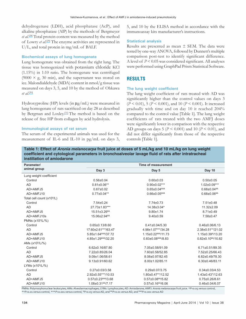
The lung weight coefficientThe lung weight coefficient of rats treated with AD was significantly higher than the control values on days 3 (P < 0.01), 5 (P < 0.001), and 10 (P < 0.001). It increased gradually with time and on day 10 it reached 204% compared to the control value [Table 1]. The lung weight coefficients of rats treated with the two AMFJ doses were significantly lower in comparison with the respective AD groups on days 5 (P < 0.001) and 10 (P < 0.01), and did not differ significantly from those of the respective controls [Table 1].
Table 1: Effect of Aronia melanocarpa fruit juice at doses of 5 mL/kg and 10 mL/kg on lung weight coefficient and cytological parameters in bronchoalveolar lavage fluid of rats after intratracheal instillation of amiodaroneParameter/ animal group
Time of measurementDay 3 Day 5 Day 10
LungweightcoefficientControl 0.56±0.04 0.60±0.03 0.50±0.05AD 0.81±0.06** 0.90±0.02*** 1.02±0.09***AD+AMFJ5 0.67±0.02 0.65±0.04&&& 0.68±0.04&&
AD+AMFJ10 0.77±0.04** 0.66±0.05&&& 0.68±0.06&&
Total cell count (x105/L)Control 7.54±0.24 7.74±0.73 7.51±0.48AD 27.73±1.83*** 14.38±3.04* 11.32±2.09AD+AMFJ5 15.51±3.26&& 9.80±1.74 8.71±0.49AD+AMFJ10s 15.06±2.94&& 9.40±0.59 7.58±0.47
PMNs (x105/L/%)Control 0.65±0.13/8.60 0.41±0.04/5.30 0.46±0.06/6.13AD 17.60±2.61***/63.47 4.98±1.07***/34.28 2.38±0.51**/21.02AD+AMFJ5 5.85±1.64&&&/37.72 1.15±0.22&&&/11.73 1.15±0.39&/13.20AD+AMFJ10 4.85±1.29&&&/32.20 0.83±0.08&&&/8.83 0.82±0.10&&/10.82
AMs (x105/L/%)Control 6.62±0.16/87.80 7.05±0.58/91.09 6.71±0.51/89.35AD 7.22±0.85/26.04 7.60±0.58/52.85 7.52±0.25/66.43AD+AMFJ5 9.09±1.06/58.61 8.08±0.97/82.45 6.82±0.49/78.30AD+AMFJ10 9.13±0.91/60.62 8.00±1.02/85.11 6.30±0.46/83.11
LYMs (x105/L/%)Control 0.27±0.03/3.58 0.29±0.07/3.75 0.34±0.03/4.53AD 2.92±0.55***/10.53 1.80±0.47**/12.52 1.43±0.43*/12.63AD+AMFJ5 0.57±0.23&&&/3.68 0.57±0.08&&/5.82 0.75±0.26/8.61AD+AMFJ10 1.08±0.31&&/7.17 0.57±0.16&&/6.06 0.46±0.04/6.07
PMNs: Polymorphonuclear leukocytes; AMs: Alveolarmacrophages; LYMs: Lymphocytes; AD: Amiodarone; AMFJ: Aronia melanocarpa fruit juice. *P<0.05 versus control, **P<0.01 versus control, ***P<0.001 versus control, &P<0.05 versus AD, and &&P<0.01 versus AD, and &&&P<0.001 versus AD
Valcheva‑Kuzmanova, et al.: Effect of AMFJ in amiodarone‑induced pneumotoxicity
Pharmacognosy Magazine | April-June 2014 | Vol 10 | Issue 38 135
Cytological assays of BALFThe total cell count in BALF of rats from AD group increased dramatically on day 3 (368% of the control value), then gradually decreased. On day 5, it was 186% of the control level and on day 10 it was still higher (but not significantly) than the control value [Table 1]. AMFJ at both doses effectively antagonized that effect of AD, and in AMFJ‑treated rats the total cell counts on days 3, 5, and 10 did not differ significantly from those of the respective controls [Table 1].
AD caused a dramatic increase of the polymorphonuclear leukocytes (PMNs) on day 3 (27 times higher than those of the control group; P < 0.001 vs. control). After that PMNs gradually decreased, but remained about 12 times higher than the control value on day 5 (P < 0.001 vs. control) and about 5 times higher than the control on day 10 (P < 0.01 vs. control) [Table 1]. AD increased also the percentage of PMNs from the total cell count in BALF on days 3, 5, and 10 to values 63.47%, 34.28%, and 21.02%, respectively, while the control values at these time points were 8.60%, 5.30%, and 6.13% [Table 1]. AMFJ treatment significantly attenuated the effect of AD on PMNs. In AMFJ‑treated rats, the counts of PMNs did not differ significantly from the respective controls on days 3, 5, and 10, and were significantly lower than those of the respective AD groups [Table 1]. For these rats, the percentage of PMNs from the total cell count at all‑time points was considerably lower in comparison with the respective AD groups [Table 1].
Neither AD, nor AMFJ treatment significantly affected the number of alveolar macrophages (AMs) in BALF [Table 1]. In AD group, the percentages of these cells from the total cell count on days 3, 5, and 10 were 26.04%, 52.85%, and 66.43%, respectively. They were considerably lower in comparison with the control values, which at these time points were 87.80%, 91.09%, and 89.35% [Table 1]. In AMFJ‑treated rats, the percentage of AMs was decreased to a certain extent on day 3 and almost returned to the control values on days 5 and 10 [Table 1].
In AD‑treated rats, the lymphocyte (LYM) count in BALF significantly increased on days 3 (P < 0.001 vs. control), 5 (P < 0.01 vs. control) and 10 (P < 0.05 vs. control) [Table 1]. Thus, AD caused an elevation of the percentage of LYMs from the total cell count in BALF on days 3, 5, and 10 to values of 10.53%, 12.52%, and 12.63%, respectively, while the control values for these time points were 3.58%, 3.75%, and 4.53% [Table 1]. The number of LYMs in AD + AMFJ5 and AD + AMFJ 10 groups did not differ significantly from that of the respective controls and their percentage from the total number of cells was similar to that of the controls [Table 1].
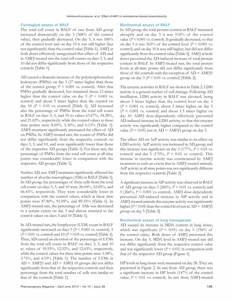
Biochemical assays of BALFIn AD group, the total protein content in BALF increased abruptly and on day 3 it was 518% of the control value (P < 0.001 vs. control). It gradually decreased, so that on day 5 it was 365% of the control level (P < 0.001 vs. control) and on day 10 it was still higher, but did not differ significantly from the control value [Table 2]. AMFJ at both doses prevented the AD‑induced increase of total protein content in BALF. In AMFJ‑treated rats, the total protein levels at all‑time points did not differ significantly from those of the controls with the exception of AD + AMFJ5 group on day 3 (P < 0.01 vs. control) [Table 2].
The enzyme activities in BALF are shown in Table 2. LDH activity is a general marker of cell damage. Following AD instillation, LDH activity in BALF was elevated, being about 3 times higher than the control level on day 3 (P < 0.001 vs. control), about 2 times higher on day 5 (P < 0.001 vs. control) and about 1.5 times higher on day 10. AMFJ dose‑dependently effectively prevented AD‑induced increase in LDH activity, so that this enzyme activity was significantly higher compared to the control value (P < 0.05) just in AD + AMFJ5 group on day 3.
The effect AD on AcP activity was similar to its effect on LDH activity. AcP activity was increased in AD group, and this increase was significant on day 3 (177%, P < 0.01 vs. control) and day 5 (170%, P < 0.01 vs. control). This increase in enzyme activity was counteracted by AMFJ treatment to such an extent that in AMFJ‑treated animals, AcP activity at all‑time points was not significantly different from the respective controls [Table 2].
A significant increase in AlP activity was observed in BALF of AD group on days 3 (260%, P < 0.01 vs. control) and 5 (266%, P < 0.001 vs. control). AMFJ dose‑dependently prevented AD‑induced increase in AlP activity and in AMFJ‑treated animals this enzyme activity was significantly higher (P < 0.05) than the control level just in AD + AMFJ5 group on day 5 [Table 2].
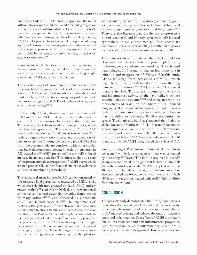
Biochemical assays of lung homogenateAD caused an increase in MDA content in lung tissue, which was significant (P < 0.01) on day 5 (196% of the control value). Both doses of AMFJ prevented this increase. On day 5, MDA level in AMFJ‑treated rats did not differ significantly from the respective control value and was significantly lower (P < 0.01) in comparison with that of the respective AD group [Figure 1].
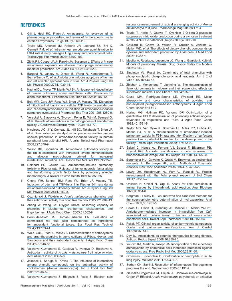
HP levels in lung tissue were measured on day 28. They are presented in Figure 2. In rats from AD group, there was a significant increase in HP levels (147% of the control value; P < 0.01 vs. control). In rats from AMFJ‑treated
Valcheva‑Kuzmanova, et al.: Effect of AMFJ in amiodarone‑induced pneumotoxicity
136 Pharmacognosy Magazine | April-June 2014 | Vol 10 | Issue 38
groups (AD + AMFJ5 and AD + AMFJ10) the HP levels did not differ significantly from the control value. In rats from AD + AMFJ10 group, the HP level was significantly lower (P < 0.05) than that of AD group.
Immunological assays of rat serumAD instillation resulted in an elevation of IL‑6 in rat serum which was significant on day 3 (136% of the control; P < 0.01 vs. control) and day 5 (116% of the control; P < 0.05 vs. control) [Figure 3]. AMFJ dose (10 mL/kg), which was tested for this effect, prevented AD‑induced elevation of IL‑6. In AMFJ‑treated animals, the levels of IL‑6 were significantly lower than those of AD group (P < 0.05 on days 3 and 5; P < 0.001 on day 10).
IL‑6 level on day 10 was even significantly lower than the control value (P < 0.001).
In AD group, a reduction of IL‑10 in rat serum was observed on days 3, 5, and 10 [Figure 4]. This reduction was statistically significant on day 3 (P < 0.05 vs. control). The tested dose of AMFJ (10 mL/kg) antagonized that effect of AD on IL‑10. In AMFJ‑treated animals, IL‑10 levels did not differ significantly from the control value [Figure 4].
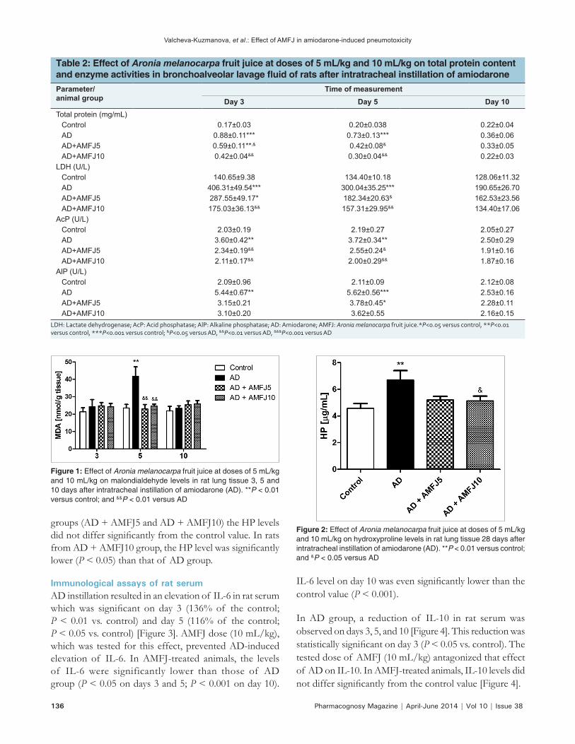
Table 2: Effect of Aronia melanocarpa fruit juice at doses of 5 mL/kg and 10 mL/kg on total protein content and enzyme activities in bronchoalveolar lavage fluid of rats after intratracheal instillation of amiodaroneParameter/ animal group
Time of measurementDay 3 Day 5 Day 10
Total protein (mg/mL)Control 0.17±0.03 0.20±0.038 0.22±0.04AD 0.88±0.11*** 0.73±0.13*** 0.36±0.06AD+AMFJ5 0.59±0.11**,& 0.42±0.08& 0.33±0.05AD+AMFJ10 0.42±0.04&& 0.30±0.04&& 0.22±0.03
LDH (U/L)Control 140.65±9.38 134.40±10.18 128.06±11.32AD 406.31±49.54*** 300.04±35.25*** 190.65±26.70AD+AMFJ5 287.55±49.17* 182.34±20.63& 162.53±23.56AD+AMFJ10 175.03±36.13&& 157.31±29.95&& 134.40±17.06
AcP (U/L)Control 2.03±0.19 2.19±0.27 2.05±0.27AD 3.60±0.42** 3.72±0.34** 2.50±0.29AD+AMFJ5 2.34±0.19&& 2.55±0.24& 1.91±0.16AD+AMFJ10 2.11±0.17&& 2.00±0.29&& 1.87±0.16
AlP (U/L)Control 2.09±0.96 2.11±0.09 2.12±0.08AD 5.44±0.67** 5.62±0.56*** 2.53±0.16AD+AMFJ5 3.15±0.21 3.78±0.45* 2.28±0.11AD+AMFJ10 3.10±0.20 3.62±0.55 2.16±0.15
LDH: Lactate dehydrogenase; AcP: Acid phosphatase; AlP: Alkaline phosphatase; AD: Amiodarone; AMFJ: Aronia melanocarpa fruit juice.*P<0.05 versus control, **P<0.01 versus control, ***P<0.001 versus control; &P<0.05 versus AD, &&P<0.01 versus AD, &&&P<0.001 versus AD
Figure 1: Effect of Aronia melanocarpa fruit juice at doses of 5 mL/kg and 10 mL/kg on malondialdehyde levels in rat lung tissue 3, 5 and 10 days after intratracheal instillation of amiodarone (AD). **P < 0.01 versus control; and &&P < 0.01 versus AD
Figure 2: Effect of Aronia melanocarpa fruit juice at doses of 5 mL/kg and 10 mL/kg on hydroxyproline levels in rat lung tissue 28 days after intratracheal instillation of amiodarone (AD). **P < 0.01 versus control; and &P < 0.05 versus AD

Valcheva‑Kuzmanova, et al.: Effect of AMFJ in amiodarone‑induced pneumotoxicity
Pharmacognosy Magazine | April-June 2014 | Vol 10 | Issue 38 137
DISCUSSION
Both indirect and direct toxic effects on target cells have been proposed for explanation of AD‑induced pulmonary toxicity. AD has been demonstrated to cause membrane destabilization and increased intracellular influx of Ca2+,[32] mitochondrial dysfunction,[6] and overproduction of reactive oxygen species.[7,8] Numerous data show the role of oxidative stress in AD‑induced lung fibrosis.[33] Free radical reactions have been suggested to play a contributory role in the fibrogenesis either directly or through inflammatory stimuli. Lipid peroxidation and certain lipid peroxidation products induce genetic overexpression of fibrogenic cytokines, the key molecules in the mechanisms of fibrosis, as well as increased transcription and synthesis of collagen.[34] Both these events can be down‑regulated, at least in experimental models, by the use of antioxidants. Consequently, both catalytic and scavenger antioxidants have been shown to attenuate AD‑induced lung injury and fibrosis in animals.[34] Other findings show that activation of macrophages and release of inflammatory and cytotoxic mediators drive AD‑induced lung fibrosis.[10,11]
The present study investigated the effect of AMFJ in pulmonary tissue in a rat model of AD‑induced pneumotoxicity. All cytological investigations of BALF, as well as the biochemical investigations of BALF and lung homogenate, showed a well‑expressed protective effect of AMFJ.
Taylor et al.[2] have demonstrated that AD instillation produces a rapid and massive damage to the alveolar‑capillary barrier and damage or death to lung airway and parenchymal cells. In this experiment, AD‑induced lung impairment
increased the permeability of pulmonary epithelium and endothelium, resulting in extravasation of plasma proteins. Thus, AD caused a significant elevation of the protein content of cell‑free BALF on days 3 and 5 to levels, which were respectively 5 and almost 4 times higher than the control ones. AMFJ prevented the disturbance of alveolar‑capillary barrier permeability, assessed by the level of protein in BALF. The protein content in BALF of AMFJ‑treated rats was comparable to the respective control levels. This effect of AMFJ might be a result of the incorporation of its biologically active polyphenols into vascular walls. This suggestion is in coincidence with the finding that vascular endothelial cells can incorporate anthocyanins into the membrane and cytosol, conferring significant protective effects against oxidative insult.[35]
The cytologic investigation of BALF suggested that AD caused substantial changes. They were most prominent on days 3 and 5 and resolved thereafter. AD caused an increase in the total cell count in BALF due to the rapid and massive damage to the alveolar‑capillary barrier. AMFJ prevented that effect. In AD‑treated rats, the number of PMNs dramatically increased. The influx of PMNs (marker of inflammation) into the interstitium and the bronchoalveolar space is considered to play a key role in the progression of acute lung injury.[36] PMNs are the main participants in the acute inflammatory response in tissues, being recruited from the circulation when local defenses are overwhelmed. Stimulated neutrophils are cleared from the inflamed sites by apoptosis (programmed cell death) and following macrophage phagocytosis, which is the key process in the inflammation resolution.[37] PMNs can possibly play a role in the inflammatory reaction by releasing growth factors and cytokines, as well as by releasing oxidants as a part of the host‑defense system. In this study, the cytological investigations showed that AMFJ markedly diminished the
Figure 3: Effect of Aronia melanocarpa fruit juice at a dose of 10 mL/kg on IL-6 levels in rat serum 3, 5 and 10 days after intratracheal instillation of amiodarone (AD). *P < 0.05 versus control; **P < 0.01 versus control; ***P < 0.001 versus control; P < 0.05 versus AD (Day 3); P < 0.05 versus AD (Day 5); and &&&P < 0.001 versus AD (Day 10)
Figure 4: Effect of Aronia melanocarpa fruit juice at a dose of 10 mL/kg on IL-10 levels in rat serum 3, 5 and 10 days after intratracheal instillation of amiodarone. *P < 0.05 versus control
Valcheva‑Kuzmanova, et al.: Effect of AMFJ in amiodarone‑induced pneumotoxicity
138 Pharmacognosy Magazine | April-June 2014 | Vol 10 | Issue 38
number of PMNs in BALF. Thus, it suppressed the initial inflammatory response induced by AD, including migration and activation of inflammatory cells and disruption of the alveolar‑capillary barrier. Acting on early, transient inflammation and damage of alveolar‑capillary barrier, AMFJ could protect from further development of lung injury and fibrosis. Other investigations have demonstrated that the juice possesses also a pro‑apoptotic effect in neutrophils by increasing caspase‑3 activity, a marker of apoptosis execution.[38]
Consistent with the development of pulmonary inflammation and edema, i.t. AD administration was accompanied by a progressive increase in the lung weight coefficient. AMFJ prevented that increase.
The elevated levels of some enzyme activities in BALF have long been recognized as markers of toxic pulmonary lesions: LDH ‑ of increased membrane permeability and frank cell lysis, AlP ‑ of toxic damage or proliferation of pneumocytes type 2, and AcP ‑ of enhanced phagocytic activity or cell killing.[39,40]
In this study, AD significantly increased the activity of LDH and AcP in BALF on days 3 and 5, and these results confirmed its pneumotoxic effect shortly after treatment. The enzymes leak from dead or damaged cells, when membrane integrity is lost. The activity of AlP in BALF was also elevated on days 3 and 5 in AD‑treated rats. This finding suggests early toxic effects and proliferation of pneumocytes type 2 during the recovery period. The data from the present study are consistent with other studies that have demonstrated elevated levels of enzymes in AD‑treated rats.[26] AMFJ prevented the early AD‑induced increase in enzyme activities. This effect might be a result of the potent antioxidant properties of AMFJ due to which it could protect cellular membranes from oxidative damage and reduce membrane permeability.
The oxidative damage induced by AD was demonstrated by the increased lipid peroxidation measured by MDA levels, which were significantly elevated on day 5. AMFJ entirely prevented that effect of AD probably due to its pronounced antioxidant and radical scavenging activities, demonstrated by many authors[12‑18] and reviewed by Chrubasik et al.[41] and Kokotkiewicz et al.[42] The experiments of Zielinska‑Przyjemska et al.[37] have shown that Aronia juice and its active fractions significantly decrease the oxidative metabolism of PMNs. As free radicals play a crucial role in the pathogenesis of AD toxicity,[7] we could suppose that the protective effect of AMFJ in this experiment might be predominantly due to its antioxidant and free radical scavenging properties. These findings are in accordance with other investigations demonstrating that other phenolic
antioxidants (butylated hydroxyanisole, curcumin, grape seed procyanidins) are effective in limiting AD‑induced reactive oxygen species generation and lung injury.[43‑45]
There are also literature data for the the cytoprotective role of vitamin C and N‑acetyl cysteine in AD‑induced cytotoxicity on cell culture media.[46] Both agents are commonly used in the clinical setting for different purposes incontext of their well‑known antioxidant actions.[46]
There are no literature data on the effect of AD on IL‑6 and IL‑10 levels. IL‑6 is a potent, pleiotropic, inf lammatory cytokine secreted by T cells and macrophages. IL‑6 seems to play an important role in initiation and progression of fibrosis.[47] In this study, AD caused a significant increase of serum IL‑6, which might be a result of IL‑6 translocation from the lung tissue to the circulation.[48] AMFJ prevented AD‑induced increase in IL‑6. This effect is consistent with the anti‑inflammatory activity of the flavonoids, which are essential juice constituents,[19,20] and correlates with the other effects of AMFJ on the indices of AD‑induced lung injury. IL‑10 is one of the most important cytokines with anti‑inflammatory properties. Today it is known that the ability to synthesize IL‑10 is not limited to certain T cell subsets, but is a characteristic of almost all leukocytes.[49] Synthesis of IL‑10 usually occurs as a consequence of acute and chronic inflammatory responses, and neutralization of IL‑10 often exacerbates inflammatory lesions.[50] AD induced a decrease of IL‑10 in rat serum while AMFJ antagonized that effect of AD.
Since, the lung HP is almost exclusively derived from collagen,[51] whole lung collagen content was estimated by measuring HP levels. The fibrotic response in the AD group was confirmed by a significant increase in lung HP above the control value on day 28. AMFJ applied in the first 10 days not only reduced the signs of inflammation, but also suppressed the fibrotic response as a result of which HP levels in rat groups treated with AMFJ did not differ from the control one.
CONCLUSION
The present study demonstrated that AMFJ could have a protective effect in a rat model AD‑induced pneumotoxicity. It increased the resistance of alveolar‑capillary membrane to AD‑induced damage and reduced the signs of oxidative stress and inflammation. That effect of AMFJ is probably due to its antioxidant and anti‑inflammatory properties. Administered in the early inflammatory phase, AMFJ could protect the animals against AD‑induced pulmonary fibrosis.
Valcheva‑Kuzmanova, et al.: Effect of AMFJ in amiodarone‑induced pneumotoxicity
Pharmacognosy Magazine | April-June 2014 | Vol 10 | Issue 38 139
REFERENCES
1. Gill J, Heel RC, Fitton A. Amiodarone. An overview of its pharmacological properties, and review of its therapeutic use in cardiacarrhythmias.Drugs1992;43:69-110.
2. Taylor MD, Antonini JM, Roberts JR, Leonard SS, Shi X, Gannett PM, et al. Intratracheal amiodarone administration to F344 rats directly damages lung airway and parenchymal cells. ToxicolApplPharmacol2003;188:92-103.
3. Zitnik RJ, Cooper JA Jr, Rankin JA, Sussman J. Effects of in vitro amiodarone exposure on alveolar macrophage inflammatorymediatorproduction.AmJMedSci1992;304:352-6.
4. Bargout R, Jankov A, Dincer E, Wang R, Komodromos T, Ibarra-Sunga O, et al. Amiodarone induces apoptosis of human and rat alveolar epithelial cells in vitro. Am J Physiol Lung Cell MolPhysiol2000;278:L1039-44.
5. Kachel DL, Moyer TP, Martin WJ 2nd. Amiodarone-induced injury of human pulmonary artery endothelial cells: Protection by alpha-tocopherol.JPharmacolExpTher1990;254:1107-12.
6. Bolt MW, Card JW, Racz WJ, Brien JF, Massey TE. Disruption of mitochondrial function and cellular ATP levels by amiodarone and N-desethylamiodarone in initiation of amiodarone-induced pulmonarycytotoxicity.JPharmacolExpTher2001;298:1280-9.
7. Vereckei A, Blazovics A, Gyorgy I, Feher E, Toth M, Szenasi G, et al. The role of free radicals in the pathogenesis of amiodarone toxicity.JCardiovascElectrophysiol1993;4:161-77.
8. Nicolescu AC, Ji Y, Comeau JL, Hill BC, Takahashi T, Brien JF, et al. Direct mitochondrial dysfunction precedes reactive oxygen species production in amiodarone-induced toxicity in human peripheral lung epithelial HPL1A cells. Toxicol Appl Pharmacol 2008;227:370-9.
9. Wilson BD, Lippmann ML. Amiodarone pulmonary toxicity in the rat is associated with increased lavage immunoglobulin and alveolar macrophages primed for increased interleukin-1secretion.AmJRespirCellMolBiol1993;9:295-9.
10. Reinhart PG, Gairola CG. Amiodarone-induced pulmonary toxicity in Fischer rats: Release of tumor necrosis factor alpha and transforming growth factor beta by pulmonary alveolar macrophages.JToxicolEnvironHealth1997;52:353-65.
11. Chung WH, Bennett BM, Racz WJ, Brien JF, Massey TE. Induction of c-jun and TGF-beta 1 in Fischer 344 rats during amiodarone-inducedpulmonaryfibrosis.AmJPhysiolLungCellMolPhysiol2001;281:L1180-8.
12. Oszmianski J, Wojdylo A. Aronia melanocarpa phenolics and theirantioxidantactivity.EurFoodResTechnol2005;221:809-13.
13. Zheng W, Wang SY. Oxygen radical absorbing capacity of phenolics in blueberries, cranberries, chokeberries, and lingonberries.JAgricFoodChem2003;51:502-9.
14. Bermudez-Soto MJ, Tomas-Barberan FA. Evaluation of commercial red fruit juice concentrates as ingredients for antioxidant functional juices. Eur Food Res Technol 2004;219:133-41.
15. Wu X, Gu L, Prior RL, McKay S. Characterization of anthocyanins and proanthocyanidins in some cultivars of Ribes, Aronia, and Sambucus and their antioxidant capacity. J Agric Food Chem 2004;52:7846-56.
16. Valcheva-Kuzmanova S, Gadjeva V, Ivanova D, Belcheva A. Antioxidant activity of Aronia melanocarpa fruit juice in vitro. ActaAliment2007;36:425-8.
17. Jakobek L, SerugaM, Krivak P. The influence of interactionsamong phenolic compounds on the antiradical activity of chokeberries (Aronia melanocarpa). Int J Food Sci Nutr 2011;62:345-52.
18. Valcheva-Kuzmanova S, Blagović B, Valić S. Electron spin
resonance measurement of radical scavenging activity of Aronia melanocarpafruitjuice.PharmacognMag2012;8:171-4.
19. Tsuda T, Horio F, Osawa T. Cyanidin 3-O-beta-D-glucoside suppresses nitric oxide production during a zymosan treatment inrats.JNutrSciVitaminol(Tokyo)2002;48:305-10.
20. Gauliard B, Grieve D, Wilson R, Crozier A, Jenkins C, Mullen WD, et al. The effects of dietary phenolic compounds on cytokine and antioxidant production by A549 cells. J Med Food 2008;11:382-4.
21. Moeller A, Rodriguez-Lecompte JC, Wang L, Gauldie J, Kolb M. Models of pulmonary fibrosis. DrugDiscovTodayDisModels2006;3:243-9.
22. Singleton VL, Rossi JA. Colorimetry of total phenolics with phosphomolybdic phosphotungstic acid reagents. Am J Enol Vitic1965;16:144-58.
23. Zhishen J, Mengcheng T, Jianming W. The determination of flavonoidcontents inmulberryandtheirscavengingeffectsonsuperoxideradicals.FoodChem1999;64:555-9.
24. Giusti MM, Rodríguez-Saona LE, Wrolstad RE. Molar absorptivity and color characteristics of acylated and non-acylated pelargonidin-based anthocyanins. J Agric Food Chem1999;47:4631-7.
25. Hertog MG, Hollman PC, Vanema DP. Optimization of a quantitative HPLC determination of potentially anticarcinogenic flavonoids in vegetables and fruits. J Agric Food Chem1992;40:1591-8.
26. Taylor MD, Van Dyke K, Bowman LL, Miles PR, Hubbs AF, Mason RJ, et al. A characterization of amiodarone-induced pulmonary toxicity inF344 ratsand identificationof surfactantprotein-D as a potential biomarker for the development of the toxicity.ToxicolApplPharmacol2000;167:182-90.
27. Saltini C, Hance AJ, Ferrans VJ, Basset F, Bitterman PB, Crystal RG. Accurate quantification of cells recovered bybronchoalveolarlavage.AmRevRespirDis1984;130:650-8.
28. Bergmeyer HU, Gawehn K, Grass M. Enzymes as biochemical reagents. In: Bergmeyer HU, editor. Methods of Enzymatic Analysis.NewYork:AcademicPress;1974.p.425-522.
29. Lowry OH, Rosebrough NJ, Farr AL, Randall RJ. Protein measurement with the Folin phenol reagent. J Biol Chem 1951;193:265-75.
30. Ohkawa H, Ohishi N, Yagi K. Assay for lipid peroxides in animal tissues by thiobarbituric acid reaction. Anal Biochem 1979;95:351-8.
31. BergmanI,LoxleyR.Twoimprovedandsimplifiedmethodsforthe spectrophotometric determination of hydroxyproline. Anal Chem1963;35:1961-5.
32. Powis G, Olsen R, Standing JE, Kachel D, Martin WJ 2nd. Amiodarone-mediated increase in intracellular free Ca2+
associated with cellular injury to human pulmonary artery endothelialcells.ToxicolApplPharmacol1990;103:156-64.
33. Pollak PT. Clinical organ toxicity of antiarrhythmic compounds: Ocular and pulmonary manifestations. Am J Cardiol 1999;84:37R-45.
34. DayBJ.Antioxidantsaspotentialtherapeuticsforlungfibrosis.AntioxidRedoxSignal2008;10:355-70.
35. Youdim KA, Martin A, Joseph JA. Incorporation of the elderberry anthocyanins by endothelial cells increases protection against oxidativestress.FreeRadicBiolMed2000;29:51-60.
36. Grommes J, Soehnlein O. Contribution of neutrophils to acute lunginjury.MolMed2011;17:293-307.
37. SerhanCN,SavillJ.Resolutionofinflammation:Thebeginningprogramstheend.NatImmunol2005;6:1191-7.
38. Zielinska-Przyjemska M, Olejnik A, Dobrowolska-Zachwieja A, Grajek W. Effect of Aronia melanocarpa polyphenols on oxidative
Valcheva‑Kuzmanova, et al.: Effect of AMFJ in amiodarone‑induced pneumotoxicity
140 Pharmacognosy Magazine | April-June 2014 | Vol 10 | Issue 38
metabolism and apoptosis of neutrophils from obese and non- obeseindividuals.ActaSciPolTechnolAliment2007;6:75-87.
39. Dusinská M, Kovaciková Z, Vallová B, Collins A. Responses of alveolar macrophages and epithelial type II cells to oxidative DNA damage caused by paraquat. Carcinogenesis 1998;19:809-12.
40. Tsukamoto M, Tampo Y, Sawada M, Yonaha M. Paraquat-induced membrane dysfunction in pulmonary microvascular endothelial cells.PharmacolToxicol2000;86:102-9.
41. Chrubasik C, Li G, Chrubasik S. The clinical effectiveness of chokeberry:Asystematicreview.PhytotherRes2010;24:1107-14.
42. Kokotkiewicz A, Jaremicz Z, Luczkiewicz M. Aronia plants: A review of traditional use, biological activities, and perspectives formodernmedicine.JMedFood2010;13:255-69.
43. Leeder RG, Brien JF, Massey TE. Investigation of the role of oxidative stress in amiodarone-induced pulmonary toxicity in the hamster.CanJPhysiolPharmacol1994;72:613-21.
44. Punithavathi D, Venkatesan N, Babu M. Curcumin inhibition of bleomycin-induced pulmonary fibrosis in rats.Br JPharmacol2000;131:169-72.
45. Bagchi D, Bagchi M, Stohs SJ, Ray SD, Sen CK, Preuss HG. Cellular protection with proanthocyanidins derived from grape seeds.AnnNYAcadSci2002;957:260-70.
46. Durukan AB, Erdem B, Durukan E, Sevim H, Karaduman T, Gurbuz HA, et al. May toxicity of amiodarone be prevented by antioxidants? A cell-culture study. J Cardiothorac Surg 2012;7:61.
47. Delaunois LM. Mechanisms in pulmonary toxicology. Clin Chest Med2004;25:1-14.
48. Kido T, Tamagawa E, Bai N, Suda K, Yang HH, Li Y, et al. Particulate matter induces translocation of IL-6 from the lung to the systemiccirculation.AmJRespirCellMolBiol2011;44:197-204.
49. Wolk K, Kunz S, Asadullah K, Sabat R. Cutting edge: Immune cells as sources and targets of the IL-10 family members? J Immunol2002;168:5397-402.
50. Avdiushko R, Hongo D, Lake-Bullock H, Kaplan A, Cohen D. IL-10 receptor dysfunction in macrophages during chronic inflammation.JLeukocBiol2001;70:624-32.
51. CrystalRG.Lungcollagen:Definition,diversityanddevelopment.FedProc1974;33:2248-55.
Cite this article as: Valcheva-Kuzmanova S, Stavreva G, Dancheva V, Terziev L, Atanasova M, Stoyanova A, et al. Effect of Aronia melanocarpa fruit juice on amiodarone-induced pneumotoxicity in rats. Phcog Mag 2014;10:132-40.
Source of Support: Nil, Conflict of Interest: None declared.