Traditionally Fermented Alcoholic Beverages in Sub-Saharan ...
Upload
khangminh22Category
view
0download
0
nutrients
Article
Effects of Cabbage-Apple Juice Fermented byLactobacillus plantarum EM on Lipid ProfileImprovement and Obesity Amelioration in Rats
Sihoon Park †, Hee-Kyoung Son †, Hae-Choon Chang and Jae-Joon Lee *
Department of Food and Nutrition, Chosun University, Gwangju 61452, Korea; [email protected] (S.P.);[email protected] (H.-K.S.); [email protected] (H.-C.C.)* Correspondence: [email protected]; Tel.: +82-62-230-7725† These authors contributed equally to this work.
Received: 24 March 2020; Accepted: 16 April 2020; Published: 18 April 2020�����������������
Abstract: This study aimed to investigate the potential of cabbage-apple juice, fermented byLactobacillus plantarum EM isolated from kimchi, to protect against obesity and dyslipidemia thatare induced by a high-fat diet in a rat model. Male rats were fed a modified AIN-93M high-fatdiet (HFD), the same diet supplemented with non-fermented cabbage-apple juice, or the same dietsupplemented with fermented cabbage-apple juice for eight weeks. In the HFD-fermented cabbage-apple juice administered groups the following parameters decreased: body weight, liver and whitefat pad weights, serum triglyceride (TG), total cholesterol (TC), LDL-cholesterol, insulin, glucoseand leptin levels, TG levels, while HDL-C and adiponectin levels in serum increased as comparedwith the HFD group. The HFD-fed rats that were supplemented with fermented cabbage-apple juiceexhibited significantly lower fatty acid synthase (FAS), acetyl-CoA carboxylase (ACC), and malicenzyme gene expression levels when compared to the exclusively HFD-fed rats. The anti-obesity andhypolipidemic effects were marginally greater in the fermented juice administered group than in thenon-fermented juice administered group. These results suggest that cabbage-apple juice—especiallyfermented cabbage-apple juice—might have beneficial effects on lipid metabolism dysfunction andobesity-related abnormalities. However, further studies are necessary for analyzing the biochemicalregulatory mechanisms of fermented juice for obesity amelioration and lipid metabolic homeostasis.
Keywords: Lactobacillus plantarum EM; cabbage-apple juice; high-fat diet; anti-obesity; hypolipidemic
1. Introduction
An increase in fruit and vegetable intake has been consistently reported to reduce mortalitydue to cardiovascular disease and the risk of hypertension and stroke [1–3]. Fruits and vegetablesare rich in potassium, folic acid, vitamins, dietary fiber, and phenol compounds. These compoundssupport the homeostasis regulation by decreasing oxidative stress, enhancing blood lipid metabolism,reducing blood pressure, and increasing insulin resistance [4–8]. It is recommended a minimum intakeamounting to 1/5 of a daily diet to achieve the health-promoting effects of fruits and vegetables.
The main components of apples—the fruit of the apple tree, Malus domestica, a species of deciduoustrees of the family Rosaceae of the order Rosales that belongs to dicotyledonous plants—are sugarsand organic acids that have organoleptic qualities. Among the sugar components, 11–12% consists ofoligosaccharides. In addition, the fruit contains a rich content of carotenoids, dietary fibers, vitamins,minerals, and antioxidant substances. The content of phenolic compounds, including procyanin,hydroxycinnamic acid, and its derivatives, phloridzin, chlorogenic acid, caffeic acid, catechins, andepicatechins is especially high, contributing to the preventive effects of apples on cardiovascular
Nutrients 2020, 12, 1135; doi:10.3390/nu12041135 www.mdpi.com/journal/nutrients

Nutrients 2020, 12, 1135 2 of 20
disease, diabetes, hypertension, and cancer [9–11]. Apple intake is known to have an effect on bodyweight reduction, not only in humans, but also in experimental animals [12–15].
Cabbage (Brassica oleraces L.) is a vegetable of the family cruciferous with a long history ofcultivation. It is known to be particularly rich in nutrients, such as lysine, linolenic acid, β-carotene,vitamin C, dietary fiber, lutein, and zeaxanthin, as well as glucosinolates—the bioactive substancesknown to prevent cancer, enhance immune function [16,17], and reduce cholesterol or lipid levels [18].It is also enriched with natural polyphenol compounds, including caffeic acid, ferulic acid, ρ-coumaricacid, phenolic acids, flavonols, and anthocyanidins [19]. When compared to the other vegetablesof the same family, cabbage contains high levels of S-methylmethionine (SMM), which has beenreported to suppress the secretion of gastric juice while facilitating cellular regeneration in tumortissues and promoting anti-inflammation, pain inhibition, and the prevention of lipid accumulation [20].S-methyl-l-cysteine sulfoxide in cabbages has also been reported to have an effect on reducing serumcholesterol levels [21]. In addition, recent research has shown the possible preventive and protectiveeffects of β-carotene on hepatic steatosis, liver damage, dyslipidemia, diet-induced obesity, oxidativestress, inflammation, and fibrosis [22,23].
Fermentation proceeds with the addition of sugars, yeast, or microorganisms, such as lacticacid bacteria (LAB) to raw material, leading to the activation of a diversity of enzymes in theraw material and the consequent production of various functional substances, while the nutrientscontained in the raw material are converted to a more easily digestible and absorbable form [24]. Thus,numerous studies have focused on the fermentation of natural food ingredients and its use in thedevelopment of functional foods with health benefits. Recently, various studies have been conductedon the development and functionality of fruit or vegetable juices fermented with LAB that exhibit anabundance of diverse bioactive substances [9,25–28]. Among the LAB used in fermenting fruits andvegetables, Lactobacillus plantarum (L. plantarum) is one of the most common species used as a probiotic,which has been reported to reduce body fat in mice [29] and exert inhibitory effects on adipogenesis in3T3-L1 cells [30]. Apple juice fermented with L. plantarum ATCC14917 has been shown to increase thecytoprotective effects against oxidative stress by enhancing the bioavailability of phenolic substances,in contrast to non-fermented apple juice [9], while carrot juice fermented with L. plantarum NCU116has been shown to exert preventive effects on type 2 diabetes [26]. In addition, when compared to rawcabbage juice, lactic acid-fermented sauerkraut juice increases the activity and gene expression levelsof antioxidant enzymes in the liver [31].
L. plantarum is generally recognized as being safe on the basis of the long history of humanconsumption of Lactobacilli in food. Among the LAB isolated from kimchi, L. plantarum EM exhibitsexcellent survival and adhesion in the gut without developing resistance to antibiotics, and shows highcholesterol revomal by growing, resting, and even dead cells based on the high cholesterol-bindingcapacity of its cell wall fraction [32]. The experimental animals fed a high-fat diet acquired diet-inducedobesity with consequent visceral fat increase, dyslipidemia, hyperinsulinemia, and/or fatty liver—aphenomenon that is known to occur similarly in the human body [33]. This implies that a study usinga rat model with high-fat diet-induced obesity to explore the prevention of obesity and hyperlipidemia,as well as the efficacy of therapeutic supplements, is likely to yield significant findings that can alsobe applied to the human body. Thus, this study used a rat model, in which obesity was inducedby a diet with 45% of total kcal from a high-fat diet, with the aim to assess the anti-obesity andlipid metabolism-enhancing effects of cabbage-apple juice and compare the effects with or withoutfermentation by L. plantarum EM.
2. Materials and Methods
2.1. Preparation of Fermented Cabbage-Apple Juice
Cabbage (Brassica oleracea var. Capitata) and apple (Malus pumila var. dulcissima Koidz) werecleaned under running water, and then pressed, separately, while using a juice extractor (HD-RBF09;

Nutrients 2020, 12, 1135 3 of 20
Hurom, Gimhae, Korea). The two juices were then blended together in equal volumes. When wetested effect of various combinations of cabbage and apple juice to sensory panels, 1:1 ratio showed thehighest score (data not shown). Thus, we selected the 1:1 ratio in this study. L. plantarum EM culturedovernight in MRS broth was centrifuged at 10100× g for 15 min at 4 ◦C (Hanil Science, Incheon, Korea),resuspended in sterilized distilled water, and then inoculated (up to 107 CFU/mL) into the juice. Theresultant preparation was designated as fermented cabbage-apple (FCA) juice in this study. The juicewithout L. plantarum EM inoculum was designated as non-fermented cabbage-apple (CA) juice. Theprepared juice was fermented at 15 ◦C for five days; thereafter, the juice was stored at 4 ◦C for 21 days.The FCA juice contained approximately 9.01~9.11 log CFU of L. plantarum EM/mL, and the strain wasnot found in the CA juice.
2.2. Sample Analyses
The pH values of the samples were determined using a pH meter (Denver, Arvada, CO, USA).The total acidity of the samples was analyzed by titrating the diluted sample with 100 mmol/L NaOHuntil pH 8.3. Sugar contents of the samples were investigated using a saccharimeter (Atago pocketPAL-3, Atago Co., Ltd, Tokyo, Japan). Protein, fat, ash, moisture, and diatary fiber contents weredetermined using A.O.A.C methods [34]. The organic acids contents were analyzed according to themethod described by Sturm et al. [35] using high-performance liquid chromatography (HPLC; ThermoScientific, Finnigan Spectra System, Waltham, MA, USA). The free sugar contents in the samples weredetermined using HPLC. The HPLC conditions described by Richmond et al. [36] were used withsome modifications. Total polyphenol contents were identified by the Folin–Ciocalten method [37],while using tannic acid as a standard. The absorbance was read at 725 nm. All of the experiments wereperformed in triplicate. The content of total glucosinolates in samples was analyzed by HPLC (HPLC;Thermo Scientific, Finnigan Spectra System, Waltham, MA, USA) according to the method of ISO [38]with slight modification.
2.3. Animals and Experimental Design
The experimental animals consisted of 24 male, five-week old Sprague Dawley rats purchasedfrom Central Lab. Animal, Inc. (Seoul, Korea). After a week of adaptation to solid formula feed(Research Diets, Inc., New Brunswick, NJ, USA) at the Lab Animal Center of Chosun University, therats were divided among each test group based on a randomized block design, with eight rats allocatedto each group; each rat was isolated and maintained in a stainless steel cage. The test groups were,as follows: (1) high-fat diet group (HFD); (2) high-fat diet and cabbage-apple juice administrationgroup (HFD-CA group); and, (3) high-fat diet and fermented cabbage-apple juice administrationgroup (HFD-FCA group). For the high-fat diet, the AIN-93G diet (D12451; Research Diets, Inc. NewBrunswick, NJ, USA) was used to ensure that a fat content of 45% per calorie was supplied. Forthe HFD-CA and HFD-FCA groups, the respective juice was administrated daily by oral gavagein 10 mL/kg of body weight (Zonde needle, JD-S-124; Jeungdo B&P Co., Ltd., Seoul, Korea) andconcurrently fed HFD for eight weeks. For the HFD group, the rats were administered with an equalvolume of physiological saline instead of the juice. The lighting was controlled on a 12 h light/darkcycle (lights on from 08:00–20:00) and the temperature of the feeding room was maintained at 18 ± 2 ◦C.Body weight and food intake were measured once weekly at the same fixed time and the rate of bodyweight gain was calculated by subtracting the weight before the experiment from the final weight andthen dividing it by the weight before the experiment. The food intake and water consumption weremonitored daily. The Institutional Animal Care and Use Committee of Chosun University approvedthe animal experimental protocol used in this study (CIACUC2019-A0003).
2.4. Blood and Tissue Sample Processing
After the eight-week feeding regimen, the rats were fasted for 12 h after oral administration, andthen sacrificed by decapitation. The collected blood was centrifuged at 1100× g at 4 ◦C for 15 min to

Nutrients 2020, 12, 1135 4 of 20
isolate the serum for storage at −70 ◦C until subsequent analysis. The liver and white fat pads (i.e.,epididymal, mesenteric, retroperitoneal, and perirenal fat pads) were immediately extracted after theblood collection and their weights were measured immediately. The tissue weight was calculated as arelative weight per 100 g post-fasting body weight prior to autopsy. The tissue samples were stored at−70 ◦C until subsequent analysis to measure the lipid content.
2.5. Serum Biomarkers and Hepatic and Adipose Tissue Lipids
Triglycerides (TGs), total cholesterol (TC), and HDL-cholesterol in the serum were measuredwhile using a blood biochemical analyzer (Fuji Dri-Chem 3500, Fujifilm, Tokyo, Japan). An enzymeassay kit (Biovision Inc., Mountain View, CA, USA) was used to measure LDL/VLDL-cholesterol. Forthe lipid contents in the liver and the white fat pads, the lipids were extracted while using the Folchmethod [39], a portion of which were used to measure the TG and TC contents following the methodsof Biggs et al. [40] and Zlatkis and Zak [41], respectively.
2.6. Serum Biochemical Parameters
Glucose content and the activities of alanine aminotransferase (ALT), aspartate aminotransferase(AST), alkaline phosphatase (ALP), and lactate dehydrogenase (LDH) in the serum, were measuredusing the blood biochemical analyzer (Fuji Dri-Chem 3500). The levels of leptin and adiponectin in theserum secreted by adipose tissue were measured while using a leptin mouse/rat enzyme immunoassay(EIA) kit (Quantikine & Immuno Assay kit, R&D Systems, Minneapolis, MN, USA) and the adiponectinrat EIA (ALPCO Diagnostics, Salem, NH, USA), respectively, based on a sandwich-type enzyme-linkedimmunosorbent assay (ELISA), and then analyzed at 450 nm using a plate reader (Spectra Max250; Molecular Devices, San Jose, CA, USA). Serum insulin levels were measured using an insulinradioimmunoassay kit (Eiken Chemical Co., Ltd., Tokyo, Japan).
2.7. Hepatic RNA Extraction and Reverse Transcription-Polymerase Chain Reaction (RT-PCR) Analysis
An RNeasy® Mini Kit (Qiagen, Hilden, Germany) was used to isolate RNA from the liverand reverse transcribed by using AccuPower RT Premix (BIONEER Corp., Daejeon, Korea),according to the manufacturer’s instructions. A RT-PCR analysis (TaKaRa Biochemicals, Tokyo,Japan) was performed using the forward primer F (5′-CAACGCCTTCACACCACCTT-3′) andreverse primer R (5′-AGCCCATTACTTCATCAAAGATCCT-3′) for acetyl-CoA carboxylase (ACC); F(5′-TGCTCCCAGCTGCAAG-3′) and R (5′-GTATCCTCGGGACCGGTTAT-3′) for fatty acid synthase(FAS); F (5′-CGACCAG-CAAAGCTGAGTGTT-3′) and R (5′-CTGCCGCTGGCAAAGATC-3′) malicenzyme (ME); F (5′-GTTTGGCAGCGGCAACTAA-3′) and R (5′- GGCATCACCCTGGTACAACTC-3′)for glucose 6-phosphate dehydrogenase (G6PDH); and, F 5′-GTGGGGCGCCCCAGGCACCAGGGC-3′
and R (5′-CTCCTTAATGTCACGCACGATTTC-3′ for β-actin. One microliter of oligo (dT)(Invitrogen/Thermo Fisher Scientific, Carlsbad, CA, USA) and DEPC were added to 1 µg of theisolated RNA to make a 20 µL mixture; this was placed in AccuPower® RT-premix (Bioneer, Seoul,Korea) for cDNA synthesis with the following reaction conditions: 42 ◦C for 60 min and 94 ◦C for 5 minPCR conditions were as follows: 94 ◦C for 3 min., 30 s at 94 ◦C (denaturation); 30 s at 62 ◦C (annealing);45 s at 72 ◦C (extension) × 30 cycles, 72 ◦C for 10 min., with maintenance at 4 ◦C thereafter. The PCRproducts were analyzed via 2% agarose gel electrophoresis in order to detect the expression of eachgene and the house-keeping gene β-actin was used as the control for mRNA levels. The data wereanalyzed using the Alpha Ease FC software (Alpha Innotech Corporation, San Leandro, CA, USA).
2.8. Histopathological Analysis of Hepatic Tissue and Adipocytes in the Epididymal Adipose Tissue
The samples of liver tissue extracted immediately after the autopsy of rats were collected and fixedusing 4% paraformaldehyde solution. Next, using the Cryocut Microtome (Leica CM1800; Wetzler,Germany) at −25 ◦C, 3–4 µm thick sections were prepared and then attached to the slide for drying.After Oil-Red O staining followed by the sequential steps of washing, neutralization, and dehydration,

Nutrients 2020, 12, 1135 5 of 20
the slide was sealed with a mounting agent. The state of the tissue was then observed under thelight microscope.
The epididymal fat pads of rats were cut to equal sizes, and then fixed using 10% formalin for24 h. Under running water, any excess fixing agents were removed. The moisture in the tissue wasremoved using ethyl alcohol and the alcohol in the tissue was removed using xylene, after which thetissue space was filled through paraffin treatment. The slides were prepared using 5 µm microsections;after hematoxylin and eosin (H&E) staining, images of the adipocytes were captured using an opticalmicroscope (TS100; Nikon, Tokyo, Japan), and the hepatic adipocytes from each test group werecompared with respect to size, using an image analyzer program (National Institute of Mental Health,Bethesda, MD, USA).
2.9. Statistical Analysis
The experimental results were statistically analyzed while using the Statistical Package forSocial Science (SPSS) program (SPSS Version 21.0, IBM Corp., Armonk, NY, USA) and each groupwas expressed as mean ± standard error. One-way analysis of variance was carried out to testthe significance of the intergroup differences in average values; at p < 0.05, Tukey’s post-hoc testwas performed.
3. Results
3.1. pH, Acidity, Nutrient Components, Organic Acid and Free Sugar Compositions, Total Polyphenol, andTotal Glucosinolates Contents of Juice Samples
The results showing pH, acidity, proximate constituents, organic acid and free sugar compositions,and total polyphenol content of juice samples are listed in Table 1, respectively. The pH values of theCA juice and FCA juice were 4.08 and 3.63, respectively, showing that the FCA juice had the lowest pH.The acidity of the CA juice and FCA juice were 1.26 and 1.58%, respectively. The carbohydrate and fatcontents were higher in CA juice than in FCA juice. However, the dietary fiber contents were higher inFCA juice than in CA juice. The contents of total organic acids were significantly higher in FCA juicethan in CA juice. Acetic and lactic acid content was high in FCA juice, while citric acid and fumaric acidcontents were higher in CA juice than in FCA juice. The contents of total free sugars were significantlylower in FCA juice than CA juice. The total polyphenol content of the FCA juice were slightly higherthan that of the CA juice. However, there no significant differences in total glucosinolates contentbetween CA juice and FCA juice.
Table 1. Changes in pH value, acidity, proximate composition, organic acid, free sugar, total polyphenol,and glucosinolates contents in non-fermented cabbage-apple juice and fermented cabbage-apple juicewith L. plantarium EM.
CA Juice FCA Juice
pH 4.08 ± 0.02 *** 3.63 ± 0.03Acidity (%) 1.26 ± 0.04 ** 1.58 ± 0.04
Proximate composition (g/100 mL)
Carbohydrate 10.10 ± 0.09 9.21 ± 0.07Crude fat 3.72 ± 0.03 *** 1.52 ± 0.01
Crude protein 0.70 ± 0.02 ** 0.61 ± 0.01Moisture 89.45 ± 0.23 * 90.31 ± 0.45
Ash 0.62 ± 0.02 0.61 ± 0.01Total dietary fiber 0.91 ± 0.01 *** 1.12 ± 0.03
Organic acid (g/100 mL)
Citric acid 0.04 ± 0.00 NDMalic acid 0.56 ± 0.01 *** 0.26 ± 0.00
Fumaric acid ND ND

Nutrients 2020, 12, 1135 6 of 20
Table 1. Cont.
CA Juice FCA Juice
Acetic acid 0.32 ± 0.00 *** 0.42 ± 0.00Lactic acid ND 1.45 ± 0.01
Total organic acid 1.03 ± 0.01 *** 2.13 ± 0.01Free sugars (g/100 mL)
Sucrose 1.44 ± 0.00 *** 1.30 ± 0.30Glucose 3.22 ± 0.01 *** 2.17 ± 0.00Xylose 0.05 ± 0.00 ND
Galactose ND 0.07 ± 0.00Fructose 5.07 ± 0.01 *** 4.86 ± 0.01Sorbitol 0.20 ± 0.00 *** 0.20 ± 0.00
Total free sugar 9.98 ± 0.01 *** 8.59 ± 0.01Total polyphenol (mg TAE/100 mL) 39.88 ± 2.68 42.36 ± 3.21Total glucosinolates (mg/100 mL) 402.01 ± 20.32 365.55 ± 22.46
CA juice, non-fermented cabbage-apple juice; FCA juice, fermented cabbage-apple juice with L. plantarum EM. ND;not detected. Values are expressed as mean ± SE of experiments performed in triplicate. Significantly differentbetween CA juice and FCA juice by Student’s t-test at * p < 0.05, ** p < 0.01, *** p < 0.001.
3.2. Body Weight Gain and Food Intake
The changes in body weight and food intake of the rats fed a high-fat diet with orally administeredcabbage-apple juice or fermented cabbage-apple juice are shown in Table 2. Body weight gainsdecreased significantly in the HFD-CA and HFD-FCA groups by 20.2% and 21.9%, respectively, ascompared to the HFD group, but there was no significant difference between the juice-administratedgroups. The changes in body weight during the eight-week period indicated weight gain every weekin all test groups, with the HFD-CA and HFD-FCA groups displaying in changes body weight fromweek 4, resulting in significant differences from week 6 when compared to the HFD group (Figure 1).Food intake did not show significant differences among the test groups.Nutrients 2020, 12, x FOR PEER REVIEW 7 of 20
Figure 1. Body weight changes in rats fed experimental diets for 8 weeks. Body weight was measured weekly and presented as mean ± SE (n = 8). a,b; Bars with different letters are significantly different at p < 0.05 by Tukey’s test. Diet groups; HFD, high fat diet group: HFD-CA, HFD + cabbage-apple juice administration group: HFD-FCA, HFD + fermented cabbage-apple juice administration group.
3.3. Liver and White Fat Pad Weights
Table 3 provides the weights of the liver, each white fat pad, and total white fat pads per 100 g body weight of the rats that were fed a high-fat diet with orally administered cabbage-apple juice or fermented cabbage-apple juice. The liver weights displayed a significant decrease in the HFD-CA and HFD-FCA groups by 13.7% and 16.3%, respectively, as compared to the HFD group. The juice-supplemented groups, HFD-CA and HFD-FCA, did not differ in liver weight. The total weight of white fat pads as the sum of the weights of epididymal, mesenteric retroperitoneal, and perirenal fat pads decreased in the HFD-CA and HFD-FCA groups by 6.8% and 15.7%, respectively, as compared to the HFD group. The weights of each white fat pad—epididymal, mesenteric, and retroperitoneal, composing the visceral fat—decreased in the HFD-CA and HFD-FCA groups when compared to the HFD group by approximately 10.1–15.1%, 9.8–17.6%, and 6.7–14.5%, respectively. However, there were no significant differences in the perirenal fat pads weight among the experimental groups. When compared to the HFD group, the group administered with fermented cabbage-apple juice (HFD-FCA) showed a significant reduction in the weights of epididymal, mesenteric, and retroperitoneal fat pads, whereas the group that was administered with cabbage-apple juice (HFD-CA) showed a significant reduction in the weight of epididymal fat pads only.
Table 3. Changes in the relative weight of the liver, mesenteric, epididymal, retroperitoneal, and total adipose tissues in rats fed experimental diets.
Group Liver
White Fat Pads Epididymal
Fat Pads Mesenteric
Fat Pads Retroperitoneal
Fat Pads Perinenal Fat Pads
Total White Fat Pads
(g/100 g Body Weight)
HFD 5.33 ± 0.58 a 1.99 ± 0.33 a 1.14 ± 0.57 a 2.43 ± 0.62 a 0.75 ± 0.16 NS
6.31 ± 0.47 a
HFD-CA 4.60 ± 0.30 b 1.79 ± 0.42 b 1.03 ± 0.18 ab 2.36 ± 0.59 a 0.71 ± 0.14 5.87 ± 0.65 b HFD-FCA 4.46 ± 0.28 b 1.69 ± 0.62 b 0.94 ± 0.20 b 2.08 ± 0.40 b 0.70 ± 0.07 5.32 ± 0.51 b
Diet groups; HFD, high fat diet group: HFD-CA, HFD + cabbage-apple juice administration group: HFD-FCA, HFD + fermented cabbage-apple juice administration group. Values are mean ± SE (n = 8 rats per group). Values with different superscripts in the same column are significantly different (p < 0.05) among groups by Tukey’s test. NS: No significance.
Figure 1. Body weight changes in rats fed experimental diets for 8 weeks. Body weight was measuredweekly and presented as mean ± SE (n = 8). a,b; Bars with different letters are significantly different atp < 0.05 by Tukey’s test. Diet groups; HFD, high fat diet group: HFD-CA, HFD + cabbage-apple juiceadministration group: HFD-FCA, HFD + fermented cabbage-apple juice administration group.

Nutrients 2020, 12, 1135 7 of 20
Table 2. Changes in body weight gain and food intake of rats fed with experimental diets.
GroupBody Weight (g)
Food Intake(g/day)Initial Weight
(g)Final Weight
(g)Total Weight
Gain (g)Body WeightGain (g/day)
HFD 244.19 ± 2.29 576.90 ± 18.29 a 332.64 ± 12.36 a 5.94 ± 0.79 a 22.72 ± 2.63 NS
HFD-CA 243.19 ± 2.01 510.14 ± 15.89 b 265.49 ± 11.13 b 4.74 ± 0.47 b 22.02 ± 1.87HFD-FCA 244.25 ± 2.38 504.67 ± 14.33 b 260.45 ± 9.88 b 4.65 ± 0.38 b 22.86 ± 2.25
Diet groups; HFD, high fat diet group: HFD-CA, HFD + cabbage-apple juice administration group: HFD-FCA,HFD + fermented cabbage-apple juice administration group. Values are mean ± SE (n = 8 rats per group). Valueswith different superscripts in the same column are significantly different (p < 0.05) among groups by Tukey’s test.NS: No significance.
3.3. Liver and White Fat Pad Weights
Table 3 provides the weights of the liver, each white fat pad, and total white fat pads per100 g body weight of the rats that were fed a high-fat diet with orally administered cabbage-applejuice or fermented cabbage-apple juice. The liver weights displayed a significant decrease in theHFD-CA and HFD-FCA groups by 13.7% and 16.3%, respectively, as compared to the HFD group. Thejuice-supplemented groups, HFD-CA and HFD-FCA, did not differ in liver weight. The total weight ofwhite fat pads as the sum of the weights of epididymal, mesenteric retroperitoneal, and perirenal fatpads decreased in the HFD-CA and HFD-FCA groups by 6.8% and 15.7%, respectively, as comparedto the HFD group. The weights of each white fat pad—epididymal, mesenteric, and retroperitoneal,composing the visceral fat—decreased in the HFD-CA and HFD-FCA groups when compared to theHFD group by approximately 10.1–15.1%, 9.8–17.6%, and 6.7–14.5%, respectively. However, therewere no significant differences in the perirenal fat pads weight among the experimental groups. Whencompared to the HFD group, the group administered with fermented cabbage-apple juice (HFD-FCA)showed a significant reduction in the weights of epididymal, mesenteric, and retroperitoneal fat pads,whereas the group that was administered with cabbage-apple juice (HFD-CA) showed a significantreduction in the weight of epididymal fat pads only.
Table 3. Changes in the relative weight of the liver, mesenteric, epididymal, retroperitoneal, and totaladipose tissues in rats fed experimental diets.
Group LiverWhite Fat Pads
EpididymalFat Pads
MesentericFat Pads
RetroperitonealFat Pads
Perinenal FatPads
Total WhiteFat Pads
(g/100 g Body Weight)
HFD 5.33 ± 0.58 a 1.99 ± 0.33 a 1.14 ± 0.57 a 2.43 ± 0.62 a 0.75 ± 0.16 NS 6.31 ± 0.47 a
HFD-CA 4.60 ± 0.30 b 1.79 ± 0.42 b 1.03 ± 0.18 ab 2.36 ± 0.59 a 0.71 ± 0.14 5.87 ± 0.65 b
HFD-FCA 4.46 ± 0.28 b 1.69 ± 0.62 b 0.94 ± 0.20 b 2.08 ± 0.40 b 0.70 ± 0.07 5.32 ± 0.51 b
Diet groups; HFD, high fat diet group: HFD-CA, HFD + cabbage-apple juice administration group: HFD-FCA,HFD + fermented cabbage-apple juice administration group. Values are mean ± SE (n = 8 rats per group). Valueswith different superscripts in the same column are significantly different (p < 0.05) among groups by Tukey’s test.NS: No significance.
3.4. Biochemical Indicators of Hepatic Function
Figure 2 shows the activities of ALT, AST, ALP, and LDH in the serum of the rats fed a high-fatdiet with orally administered cabbage-apple juice or fermented cabbage-apple juice for eight weekswith orally administered cabbage-apple juice or fermented cabbage-apple juice. The serum ALPactivity did not show intergroup differences. The ALT, AST, and LDH activities decreased in theHFD-CA and HFD-FCA groups by approximately 14.4–19.7%, 7.3–16.9%, and 16.6–20.0%, respectively,as compared to the HFD group. However, a significant decrease when compared to HFD was displayedby HFD-FCA exclusively for ALT and AST and by both HFD-juice supplemented groups for LDH.

Nutrients 2020, 12, 1135 8 of 20
Nutrients 2020, 12, x FOR PEER REVIEW 8 of 20
3.4. Biochemical Indicators of Hepatic Function
Figure 2 shows the activities of ALT, AST, ALP, and LDH in the serum of the rats fed a high-fat diet with orally administered cabbage-apple juice or fermented cabbage-apple juice for eight weeks with orally administered cabbage-apple juice or fermented cabbage-apple juice. The serum ALP activity did not show intergroup differences. The ALT, AST, and LDH activities decreased in the HFD-CA and HFD-FCA groups by approximately 14.4–19.7%, 7.3–16.9%, and 16.6–20.0%, respectively, as compared to the HFD group. However, a significant decrease when compared to HFD was displayed by HFD-FCA exclusively for ALT and AST and by both HFD-juice supplemented groups for LDH.
Figure 2. Serum alanine aminotransferase (ALT) (A), aspartate aminotransferase (AST) (B), alkaline phosphatase (ALP) (C), and lactate dehydrogenase (LDH) (D) activities in rats fed experimental diets for eight weeks. Values are mean ± SE (n = 8). a,b; Bars with different letters are significantly different at p < 0.05 by Tukey’s test. NS: No significance. Diet groups; HFD, high fat diet group: HFD-CA, HFD + cabbage-apple juice administration group: HFD-FCA, HFD + fermented cabbage-apple juice administration group.
3.5. Serum Lipid Levels
Table 4 shows the changes in the serum lipid levels of the rats fed a high-fat diet with orally administered cabbage-apple juice or fermented cabbage-apple juice for eight weeks. The TG content in the serum decreased in the HFD-CA and HFD-FCA groups by 19.4% and 27.4%, respectively, compared to the HFD group. The TC content also decreased significantly in the HFD-CA and HFD-FCA groups by 19.1% and 26.5%, respectively. The LDL/VLDL-cholesterol content decreased in the HFD-CA and HFD-FCA groups by 13.6% and 23.2%, respectively, whereas the HDL-cholesterol content increased in the HFD-FCA group by 32.6%, as compared to the HFD group.
Figure 2. Serum alanine aminotransferase (ALT) (A), aspartate aminotransferase (AST) (B), alkalinephosphatase (ALP) (C), and lactate dehydrogenase (LDH) (D) activities in rats fed experimental dietsfor eight weeks. Values are mean ± SE (n = 8). a,b; Bars with different letters are significantly differentat p < 0.05 by Tukey’s test. NS: No significance. Diet groups; HFD, high fat diet group: HFD-CA,HFD + cabbage-apple juice administration group: HFD-FCA, HFD + fermented cabbage-apple juiceadministration group.
3.5. Serum Lipid Levels
Table 4 shows the changes in the serum lipid levels of the rats fed a high-fat diet with orallyadministered cabbage-apple juice or fermented cabbage-apple juice for eight weeks. The TG content inthe serum decreased in the HFD-CA and HFD-FCA groups by 19.4% and 27.4%, respectively, comparedto the HFD group. The TC content also decreased significantly in the HFD-CA and HFD-FCA groupsby 19.1% and 26.5%, respectively. The LDL/VLDL-cholesterol content decreased in the HFD-CA andHFD-FCA groups by 13.6% and 23.2%, respectively, whereas the HDL-cholesterol content increased inthe HFD-FCA group by 32.6%, as compared to the HFD group.
Table 4. Serum lipid profiles in rats fed experimental diets.
HFD HFD-CA HFD-FCA
Triglyceride (mg/dL) 101.63 ± 11.97 a 81.95 ± 8.16 b 73.79 ± 8.22 b
Total cholesterol (mg/dL) 123.51 ± 10.33 a 99.88 ± 11.92 b 90.60 ± 10.58 b
LDL/VLDL cholesterol (mg/dL) 92.68 ± 9.94 a 80.05 ± 7.02 ab 71.19 ± 8.25 b
HDL-cholesterol (mg/dL) 30.75 ± 5.97 b 34.13 ± 4.12 b 44.75 ± 4.53 a
Diet groups; HFD, high fat diet group: HFD-CA, HFD + cabbage-apple juice administration group: HFD-FCA,HFD + fermented cabbage-apple juice administration group. Values are mean ± SE (n = 8 rats per group). Valueswith different superscripts in the same column are significantly different (p < 0.05) among groups by Tukey’s test.NS: No significance.
Nutrients 2020, 12, 1135 9 of 20
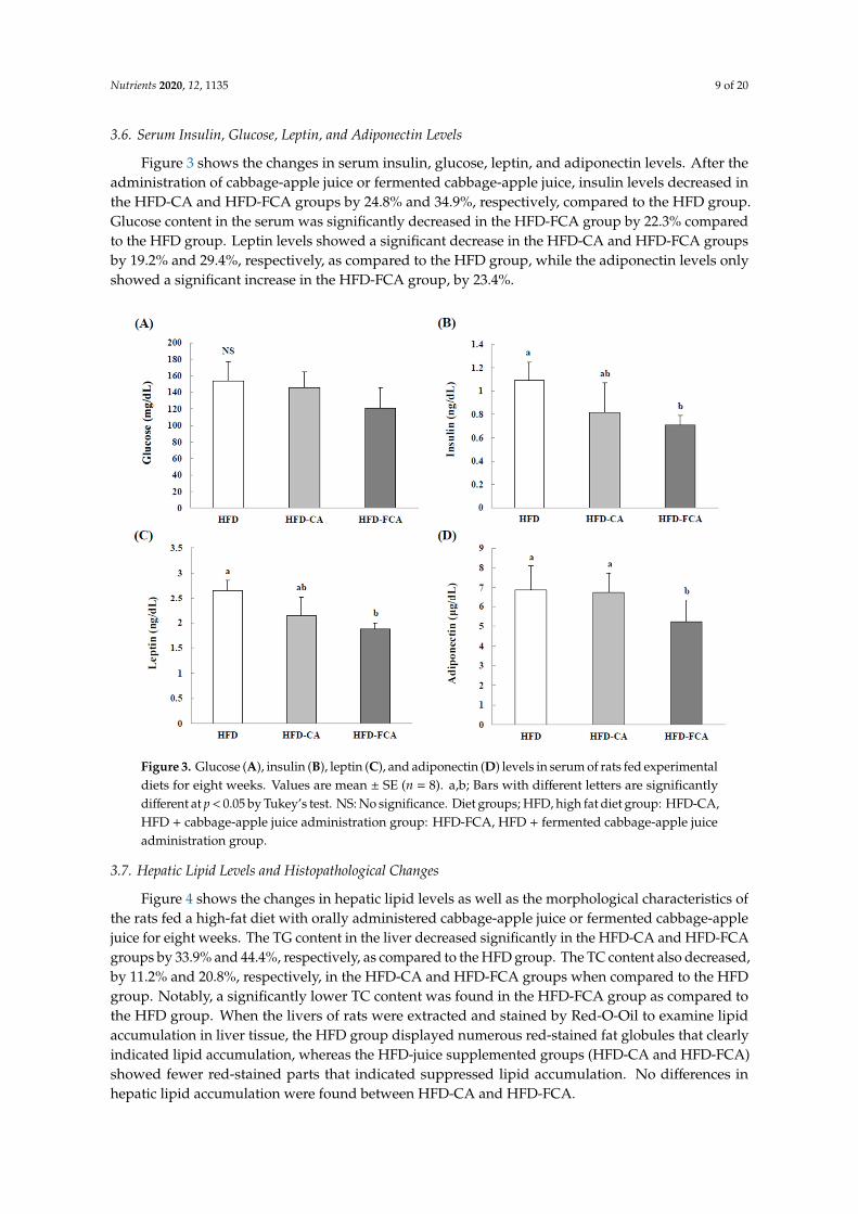
3.6. Serum Insulin, Glucose, Leptin, and Adiponectin Levels
Figure 3 shows the changes in serum insulin, glucose, leptin, and adiponectin levels. After theadministration of cabbage-apple juice or fermented cabbage-apple juice, insulin levels decreased inthe HFD-CA and HFD-FCA groups by 24.8% and 34.9%, respectively, compared to the HFD group.Glucose content in the serum was significantly decreased in the HFD-FCA group by 22.3% comparedto the HFD group. Leptin levels showed a significant decrease in the HFD-CA and HFD-FCA groupsby 19.2% and 29.4%, respectively, as compared to the HFD group, while the adiponectin levels onlyshowed a significant increase in the HFD-FCA group, by 23.4%.
Nutrients 2020, 12, x FOR PEER REVIEW 9 of 20
Table 4. Serum lipid profiles in rats fed experimental diets.
HFD HFD-CA HFD-FCA Triglyceride (mg/dL) 101.63 ± 11.97 a 81.95 ± 8.16 b 73.79 ± 8.22 b
Total cholesterol (mg/dL) 123.51 ± 10.33 a 99.88 ± 11.92 b 90.60 ± 10.58 b LDL/VLDL cholesterol (mg/dL) 92.68 ± 9.94 a 80.05 ± 7.02 ab 71.19 ± 8.25 b
HDL-cholesterol (mg/dL) 30.75 ± 5.97 b 34.13 ± 4.12 b 44.75 ± 4.53 a Diet groups; HFD, high fat diet group: HFD-CA, HFD + cabbage-apple juice administration group: HFD-FCA, HFD + fermented cabbage-apple juice administration group. Values are mean ± SE (n = 8 rats per group). Values with different superscripts in the same column are significantly different (p < 0.05) among groups by Tukey’s test. NS: No significance.
3.6. Serum Insulin, Glucose, Leptin, and Adiponectin Levels
Figure 3 shows the changes in serum insulin, glucose, leptin, and adiponectin levels. After the administration of cabbage-apple juice or fermented cabbage-apple juice, insulin levels decreased in the HFD-CA and HFD-FCA groups by 24.8% and 34.9%, respectively, compared to the HFD group. Glucose content in the serum was significantly decreased in the HFD-FCA group by 22.3% compared to the HFD group. Leptin levels showed a significant decrease in the HFD-CA and HFD-FCA groups by 19.2% and 29.4%, respectively, as compared to the HFD group, while the adiponectin levels only showed a significant increase in the HFD-FCA group, by 23.4%.
Figure 3. Glucose (A), insulin (B), leptin (C), and adiponectin (D) levels in serum of rats fed experimental diets for eight weeks. Values are mean ± SE (n = 8). a,b; Bars with different letters are significantly different at p < 0.05 by Tukey’s test. NS: No significance. Diet groups; HFD, high fat diet group: HFD-CA, HFD + cabbage-apple juice administration group: HFD-FCA, HFD + fermented cabbage-apple juice administration group.
Figure 3. Glucose (A), insulin (B), leptin (C), and adiponectin (D) levels in serum of rats fed experimentaldiets for eight weeks. Values are mean ± SE (n = 8). a,b; Bars with different letters are significantlydifferent at p < 0.05 by Tukey’s test. NS: No significance. Diet groups; HFD, high fat diet group: HFD-CA,HFD + cabbage-apple juice administration group: HFD-FCA, HFD + fermented cabbage-apple juiceadministration group.
3.7. Hepatic Lipid Levels and Histopathological Changes
Figure 4 shows the changes in hepatic lipid levels as well as the morphological characteristics ofthe rats fed a high-fat diet with orally administered cabbage-apple juice or fermented cabbage-applejuice for eight weeks. The TG content in the liver decreased significantly in the HFD-CA and HFD-FCAgroups by 33.9% and 44.4%, respectively, as compared to the HFD group. The TC content also decreased,by 11.2% and 20.8%, respectively, in the HFD-CA and HFD-FCA groups when compared to the HFDgroup. Notably, a significantly lower TC content was found in the HFD-FCA group as compared tothe HFD group. When the livers of rats were extracted and stained by Red-O-Oil to examine lipidaccumulation in liver tissue, the HFD group displayed numerous red-stained fat globules that clearlyindicated lipid accumulation, whereas the HFD-juice supplemented groups (HFD-CA and HFD-FCA)showed fewer red-stained parts that indicated suppressed lipid accumulation. No differences inhepatic lipid accumulation were found between HFD-CA and HFD-FCA.

Nutrients 2020, 12, 1135 10 of 20
Nutrients 2020, 12, x FOR PEER REVIEW 10 of 20
3.7. Hepatic Lipid Levels and Histopathological Changes
Figure 4 shows the changes in hepatic lipid levels as well as the morphological characteristics of the rats fed a high-fat diet with orally administered cabbage-apple juice or fermented cabbage-apple juice for eight weeks. The TG content in the liver decreased significantly in the HFD-CA and HFD-FCA groups by 33.9% and 44.4%, respectively, as compared to the HFD group. The TC content also decreased, by 11.2% and 20.8%, respectively, in the HFD-CA and HFD-FCA groups when compared to the HFD group. Notably, a significantly lower TC content was found in the HFD-FCA group as compared to the HFD group. When the livers of rats were extracted and stained by Red-O-Oil to examine lipid accumulation in liver tissue, the HFD group displayed numerous red-stained fat globules that clearly indicated lipid accumulation, whereas the HFD-juice supplemented groups (HFD-CA and HFD-FCA) showed fewer red-stained parts that indicated suppressed lipid accumulation. No differences in hepatic lipid accumulation were found between HFD-CA and HFD-FCA.
Figure 4. Hepatic triglyceride (A) and total cholesterol (B) levels, representative anatomical views (C), and histopathological analysis (D) in rats fed experimental diets for 8 weeks. All sections were stained with Oil Red O, ×100. Values are mean ± SE (n = 8 rats per group). a,b; Bars with different letters are significantly different at p < 0.05 by Tukey’s test. Diet groups; HFD, high fat diet group: HFD-CA, HFD + cabbage-apple juice administration group: HFD-FCA, HFD + fermented cabbage-apple juice administration group.
3.8. mRNA Expression of an Enzyme Related to Lipid Synthesis in the Liver
Figure 5 shows the effects on gene expression levels of the enzymes that are involved in hepatic lipid synthesis of the rats fed a high-fat diet with orally administered cabbage-apple juice or fermented cabbage-apple juice for eight weeks. The level of mRNA expression of ACC in the liver
Figure 4. Hepatic triglyceride (A) and total cholesterol (B) levels, representative anatomical views (C),and histopathological analysis (D) in rats fed experimental diets for 8 weeks. All sections were stainedwith Oil Red O, ×100. Values are mean ± SE (n = 8 rats per group). a,b; Bars with different letters aresignificantly different at p < 0.05 by Tukey’s test. Diet groups; HFD, high fat diet group: HFD-CA,HFD + cabbage-apple juice administration group: HFD-FCA, HFD + fermented cabbage-apple juiceadministration group.
3.8. mRNA Expression of an Enzyme Related to Lipid Synthesis in the Liver
Figure 5 shows the effects on gene expression levels of the enzymes that are involved in hepaticlipid synthesis of the rats fed a high-fat diet with orally administered cabbage-apple juice or fermentedcabbage-apple juice for eight weeks. The level of mRNA expression of ACC in the liver tissue wassignificantly lower in the HFD-CA and HFD-FCA groups than in the HFD group. On the contrary, themRNA expression levels of FAS and G6PDH was significantly lower in the HFD-FCA group only. Themalic enzyme gene expression levels in the liver did not show intergroup differences.

Nutrients 2020, 12, 1135 11 of 20
Nutrients 2020, 12, x FOR PEER REVIEW 11 of 20
tissue was significantly lower in the HFD-CA and HFD-FCA groups than in the HFD group. On the contrary, the mRNA expression levels of FAS and G6PDH was significantly lower in the HFD-FCA group only. The malic enzyme gene expression levels in the liver did not show intergroup differences.
Figure 5. mRNA expression levels of enzymes related to lipid synthesis (A) in livers of rats fed experimental diets for eight weeks. The mRNA expression levels of FAS (B), ACC (C), G6PDH (D), and malic enzyme (ME) (E) were measured by RT-PCR. In the determination of mRNA levels, β-actin served as a loading control. Values are mean ± SE (n = 8 rats per group). a,b; Bars with different letters are significantly different at p < 0.05 by Tukey’s test. Diet groups; HFD, high fat diet group: HFD-CA, HFD + cabbage-apple juice administration group: HFD-FCA, HFD + fermented cabbage-apple juice administration group.
3.9. Epididymal Adipose Tissue TG Contents and Histopathological Changes
The TG content in the epididymal adipose tissue decreased in the HFD-juice supplemented groups (HFD-CA and HFD-FCA) by approximately 14.2–28.2% as compared to the HFD group (Figure 6). When the size of epididymal adipocytes was measured, HFD displayed a marked increase in size; however, a decrease in size in the HFD-juice supplemented groups (HFD-CA and HFD-FCA) when compared to the HFD group was shown in the HFD-juice supplemented groups (HFD-CA and HFD-FCA). HFD-FCA, in particular, showed a significantly reduced adipocyte size when compared to HFD-CA.
Figure 5. mRNA expression levels of enzymes related to lipid synthesis (A) in livers of rats fedexperimental diets for eight weeks. The mRNA expression levels of FAS (B), ACC (C), G6PDH (D),and malic enzyme (ME) (E) were measured by RT-PCR. In the determination of mRNA levels, β-actinserved as a loading control. Values are mean ± SE (n = 8 rats per group). a,b; Bars with different lettersare significantly different at p < 0.05 by Tukey’s test. Diet groups; HFD, high fat diet group: HFD-CA,HFD + cabbage-apple juice administration group: HFD-FCA, HFD + fermented cabbage-apple juiceadministration group.
3.9. Epididymal Adipose Tissue TG Contents and Histopathological Changes
The TG content in the epididymal adipose tissue decreased in the HFD-juice supplemented groups(HFD-CA and HFD-FCA) by approximately 14.2–28.2% as compared to the HFD group (Figure 6).When the size of epididymal adipocytes was measured, HFD displayed a marked increase in size;however, a decrease in size in the HFD-juice supplemented groups (HFD-CA and HFD-FCA) whencompared to the HFD group was shown in the HFD-juice supplemented groups (HFD-CA andHFD-FCA). HFD-FCA, in particular, showed a significantly reduced adipocyte size when comparedto HFD-CA.

Nutrients 2020, 12, 1135 12 of 20Nutrients 2020, 12, x FOR PEER REVIEW 12 of 20
Figure 6. Epidydimal triglyceride content (A), representative findings (B), and adipocyte size (C) in rats fed experimental diets for 8 weeks. Epididymal fat tissues were visualized by hematoxylin and eosin staining. Adipocyte size was measured using a microscope and quantified using an image analyzer. Values are mean ± SE (n = 8 rats per group). a,b; Bars with different letters are significantly different at p < 0.05 by Tukey’s test. Diet groups; HFD, high fat diet group: HFD-CA, HFD + cabbage-apple juice administration group: HFD-FCA, HFD + fermented cabbage-apple juice administration group.
4. Discussion
The natural polyphenol compounds found in fruits and vegetables are known to exhibit anti-obesity effects by altering signal transduction in target cells, such as adipocytes, regulating gene expression, and enhancing free radical scavenging activity [12]. The polyphenol or flavonoid compounds abundant in apples include procyanidin, hydroxycinnamic acid and its derivatives, chlorogenic acid, caffeic acid, and epicatechin [9–11], while those that are abundant in cabbages include phenolic acids, flavonols, and anthocyanidines [19]. In addition, apples and cabbages are both rich in dietary fiber; notably, apples contain a high level of pectin among its dietary fiber that has been shown to act as a prebiotic in an in vivo study [42]. Cruciferous vegetables that belong to the family of cruciferous, such as cabbage, are rich in glucosinolates, carotenoids, and vitamin C,
Figure 6. Epidydimal triglyceride content (A), representative findings (B), and adipocyte size (C) in ratsfed experimental diets for 8 weeks. Epididymal fat tissues were visualized by hematoxylin and eosinstaining. Adipocyte size was measured using a microscope and quantified using an image analyzer.Values are mean ± SE (n = 8 rats per group). a,b; Bars with different letters are significantly different atp < 0.05 by Tukey’s test. Diet groups; HFD, high fat diet group: HFD-CA, HFD + cabbage-apple juiceadministration group: HFD-FCA, HFD + fermented cabbage-apple juice administration group.
4. Discussion
The natural polyphenol compounds found in fruits and vegetables are known to exhibit anti-obesityeffects by altering signal transduction in target cells, such as adipocytes, regulating gene expression,and enhancing free radical scavenging activity [12]. The polyphenol or flavonoid compounds abundantin apples include procyanidin, hydroxycinnamic acid and its derivatives, chlorogenic acid, caffeic acid,and epicatechin [9–11], while those that are abundant in cabbages include phenolic acids, flavonols,and anthocyanidines [19]. In addition, apples and cabbages are both rich in dietary fiber; notably,apples contain a high level of pectin among its dietary fiber that has been shown to act as a prebiotic inan in vivo study [42]. Cruciferous vegetables that belong to the family of cruciferous, such as cabbage,

Nutrients 2020, 12, 1135 13 of 20
are rich in glucosinolates, carotenoids, and vitamin C, which play a major role in the modulation oflipid metabolism in vivo and in vitro [18]. Carotenoids have been reported to possess anti-obesity andanti-inflammatory abilities [43], and a hepatoprotective effect [22,23]. Such components are known toexert anti-cholesterol and anti-obesity activities [9–11,19]. In a previous study, when obese individualswere administered a 240–720 mg/kg of apple (fruits or juice) daily, body weight loss was observed; inexperimental animals, a daily intake of 1–10 mg/kg of apples was shown to lead to body weight loss [12].Consequently, we prepared a mixed juice containing equal amounts of apples and cabbages withknown preventive effects on metabolic diseases that are attributed to obesity; next, by administeringthe juice to rats on a high-fat diet, we investigated the changes in body weight, liver and white fatpad weights, serum and hepatic lipid profiles, gene expression related to hepatic lipid metabolism,and adipocyte size. In our study, we have a limitation in vehicle control, since we administered salineand fiber into AIN-93M diet. The administration of saline and fiber cannot fully account for fiber andbioactive components in other groups.
The anti-obesity and hypolipidemic effects of vegetable and fruit juice fermented by LAB havebeen reported by several investigators [25–28,44]. LAB fermentation produces a variety of organic acids,short-chain fatty acids (SCFAs), amino acids, and secondary metabolite compounds [18,24,26,27,45,46].Among the organic acids, SCFAs and amino acids showing anti-obesity properties in experimentalanimals are acetic acid [26], propionic acid [26,45], and ornithine [46]. In this study, L. plantarum EMfermentation showed an increase in dietary fiber, acetic acid, lactic acid, total organic acid, and totalpolyphenol contents, and a decrease in the crude fat and total free sugar contents of cabbage-apple juice.
Although soluble polyphenols are rapidly absorbed in the small intestine, most show a lowabsorption rate in the colon. The polyphenols contained in apples, in particular, are found in theform of aglycones and glucoside conjugates with low bioavailability [9,47]. Thus, fermented naturalproducts have gained considerable attention because the fermentation of natural food ingredients withLAB has been shown to increase the bioactivity of nutrients through biotransformation or probioticeffects [9,48,49]. LAB converts the phenol compounds in fruits and vegetables to a more absorbable formin the human intestines, thereby maximizing the absorption rate and bioavailability [9,49]. Therefore,we conducted an experiment to compare non-fermented cabbage-apple juice and cabbage-apple juicefermented with kimchi-isolated L. plantarum EM [32] with respect to the anti-obesity effects and positiveeffects on lipid metabolism.
In this study, five-week old Sprague Dawley rats were fed a high-fat diet for eight weeks, whichled to increased body weight, increased weights of liver and white fat pads, and increased levelsof serum TG, TC, and LDL-cholesterol. The levels of TG and TC in liver tissue also increased, withincreased expression levels of FSA, ACC, malic enzyme, and G6PDH genes that code for enzymesthat are related to lipid synthesis, which confirmed body fat accumulation and dyslipidemia. Thesecharacteristics indicate that a high-fat diet induces obesity and hyperlipidemia, a result that coincideswith previous studies on obesity [50]. In addition, the histopathological tests on liver tissue showedan increase in fat granules and lipid accumulation (hepatic steatosis). Epididymal fat pad size wasalso markedly increased. These phenomena resulted in significant increases in body weight and liverweight in experimental animals fed a high-fat diet [51]. The weight of the organs including the liverincreased when high-fat diet caused unbalanced glycometabolism, inflow of excessively producedglucose, and abnormal RNA and DNA synthesis [52]. Obesity is caused by an increase in body fat,rather than in body weight, and an increase in the weight of adipose tissue leads to lipid accumulation,such that the higher the content, the higher the risk of metabolic disease. In particular, it is known that,despite equal body fat content, increased visceral fat rather than subcutaneous fat poses a health hazard;the higher the content of visceral fat, the higher the incidence of metabolic complications includingchanges in hypertension, dyslipidemia, and inflammatory cytokines, as well as in hyperinsulinemiaresistance [53,54]. However, body weight loss in obesity improves obesity-associated diseases andmetabolic disorders [55]. In this study, an inhibitory effect of abdominal obesity, as well as a reducedrisk of metabolic disease, was observed, based on the decrease in not only body weight and the weights

Nutrients 2020, 12, 1135 14 of 20
of the liver and white fat pads, but also in hepatic lipid accumulation and adipocyte size after theadministration of cabbage-apple juice or fermented cabbage-apple juice. Such findings may suggestthat obesity in mice can be prevented by non-absorbable procyanidins, a type of flavonoid found inapples [56], while apple-derived pectin attenuates metabolic endotoxemia in rats with diet-inducedobesity, thereby reducing body weight and inhibiting body fat accumulation [42]. Furthermore,procyanidins, a component in apples consisting of various polymerized catechins, are known tosuppress pancreatic lipase inhibitory activity and TG absorption [57]. The bioactive compounds, suchas polyphenols or flavonoid, and the dietary fibers contribute to the decrease in body weight and bodyfat content.
Serum ALT and AST are distributed in liver tissue; as enzymes that are involved in amino acidbiosynthesis, their activities are promoted upon damage to the liver due to drugs or stress, such thatthey are used as indicators of liver damage [58]. These enzymes show increased activities in obesitybecause the condition leads to lipid accumulation in liver tissue and the production of lipid peroxidesthat in turn produce reactive oxygen species, which together damage the liver [59]. Serum ALP activityincreases with hyperlipidemia or related complications, hepatobiliary obstruction, and liver diseases.Advanced injury to hepatocytes leads to increased ALP activity and consequent disturbance to bileacid excretion in the liver and intestines, which is known to increase serum cholesterol levels [60].In addition, LDH activity changes upon the disturbances to bile secretion that is caused by the onsetof hypercholesterolemia or lipid accumulation in the liver and intestines [61]. In general, an inputof excess TG or cholesterol to the liver as part of dietary intake is known to result in fatty liverand damage to hepatocytes, as the excess TG or cholesterol binds to lipid acceptor apoprotein toform lipoprotein that cannot be excreted. It is presumed that either juice may improve serum orliver lipid metabolism and delay injury to hepatocytes, thereby brining about positive effects on therecovery and maintenance of liver function, based on these results and the findings of this studydemonstrating that the activities of AST, ALT, ALP, and LDH were increased by high-fat diet andreduced by administering cabbage-apple juice or fermented cabbage-apple juice. In an animal modelwith acetaminophen-induced liver damage, cabbage extract effectively lowered the activities of ALTand AST to exert an enhancing effect on liver protection and liver function [62]. In aerobic condition,lactic acid is increased to produce cellular energy via increasing LDH activity. The elevation of LDHactivity is a pathological biomarker in cancer [63]. Therefore, the consumption of lactic acid should becarefully evaluated, since it might increase LDH activity because the fermented cabbage and applejuice inherently has higher lactate. Interestingly, one of clinical study demonstrated that short-terminfusion of lactate did not alter metabolic rate and cytokine significantly [64]. Moreover, long-termexposure of lactate decreased LPS-inducible cytokine expression [64]. Therefore, dietary lactic acidmight act differently when compared to endogenous lactic acid. However, further intensive studies arerequired to examine the potential net benefic and side effect in lactic acid consumption.
High-fat diet increases the incidence of atherosclerotic coronary artery disease and cardiovasculardisease, while facilitating the induction of atherosclerosis and other complications [59]. However, theserum TG, TC, and LDL-cholesterol levels that increased in response to a high-fat diet were restored tohealthy levels by administering cabbage-apple juice or fermented cabbage-apple juice since a reductionin serum lipid levels is known to reduce the risk of atherosclerotic cardiovascular disease [65]. Thepolyphenol compounds in fruits and vegetables play a beneficial role in preventing cardiovasculardisease as they change the serum lipid levels [66]; furthermore, studies report that the higher the intakeof polyphenol compounds, including flavonoids, the lower the mortality risk due to cardiovasculardisease [67]. In a study that fed corn oil-loaded mice with a diet containing 60 mg of apple polyphenol,reduced TG absorption and serum TG content were observed [57]. Procyanidins, in particular, as acomponent of apple polyphenol, has been shown to exert anti-atherosclerosis and cholesterol-loweringeffects in rats [68]. Cabbage leaf protein concentrate was also shown to have enhancing effects on serumlipid metabolism [69]. Moreover, cabbage extract, as well as the S-methyl-l-cysteine sulfoxide found in

Nutrients 2020, 12, 1135 15 of 20
cabbages, were shown to inhibit hypercholesterolemia in hepatoma-bearing rats, which was attributedto a reduction in the serum cholesterol level and an increase in bile acid excretion in feces [21].
The liver is an organ that plays a crucial role in regulating the serum TG and TC levels bymediating the biosynthesis of TG and TC and their secretion to the circulatory system in lipoproteinforms. This renders TG and TC levels in the liver a key indicator of circulatory disease. The TGcontent in the liver depends on various interactions, such as the input, biosynthesis, oxidation, andrelease of fatty acids in VLDL form. High-fat diet causes excessive fatty acid input to the liver toinduce lipid accumulation and a direct correlation between the accumulation of TG in the liver andinsulin resistance has been reported [70]. The accumulation of TG and TC in the liver is known toincrease liver weight. The high-fat diet applied in this study was shown to result in hepatic lipidaccumulation that was based on increased TG, TC, and LDL/VLDL-cholesterol contents in liver tissue.Exposure to a high-fat diet was also shown to induce typical lesions in non-alcoholic fatty liverdisease; upon histopathological observation, a myriad of isolated forms of lipid accumulation werefound in the cytoplasm of hepatocytes. However, the TG, TC, and LDL/VLDL-cholesterol levels inliver tissue decreased when cabbage-apple juice or fermented cabbage-apple juice was administered,which implied that these juices might exert preventive effects on hepatic lipid accumulation. Theformation of TG in the liver is based on synthesis mediated by several key enzymes such as ACC,FAS, and G6PDH; hence, gene expression levels for these lipid synthesis-related enzymes were alsomeasured. Although high-fat diet increased the gene expression levels of ACC, FAS, malic enzyme,and G6PDH in liver tissue, administering cabbage-apple juice or fermented cabbage-apple juice led toa decrease in the expression of these genes. Thus, the results of reduced liver weight and TG content,as well as the inhibition of hepatic lipid accumulation by the administration of cabbage-apple juice orfermented cabbage-apple juice, seem to be the result of reduced gene expression levels of the lipidsynthesis-related enzymes—malic enzyme, ACC, and FAS—and not to food intake.
The long-term intake of a high-fat diet induces elevated blood glucose levels and eventually leadsto insulin resistance. In the human body, when the blood glucose level increases after a meal, thepancreas secrets insulin to lower the glucose level; however, in the case of excessive accumulation ofbody fat, hyperinsulinemia due to insulin resistance persists for a long time. Adipose tissue providesstorage for excess energy, while it is also known to act as an endocrine organ that regulates body fatcontent and nutrient metabolism. An overabundance of adipose tissue and consequent dysfunctionlead to a regulatory disorder of adipokine secretion that contributes to inflammatory reactions aswell as changes in glucose and lipid metabolism, thereby resulting in obesity-associated metabolicdiseases, including dyslipidemia, non-alcoholic fatty liver, insulin resistance, and type 2 diabetes [71,72].Leptin is secreted from adipose tissue. It regulates appetite by stimulating the hypothalamus in thebrain and glucose and lipid metabolism by increasing thermogenesis. The concentration of leptinis closely related to body fat content; in overweight or obese people, despite increased leptin levels,leptin resistance leads to the excess accumulation of TG in adipose tissue, liver, muscle, and pancreas,whereby insulin sensitivity and secretion are damaged [73]. In contrast, plasma adiponectin showsan inverse correlation with body fat content, such that its level decreases in obesity; moreover, withregard to insulin sensitivity, a positive correlation has been found [74]. Plasma adiponectin has alsobeen reported to exert a positive effect on lipid metabolism, whereby plasma TG decreases but HDL-Cincreases, and the oxidation of fatty acids in the liver and muscle is facilitated along with lipoproteinlipase activity that decomposes VLDL to reduce the serum TG content [75,76]. A reduction in plasmaadiponectin level has been shown to increase the risk of dyslipidemia and cardiovascular diseasein experimental animals as well as humans [77]. In this study, although food intake did not takeleptin function into account, the FGA-HFD group that was fed a high-fat diet and administered withfermented cabbage-apple juice showed a significant reduction in total fat content along with an increasein serum leptin levels, but a decrease in adiponectin levels as compared to the HFD group fed ahigh-fat diet only, suggesting that fermented cabbage-apple juice acted to reduce body fat content. The

Nutrients 2020, 12, 1135 16 of 20
juice was also shown to enhance serum glucose and insulin levels in addition to adipokines, whichsuggested that it might improve obesity-induced metabolic diseases.
Fermented fruit-vegetable juice, as compared to non-fermented fruit-vegetable juice, has beenreported to exhibit diverse health-promoting effects, such as enhancing the nutritional values, thebioavailability of phenolics, the contents and composition of secondary metabolite compounds,as well as antioxidant effects [9,26,27,44–46,48,49]. Such findings indicate that the enhanced lipidmetabolism and anti-obesity effects are based on the compositional changes in natural polyphenolcompounds [9–11], glucosinolates [18], and carotenoids [22,23,26,43] that are found in fruits andvegetables, the source materials of the juice, and dietary fiber [42], as well as organic acids [26,45],and amino acids [46] produced during LAB fermentation. Li et al. [26] reported that carrot juice thatwas fermented by L. plantarum NCU116 had greater acetic acid, propionic acid, lactic acid, β-carotene,and amino acid contents, and anti-diabetic, anti-oxidative, and lipid-lowering effects than those ofthe non-fermented carrot juice. Thus, when fermented cabbage-apple juice was compared withnon-fermented cabbage-apple juice in this study, improvements in the compositional changes in serumlipids, gene expression regulation for enzymes engaged in lipid synthesis, and inhibitory effects onleptin appear to be the result of increased content or bioavailability of the functional ingredients in applesand cabbages after fermentation. Based on these findings, the different types of polyphenol compounds,dietary fibers, organic acids, and SCFAs contained in fermented cabbage-apple juice are presumed tohave an effect in preventing obesity and improving lipid metabolism. Nevertheless, further studies arenecessary for identifying the components and elucidating their effects on metabolic mechanisms.
5. Conclusions
Based on our findings, the intake of cabbage-apple juice or fermented cabbage-apple juice alongwith a high-fat diet appears to be effective in preventing various metabolic disorders that are caused byobesity, as the juice effectively regulates body weight and the weights of liver and white fat pads in ratswith high-fat diet-induced obesity. Furthermore, it reduces the levels of serum leptin and insulin whileincreasing the level of adiponectin and altering gene expression for enzymes that are related to hepaticlipid metabolism to improve serum lipid levels. The anti-obesity effects and positive effects on lipidmetabolism were shown to be more substantial in fermented cabbage-apple juice than non-fermentedcabbage-apple juice. The cabbage-apple juice that was fermented with L. plantarum EM was shown tofurther enhance the beneficial effects of cabbage-apple juice on obesity-induced metabolic syndrome,at least under the conditions provided in this study.
Author Contributions: Conceptualization, H.-C.C. and J.-J.L.; methodology, H.-K.S. and J.-J.L.; software, S.P. andH.-K.S.; validation, S.P., H.-K.S., H.-C.C. and J.-J.L.; formal analysis, S.P. and H.-K.S.; investigation, S.P., H.-K.S.,H.-C.C. and J.-J.L.; resources, H.-C.C. and J.-J.L.; data curation, S.P., H.-K.S., H.-C.C. and J.-J.L.; writing—originaldraft preparation, S.P., H.-K.S., H.-C.C. and J.-J.L.; writing—review and editing, H.-K.S., H.-C.C. and J.-J.L;visualization, S.P. and H.-K.S.; supervision, H.-C.C. and J.-J.L.; project administration, H.-C.C. and J.-J.L.; fundingacquisition, H.-C.C. and J.-J.L. All authors have read and agree to the published version of the manuscript.
Funding: This work was supported by Koran Institute of Planning and Evaluation for Technology in Food,Agriculture, Forestry and Fisheries (IPET) through (Agricultural Microbiome R&D Program), funded by Ministryof Agriculture, Food and Rural Affairs (MAFRA) (918005-4).
Conflicts of Interest: The authors declare no conflicts of interest.
References
1. Boheing, H.; Bechthold, A.; Bub, A.; Ellinger, S.; Haller, D.; Kroke, A.; Leschik-Bonnet, E.; Müller, M.J.;Oberritter, H.; Schulze, M.; et al. Critical review: Vegetables and fruit in the prevention of chronic diseases.Eur. J. Nutr. 2012, 51, 637–663. [CrossRef]
2. Rimm, E.B.; Ascherio, A.; Giovannucci, E.; Spiegelman, D.; Stampfer, M.J.; Willett, W.C. Vegetable, fruit, andcereal fiber intake and risk of coronary heart disease among men. JAMA 1996, 275, 447–451. [CrossRef]
3. Slavin, J.L.; Lloyd, B. Health benefits of fruits and vegetables. Adv. Nutr. 2012, 3, 506–516. [CrossRef][PubMed]

Nutrients 2020, 12, 1135 17 of 20
4. Carter, P.; Gray, L.J.; Troughton, J.; Khunti, K.; Davies, M.J. Fruit and vegetable intake and incidence of type 2diabetes mellitus: Systematic review and meta-analysis. BMJ 2010, 341, c4229. [CrossRef] [PubMed]
5. Hung, H.C.; Joshipura, K.J.; Jiang, R.; Hu, F.B.; Hunter, D.; Smith-Warner, S.A.; Colditz, G.A.; Rosner, B.;Spiegelman, D.; Willett, W.C. Fruit and vegetable intake and risk of major chronic disease. J. Natl. CancerInst. 2004, 96, 1577–1584. [CrossRef] [PubMed]
6. Koryachkina, S.Y.; Ladnova, O.L.; Godunov, O.A.; Kholodova, E.N.; Lazareva, T.N. The study of physiologicaleffect of fruit and vegetable powders in animal experiment. Vopr. Pitan. 2016, 85, 48–56. [PubMed]
7. Peluso, I.; Raguzzini, A.; Catasta, G.; Cammisotto, V.; Perrone, A.; Tomino, C.; Toti, E.; Serafini, M. Effects ofhigh consumption of vegetables on clinical, immunological, and antioxidant markers in subjects at risk ofcardiovascular diseases. Oxid. Med. Cell Longev. 2018, 5417165. [CrossRef]
8. Lichtenthäler, R.; Marx, F. Total oxidant scavenging capacities of common European fruit and vegetablejuices. J. Agric. Food Chem. 2005, 53, 103–110. [CrossRef]
9. Li, Z.; Teng, J.; Lyu, Y.; Hu, X.; Zhao, Y.; Wang, M. Enhanced antioxidant activity for apple juice fermentedwith Lactobacillus plantarum ATCC14917. Molecules Dec. 2019, 24, 51. [CrossRef]
10. Trošt, K.; Ulaszewska, M.M.; Stanstrup, J.; Albanese, D.; De Filippo, C.; Tuohy, K.M.; Natella, F.; Scaccini, C.;Mattivi, F. Host: Microbiome co-metabolic processing of dietary polyphenols—An acute, single blinded,cross-over study with different doses of apple polyphenols in healthy subjects. Food Res. Int. 2018, 112,108–128. [CrossRef]
11. Zielinski, A.A.; Zardo, D.M.; Alberti, A.; Bortolini, D.G.; Benvenutti, L.; Demiate, I.M.; Nogueira, A. Effect ofcryoconcentration process on phenolic compounds and antioxidant activity in apple juice. J. Sci. Food Agric.2019, 99, 2786–2792. [CrossRef] [PubMed]
12. Asgary, S.; Rastqar, A.; Keshvari, M. Weight loss associated with consumption of apples: A review. J. Am.Coll. Nutr. 2018, 37, 627–639. [CrossRef] [PubMed]
13. O’Neil, C.E.; Nicklas, T.A.; Fulgoni, V.L. Consumption of apples is associated with a better diet qualityand reduced risk of obesity in children: National Health and Nutrition Examination Survey (NHANES)2003–2010. Nutr. J. 2015, 14, 48. [CrossRef] [PubMed]
14. Wang, Q.; Wang, X.; Gong, G.; Li, G.; Li, C. Consumption of fruit, but not vegetable, may reduce risk ofgastric cancer: Results from a meta-analysis of cohort studies. Eur. J. Cancer 2014, 50, 1498–1509. [CrossRef]
15. Samout, N.; Bouzenna, H.; Dhibi, S.; Ncib, S.; ElFeki, A.; Hfaiedh, N. Therapeutic effect of apple pectin inobese rats. Biomed. Pharmacother. 2016, 83, 1233–1238. [CrossRef]
16. Anubhuti, S.H.; Ashok, S.H.; Prashant, Y.; Dhiraj, S. Isothiocyanates in Brassica: Potential anticancer agents.Asian Pac. J. Cancer Prev. 2016, 17, 4507–4510.
17. Shim, K.H.; Sung, N.K.; Kang, K.S.; Ahn, C.W.; Seo, K.I. Analysis of glucosinolates and the change of contentsduring processing and storage in Cruciferous vegetables. J. Korean Soc. Food Sci. Nutr. 1992, 21, 43–48.
18. Rodríguez-Cantú, L.N.; Gutiérrez-Uribe, J.A.; Arriola-Vucovich, J.; Díaz-De La Garza, R.I.; Fahey, J.W.;Serna-Saldivar, S.O. Broccoli (Brassica oleracea var. italica) sprouts and extracts rich in glucosinolates andisothiocyanates affect cholesterol metabolism and genes involved in lipid homeostasis in hamsters. J. Agric.Food Chem. 2011, 59, 1095–1103.
19. Jahangir, M.; Kim, H.K.; Choi, Y.H.; Verpoorte, R. Health-affecting compounds in Brassicaceae. Comp. Rev.Food Sci. Food Saf. 2009, 8, 31–43. [CrossRef]
20. Gessler, N.N.; Bezzubov, A.A.; Podlepa, E.M.; Bykhovski, V.Y. S-methylmethionine (vitamin U) metabolismin plants. Appl. Biochem. Micro. 1991, 27, 275–280.
21. Komatsu, W.; Miura, Y.; Yagasaki, K. Suppression of hypercholesterolemia in hepatoma-bearing rats bycabbage extract and its component, S-methyl-L-cysteine sulfoxide. Lipids 1998, 33, 499–503. [CrossRef][PubMed]
22. Elvira-Torales, L.I.; García-Alonso, J.; Periago-Castón, M.J. Nutritional importance of carotenoids and theireffect on liver health: A review. Antioxidants (Basel) 2019, 8, 229. [CrossRef] [PubMed]
23. Yilmaz, B.; Sahin, K.; Bilen, H.; Bahcecioglu, I.B.; Bilir, B.; Ashraf, S.; Halazun, K.J.; Kucuk, O. Carotenoidsand non-alcoholic fatty liver disease. Hepatobiliary Surg Nutr. 2015, 4, 161–171.
24. Hubert, J.; Berger, M.; Mepveu, F.; Paul, F.; Dayd, J. Effects of fermentation on the phytochemical compositionand antioxidant properties of soy germ. Food Chem. 2008, 109, 709–721. [CrossRef] [PubMed]

Nutrients 2020, 12, 1135 18 of 20
25. Foo, H.L.; Loh, T.C.; Lim, Y.S.; Shukriyah, M.H.; Kufli, C.N.; Law, F.L. Effects of fermented fruits on growthperformance, shedding of Enterobacteriaceae and lactic acid bacteria and plasma cholesterol in rats. Pakistan J.Nutr. 2003, 2, 228–233.
26. Li, C.; Ding, Q.; Nie, S.P.; Zhang, Y.S.; Xiong, T.; Xie, M.Y. Carrot juice fermented with Lactobacillus plantarumNCU116 ameliorates type 2 diabetes in rats. J. Agri. Food Chem. Dec. 2014, 62, 11884–11891. [CrossRef][PubMed]
27. Mauro, C.S.I.; Guergoletto, K.B.; Garcia, S. Development of blueberry and carrot juice blend fermented byLactobacillus reuteri LR92. Beverages 2016, 2, 37. [CrossRef]
28. Chu, H.; Kim, J. Anti-obesity effect of Fructus pyri Pyrifoliae extract fermented by lactic-acid bacteria on rats.Appl. Microsc. 2018, 48, 62–72. [CrossRef]
29. Takemura, N.; Okubo, T.; Sonoyama, K. Lactobacillus plantarum strain No. 14 reduces adipocyte size in micefed high-fat diet. Exp. Biol. Med (Maywood) 2010, 235, 849–856. [CrossRef]
30. Park, D.Y.; Ahn, Y.T.; Huh, C.S.; Jeon, S.M.; Choi, M.S. The inhibitory effect of Lactobacillus plantarum KY1032cell extract on the adipogenesis of 3T3-L1 cells. J. Med. Food 2011, 14, 670–675. [CrossRef]
31. Krajka-Kuzniak, V.; Szaefer, H.; Bartoszek, A.; Baer-Dubowska, W. Modulation of rat hepatic and kidneyphase II enzymes by cabbage juices: Comparison with the effects of indole-3-carbinol and phenethylisothiocyanate. Br. J. Nutr. 2011, 105, 816–826. [CrossRef] [PubMed]
32. Choi, E.A.; Chang, H.C. Cholesterol-lowering effects of a putative probiotic strain Lactobacillus plantarum EMisolated from kimchi. LWT Food Sci. Technol. 2015, 62, 210–217. [CrossRef]
33. Heydemann, A. An overview of murine high fat diet as a model for type 2 diabetes mellitus. J. Diabetes Res.2016, 2902351. [CrossRef] [PubMed]
34. A.O.A.C. Official Methods of Analysis, 17th ed.; Association of Official Analytical Chemist: Gaithersburg, MD,USA, 2000.
35. Sturm, K.; Koron, D.; Stampar, F. The composition of fruit of different strawberry varieties depending onmaturity stage. Food Chem. 2003, 83, 417–422. [CrossRef]
36. Richmond, M.L.; Brandao, S.C.; Gray, J.I.; Markakis, P.; Stine, C.M. Analysis of simple sugars and sorbitol infruit by high-performance liquid chromatography. J. Agric. Food Chem. 1981, 29, 4–7. [CrossRef]
37. Singleton, V.L.; Orthofer, R.; Lamuela-Ravebtos, R.M. Analysis of total phenols and others oxidation substratesand antioxidants by means of Folin-Ciocalteu reagent. Methods Enzymol. 1999, 299, 152–178.
38. International Standard Organization. Rapeseed: Determination of Glucosinolates Content-Part I: Method UsingHigh Performance Liquid Chromatography; ISO 9167-1. 1992; International Standard Organization: Geneva,Switzerland, 1992; pp. 1–9.
39. Folch, J.; Lees, M.; Sloane-Stanley, G. A simple method for the isolation and purification of total lipids fromanimal tissues. J. Biol. Chem. 1957, 226, 497–509.
40. Biggs, H.G.; Erikson, T.M.; Moorehead, W.R. A mannual colorimetric assay of triglyceride in serum. Clin.Chem. 1975, 21, 437. [CrossRef]
41. Zlatkis, A.; Zak, B. Study of a new cholesterol reagent. Anal. Biochem. 1969, 29, 143–148. [CrossRef]42. Jiang, T.; Gao, X.; Wu, C.; Tian, F.; Lei, Q.; Bi, J.; Xie, B.; Wang, H.Y.; Chen, S.; Wang, X. Apple-derived pectin
modulates gut microbiota, improves gut barrier function, and attenuates metabolic endotoxemia in rats withdiet-induced obesity. Nutrients 2016, 8, 126. [CrossRef]
43. Mounien, L.; Tourniaire, F.; Landrier, J.F. Anti-obesity effect of carotenoids: Direct impact on adipose tissueand adipose-tissue-driven indirect effects. Nutrients 2019, 11, 1562. [CrossRef] [PubMed]
44. Jeon, J.H.; Kim, B.; Mun, E.G.; Cha, Y.S.; Yu, O.K. Effects of fermented blueberry liquid in high-fat diet-inducedobese C57BL/6J mice. J. Nutr. Health 2017, 50, 543–551. [CrossRef]
45. Parvez, S.; Malik, K.A.; Ah Kang, S.; Kim, H.Y. Probiotics and their fermented food products are beneficialfor health. J. Appl. Microbiol. 2006, 100, 1171–1185. [CrossRef] [PubMed]
46. Moon, Y.J.; Soh, J.R.; Yu, J.J.; Shon, H.S.; Cha, Y.S.; Oh, S.H. Intracellular lipid accumulation inhibitory effectof Weissella koreensis OK1-6 isolated from Kimchi on differentiating adipocyte. J. Appl. Microbiol. 2012, 113,652–658. [CrossRef] [PubMed]
47. Borges, G.; Lean, M.E.; Roberts, S.A.; Crozier, A. 2013, Bioavailability of dietary (poly)phenols: A study withileostomists to discriminate between absorption in small and large intestine. Food Funct. 2013, 4, 754–762.[CrossRef]

Nutrients 2020, 12, 1135 19 of 20
48. Trinh, H.T.; Han, S.J.; Kim, S.W.; Lee, Y.C.; Kim, D.H. Bifidus fermentation increases hypolipidemic andhypoglyccemic effects of red ginseng. J. Miocrobiol. Biotechnol. 2007, 17, 1127–1133.
49. Valero-Cases, E.; Nuncio-Jáuregui, N.; Frutos, M.J. Influence of fermentation with different lactic acid bacteriaand in vitro digestion on the biotransformation of phenolic compounds in fermented pomegranate juices.J. Agric. Food Chem. 2017, 65, 6488–6496. [CrossRef]
50. Kim, J.Y.; Nolte, L.A.; Hansen, P.A.; Han, D.H.; Ferguson, K.; Thompson, P.A.; Holloszy, J.O. High-fatdiet-induced muscle insulin resistance: Relationship to visceral fat mass. Am. J. Physiol. Regul. Integr. Comp.Physiol. 2000, 279, R2057–R2065. [CrossRef]
51. Ha, S.K.; Chae, C. Inducible nitric oxide distribution in the fatty liver of a mouse with high fat diet-inducedobesity. Exp, Anim. 2010, 59, 595–604. [CrossRef]
52. Wu, Y.G.; Xia, L.L.; Lin, H.; Zhou, D.; Qian, H.; Lin, S.T. Prevention of early liver injury by breviscapine instreptozotocin-induced diabetic rats. Planta Med. 2007, 73, 433–438. [CrossRef]
53. Despres, J.P. Abdominal obesity as important component of insulin-resistant syndrome. Nutrition 1993, 19,452–459.
54. Kunitomi, M.; Wada, J.; Takahashi, K.; Tsuchiyama, Y.; Mimura, Y.; Hida, K.; Miyatake, N.; Fujii, M.; Kira, S.;Shikata, K.; et al. Relationship between reduced serum IGF-I levels and accumulation of visceral fat inJapanese men. Int. J. Obes. Relat. Metab. Disord. 2002, 26, 361–369. [CrossRef] [PubMed]
55. Yoo, S.J. Pharmacological treatment of obesity. J. Korean Endocrine. Soc. 2008, 23, 223–233. [CrossRef]56. Masumoto, S.; Terao, A.; Yamamoto, Y.; Mukai, T.; Miura, T.; Shoji, T. Non-absorbable apple procyanidins
prevent obesity associated with gut microbial and metabolomic changes. Sci. Rep. 2016, 6, 31208. [CrossRef]57. Sugiyama, H.; Akazome, Y.; Shoji, T.; Yamaguchi, A.; Yasue, M.; Kanda, T.; Ohtake, Y. Oligomeric procyanidins
in apple polyphenol are main active components for inhibition of pancreatic lipase and triglyceride absorption.J. Agric. Food Chem. 2007, 55, 4604–4609. [CrossRef]
58. Plaa, G.L.; Charbonneau, M. Detection and evaluation of chemically induced liver injury. In Principles andMethods of Toxicology; Hayes, A.W., Ed.; Raven Press: New York, NY, USA, 1994; pp. 839–870.
59. Reitman, S.; Frankel, S. A colorimetric method for the determination of serum glutamic oxalacetic andglutamic pyruvic transaminase. Am. J. Clin. Pathol. 1957, 2891, 56–63. [CrossRef]
60. Corinne, H.R.; Emma, S.W. Basic Nutrition and Diettherapy, 5th ed.; Macmillan Pub. Co.: New York, NY, USA,1984; pp. 37–44.
61. Lim, S.S.; Kim, M.H.; Lee, J.H. Effect of Artemisia princeps var orientalis and Circium japonicum var ussurienseon liver function body lipid and bile acid of hyperlipidemic rat. Korean J. Nutr. 1997, 30, 797–802.
62. Kim, H.K. The effects of protecting liver and improving liver function on cabbage extract. JCCT 2019, 5,389–395.
63. Jurisic, V.; Radenkovic, S.; Konjevic, G. The actual role of LDH as tumor marker, biochemical and clinicalaspects. Adv. Exp. Med. Biol. 2015, 867, 115–124.
64. Ratter, J.M.; Rooijackers, H.M.M.; Hooiveld, G.J.; Hijmans, A.G.M.; De Galan, B.E.; Tack, C.J.; Stienstra, R.In vitro and in vivo effects of lactate on metabolism and cytokine production of human primary PBMCs andmonocytes. Front Immunol. 2018, 12, 2564. [CrossRef]
65. Davignon, J.; Cohn, J.S. Triglyceride: A risk factor for coronary heart disease. Atherosclerosis 1996, 124,S57–S64. [CrossRef]
66. Yugarani, T.; Tan, B.K.; Teh, M.; Das, N.P. Effects of polyphenolic natural products on the lipid profiles of ratsfed high fat diets. Lipids 1992, 27, 181–186. [CrossRef] [PubMed]
67. Hertog, M.G.; Feskens, E.J.; Hollman, P.C.; Katan, M.B.; Kromhout, D. Dietary antioxidant flavonoids andrisk of coronary heart disease: The Zutphen elderly study. Lancet 1993, 342, 1007–1011. [CrossRef]
68. Wang, L.; Fumoto, T.; Masumoto, S.; Shoji, T.; Miura, T.; Naraoka, M.; Matsuda, N.; Imaizumi, T.; Ohkuma, H.Regression of atherosclerosis with apple procyanidins by activating the ATP-binding cassette subfamily Amember 1 in a rabbit model. Atherosclerosis 2017, 258, 56–64. [CrossRef] [PubMed]
69. Igarashi, K.; Satoh, A.; Numazawa, S.; Takahashi, E. Effects of cabbage leaf protein concentrate on the serumand liver lipid concentrations in rats. J. Nutr. Sci. Vitaminol. (Tokyo) 1997, 43, 261–270. [CrossRef] [PubMed]
70. Korenblat, K.M.; Fabbrini, E.; Mohammed, B.S.; Klein, S. Liver, muscle, and adipose tissue insulin action isdirectly related to intrahepatic triglyceride content in obese subjects. Gastroenterology 2008, 134, 1369–1375.[CrossRef] [PubMed]

Nutrients 2020, 12, 1135 20 of 20
71. Fain, J.N. Release of inflammatory mediators by human adipose tissue is enhanced in obesity and primarilyby the nonfats cells: A review. Mediators Inflamm. 2010, 513948. [CrossRef]
72. Jung, U.J.; Choi, M.S. Obesity and its metabolic complications: The role of adipokines and the relationshipbetween obesity, inflammation, insulin resistance, dyslipidemia and nonalcoholic fatty liver disease. Int. J.Mol. Sci. 2014, 15, 6184–6223. [CrossRef]
73. Considine, R.V.; Sinha, M.K.; Heiman, M.L.; Kriauciunas, A.; Stephens, T.W.; Nyce, M.R.; Ohannesian, J.P.;Marco, C.C.; McKee, L.J.; Bauer, T.L.; et al. Serum immunoreactive-leptin concentrations in normal-weightand obese humans. N. Engl. J. Med. 1996, 334, 292–295. [CrossRef]
74. Arita, Y.; Kihara, S.; Ouchi, N.; Takahashi, M.; Maeda, K.; Miyagawa, J.; Hotta, K.; Shimomura, I.; Nakamura, T.;Miyaoka, K.; et al. Paradoxical decrease of an adipose-specific protein, adiponectin, in obesity. Biochem.Biophys. Res. Commun. 1999, 257, 79–83. [CrossRef]
75. Yamauchi, T.; Kamon, J.; Minokoshi, Y.; Ito, Y.; Waki, H.; Uchida, S.; Yamashita, S.; Noda, M.; Kita, S.; Ueki, K.;et al. Adiponectin stimulates glucose utilization and fatty-acid oxidation by activating AMP-activatedprotein kinase. Nat. Med. 2002, 8, 1288–1295. [CrossRef] [PubMed]
76. Berneis, K.K.; Krauss, R.M. Metabolic origins and clinical significance of LDL heterogeneity. J. Lipid Res.2002, 43, 1363–1379. [CrossRef] [PubMed]
77. Kadowaki, T.; Yamauchi, T. Adiponectin and adiponectin receptors. Endocr. Rev. 2005, 26, 439–451. [CrossRef][PubMed]
© 2020 by the authors. Licensee MDPI, Basel, Switzerland. This article is an open accessarticle distributed under the terms and conditions of the Creative Commons Attribution(CC BY) license (http://creativecommons.org/licenses/by/4.0/).
Copyright © 2022 FDOKUMEN