Effect of acylation on the interaction of the N-Terminal segment of pulmonary surfactant protein...
9
Effect of acylation on the interaction of the N-Terminal segment of pulmonary surfactant protein SP-C with phospholipid membranes I. Plasencia a, 1 ,2 , F. Baumgart 1 , D. Andreu b , D. Marsh c , J. Pérez-Gil a, ⁎ a Departamento de Bioquímica y Biología Molecular I, Facultad de Biología, Universidad Complutense, 28040 Madrid, Spain b Universidad Pompeu Fabra, 08033 Barcelona, Spain c Max-Planck-Institut für biophysikalische Chemie, Abt. Spektroskopie, 37070 Göttingen, Germany article info abstract Article history: Received 28 September 2007 Received in revised form 22 January 2008 Accepted 7 February 2008 Available online 21 February 2008 SP-C, the smallest pulmonary surfactant protein, is required for the formation and stability of surface-active films at the air–liquid interface in the lung. The protein consists of a hydrophobic transmembrane α-helix and a cationic N-terminal segment containing palmitoylated cysteines. Recent evidence suggests that the N-terminal segment is of critical importance for SP-C function. In the present work, the role of palmitoylation in modulating the lipid– protein interactions of the N-terminal segment of SP-C has been studied by analyzing the effect of palmitoylated and non-palmitoylated synthetic peptides designed to mimic the N-terminal segment on the dynamic properties of phospholipid bilayers, recorded by spin-label electron spin resonance (ESR) spectroscopy. Both palmitoylated and non-palmitoylated peptides decrease the mobility of phosphatidylcholine (5-PCSL) and phosphatidylglycerol (5-PGSL) spin probes in dipalmitoylphosphatidylcholine (DPPC) or dipalmitoylphosphatidylglycerol (DPPG) bilayers. In zwitterionic DPPC membranes, both peptides have a greater effect at temperatures below than above the main gel-to-liquid-crystalline phase transition, the palmitoylated peptide inducing greater immobilisation of the lipid than does the non-palmitoylated form. In anionic DPPG membranes, both palmitoylated and non- palmitoylated peptides have similar immobilizing effects, probably dominated by electrostatic interactions. Both palmitoylated and non-palmitoylated peptides have effects comparable to whole native SP-C, as regards improving the gel phase solubility of phospholipid spin probes and increasing the polarity of the bilayer surface monitored by pK shifts of fatty acid spin probes. This indicates that a significant part of the perturbing properties of SP-C in phospholipid bilayers is mediated by interactions of the N-terminal segment. The effect of SP-C N-terminal peptides on the chain flexibility gradient of DPPC and DPPG bilayers is consistent with the existence of a peptide-promoted interdigitated phase at temperatures below the main gel-to-liquid-crystalline phase transition. The palmitoylated peptide, but not the non-palmitoylated version, is able to stably segregate interdigitated and non-interdigitated populations of phospholipids in DPPC bilayers. This feature suggests that the palmitoylated N-terminal segment stabilizes ordered domains such as those containing interdigitated lipids. We propose that palmitoylation may be important to promote and facilitate association of SP-C and SP-C- containing membranes with ordered lipid structures such as those potentially existing in highly compressed states of the interfacial surfactant film. © 2008 Elsevier B.V. All rights reserved. Keywords: Protein palmitoylation Lipid–protein interactions Interdigitated phase Lipid domains Monolayer Surface tension 1. Introduction S-acylation of cysteine residues in proteins is decisive to function, e.g., by modulating membrane association and dynamics, and by influencing protein stability. In several cases, acylation is necessary for complete association of the protein, or its domains, with the mem- brane, and it additionally modulates lipid–protein interactions [1]. Palmitoylation of proteins with only one transmembrane domain usually occurs juxtaposed with the transmembrane region, as is the case with the pulmonary surfactant protein SP-C [2]. SP-C is not strictly required to initiate respiratory function at birth, as shown by survival of SP-C knock-out mice [3]. However, the lungs of these SP-C-deficient animals appear to be intrinsically unstable. Consistent with this, genetic deficiencies in the structure and expres- sion of the SP-C gene have been found in patients developing chronic respiratory diseases [4]. In vitro, SP-C has been shown to promote interfacial phospholipid adsorption and to stabilize interfacial mono- layers that are subjected to dynamic compression–expansion cycling [5,6]. SP-C seems also to participate in generating the surface-attached reservoir of surfactant that provides a continuous supply of surface- active molecules to replenish the interfacial film during successive Biochimica et Biophysica Acta 1778 (2008) 1274–1282 ⁎ Corresponding author. Departamento de Bioquímica y Biología Molecular I, Facultad de Biología, Universidad Complutense, 28040 Madrid, Spain. Tel.: +34 91 3944994; fax: +34 91 3944672. E-mail address: [email protected] (J. Pérez-Gil). 1 These authors contributed equally to this work. 2 Present address: MEMPHYS — Center for Biomembrane Physics. Physics Depart- ment, University of Southern Denmark, Campusvej 55, DK-5230 Odense M, Denmark. 0005-2736/$ – see front matter © 2008 Elsevier B.V. All rights reserved. doi:10.1016/j.bbamem.2008.02.004 Contents lists available at ScienceDirect Biochimica et Biophysica Acta journal homepage: www.elsevier.com/locate/bbamem
Transcript of Effect of acylation on the interaction of the N-Terminal segment of pulmonary surfactant protein...
Biochimica et Biophysica Acta 1778 (2008) 1274–1282
Contents lists available at ScienceDirect
Biochimica et Biophysica Acta
j ourna l homepage: www.e lsev ie r.com/ locate /bbamem
Effect of acylation on the interaction of the N-Terminal segment of pulmonarysurfactant protein SP-C with phospholipid membranes
I. Plasencia a,1,2, F. Baumgart 1, D. Andreu b, D. Marsh c, J. Pérez-Gil a,⁎a Departamento de Bioquímica y Biología Molecular I, Facultad de Biología, Universidad Complutense, 28040 Madrid, Spainb Universidad Pompeu Fabra, 08033 Barcelona, Spainc Max-Planck-Institut für biophysikalische Chemie, Abt. Spektroskopie, 37070 Göttingen, Germany
a r t i c l e i n f o
⁎ Corresponding author. Departamento de BioquímFacultad de Biología, Universidad Complutense, 280403944994; fax: +34 91 3944672.
E-mail address: [email protected] (J. Pérez-Gil).1 These authors contributed equally to this work.2 Present address: MEMPHYS — Center for Biomemb
ment, University of Southern Denmark, Campusvej 55, D
0005-2736/$ – see front matter © 2008 Elsevier B.V. Adoi:10.1016/j.bbamem.2008.02.004
a b s t r a c t
Article history:Received 28 September 2007Received in revised form 22 January 2008Accepted 7 February 2008Available online 21 February 2008
SP-C, the smallest pulmonary surfactant protein, is required for the formation and stability of surface-active filmsat the air–liquid interface in the lung. Theprotein consists of a hydrophobic transmembraneα-helix and a cationicN-terminal segment containingpalmitoylated cysteines. Recent evidence suggests that theN-terminal segment isof critical importance for SP-C function. In the present work, the role of palmitoylation in modulating the lipid–protein interactions of the N-terminal segment of SP-C has been studied by analyzing the effect of palmitoylatedand non-palmitoylated synthetic peptides designed tomimic the N-terminal segment on the dynamic propertiesof phospholipid bilayers, recorded by spin-label electron spin resonance (ESR) spectroscopy. Both palmitoylatedandnon-palmitoylatedpeptides decrease themobility of phosphatidylcholine (5-PCSL) and phosphatidylglycerol(5-PGSL) spin probes in dipalmitoylphosphatidylcholine (DPPC) or dipalmitoylphosphatidylglycerol (DPPG)bilayers. In zwitterionic DPPCmembranes, both peptides have a greater effect at temperatures below than abovethemain gel-to-liquid-crystalline phase transition, the palmitoylated peptide inducing greater immobilisation ofthe lipid than does the non-palmitoylated form. In anionic DPPG membranes, both palmitoylated and non-palmitoylated peptides have similar immobilizing effects, probably dominated by electrostatic interactions. Bothpalmitoylated and non-palmitoylated peptides have effects comparable to whole native SP-C, as regardsimproving the gel phase solubility of phospholipid spin probes and increasing the polarity of the bilayer surfacemonitored by pK shifts of fatty acid spin probes. This indicates that a significant part of the perturbing propertiesof SP-C in phospholipid bilayers is mediated by interactions of the N-terminal segment. The effect ofSP-C N-terminal peptides on the chain flexibility gradient of DPPC and DPPG bilayers is consistent with theexistence of a peptide-promoted interdigitated phase at temperatures below the main gel-to-liquid-crystallinephase transition. The palmitoylated peptide, but not the non-palmitoylated version, is able to stably segregateinterdigitated and non-interdigitated populations of phospholipids in DPPC bilayers. This feature suggests thatthe palmitoylated N-terminal segment stabilizes ordered domains such as those containing interdigitated lipids.We propose that palmitoylation may be important to promote and facilitate association of SP-C and SP-C-containing membranes with ordered lipid structures such as those potentially existing in highly compressedstates of the interfacial surfactant film.
© 2008 Elsevier B.V. All rights reserved.
Keywords:Protein palmitoylationLipid–protein interactionsInterdigitated phaseLipid domainsMonolayerSurface tension
1. Introduction
S-acylation of cysteine residues in proteins is decisive to function,e.g., by modulating membrane association and dynamics, and byinfluencing protein stability. In several cases, acylation is necessary forcomplete association of the protein, or its domains, with the mem-
ica y Biología Molecular I,Madrid, Spain. Tel.: +34 91
rane Physics. Physics Depart-K-5230 Odense M, Denmark.
ll rights reserved.
brane, and it additionally modulates lipid–protein interactions [1].Palmitoylation of proteins with only one transmembrane domainusually occurs juxtaposed with the transmembrane region, as is thecase with the pulmonary surfactant protein SP-C [2].
SP-C is not strictly required to initiate respiratory function at birth,as shown by survival of SP-C knock-out mice [3]. However, the lungs ofthese SP-C-deficient animals appear to be intrinsically unstable.Consistent with this, genetic deficiencies in the structure and expres-sion of the SP-C gene have been found in patients developing chronicrespiratory diseases [4]. In vitro, SP-C has been shown to promoteinterfacial phospholipid adsorption and to stabilize interfacial mono-layers that are subjected to dynamic compression–expansion cycling[5,6]. SP-C seems also to participate in generating the surface-attachedreservoir of surfactant that provides a continuous supply of surface-active molecules to replenish the interfacial film during successive

Fig. 1. ESR spectra of a 5-PCSL spin-label in dipalmitoylphosphatidylcholine (DPPC) bilayers, in the presence or in the absence (dashed line) of 40 wt% of the acylated peptide pSP-C(Palm)2 (solid black line) or the non-palmitoylated peptide pSP-C(free) (solid grey line), at the temperatures indicated. Lines connecting the different spectra are visual guides toillustrate temperature-dependent reduction in the total spectral width. The total scan range was 100 G. Inserts on the left- and right-hand sides show expanded details of the low-and high-field peaks of the ESR spectra in the absence (dashed line) or presence (solid line) of the palmitoylated or non-palmitoylated peptide, as indicated.
1275I. Plasencia et al. / Biochimica et Biophysica Acta 1778 (2008) 1274–1282
respiratory cycles [7]. All SP-C sequences identified so far possess oneor two cysteines at the N-terminal region, including the very recentlyreported SP-C sequence from the toad Xenopus [8–10], and in all casesinvestigated these N-terminal cysteines are stoichiometrically palmi-toylated. Numerous biophysical studies of SP-C have indicated thatpalmitoylation of the N-terminal region increases hydrophobicity,which is important for stabilization of the α-helical structure [11,12],favoring the interaction of the protein with surfactant phospholipids.Moreover, palmitoylation plays an important role in the correctadsorption and reincorporation of surfactant material into the sur-face-active film [13,14], and in the stability of these films when sub-jected to repetitive compression–expansion cycling [15]. In contrast,other studies did not find significant differences when comparing thebehaviour of acylated and deacylated forms of SP-C in interfacial films[16,17]. On the other hand, non-palmitoylated recombinant forms ofSP-C produced in bacteria show similar, if not better, ability to promoteformation of phospholipid-based interfacial films than does the nativepalmitoylated protein purified from mammalian lungs [18]. In thesame direction, surfactant preparations containing non-palmitoylatedrecombinant SP-C forms as the only protein component have shownefficiency in restoring pulmonary function in vivo [19–21].
In recent years, studies with synthetic peptides mimicking thesequence of the N-terminal segment of SP-C have shown that thisregion of the protein possesses intrinsic capability to interact with andto perturb phospholipid bilayers [22] andmonolayers [23], even in theabsence of palmitoylation. The question is, therefore, how palmitoyla-tion modulates the conformation and/or the manner in which the N-terminal segment interacts with phospholipids. Palmitoylation of theSP-C N-terminal segment, for instance, seems to be important inmaintaining the association of this protein segment with highlycompressed interfacialfilms [24,25], such as those formed at the end ofexhalation during respiration. Whether association with compressedstates requires a particular conformation or disposition of thepalmitoylated segment in the lipid layers, or is maintained exclusivelythrough the interfacial insertion of the protein acyl chains, is not clear.A major objective of the present work is to obtain further insight intothe effect of palmitoylation on the mode and extent of lipid–proteininteractions established by the N-terminal segment of SP-C in phos-
pholipid bilayers, by using spin-label electron spin resonance (ESR)spectroscopy.
2. Materials and methods
2.1. Materials
Chloroform (Chl) and methanol (MeOH) were HPLC-grade solvents from Scharlau(Barcelona, Spain). The lipids, dipalmitoylphosphatidylcholine (DPPC) and dipalmi-toylphosphatidylglycerol (DPPG), were purchased from Avanti Polar Lipids (Birming-ham, AL, USA). Spin-labelled phosphatidylcholine and phosphatidylglycerol with thenitroxide group at different positions in the sn-2 acyl chain were synthesized asdescribed by Marsh and Watts [26]. The spin-labels were stored at −20 °C in Chl/MeOH(2/1, v/v) solutions at a concentration of 1 mg/ml.
2.2. Peptide synthesis and purification
13-residue peptides were synthesized by Fmoc chemistry, as described elsewhere[24,27], taking the sequence of porcine SP-C as a template. The peptide named pSP-C(free) (NH2-L-R-I-P-C-C-P-V-N-L-K-R-L-CONH2), has the two cysteines of the nativesequence in free sulfhydryl form. The other peptide, named pSP-C(Palm)2 (NH2-L-R-I-P-CPalm-CPalm-P-V-N-L-K-R-L-CONH2) is palmitoylated at Cys5 and Cys6, as in the N-terminal segment of native SP-C. Both synthetic products were purified to N95% (HPLC)homogeneity by semipreparative HPLC on C8-silica linear acetonitrile–water gradients[24,27]. The purified materials had the correct amino acid analysis and gave theexpected molecular mass by MALDI-TOF mass spectrometry.
2.3. Lipid/peptide samples
As a membrane model, lipid and lipid/peptide bilayers were prepared using eitherDPPC or DPPG as a host phospholipid matrix. These species have been widely used asmodels of pulmonary surfactant. DPPC is the main phospholipid species in surfactantand also that providing the main surface activity [6,28]. Phosphatidylglycerol (PG) is themain anionic phospholipid in surfactant, bearing also a fair proportion of saturated acylchains. Although pulmonary surfactant also contains a substantial proportion ofunsaturated phospholipids, it has been found that saturated PC and PG species se-gregate in surfactant membranes and films [29–31]. Pure DPPC or DPPG bilayers showwell-defined thermotropic behaviour, including sharp temperature-driven gel-to-fluidphase transitions, which allow direct determination of the effect of proteins andpeptides on both gel and fluid membrane phases. For ESR experiments, appropriateamounts of DPPC or DPPG and the selected spin-labelled lipid (1 mol%) were dissolvedin Chl/MeOH (2/1, v/v), and mixed with the desired amount of peptide. Organic solventwas then removed, first under a N2 stream, and later by placing the samples undervacuum overnight. Multilamellar suspensions were prepared by hydrating the samplesin 100 μl of buffer (50 mM HEPES, 150 mM NaCl, pH 7), and the reconstituted material
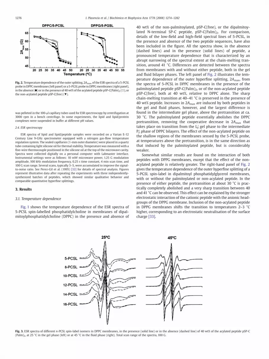
Fig. 2. Temperature dependenceof the outer splitting, 2Amax, of the ESR spectra of a 5-PCSLprobe inDPPCmembranes (left panel) or a5-PGSLprobe inDPPGmembranes (rightpanel),in the absence (●) or in the presence of 40wt%of the acylatedpeptide pSP-C(Palm)2 (○), orthe non-acylated peptide pSP-C(free) (▼).
1276 I. Plasencia et al. / Biochimica et Biophysica Acta 1778 (2008) 1274–1282
was pelleted in the 100-μl capillary tubes used for ESR spectroscopy by centrifugation at3000 rpm in a bench centrifuge. In some experiments, the lipid and lipid/proteincomplexes were suspended in buffer at different pH values.
2.4. ESR spectroscopy
ESR spectra of lipid and lipid/peptide samples were recorded on a Varian E-12Century Line 9-GHz spectrometer equipped with a nitrogen gas-flow temperatureregulation system. The sealed capillaries (1-mm outer diameter) were placed in a quartztube containing light silicone oil for thermal stability. Temperaturewasmeasuredwith afine-wire thermocouple positioned in the silicone oil at the top of the microwave cavity.Spectra were collected digitally on a personal computer with Labmaster interface.Instrumental settings were as follows: 10 mW microwave power, 1.25 G modulationamplitude, 100 kHz modulation frequency, 0.25 s time constant, 4 min scan time, and100 G scan range. Several scans, typically 3–5, were accumulated to improve the signal-to-noise ratio. See Perez-Gil et al. (1995) [32] for details of spectral analysis. Figuresrepresent illustrative data after repeating the experiments with three independently-synthesized batches of peptides, which showed similar qualitative behavior andcomparable quantitative hyperfine splittings.
3. Results
3.1. Temperature dependence
Fig. 1 shows the temperature dependence of the ESR spectra of5-PCSL spin-labelled phosphatidylcholine in membranes of dipal-mitoylphosphatidylcholine (DPPC) in the presence and absence of
Fig. 3. ESR spectra of different n-PCSL spin-label isomers in DPPC membranes, in the presen(Palm)2, at 25 °C in the gel phase (left) or at 45 °C in the fluid phase (right). Total scan rang
40 wt% of the non-palmitoylated, pSP-C(free), or the dipalmitoy-lated N-terminal SP-C peptide, pSP-C(Palm)2. For comparison,details of the low-field and high-field spectral lines of 5-PCSL, inthe presence and absence of the two peptide sequences, have alsobeen included in the figure. All the spectra show, in the absence(dashed lines) and in the presence (solid lines) of peptide, apronounced temperature dependence that is characterized by anabrupt narrowing of the spectral extent at the chain-melting tran-sition, around 41 °C. Differences are detected between the spectrafrom membranes with and without either peptide, both in the geland fluid bilayer phases. The left panel of Fig. 2 illustrates the tem-perature dependence of the outer hyperfine splitting, 2Amax, fromthe spectra of 5-PCSL in DPPC membranes in the presence of thepalmitoylated peptide pSP-C(Palm)2, or of the non-acylated peptidepSP-C(free), both at 40 wt%, relative to DPPC alone. The sharpchain-melting transition at 40–41 °C is preserved in the presence of40 wt% peptide. Increases in 2Amax are induced by both peptides inthe gel and fluid phases, however, and the largest difference isfound in the intermediate gel phase, above the pretransition at ca.30 °C. The palmitoylated peptide essentially abolishes the DPPCpretransition, removing the cooperative decrease in 2Amax thattakes place on transition from the Lβ′ gel phase to the intermediatePβ′ phase of DPPC bilayers. The effect of the non-acylated peptide onthe shallow regions of the membranes sensed by the 5-PCSL probe,at temperatures above the pretransition, is in the same direction asthat induced by the palmitoylated peptide, but is considerablyweaker.
Somewhat similar results are found on the interaction of bothpeptides with DPPG membranes, except that the effect of the non-acylated peptide is relatively greater. The right-hand panel of Fig. 2gives the temperature dependence of the outer hyperfine splitting of a5-PGSL spin-label in dipalmitoyl phosphatidylglycerol membranes,with or without the palmitoylated or non-acylated peptide. In thepresence of either peptide, the pretransition at about 30 °C is prac-tically completely abolished and a very sharp transition between 40and 41 °C can be observed. This effect can be explained by the strongerelectrostatic interaction of the cationic peptide with the anionic head-groups of the DPPG membrane. Inclusion of the non-acylated peptidein DPPG membranes shifts the transition to temperatures 2–3 °Chigher, corresponding to an electrostatic neutralisation of the surfacecharge [33].
ce (solid line) or in the absence (dashed line) of 40 wt% of the acylated peptide pSP-Ce of the spectra, 100 G.
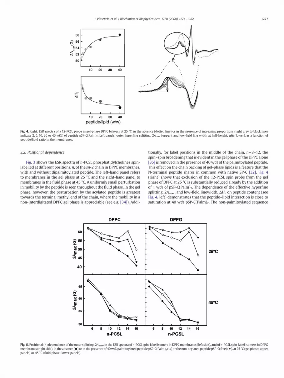
Fig. 4. Right: ESR spectra of a 12-PCSL probe in gel-phase DPPC bilayers at 25 °C, in the absence (dotted line) or in the presence of increasing proportions (light grey to black linesindicate 2, 5, 10, 20 or 40 wt%) of peptide pSP-C(Palm)2. Left panels: outer hyperfine splitting, 2Amax (upper), and low-field line width at half-height, ΔHl (lower), as a function ofpeptide/lipid ratio in the membranes.
1277I. Plasencia et al. / Biochimica et Biophysica Acta 1778 (2008) 1274–1282
3.2. Positional dependence
Fig. 3 shows the ESR spectra of n-PCSL phosphatidylcholines spin-labelled at different positions, n, of the sn-2 chain in DPPCmembranes,with and without dipalmitoylated peptide. The left-hand panel refersto membranes in the gel phase at 25 °C and the right-hand panel tomembranes in the fluid phase at 45 °C. A uniformly small perturbationinmobility by the peptide is seen throughout the fluid phase. In the gelphase, however, the perturbation by the acylated peptide is greatesttowards the terminal methyl end of the chain, where the mobility in anon-interdigitated DPPC gel phase is appreciable (see e.g. [34]). Addi-
Fig. 5. Positional (n) dependence of the outer splitting, 2Amax, in the ESR spectra of n-PCSL spinmembranes (right side), in the absence (●) or in the presence of 40wt% palmitoylated peptidepanels) or 45 °C (fluid phase; lower panels).
tionally, for label positions in the middle of the chain, n=8–12, thespin–spin broadening that is evident in the gel phase of the DPPC alone[35] is removed in the presence of 40wt% of the palmitoylated peptide.This effect on the chain packing of gel-phase lipids is a feature that theN-terminal peptide shares in common with native SP-C [32]. Fig. 4(right) shows that exclusion of the 12-PCSL spin probe from the gelphase of DPPC at 25 °C is substantially reduced already by the additionof 1 wt% of pSP-C(Palm)2. The dependence of the effective hyperfinesplitting, 2Amax, and low-field linewidth, ΔHl, on peptide content (seeFig. 4, left) demonstrates that the peptide–lipid interaction is close tosaturation at 40 wt% pSP-C(Palm)2. The non-palmitoylated sequence
-label isomers in DPPCmembranes (left side), and of n-PGSL spin-label isomers in DPPGpSP-C(Palm)2 (○) or the non-acylated peptide pSP-C(free) (▼), at 25 °C (gel phase; upper

Fig. 6. ESR spectra of different n-PCSL spin-label isomers in DPPC membranes at 37 °C, in the absence (dotted lines) or in the presence (solid lines) of 40 wt% of the palmitoylatedpeptide pSP-C(Palm)2 (left column) or the non-acylated peptide pSP-C(free) (right column). Total scan width: 100 G. Arrows indicate the position of the immobilized spectralcomponent that is attributed to a lipid population in an interdigitated (Lβi ) phase.
1278 I. Plasencia et al. / Biochimica et Biophysica Acta 1778 (2008) 1274–1282
had similar qualitative effects in terms of improving the solubility ofthe spin probe in the gel phase (not shown).
Fig. 5 (lower left-hand panel) shows the dependence of the n-PCSLouter hyperfine splitting, 2Amax, on position, n, of chain spin-labelling for fluid DPPC bilayers, in the presence and absence of thedipalmitoylated or non-acylated peptide. The chain flexibility profilewith increasing n, which is characteristic of spin-labelled lipid chainsin fluid phase membranes, is preserved in the presence of both pep-tides. Only modest increases in 2Amax are found at any chain positionin the fluid phase. In the gel phase at 25 °C, both peptides perturb themobility profile, in the sense that the values of Amax for n=14 becomemore comparable to those for n=5, in the presence of peptide (seeFig. 5, upper left). This removal of the mobility gradient is a featureassociated with the formation of an interdigitated gel phase, Lβi , inDPPC bilayers (see e.g. [34,36]). The right-hand panels of Fig. 5 give thepositional dependences of the outer hyperfine splittings of the n-PGSLspin-labels in gel and fluid DPPG membranes, with and without thepalmitoylated or non-acylated peptides. Overall, much the samedifferential features are exhibited in the interaction of the two pep-tides as with membranes of DPPC, except that the perturbations ofDPPGmembranes by the peptides are considerably larger, particularlyin the fluid phase. As already mentioned above, it is possible that inthe absence of palmitoylation the electrostatic interactions betweenthe cationic peptide and the anionic phospholipid headgroups domi-nate and cause the peptide to interact differently with anionic bi-layers, as compared with zwitterionic bilayers.
3.3. Phase coexistence
Fig. 6 shows the ESR spectra of different n-PCSL spin-labelledpositional isomers in DPPC membranes at 37 °C, i.e., corresponding tothe intermediate Pβ′ phase in the absence of peptide. A notable feature
Table 1Outer hyperfine splittings, in the presence and absence of peptide pSP-C(Palm)2 or pSP-C(free), of phosphatidylcholine spin-labelled at the 14th or 16th position of the sn-2acyl chain, incorporated in DPPC bilayers at 37 °C
2Amax (gauss)
DPPC DPPC/pSP-C(free) DPPC/pSP-C(palm)
14-PCSL 33.8 48.3 59.6, 34.216-PCSL 32.3 32.7 43.3, 32.5
is the coexistence of two spectral components for spin-labels situatedtowards the terminal methyl position (i.e., at n=14–16), in thepresence of the palmitoylated peptide, pSP-C(Palm)2. This suggeststhat, at 37 °C, the membranes containing the dipalmitoylated peptideare in a state of coexistence between interdigitated and non-interdigitated gel phases, because the outer spectral component thatis characteristic of the Lβi -phase is more strongly immobilised (2Amax
~60 G for 14-PCSL, see Table 1) than in the peptide-free Pβ′-phase
Fig. 7. ESR spectra of a 14-PCSL spin probe in DPPC membranes at 37 °C in the presenceof increasing amounts (wt%) of the palmitoylated peptide pSP-C(Palm)2. Forcomparison, the ESR spectrum of membranes containing 40 wt% of the non-palmitoylated peptide is also included (dotted line). Total scan width: 100 G. Arrowsindicate the position of a second, more immobilized, spectral component. Inset: ratio oflow-field lineheights of the immobilized relative to the non-immobilized component,with the latter normalized with respect to the peak-to-peak linewidth squared, versusthe peptide to lipid ratio.
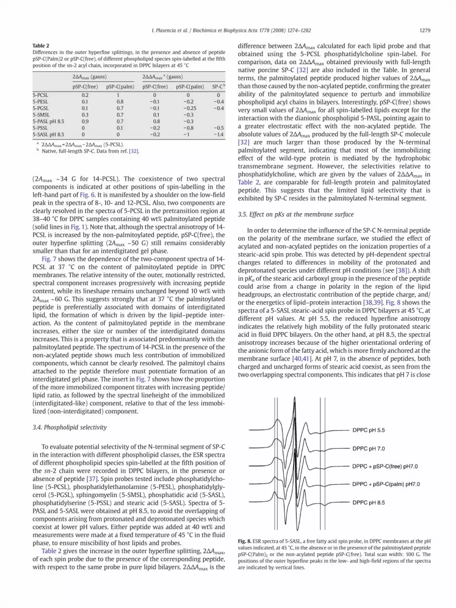
Table 2Differences in the outer hyperfine splittings, in the presence and absence of peptidepSP-C(Palm)2 or pSP-C(free), of different phospholipid species spin-labelled at the fifthposition of the sn-2 acyl chain, incorporated in DPPC bilayers at 45 °C
2ΔAmax (gauss) 2ΔΔAmaxa (gauss)
pSP-C(free) pSP-C(palm) pSP-C(free) pSP-C(palm) SP-C b
5-PCSL 0.2 1 0 0 05-PESL 0.1 0.8 −0.1 −0.2 −0.45-PGSL 0.1 0.7 −0.1 −0.25 −0.45-SMSL 0.3 0.7 0.1 −0.35-PASL pH 8.5 0.9 0.7 0.8 −0.35-PSSL 0 0.1 −0.2 −0.8 −0.55-SASL pH 8.5 0 0 −0.2 −1 −1.4
a 2ΔΔAmax=2ΔAmax−2ΔAmax (5-PCSL).b Native, full-length SP-C. Data from ref. [32].
Fig. 8. ESR spectra of 5-SASL, a free fatty acid spin probe, in DPPC membranes at the pHvalues indicated, at 45 °C, in the absence or in the presence of the palmitoylated peptidepSP-C(Palm)2 or the non-acylated peptide pSP-C(free). Total scan width: 100 G. Thepositions of the outer hyperfine peaks in the low- and high-field regions of the spectraare indicated by vertical lines.
1279I. Plasencia et al. / Biochimica et Biophysica Acta 1778 (2008) 1274–1282
(2Amax ~34 G for 14-PCSL). The coexistence of two spectralcomponents is indicated at other positions of spin-labelling in theleft-hand part of Fig. 6. It is manifested by a shoulder on the low-fieldpeak in the spectra of 8-, 10- and 12-PCSL. Also, two components areclearly resolved in the spectra of 5-PCSL in the pretransition region at38–40 °C for DPPC samples containing 40 wt% palmitoylated peptide(solid lines in Fig. 1). Note that, although the spectral anisotropy of 14-PCSL is increased by the non-palmitoylated peptide, pSP-C(free), theouter hyperfine splitting (2Amax ~50 G) still remains considerablysmaller than that for an interdigitated gel phase.
Fig. 7 shows the dependence of the two-component spectra of 14-PCSL at 37 °C on the content of palmitoylated peptide in DPPCmembranes. The relative intensity of the outer, motionally restricted,spectral component increases progressively with increasing peptidecontent, while its lineshape remains unchanged beyond 10 wt% with2Amax ~60 G. This suggests strongly that at 37 °C the palmitoylatedpeptide is preferentially associated with domains of interdigitatedlipid, the formation of which is driven by the lipid–peptide inter-action. As the content of palmitoylated peptide in the membraneincreases, either the size or number of the interdigitated domainsincreases. This is a property that is associated predominantly with thepalmitoylated peptide. The spectrum of 14-PCSL in the presence of thenon-acylated peptide shows much less contribution of immobilizedcomponents, which cannot be clearly resolved. The palmitoyl chainsattached to the peptide therefore must potentiate formation of aninterdigitated gel phase. The insert in Fig. 7 shows how the proportionof the more immobilized component titrates with increasing peptide/lipid ratio, as followed by the spectral lineheight of the immobilized(interdigitated-like) component, relative to that of the less immobi-lized (non-interdigitated) component.
3.4. Phospholipid selectivity
To evaluate potential selectivity of the N-terminal segment of SP-Cin the interaction with different phospholipid classes, the ESR spectraof different phospholipid species spin-labelled at the fifth position ofthe sn-2 chain were recorded in DPPC bilayers, in the presence orabsence of peptide [37]. Spin probes tested include phosphatidylcho-line (5-PCSL), phosphatidylethanolamine (5-PESL), phosphatidylgly-cerol (5-PGSL), sphingomyelin (5-SMSL), phosphatidic acid (5-SASL),phosphatidylserine (5-PSSL) and stearic acid (5-SASL). Spectra of 5-PASL and 5-SASL were obtained at pH 8.5, to avoid the overlapping ofcomponents arising from protonated and deprotonated species whichcoexist at lower pH values. Either peptide was added at 40 wt% andmeasurements were made at a fixed temperature of 45 °C in the fluidphase, to ensure miscibility of host lipids and probes.
Table 2 gives the increase in the outer hyperfine splitting, 2ΔAmax,of each spin probe due to the presence of the corresponding peptide,with respect to the same probe in pure lipid bilayers. 2ΔΔAmax is the
difference between 2ΔAmax calculated for each lipid probe and thatobtained using the 5-PCSL phosphatidylcholine spin-label. Forcomparison, data on 2ΔΔAmax obtained previously with full-lengthnative porcine SP-C [32] are also included in the Table. In generalterms, the palmitoylated peptide produced higher values of 2ΔAmax
than those caused by the non-acylated peptide, confirming the greaterability of the palmitoylated sequence to perturb and immobilizephospholipid acyl chains in bilayers. Interestingly, pSP-C(free) showsvery small values of 2ΔAmax for all spin-labelled lipids except for theinteraction with the dianionic phospholipid 5-PASL, pointing again toa greater electrostatic effect with the non-acylated peptide. Theabsolute values of 2ΔAmax produced by the full-length SP-C molecule[32] are much larger than those produced by the N-terminalpalmitoylated segment, indicating that most of the immobilizingeffect of the wild-type protein is mediated by the hydrophobictransmembrane segment. However, the selectivities relative tophosphatidylcholine, which are given by the values of 2ΔΔAmax inTable 2, are comparable for full-length protein and palmitoylatedpeptide. This suggests that the limited lipid selectivity that isexhibited by SP-C resides in the palmitoylated N-terminal segment.
3.5. Effect on pKs at the membrane surface
In order to determine the influence of the SP-C N-terminal peptideon the polarity of the membrane surface, we studied the effect ofacylated and non-acylated peptides on the ionization properties of astearic-acid spin probe. This was detected by pH-dependent spectralchanges related to differences in mobility of the protonated anddeprotonated species under different pH conditions (see [38]). A shiftin pKa of the stearic acid carboxyl group in the presence of the peptidecould arise from a change in polarity in the region of the lipidheadgroups, an electrostatic contribution of the peptide charge, and/or the energetics of lipid–protein interaction [38,39]. Fig. 8 shows thespectra of a 5-SASL stearic-acid spin probe in DPPC bilayers at 45 °C, atdifferent pH values. At pH 5.5, the reduced hyperfine anisotropyindicates the relatively high mobility of the fully protonated stearicacid in fluid DPPC bilayers. On the other hand, at pH 8.5, the spectralanisotropy increases because of the higher orientational ordering ofthe anionic form of the fatty acid, which is more firmly anchored at themembrane surface [40,41]. At pH 7, in the absence of peptides, bothcharged and uncharged forms of stearic acid coexist, as seen from thetwo overlapping spectral components. This indicates that pH 7 is close
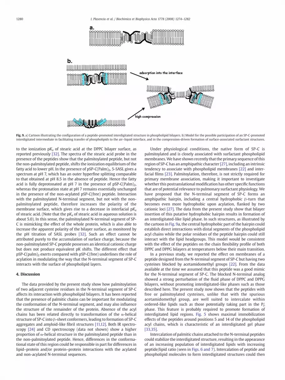
Fig. 9. a) Cartoon illustrating the configuration of a peptide-promoted interdigitated structure in phospholipid bilayers. b) Model for the possible participation of an SP-C-promotedinterdigitated intermediate in facilitating transfer of phospholipids to the air–liquid interface, and in the compression-driven formation of surface-associated surfactant structures.
1280 I. Plasencia et al. / Biochimica et Biophysica Acta 1778 (2008) 1274–1282
to the ionization pKa of stearic acid at the DPPC bilayer surface, asreported previously [32]. The spectra of the stearic acid probe in thepresence of the peptides show that the palmitoylated peptide, but notthe non-palmitoylated peptide, shifts the ionization equilibrium of thefatty acid to lower pH. In the presence of pSP-C(Palm)2, 5-SASL gives aspectrum at pH 7, which has an outer hyperfine splitting comparableto that obtained at pH 8.5 in the absence of peptide. Hence the fattyacid is fully deprotonated at pH 7 in the presence of pSP-C(Palm)2,whereas the protonation state at pH 7 remains essentially unchangedin the presence of the non-acylated pSP-C(free) peptide. Interactionwith the palmitoylated N-terminal segment, but not with the non-palmitoylated peptide, therefore increases the polarity of themembrane surface, which gives rise to a decrease in interfacial pKa
of stearic acid. (Note that the pKa of stearic acid in aqueous solution isabout 5.0). In this sense, the palmitoylated N-terminal segment of SP-C is mimicking the effect of the whole protein, which is also able toincrease the apparent polarity of the bilayer surface, as monitored bythe pH titration of SASL probes [32]. Such an effect cannot beattributed purely to the accumulation of surface charge, because thenon-palmitoylated SP-C peptide possesses an identical cationic chargebut does not produce equivalent pK shifts. The different effect thatpSP-C(palm)2 exerts compared with pSP-C(free) underlines the role ofacylation in modulating the way that the N-terminal segment of SP-Cinteracts with the surface of phospholipid layers.
4. Discussion
The data provided by the present study show how palmitoylationof two adjacent cysteine residues in the N-terminal segment of SP-Caffects its interaction with phospholipid bilayers. It has been reportedthat the presence of palmitic chains can be important for modulatingthe conformation of the N-terminal segment, and may also influencethe structure of the remainder of the protein. Absence of the acylchains has been related directly to transformation of the α-helicalstructure of SP-C into β-sheet conformers, leading to formation of SP-Caggregates and amyloid-like fibril structures [11,12]. Both IR spectro-scopy [24] and CD spectroscopy (data not shown) show a higherproportion of α-helical structure in the palmitoylated peptide than inthe non-palmitoylated peptide. Hence, differences in the conforma-tional state of this region could be responsible in part for differences inlipid–protein and/or protein–protein interactions with the acylatedand non-acylated N-terminal sequences.
Under physiological conditions, the native form of SP-C ispalmitoylated and is closely associated with surfactant phospholipidmembranes.Wehave shown recently that the primary sequence of thisregion of SP-C has an amphipathic character [27], including an intrinsictendency to associate with phospholipid membranes [22] and inter-facial films [23]. Palmitoylation, therefore, is not strictly required forprimary membrane association, making it important to investigatewhether this postranslationalmodification has other specific functionsthat are of potential relevance to pulmonary surfactant physiology.Wehave proposed that the N-terminal segment of SP-C forms anamphipathic hairpin, including a central hydrophobic β-turn thatbecomes even more hydrophobic upon acylation, flanked by twocationic loci [27]. The data from the present study show that bilayerinsertion of this putative hydrophobic hairpin results in formation ofan interdigitated-like lipid phase. In such structures, as illustrated bythe cartoon in Fig. 9a, the central hydrophobic part of the hairpin couldestablish direct interactions with distal segments of the phospholipidacyl chains while the polar residues of the peptide hairpin could stillinteract with the lipid headgroups. This model would be consistentwith the effect of the peptides on the chain flexibility profile of bothDPPC and DPPG bilayers at temperatures below their main transition.
In a previous study, we reported the effect on membranes of apeptide designed from the N-terminal segment of SP-C but having twocysteines blocked by acetamidomethyl groups [22]. From the dataavailable at the time we assumed that this peptide was a good mimicfor the N-terminal segment of SP-C. The blocked N-terminal analogshowed a strong perturbation of the fluid phase of DPPC and DPPGbilayers, without promoting interdigitated-like phases such as thosedescribed here. The present study now shows that the peptides withfree or palmitoylated cysteines, unlike that with the spuriousacetamidomethyl group, are well suited to intercalate withinordered-like lipids such as those potentially taking part in the Pβ′phase. This feature is probably required to promote formation ofinterdigitated lipid regions. Fig. 5 shows maximal immobilizationeffects of the peptides around positions 5 and 14 of the phospholipidacyl chains, which is characteristic of an interdigitated gel phase[33,35].
Intercalation of palmitic chains attached to theN-terminal peptidescould stabilize the interdigitated structure, resulting in the appearanceof an increasing population of interdigitated lipids with increasingpeptide/lipid ratio (seen in Figs. 6 and 7). Intercalation of peptide andphospholipid molecules to form interdigitated structures could then

1281I. Plasencia et al. / Biochimica et Biophysica Acta 1778 (2008) 1274–1282
inhibit the Lβ→Pβ′ transition, maintaining maximal order of the acylchains up to temperatures close to the main gel-to-fluid phasetransition. Interestingly, a recent study has proposed that the Pβ′ ripplephase might consist of a coexistence of bilayered and interdigitatedlipid phases [42]. Palmitoylation could thenpromote stable associationof the N-terminal segment with interdigitated patches that mightalready exist in the ripple phase and promote their spatial extension.This property could be important to ensure maximal stability of DPPC-enriched domains segregated in pulmonary surfactant films at physio-logical temperatures [30,31]. These domains might have order andpacking properties similar to those exhibited by DPPC bilayers at 37 °C,the temperature at which stabilization of interdigitated-like mem-brane regions by the N-terminal segment seems to be maximal. In oneof the few studies published so far on lipid–protein interactions ofacylated peptides, a specific effect of an acylated peptide on the Pβ′ripple phase of DPPC bilayers has also been reported [43], suggestingthat association with and possible stabilization of interdigitated-likeregions could be a general feature favoured by palmitoylated segmentsof proteins. In the pure Lα phase, the lipid chains are too disordered toallow organization of well-defined interdigitated structures, and thepeptides are then associated with normal bilayer structures.
Protein-attached saturated palmitic chains are particularly wellsuited to insert into ordered membrane structures, such as the inter-digitated structures proposed here, the condensed interfacial phasesreached at the end of expiration, and the liquid-ordered regionsadopted by raft-likemembrane domains enriched in sphingolipids andcholesterol. Several examples have been reported in the literatureshowing that palmitoylation may promote association of certainproteins with ordered membrane domains [44–47]. Our results alsosuggest that palmitoylated protein segments could contribute tonucleate ordered membrane regions, the size and stability of whichstill remains to be evaluated. We have observed similar qualitativeeffects in samples containing peptides over the whole range 1–40%protein/lipid (w/w). This includes the peptide-induced increase in2Amax, the reduction of spin–spin broadening, and the segregation ofan immobilized lipid component (see ESR spectra at different peptide/lipid ratios in Figs. 4 and 7). No discontinuities were detected in theprogressive effects caused by increasing peptide contents, which couldhave been indicative of phase transformation, e.g., micellisation in themembranes at the highest peptide/lipid ratios.
The cartoon in Fig. 9b illustrates the possible participation of thelipid/protein structures that are promoted by the palmitoylated N-terminal segment of SP-C in the activities of pulmonary surfactant atthe respiratory air–liquid interface. SP-C promotes transfer ofphospholipids from phospholipid bilayers to the air–liquid interface[11,18], independent of its acylation state. Simultaneous perturbationof membranes [22] and monolayers [23] by the N-terminal segmentof SP-C could permit transient exposure of phospholipid molecules,hence accelerating interfacial adsorption. An SP-C-promoted transi-tion of surfactant structures from non-interdigitated to interdigi-tated bilayer-like arrangements, initiated at locations where SP-C isaccumulated [29,48,49], might accelerate bilayer/monolayer conver-sions. On the other hand, compression of SP-C-containing interfaciallipid–protein films could induce a progressive accumulation of SP-Cin defined locations of the surface layers [48,50]. Sufficiently highprotein density could favour formation of an ordered lipid/proteinphase, where the palmitic chains of the N-terminal segment of SP-Ccould be intercalated with the phospholipid chains, in a mannersimilar to the interdigitated structure proposed here. These surfacestructures could be particularly favourable for initiating transitionsthat fold the interfacial film towards the bulk phase, and so induceformation of the surface-associated multilayered structures that areknown to be promoted by SP-C [25,51]. Such multilayered structuresassociated with surfactant interfacial films have been demonstratedin recent years [52,53], and are now a central part of current mole-cular models of pulmonary surfactant action [54]. Surface-associated
surfactant reservoirs would have the dual role of improving mechan-ical stability of highly compressed films at the end of expiration, andof promoting replenishment of the surface film with new surfactantmolecules that are inserted into the interface during inspiration.Palmitoylation could prevent total dissociation of the multilayeredstructures from the highly packed interfacial film [24]. This possi-bility could explain why surfactant obtained from SP-C knock-outmice forms intrinsically unstable films [3], which could be respon-sible at least in part for the development of chronic respiratoryproblems in animals and patients whose lungs lack SP-C [4]. Theability of the SP-C N-terminal segment to maintain association withhighly compressed films would be especially important at the endof exhalation, when the highest surface pressures (lowest surfacetensions) must be supported.
Nevertheless, it is important to consider that the nature of thestructures promoted by SP-C and its N-terminal segment in surfactantcomplexes that are reconstituted in vitro from lipid/protein or lipid/peptide organic solutionsmay differ from the structure and dispositionof surfactant structures that are assembled in vivo into the lamellarbodies of pneumocytes. Very little is known about the orientation ofSP-C in the membrane structures reaching the pulmonary interface.It is possible that SP-C-promoted membrane perturbations when theN-terminal segment has access to both sides of the membrane – aspresumably occurs in complexes reconstituted in vitro – could differfrom those when SP-C perturbs the membranes from a single side,either alone or in concert with other surfactant proteins. Furtherstudies with native surfactant complexes are still required beforewe shall be able to model fully functional pulmonary surfactantcomplexes.
Acknowledgements
We are grateful to Frau B. Angerstein for spin-label phospholipidsynthesis. This research was supported by grants from the SpanishMinistry of Science (BIO2006-03130, CONSOLIDER CSD2007-00010),Community of Madrid (S0505/MAT/0283) and the Marie Curie Pro-gramme (CT04-007931).
References
[1] M.J. Nadolski, M.E. Linder, Protein lipidation, FEBS J. 274 (2007) 5202–5210.[2] A. ten Brinke, J.J. Batenburg, B.M. Gadella, H.P. Haagsman, A.B. Vaandrager, L.M. van
Golde, The juxtamembrane lysine and arginine residues of surfactant protein Cprecursor influence palmitoylation via effects on trafficking, Am. J. Respir. Cell Mol.Biol. 25 (2001) 156–163.
[3] S.W. Glasser, M.S. Burhans, T.R. Korfhagen, C.L. Na, P.D. Sly, G.F. Ross, M. Ikegami, J.A. Whitsett, Altered stability of pulmonary surfactant in SP-C-deficient mice, Proc.Natl. Acad. Sci. U. S. A. 98 (2001) 6366–6371.
[4] L.M. Nogee, Alterations in SP-B and SP-C expression in neonatal lung disease,Annu. Rev. Physiol. 66 (2004) 601–623.
[5] J. Perez-Gil, J. Tucker, G. Simatos, K.M. Keough, Interfacial adsorption of simplelipid mixtures combined with hydrophobic surfactant protein from pig lung,Biochem. Cell Biol. 70 (1992) 332–338.
[6] E.J. Veldhuizen, H.P. Haagsman, Role of pulmonary surfactant components insurface film formation and dynamics, Biochim. Biophys. Acta 1467 (2000)255–270.
[7] E.J. Veldhuizen, R.V. Diemel, G. Putz, L.M. van Golde, J.J. Batenburg, H.P. Haagsman,Effect of the hydrophobic surfactant proteins on the surface activity of spread filmsin the captive bubble surfactometer, Chem. Phys. Lipids 110 (2001) 47–55.
[8] N.J. Foot, S. Orgeig, S. Donnellan, T. Bertozzi, C.B. Daniels, Positive selection inthe N-terminal extramembrane domain of lung surfactant protein C (SP-C) inmarine mammals, J. Mol. Evol. 65 (2007) 12–22.
[9] B.A. Hyatt, E.R. Resnik, N.S. Johnson, J.L. Lohr, D.N. Cornfield, Lung specific devel-opmental expression of the Xenopus laevis surfactant protein C and B genes,Gene Expr. Patterns 7 (2007) 8–14.
[10] J. Johansson, Structure and properties of surfactant protein C, Biochim. Biophys.Acta 1408 (1998) 161–172.
[11] R.A. Dluhy, S. Shanmukh, J.B. Leapard, P. Kruger, J.E. Baatz, Deacylated pulmonarysurfactant protein SP-C transforms from alpha-helical to amyloid fibril structurevia a pH-dependent mechanism: an infrared structural investigation, Biophys. J. 85(2003) 2417–2429.
[12] M. Gustafsson, W.J. Griffiths, E. Furusjo, J. Johansson, The palmitoyl groups of lungsurfactant protein C reduce unfolding into a fibrillogenic intermediate, J. Mol. Biol.310 (2001) 937–950.
1282 I. Plasencia et al. / Biochimica et Biophysica Acta 1778 (2008) 1274–1282
[13] M. Gustafsson, M. Palmblad, T. Curstedt, J. Johansson, S. Schurch, Palmitoylation ofa pulmonary surfactant protein C analogue affects the surface associated lipidreservoir and film stability, Biochim. Biophys. Acta 1466 (2000) 169–178.
[14] G. Putz, M. Walch, M. Van Eijk, H.P. Haagsman, Hydrophobic lung surfactantproteins B and C remain associated with surface film during dynamic cyclic areachanges, Biochim. Biophys. Acta 1453 (1999) 126–134.
[15] J. Ding, D.Y. Takamoto, A. von Nahmen, M.M. Lipp, K.Y. Lee, A.J. Waring, J.A.Zasadzinski, Effects of lung surfactant proteins, SP-B and SP-C, and palmitic acid onmonolayer stability, Biophys. J. 80 (2001) 2262–2272.
[16] L.A. Creuwels, R.A. Demel, L.M. van Golde, B.J. Benson, H.P. Haagsman, Effect ofacylation on structure and function of surfactant protein C at the air–liquidinterface, J. Biol. Chem. 268 (1993) 26752–26758.
[17] C.R. Flach, A. Gericke, K.M. Keough, R. Mendelsohn, Palmitoylation of lungsurfactant protein SP-C alters surface thermodynamics, but not protein secondarystructure or orientation in 1,2-dipalmitoylphosphatidylcholine langmuir films,Biochim. Biophys. Acta 1416 (1999) 11–20.
[18] D. Lukovic, I. Plasencia, F.J. Taberner, J. Salgado, J.J. Calvete, J. Perez-Gil, I. Mingarro,Production and characterisation of recombinant forms of human pulmonarysurfactant protein C (SP-C): structure and surface activity, Biochim. Biophys. Acta1758 (2006) 509–518.
[19] S. Hawgood, A. Ogawa, K. Yukitake, M. Schlueter, C. Brown, T. White, D. Buckley,D. Lesikar, B. Benson, Lung function in premature rabbits treated withrecombinant human surfactant protein-C, Am. J. Respir. Crit. Care Med. 154(1996) 484–490.
[20] A. Hilgendorff, M. Doerner, D. Rawer, J. Leick, A. Trotter, M. Ebsen, C. Ruppert, A.Gunther, L. Gortner, I. Reiss, Effects of a recombinant surfactant protein-C-basedsurfactant on lung function and the pulmonary surfactant system in a model ofmeconium aspiration syndrome, Crit. Care Med. 34 (2006) 203–210.
[21] R.G. Spragg, R.M. Smith, K. Harris, J. Lewis, D. Hafner, P. Germann, Effect ofrecombinant SP-C surfactant in a porcine lavagemodel of acute lung injury, J. Appl.Physiol. 88 (2000) 674–681.
[22] I. Plasencia, L. Rivas, K.M. Keough, D. Marsh, J. Perez-Gil, The N-terminal segmentof pulmonary surfactant lipopeptide SP-C has intrinsic propensity to interact withand perturb phospholipid bilayers, Biochem. J. 377 (2004) 183–193.
[23] I. Plasencia, K.M. Keough, J. Perez-Gil, Interaction of the N-terminal segment ofpulmonary surfactant protein SP-C with interfacial phospholipid films, Biochim.Biophys. Acta 1713 (2005) 118–128.
[24] X. Bi, C.R. Flach, J. Perez-Gil, I. Plasencia, D. Andreu, E. Oliveira, R. Mendelsohn,Secondary structure and lipid interactions of the N-terminal segment of pul-monary surfactant SP-C in Langmuir films: IR reflection-absorption spectroscopyand surface pressure studies, Biochemistry 41 (2002) 8385–8395.
[25] P. Na Nakorn, M.C. Meyer, C.R. Flach, R. Mendelsohn, H.J. Galla, Surfactant protein Cand lung function: new insights into the role of alpha-helical length andpalmitoylation, Eur. Biophys. J. 36 (2007) 477–489.
[26] D. Marsh, A. Watts, Spin-Labelling and Lipid–Protein Interactions in Membranes,Wiley Interscience, New York, 1982.
[27] I. Plasencia, L. Rivas, C. Casals, K.M. Keough, J. Perez-Gil, Intrinsic structural dif-ferences in the N-terminal segment of pulmonary surfactant protein SP-C fromdifferent species, Comp. Biochem. Physiol. A. Mol. Integr. Physiol. 129 (2001)129–139.
[28] O. Blanco, J. Perez-Gil, Biochemical and pharmacological differences betweenpreparations of exogenous natural surfactant used to treat Respiratory DistressSyndrome: role of the different components in an efficient pulmonary surfactant,Eur. J. Pharmacol. 568 (2007) 1–15.
[29] J. Bernardino de la Serna, J. Perez-Gil, A.C. Simonsen, L.A. Bagatolli, Cholesterolrules: direct observation of the coexistence of two fluid phases in nativepulmonary surfactant membranes at physiological temperatures, J. Biol. Chem.279 (2004) 40715–40722.
[30] B.M. Discher, K.M. Maloney,W.R. Schief Jr., D.W. Grainger, V. Vogel, S.B. Hall, Lateralphase separation in interfacial films of pulmonary surfactant, Biophys. J. 71 (1996)2583–2590.
[31] K. Nag, J. Perez-Gil, M.L. Ruano, L.A. Worthman, J. Stewart, C. Casals, K.M. Keough,Phase transitions in films of lung surfactant at the air–water interface, Biophys. J.74 (1998) 2983–2995.
[32] J. Perez-Gil, C. Casals, D. Marsh, Interactions of hydrophobic lung surfactantproteins SP-B and SP-C with dipalmitoylphosphatidylcholine and dipalmitoylpho-sphatidylglycerol bilayers studied by electron spin resonance spectroscopy,Biochemistry 34 (1995) 3964–3971.
[33] G. Cevc, A. Watts, D. Marsh, Non-electrostatic contribution to the titration of theordered-fluid phase transition of phosphatidylglycerol bilayers, FEBS Lett. 120(1980) 267–270.
[34] R. Bartucci, T. Pali, D. Marsh, Lipid chain motion in an interdigitated gel phase:conventional and saturation transfer ESR of spin-labeled lipids in dipalmitoylpho-sphatidylcholine-glycerol dispersions, Biochemistry 32 (1993) 274–281.
[35] P. Fajer, A. Watts, D. Marsh, Saturation transfer, continuous wave saturation, andsaturation recovery electron spin resonance studies of chain-spin labeled phos-phatidylcholines in the low temperature phases of dipalmitoyl phosphatidylcho-line bilayers. Effects of rotational dynamics and spin–spin interactions, Biophys. J.61 (1992) 879–891.
[36] M.J. Swamy, D. Marsh, Spin-label electron spin resonance studies on the dynamicsof the different phases of N-biotinylphosphatidylethanolamines, Biochemistry 33(1994) 11656–11663.
[37] M.B. Sankaram, B. De Kruijff, D. Marsh, Selectivity of interaction of spin-labelledlipids with peripheral proteins bound to dimyristoylphosphatidylglycerol bilayers,as determined by ESR spectroscopy, Biochim. Biophys. Acta 986 (1989) 315–320.
[38] J. Miyazaki, K. Hideg, D. Marsh, Interfacial ionization and partitioning ofmembrane-bound local anesthetics, Biochim. Biophys. Acta 1103 (1992) 62–68.
[39] M.B. Sankaram, P.J. Brophy, W. Jordi, D. Marsh, Fatty acid pH titration and theselectivity of interaction with extrinsic proteins in dimyristoylphosphatidylgly-cerol dispersions. Spin label ESR studies, Biochim. Biophys. Acta 1021 (1990)63–69.
[40] M. Esmann, D. Marsh, Spin-label studies on the origin of the specificity of lipid–protein interactions in Na+,K+-ATPase membranes from Squalus acanthias,Biochemistry 24 (1985) 3572–3578.
[41] Y.V. Rama Krishna, D. Marsh, Spin label ESR and 31P-NMR studies of the cubic andinverted hexagonal phases of dimyristoylphosphatidylcholine/myristic acid(1:2, mol/mol) mixtures, Biochim. Biophys. Acta 1024 (1990) 89–94.
[42] A.H. de Vries, S. Yefimov, A.E. Mark, S.J. Marrink, Molecular structure of the lecithinripple phase, Proc. Natl. Acad. Sci. U. S. A. 102 (2005) 5392–5396.
[43] T.B. Pedersen, T. Kaasgaard, M.O. Jensen, S. Frokjaer, O.G. Mouritsen, K. Jorgensen,Phase behavior and nanoscale structure of phospholipid membranes incorporatedwith acylated C14-peptides, Biophys. J. 89 (2005) 2494–2503.
[44] M. Higuchi, K.M. Izumi, E. Kieff, Epstein-Barr virus latent-infection membraneproteins are palmitoylated and raft-associated: protein 1 binds to the cytoskeletonthrough TNF receptor cytoplasmic factors, Proc. Natl. Acad. Sci. U. S. A. 98 (2001)4675–4680.
[45] S. Moffett, D.A. Brown, M.E. Linder, Lipid-dependent targeting of G proteins intorafts, J. Biol. Chem. 275 (2000) 2191–2198.
[46] H. Shogomori, A.T. Hammond, A.G. Ostermeyer-Fay, D.J. Barr, G.W. Feigenson, E.London, D.A. Brown, Palmitoylation and intracellular domain interactions bothcontribute to raft targeting of linker for activation of T cells, J. Biol. Chem. 280(2005) 18931–18942.
[47] N. Tanimura, S. Saitoh, S. Kawano, A. Kosugi, K. Miyake, Palmitoylation of LATcontributes to its subcellular localization and stability, Biochem. Biophys. Res.Commun. 341 (2006) 1177–1183.
[48] K. Nag, S.G. Taneva, J. Perez-Gil, A. Cruz, Keough, Combinations of fluorescentlylabeled pulmonary surfactant proteins SP-B and SP-C in phospholipid films,Biophys. J. 72 (1997) 2638–2650.
[49] R.Wustneck, J. Perez-Gil, N.Wustneck, A. Cruz, V.B. Fainerman, U. Pison, Interfacialproperties of pulmonary surfactant layers, Adv. Colloid Interface Sci. 117 (2005)33–58.
[50] A. Kramer, A. Wintergalen, M. Sieber, H.J. Galla, M. Amrein, R. Guckenberger,Distribution of the surfactant-associated protein C within a lung surfactantmodel film investigated by near-field optical microscopy, Biophys. J. 78 (2000)458–465.
[51] A. von Nahmen, A. Post, H.J. Galla, M. Sieber, The phase behavior of lipidmonolayers containing pulmonary surfactant protein C studied by fluorescencelight microscopy, Eur Biophys. J. 26 (1997) 359–369.
[52] D. Follows, F. Tiberg, R.K. Thomas, M. Larsson, Multilayers at the surface ofsolutions of exogenous lung surfactant: direct observation by neutron reflection,Biochim. Biophys. Acta 1768 (2007) 228–235.
[53] S. Malcharek, A. Hinz, L. Hilterhaus, H.J. Galla, Multilayer structures in lipidmonolayer films containing surfactant protein C: effects of cholesterol and POPE,Biophys. J. 88 (2005) 2638–2649.
[54] A.G. Serrano, J. Perez-Gil, Protein–lipid interactions and surface activity in thepulmonary surfactant system, Chem. Phys. Lipids 141 (2006) 105–118.