Titrimetric determination of anionic surfactant content in ...
Upload
independentCategory
view
3download
0
New Mass-Spectrometry-Compatible Degradable Surfactant forTissue ProteomicsYing-Hua Chang,† Zachery R. Gregorich,†,‡ Albert J. Chen,† Leekyoung Hwang,§ Huseyin Guner,∥
Deyang Yu,†,⊥ Jianyi Zhang,# and Ying Ge*,†,‡,§,∥,⊥
†Department of Cell and Regenerative Biology, ‡Molecular and Cellular Pharmacology Program, §Department of Chemistry, ∥HumanProteomics Program, and ⊥Molecular and Environmental Toxicology Program, University of Wisconsin-Madison, 1300 UniversityAvenue, Madison 53706, Wisconsin,United States#Division of Cardiology, Department of Medicine, Department of Biomedical Engineering, Stem Cell Institute, University ofMinnesota, 2001 6th Street SE, Minneapolis, Minnesota 55455, United States
*S Supporting Information
ABSTRACT: Tissue proteomics is increasingly recognized forits role in biomarker discovery and disease mechanisminvestigation. However, protein solubility remains a significantchallenge in mass spectrometry (MS)-based tissue proteomics.Conventional surfactants such as sodium dodecyl sulfate(SDS), the preferred surfactant for protein solubilization, arenot compatible with MS. Herein, we have screened a library ofsurfactant-like compounds and discovered an MS-compatibledegradable surfactant (MaSDeS) for tissue proteomics thatsolubilizes all categories of proteins with performancecomparable to SDS. The use of MaSDeS in the tissueextraction significantly improves the total number of proteinidentifications from commonly used tissues, including tissuefrom the heart, liver, and lung. Notably, MaSDeS significantly enriches membrane proteins, which are often under-represented inproteomics studies. The acid degradable nature of MaSDeS makes it amenable for high-throughput MS-based proteomics. Inaddition, the thermostability of MaSDeS allows for its use in experiments requiring high temperature to facilitate proteinextraction and solubilization. Furthermore, we have shown that MaSDeS outperforms the other MS-compatible surfactants interms of overall protein solubility and the total number of identified proteins in tissue proteomics. Thus, the use of MaSDeS willgreatly advance tissue proteomics and realize its potential in basic biomedical and clinical research. MaSDeS could be utilized in avariety of proteomics studies as well as general biochemical and biological experiments that employ surfactants for proteinsolubilization.
KEYWORDS: surfactant, proteomics, membrane proteins, mass spectrometry, tissue
■ INTRODUCTION
Mass spectrometry (MS)-based tissue proteomics is increas-ingly recognized as a highly valuable tool for biomarkerdiscovery and mechanistic investigation because tissues near thedisease source contain the highest concentration of potentialdisease markers.1−11 Nonetheless, there are still significantchallenges in tissue proteomics such as protein solubility issues,particularly for membrane proteins.12−15 Membrane proteinscomprise approximately one-third of the proteome,12−14,16−18
playing an important role in many essential cellular functionsand accounting for a significant proportion of drug targets.19,20
Surfactants (surface-acting agents, also known as detergents)are commonly used in biochemical research to effectivelyextract proteins from tissues.21 Sodium dodecyl sulfate (SDS),owing to its outstanding performance in solubilizing proteins, isthe most widely used surfactant.13,21−24 Unfortunately, evenvery low concentrations (<0.01%) of SDS can severely suppress
the ionization of proteins/peptides.21,23,25 Consequently,numerous methods have been developed to remove SDSprior to MS analyses.21,22,26−30 Although gel-based methodscan effectively remove surfactants, the protein coverage,sensitivity, and reproducibility of protein identification/quantification using these methods are relatively low incomparison with methods using direct in-solution digestionliquid chromatography (LC)−MS-based shotgun method.13
Unfortunately, in-solution removal of SDS has been known tobe difficult and require multiple cleanup steps that often resultin protein loss, degradation, and are also time-consum-ing.21,22,27,28 Some nonionic detergents such as the nonionicsaccharides are MS-compatible at relatively low concentrations(<0.1%), but these surfactants disrupt lipid−lipid and lipid−
Received: December 10, 2014Published: January 15, 2015
Article
pubs.acs.org/jpr
© 2015 American Chemical Society 1587 DOI: 10.1021/pr5012679J. Proteome Res. 2015, 14, 1587−1599

protein interactions instead of protein−protein interactionsand, therefore, are considered relatively mild surfactants.23
To address these problems, acid-labile MS-compatible ionicsurfactants have been developed.31−33 These surfactantscontain an acid-labile functional group between the hydrophilichead and hydrophobic tail of the surfactant molecule. Upon theaddition of acid to the surfactant-containing sample, thesurfactant quickly degrades into innocuous nonsurfactantbyproducts, thus eliminating the need to remove the detergentprior to MS analysis.31,33,34 However, commonly used acid-labile surfactants appear to be only mild denaturants and,consequently, are not as effective as SDS at solubilizingproteins.33,34 Consequently, SDS still remains the surfactant ofchoice for the total solubilization of tissues and cells.21,22,28
Herein, we aimed to identify a strong MS-compatibledegradable ionic surfactant that can effectively solubilizeproteins during sample preparation with similar performanceto SDS. We have screened a library of 43 surfactant-likecompounds and discovered a mass-spectrometry-compatibledegradable surfactant (MaSDeS) that can solubilize allcategories of proteins with comparable performance to SDSat the concentrations used in this study. Importantly, MaSDeSsignificantly enriched membrane proteins, which are oftenunder-represented in proteomics studies. In addition, MaSDeSis slowly degradable under acidic conditions, which obviates theneed to remove the surfactant prior to MS analysis and enablethe high-throughput proteomics analysis. Furthermore, it isthermostable and thus can be used in experiments that requirerelatively high temperature to facilitate protein extraction andsolubilization. These favorable characteristics make MaSDeS anideal MS-compatible surfactant for proteomics studies. Here wehave shown that MaSDeS outperforms other MS-compatiblesurfactants, including RapiGest (RG), PPS Silent Surfactant(PPS), ProteaseMAX (PM), octyl β-D-glucopyranoside (OG),n-dodecyl β-D-maltoside (DDM), and digitonin (DGT), byboth improving protein solubilization and increasing thenumber of identified proteins in tissue proteomics.
■ EXPERIMENTAL METHODS
Sequential Tissue Extraction
All experiments involving animals were conducted inaccordance with the NIH Guide for the Care and Use ofLaboratory Animals and using protocols approved by theUniversity of Wisconsin Institutional Animal Care and UseCommittee. Swine heart, liver, and lung tissue samples wereexcised from healthy Yorkshire domestic pigs, snap-frozen inliquid N2, and stored under −80 °C before use. All steps of thesequential tissue extraction procedure were performed in a coldroom (4 °C). First, the frozen pig tissue samples (approx-imately 200−300 mg) were cut into small pieces (∼2 mm3) andimmediately washed twice with cold PBS buffer containing aprotease inhibitor cocktail (Roche, Switzerland). The washedtissue pieces were transferred to HEPES buffer (0.25 Msucrose, 25 mM HEPES at pH 7.4, 50 mM NaF, 0.25 mMNa3VO4, 0.25 mM PMSF, 2.5 mM EDTA, and 1 mg/mLprotease inhibitor cocktail) and homogenized five to six timesusing a Polytron electric homogenizer (model PRO200, PROScientific, Oxford, CT) for 5−7 s on ice. The homogenate wascentrifuged at 120 000g for 30 min at 4 °C, and the supernatantwas removed and saved as the “pre-extract”. The remainingpellet was rehomogenized in HEPES buffer with or without(control) surfactant. After homogenization, the samples were
incubated at 4 °C and placed in a vortex at 900 rpm for 30 min,followed by 30 min of centrifugation at 16 000g at 4 °C. Allsupernatants were collected and stored at −80 °C for laterstudy.Protein Solubility Evaluation
After the first extraction previously described, the homogenizedtissue samples were evenly divided into smaller aliquots. In onealiquot, 25 mM ammonium bicarbonate (ABC) buffer was usedas a control. Different surfactants were individually added to theother aliquots. The final concentrations of the surfactants in thealiquots were 0.1, 0.2, and 0.5%, respectively. The 25 mM ABCbuffer was used to equalize the volume in all of the aliquots. Anequal volume of each aliquot was subsequently resolved using12.5% SDS-polyacrylamide gel electrophoresis (PAGE) with avoltage of 55 V for 30 min and 120 V for an additional 1.5 h.Visuals of separated protein bands were created usingCoomassie Brilliant Blue R-250. The protein concentration ofeach sample was determined using the Bradford assay (Bio-Rad,Hercules, CA) in accordance with the manufacturer’s protocol.Measurement of Critical Micelle Concentration
The critical micelle concentration (CMC) was determinedusing the Optimizer-BlueBALLS kit (G-Biosciences, St. Louis,MO). In brief, 0.25 mL of different concentrations (mg/mL, %)of MaSDeS and SDS, from 0.0003 to 5%, was added to 1Optimizer-blueBALL and then incubated for 4 h at roomtemperature. During the 4 h incubation, all samples wereperiodically vortexed. At the end of the incubation period, eachsample was centrifuged at 6000g for 15 min. The supernatantswere then transferred to a 96-well plate, and the optical densitywas read at 650 nm.Comparison of Degradation Rates
Surfactants were dissolved in 25 mM ABC buffer to a finalconcentration of 0.2% and incubated in acid (10%) or at hightemperature (65 and 80 °C) to investigate their degradationrates. Each sample was diluted 50 times with isopropyl alcohol(IPA) buffer (IPA/acetonitrile/acetic acid/water 20:40:1:39)and methanol with 1% ammonium hydroxide (1:2 v/v). Allsamples were analyzed using a 7T linear ion trap/Fouriertransform ion cyclotron resonance (FT-ICR) mass spectrom-eter (LTQ/FT Ultra, Thermo Scientific, Bremen, Germany)equipped with an automated chip-based nano ESI source(Triversa NanoMate; Advion Bioscience, Ithaca, NY). Eachsample was introduced into the mass spectrometer using aspray voltage of −1.5 kV and a gas pressure of 0.5, resulting in aflow rate of 50−200 nL/min. Survey MS spectra in the massrange of m/z 120−450 were acquired in the FT-ICR with aresolution of 10 000 at 400 m/z and the maximum injectiontime was set at 3000 ms. The degradation rate (%) was thesignal intensity of the hydrophilic head (degradation product)divided by the signal intensity of the hydrophilic head plus thesignal of the intact surfactant.Protein Digestion
Each protein sample (20 μg for equal amount loading and 20μL for equal volume loading experiments) was reduced with 20mM dithiothreitol (DTT) at 65 °C and alkylated with 25 mMiodoacetamide (IAA) at room temperature, with each steprequiring 1 h. The samples were all adjusted to a pH of 8.Modified trypsin [1:50 (w/w, trypsin/protein)] was added toeach sample, and the samples were incubated at 37 °C for 20 hto allow for complete digestion. Subsequently, 5% ofacetonitrile with 1% of formic acid was added to each sample
Journal of Proteome Research Article
DOI: 10.1021/pr5012679J. Proteome Res. 2015, 14, 1587−1599
1588

to stop the reaction. The samples were then centrifuged at16 000g at 4 °C for 1 h before the supernatants were collectedfor MS analysis.MS Analysis of Tissue Samples (Q Exactive Orbitrap)
A Q Exactive mass spectrometer (Thermo Scientific, Bremen,Germany) with a nanoelectrospray ion source coupled to anano flow UPLC (Waters LC nanoACQUITY, Milford, MA)was used to perform MS analyses. A 180 μm × 20 mm trapcolumn (Symmetry C18) and a 75 μm × 100 mm Waters C18analytical column (1.7 μm particle BEH) with mobile phases A(0.1% formic acid in water) and B (0.1% formic acid inacetonitrile) were also used. The pump flow rate was set to 0.35μL/min, and peptides (1 μg) were contained in the trapcolumn for 10 min before elution was achieved. A lineargradient of 5−35% B was used for the first 130 min, followedby a rapid increase to 95% B over the following 20 min. Thecolumn temperature was kept at a constant 28 °C. Theelectrospray voltage was set at 1.9 kV. The Orbitrap massanalyzer performed full MS scans over the range m/z 300−2000 with a mass resolution of 70 000 (at m/z 200). Theautomatic gain control target value was 1.00 × 10⁶ with amaximum injection time set at 100 ms. The 15 most intenseions with charge states ≥2 were fragmented in the HCDcollision cell using normalized collision energy of 30%. Tandem
mass spectra were acquired in the Orbitrap mass analyzer set toa mass resolution of 17 500 at m/z 200 (automatic gain controltarget 1.00 × 10⁵, 30 ms maximum injection time). The ionselection threshold was 3300 for MS/MS. Dynamic exclusion ofthe sequenced peptides for 20 s was used to minimize therepeat sequencing of peptides.Protein Identification and Quantification
The SEQUEST-based Proteome Discoverer (Thermo Scien-tific; version 1.4) was used to analyze all MS/MS samples andwas set to search Sus scrofa database (24,476 entries), whichwas downloaded from NCBI (ftp://ftp.ncbi.nih.gov/genomes/Sus_scrofa/protein/)35 with trypsin as the enzyme. The searchsettings that were used allowed for data containing two missedcleavages and the mass tolerances for the precursor andfragment ion masses were 10 ppm and 0.02 Da, respectively.The carbamidomethyl of cysteine was specified as a fixedmodification, whereas deamidated asparagine and glutamine aswell as oxidated methionine were set as variable modifications.The data were further searched against a decoy database andfiltered using a 1% false discovery rate. Peptides with highconfidence, rank 1, and delta Cn < 0.1 were accepted. Theproteins identified as common trypsin autolysis peaks as well askeratin contamination were excluded. The function thatincluded distinct proteins in the search was enabled. The
Figure 1. Identification and characterization of an MS-compatible degradable surfactant (MaSDeS) with comparable performance to SDS. (A)Comparison of the structures of SDS and MaSDeS. Mr, most abundant molecular weight. (B) MaSDeS degrades under acidic conditions (10%formic acid in 25 mM ABC buffer) at 37 °C. (C) Determination of the CMC of MaSDeS and SDS at room temperature. (D) Schematic depictingthe sequential tissue extraction procedure. Pre-extract, first HEPES extraction; control, second HEPES extraction without the addition of surfactant;MaSDeS and SDS, second HEPES extraction with buffer containing MaSDeS or SDS, respectively. (E) SDS-PAGE analysis and (F) Bradford proteinassay results of extracts from heart tissue using 0.1, 0.2, or 0.5% of MaSDeS or SDS, respectively. An equal volume of each extract was evaluated bySDS-PAGE and Bradford protein assay. Both SDS-PAGE and Bradford protein assay results show that protein solubilization with MaSDeS iscomparable to that obtained with SDS.
Journal of Proteome Research Article
DOI: 10.1021/pr5012679J. Proteome Res. 2015, 14, 1587−1599
1589

Proteome Discoverer plugin, InforSense, was used forcollecting Gene Ontology data. Further information regardingother relevant biological processes for each protein wasretrieved from the AmiGo database. For quantification,normalized peptide intensity was used to adjust for mediansignal variances from run-to-run. All peptides corresponding totheir respective proteins in each run were calculated for theirgeometric mean, which provided the protein’s final abundance.The lowest protein intensity divided by 10 was assigned tounidentified proteins.
Bioinformatics and Statistical Analyses
The protein information, such as subcellular locations andbiological processes, was classified during the database searchby Proteome Discoverer. If the proteins could not be classifiedby Proteome Discoverer, the Gene Ontology and Uniprotdatabases were manually checked for those proteins. TMHMMwas performed to predict transmembrane helices in proteins.36
STRING database/software was used to perform functionenrichment and interactome prediction.37 This study usedSPSS software (SPSS 18.0, Chicago, IL) and two-tailed tests,with a statistically significant p value of <0.05, which wasapplied to all tests. The Mann−Whitney U and Kruskal−Wallistests were used to determine the differences between groups.
Western Blot
Equal amounts of protein (50 μg) from different samples wereloaded and resolved on 12.5% SDS-PAGE gels. Whentransferring the proteins to a PVDF membrane, a Fast Semi-Dry Blotter (Fisher Scientific, Waltham, MA) was used inaccordance with the manufacturer’s protocol. The membranewas placed in a protein-free blocking buffer (Fisher Scientific)for 1 h at room temperature and incubated with primaryantibodies overnight (4 °C). The membranes were thenwashed with TBST five times before incubation withhorseradish peroxidase-conjugated secondary antibodies for50 min at room temperature. Before the membranes weredeveloped using enhanced chemiluminescence detection, theywere washed with TBST five more times.
■ RESULTS
Identification of a Mass-Spectrometry-CompatibleDegradable Surfactant (MaSDeS)
To identify an optimized surfactant for tissue proteomics, wescreened a library of 43 surfactant-like compounds (synthesizedby Promega Corporation; structural and other informationregarding these surfactants can be found in the patent)38 basedon their effectiveness in solubilizing proteins from tissueextracts and discovered a compound, sodium 3-((((1-(thiophen-3-yl)undecyl)oxy)carbonyl)amino)propane-1-sulfo-nate) (Figure 1A), as the best candidate. This surfactant hashigh structural similarity to SDS including a sulfonate anion asthe hydrophilic head and a long alkyl chain as the hydrophobictail. In addition to the traditional features of surfactantmolecules, it has an acid-labile functional group (thiophenylcarbamate), which separates the hydrophilic head and hydro-phobic tail, and plays an essential role in the acid degradabilityof the surfactant. Under acidic conditions, the surfactant willundergo acid hydrolysis resulting in a neutral compound and asmall zwitterionic species (Figure 1A) (Supplemental Scheme 1in the Supporting Information (SI)).Next, we assessed the degradation rate of this surfactant
under acidic conditions (pH 2 to 3) and found that it is
progressively degraded over the course of 24 h (and thusreferred to as mass spectrometry-compatible degradablesurfactant, MaSDeS, hereafter) (Figure 1B and SupplementalFigure S1 in the SI). Subsequently, we determined the CMC(the minimal surfactant concentration at which micelles areobserved) of this surfactant to be 0.007% in comparison with0.03% for SDS at room temperature, confirming that very lowconcentrations of MaSDeS are sufficient to form micelles insolution (Figure 1C). Furthermore, we found that MaSDeS isthermostable. After incubation at either 65 or 80 °C for 24 h,only 4 and 6% of degradation products were detected by FT-ICR MS, respectively (Supplemental Figures S2−S3 in the SI).It is important to note that MaSDeS, at concentrations up to0.5% surfactant, had minimal effects on digestion efficiency andMS analysis, proving the MS compatibility of this surfactant(Supplemental Methods and Supplemental Figure S4 in theSI).
Effectiveness of MaSDeS for Solubilizing Proteins fromCommonly Used Tissues
Given the favorable characteristics of MaSDeS, we nextinvestigated the effectiveness of this surfactant for solubilizingproteins from commonly used tissues (heart, liver, and lung)with a fast and simple sequential extraction method to minimizecontamination from highly abundant blood proteins (Figure1D, Supplemental Results in the SI). After the firsthomogenization, the pre-extract contained highly abundantsoluble proteins such as albumin and other blood proteins(Supplemental Figure S5 in the SI). The pellets from the pre-extraction were then homogenized again with and withoutsurfactant (Figure 1D). We found that the inclusion ofMaSDeS in the extraction buffer greatly improved thesolubilization of proteins in all three types of tissue (heart,liver, and lung) extracts and performed at a level comparable toSDS, as evidenced by SDS-PAGE (Figure 1E and SupplementalFigure S6A,B in the SI) and Bradford protein assay (Figure 1Fand Supplemental Figure S6C,D in the SI) at 0.1, 0.2, and 0.5%of the surfactant concentration. Subsequent LC−MS/MSanalysis of equal volumes of swine tissue extracts, from allthree tissues, confirmed the increase in the total amount ofprotein in samples containing MaSDeS. Each type of tissue washomogenized and equally divided, and then either MaSDeS or25 mM of ABC buffer (for control) was added. The equalvolume experiment allowed us to assess the effectiveness ofMaSDeS for solubilizing and identifying proteins during MSanalyses. Each sample was run in triplicate (SupplementalTables S1−S3 in the SI). Over 90% of proteins could beidentified in at least two runs showcasing the highreproducibility of this extraction method. The number ofproteins identified in swine heart, lung, and liver extracts,generated with extraction buffer containing MaSDeS, weresignificantly improved compared with the control extracts(Supplemental Figure S7 in the SI). In the MaSDeS-containingheart tissue extracts, the results represented a greater than two-fold increase in the number of proteins identified (Supple-mental Figure S7A in the SI). The protein intensity distributionand the sum of the protein intensities in each extract revealedthat tissue extracts containing MaSDeS have much higherprotein intensity profiles than their respective controls(Supplemental Figure S7B,C in the SI), suggesting an increasein the total amount of proteins solubilized in MaSDeS-containing extracts. These results further underscore the
Journal of Proteome Research Article
DOI: 10.1021/pr5012679J. Proteome Res. 2015, 14, 1587−1599
1590

effectiveness of MaSDeS for solubilizing proteins, which greatlyimproves protein identification.
Improvement of Protein Identification Using MaSDeS
The swine heart tissue served as a model to investigate theimprovement of protein identification using MaSDeS. An equalamount of protein with or without 0.2% MaSDeS (MaSDeSand control, respectively) was analyzed by LC−MS/MS; weobserved an increase in the total number of proteins identifiedin MaSDeS-containing tissue extracts. A total of 1113 proteinswere identified in samples containing MaSDeS versus 778proteins identified in control in triplicate analyses (note thatthe reason the number of identified proteins is lower than othertissue proteomics studies is due to the fact that the swineprotein database remains incomplete) (Figure 2A, Supplemen-tal Figures S5A and S8, Supplemental Table S4 in the SI). Highreproducibility was observed across MS technical replicateswith 80.5 ± 12.3 and 79.6 ± 2.0% of the proteins identified incontrol and MaSDeS extracts, respectively, identified in allthree replicates. Additionally, the coefficient of variation (CV)
for the protein intensities in different MS technical replicateswas <30% for ∼80% of the proteins identified in the controland MaSDeS-containing extracts, allowing for increasedconfidence in protein quantification (Supplemental FigureS8C in the SI).Next, to determine what categories of proteins were
solubilized by MaSDeS, we performed Gene Ontology analysisto investigate the subcellular locations of the proteins identifiedin heart tissue extracts with and without MaSDeS (Figure 2B).A total of 481 membrane proteins (43% of the total identifiedproteins) were identified in swine heart tissue samples extractedwith buffer containing MaSDeS, which is nearly a two-foldincrease compared with control. In addition, the summedintensities of the membrane proteins identified in swine hearttissue extracts containing MaSDeS were approximately twotimes higher than in control (Figure 2C). Furthermore,proteins identified in MaSDeS-containing extracts displayedthe highest frequency of transmembrane helices (Figure 2D).
Figure 2. Use of MaSDeS significantly improved the detection of membrane proteins. (A) Venn diagram showing the number of proteins identifiedin heart tissue extracts without (Control) and with 0.2% MaSDeS as well as the overlap between these extractions. (B) Gene Ontology annotationsfor the subcellular localizations of the identified proteins in each extract. (C) Summed intensities of the identified membrane proteins demonstratethat extracts generated with buffer containing MaSDeS have a greater abundance of membrane proteins than the Control. (D) TMHMM software,which uses the Hidden Markov Model algorithm, was utilized to predict the number of transmembrane helices in each identified protein based ontheir protein sequence. The data show that the proteins identified in extracts containing MaSDeS have a higher frequency of transmembrane helicesthan the proteins identified in the Control. (E) Western blot and (F−J) quantitative MS results demonstrate that membrane proteins were present inthe greatest amount in extracts generated with buffer containing MaSDeS. The abundance of membrane proteins in heart tissue extracts containing0.2% MaSDeS was similar to that in extracts containing 0.2% SDS. *, p < 0.05; N.S., not significant.
Journal of Proteome Research Article
DOI: 10.1021/pr5012679J. Proteome Res. 2015, 14, 1587−1599
1591
To further validate these findings, we confirmed the presenceof well-known membrane proteins, such as the Na+-K+ ATPase,voltage-dependent anion channel 1 (VDAC1), and phospho-lamban (PLN), in the extracts by Western blot (Figure 2E) andlabel-free quantitative MS (Figure 2F−J and SupplementalFigure S9). Notably, the enrichment of these proteins inMaSDeS-containing extracts was comparable to that observedin samples with SDS, as shown in the Western blot data (Figure2E).
Characterization of Proteins Uniquely Identified inMaSDeS-Containing Extracts
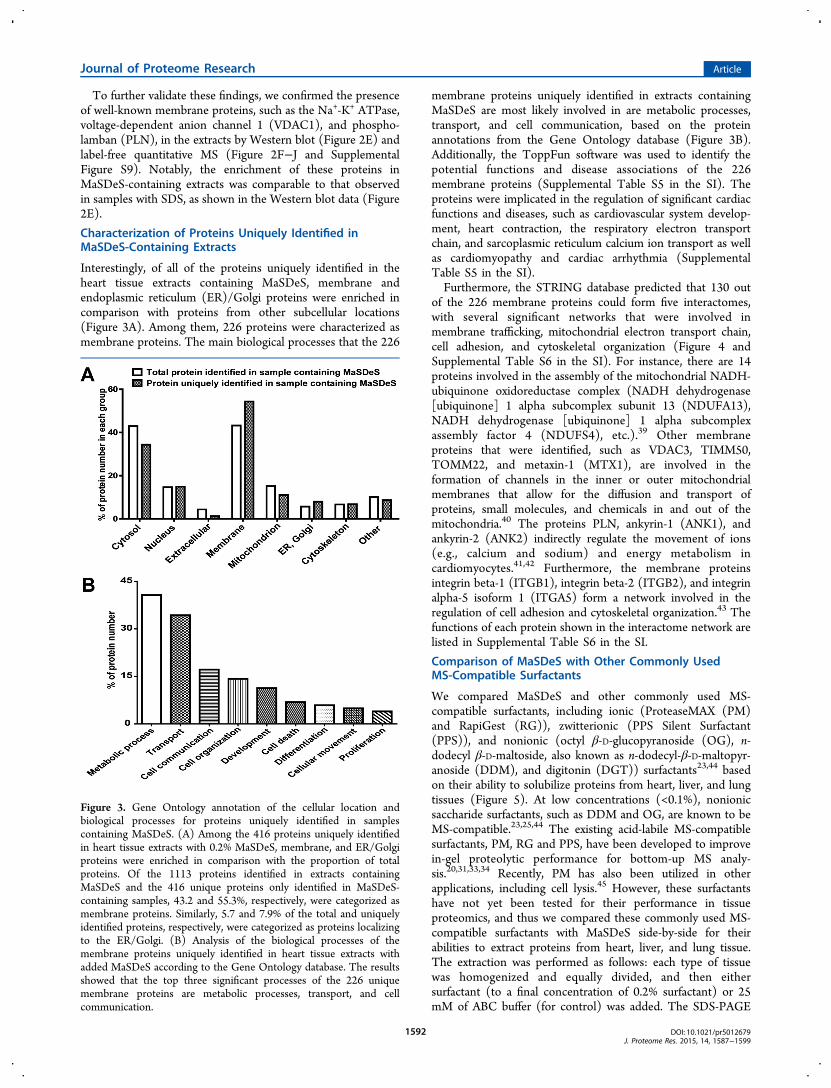
Interestingly, of all of the proteins uniquely identified in theheart tissue extracts containing MaSDeS, membrane andendoplasmic reticulum (ER)/Golgi proteins were enriched incomparison with proteins from other subcellular locations(Figure 3A). Among them, 226 proteins were characterized asmembrane proteins. The main biological processes that the 226
membrane proteins uniquely identified in extracts containingMaSDeS are most likely involved in are metabolic processes,transport, and cell communication, based on the proteinannotations from the Gene Ontology database (Figure 3B).Additionally, the ToppFun software was used to identify thepotential functions and disease associations of the 226membrane proteins (Supplemental Table S5 in the SI). Theproteins were implicated in the regulation of significant cardiacfunctions and diseases, such as cardiovascular system develop-ment, heart contraction, the respiratory electron transportchain, and sarcoplasmic reticulum calcium ion transport as wellas cardiomyopathy and cardiac arrhythmia (SupplementalTable S5 in the SI).Furthermore, the STRING database predicted that 130 out
of the 226 membrane proteins could form five interactomes,with several significant networks that were involved inmembrane trafficking, mitochondrial electron transport chain,cell adhesion, and cytoskeletal organization (Figure 4 andSupplemental Table S6 in the SI). For instance, there are 14proteins involved in the assembly of the mitochondrial NADH-ubiquinone oxidoreductase complex (NADH dehydrogenase[ubiquinone] 1 alpha subcomplex subunit 13 (NDUFA13),NADH dehydrogenase [ubiquinone] 1 alpha subcomplexassembly factor 4 (NDUFS4), etc.).39 Other membraneproteins that were identified, such as VDAC3, TIMM50,TOMM22, and metaxin-1 (MTX1), are involved in theformation of channels in the inner or outer mitochondrialmembranes that allow for the diffusion and transport ofproteins, small molecules, and chemicals in and out of themitochondria.40 The proteins PLN, ankyrin-1 (ANK1), andankyrin-2 (ANK2) indirectly regulate the movement of ions(e.g., calcium and sodium) and energy metabolism incardiomyocytes.41,42 Furthermore, the membrane proteinsintegrin beta-1 (ITGB1), integrin beta-2 (ITGB2), and integrinalpha-5 isoform 1 (ITGA5) form a network involved in theregulation of cell adhesion and cytoskeletal organization.43 Thefunctions of each protein shown in the interactome network arelisted in Supplemental Table S6 in the SI.
Comparison of MaSDeS with Other Commonly UsedMS-Compatible Surfactants
We compared MaSDeS and other commonly used MS-compatible surfactants, including ionic (ProteaseMAX (PM)and RapiGest (RG)), zwitterionic (PPS Silent Surfactant(PPS)), and nonionic (octyl β-D-glucopyranoside (OG), n-dodecyl β-D-maltoside, also known as n-dodecyl-β-D-maltopyr-anoside (DDM), and digitonin (DGT)) surfactants23,44 basedon their ability to solubilize proteins from heart, liver, and lungtissues (Figure 5). At low concentrations (<0.1%), nonionicsaccharide surfactants, such as DDM and OG, are known to beMS-compatible.23,25,44 The existing acid-labile MS-compatiblesurfactants, PM, RG and PPS, have been developed to improvein-gel proteolytic performance for bottom-up MS analy-sis.20,31,33,34 Recently, PM has also been utilized in otherapplications, including cell lysis.45 However, these surfactantshave not yet been tested for their performance in tissueproteomics, and thus we compared these commonly used MS-compatible surfactants with MaSDeS side-by-side for theirabilities to extract proteins from heart, liver, and lung tissue.The extraction was performed as follows: each type of tissuewas homogenized and equally divided, and then eithersurfactant (to a final concentration of 0.2% surfactant) or 25mM of ABC buffer (for control) was added. The SDS-PAGE
Figure 3. Gene Ontology annotation of the cellular location andbiological processes for proteins uniquely identified in samplescontaining MaSDeS. (A) Among the 416 proteins uniquely identifiedin heart tissue extracts with 0.2% MaSDeS, membrane, and ER/Golgiproteins were enriched in comparison with the proportion of totalproteins. Of the 1113 proteins identified in extracts containingMaSDeS and the 416 unique proteins only identified in MaSDeS-containing samples, 43.2 and 55.3%, respectively, were categorized asmembrane proteins. Similarly, 5.7 and 7.9% of the total and uniquelyidentified proteins, respectively, were categorized as proteins localizingto the ER/Golgi. (B) Analysis of the biological processes of themembrane proteins uniquely identified in heart tissue extracts withadded MaSDeS according to the Gene Ontology database. The resultsshowed that the top three significant processes of the 226 uniquemembrane proteins are metabolic processes, transport, and cellcommunication.
Journal of Proteome Research Article
DOI: 10.1021/pr5012679J. Proteome Res. 2015, 14, 1587−1599
1592

and protein assay results showed that MaSDeS performssignificantly better in solubilizing proteins than thesecommonly used surfactants, RG, PM, PPS, DDM, OG, andDGT, in all three types of tissues (Figure 5). PM and RGdemonstrated much better protein solubilization ability thanPPS, OG, DDM, and DGT.We next compared the degradation rate of MaSDeS with the
two leading commercially available acid-labile surfactants, RGand PM, under acidic conditions (pH 2 to 3) and found thatMaSDeS degrades much slower than PM and RG (Supple-mental Figure S10 in the SI). Subsequently, LC−MS/MSanalyses of equal volume experiments were performed to assessthe effectiveness of MaSDeS, PM, and RG for identifying
proteins during MS analyses. Each sample was run in triplicate
(Supplemental Tables S1−S3 in the SI). Over 90% of proteins
could be identified in at least two runs showcasing the high
reproducibility of this extraction method. The number and
intensity of proteins identified in swine heart, lung, and liver
tissue extracts generated with extraction buffer containing
MaSDeS were significantly increased (Figure 6, Supplemental
Figure S11 in the SI) compared with the control, PM, and RG
extracts.
Figure 4. Interactome analysis of the 226 membrane proteins uniquely identified in heart tissue extracts containing MaSDeS. The proteins in thepink circles serve as channels in the inner or outer mitochondrial membranes, allowing for the diffusion and transport of proteins, small molecules,and chemicals in and out of the mitochondria. The proteins in the blue circles are involved in the regulation of cell adhesion or cytoskeletonorganization/assembly. The proteins in the orange circles are well-known proteins involved in excitation-contraction coupling in the heart.
Journal of Proteome Research Article
DOI: 10.1021/pr5012679J. Proteome Res. 2015, 14, 1587−1599
1593

■ DISCUSSION
MaSDeS is a Strong MS-Compatible Surfactant for TissueProteomics
Protein solubility is a major challenge in tissue proteomics,particularly for membrane proteins.12−14 In this study, we haveidentified an MS-compatible degradable surfactant (MaSDeS),which can effectively solubilize all categories of proteins,including membrane proteins, from tissue sources commonlyused in proteomic analyses. As demonstrated, the performanceof this surfactant was comparable to the strongest surfactant,SDS, in solubilizing proteins from tissue extracts. It is MS-
compatible and greatly outperforms the existing MS-compatiblesurfactants.In general, surfactants (also known as detergents) can be
categorized into three major groups: ionic, nonionic, andzwitterionic.23 Ionic surfactants, such as the classical anionicsurfactant SDS, are strong denaturants and highly efficient atsolubilizing proteins.24 This stems from two features of SDS: along hydrocarbon tail that interacts with polypeptides andbreaks intraprotein interactions and an anionic headgroup thatcan interact with positively charged amino acids in thepolypeptide chain. The interaction between the anionicheadgroup and the polypeptide chain disrupts inter- andintraprotein electrostatic interactions, thereby promoting the
Figure 5. Comparison of MaSDeS with other leading MS-compatible surfactants in protein solubilization. The commonly used MS-compatiblesurfactants, including ionic (ProteaseMAX (PM), RapiGest (RG), PPS Silent Surfactant (PPS)) and nonionic (octyl β-D-glucopyranoside (OG), n-dodecyl β-D-maltoside (DDM), and digitonin (DGT)) surfactants, were investigated for their ability to solubilize proteins when compared withMaSDeS and SDS. Protein solubilization from three commonly used tissue sources were used for testing. An equal volume of each sample wasevaluated by the SDS-PAGE results: (A) heart tissue, (C) liver tissue, and (E) lung tissue as well as protein assay with (B) heart tissue, (D) livertissue, and (F) lung tissue. Both SDS-PAGE and protein assay results showed that MaSDeS possessed the highest capability for solubilizing proteins.All of samples have significant differences with control. * indicates the differences with MaSDeS. *p < 0.05, **p < 0.01, N.S. not significant.
Journal of Proteome Research Article
DOI: 10.1021/pr5012679J. Proteome Res. 2015, 14, 1587−1599
1594

disruption of protein tertiary structure and preventing proteinaggregation.23 Ionic surfactants such as SDS, despite being thestrongest surfactants, are unfortunately not compatible with MSanalyses since they can inhibit enzymatic digestion and severelysuppress the ionization of proteins/peptides.21,23,25 Nonionicsurfactants, such as OG, DDM, DGT, Triton X-100, and NP-40, also contain a hydrocarbon tail but, unlike ionic detergents,lack a charged headgroup. Consequently, these surfactantscannot effectively disrupt protein−protein interactions andtherefore are considered relatively mild solubilizing agents.23
Although some of the nonionic saccharide surfactants such asDDM and DG are MS-compatible at low concentrations(<0.1%),44,46,47 other polymer-based nonionic surfactants suchas Triton X-100 and NP-40 can cause significant signalsuppression and high polymer background in addition to theformation of adducts with the proteins25 and thus are not
selected in this study. Zwitterionic surfactants, such as CHAPS,are also MS-compatible at low concentrations (<0.1%), butunfortunately they also lead to adduct formation and signalsuppression.25 Zwitterionic surfactants have intermediateprotein solubilizing abilities, being stronger than nonionicdetergents but not as strong as ionic surfactants.23
Subsequently, MS-compatible surfactants have been devel-oped such as RG and PM, which are ionic surfactants,31−33 inaddition to PPS, which is zwitterionic. These surfactants differfrom conventional surfactants in that they contain an acid-labilefunctional group. Thus, upon addition of acid to the surfactant-containing sample, the surfactant quickly degrades intoinnocuous nonsurfactant byproducts, eliminating the need toremove the detergent prior to MS analysis.31−33 It should benoted that while these surfactants are supposed to enhancetrypsin digestion efficiency, they are frequently used in
Figure 6. Comparison of PM, RG, and MaSDeS on protein identification. Heart (A,B), liver (C,D), and lung (E,F) tissues were homogenized withand without 0.2% PM, RG, or MaSDeS to evaluate protein identification under equal volume loading. (A,C,E) Identified protein number. (B,D,F)Sum of protein intensity. Each sample was run in triplicate. All of the given protein intensities were presented in Log10 form. MaSDeS containingsamples showed higher number of protein identifications and higher sum of intensities compared with control and PM- and RG-containing samplesin all three types of tissues. All of surfactant-containing samples have significant differences with control. * means the sample has a statisticallysignificant difference from MaSDeS sample. *, p < 0.05. **, p < 0.01. # means the sample has a statistically significant difference from control sample.#, p < 0.05. ##, p < 0.01.
Journal of Proteome Research Article
DOI: 10.1021/pr5012679J. Proteome Res. 2015, 14, 1587−1599
1595

combination with gel-based methods.33,48,49 This may bepartially explained by the fact that higher concentrations ofRG (>0.5%) have been shown to influence the digestionefficiency.31 The data presented here showed that PM and RGperform at a similar level in solubilizing proteins but are inferiorto MaSDeS and SDS (Figure 5). The zwitterionic surfactantPPS was not as effective as RG or PM for protein solubilization,which is consistent with previous studies comparing RG andPPS.50 In addition, nonionic surfactants, such as DDM, havealso been utilized to solubilize integral membrane proteins forthe MS analysis of protein complexes.44 Nevertheless, thecommonly used MS-compatible nonionic surfactants, OG,DDM, and DGT, poorly solubilized proteins in aqueoussolution compared with ionic surfactants (Figure 5), which isconsistent with previous studies.23 Thus, our data demonstratethat MaSDeS’s protein solubilizing capability is greater thanthose of currently available MS-compatible surfactants and is onpar with those of SDS, the gold standard surfactant for proteinsolubilization (Figures 1E and 5). Furthermore, the CMC ofthis surfactant is very low compared with SDS at roomtemperature (Figure 1C). Low CMC provides an importantadvantage of MaSDeS as less surfactant is needed for proteinsolubilization.
Unique Characteristics of MaSDeS: Slow Degradation andThermostability
Unlike PM and RG, which degrade rapidly under acidicconditions (both PM and RG were completely degraded within30 min under acidic conditions), MaSDeS started degradingafter 1 h of incubation at pH 2 to 3 (Supplemental Figure S10in the SI). The observed differences in the degradation timesfor these surfactants under acidic conditions are likely due tothe differences in their chemical properties. RG contains a ketalfunctional group, which is highly susceptible to hydrolysisunder acidic conditions (Supplemental Scheme 1A in the SI).Consequently, at pH <5, this functional group is rapidlycleaved, generating a hydrophilic anionic molecule and ahydrophobic molecule (CH3 (CH2)10COCH3).
31 MaSDeS hasa similar molecular structure to PM with the exception that itcontains a thiophene ring in place of the furan ring in PM(Supplemental Scheme 1B in the SI). Under acidic conditions(pH <2), the carbonyl oxygen atom would be protonated andthe C−O σ bond would be cleaved, forming a carbocation thatis stabilized by a furan ring (or thiophene ring) as anintermediate. Compared with the thiophene group (inMaSDeS), the furanyl group (in PM) is a better electron-donating group and therefore promotes rapid cleavage of theC−O σ-bond. (See mechanistic details in the SupplementaryScheme 1B in the SI.) Subsequently, the resulting carbamatespecies will undergo decarboxylation [loss of carbon dioxide(CO2)]. This provides a strong driving force for degradation ofthe surfactant into CO2 and a zwitterionic species, similarly asproposed for PM.33 Therefore, it is expected that MaSDeS,with its thiophene ring, is slowly degraded into an alcohol, CO2,and a zwitterionic species (Figure 1A), compared with PM,which will degrade more quickly due to the furan ring(Supplemental Scheme 1B in the SI). This slow degradationunder acidic conditions will allow membrane proteins to remainin solution for a longer time and thus may have contributed tothe better solubilization and increase in membrane proteinidentifications in MaSDeS-containing samples (SupplementalFigure S10 in the SI).
In addition to relatively slow degradation under acidconditions, MaSDeS was also found to be thermostable, withminimal degradation observed up to 12 h after incubation at 80°C (Supplementary Figure S2 in the SI). This is likely related tothe fact that the furan ring is a better electron-donating groupthan the thiophene ring as previously discussed. The thermo-stability allows MaSDeS to be used in experiments that requirerelatively high temperature to facilitate protein extraction andsolubilization. For example, some membrane proteins aredifficult to be solubilized at room temperature even in thepresence of high concentrations of SDS.51 These uniquecharacteristics of MaSDeS, thermostability and relatively slowdegradation under acidic conditions (Supplemental Figures S2and S3 and Supplemental Figure S10 in the SI), may makeMaSDeS useful in a wide range of tissue proteomicsapplications. For instance, histones and myofilament proteinsare extracted using acidic conditions,7,52−54 and thus theaddition of MaSDeS could help maintain or increase proteinsolubility during the extraction process. It has also beendemonstrated that high temperature (60 °C) chromatographicseparation dramatically improved the recovery of hydrophobicpeptides derived from the transmembrane domains of integralmembrane proteins.12,23
MaSDeS Improves Membrane Protein Identification inTissue Proteomics
Membrane proteins play an important role in many essentialcellular functions and account for a significant proportion ofdrug targets.20,55−59 However, membrane proteins are notori-ously difficult to study due to their hydrophobic nature, whichmakes them difficult to solubilize. Consequently, membraneproteins are under-represented in proteomics stud-ies.12,13,16,23,60 As demonstrated in this study, the use ofMaSDeS during tissue extraction leads to a significantenrichment of membrane proteins. We have shown that thenumber of membrane proteins identified in extracts generatedusing buffer containing MaSDeS was greater than for controlextracts (no surfactant) (Figure 2B and Supplemental Table S4in the SI). Notably, 43% of the total identified proteins in theswine heart tissue extract are membrane proteins. Additionally,on the basis of the quantitative MS results, the total amount ofmembrane protein identified in MaSDeS-containing extractswas significantly greater than in the pre-extract and controlextracts (Figure 2C and Supplemental Figure S9 in the SI).Furthermore, Western blot analysis of membrane proteinrevealed that MaSDeS significantly enriched membraneproteins comparable to SDS (Figure 2E). The levels of well-known membrane proteins were significantly enriched in hearttissue extracts containing MaSDeS in comparison with controlextracts. Such membrane proteins included the Na+-K+ ATPase,VDAC1, and PLN, which are essential membrane proteinsinvolved in excitation−contraction coupling in the heart61 aswell as cadherin, a transmembrane protein involved in cell−celladhesion.62
The 226 membrane proteins uniquely identified in the swineheart tissue extracts containing MaSDeS formed interactomesthat are involved in the regulation of cardiac function. Our datasuggest that the inclusion of MaSDeS in the extraction bufferallowed for the identification of these important membraneproteins, which would have otherwise gone undetected withoutthis surfactant. Thus, MaSDeS has the potential to be highlyuseful for membrane proteome analysis.
Journal of Proteome Research Article
DOI: 10.1021/pr5012679J. Proteome Res. 2015, 14, 1587−1599
1596

■ CONCLUSIONS
To recapitulate, we have identified an MS-compatiblesurfactant, MaSDeS, which can effectively solubilize allcategories of proteins, including membrane proteins, withperformance that is comparable to the strongest surfactant,SDS, at the concentrations employed in this study. The use ofMaSDeS greatly simplifies the sample preparation process fortissue proteomic analyses because it is acid-labile and thereforedoes not need to be removed prior to MS analysis. This makesMaSDeS amenable for high-throughput proteomic analyses.Moreover, the incorporation of MaSDeS in the sequentialtissue extraction leads to a significant enrichment of membraneproteins, which are often under-represented in proteomicstudies. Furthermore, MaSDeS is thermostable and, con-sequently, can be used in cases where high temperature isneeded to facilitate protein extraction and solubilization. Thesefavorable characteristics of MaSDeS will make it a powerful toolfor the analysis of tissues and other challenging biologicalsamples. We expect it will expedite the use of tissue proteomicsin clinical diagnosis and can be applicable to a wide range ofbiological applications.
■ ASSOCIATED CONTENT
*S Supporting Information
Supplemental Scheme 1. Comparison of the degradationmechanisms of RapiGest (RG), ProteaseMAX (PM) andMaSDeS under acidic conditions. Supplemental Figure S1.High-resolution FT-ICR MS analysis of MaSDeS degradationin 10% FA. Supplemental Figure S2. Assessment of MaSDeSthermostability. Supplemental Figure S3. MS evaluation ofMaSDeS stability at 65 °C. Supplemental Figure S4. Evaluationof the influence of MaSDeS on enzymatic digestion and MSanalysis. Supplemental Figure S5. The sequential tissueextraction method reduces blood protein contamination inthe second heart tissue extract. Supplemental Figure S6. SDS-polyacrylamide gel electrophoresis (PAGE) and protein assayanalyses of sequential extracts from liver and lung. Supple-mental Figure S7. The effect of MaSDeS on proteinidentification. Supplemental Figure S8. MS analysis of equalprotein amounts (1 μg) from pig heart tissue extracts (pre-extract, control, and 0.2% MaSDeS). Supplemental Figure S9.Quantitative MS results showing that the use of MaSDeSsignificantly improved the detection of membrane proteins inheart tissue extracts. Supplemental Figure S10. MaSDeSdegrades slowly under acidic conditions at 37 °C, in contrastwith the rapid degradation observed for the two commerciallyavailable acid-labile surfactants, RapiGest (RG), and Protease-MAX (PM). Supplemental Figure S11. Buffer containing 0.2%of PM, RG, or MaSDeS was used to homogenize pig heart,liver, and lung tissue. Supplemental Table S1. Protein/peptidelist: equal volume loading test in heart tissue. SupplementalTable S2. Protein/peptide list: equal volume loading test inliver tissue. Supplemental Table S3. Protein/peptide list: equalvolume loading test in lung tissue. Supplemental Table S4.Protein/peptide list: equal amount loading test in heart tissue.Supplemental Table S5. Enriched function and human diseaselist. Supplemental Table S6. Protein information on inter-actome. This material is available free of charge via the Internetat http://pubs.acs.org.
■ AUTHOR INFORMATION
Corresponding Author
*E-mail: [email protected]. Tel: 608-263-9212. Fax: 608-265-5512.
Notes
The authors declare no competing financial interest.
■ ACKNOWLEDGMENTS
We acknowledge the grants from the U.S. National Institutes ofHealth, R01 HL096971, HL109810 (to Y.G.), and R01HL114120 (to J.Z.). We thank Yoonkyu (Ryan) Lee fortechnical assistance, Serife Ayaz-Guner for critical reading ofthis manuscript, and Prof. Weiping Tang for helpfuldiscussions. We also thank Sergei Saveliev and Marjeta Urhfrom Promega for kindly providing the surfactant compoundlibrary and for helpful discussions.
■ ABBREVIATIONS
ABC, ammonium bicarbonate; BSA, bovine serum albumin;CMC, critical micelle concentration; CV, coefficient ofvariation; DDM, n-dodecyl β-D-maltoside; DGT, digitonin;DTT, dithiothreitol; ER, endoplasmic reticulum; FA, formicacid; FT-ICR, Fourier transform ion cyclotron resonance; IAA,iodoacetamide; IPA, isopropyl alcohol; MaSDeS, massspectrometry-compatible degradable surfactant; MS, massspectrometry; MS/MS, tandem mass spectrometry; OG, octylβ-D-glucopyranoside; PLN, phospholamban; PM, Protease-MAX; PPS, PPS silent surfactant; RG, RapiGest; SDS, sodiumdodecyl sulfate; SDS-PAGE, sodium dodecyl sulfate-polyacry-lamide gel electrophoresis
■ REFERENCES(1) Veenstra, T. D.; Zhou, M. Tissue Proteomics and Metabolomics:An Excellent Start and a Promising Future. J. Proteome Res. 2009, 8(4), 1617.(2) Zhang, J.; Guy, M. J.; Norman, H. S.; Chen, Y.-C.; Xu, Q.; Dong,X.; Guner, H.; Wang, S.; Kohmoto, T.; Young, K. H.; Moss, R. L.; Ge,Y. Top-Down Quantitative Proteomics Identified Phosphorylation ofCardiac Troponin I as a Candidate Biomarker for Chronic HeartFailure. J. Proteome Res. 2011, 10 (9), 4054−4065.(3) Kim, M. S.; Pinto, S. M.; Getnet, D.; Nirujogi, R. S.; Manda, S. S.;Chaerkady, R.; Madugundu, A. K.; Kelkar, D. S.; Isserlin, R.; Jain, S.;Thomas, J. K.; Muthusamy, B.; Leal-Rojas, P.; Kumar, P.;Sahasrabuddhe, N. A.; Balakrishnan, L.; Advani, J.; George, B.;Renuse, S.; Selvan, L. D.; Patil, A. H.; Nanjappa, V.; Radhakrishnan, A.;Prasad, S.; Subbannayya, T.; Raju, R.; Kumar, M.; Sreenivasamurthy, S.K.; Marimuthu, A.; Sathe, G. J.; Chavan, S.; Datta, K. K.; Subbannayya,Y.; Sahu, A.; Yelamanchi, S. D.; Jayaram, S.; Rajagopalan, P.; Sharma,J.; Murthy, K. R.; Syed, N.; Goel, R.; Khan, A. A.; Ahmad, S.; Dey, G.;Mudgal, K.; Chatterjee, A.; Huang, T. C.; Zhong, J.; Wu, X.; Shaw, P.G.; Freed, D.; Zahari, M. S.; Mukherjee, K. K.; Shankar, S.;Mahadevan, A.; Lam, H.; Mitchell, C. J.; Shankar, S. K.;Satishchandra, P.; Schroeder, J. T.; Sirdeshmukh, R.; Maitra, A.;Leach, S. D.; Drake, C. G.; Halushka, M. K.; Prasad, T. S.; Hruban, R.H.; Kerr, C. L.; Bader, G. D.; Iacobuzio-Donahue, C. A.; Gowda, H.;Pandey, A. A draft map of the human proteome. Nature 2014, 509(7502), 575−581.(4) Xiao, Y.; Guo, L.; Wang, Y. A Targeted Quantitative ProteomicsStrategy for Global Kinome Profiling of Cancer Cells and Tissues. Mol.Cell. Proteomics. 2014, 13 (4), 1065−1075.(5) Tu, C.; Li, J.; Jiang, X.; Sheflin, L. G.; Pfeffer, B. A.; Behringer,M.; Fliesler, S. J.; Qu, J. Ion-current-based proteomic profiling of theretina in a rat model of Smith-Lemli-Opitz syndrome. Mol. Cell.Proteomics 2013, 12 (12), 3583−3598.
Journal of Proteome Research Article
DOI: 10.1021/pr5012679J. Proteome Res. 2015, 14, 1587−1599
1597

(6) Qu, J.; Young, R.; Page, B. J.; Shen, X.; Tata, N.; Li, J.; Duan, X.;Fallavollita, J. A.; Canty, J. M., Jr. Reproducible Ion-Current-BasedApproach for 24-Plex Comparison of the Tissue Proteomes ofHibernating versus Normal Myocardium in Swine Models. J. ProteomeRes. 2014, 13 (5), 2571−2584.(7) Peng, Y.; Gregorich, Z. R.; Valeja, S. G.; Zhang, H.; Cai, W.;Chen, Y. C.; Guner, H.; Chen, A. J.; Schwahn, D. J.; Hacker, T. A.; Liu,X.; Ge, Y. Top-down proteomics reveals concerted reductions inmyofilament and Z-disc protein phosphorylation after acutemyocardial infarction. Mol. Cell. Proteomics 2014, 13 (10), 2752−2764.(8) Gregorich, Z. R.; Ge, Y. Top-down proteomics in health anddisease: Challenges and opportunities. Proteomics 2014, 14 (10),1195−1210.(9) Dong, X.; Sumandea, C. A.; Chen, Y.-C.; Garcia-Cazarin, M. L.;Zhang, J.; Balke, C. W.; Sumandea, M. P.; Ge, Y. AugmentedPhosphorylation of Cardiac Troponin I in Hypertensive Heart Failure.J. Biol. Chem. 2012, 287 (2), 848−857.(10) Atrih, A.; Mudaliar, M. A. V.; Zakikhani, P.; Lamont, D. J.;Huang, J. T. J.; Bray, S. E.; Barton, G.; Fleming, S.; Nabi, G.Quantitative proteomics in resected renal cancer tissue for biomarkerdiscovery and profiling. Br. J. Cancer. 2014, 110 (6), 1622−1633.(11) Yi, J. K.; Bae, J. Y.; Kim, D. H.; Lee, J. W.; Han, W. S.; Shin, H.J.; Lee, C. J.; Noh, D. Y. Novel protein markers identified byproteomics technique comparing breast cancer tissues against normaltissues. Mol. Cell. Proteomics 2006, 5 (10), S211−S211.(12) Whitelegge, J. P. Integral membrane proteins and bilayerproteomics. Anal. Chem. 2013, 85 (5), 2558−2568.(13) Wu, C. C.; Yates, J. R., 3rd. The application of massspectrometry to membrane proteomics. Nat. Biotechnol. 2003, 21(3), 262−267.(14) Whitelegge, J. P.; le Coutre, J.; Lee, J. C.; Engel, C. K.; Prive, G.G.; Faull, K. F.; Kaback, H. R. Toward the bilayer proteome,electrospray ionization-mass spectrometry of large, intact trans-membrane proteins. Proc. Natl. Acad. Sci. U. S. A. 1999, 96 (19),10695−10698.(15) Barrera, N. P.; Robinson, C. V. Advances in the massspectrometry of membrane proteins: from individual proteins tointact complexes. Annu. Rev. Biochem. 2011, 80, 247−271.(16) Wu, C. C.; MacCoss, M. J.; Howell, K. E.; Yates, J. R., 3rd. Amethod for the comprehensive proteomic analysis of membraneproteins. Nat. Biotechnol. 2003, 21 (5), 532−538.(17) Krogh, A.; Larsson, B.; von Heijne, G.; Sonnhammer, E. L.Predicting transmembrane protein topology with a hidden Markovmodel: application to complete genomes. J. Mol. Biol. 2001, 305 (3),567−580.(18) Carroll, J.; Altman, M. C.; Fearnley, I. M.; Walker, J. E.Identification of membrane proteins by tandem mass spectrometry ofprotein ions. Proc. Natl. Acad. Sci. U. S. A. 2007, 104 (36), 14330−14335.(19) Rasmussen, S. G.; Choi, H. J.; Rosenbaum, D. M.; Kobilka, T.S.; Thian, F. S.; Edwards, P. C.; Burghammer, M.; Ratnala, V. R.;Sanishvili, R.; Fischetti, R. F.; Schertler, G. F.; Weis, W. I.; Kobilka, B.K. Crystal structure of the human beta2 adrenergic G-protein-coupledreceptor. Nature 2007, 450 (7168), 383−387.(20) Overington, J. P.; Al-Lazikani, B.; Hopkins, A. L. How manydrug targets are there? Nat. Rev. Drug Discovery 2006, 5 (12), 993−996.(21) Wisniewski, J. R.; Zougman, A.; Nagaraj, N.; Mann, M.Universal sample preparation method for proteome analysis. Nat.Methods 2009, 6 (5), 359−362.(22) Bereman, M. S.; Egertson, J. D.; MacCoss, M. J. Comparisonbetween procedures using SDS for shotgun proteomic analyses ofcomplex samples. Proteomics 2011, 11 (14), 2931−2935.(23) Speers, A. E.; Wu, C. C. Proteomics of integral membraneproteins–theory and application. Chem. Rev. 2007, 107 (8), 3687−3714.(24) Masuda, T.; Tomita, M.; Ishihama, Y. Phase transfer surfactant-aided trypsin digestion for membrane proteome analysis. J. ProteomeRes. 2008, 7 (2), 731−740.
(25) Loo, R. R.; Dales, N.; Andrews, P. C. Surfactant effects onprotein structure examined by electrospray ionization mass spectrom-etry. Protein Sci. 1994, 3 (11), 1975−1983.(26) Didangelos, A.; Yin, X.; Mayr, M. Method for proteinsubfractionation of cardiovascular tissues before DIGE analysis.Methods Mol. Biol. 2012, 854, 287−297.(27) Duan, X.; Young, R.; Straubinger, R. M.; Page, B.; Cao, J.; Wang,H.; Yu, H.; Canty, J. M.; Qu, J. A straightforward and highly efficientprecipitation/on-pellet digestion procedure coupled with a longgradient nano-LC separation and Orbitrap mass spectrometry forlabel-free expression profiling of the swine heart mitochondrialproteome. J. Proteome Res. 2009, 8 (6), 2838−2850.(28) Botelho, D.; Wall, M. J.; Vieira, D. B.; Fitzsimmons, S.; Liu, F.;Doucette, A. Top-down and bottom-up proteomics of SDS-containingsolutions following mass-based separation. J. Proteome Res. 2010, 9 (6),2863−2870.(29) Doucette, A.; Craft, D.; Li, L. Protein concentration and enzymedigestion on microbeads for MALDI-TOF peptides mass mapping ofproteins from dilute solutions. Anal. Chem. 2000, 72 (14), 3355−3362.(30) Manza, L. L.; Stamer, S. L.; Ham, A. J.; Codreanu, S. G.; Liebler,D. C. Sample preparation and digestion for proteomic analyses usingspin filters. Proteomics 2005, 5 (7), 1742−1745.(31) Yu, Y. Q.; Gilar, M.; Lee, P. J.; Bouvier, E. S.; Gebler, J. C.Enzyme-friendly, mass spectrometry-compatible surfactant for in-solution enzymatic digestion of proteins. Anal. Chem. 2003, 75 (21),6023−6038.(32) Chen, E. I.; McClatchy, D.; Park, S. K.; Yates, J. R., 3rd.Comparisons of mass spectrometry compatible surfactants for globalanalysis of the mammalian brain proteome. Anal. Chem. 2008, 80 (22),8694−8701.(33) Saveliev, S. V.; Woodroofe, C. C.; Sabat, G.; Adams, C. M.;Klaubert, D.; Wood, K.; Urh, M. Mass spectrometry compatiblesurfactant for optimized in-gel protein digestion. Anal. Chem. 2013, 85(2), 907−14.(34) Chen, E. I.; Cociorva, D.; Norris, J. L.; Yates, J. R., 3rd.Optimization of mass spectrometry-compatible surfactants for shotgunproteomics. J. Proteome Res. 2007, 6 (7), 2529−2538.(35) Collins, J. W.; Coldham, N. G.; Salguero, F. J.; Cooley, W. A.;Newell, W. R.; Rastall, R. A.; Gibson, G. R.; Woodward, M. J.; LaRagione, R. M. Response of porcine intestinal in vitro organ culturetissues following exposure to Lactobacillus plantarum JC1 andSalmonella enterica serovar Typhimurium SL1344. Appl. Environ.Microbiol. 2010, 76 (19), 6645−6657.(36) Moller, S.; Croning, M. D.; Apweiler, R. Evaluation of methodsfor the prediction of membrane spanning regions. Bioinformatics 2001,17 (7), 646−653.(37) Franceschini, A.; Szklarczyk, D.; Frankild, S.; Kuhn, M.;Simonovic, M.; Roth, A.; Lin, J.; Minguez, P.; Bork, P.; von Mering, C.;Jensen, L. J. STRING v9.1: protein-protein interaction networks, withincreased coverage and integration. Nucleic Acids Res. 2013, 41(Database issue), D808−D815.(38) Saveliev, S.; Simpson, D.; Wood, K. V. Cleavable Surfactants.Patent WO 2009048611 A2, 2009.(39) Kussmaul, L.; Hirst, J. The mechanism of superoxide productionby NADH:ubiquinone oxidoreductase (complex I) from bovine heartmitochondria. Proc. Natl. Acad. Sci. U. S. A. 2006, 103 (20), 7607−7612.(40) Zhang, J.; Liem, D. A.; Mueller, M.; Wang, Y.; Zong, C.; Deng,N.; Vondriska, T. M.; Korge, P.; Drews, O.; Maclellan, W. R.; Honda,H.; Weiss, J. N.; Apweiler, R.; Ping, P. Altered proteome biology ofcardiac mitochondria under stress conditions. J. Proteome Res. 2008, 7(6), 2204−2214.(41) Cunha, S. R.; Mohler, P. J. Cardiac ankyrins: Essentialcomponents for development and maintenance of excitable membranedomains in heart. Cardiovasc. Res. 2006, 71 (1), 22−29.(42) MacLennan, D. H.; Kranias, E. G. Phospholamban: a crucialregulator of cardiac contractility. Nat. Rev. Mol. Cell Biol. 2003, 4 (7),566−577.
Journal of Proteome Research Article
DOI: 10.1021/pr5012679J. Proteome Res. 2015, 14, 1587−1599
1598

(43) Hood, J. D.; Cheresh, D. A. Role of integrins in cell invasion andmigration. Nat. Rev. Cancer 2002, 2 (2), 91−100.(44) Laganowsky, A.; Reading, E.; Hopper, J. T.; Robinson, C. V.Mass spectrometry of intact membrane protein complexes. Nat. Protoc2013, 8 (4), 639−651.(45) Pirmoradian, M.; Budamgunta, H.; Chingin, K.; Zhang, B.;Astorga-Wells, J.; Zubarev, R. A. Rapid and Deep Human ProteomeAnalysis by Single-dimension Shotgun Proteomics. Mol. Cell.Proteomics 2013, 12 (11), 3330−3338.(46) Barrera, N. P.; Di Bartolo, N.; Booth, P. J.; Robinson, C. V.Micelles protect membrane complexes from solution to vacuum.Science 2008, 321 (5886), 243−246.(47) Zhou, M.; Morgner, N.; Barrera, N. P.; Politis, A.; Isaacson, S.C.; Matak-Vinkovic, D.; Murata, T.; Bernal, R. A.; Stock, D.; Robinson,C. V. Mass spectrometry of intact V-type ATPases reveals bound lipidsand the effects of nucleotide binding. Science 2011, 334 (6054), 380−385.(48) Zhou, J.; Huang, S.; Bi, D.; Zhang, H.; Li, J.; Lin, Y.; Chen, P.;Wang, X.; Liang, S. Analysis of integral membrane proteins by heat gel-embedment combined with improved in-gel digestions. Electrophoresis2009, 30 (23), 4109−4117.(49) Nomura, E.; Katsuta, K.; Ueda, T.; Toriyama, M.; Mori, T.;Inagaki, N. Acid-labile surfactant improves in-sodium dodecyl sulfatepolyacrylamide gel protein digestion for matrix-assisted laserdesorption/ionization mass spectrometric peptide mapping. J. MassSpectrom. 2004, 39 (2), 202−207.(50) Wu, F.; Sun, D.; Wang, N.; Gong, Y.; Li, L. Comparison ofsurfactant-assisted shotgun methods using acid-labile surfactants andsodium dodecyl sulfate for membrane proteome analysis. Anal. Chim.Acta 2011, 698 (1−2), 36−43.(51) Deal, C. D.; Kaplan, S. Solubilization, isolation, andimmunochemical characterization of the major outer membraneprotein from Rhodopseudomonas sphaeroides. J. Biol. Chem. 1983,258 (10), 6524−6529.(52) Lin, S.; Wein, S.; Gonzales-Cope, M.; Otte, G. L.; Yuan, Z.-F.;Afjehi-Sadat, L.; Maile, T.; Berger, S. L.; Rush, J.; Lill, J. R.; Arnott, D.;Garcia, B. A. Stable-isotope-labeled Histone Peptide Library forHistone Post-translational Modification and Variant Quantification byMass Spectrometry. Mol. Cell. Proteomics 2014, 13 (9), 2450−2466.(53) Garcia, B. A.; Pesavento, J. J.; Mizzen, C. A.; Kelleher, N. L.Pervasive combinatorial modification of histone H3 in human cells.Nat. Methods 2007, 4 (6), 487−489.(54) Shechter, D.; Dormann, H. L.; Allis, C. D.; Hake, S. B.Extraction, purification and analysis of histones. Nat. Protoc. 2007, 2(6), 1445−1457.(55) Borjesson, S. I.; Elinder, F. Structure, function, and modificationof the voltage sensor in voltage-gated ion channels. Cell Biochem.Biophys. 2008, 52 (3), 149−174.(56) D’Angelo, M. A.; Hetzer, M. W. Structure, dynamics andfunction of nuclear pore complexes. Trends Cell Biol. 2008, 18 (10),456−466.(57) Ishibashi, K.; Hara, S.; Kondo, S. Aquaporin water channels inmammals. Clin. Exp. Nephrol. 2009, 13 (2), 107−117.(58) Kawai, T.; Akira, S. Toll-like receptor and RIG-I-like receptorsignaling. Ann. N.Y. Acad. Sci. 2008, 1143, 1−20.(59) Carberry, S.; Zweyer, M.; Swandulla, D.; Ohlendieck, K.Comparative proteomic analysis of the contractile-protein-depletedfraction from normal versus dystrophic skeletal muscle. Anal. Biochem.2014, 446, 108−115.(60) Tan, S.; Tan, H. T.; Chung, M. C. Membrane proteins andmembrane proteomics. Proteomics 2008, 8 (19), 3924−3932.(61) Bers, D. M. Cardiac excitation-contraction coupling. Nature2002, 415 (6868), 198−205.(62) Cavallaro, U.; Christofori, G. Cell adhesion and signalling bycadherins and Ig-CAMs in cancer. Nat. Rev. Cancer 2004, 4 (2), 118−132.
Journal of Proteome Research Article
DOI: 10.1021/pr5012679J. Proteome Res. 2015, 14, 1587−1599
1599
Copyright © 2022 FDOKUMEN