Fluorinated surfactant templating of vinyl-functionalized nanoporous silica
Upload
independentCategory
view
1download
0
COVER SHEET
Frost, Ray and He, Hongping and Kloprogge, Theo and Bostrom, Thor and Duong, Loc and Yuan, Peng and Xi, Yunfei and Yang, Dan (2006) Changes in the morphology of organoclays with HDTMA+ surfactant loading. Applied Clay Science 31(3-4):pp. 262-271. Copyright 2006 Elsevier. Accessed from: http://eprints.qut.edu.au/archive/00003646

1
Changes In The Morphology Of Organoclays With
HDTMA+ Surfactant Loading.
Hongping He 1,2, Ray L. Frost 2,* Thor Bostrom 2, Peng Yuan 1, Loc Duong 2, Dan Yang 1, Yunfei Xi 2 and J. Theo Kloprogge 2
1 Guangzhou Institute of Geochemistry, Chinese Academy of Sciences, Wushan,
Guangzhou 510640, China 2 Inorganic Materials Research Program, School of Physical and Chemical Sciences,
Queensland University of Technology, GPO Box 2434, Brisbane, QLD 4001, Australia
* Correspondence to: Ray L. Frost, Inorganic Materials Research Program, School of Physical and Chemical Sciences, Queensland University of Technology, GPO Box 2434, Brisbane, QLD 4001, Australia. E-mail: [email protected]

2
Abstract The detailed understanding of the interlayer structure of organoclays is of
importance in the design of organoclay based materials and their industrial applications. In this study, Transmission Electron Microscopy (TEM), Scanning Electron Microscopy (SEM) and X-Ray Diffraction (XRD) have been used to provide new insights into the interlayer structure and morphology of HDTMA+/montmorillonite organoclays. XRD patterns show that thermal treatment has an important effect on the stability of organoclays, reflected by significant changes in the basal spacing. TEM and SEM micrographs demonstrate that the organoclays with lower surfactant packing density are mainly composed of irregular layer stacking with a number of curved organoclay layers, while those with higher surfactant packing density are mainly composed of regularly intercalated and flat layers. Variations of the interlayer distances exist in all organoclays and are more pronounced in the organoclays with lower surfactant packing density. This study demonstrates that not only the arrangement model of surfactant but also the morphology of organoclay strongly depend on the surfactant packing density within the montmorillonite interlayer space. Keywords: Intercalation; Organoclay; Morphology; Transmission electron microscopy;
Scanning electron microscopy; Nanochemistry

3
Introduction The study of organoclays is a vital subject in current research since various
organoclays are widely used as nanocomposite precursors [1,2], adsorbents for organic pollutants [3], rheological control agents [4] and electric materials [5]. The combination of the hydrophobic nature of the surfactant and the layered structure of the silicate layers leads to unique physicochemical properties. In these applications, the behavior and properties of the organoclays strongly depend on the structure and the molecular environment of the organic molecules within the galleries.
During the intercalation of the surfactants, the interlayer of the clay minerals is expanded as shown by the XRD patterns. A number of previous studies have demonstrated that the d-spacings of the organoclays depend on the length of the alkyl chains and the packing density of the surfactants within the galleries of clay minerals [6-11]. Accordingly, various models for the surfactant structural arrangements have been proposed chiefly based on XRD results. Recently, new approaches to the study of the surfactant structural arrangements including Fourier Transform Infrared (FTIR) spectroscopy, Raman spectroscopy, Differential Scanning Calorimetry (DSC) and Magic-Angle-Spinning Nuclear-Magic-Resonance (MAS NMR) have been introduced to study the microstructure of organoclays and provided detailed conformational information [12-20]. However, these studies could not provide information about morphology and homogeneity of the organoclays.
TEM complemented with SEM are the best techniques to investigate the
morphology of clay minerals. TEM and SEM have been widely used in the study of organoclay based nanocomposites [1,2,21,22]. In this research, the principal focus was on the distribution of organoclays in the nanocomposites and their effect on the nanocomposite properties. However, few studies have been performed on the microstructure and morphology of the organoclays [23,24]. Hoffmann et al. [21] revealed that low molecular weight modifiers only promoted intercalation and failed to exfoliate the silicate particles during the synthesis of nanocomposites, whereas organophilic modification of clay with bulky surfactant molecules affords completely exfoliated silicate particles in the nanocomposites. This means the intercalated species of organoclay have an important effect on the properties of the organoclay and, therefore, nanocomposite. Meanwhile, Lee and Kim [23,24] proposed that the charge value or its distribution on clay layer surface might be an important factor in expansion. Such characteristics of the organoclay would affect the function of the organoclay as a precursor in nanocomposite formation. The exfoliation of the organoclay will affect the properties of the resultant clay nanocomposites. One of the most important applications of organoclays is in the absorption of recalcitrant organics in water. These absorption processes are likely to be effected by the morphology and in particular the curvature of the organoclays. Industrial applications of organoclays are in the use as filter beds in which large volumes of water containing low concentrations of organic impurities are passed through the filter beds. The organoclay will remove the organic pollutants from the water.
In this study, HDTMA+/montmorillonite organoclays were prepared at different
surfactant concentrations and investigated using TEM, SEM and XRD, to elucidate

4
the relation between the morphology of organoclay and the surfactant packing density within the montmorillonite galleries. Such information is vital for the preparation of organoclay-based nanocomposites.
Materials and Methods
The montmorillonite used in this study originated from Hebei province, China.
The sample was purified by sedimentation and the <2µm fraction was collected and
dried at 90 oC. The sample was ground and sieved through 200 mesh and sealed in a glass tube for use. The collected montmorillonite contains very low concentrations of illite and kaolinite [19]. The cation exchange capacity (CEC) was 57.9 mmol/100g. The chemical composition of the montmorillonite was determined by wet chemical analysis.
Its structural formula is [Na0.05Ca0.18Mg0.10][Al1.58Fe0.03Mg0.39][Si3.77Al0.23]O10(OH)2·nH2O. The surfactant used in this study is hexadecyltrimethylammonium bromide (HDTMAB) with a purity of 99%, and was obtained from YuanJu Chem. Co., China. The surfactant was used without any further purification. The preparation of Na-montmorillonite (Na-Mont) was carried out by the following procedure. 10 g of the mixture of montmorillonite (9.4 g) and Na2CO3 (0.6 g) was added into 100ml of deionized water and stirred at 80 oC for 3 h. Na-Mont was collected by centrifugation and washed with deionized water until the pH of the solution of Na-Mont was 7. The Na-Mont was dried at 105 oC, ground and sieved through 200 mesh and kept in a sealed bottle.
The synthesis of organoclay was performed by the following procedure: 2.5 g of Na-montmorillonite was first dispersed in about 300 ml of deionized water, to which a desired amount of hexadecyltrimethylammonium bromide was slowly added. The concentrations of HDTMA+ were 0.5 CEC, 1.5 CEC and 5.0 CEC of montmorillonite, respectively. The reaction mixtures were stirred for 10 h at 80 oC. All products were washed free of bromide anions (tested by AgNO3), dried at 90 oC and ground in an agate mortar. The HDTMA+/montmorillonite organoclays prepared at the concentrations of 0.5 CEC, 1.5 CEC and 5.0 CEC were labeled as OM1, OM2 and OM3, respectively. The corresponding amounts of the loaded surfactant are 0.46 CEC, 0.93 CEC and 1.58 CEC, respectively, determined by thermogravemetric analyses.
For TEM observation, small drops of dilute suspensions of 0.1 g of the clays in 5
cm3 of doubly distilled water were placed on Cu mesh grids that had been coated with a thin carbon film. The grids were air dried then briefly placed in a 60 °C oven to ensure complete drying prior to insertion into the instrument. The specimens were examined in a Philips CM200 transmission electron microscope operated at an accelerating voltage of 200 kV. Though this preparation is not generally optimal for high resolution imaging as compared to thinned or sectioned samples, the montmorillonite crystallite layers were thin enough that imaging of the interlayer spacings could be performed where suitable orientations could be obtained. The surface morphology of the prepared organoclays was examined by SEM. Small

5
amounts of the dried powders (approximately 0.01 g) were placed on sticky carbon tape on standard Al mounts, then sputter coated with a thin conductive layer of gold. The samples were viewed in an FEI Quanta 200 scanning electron microscope at 20 kV.
For XRD analysis, the organoclays were first dispersed in the deionized water,
then dropped on the glass slides and dried in the air. XRD patterns were recorded between 2 and 20° (2θ) at a scanning speed of 2 °/min., using Rigaku D/max-1200 diffractometer with CuKα radiation. In addition, the XRD analysis of the organoclay powder (before being dispersed in water) was performed under the same conditions as above-mentioned, to elucidate the effect resulted from the dispersion of organoclay in water.
Results and Discussion D-spacing of organoclay and corresponding conformations of interlayer HDTMA
Figure 1 shows the XRD patterns of Na-montmorillonite and the three organoclays prepared at different surfactant concentrations. Upon intercalation, the basal spacings are expanded as expected depending on the surfactant concentrations. The d(001) values of OM1 and OM3 are at 1.48 nm and 2.26 nm, respectively, while OM2 shows the main peak at 1.98 nm with a shoulder at 2.22 nm.
As shown in Figure 2, the shape of a perfectly straight-chain of HDTMA+ looks
like a ‘nail’, where the long alkyl chain is the ‘nail-body’ and the chain end holding three methyls is the ‘nail-head’. Based on the data of van der Waals radius, covalent bond radius and bond angle, the parameters of HDTMA+ were calculated and are shown in Figure 2. When the HDTMA+ lies stretched out, the length of the ‘nail’ is ~2.53 nm, consisting of the ‘nail-head’ (0.43 nm) and ‘nail-body’ (2.1 nm). Our calculation result is similar to that in the literature [11]. However, the height of the HDTMA+ cation will vary with its orientation. When the plane of the zig-zag arrangement of the carbon atoms of HDTMA+ is perpendicular to the plane of the silicate layer as shown in Figure 2a, the height of the ‘nail-body’ is ~0.46 nm and that of the ‘nail-head’ is ~0.51 nm. However the heights of ‘nail-body’ and ‘nail-head’ are 0.41 nm and 0.67 nm, respectively, when the plane of the zig-zag arrangement of the carbon atoms of HDTMA+ is parallel to the plane of the silicate layer as shown in Figure 2b. From the literature [14], the thickness of a single montmorillonite’s TOT layer is 0.97nm. The basal spacing for OM1 is 1.48 nm as shown in Figure 1. Therefore, the interlayer distance is 0.51 nm. This value agrees well with the height of the ‘nail-head’ of HDTMA+ when the plane of the zig-zag arrangement of the carbon atoms of HDTMA+ is perpendicular to the plane of the montmorillonite layer. This implies that a lateral-monolayer arrangement of HDTMA+ within the interlayer space of montmorillonite and the plane of the zig-zag arrangement of the carbon atoms of HDTMA+ is perpendicular to the plane of the montmorillonite layer. For OM3, the basal spacing of 2.26 nm reflects a paraffin-type monomolecular arrangement. Taking the length of HTDMA+ and the thickness of TOT layer into account, it can be

6
calculated that the angle (α) between the alkyl chain and basal surface is 31°. In the XRD pattern of OM2, the main peak shifts to 1.98 nm, while a weak
diffraction peak at 2.22 nm, corresponding to a paraffin-type monomolecular arrangement, is recorded. The basal spacing of 1.98 nm indicates that there is an interlayer spacing of 1.01 nm. The pseudotrilayer arrangement can well explain the basal reflection at 1.98 nm. Beneke and Lagaly [7], Brindley and Moll [25] proposed a model for the arrangement of alkyl chains based upon mutual interlocking. In the parallel packing of the chains it was assumed that a CH2 group of one chain lies between similar groups of neighboring chains. In this case, the height of the bilayer with interlocking chains should be smaller than that without interlocking. The decrease of the height is 0.1 nm when adding one layer with interlocking, compared with that without interlocking. Therefore, the height of pseudotrilayer should be 1.0 nm. This value is similar to our experimental results and that reported previously by other researchers [26].
Layer Stacking regularity as revealed by high resolution Transmission Electron Microscopy
Based on the variable basal spacings and the broad profiles of XRD patterns, it is proposed that the HDTMA+/montmorillonite organoclays represent multi-phase systems consisting of both regularly and randomly intercalated layers. This is clearly demonstrated by the TEM micrographs as shown in Figures 3-5.
The morphologies of OM3 are shown in Figure 3. Generally, OM3 is mainly composed of regularly intercalated layers as shown in Figure 3a in spite of a slight difference in the layer-to-layer distance for different intercalated clay particles (see Figures 3a, b and c). This kind of difference does exist even for the neighboring interlayer spaces in the same particles (shown in Figure 3a) and for different parts in the same interlayer space (shown in Figure 3b). However, most layer-to-layer distances are between 2.2 – 2.3 nm, corresponding to relatively narrow basal reflection centered at 2.26 nm. We can also observe a characteristic swelling of montmorillonite containing termination of layers as shown by the arrows in Figure 3b. This suggests that the swelling of silicate layers may be augmented by defects of the clay structure [23]. In sample OM3, most layers are flat with few curved layers.
In sample OM2, irregularly intercalated layers can always be observed as shown
in Figures 4a and b. However, some regularly intercalated layers only occur in a small area (Figure 4c). In addition, the slight difference of layer height for the regularly intercalated layers still can be observed as in OM3. Compared with sample OM3, the morphologies of OM2 show that there are much more variations for layer height of the organoclay, and the layer height is obviously decreased with most in the range of 1.9 – 2.3 nm. This is in accordance with the XRD result for OM2, in which a broad basal reflection at 1.98 nm and a weak shoulder at 2.22 nm are recorded. Meanwhile, some curved and irregular organoclay layers can be found in OM2 (Figure 4e). This is obviously different from sample OM3.

7
For sample OM1, it proved difficult to obtain detailed TEM micrographs since
the small amount of surfactants within the montmorillonite interlayer made the sample susceptible to beam damage or radiolysis when electron beam was focused on the sample for any period of time. It must be kept in mind that when measuring images with the TEM, the high vacuum will cause the evaporation of the intercalate. This means the images must be taken quickly. The problem is greater for the low concentration intercalated surfactants. Figure 5 shows the morphologies of sample OM1. Generally, they are similar to those of sample OM2, being mainly composed of regularly intercalated layers with regular layer-to-layer distance. In some area, the layer height remained about 1.3 nm, similar to that of Na-montmorillonite. This indicates that not all the interlayers are intercalated by surfactants, resulting from lower surfactant concentration and/or irregular intercalation. Compared with OM2 and OM3, more curved layers can be found in sample OM1, similar to natural montmorillonite [27]. This should be the result of the lower packing density of surfactants within the galleries.
Scanning Electron Microscopy Morphology
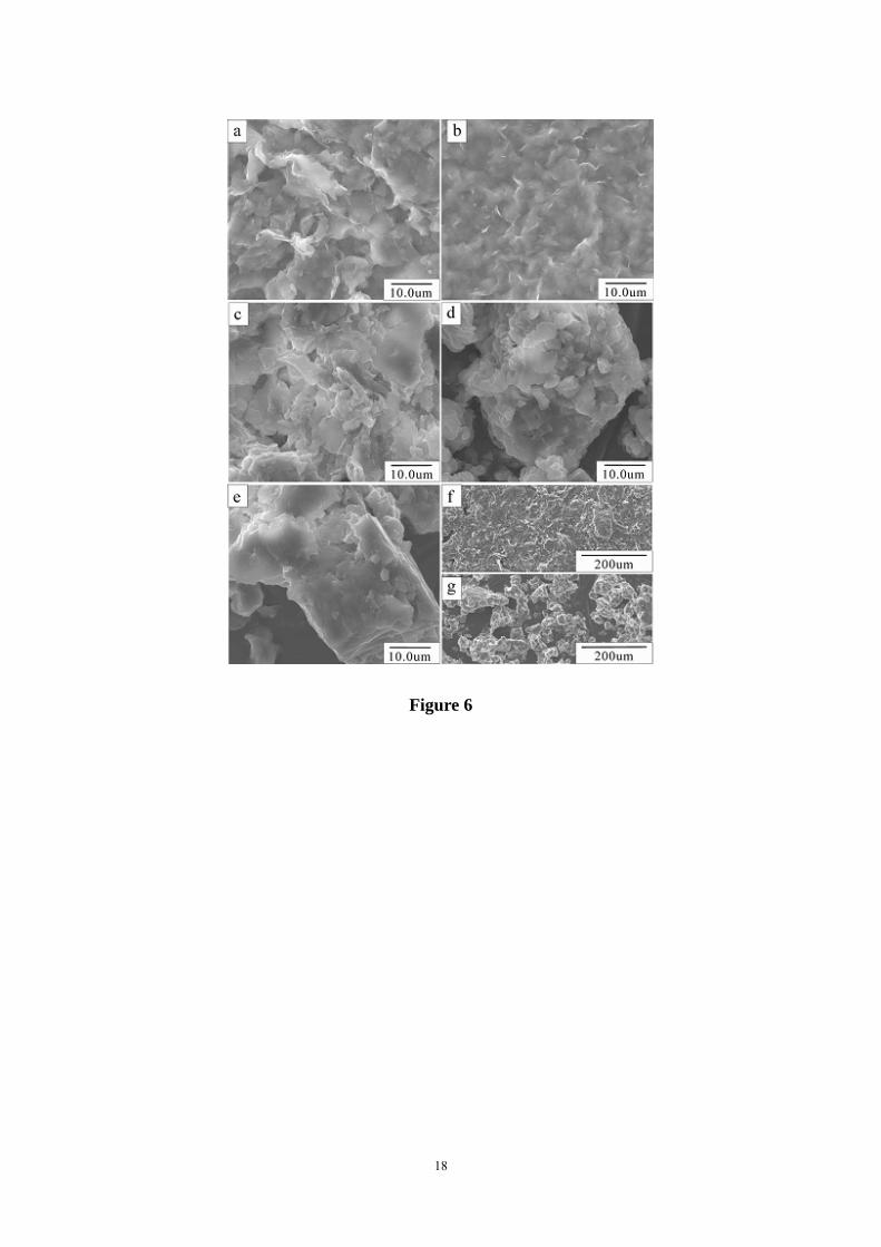
SEM micrographs (Figure 6) show surface morphology of montmorillonite and
the organoclay samples. It can be seen that the original Ca- and Na- montmorillonites have massive and curved plates (Figures 6a, b and f), especially for Na- montmorillonite [23]. The clay treated with organic surfactant shows significant changes in morphology. Compared with the morphology of the Na-montmorillonite, there are many small and aggregated particles and the plates become relatively flat in OM1 and OM2 (Figures 6c and d), while OM3 demonstrates mostly flat and aggregated morphology (Figures 6e and g). This is similar to that observed in the TEM micrographs in the present study and indicates that, with the increase of surfactant packing density in the interlayer, the curved plates in Na-montmorillonite will transform to flat layers. Hence, the present study shows that not only the basal spacing but also the morphology of the organoclay strongly depends on the packing density of surfactant within the montmorillonite interlayer space.
Montmorillonite Layer Flattening By Increasing Surfactant Loading
A number of previous XRD studies have shown that the arrangement models of
surfactant within the clay mineral galleries and the basal reflections strongly depend on the surfactant packing density. However, XRD patterns cannot provide the detailed information about the intercalated layers, including whether they are regularly intercalated layers or not. Our TEM micrographs for HDTMA+/montmorillonite organoclays prepared at higher surfactant concentrations have clearly demonstrated that they are mainly composed of regularly intercalated layers as proposed in the literature [10]. Meanwhile, an obviuous difference of layer height does exist from layer to layer. This can well explain the observations in the literature that the basal reflections for organoclays are always relatively broad. The organoclays, prepared at lower surfactant concentrations, are mainly composed of randomly intercalated layers.

8
The Na-montmorillonite layer may be retained since there is not enough surfactant provided to exchange the sodium cations in the montmorillonite interlayer. In the study of fluorohectorite intercalated with quaternary phosphonium salt [C16H33P(C4H9)3
+]Br at 0.5 CEC concentration, Ijdo and Pinnavaia [28] proposed that the organoclay is composed of regular stacking of two types of galleries, C16H33P(C4H9)3
+ fluorohectorite and Na+ fluorohectorite. However, such regular stacking of two types of galleries, surfactant-montmorillonite and Na-montmorillonite, does not occur in HDTMA+/montmorillonite organoclays. Our present study demonstrates that, at lower surfactant concentration, the intercalation of surfactant is random and the increase of surfactant packing density will result in the regular stacking of surfactant-montmorillonite galleries. This may be different from the previous study reported by Lee and Kim [23,24], in which they found many disconnected, thin, relatively short and strongly disrupted layer stacks in the organoclay prepared at 2.5 CEC while the other organoclay prepared at 1.0 CEC shows a well preserved repetitive layer-to-layer distance. As indicated in their paper, ultrasonic dispersion was used in the preparation of oriented films for XRD and alcohol used for the sample preparation for TEM. However, ultrasonic method can affect the microstructure of intercalated clays [29,30] and alcohol is organic solvent, which might also influence the structure of the organo-clays. In addition, Lee and Kim [23,24] also proposed that the charge value or its distribution on clay mineral might be an important factor for expansion characteristics of organoclay. Hence, we believe the difference between our result and previous study might be resulted from the different experimental conditions and characteristics of montmorillonite sample.
Our present study also indicates that the morphologies of the organoclays
strongly depend on the surfactant packing density within the interlayer, i.e., many curved plates in the organoclays prepared at lower surfactant concentrations while fewer occur in the ones prepared at higher surfactant concentrations. Figure 7 shows schematically how the curved plates transform to flat ones with the increase of surfactant packing density. After alkylammonium cations enter into the silicate interlayers, they experience the interactions between the head group of alkylammonium cation ─ silicate surface, the hydrocarbon chain of alkylammonium cation ─ silicate surface and the hydrocarbon chain ─ hydrocarbon chain of alkylammonium cations. The latter comprises the nonbonded van der Waals interactions, which is a function of alkylammonium concentration [31]. The strong electrostatic interaction between negative charged clay surface and the positively charged head group of alkylammonium cation will hold the head group of alkylammonium cation close to the clay surface as indicated by molecular simulation [32,33]. On the other hand, since the silicate surface of clay is hydrophilic whereas the hydrocarbon chains of alkylammonium cation are hydrophobic, the silicate surface ─ hydrocarbon chain interaction is of a repulsive nature. The transformation of the curved plates to flat ones might be mainly controlled by the interactions between the hydrocarbon chain ─ silicate surface and the hydrocarbon chain ─ hydrocarbon chain of alkylammonium cations. Previous reports [10,17,32] have demonstrated that, in the organoclays with lower surfactant packing density, the alkyl chains within the interlayer space are parallel within the interlayer space and are

9
individually separated. In this case, the repulsive interaction between the hydrocarbon chain ─ silicate surface is dominant whereas the interaction among the hydrocarbon chains is very weak. This repulsive interaction is an important factor to result in the transformation from the curved plates to flat ones. In addition, Li and Ishida [17] proposed that alkyl chains have the tendency to take more ordered trans conformers to minimize the total energy of organoclay. However, the bent clay plates will result in the increase of gauche conformers, which will increase the total energy of organoclays. Accordingly, the transformation from the curved plates to flat ones will decrease the total energy of organoclays. Here, we can find that both of the two above mentioned factors would lead to flattening the organoclay plates.
With the increase of surfactant packing density, the interchain interaction among
the alkylammonium cations becomes the dominant force and the orientation of the hydrocarbon tail changes from parallel to the silicate surface within the interlayer space to parallel but at an angle to the silicate surface as shown by our XRD patterns and previous studies [17]. The force from the silicate surface to the confined alkyl chains is limited by the distance [31]. Accordingly, when alkyl chains radiate at an angle away from the silicate surface, the interaction among hydrocarbon chains is relatively strong while that between the hydrocarbon chain ─ silicate surface is very weak or does not exist any more. The interaction among alkyl chains will increase with the increase of the surfactant packing density [23] and this will result in the ordered packing of the alkyl chains as indicated by FTIR and Raman [12,17,34,35]. Reasonably, the stacking of the organoclay layers will be regular and flat rather than random and curved as shown in the TEM and SEM micrographs of OM3.
Conclusions XRD patterns indicate that the surfactants in OM1 and OM3 adopt
lateral-monolayer and paraffin-type monomolecular arrangement models, respectively, while pseudotrilayer and paraffin-type monomolecular arrangements coexist in OM2. The surfactant arrangement model strongly depends on the packing density of surfactant within the montmorillonite galleries.
TEM and SEM micrographs demonstrate that the organoclays with lower
surfactant packing density are mainly composed of irregular layer stacking with a number of curved organoclay layers while those with higher surfactant packing density are mainly composed of regular layer stacking with fewer curved organoclay layers. The variation of the layer height from layer to layer exists in all three organoclays while more obvious variation is found in the organoclays with lower surfactant packing density than the one with higher packing density. This can well explain the broad basal reflections reported in the present study and in literature. Our present study demonstrates that not only the arrangement model of surfactant but also the morphology of the organoclay strongly depend on the surfactant packing density within the montmorillonite interlayer space.
Acknowledgments
The financial and infra-structural support from the National Natural Science

10
Foundation of China (Grant No. 40372029), the Inorganic Materials Research Program, Queensland University of Technology and Guangzhou Institute of Geochemistry (International Cooperation Research Program) are gratefully acknowledged.

11
References 1. A. J. Gu, S. W. Kuo, F. C. Chang, J. Appl. Polym. Sci. 79 (2001) 1902. 2. P. Maiti, K. Yamada, M. Okamoto, K. Ueda, K. Okamoto, Chem. Mater. 14
(2002) 4654. 3. L. Z. Zhu, Y. H. Su, Clays Clay Miner. 49 (2002) 421. 4. E. Manias, G. Hadziioannou, G. Brinke, Langmuir 12 (1996) 4587. 5. J. H. Wu, M. M. Lerner, Chem. Mater. 5 (1993) 835. 6. G. Lagaly, Clay Miner. 16 (1981) 1. 7. K. Beneke, G. Lagaly, Clay Miner. 17 (1982) 175. 8. H. Favre, G. Lagaly, Clay Miner. 26 (1991) 19. 9. K. Tamura, H. Nakazawa, Clays Clay Miner. 44 (1996) 501. 10. Z. Klapyta, T. Fujita, N. Iyi, Appl. Clay Sci. 19 (2001) 5. 11. T. Yui, H. Yoshida, H. Tachibana, D. Tryk, H. Inoue, Langmuir 18 (2002) 891. 12. R. A. Vaia, R. K. Teukolsky, E. P. Giannelis, Chem. Mater. 6 (1994) 1017. 13. K. Khatib, M. Francois, P. Tekely, L. J. Michot, J. Y. Bottero, I. Baudin, J. Colloid
Interf. Sci. 183 (1996) 148. 14. D. J. Harris, T. J. Bonagamba, K. Schmidt-Rohr, Macromolecules, 32 (1999)
6718. 15. L. Q. Wang, J. Liu, G. J. Exarhos, K. Y. Flanigan, R. Bordia, J. Phys. Chem. B
104 (2000) 2810. 16. Y. Q. Li, H. Ishida, Chem. Mater. 14 (2002) 1398. 17. Y. Q. Li, H. Ishida, Langmuir 19 (2003) 2479. 18. J. Madejova, Vib. Spectrosc. 31 (2003) 1. 19. J. X. Zhu, H. P. He, J. G. Guo, D. Yang, X. D. Xie, Chinese Sci. Bull. 48 (2003)
368. 20. H. P. He, R. L. Frost, F. Deng, J. X. Zhu, X. Y. Wen, P. Yuan, Clays Clay Miner.
52 (2004) 350. 21. B. Hoffmann, C. Dietrich, R. Thomann, C. Friedrich, R. Mülhaupt, Macromol.
Rapid Commun. 21 (2000) 57. 22. W. Xie, J. M. Hwu, G. J. Jiang, T. M. Buthelezi, W. P. Pan, Polym. Eng. Sci. 43
(2003) 214. 23. S. Y. Lee, S. J. Kim, Colloid Surface A 211 (2002) 19. 24. S. Y. Lee, S. J. Kim, Clays Clay Miner. 50 (2002) 435. 25. G. Brindley, W. Moll, Am. Miner. 50 (1965) 1355. 26. J. Choy, S. Kwak, Y. Han, B. Kim, Mater. Lett. 33 (1997) 143. 27. B. Bauluz, D. R. Peacor, R. F. Ylagan, Clays Clay Miner. 50 (2002) 157. 28. W. Ijdo, T. Pinnavaia, Chem. Mater. 11 (1999) 3227. 29. P. S. Katdare, V. Ramaswamy, A. V. Ramaswamy, Catal. Today 49 (1999) 313. 30. P. S. Katdare, V. Ramaswamy, A. V. Ramaswamy, Micropor. Mesopor. Mater. 37
(2000) 329. 31. S. Gupta, D. C. Koopman, G. B. Westermann-Clark, I. A. Bitsanis, J. Chem. Phys.
100 (1994) 8444. 32. Q. H. Zeng, A. B. Yu, G. Q. Lu, R. K. Standish, Chem. Mater. 15 (2003) 4732. 33. H. P. He, J. Galy, J. F. Gerard, J. Phys. Chem. B. 109 (2005) 13301. 34. H. P. He, L. R. Frost, J. X. Zhu, Spectrochim. Acta A 60 (2004) 2853. 35. H. P. He, R. L. Frost, Y. F. Xi, J. X. Zhu, J. Raman Spectros. 35 (2004) 316.

12
Figure Captions
Figure 1. XRD patterns of Na-montmorillonite (Na-mont) and HDTMA+/montmorillonite
organoclays.
Figure 2. Molecular conformation of HDTMA+. a: the plane of the zig-zag arrangement of the carbon atoms of HDTMA+ is perpendicular to
the plane of the montmorillonite layer; b: the plane of the zig-zag arrangement of the carbon atoms of HDTMA+ is parallel to the
plane of the montmorillonite layer. Figure 3. TEM micrographs of OM3 with generally regular stacking of intercalated layers with a
slight difference of layer height from layer to layer (a) and coexistence of regularly and irregularly intercalated layers (b and c).
Figure 4. TEM micrographs of OM2.
a: curved and irregularly intercalated layers; b: irregular stacking of intercalated layers; c: relatively regular stacking of intercalated layers in a small area; d and e: curved and irregular stacking of intercalated layers.
Figure 5. TEM micrographs of OM1 with curved and irregularly intercalated layers. Figure 6. SEM micrographs of Ca-montmorillonite (a, g), Na-montmorillonite (b), OM1 (c), OM2
(d) and OM3 (e, f). Figure 7. The schematic diagram for the curved plates transforming to flat ones with the increase
of surfactant packing density and alkyl chain aggregation in the interlayers: (a) Na-montmorillonite; (b) lateral monolayer (OM1); (c) pseudotrilayer with a small amount of paraffin-type monolayer (OM2); (d) paraffin-type monolayer (OM3).
The hydrated sodium cations are represented by special circles ( ) while single open circles (○) represent methyls and nitrogen atoms in cationic head groups are represented by filled circles (●).

13
Figure 1

14
Figure 2

15
Figure 3

16
Figure 4

17
Figure 5
18
Figure 6

19
Figure 7
Copyright © 2022 FDOKUMEN